VisualSonics White Paper · VisualSonics White Paper: Nonlinear Contrast Agent Imaging with a High...
22
Andrew Needles April 14, 2009 Tonya Coulthard Version 1.0 Catherine Theodoropoulos Stuart Foster VisualSonics White Paper: Nonlinear Contrast Agent Imaging with a High Frequency Linear Array Based System
Transcript of VisualSonics White Paper · VisualSonics White Paper: Nonlinear Contrast Agent Imaging with a High...

Andrew Needles April 14, 2009 Tonya Coulthard Version 1.0 Catherine Theodoropoulos Stuart Foster
VisualSonics White Paper: Nonlinear Contrast Agent Imaging with a High Frequency Linear
Array Based System

VSI White Paper: Nonlinear Contrast Agent Imaging with a High Frequency Linear Array Based System
Table of Contents Summary .............................................................................................................. 1
Introduction ......................................................................................................... 1
Nonlinear Ultrasound ............................................................................................ 2
(a) Nonlinear Properties of Microbubbles................................................................... 2
(b) Nonlinear Properties of Tissue............................................................................ 3
The Importance of Imaging Microbubbles at Low Acoustic Pressures .................... 4
Nonlinear Ultrasound Contrast Agent Imaging Techniques .................................... 5
(a) Pulse Inversion................................................................................................... 5
Figure 2: Pulse Inversion in Tissue ............................................................................. 5
Figure 3: Pulse Inversion with microbubbles ................................................................ 6
(b) Amplitude Modulation ....................................................................................... 6
Figure 4: Amplitude Modulation with Tissue ................................................................. 7
Figure 5: Amplitude Modulation with Microbubbles ........................................................ 7
Choosing a Nonlinear Imaging Technique for High Frequencies............................. 8
In vivo Protocols for Quantification of Nonlinear Contrast Imaging ..................... 10
Perfusion Imaging for Organs, Tissues, and Tumors............................................ 10
Bolus Injection Protocols ...................................................................................... 10
Disruption-Replenishment Protocols ....................................................................... 12
Targeted Molecular Imaging ............................................................................... 13
Conclusions......................................................................................................... 15
References.......................................................................................................... 16
Glossary.............................................................................................................. 19

VSI White Paper: Nonlinear Contrast Agent Imaging with a High Frequency Linear Array Based System Page 1
Summary
Ultrasound contrast agents act as an intravascular tracer, allowing for the visualization of blood flow inside living tissue. These micron-sized gas filled bubbles, when excited with ultrasound, can be preferentially detected over surrounding tissue, proving high sensitivity to blood signals in the microcirculation. Using the new Vevo 2100 array-based system, linear measurements of microbubble echo power can be made, which correlate with blood flow characteristics. Alternatively, microbubbles can be labeled with ligands capable of binding to cellular receptors. This enables the use of micro-ultrasound for molecular imaging; the detection of disease states in various tissue types. Microbubble contrast agents, therefore, are a powerful tool that enables the pre-clinical researcher to monitor the functional dynamics of animal models of human disease.
Introduction
Recent developments in linear array technology have pushed traditional ultrasound imaging frequencies higher, into the range of 15-70 MHz (Ritter et al. 2002; Lukacs et al. 2006; Brown et al. 2007). Many of these devices have been fabricated, however, an important requirement for any array based imaging system, the beamforming electronics, has been lacking. Recently, VisualSonics launched the Vevo 2100 linear array based micro-ultrasound imaging system (Foster et al. 2009). It has a fully developed, 64 channel, high frequency beamformer, capable of driving linear arrays in the 12-70 MHz range. This is a giant technological breakthrough for the field of micro-ultrasound. A linear array brings the advantage of improved depth-of-field, significantly improving image quality over comparable single-element transducers found on the Vevo 2100’s predecessor, the Vevo 770. Additionally, the linear array provides high frame rate (>30 Hz) 2D blood flow imaging (known as Power and Color Doppler), something not generally possible with a single-element transducer (Needles et al. 2007).
Microbubble contrast agents have been used in ultrasound imaging as a means of improving the visualization of blood flow with respect to the surrounding tissue beyond the sensitivity of Power and Color Doppler imaging (Becher and Burns 2000). These micron-sized particles (~1-10 μm, on the order of the size of a red blood cell) consist of a gas core surrounded by a lipid shell, and are injected in very minute volumes into the circulatory system. VisualSonics distributes a contrast agent called MicroMarker, which is optimized for high frequencies. It contains a perfluorobutane gas core encapsulated by a phospholipid shell. MicroMarker is manufactured by the Bracco Group, one of the largest manufacturers of diagnostic contrast agents globally. In standard B-Mode (gray-scale) imaging, the MicroMarker microbubbles can be visualized due to their high echogenicity from incident high-frequency ultrasound waves. With post-processing algorithms, these enhanced echoes from bubbles can be segmented from tissue. This is the approach used on the VisualSonics’

VSI White Paper: Nonlinear Contrast Agent Imaging with a High Frequency Linear Array Based System Page 2
previous generation system, the Vevo 770 (Rychak et al. 2007; Lyshchik et al. 2007, Willmann et al. 2008), and will be referred to henceforth as Linear Contrast Mode. Linear Contrast Mode is also available on the Vevo 2100. The challenge, however, is that in many cases the microbubbles and tissue are comparably bright, resulting in poor contrast between them. This makes visualization of the microbubbles difficult, even after post-processing. Understanding the patterns of blood flow in the microcirculation is a powerful tool for evaluating the differences between normal and pathological tissues, particularly in the areas of cancer (Folkman 1995; Carmeliet and Jain 2000), and sites of inflammation (Kumamoto et al. 1995). In addition to visualizing and quantifying blood flow in the microcirculation, targeting microbubbles to cellular receptors and detecting them with ultrasound can give valuable insight about the molecular state of small animal models of human disease (Lindner 2004). In order to improve the sensitivity to microbubbles, the surrounding tissue must be removed from the received ultrasound signals. Additionally, eliminating post-processing techniques allows the visualization of contrast agent to occur in real-time.
Nonlinear Ultrasound
In general, a linear system has the property that its inputs and outputs are proportional to each other, and can be expressed through simple algebraic expressions. For example, when listening to the radio with the volume set to 25% and then changing it to 50%, the sound from the speaker would be twice as loud. Now consider that as the radio volume is slowly increased towards 100%, the sound becomes extremely loud with crackling and hissing. The sound coming out of the radio is no longer increasing linearly; it is undergoing nonlinear distortion which is being detected by the human ear. Similar effects occur with ultrasound, however, the human ear could now be thought of as the ultrasound transducer and the speaker system would be replaced by the biological tissues being imaged. As the “volume” of the ultrasound is increased (i.e. the acoustic power) the response from the tissue system begins to respond nonlinearly, which in turn is detected by the ultrasound transducer. Both tissue and microbubbles will scatter ultrasound nonlinearly. The following sections outline both of these processes.
(a) Nonlinear Properties of Microbubbles
Due to their high compressibility, microbubbles will oscillate nonlinearly in an ultrasound field, at acoustic pressures on the order of 100-500 kPa (Leighton, 1994). A nonlinear oscillation occurs because the radial expansion of the microbubbles differs from the radial contraction. During the expansion phase of oscillation, the radius of the gas microbubble can increase by several times its original size. During the contraction phase of the oscillation the radius is limited since the gas molecules are forced closer together, making it harder to compress. The oscillation, therefore, becomes asymmetrical, resulting in the scattered ultrasound echo containing nonlinear energy (Goertz et al. 2005). Most microbubble

VSI White Paper: Nonlinear Contrast Agent Imaging with a High Frequency Linear Array Based System Page 3
imaging techniques try to preferentially detect this nonlinear energy from the bubbles in order to segment the bubble signals from tissue (Deng and Lizzi 2002).
(b) Nonlinear Properties of Tissue
Tissue will also respond nonlinearly to ultrasound as the acoustic pressure is increased (Hamilton and Blackstock 1998). This essentially forms the “noise” in the contrast image, as it is the baseline signal above which we want to detect contrast agent. In practice, there will always be some small amount of residual nonlinear tissue signal detected. For the purposes of the discussion here, however, we will assume that a low enough acoustic pressure is used to limit the nonlinear response in tissue so that we can assume that any received signal from tissue is completely linear.
A nonlinear signal, when analyzed in terms of its frequency content, can be shown to possess harmonic components of the original fundamental frequency (fo) which comprised the signal before undergoing nonlinear distortion. By definition, a harmonic is a multiple of the center frequency of the incident ultrasound wave. For example, a high frequency ultrasound wave with a center frequency of 20 MHz, after scattering nonlinearly will have harmonics at twice the center frequency (second harmonic), and above (third harmonic, fourth harmonic et cetera)(de Jong et al. 1994). If the nonlinear scattering is due to a microbubble then there may also be energy at half of the center frequency (subharmonic) (Forsberg et al. 2000). Depending on the bandwidth of the ultrasound transducer used, different harmonic components can be detected. Interestingly, there may also be nonlinear energy at the fundamental frequency (i.e. the fundamental harmonic), which will be most easily detected if this corresponds to the center of the transducer’s bandwidth (Haider and Chiao 1999).

VSI White Paper: Nonlinear Contrast Agent Imaging with a High Frequency Linear Array Based System Page 4
Figure 1: Example of nonlinear scattering from microbubbles with energy at various harmonic components.
The Importance of Imaging Microbubbles at Low Acoustic Pressures
Traditional microbubble imaging techniques generally used high acoustic pressures to detect microbubbles (Becher and Burns 2000). As microbuble and ultrasound transducer technology were improved over time, however, lower acoustic pressure (i.e lower Mechanical Index or MI) imaging techniques were developed. Imaging microbubbles with low acoustic pressures has two advantages:
- nonlinearities in tissue are minimized
- microbubbles are not ruptured or disrupted during imaging
Using low acoustic pressures for imaging contrast agents, therefore, has the advantage of improving the contrast-to-tissue ratio (CTR) by reducing residual tissue signal and maximizing agent detection by not disrupting it. Additionally, applying quantitative techniques such as monitoring the rate of inflow/outflow of agent, or detecting targeted microbubbles, are possible since the agent is not being disrupted.
VSI White Paper: Nonlinear Contrast Agent Imaging with a High Frequency Linear Array Based System Page 5
Nonlinear Ultrasound Contrast Agent Imaging Techniques
Many techniques have been proposed for imaging ultrasound contrast agents nonlinearly (Burns et al. 1994; de Jong et al. 2000; Chang et al. 1995; Hope-Simpson et al. 1999). Some of these techniques send multiple ultrasound pulses down a single line and apply signal processing. This section will outline two of the most basic multi-pulse techniques, Pulse Inversion and Amplitude Modulation, which form the basis for modern day nonlinear contrast agent detection. Beyond the information presented here, readers are referred to a review of these techniques by Eckersley et al. (2005).
(a) Pulse Inversion
Pulse inversion is the classic example of a nonlinear microbubble imaging technique. In its simplest form, two ultrasound pulses are fired consecutively into tissue, with the second pulse being an inverted copy of the first. Based on the properties of a linear system and assuming that the tissue is completely linear, the received pulses from the tissue can be summed together, resulting in a complete cancellation of the tissue signal. Figure 2 illustrates this process:
Incident Pulse Tissue Response
Tissue
+ =
Pulse 1
Pulse 2
+1
-1
Figure 2: Pulse Inversion in Tissue
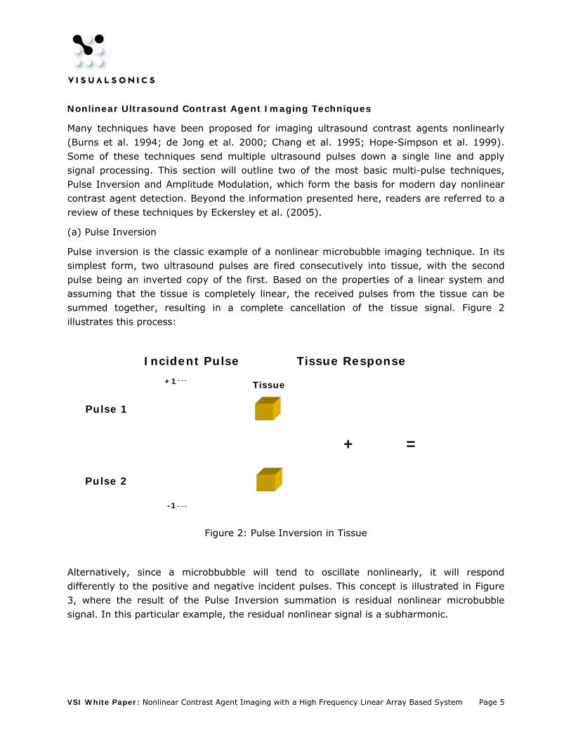
Alternatively, since a microbbubble will tend to oscillate nonlinearly, it will respond differently to the positive and negative incident pulses. This concept is illustrated in Figure 3, where the result of the Pulse Inversion summation is residual nonlinear microbubble signal. In this particular example, the residual nonlinear signal is a subharmonic.
VSI White Paper: Nonlinear Contrast Agent Imaging with a High Frequency Linear Array Based System Page 6
-1
Incident Pulse Bubble Response
Bubble
+ =
Pulse 1
Pulse 2
+1
Figure 3: Pulse Inversion with microbubbles
The example of Pulse Inversion illustrates how the properties of a linear system can be exploited to differentiate signal from microbbubles in the presence of tissue. Since in reality the received echoes will be a combination of both tissue and microbubble, after applying Pulse Inversion summation, any residual signal will be nonlinear microbubble signal. These microbubble signals can then be presented on the ultrasound display, giving excellent contrast between the regions of vascularity and the background tissue signal.
(b) Amplitude Modulation
Amplitude modulation is another nonlinear imaging technique which uses multiple pulse firings to differentiate linear and nonlinear ultrasound signals. Again, in the simplest form it can be illustrated with two incident pulses being exposed to tissue, where the first pulse is scaled by a factor of 2 relative to the second pulse. When the incident pulses respond to linear tissue, the received echoes can be subtracted, after scaling the second pulse by a factor of 2. Assuming that the tissue response is linear, this subtraction will produce a complete cancellation of the tissue, illustrated in Figure 4.

VSI White Paper: Nonlinear Contrast Agent Imaging with a High Frequency Linear Array Based System Page 7
Incident Pulse Tissue Response
Tissue
- =2x
Pulse 1
Pulse 2
+1
+½
Figure 4: Amplitude Modulation with Tissue
When considering the same sequence of two amplitude modulated pulses, except with microbubbles instead of tissue, the response of the bubble can be seen to change between the two different incident pulse amplitudes. Compare this to the tissue example in Figure 4 which was representative of a linear system. In Figure 4, the received pulses could be scaled linearly and cancelled. Microbubbles, however, will respond differently to different amplitudes of incident ultrasound. This is due to the nonlinear response of the bubbles. Amplitude modulation is a technique which exploits this property of microbubbles and is illustrated in Figure 5.
Incident Pulse Bubble Response
Bubble
- =2x
Pulse 1
Pulse 2
+1
+½
Figure 5: Amplitude Modulation with Microbubbles

VSI White Paper: Nonlinear Contrast Agent Imaging with a High Frequency Linear Array Based System Page 8
Choosing a Nonlinear Imaging Technique for High Frequencies
On the VisualSonics Vevo 2100 micro-ultrasound system the main nonlinear signals of interest are the subharmonic and nonlinear fundamental harmonic. The subharmonic has the unique property that it is generated though nonlinear scattering from microbubbles, but not from tissue. In general, lower frequency contrast agent imaging methods have utilized nonlinear energy at the second harmonic. This approach is less desirable at higher frequencies for two main reasons. First of all, the amount of nonlinear tissue signal at the second harmonic, even for relatively low MI imaging, is significant at high frequencies (Goertz et al. 2005). This suggests that there is a frequency dependence of nonlinear propagation in tissue. The second reason is that since ultrasound attenuation increases as a function of frequency, the second harmonic at high frequencies will suffer from more frequency dependent attenuation than at lower frequencies. For these reasons, the Vevo 2100 implementation has focused on utilizing nonlinear fundamental and subharmonic energy over second harmonic energy. Both the Pulse Inversion (PI) and Amplitude Modulation (AM) techniques described earlier are sensitive to detecting nonlinear fundamental and subharmonic energy.
AM allows the separation of linear and nonlinear energy at the fundamental frequency. The combination of detecting both subharmonic and nonlinear fundamental energy makes the AM technique very attractive as a high frequency microbubble imaging strategy. Figure 6 shows real data collected with the MS-250 linear array at a transmit frequency of 24 MHz. The data is from VisualSonics’ MicroMarker high frequency contrast agent flowing through a tissue mimicking medium, for both PI and AM processing. Figure 6 is a frequency plot of received ultrasound echoes, with all curves referenced to the raw unprocessed data (not shown).

VSI White Paper: Nonlinear Contrast Agent Imaging with a High Frequency Linear Array Based System Page 9
Figure 6: 24 MHz in vitro PI and AM frequency spectra for both bubbles and tissue
In Figure 6 it is clear that both PI and AM detect nonlinear subharmonic energy at 12 MHz as expected. In the case of AM, additional nonlinear energy is detected at the fundamental frequency (24 MHz). Other interesting aspects of this data are that PI is much better at suppressing tissue, particularly in the fundamental band. The residual tissue signal at the fundamental frequency detected by AM, is nonlinear in nature, despite the fact that this data was collected at relatively low acoustic pressure (350 kPa). This is one drawback of the AM technique, especially as the transmit frequency is increased and nonlinear propagation in tissue becomes more problematic as a function of frequency. By using PI and isolating subharmonic energy though bandpass filtering, one could take advantage of the improved tissue suppression offered by PI, particularly as transmit frequencies are increased (30 MHz and above) and nonlinear tissue signal becomes problematic. Still, for frequencies in the 15-30 MHz range, the amount of nonlinear tissue signal can be tolerated, so the benefits of detecting both subharmonic and nonlinear fundamental with AM are the most attractive.

VSI White Paper: Nonlinear Contrast Agent Imaging with a High Frequency Linear Array Based System Page 10
Thus, the current implementation of the new Nonlinear Contrast Mode on the Vevo 2100 uses the MS-200 (15 MHz centre frequency) and the MS-250 (21 MHz centre frequency) and is based on a form of AM processing. Future development will look to expanding the nonlinear contrast imaging functionality to higher frequency transducers (MS-400, MS-550(S/D), MS-700), using PI subharmonic imaging.
In vivo Protocols for Quantification of Nonlinear Contrast Imaging
The following sections will highlight two distinct methods of microbubble imaging in mice, using the MS-250 transducer and MicroMarker high frequency contrast agent. These methods are geared towards not only imaging, but generating quantitative analysis for in vivo experiments. For further detailed information about these methods please refer to the VisualSonics imaging protocols.
Perfusion Imaging for Organs, Tissues, and Tumors
There are two main protocols that can be used to assess perfusion. The first one involves performing a bolus injection of contrast agents and the second one requires a continuous infusion of contrast agents.
Bolus Injection Protocols
A typical bolus injection involves inserting a catheter into the tail vein of the mouse (any type of venous access may be used). Typically, 50 µL of contrast agent is injected into the animal. This corresponds to a dose ranging from 107 to 108 bubbles of MicroMarker contrast agent, depending on the target of interest. The inflow and outlfow of the agent can be tracked in the imaging plane over time and plotted as a Time-Intensity curve, with the linear data used to generate the ultrasound image. Figure 7 illustrates the time course of a microbubble bolus within the abdomen of the mouse, showing both anatomical B-Mode images on the left and Nonlinear Contrast images on the right. Before the injection of agent (time = 0 sec), the abdominal region is void of signal in the Nonlinear Contrast image, as the tissue signals (with the exception of the skin line) have been suppressed. Subsequently, the agent can be seen to fill the abdominal organs, starting with the major vessels (time = 1.5 sec). After 5 seconds all of the major organs within the image are filled.

VSI White Paper: Nonlinear Contrast Agent Imaging with a High Frequency Linear Array Based System Page 11
Figure 7: Abdominal cavity of mouse showing the kidney (K), pancreas (P), and spleen (S) at various time points following a bolus injection. B-Mode images (left) are displayed simultaneously with the Contrast image, displayed in copper (right).
Regions of interest can be drawn on the images to segment particular organs. Within the region, the linear mean echo power from the ultrasound data can be tracked as a function of time. This is illustrated in Figure 8. The dynamics of these curves can give valuable

VSI White Paper: Nonlinear Contrast Agent Imaging with a High Frequency Linear Array Based System Page 12
information about the relative blood flow characteristics of the tissue in questions, particularly when comparing regions of normal and pathological tissues. Examples of relevant parameters include: Time-to-Peak, Peak Enhancement, Wash-In Rate, Wash-Out Rate, and Area Under the Curve, to name a few.
Figure 8: Abdominal cavity of mouse at 3.7 s after bolus injection, showing the kidney (green), medulla (blue), pancreas (red), and spleen (magenta). Curves in (a) are generated from the contrast signal regions-of-interest, shown within the nonlinear contrast image in (c).
Disruption-Replenishment Protocols
While bolus injections are relatively easy to perform, there are limitations with analyzing the inflow portion of the curve. This is due to the fact that the rate of inflow can be influenced by the path that the agent takes through the animal before filling the imaging plane. Disruption-replenishment techniques have been developed as a better solution. This technique uses a continuous infusion of agent, generally with a syringe pump, and relies on a high power “burst” of ultrasound to clear the imaging plane of microbubbles by disrupting them. This process of disruption essentially creates a “negative bolus”, that is independent of any path length to replenish the agent. Figure 9a shows a mouse heart after the infusion of microbubbles into the circulatory system. After applying the high-powered burst, regions
Whole Kidney
Kidney Medulla
Pancreas
Spleen
(a) (b)
(c)

VSI White Paper: Nonlinear Contrast Agent Imaging with a High Frequency Linear Array Based System Page 13
of capillary flow become very dark, as the contrast agent is destroyed. Figure 9b shows the heart immediately following the ultrasound burst. The anterior wall (AW) and posterior wall (PW) of the myocardium are clearly delineated from the left ventricle (LV). The subsequent refilling of the agent can be tracked over time, giving valuable functional information the perfusion of tissue, in this case the heart wall (Arditi et al. 2006).
Figure 9: Disruption-Replenishment imaging in the mouse heart. Nonlinear contrast images before high-power ultrasound exposure (a) and after (b). The high-power ultrasound disrupts microbubbles, which in turn delineates the anterior wall (AM) and posterior wall (PW) of the myocardium from the left ventricle (LV). Subsequent refilling of the myocardium is illustrated in (c).
Targeted Molecular Imaging
A different approach to imaging microbubbles is to use the contrast agent as a labeler of a particular disease state. This is accomplished by using the MicroMarker agent that is coated with streptavidin. By using biotinylated antibodies, which can be linked to the avidinated microbubble though the avid-biotin complex (one of the strongest bonds in nature), these targeted microbubbles can be attached to selected cellular receptors. A common example

VSI White Paper: Nonlinear Contrast Agent Imaging with a High Frequency Linear Array Based System Page 14
found in research, is targeting for Vascular Endothelial Growth Factor Receptor2 (VEGFR-2) in subcutaneous tumors implanted in the hind limb of a mouse. Figure 10 shows images from the control microbubble (a) and the targeted microbubble (b), 4 minutes after injection of the agent into a subcutaneous human hepatoma tumor. Figure 10c demonstrates the enhancement of detected microbubbles targeted for VEGFR-2, present in a region within the cancerous tissue.
Figure 10: Nonlinear contrast images of a subcutaneous hepatoma tumor on the hind limb of a mouse. Both control microbubbles lacking the targeting ligands (a) and microbubbles targeted to VEGFR-2 (b) are shown. The enhancement in signal with the targeted bubbles is shown in (c).
VSI White Paper: Nonlinear Contrast Agent Imaging with a High Frequency Linear Array Based System Page 15
Conclusions
The use of micro-ultrasound in pre-clinical research is becoming increasingly more widespread. In addition to anatomical imaging, functional imaging modes like color and power Doppler add information to the ultrasound examinations in small animals. Beyond the information conveyed through these modes, high frequency contrast agents can enhance the sensitivity to low level blood signals in the microcirculation, as well as act as a mechanism for targeting and identifying diseased tissues. Using nonlinear signal processing allows for these contrast agents to be detected in the presence of the surrounding tissue. Using the Vevo 2100 micro-ultrasound system now allows for microbubble specific contrast agent detection at high ultrasound frequencies (> 15 MHz). The result is a robust, real-time imaging platform that will enable the preclinical researcher to conduct accurate anatomical, functional, and molecular experiments in small animals.
VSI White Paper: Nonlinear Contrast Agent Imaging with a High Frequency Linear Array Based System Page 16
References
Arditi M, Frinking JA, Zhou X, Rognin NG. A new formalism for the quantification of tissue perfusion by the destruction-replenishment method in contrast ultrasound imaging. IEEE Trans Ultrason Ferroelec Freq Contr 2006;53:1118-1129.
Becher H, Burns PN. Handbook of Contrast Echocardiography. Berlin: Springer, 2000.
Burns PN, Powers JE, Hope-Simpson D, et al. Harmonic power mode Doppler using contrast agents: An improved method for small vessel flow imaging. J Ech Med Ultra 1994;16:132–142.
Brown JA, Foster FS, Needles A, Cherin E, Lockwood GR. Fabrication and Performance of a 40-MHz Linear Array Based on a 1-3 Composite with Geometric Elevation Focusing. IEEE Trans Ultrason Ferroelectr Freq Cont 2007;54:1888-1894.
Carmeliet P, Jain RK. Angiogenesis in cancer and other diseases. Nature 2000;407:249–257.
Chang PH, Shung KK, Levine HB. Second harmonic imaging and harmonic Doppler measurements with Albunex. IEEE Trans Ultrason Ferroelect Freq Contr 1995;42:1020–1027.
Deng CX, Lizzi FL. A review of physical phenomena associated with ultrasonic contrast agents and illustrative clinical phenomena. Ultrasound Med Biol 2002;28:277–286.
de Jong N, Cornet R, Lancee CT. Higher harmonics of vibrating gas-filled microspheres. Part Two: Measurement. Ultrasonics 1994;32:455–459.
de Jong N, Frinking PJA, Buoakaz A, Ten Cate FJ. Detection procedures of ultrasound contrast agents. Ultrasonics 2000;38:87–92

VSI White Paper: Nonlinear Contrast Agent Imaging with a High Frequency Linear Array Based System Page 17
Eckersley RJ, Chin CT, Burns PN. Optimising phase and amplitude modulation schemes for imaging microbubble contrast agents at low acoustic power. Ultra. Med. Biol. 2005;31:213-219.
Folkman J. Tumor angiogenesis. In: Mendelsohn J, Howley PM, Israel MA, Liotta LA, eds. The molecular basis of cancer. Philidalphia, PA: W. B. Saunders Company, 1995.
Foster FS, Mehi J, Lukacs M, et al. High frequency array based micro-ultrasound for preclinical imaging. Ultrasound Med Biol. In Press.
Forsberg F, Shi WT, Goldberg BB. Subharmonic imaging of contrast agents. Ultrasonics 2000;38:93-98
Goertz DE, Cherin E, Needles A, et al. High frequency nonlinear B-Scan imaging of microbubble contrast agents. IEEE Trans Ultrason Ferroelectr Freq Cont 2005;52:65-79.
Haider B, Chiao RY. High order nonlinear ultrasonic imaging. Proc IEEE Ultrason Symp 1999;2:1527–1531.
Hamilton MF, Blackstock DT. Nonlinear Acoustics: Theory and Applications. Academic Press, 1998.
Hope-Simpson D, Chin CT, Burns PN. Pulse inversion Doppler: A new method for detecting non-linear echoes from microbubble contrast agents. IEEE Trans Ultrason Ferroelect Freq Contr 1999;46:372–382.
Kumamoto M, Nakashimi Y, Sueishi K. Intimal neovascularization in human coronary atherosclerosis—its origin and pathophysiological significance. Human Pathol. 1995;26:450–456.
Leighton, T. The Acoustic Bubble. Academic Press, 1997.

VSI White Paper: Nonlinear Contrast Agent Imaging with a High Frequency Linear Array Based System Page 18
Lindner JR. Molecular imaging with contrast ultrasound and targeted microbubbles. J Nucl Cardiol 2004; 11:215-221.
Lukacs M, Yin J, Pang G, et al. Performance and Characterization of New Micromachined High-Frequency Linear Arrays. IEEE Trans Ultrason Ferroelec Freq Contr 2006;53:1719-1729.
Lyshchik A, Fleischer AC, Huamani J, et al. Molecular Imaging of Vascular Endothelial Growth Factor Receptor 2 Expression Using Targeted Contrast-Enhanced High-Frequency Ultrasonography. J Ultrasound Med 2007;26:1575-1586
Needles A, Goertz DE, Cheung AM, Foster FS. Interframe clutter filtering for high frequency flow imaging. Ultrasound Med Biol 2007;33:591-600.
Ritter TA, Shrout TR, Tutweiler R, Shung KK. A 30 MHz peizo-composite ultrasound array for medical imaging applications. IEEE Ultrason Ferroelec Freq Cont 2002;49:217-230.
Rychak JJ, Graba J, Cheung AM, et al. Microultrasound molecular imaging of vascular endothelial growth factor receptor 2 in a mouse model of tumor angiogenesis. Mol Imaging 2007;6:289-296.
Willmann JK, Paulmurugan R, Chen K, et al. US imaging of tumor angiogenesis with microbubbles targeted to vascular endothelial growth factor receptor type 2 in mice. Radiology 2008;246:508-518.

VSI White Paper: Nonlinear Contrast Agent Imaging with a High Frequency Linear Array Based System Page 19
Glossary
Acoustic Pressure: The localized pressure of a sound wave, where pressure is defined as the force per unit area applied to an object. The pressure is represented as the amplitude of the time-varying acoustic wave.
Amplitude Modulation: A nonlinear microbubble imaging technique. In its simplest form, two ultrasound pulses are fired consecutively into tissue, with the amplitude of the second pulse being a scaled copy of the first. Based on the properties of a linear system the received pulses from linear scatterers can be subtracted after re-scaling the second pulse, resulting in a complete cancellation, whereas nonlinear signals from microbubbles will not cancel. The nonlinear signal retained is from the even harmonics (i.e. subharmonic, second harmonic, fourth harmonic etc.) and nonlinear fundamental.
Beamformer: The electronic circuitry connected to a transducer array, capable of transmitting on arbitrary sequences of transducer elements to form an ultrasound beam with a given focal depth, width, and direction.
Bolus: An injection of a fixed amount of contrast agent into the bloodstream (typically on the venous side).
“Burst”: A term used on the Vevo 2100 to describe the process of sending high power ultrasound to disrupt microbubbles. This process clears the imaging plane of contrast agent allowing for the subsequent refill to be tracked. The high power ultrasound can be initiated on the Vevo 2100, for a selectable time period, by pressing the “Burst” key.
Harmonic: A multiple of the center frequency of an incident ultrasound wave that is generated through the scattering of ultrasound off of a reflector.
Linear Array: An ultrasound transducer that has a series of small elements (256 on the Vevo 2100) lined up in parallel to one another on a rectangular surface. These elements can be excited individually or in groups by an electronic voltage source to create ultrasound beams with variable focal properties, widths and directions.

VSI White Paper: Nonlinear Contrast Agent Imaging with a High Frequency Linear Array Based System Page 20
Linear: Describes a relation between two quantities that vary proportionally to one another.
Linear Contrast Mode: A software feature on the Vevo 770 and the Vevo 2100 that collects a series of B-Mode images before and after an injection of contrast agent, and highlights the regions of contrast agent with a colored overlay, by performing a frame subtraction technique through post-processing. It was known simply as Contrast Mode before the advent of Nonlinear Contrast Mode.
“Negative Bolus”: The result of applying a sequence of high power ultrasound pulses, which clears the imaging plane of microbubbles. The subsequent refill of the microbubbles can then be tracked, similar to performing a bolus injection.
Nonlinear: Describes a relation between two quantities that do not vary proportionally to one another. In the case of a microbubble, this describes the process by which the radial expansion and contraction of the bubble begin to differ as the amplitude of the incident ultrasound is increased.
Nonlinear Contrast Mode: A new software feature on the Vevo 2100 which uses nonlinear ultrasound signals from microbubble contrast agents to differentiate the agent from the surrounding tissue, in real-time.
MicroMarker Contrast Agents: A group of micron-sized microbubble contrast agents with a perfluorobutane gas core and phospholipid shell that are optimized for high frequencies and manufactured exclusively for VisualSonics.
Subharmonic: A nonlinear signal, generated though the scattering of sound from a microbubble, that has a frequency equal to one half of the original incident ultrasound wave. The subharmonic signal will be generated from microbubbles but not from tissue.
Pulse Inversion: A nonlinear microbubble imaging technique. In its simplest form, two ultrasound pulses are fired consecutively into tissue, with the second pulse being an inverted copy of the first. Based on the properties of a linear system the received pulses from linear scatterers can be summed together, resulting in a complete cancellation, whereas nonlinear signals from microbubbles will not cancel. The nonlinear signal retained is from the even harmonics (i.e. subharmonic, second harmonic, fourth harmonic etc.).