
VISUAL ACUITY: THE CRITICAL MEASURE! - JCAHPOdocuments.jcahpo.org/atpo/pdf/atpo_articles/Visual...
18
VISUAL ACUITY: THE CRITICAL MEASURE! Krissa Lyse B. Drentlaw, COT This article and accompanying quiz are worth 1 JCAHPO Group A continuing education credit. CONTINUING EDUCATION CREDITS ARE SUBJECT TO CHANGE. Internet Articles are reviewed on a regular basis for content and amount of time needed to complete the article and quiz. We advise you to print the article and take the quiz within 30 days. Ample notice is provided when CE credits change - please check the Web site frequently for any updates and/or changes. We will accept articles for full credit within a 30-day time period when CE credits have changed or when an article has been removed from the Web site. INTRODUCTION Although vision is often taken for granted, the assessment of it should not be. The measure of visual acuity is intended to elicit information concerning the integrity of the visual system. Ophthalmologists rely on a variety of testing methods and the technical personnel who perform them to determine a patient’s particular acui ty problem. The following information is intended to provide an overview of the procedures and techniques that can be used in the consistent and concise assessment of visual acuity. OBJECTIVE/OBJECTIVES To provide the basis in setting a standard for visual acuity assessment in a multi- floor/multi-technical staff clinical practice. After completion of this article the ophthalmic medical professional should: Understand the necessity for and how to measure visual acuity. Discuss various methods of measuring visual acuity. Understand why and when to retest vision with a pinhole. Understand how to record visual acuity. Understand when and how to test for potential visual acuity in the preoperative patient with decreased vision. Explain contrast sensitivity and glare as they pertain to visual acuity.
Transcript of VISUAL ACUITY: THE CRITICAL MEASURE! - JCAHPOdocuments.jcahpo.org/atpo/pdf/atpo_articles/Visual...
VISUAL ACUITY: THE CRITICAL MEASURE!
Krissa Lyse B. Drentlaw, COT
This article and accompanying quiz are worth 1 JCAHPO Group A continuing education credit.
CONTINUING EDUCATION CREDITS ARE SUBJECT TO CHANGE. Internet Articles are reviewed on a regular basis for content and amount of time needed to complete the article and quiz. We advise you to print the article and take the quiz within 30 days. Ample notice is provided when CE credits change - please check the Web site frequently for any updates and/or changes. We will accept articles for full credit within a 30-day time period when CE credits have changed or when an article has been removed from the Web site.
INTRODUCTION
Although vision is often taken for granted, the assessment of it should not be. The
measure of visual acuity is intended to elicit information concerning the integrity of the
visual system. Ophthalmologists rely on a variety of testing methods and the technical
personnel who perform them to determine a patient’s particular acuity problem. The
following information is intended to provide an overview of the procedures and
techniques that can be used in the consistent and concise assessment of visual acuity.
OBJECTIVE/OBJECTIVES
To provide the basis in setting a standard for visual acuity assessment in a multi-
floor/multi-technical staff clinical practice. After completion of this article the ophthalmic
medical professional should:
Understand the necessity for and how to measure visual acuity.
Discuss various methods of measuring visual acuity.
Understand why and when to retest vision with a pinhole.
Understand how to record visual acuity.
Understand when and how to test for potential visual acuity in the
preoperative patient with decreased vision.
Explain contrast sensitivity and glare as they pertain to visual acuity.

VISUAL ACUITY: THE CRITICAL MEASURE! Vision is a complex human sense that consists of, but is not limited to, visual acuity,
visual field, and contrast sensitivity. This review will specifically address the subjective
clinical measurement of visual acuity. By definition, “the term visual acuity refers to an
angular measurement relating testing distance to the minimal object size resolvable at
that distance” (Wilson, 1996, p. 32). Simply, it is the measure of the eyes’ ability to
distinguish object details and shape at a given distance. Although visual acuity testing
assesses only one aspect of visual function, it is the one test, when administered in a
concise and consistent manner, which can detect changes in the integrity of the visual
system. Therefore, it is important that all ophthalmic personnel understand how to
accurately measure visual acuity and determine the most appropriate method of
assessment for individual patients. Visual acuity must be accurately documented. A
variety of optotype targets and methods of visual acuity assessment and clinical chart
notation will be presented to further ophthalmic technical staff knowledge in the task of
visual acuity assessment.
Historically, there has been debate over whether to use letters of the alphabet in
obtaining visual acuity, letters require cognitive recognition and may not be useful for
children under school age. All optotypes used in visual acuity assessment must subtend
five minutes of arc, with each section of the optotype subtending one minute of arc.
Although a variety of testing targets have been developed, those most frequently used
are the Snellen letter, tumbling E, and Allen picture chart.
Most letter charts (including the Snellen) require some degree of literacy and
verbalization skills. The tumbling E and Allen pictures can each be performed on a
matching basis. A technician must use clinical judgment to determine which optotypes
best achieve accurate visual acuity measurement in an individual patient. The Snellen
chart is commonly used as part of the comprehensive examination in literate adults and
children. Use of the tumbling E and Allen pictures may be indicated in the visual acuity
assessment of patients with limited cognitive skills.
Measurement of visual acuity is necessary to determine the integrity of the visual
system. When performed as part of the comprehensive ocular examination, visual
acuity assessment can reveal refractive errors, optical disorders, and ocular disease.

Visual acuity should be evaluated both uncorrected and corrected, at distance and near,
with each eye being tested independently. Binocular acuity is indicated in specific
situations such as nystagmus (repetitive involuntary eye movements). Special
techniques for testing vision in young children and infants will be discussed separately
in later paragraphs (Stein et al., 1994, p. 135 / Stamper & Wasson, 1994, p. 75).
The distance acuity is typically measured at twenty feet (six meters) and most optotype
charts have been calibrated to this standard. At twenty feet the rays of light from a
distant object are practically parallel, and little accommodation is required. If the
examination lane is shorter than twenty feet, mirrors and a reflective system are used to
achieve the appropriate test distance. In some instances an optotype chart may be
recalibrated for shorter testing distances.
At the left of most rows of the Snellen chart is a numeric notation that can be expressed
as a fraction in which the numerator denotes the distance the patient is from the chart
and the denominator denotes the distance at which an emmetropic eye could see the
optotype on the chart. For example, a patient with a visual acuity of 20/50 sees at
twenty feet what the patient with no refractive error or ocular pathology would see at fifty
feet. The Snellen notation is internationally recognized in feet or meters. In the
Western Hemisphere technicians tend to express visual acuity measure in feet,
whereas in Europe the metric system is utilized (Stein et al., 1994, p. 136). These two
notations can be converted one to the other, with the use of simple math. If a patient’s
visual acuity is expressed as 6/6, divide the denominator by the numerator and multiply
by 20 to calculate the new denominator. Therefore, 6/6 vision is equivalent to 20/20. (6
6 = 1; then multiply 20 x 1 = 20) (Further example 6/18, 18 6 = 3 so 20 x 3 = 60, the
equivalent visual acuity is 20/60) Other expressions of acuity that may be used include
the Decimal and Jaeger (J) notations. (Figure 1.1 Visual acuity notations).

Visual Acuity in Different Notations
Feet Meters Decimal Jaeger
20/20 6/6 1.0 J1+
20/25 6/7.5 0.8 J1
20/30 6/9 0.7 J2
20/40 6/12 0.5 J3
20/50 6/15 0.4 J5
20/70 6/21 N/A J7
20/80 6/24 N/A N/A
20/100 6/30 0.2 J10
20/150 6/45 N/A N/A
20/200 6/60 0.1 J16
20/400 6/120 0.05 N/A
Figure 1.1. Visual Acuity Notations (Stein, Cassin, Wilson).
In addition to Snellen and other numeric notations, there are several abbreviations used
to record visual acuity in a patient’s medical record. (Figure 1.2 Visual acuity notations
and abbreviations). Much attention should be given to the appropriate use of these
abbreviations, because the patient’s medical record not only provides information to the
physician, but is also a legal document of care subject to review.
Visual Acuity Notations and Abbreviations
VA or V.………………………………………………….Visual Acuity c.c. .. ………………………………………………….With correction s.c. …………………………………………………Without correction N…….………………………………………………………...……Near D…….………………………………………………………….Distance PH……………………………………………………………….Pinhole OD or RE…………………………………………………….Right Eye OS or LE……………………………………………………….Left eye OU…..……………………………………………Both eyes (together) J………..………………………………………………Jaeger notation E…………………………………………Tumbling E chart or E game CF or FC….………………………...Count fingers or finger counting HM……………………………………………………...…Hand motion LP……………………………………………………...Light perception LP c pro……...…………..………….Light perception with projection NLP………………………………………………...No light perception F/F…………………………………………………………...Fix/Follow 20/40-2…………………………Missed two letters on the 20/40 line 20/50+2………….read 2 letters on the line following the 20/50 line + Entopic response

Figure 1.2. Visual Acuity Notations and Abbreviations (Wilson, Stein, Cassin,
Stamper). These abbreviations are a guideline to visual acuity notation. Each
ophthalmologic practice may have additional abbreviations in use. A standard list of
abbreviations used in the practice should always be on file for medical/legal review.
Procedures for assessment of distance visual acuity follow these basic steps (methods
may vary from practice to practice):
Position the patient at the appropriate distance.
Occlude (cover) the eye not being evaluated.
Ask the patient to read the optotypes from right to left.
Continually decrease the size of the optotype until the patient is unable to
read or misses more than half the characters on any one line.
Note the line read and record the result in the patient chart.
Repeat above procedure for the fellow eye.
Errors that can occur during distance visual acuity testing include peaking, pressure on
the globe from improper occlusion, patient fixation through wrong position of multifocal
lenses, and use of an inappropriate optotype. A technician will learn with experience
how to avoid and/or watch for these errors.
On occasion, an ophthalmic technician will not be able to use the standard testing
distance and chart. For example, if a patient cannot read the largest optotype on a
given chart, a hand held 20/400 or 20/200 “E” may be brought toward the patient until it
is seen. If a patient with low visual acuity is not able to resolve the largest optotype at
their face, the evaluator will then ask the patient to count fingers at the greatest distance
possible. If the patient cannot distinguish fingers, then the ability to see hand motion is
assessed by moving the examiners hand in front of the eye being evaluated. When
hand motion is absent, the technician should proceed with evaluation for light
perception. To examine for light perception vision, ensure that the fellow eye is
completely masked, and shine a penlight into the patient’s eye being evaluated. Ask the
patient to state when the light is on or off. A further evaluation of light perception can
include asking the patient to identify from which quadrant they believe the light is
coming. If the patient can point to or verbalize the light position accurately, the acuity is
recorded as light projection. If no light is perceived then the statement no light

perception is recorded in the medical record (Stein et al., 1994, p. 137 / Wilson, 1996, p.
36). (Figure 1.3 Varied medical chart documentation of visual acuity).
Visual Acuity Documentation
Patient with distance acuity of 20/20 in the right eye and 20/40 in the left eye who wears corrective lenses.
VA cc 20/20 20/40
Patient with distance acuity of 20/100 in the right eye and stated visualization of the 20/200 E in the left eye at two feet who does not wear corrective lenses. DVA sc 20/100 2/200 E
Patient with distance acuity of 20/20 in the right eye and count fingers at one foot in the left eye who does not wear corrective lenses. VA sc 20/20 CF @ 1’
Figure 1.3. Varied Visual Acuity Notations (Stamper, Stein, Herrin). It should be noted
that visual acuity is expressed with the right eye on top. Methods of visual acuity
documentation vary from practice to practice.
In a patient with “no light perception,” the technician may choose to evaluate for entopic
response. This can be accomplished by placing a transilluminator on the closed lid of a
patient and asking, “can you see any flashes of light” (Herrin, 1990, p. 26). Entopic
phenomenon is a visual sensation that arises from stimulation of the retina; it can be
caused by physical pressure on the side of the eyeball. In the case of visual acuity
assessment, it is the brightness of the transilluminator that produces an after-image.
This after-image thus allows visualization by an individual of his/her own retinal blood
vessels (Cassin, 1990, p. 98).
In some instances a technician may detect a below-normal visual acuity that is due to a
refractive error. An effective tool to determine if distance visual acuity can be corrected
with a change of glasses or contact lenses is the pinhole disk. The pinhole disk admits
only central rays of light, that do not need to be refracted by the cornea or lens to fall to
a point on the retina as opposed to a circle of blur. A single pinhole of not more than
2.4mm or a multiple circular arrangement of 1.0mm pinholes can be used. Upon
reevaluation with the pinhole, if a patient’s visual acuity improves two or more lines,
there is probably a refractive error present, and refraction (evaluation for corrective
lenses) should be performed before any further testing. If the acuity is not improved

with pinhole evaluation, it is likely that the cause of the decreased visual acuity is not
refractive, and further ophthalmologic evaluation is indicated (Stamper & Wasson, 1994,
p. 78 / Herrin, 1990, p. 27).
Performance of the Pinhole Acuity should include the following steps:
Position the patient with corrective lenses (if corrective lenses are worn)
Occlude the eye not being evaluated and position the pinhole(s) in front of the
eye being evaluated.
Instruct the patient to look through the pinhole(s) at the distance chart.
Instruct the patient to use small movements of the paddle to align the pinhole(s)
so they can read the optotypes from right to left.
Have the patient begin reading on the last line that was seen without the
pinhole(s)
The smallest line read from the distance Snellen chart is recorded in the patient’s
medical record following the already recorded distance acuity. (Figure 1.4
Recording of pinhole acuity).
Repeat above procedure for the fellow eye.
Recording of Pinhole Visual Acuity
Patient with distance visual acuity of 20/20 in the right eye and 20/40 in the left eye who wears corrective lenses and has had pinhole acuity performed. DVA cc 20/20 20/40 c PH 20/25
Patient with distance acuity of 20/100 in the right eye and 20/80 in the left eye who does not wear corrective lenses and has had a pinhole acuity performed without improvement in the right eye and with improvement in the left eye to 20/50. DVA sc 20/100 PHNI 20/80 c PH 20/50
Figure 1.4. Recording of Pinhole Acuity (Stein, Herrin). Improved pinhole visual acuity
can be indicative of an uncorrected refractive error. It is another tool that aids in
ophthalmologic evaluation.
With the distance and pinhole visual acuity recorded, the ophthalmic technician is ready
to assess the patient’s near visual acuity. Near visual acuity is most often assessed at
the new patient’s first visit and thereafter if a patient complains of difficulty with reading.
A near visual acuity is recorded as a Snellen fraction (distance equivalent), in Jaeger

notation such as J1+, or in Snellen M units in the case of low vision. Most offices utilize
the Snellen distance equivalents or Jaeger notation, found on the side of reading cards
adjacent to the line of print.
In performing the near visual acuity assessment it is of great importance to note at what
distance the chart is to be held from the patient. Some charts are calibrated for 14, 16,
or 22 inch testing distances. Patients should be wearing their corrective lenses even if
they are for distance viewing. If the patient wears specific reading glasses, they should
be worn rather than the distance glasses. If the patient wears multifocal lenses, ensure
that they are using the reading portion of their lens, not the distance or middle segment.
These steps can be followed for evaluation of near visual acuity:
Position reading card at the appropriate test distance.
Occlude the eye not being evaluated.
Ask the patient to read the optotypes from left to right until the smallest viewable
optotype is reached.
Note the smallest line read and record the result in the patient’s medical record.
(Figure 1.5 Recording of near visual acuity).
Repeat above procedure when evaluating the fellow eye.
Near Visual Acuity Documentation
Patient with distance visual acuity of 20/20 in the right eye and 20/20 in the left eye, with a near acuity of 20/40 in the right eye and 20/40 in the left eye who does not wear corrective lenses. DVA sc 20/20 NVA sc 20/40 20/20 20/40
Patient with near acuity of 20/20 in the right eye and NLP in the left eye due to trauma. This patient wears corrective reading lenses as needed. NVA sc 20/20 NVA cc J1+ NLP NLP It should be noted that these two acuities with and without correction are equal. The first notation is in the Snellen distance equivalent and the second is in Jaeger notation.
Figure 1.5. Recording of near visual acuity (Stein, Stamper).
With consistent technique the ophthalmic technician will become both proficient and
accurate in assessing the visual acuity of the cooperative, literate patient.

Not every patient is cooperative, literate, and sighted. Infants and children, individuals
with low vision, and patients with other conditions can require special attention and
testing methods, in addition to adequate time to obtain visual information.
Pediatric patients present a special challenge even to experienced technicians. Young
children quickly become disinterested in visual acuity assessment. If a child loses
interest or becomes hesitant, another means of evaluation can be successful. At times
the ophthalmic technician may need to make a game of visual acuity assessment in
order to achieve accurate results with a pediatric patient. To begin, ensure that the
pediatric patient is familiar with or can identify the optotype being used. This can be
accomplished by using a pointing card with pictures of each of the characters the
patient will be asked to identify. (This card is also helpful for evaluation of the non-
verbal patient.) Children need reassurance, praise, and positive reinforcement even
though they may make mistakes or are unable to do what is asked. This positive effort
on the technician’s part will foster trust and further cooperation by the pediatric patient
toward the evaluating ophthalmologist during the ocular examination.
In assessing visual acuity in the pediatric patient, it is of the utmost importance that the
fellow eye not being evaluated is completely occluded. Most children will cooperatively
use the occluder or allow a parent to cover the fellow eye. In some cases the technician
may choose to use an orthoclude patch (adhesive/bandage patch) to ensure that the
child is only using the test eye to see. A full twenty-foot lane is most accurate to test
children; however, a ten-foot chart can be used. Once a technician has the child’s
attention, the left eye occluded, and a co-workers assistance . . . the name of the game
is ready, set, go!
An ophthalmic technician only gets one chance with a pediatric patient to follow these
basic rules of visual acuity measurement:
Encourage the child to read the smallest line possible (only show children one
line of the chart at a time).
If the child quickly offers verbal response to a large optotype, decrease the
size of the optotype by more than one line.
Praise the child and give positive encouragement!

Obtain the visual acuity and record the results in the patient’s medical record
(make a special effort to note what optotype was used for future visual acuity
assessment).
Repeat the appropriate occlusion, directions, encouragement, and praise to
obtain the visual acuity of the fellow eye and again record the results.
In children suspected of or known to have amblyopia (decreased vision in one or both
eyes without detectable anatomic damage of the globe or visual pathway), the vision
should be assessed in a linear manner as well as with isolated letters. “Studies have
proven that a child with amblyopia will exhibit improved visual acuity when shown
isolated letters;” this phenomenon is known as crowding (Herrin, 1990, p.31). If visual
acuity is performed using isolated letters, this should be documented in the chart so as
not to mislead the physician into believing that better visual acuity was achieved,
particularly in an amblyopic patient. It should be noted that during pediatric
assessment, the comparison of visual acuity between the child’s two eyes is important.
The ophthalmic technician must remember that children are not small adults in that they
require their own testing procedures and time. If one is a new technician in a pediatric
practice or senior technician that does not often work with children, ask the attending
ophthalmologist how they prefer to evaluate children.
The infant requires special procedures for visual acuity. Although “infants can not
undergo formal testing of visual acuity,” the ophthalmic technician should measure the
function of the visual system. (Wilson, 1996, p. 39). An infant can fix and follow a bright
colored silent test object at two to three months of age. In a newborn, the technician
can look for a “consistent blink reflex and response to a penlight” (Wilson, 1996, p. 39).
In most cases of infant evaluation the ophthalmic technician is required to record the
case history, and the evaluating ophthalmologist will complete the comprehensive
ocular examination.
Patients with low vision deserve special consideration and time devoted to visual acuity
assessment. If a patient with low visual acuity is unable to read the standard twenty-
foot Snellen chart successfully, move large optotypes toward the patient until a
quantitative and qualitative visual acuity is achieved. In the case of a low vision patient
that can be helped with optical aids, it is inappropriate to use count fingers and hand
motion assessment unless these results are truly all that remains of the intact visual
system. Measure visual acuity with the optical aids.
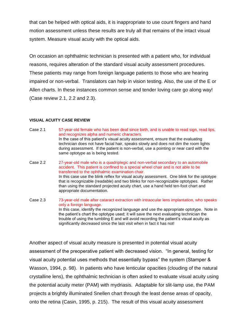
On occasion an ophthalmic technician is presented with a patient who, for individual
reasons, requires alteration of the standard visual acuity assessment procedures.
These patients may range from foreign language patients to those who are hearing
impaired or non-verbal. Translators can help in vision testing. Also, the use of the E or
Allen charts. In these instances common sense and tender loving care go along way!
(Case review 2.1, 2.2 and 2.3).
VISUAL ACUITY CASE REVIEW
Case 2.1 57-year-old female who has been deaf since birth, and is unable to read sign, read lips, and recognizes alpha and numeric characters.
In the case of this patient’s visual acuity assessment, ensure that the evaluating technician does not have facial hair, speaks slowly and does not dim the room lights during assessment. If the patient is non-verbal, use a pointing or near card with the same optotype as is being tested.
Case 2.2 27-year-old male who is a quadriplegic and non-verbal secondary to an automobile
accident. This patient is confined to a special wheel chair and is not able to be transferred to the ophthalmic examination chair.
In this case use the blink reflex for visual acuity assessment. One blink for the optotype that is recognizable (readable) and two blinks for non-recognizable optotypes. Rather than using the standard projected acuity chart, use a hand held ten-foot chart and appropriate documentation.
Case 2.3 73-year-old male after cataract extraction with intraocular lens implantation, who speaks
only a foreign language. In this case, identify the recognized language and use the appropriate optotype. Note in
the patient’s chart the optotype used; it will save the next evaluating technician the trouble of using the tumbling E and will avoid recording the patient’s visual acuity as significantly decreased since the last visit when in fact it has not!
Another aspect of visual acuity measure is presented in potential visual acuity
assessment of the preoperative patient with decreased vision. “In general, testing for
visual acuity potential uses methods that essentially bypass” the system (Stamper &
Wasson, 1994, p. 98). In patients who have lenticular opacities (clouding of the natural
crystalline lens), the ophthalmic technician is often asked to evaluate visual acuity using
the potential acuity meter (PAM) with mydriasis. Adaptable for slit-lamp use, the PAM
projects a brightly illuminated Snellen chart through the least dense areas of opacity,
onto the retina (Casin, 1995, p. 215). The result of this visual acuity assessment

provides information of the visual system to the examining ophthalmologist. It can
indicate the potential visual acuity achievable should the patient undergo cataract
surgery. It should be understood that the PAM might also be used in the evaluation of
patients with corneal scars that intersect the visual axis. If visual acuity is improved
during evaluation of a patient with central corneal scaring, the attending ophthalmologist
might elect to perform a corneal transplant.
Whether a patient is being evaluated in a base-line, preoperative, or postoperative
examination, the assessment of visual acuity can be altered by two additional elements
that are often overlooked: contrast-sensitivity and glare. Contrast-sensitivity refers to
the ability to discriminate relative darkness and brightness. It is a function of the visual
system to “detect detail having subtle gradations in grayness between the test target
and background” (Cassin, 1995, p. 73). Contrast-sensitivity can be impaired even in the
patient who has excellent Snellen acuity. A patient who presents with the chief
complaint of trouble seeing at night, in dim light, or on cloudy days should be evaluated
for contrast sensitivity. There are many tests available for this purpose, most “use
sinusoidal wave gratings that vary in both contrast and spatial frequency” (Wilson, 1996,
p. 167). At low spatial frequency, the stripes are wide and widely spaced. In high
spatial frequencies the stripes are narrower and closer together. These sine wave bars
of various frequencies slowly increase in contrast. The gratings are presented in
varying orientations and the patient is asked to identify the direction of the lines.
(Figure1.6 Sinusoidal gratings).
Figure 1.6. Sinusoidal gratings (Simulated). Low-contrast to the left and medium-
contrast to the right.
Practical application of contrast-sensitivity as it pertains to visual acuity, the patient
being assessed, and the evaluating technician in the clinical setting, can be reduced to
a few simple but important points:
Ensure appropriate standardized illumination of Snellen chart/reading card.

Keep all charts, projectors, screens, and reflecting mirrors clean.
If reading cards are “pocket kept” they should be replaced frequently as to
avoid wear and decreased contrast between the optotype and the card
background.
The final element of visual acuity assessment is glare. Since visual acuity can degrade
considerably in the presence of bright light, glare testing assesses the patient’s vision in
the presence of such light. The Brightness Acuity Tester (BAT) developed by Jack
Holladay, uses three controlled degrees of light that can be utilized in conjunction with
the Snellen acuity chart. While the fellow eye is occluded, the BAT is placed over the
viewing eye at low intensity and the patient is asked to read the smallest line they can
on the Snellen chart. The procedure is then repeated with medium and high intensity,
all results are then recorded in the patient’s medical record. (Figure1.7 Documentation
of BAT examination).
Documentation of BAT Visual Acuity
Patient with distance acuity of 20/40 in the right eye that is known to have cataract. The patient does wear
corrective lenses.
DVA OD cc 20/40 BAT 20/40 low
20/60 med
20/80 high
Patient with distance acuity of 20/50 OU due to central corneal scaring. The patient does not wear glasses and is
presented for BAT evaluation only.
BAT sc 20/70 low 20/80 low
20/100 med 20/100 med
20/200 high 20/800 high
Figure 1.7. Documentation of BAT Examination.
SUMMARY
Visual acuity is measured to evaluate the status of the visual system! Although visual
acuity testing measures only one aspect of visual function, it is the one test when
administered in a concise, consistent manner by a trained, caring ophthalmic technician
that can detect changes in the integrity of the visual system. It is of significant
importance that the evaluating ophthalmic technician uses the appropriate equipment
and technique to assess visual acuity in every ophthalmic patient. A pseudo-visual
acuity under any circumstance sets the stage for inappropriate patient care and is in
sharp contrast to what a sensitive, caring ophthalmologic staff should provide.

REFERENCE Cassin, Barbara and Sheila A. B. Solomon (1990). Dictionary of Eye Terminology (2
nd ed.) (M.L.
Rubin, MD, Ed.). Gainesville: Triad Publishing Company.
Cassin, Barbara, MEd, CO, COMT (1995). Ophthalmic Technical Personnel. Philadelphia : W.B. SaundersCompany.
Herrin, Michelle Pett, COMT (1990). Ophthalmic Examination and Basic Skills. New Jersey: Slack Incorporated.
Stamper, R.L., MD and Wasson, P.J., MD, (Eds.). 1994. Ophthalmic Medical Assisting: an Independent Study Course Second Edition. San Francisco: American Academy of Ophthalmology.
Stein, Harold A., MD, Slat, Bernard J., MD and Stein, Raymond M., MD, (1994). The Ophthalmic Assistant: A Guide for Ophthalmic Medical Personnel (6
th ed.). St. Louis: C.V.
Mosby.
Wilson, Fred M., II, MD, (Ed.). (1996). Practical Ophthalmology: A Manual for Beginning Residents. San Francisco: American Academy of Ophthalmology.

VISUAL ACUITY: THE CRITICAL MEASURE!
This article and accompanying quiz are worth 1 JCAHPO Group A continuing education credit.
1. A patient’s vision may decrease considerably in the presence of bright light. Glare testing can best be achieved using a
a. Potential Acuity Meter (PAM). b. Transilluminator. c. Brightness Acuity Tester (BAT). d. Pocket acuity tester.
2. Which device is helpful in evaluating the visual acuity in a non-verbal patient?
a. A color plate b. A Titmus stereo fly c. A pointing card d. A horizontal prism bar
3. Assessment of binocular acuity may be indicated when is present.
a. amblyopia b. nystagmus c. strabismus d. myopia
4. When evaluating a patient’s near vision what position of a progressive multifocal lens should the
patient be viewing through the a. distance position. b. intermediate position. c. inferior intermediate position. d. lowest position of the progressive segment.
5. If a patient’s visual acuity improves two or more lines upon evaluation with the pinhole, there is
probably a a. cortical cataract present. b. refractive error present. c. macular degeneration present. d. diabetic retinopathy present.
6. The purpose of visual acuity measurement is to
a. document the presence of ocular pathology. b. determine the near point of convergence. c. document ocular binocularity at distance. d. determine the integrity of the visual system.
7. Vision (visual integrity) in the newborn can be evaluated by observation of
a. an ability to fix and follow. b. an alertness to bright colored toys. c. a consistent blink reflex. d. a binocular eye movement.
8. The visual acuity 6/18 is equivalent to
a. 20/20. b. 20/40. c. 20/60. d. 20/80.

9. In a child known to have amblyopia the visual acuity should be assessed a. in a binocular manner. b. in a linear manner. c. in an isolated manner. d. both b and c above.
10. When the visual acuity is documented as 20/50 +2, it means
a. the patient read the 20/50 line and two letters on the following line. b. the patient read two letters of the 20/50 line and two letters on the following line. c. the patient read the 20/50 line and two letters of the previous line. d. the patient read the 20/50 line and two letters of the previous and following line.

Visual Acuity: The Critical Measure
(#10)
COMPLIMENTARY QUIZ ANSWER SHEET
This quiz is worth 1.0 JCAHPO Group A continuing education credits
You must complete the enclosed quiz on your own and may not seek assistance from other individuals. You may, however, seek assistance from other individuals for clarification and understanding of the article content. Failure to comply with this policy may result in the revocation of credit.
Name JCAHPO ID #
Home Address
City / State / Zip
Home Phone Work Phone
E-mail Address Signature
A total of 75% or more correct is a passing score.
ATPO is not responsible for Answer Sheetsnot received at the ATPO offices.
I attest that I have completed this quiz on my own. (SIGNATURE)
______________________________________________________
For Administrative Use Only:
Score: __________ Date: ___________
Return completed answer sheet and evaluation to: ATPO
2025 Woodlane Drive St. Paul, MN 55125 Fax: (651) 731-0410
Email: [email protected]
1. ________ 6. ________
2. ________ 7. ________
3. ________ 8. ________
4. ________ 9. ________
5. ________ 10. ________
ALLOW UP TO TWO (2) WEEKS FOR PROCESSING
ATPO Member – Complimentary* OR ATPO Member – Complimentary Retest*
*All articles are also available for purchase at EyeCareMarketplace
Place your answers to each question in the appropriate space adjacent to the question number.
*ATPO members may earn up to 5 free CE credits each membership year. To earn the free CE credits, you must be an ATPO member in goodstanding, then either read an article or listen to an audio tape, take the accompanying quiz and check the ATPO “complimentary” credit box on thequiz answer sheet. Once completed, email, mail or fax the quiz to ATPO. If you fail the quiz and have not used all five complimentary quizzes, you
may submit the retest as a complimentary quiz. If you fail and have used all of your complimentary quizzes, you may visit EyeCareMarketplace and take the quiz at a full-price rate.
You are only allowed to utilize each article once during your three-year recertification period.

CE Evaluation
VISUAL ACUITY: THE CRITICAL MEASURE!
Please read each question carefully. Your feedback is important to us. Thank you! 1. How long have you been employed in the field of ophthalmology? years 2. This written article was designed at a level right for me. (circle one) YES NO 3. Please read the following statements. Then, circle the number corresponding to the degree
to which you agree with each statement.
Statement Strongly
Agree Agree Disagree
Strongly Disagree
Not Applicable
1. The material was organized and presented in a clear and efficient way.
5 4 3 2 1
2. The information will be useful/relevant to me.
5 4 3 2 1
3. The material was presented at a level appropriate to my background and level.
5 4 3 2 1
4. Overall, I was satisfied with the article. 5 4 3 2 1
4. What part of the article was most useful to you?
5. What part of the article was least useful to you?
6. What suggestions do you have for improving this article?
Thank you for completing this article evaluation! ATPO values your feedback. Please return your evaluation form with the completed quiz. Return completed answer sheet and evaluation form to:
ATPO Attn: CE Dept
2025 Woodlane Drive St. Paul, MN 55125 Fax: (651) 731-0410