VISIONS - Canon Medical Systems Europe · 2016-08-08 · VISIONS magazine is a publication of...
56
Infinix-i Rite Edition Exercise Myocardial Performance in Adolescent Athletes Clinical Added Value of Volume CT Everything we do is Made For life VISIONS Magazine for Medical & Health Professionals I August 2016 14 I X-RAY 18 I CORPORATE CAMPAIGN 39 I ULTRASOUND 43 I COMPUTED TOMOGRAPHY 27
Transcript of VISIONS - Canon Medical Systems Europe · 2016-08-08 · VISIONS magazine is a publication of...

Infinix-iRite Edition
Exercise Myocardial Performance in Adolescent Athletes
Clinical Added Value of Volume CT
Everything we do is
Made For life
VISIONSMagazine for Medical & Health Professionals I August 2016
14 I X-RAY
18 I CORPORATE CAMPAIGN
39 I ULTRASOUND
43 I COMPUTED TOMOGRAPHY 27

VISIONS magazine is a publication of Toshiba Medical Systems Europe (Toshiba) and is offered free of charge to medical and health professionals. The magazine is published twice a year. Registration to access full, previously published, digital editions can be done via the web site: www.toshiba-medical.eu/visions. Toshiba stores and uses personal data of the registration to send out the magazine and inform members about new developments. Members can customize preferences or opt-out, after registration, in the online VISIONS profile. VISIONS magazine is covering Toshiba’s European region and as such reflects products, technologies and services for this particular area. The mentioned products may not be available in other geographic regions. Please consult your Toshiba representative sales office in case of any questions. No part of this publication may be reproduced in whole or in part, stored in an automated storage and retrieval system or transmitted in any manner whatsoever without written permission of the publisher. VISIONS Magazine is not responsible for any inaccuracies in this publication.
News items and articles are announced firstly, as pre-publication, via the dedicated VISIONS LinkedIn Group: https://www.linkedin.com/groups/3698045. In this group you can actively participate in discussions about the content and future direction of the magazine. Follow us also on SlideShare: http://www.slideshare.net/toshibamedical.
PublisherTOSHIBA Medical Systems Europe B.V.Zilverstraat 1NL-2718 RP ZoetermeerTel.: +31 79 368 92 22Fax: +31 79 368 94 44Web: www.toshiba-medical.euEmail: [email protected]
Editor-in-chiefJack Hoogendoorn ([email protected])
Modality coordinators and reviewersCT: Roy Irwan, Chloe StevesonUL: Jeroen UijttenhoutMR: Dirk Berneking
Design & LayoutBoerma Reclame (www.boermareclame.com)
PhotographySteven Seet, Ralf Günther (Photopage)
PrintmanagementHet Staat Gedrukt (www.hetstaatgedrukt.nl)
Text contributions and editingThe Creative Practice (www.thecreativepractice.com)
Subscription Servicewww.toshiba-medical.eu/visions
© 2016 by TOSHIBA Medical Systems EuropeAll rights reserved
ISSN 1617-2876
27The ‘made for life’ philosophy prevails as Toshiba Medical’s ongoing commitment to humanity.Read more on page 18.
All articles should be double-spaced.
FULL LENGTH ARTICLES: Full length articles should generally include the following:- Author’s full name and highest academic degree, employer
medical institution- Author’s biography (150 words)- Author’s passport-size photograph (suitable for publication);
(image of 300 dpi)- 200-word abstract- Text including headline, sub-title, introduction and sections like:
materials & methods (which should include a full description of equipment used), results, discussion and references
- Text approx 4 to 5 pages or 12.000 to 14.000 characters (not including figures, tables and photographs)
- Correspondence address- Literature (no more than 10 references)- Separate, continuous numbered image- and table captions
SHORT CONTRIBUTIONS: Short contributions should generally include the following:- Author’s full name and highest academic degree- Author’s employer medical institution- Author’s biography (150 words)- Author’s passport-size photograph (suitable for publication);
(image of 300 dpi)- Text including headline, sub-title, introduction and full descrip-
tion of methods & materials/equipment used- Case Report or description of system improvements (Technical
Notes)- Correspondence address- Literature (no more than 10 references)- Separate, continuous numbered image- and table captions
TEXTThe article should be saved in Microsoft Word (PC format) if pos-sible, and, if not, in text only.
Please indicate the software program and version used (Microsoft Word 2007, etc.) and whether it is a PC or Macintosh formatted document. If e-mailing, make sure to send it as an attachment, rather than embedded in the e-mail message.
SYMBOLS, FORMULAS AND ABBREVIATIONSSymbols, Greek letters superscripts/Subscripts must to be identi-fied clearly. Furthermore, the figure 1 (one) and the letter l (el) as well as the capital letter 0 and the figure 0 (zero) should be easy to differentiate.
All abbreviations including units of measure, chemical names, technical or medical acronyms, names of organisations or institu-tions should be defined when they first appear in the text (e.g. congestive heart failure (CHF). Please refrain from using unfamiliar abbreviations, clinical slang or jargon.
IMAGES, ART AND TABLESCite all figures and tables in text, preferably in consecutive order. Please include a caption for each figure. All captions for each figure, should be separate from the text, at the end of the manu-script on a separate page. Captions should avoid duplication of text material. Credit lines for artwork can appear at the end of the corresponding caption by stating: (Provided by first initial, last name). Black out (or give clear instructions which parts should be blackened out) of the images to not violate any data protection regulations (e.g. patient data)
Do not embed figures, charts, or graphs into your document file. Please provide them as a separate file, as well as hard copy/correct .pdf file. Please use one the following formats: EPS , TIFF or JPEG.
Arrows stuck onto images for purposes of delineation should be clearly visible and reproducible.
Authors should indicate if they would like to have artwork returned.
Each table should have a title, and all abbreviations should be spelled out or explained in a footnote.
STYLETitle page should include full names, degrees and titles of authors, and affiliations (name of institution, city and state) for use in a by-line, as well as phone and fax numbers to facilitate sending edited copy back to author for approval.
Define all symbols, abbreviations and acronyms on first reference.
All manuscripts should be written in a third-person style, unless the article is specifically an editorial or first-hand review.
REFERENCESA maximum of 10 references is suggested. Complete references should be listed in order of citation in text, NOT alphabetically. Up to four authors will be listed; if there are five or more authors, only the first three will be listed, followed by et al. Within the text, reference numbers should appear as footnotes in parentheses or in superscript text at the end of each appropriate citation. Please do not use Microsoft Words endnote feature, as this causes major problems in the editing phase.
In addition, if the reference is not in English, please indicate the language of publication.
Journal example:Oberhaensli RD, Galloway GJ, Hilton-Jones D, et al. The study of human organs by phosphorus-31 topical magnetic resonance spectroscopy. Br J Radiol 1987;60:367-373
Book example:Welch KMA, Barkley GL. Biochemistry and pharmacology of cer-ebral ischemia. In: Barnett JHM, Stein BM, Mohr JP, Yatsu FM, eds. Stroke: pathophysiology, diagnosis and management. New York: Churchill Livingstone, 1986;75-90.
Works are generally classified into two categories: Full length articles (e.g. clinical added value of new/special applications & technologies) and Short contributions (e.g. system testimonials, case reports, technical notes).
General guidelines for authors
Copy
right
© 2
015
Soci
al M
edia
Exa
min
er®
V27_Authors Guidelines.indd 55 02-08-16 11:59

VISIONS27 | 3
While working hard to get this 27th edition of VISIONS magazine to print, the countdown to the Summer
Olympic Games, in Rio de Janeiro, Brazil, runs quietly in the background. Still 15 days, 17 hours, 25 min and 36
seconds to go, as I write this text, before the official opening ceremony will start.
The modern Olympic Games are the leading international sporting event featuring Summer- and Winter sports
competitions, in which thousands of athletes from around the world participate in a variety of competitions.
The Olympic Games are considered to be the world’s foremost sports competition, with more than 206 nations
participating1.
Country teams and individual athletes from all over the world have been preparing, physically and mentally for
this event, for months in advance. Training hard, scrutinizing their nutrition and ensuring that they get enough
rest. All to reach peak condition to enable them to give their best, and, hopefully, an ultimate sports performance.
Taking part in the games should not be taken lightly. “You need to have guts when you’re up against it. You need
a dream, a goal, and above all, your family,” commented Oscar Figueroa on participating in the Olympic Games
himself2. In itself, this is quite true, but I would like to add that professional, medical counseling and monitoring
are also indispensable requirements to reach the highest goals.
Toshiba is proud to be a leading imaging expert that is heavily involved in and contributes to the optimal health
condition of athletes. Renowned football clubs, such as Manchester United, FC Barcelona, Inter Milan and AS
Monaco, use Toshiba equipment and systems to keep their players in optimal condition. It is not a coincidence
that you will find several articles in this edition of VISIONS magazine that reflect our knowledge, experience and
expertise in the field of sports medicine.
I say: “Let the games begin!”
PS: did you know we also host a VISIONS Magazine LinkedIn Group? Feel free to join us!
https://www.linkedin.com/groups/3698045
Kind regards,
Dear reader,
EDITORIAL
Jack HoogendoornSr. Manager Marketing Communications
Toshiba Medical Systems Europe BV
1 Wikipedia; The Free Encyclopedia2 Weightlifting - London 2012 Olympic silver medal winner for Colombia

©2016 TOSHIBA MEDICAL SYSTEMS4 | VISIONS27
The world’s fastest and most flexible angio suite; the Infinix-i Rite Edition.
14 Infinix-i Rite Edition – 3D Anywhere in Angiography
18 At Toshiba Medical, everything we do is made for life.
20 Karolinska University Hospital invests in TAVI examination
23 Managed Equipment Services
28 The Future of Ultrasound - Shaped by Fifty Years of Heritage
32 Hyaluronic acid protection of cartilage
14
20
18
Dr. Subhash Srivastava; Radiologist specialized in cardiovascular diagnostics at the Karolinska University Hospital.
Toshiba: Made For life
CONTENTSX-RAY
Infinix-i Rite Edition
COMPUTED TOMOGRAPHY
TAVI, Aortic Valve, Vascular examinations
ULTRASOUND
Heritage, history, introduction
MAGNETIC RESONANCE
Knee, sports medicine, MSK, cartilage
MES
MES, Equipment & Technology Management, Financial, Process & Performance optimization

VISIONS27 | 5
Dose, Metal artefact reduction
35 Superb Micro-Vascular Imaging – A new tool for the sports physician
39 Exercise Myocardial Performance in Adolescent Athletes
43 German Armed Forces and Patients Benefit from New Options for Low-Dose Volume CT
48 Initial experiences of 3D SMI in Pediatric Radiology
52 Current Status of Computed Tomography for TAVI Planning
SMI, sports medicine, neovessels
2D WMT
28
52
39
Fifty years of dedication to Ultrasound have led to Toshiba’s newest premium Ultrasound solution: The Aplio i-series.
TAVI is used to treat aortic stenosis; the most common heart valve disease in the Western World.
Set-up for simultaneous performance of exercise echocardiography and cardio-pulmonary exercise testing.
ULTRASOUND
Micro-Vascular Imaging, Pediatric
03 Editorial
06 News
17 Message from the President
ULTRASOUND
ULTRASOUND
COMPUTED TOMOGRAPHY
TAVI
COMPUTED TOMOGRAPHY

©2016 TOSHIBA MEDICAL SYSTEMS6 | VISIONS27
NEWS
TMVS wins ‘Made in Scotland - Life Sciences Company of the Year’ Award.
FC Barcelona unveils one of the most powerful magnetic resonance systems on the market
myWobble: a mobile platform for managing genetic data
Harefield Hospital partners with Toshiba
Malcolm Campbell, Vice President Engineering of Toshiba Medical
Visualitation Systems, picked up the coveted award that recognises
the ability to innovation and the contribution TMVS made in the life
sciences market in the last year. Examples of these contributions
are: Ultrasound luminance, MR Core imaging, Adaptive Motion
Correction and Cardiac Subtraction. All technologies that have found
their way to Toshiba’s global product portfolio.
The FC Barcelona Medical Centre has, as a result of its agreement with
Toshiba Medical Systems Europe, some of the finest technology in
the field of sports diagnosis. And now the facilities went one step
further with the installation of a 3 Tesla nuclear magnetic resonance
machine, the most powerful device produced by the Japanese
medical technology firm. It will be used to diagnose muscle and bone
injuries in athletes at greater speed and accuracy levels than ever.
The myWobble app
transforms genetic data
to easy-to-interpret and
visualized information for
people with or without
a science background.
Users can learn about
their health profile,
ancestry, how they might
react to pharmaceutical
drugs and which genetic
marks cause their physical
features. Users can enjoy
the app while their
genetic data remains
private and stored solely
on their own mobile
device. myWobble is
available on iTunes for $ 9.99 on iPhone and iPad and enables the
personalgenomics revolution today.
Video: https://vimeo.com/103711315
Toshiba announced the design of a new cath lab at Harefield Hospital,
the largest heart and lung centre in the United Kingdom. The Toshiba
Infinix-i™ cardiac catheterisation laboratory (Cath Lab) is unique due
to the range of movements that the equipment can make around
the patient, these unique movements enable the Cardiologist to
operate without moving the patient on the treatment table which
contributes to a make procedures quicker, safer and more efficient.
The Hospital’s dedicated heart attack centre deals with acute and
immediate emergencies from outer north-west London, providing
primary angioplasty in its four specialist catheter laboratories with
the Toshiba Infinix-i being the latest. Its arrival-to-treatment time of
26 minutes is one of the fastest in Europe: speed of treatment has
been shown to be crucial to survival in such cases.
Mark Bowers, Cardiology Service Manager at Harefield Hospital
comments: ‘The project management from design to installation was
world class. By using Toshiba’s augmented reality planning tool, we
were able to produce a truly collaborative design that allowed the
team at Harefield team to feed into and agree design ahead of build.’
Mark Hitchman, UK Managing Director for Toshiba Medical Systems
comments: ‘The Cath lab design and installation at Harefield is a
testament to Toshibaís philosophy of partnering with clinicians
to develop innovative facilities, situated within an inspirationally
designed space that enhances their ability to provide fast, efficient
and effective care for their patients’.
Malcom Campbell holding the Made in Scotland Award.

VISIONS27 | 7
FotoMed HD is
an iPad app that
allows you compare
your photo with
health and medical
images from trusted
web resources, or
compare your own
photos (such as skin
conditions) taken
at different times.
You can browse all
popular health topics
on the MedlinePlus
mobile website, a service of the U.S. National Library of Medicine.
You can also retrieve or tag interesting images of any health topics.
More information: http://fotomed.zynsoft.com
NEWS
The system will be operated by staff from Assistència Sanitària which,
like Toshiba, is also an FC Barcelona sponsor, and will be used for the
diagnosis, treatment and rehab of both professional and amateur
athletes. Since the agreement with the football club was signed,
Toshiba has already provided thirteen echography systems to the
clubís medical services. Assistència Sanitària insurance policy holders
will also be able to use the new machine, located in the Medical
Centre at the Ciutat Esportiva Joan Gamper training ground.
Medical Reference and Image Comparison
Join our Hand-On Tutorials and visit our booth at ESC 2016 (27-30
August, Rome, Italy) to learn more about our products, systems and
technologies that are Made For life and provide you clinical advantages
in your every day work. We offer the opportunity to gain in-depth
knowledge and skills on the latest cardiac imaging technologies in
our sessions which will be hosted in Room 3 and 4 throughout the
day from Saturday to Tuesday. We’re located in Hall E4, Booth M100.
Patient Centric, Application-BasedVital Images recently launched version 7 of its Vitrea advanced
visualization software. This innovative, application-based platform
delivers full-powered solutions for 2D, 3D and 4D medical imaging and
comprehensive user workflows, including CT, MR and XA applications.
‘We are excited to launch the latest version of Vitrea advanced
visualization,’ says Jim Litterer, CEO of Vital Images. ‘With this release,
clinicians have access to enhanced diagnostic and workflow tools to
help improve patient outcomes and departmental efficiencies.’
With several new applications and unified, scalable deployment
options, Vitrea software empowers CMIOs and their clinicians to grow
their applications based on current and future needs. The flexibility of
partner applications being available on all deployments and content
personalization built into Vitrea software can help CMIOs gain more
rapid physician adoption.
Vital also streamlined the user interface and integration of new
applications to ensure consistent user experience across all modalities:
CT/MR/XA/PET/SPECT. By providing advanced clinical tools within a
uniform user interface, Vitrea software enables physicians to have
meaningful interactions wherever they are.
‘The standardized platform of Vitrea software provides seamless
integration of ‘best of breed’ partner applications, including a unified
reporting database,’ says Steve Andersen, general manager, AV and
EVP at Vital Images. ‘Its scalable architecture and comprehensive
solution-set, coupled with its proven ease of use and Vitalís world-class
service, offer clinicians superior performance and flexibility.’
Meet us at ESC 2016
Vital Introduces Vitrea® 7, a New Era in Advanced Visualization

©2016 TOSHIBA MEDICAL SYSTEMS8 | VISIONS27
Measuring the Impact of Top SportThe effect of top sports on cartilage and the chondral charge was
explained by Professor Gold from Stanford University, California, US. Dr.
Bossy, from the Clinica Creu Blanca Medical Center, focused on muscle
and tendon injuries and emphasized the importance of identifying the
function of each muscle and tendon with regard to the total support
structure. To explore the internal structure of peripheral nerves, Dr.
Lefebvre, from Lille University Hospital, in France, showed high level
diffusion images with tensor reconstruction.
Dr. Padrón, from the Clinical Centro, Madrid, in Spain, presented
interesting cases on chondral and osteochondral injuries. The advanced
diagnosis using quantitative imaging techniques showed that MRI is
increasingly emerging from a qualitative technique towards quantifiable
data that can easily be compared in follow-up examinations. From
chondral pathology a small step
was made into MR Hip imaging by
Dr. Cerezal from Clinica Diagnostico
Specialists from all over Europe with an interest in top level sports
were gathered to discuss this topic and were able to explore advanced
MRI techniques in musculoskeletal imaging at Toshiba’s European MRI
User meeting. The event was held at the stadium of one of Europe’s
top professional football clubs, FC Barcelona, in Barcelona, Spain. The
meeting was organized by Toshiba in collaboration with the Clinica
Creu Blanca Medical Center in Barcelona and Olea Medical – providers
of advanced MRI and CT post-processing and visualization solutions.
Speakers at the event included a variety of researchers, radiologists and
technicians, who shared results they have achieved in this field with
Toshiba MRI systems and Olea Medical’s advanced software.
Decision-Making Based on Precise ResultsDr. Canal, Chief Medical Officer of FC Barcelona, pointed out the
importance of advanced imaging techniques in his daily practice of
managing treatment and training of top athletes. He explained the
background of decision-making based on medical imaging. One of
the physicians of the FC Barcelona Medical Team, Dr.
Til, showed the results of MRI examinations on cartilage
problems sometimes experienced by players.
Dr. Blasi from the University of Barcelona presented
on the anatomy and histology of cartilage, muscles
and tendons. To get the best views of these tissues,
correct imaging protocols are crucial. Mrs. Ferrer,
Radiographer at Clinica Creu Blanca Medical Center,
and Mrs. Fernandes, from Toshiba Medical Systems in
Spain, showed an extensive overview of optimal MRI
techniques, pinpointing important issues with regard to
optimizing 3D imaging.
NEWS
Exploring MR Applications in Top Sport Advanced Techniques in Musculoskeletal Imaging
Playing sports at the highest levels increases the chance of injuries, as the body is pushed to its limits, and sometimes beyond. High quality imaging can play a key role in managing the training and treatment of top athletes through optimization of training schedules and injury prevention, as well as revalidation after injury.

VISIONS27 | 9VISIONS27 | 9
Access to Advanced KnowledgeThrough user meetings, Toshiba aims to provide its customers with
direct access to specialists with experience in advanced techniques
and applications of Toshiba’s technology that might be valuable in their
own work.
NEWS
Medico Cantabria, Santander, Mexico. Dr. Cerezal, who is an expert in hip
pathology, shared a large variety of hip pathologies visualized with MRI.
Muscle and tendon injuries were also explored in the event. While
Dr. Blasi from the University of Barcelona, explained the anatomy and
histology of these important structures, Dr. Teixeira from the University
of Nancy, in France, demonstrated how advanced imaging techniques
can be used to observe all the features and characteristics of muscles
and tendons.
Assessing Athletic PotentialProfessor Derave from Ghent University, in Belgium, presented a novel
Muscle Talent Scan Project. The ratio between different muscle types and
their abundance is unique to the performance potential of an athlete in
a specific sport. A 20 minute MRS scan can reveal the potential of an
athlete. In addition, optimization of training schemes based on
this knowledge could play a crucial role in injury prevention.
Professor Gold Stanford University, California, US, also explored
muscle velocity and the use of phase contrast to measure this.
Fusion TechniquesDr. Til also demonstrated the technique of fusion MRI and
Ultrasound modalities. Whereas problems are encountered
in Ultrasound, due to limits to penetration depth caused by
shading, the same is not incurred in MRI. However, MRI takes
more time is less easily available. Through synchronized fusion
of the images during live
ultrasound scans, the
operator can benefit from
the best of both worlds,
as demonstrated in a live
demonstration.

©2016 TOSHIBA MEDICAL SYSTEMS10 | VISIONS27
REVOLUTIONISE INTERVENTION
Toshiba’s ground-breaking new In� nix 4DCT supports you in bridging the gap between the interventional lab and CT with one seamlessly integrated solution. The system eliminates the need to transfer patients back and forth between di� erent rooms, while minimizing dose and maintaining patient safety. Helping to save valuable time and gain e� ciencies with the ability
to plan, treat, and verify in the same room, on a single system.
One Room. One System. One Procedure.
270 DEGREES OF FREEDOM CONVENIENT C-ARM PARKING POSITIONINFINIX 4DCT : THE MOST
POWERFUL ANGIO CT SYSTEM
MEET US AT CIRSE AND
JOIN OUR SYMPOSIUMON SUNDAY 11/09/2016
16:15-16:35 HRS
More information? www.toshiba-medical.eu ULTRASOUND CT MRI X-RAY SERVICES
SEE. DIAGNOSE. TREAT.
20160609_Toshiba-advertentie Infinix 4DCT - A4.indd 1 29-07-16 12:15

VISIONS27 | 11
NEWS
Recently Toshiba announced a strategic partnership with
The Advanced Wellbeing Research Centre in Sheffield, United
Kingdom. The partnership will see Toshiba providing state-of-the-
art diagnostic imaging equipment and wearable biosensors as
well as ongoing consultancy with regard to application innovation
and development for AWRC applied research.
Much of the research expertise will come from research teams
that helped the Great Brittan Olympic Team achieve 24 medals
in London 2012 and will integrate into the National Centre for
Sport and Exercise Medicine that seeks to improve people’s lives
through physical activity.
Toshiba will also supply World class diagnostic imaging
equipment including its Aquilion ONE™ dynamic volume CT
system, which will allow researchers to successfully collect
images of athletes’ and patients’ entire organs in one rotation.
The imaging equipment comes with dynamic volumetric
acquisition protocols that can be used to review moving joint
structures in 3D, as well as dynamic blood or air flow that will
see Toshiba consultants partnering with AWRC researchers to
develop applications and best practice guidelines regarding the
early diagnosis, improved rehabilitation and preventative care for
those that are exercising both at elite athlete level and for the
local community and wider public.
Professor Steve Haake, AWRC Director, comments: “We are
delighted that Toshiba has become AWRC’s first industry
partner joining Government’s backing. Media articles about
overweight children, rising levels of obesity and diabetes appear
in a newspaper almost every day. Most recently the World Health
Organisation reported that nearly three quarters of men and two
thirds of women in the United Kingdom will be overweight by
20201. That’s less than 5 years away!
“Sedentary behaviour and a stream of rich food can lead to all sorts
of problems appearing in our lives as chronic disease. This was
highlighted by a 2011 report for the Department of Health, which
showed that increasing physical activity could reduce the risk of
type II diabetes and colon cancer by up to 50%, heart disease and
stroke by up to 35%, depression by 30% and the other scourge of
our day, Alzheimer’s disease, by 30%2.
Left to right: David Hobson - Chief Executive Legacy Park Ltd, Jane Ellison MP - Minister for Public Health, Mark Hitchman - Managing Director Toshiba
Medical Systems UK, Graham Moore - Westfield Health Chairman, Rt Hon Richard Caborn - Project Lead Legacy Park Ltd, Professor Steve Haake - AWRC
Director, Professor Karen Bryan - Pro Vice-Chancellor and Dean of the Faculty of Health and Wellbeing at Sheffield Hallam University, Professor Paul
Harrison - Pro Vice-chancellor for Research and innovation at Sheffield Hallam University.
State-of-the-art diagnostic imaging in sports medicine About the strategic partnership with The Advanced Wellbeing Research Centre (AWRC)REVOLUTIONISE
INTERVENTION
Toshiba’s ground-breaking new In� nix 4DCT supports you in bridging the gap between the interventional lab and CT with one seamlessly integrated solution. The system eliminates the need to transfer patients back and forth between di� erent rooms, while minimizing dose and maintaining patient safety. Helping to save valuable time and gain e� ciencies with the ability
to plan, treat, and verify in the same room, on a single system.
One Room. One System. One Procedure.
270 DEGREES OF FREEDOM CONVENIENT C-ARM PARKING POSITIONINFINIX 4DCT : THE MOST
POWERFUL ANGIO CT SYSTEM
MEET US AT CIRSE AND
JOIN OUR SYMPOSIUMON SUNDAY 11/09/2016
16:15-16:35 HRS
More information? www.toshiba-medical.eu ULTRASOUND CT MRI X-RAY SERVICES
SEE. DIAGNOSE. TREAT.
20160609_Toshiba-advertentie Infinix 4DCT - A4.indd 1 29-07-16 12:15

©2016 TOSHIBA MEDICAL SYSTEMS12 | VISIONS27
NEWS
“With these statistics and predictions in mind, the Advanced
Wellbeing Research Centre has been set up to become the most
advanced research and development centre for physical activity in
the world, creating ‘innovations that help people move‘ in sport,
healthcare, physical activity and leisure“.
Acting as a hothouse of innovation, the AWRC will bring
together a set of industry partners who manufacture and supply
technology and equipment including apps, activity loggers, sports
equipment, orthotics, and clinical devices. The main focus of the
AWRC research and innovation will be on how technology is used,
how people interact with technology that helps them become
more physically active, and how improvements in physical health
are captured and monitored in order to provide an evidence base
regarding the positive impact of physical activity on health.
Projects will aim to find out what works in large populations,
initially using the population of Sheffield as a field laboratory.
Additionally, the AWRC is set to become a critical assistance
partner for the National Health Service by providing an evidence
base that proves the positive impact of exercise on the health of
the population in the United Kingdom.
Mark Hitchman, Toshiba Medical Systems‘ UK Managing Director
comments: “Our partnership with AWRC forms a critical part of
Toshiba’s strategy to invest and partner in research projects that
will have considerable benefits to NHS efforts and the population
in the United Kingdom at large.
“AWRC will target wide swathes of the population, from those
who are completely sedentary to those who are most active; from
young to old; from those in the most deprived communities to
those in the most affluent; from those at home to those in work, in
education or in elite sport.
“Toshiba’s imaging equipment and exercise monitors will play a
crucial role in identifying and understanding the positive effects of
exercise on health, whilst helping to provide accurate diagnoses
and recovery paths for those that are injured.“
Mark Hitchman - Managing Director, Toshiba Medical Systems UK
(left) and Professor Steve Haake - AWRC Director (right)
As well as the AWRC, Toshiba’s radiology equipment is powering
not just some of the leading medical institutions, but also some
of the World’s leading sports organisations and community-
based sports facilities such as Manchester United Football
Club, FC Barcelona, Barnet Football Club, The Tessa Sanderson
Foundation and Academy and the recently held 2014 Glasgow
Commonwealth Games.
References1 http://gu.com/p/485k7. 2 Start Active Stay Active. Varney, Brannan and Aaltonen, 2011. Department of Health.
►Next page is part of the VISIONS Photo Page Series reflecting an eye for
the beauty of our planet, the environment and the direct surroundings
where Toshiba’s systems are installed by Toshiba and its customers. Not
the actual imaging products but photos of sceneries, cities, countries or
other cultural aspects are highlighted on this photo page.
Every reader of VISIONS can participate and get their picture published.
The submitted content should include: high resolution (300dpi) image,
photo of the hospital and a brief text, name of photographer and Toshiba
system(s) installed. The complete result is shown on the opposite page.
Send your pictures and texts to: [email protected],
Subject: Photo Page

VISIONS27 | 13
The Leibniz Institute for Zoo and Wildlife Research (IZW) is an internationally renowned research institute of the Leibniz Association. With the mission of “understanding and improving adaptability” it examines evolutionary adaptations of wildlife and its resilience to global change, and develops new concepts and measures for conservation. To achieve this, the IZW uses its broad interdisciplinary expertise in evolutionary ecology and genetics, wildlife diseases, reproductive biology and manage-ment in a close dialogue with stakeholders and the public.
Web: www.leibniz-izw.de - Text & photography: Steven Seet
The tiger (Panthera tigris) is the largest cat species, most recognisable for their pattern of dark vertical stripes on reddish-orange fur with a lighter underside. The largest tigers have reached a total body length of up to 3.38 m (11.1 ft) over curves and have weighed up to 388.7 kg (857 lb) in the wild. The species is classified in the genus Panthera with the lion, leopard, jaguar and snow leopard. Tigers are apex predators, primarily preying on ungulates such as deer and bovids. They are territorial and generally solitary but social animals, often requiring large contiguous areas of habitat that support their prey requirements.
Text Source: Wikipedia – Photography: Ralf Günther

©2016 TOSHIBA MEDICAL SYSTEMS14 | VISIONS27
Infinix-i Rite Edition – 3D Anywhere in Angiography
Around the world, providers of Interventional systems are being challenged to improve ergonomics, patient comfort and dose efficiency while at the same time reducing the cost of ownership and environmental impact.
PRODUCT X-RAY
Infinix-i Rite Edition
The world’s fastest, most flexible angio suite

VISIONS27 | 15XREU150023
By fulfilling all these demands Toshiba is proud to intro-
duce the world’s fastest and most flexible angio suite; the
Infinix-i Rite Edition, a new member of the Infinix-i family.
As its new flagship, the Infinix-i Rite Edition incorporates
state-of-the-art technologies allowing whole body cover-
age, from head to toe without any patient or table move-
ment and free head access, which is a strong demand
from interventional radiologists and anesthetists during
complex procedures, is realized through 270° isocentric
C-arm rotation. The unique lateral C-Arm stroke expands
and simplifies your access for radial approach, shunt
angio, venography and port implants.
“Our approach is simple: it’s the equipment that moves
around the patient and not the other way around. This
translates into optimized visualization and positioning
in the examination room”, says René Degros, Business
Unit Manager for X-Ray with Toshiba about this new “3D
anywhere” system as it is already being called among
physicians.

©2016 TOSHIBA MEDICAL SYSTEMS16 | VISIONS27
The system provides full automatic synchronization
between the flat panel detector & collimator for correct
head up display regardless of C-Arm position, developed
for the most challenging clinical procedures.
As procedures are getting more complex, advanced 3D
acquisition becomes a strong requirement. With the
introduction of the Infinix-i Rite Edition, you will discover
new horizons in 3D imaging.
The outstanding 3D rotation coverage of 210 degrees
with C-arm at table left/right side in combination with an
amazing speed of 80 degrees per second is the key ena-
bler for the unmatched image quality delivered by the
Infinix-i Rite Edition. Especially its unparalleled rotational
speed will result in a significant reduction of breathing
artefacts and contrast media.
Benefit from Toshiba’s Dose Rite Philosophy which brings
together what belongs together: dose saving technolo-
gies & dose awareness tools. Drastically reduce radiation
dose to your patients and yourself by applying state of
the art dose saving techniques available such as Live
Zoom, Spot Fluoroscopy, Spot ROI and our real time Dose
Tracking System.
More information: http://tinyurl.com/zd3w3dh

VISIONS27 | 17
PRESIDENT’SMESSAGE
“ Our business foundation is based on partnership.”
Ever since our foundation over 100 years ago, we have
opened new eras in various fields with our epoch-making
innovations and we are proud of many “world firsts”. It
takes strong collaboration and a dedicated team to
achieve such efforts in developing ongoing innovations.
While reflecting on our history and achievements, we
are now ready to move on to yet another new era in a
collaborative manner with the continued support of our
customers.
A final agreement concerning the sale of Toshiba Medical
Systems Corporation to Canon Inc. was reached in March.
As a result, Toshiba Medical is now independent from the
Toshiba Group, but will continue to operate as usual.
From the beginning, my aim during this acquisition phase
was to achieve smooth transition while maintaining our
core values, and network with customers that we have
built through our 100 year history.
Our business foundation is based on partnership with
customers and this will not change.
We will continue to invest in R&D, our core competencies
and our growth strategies.
While diagnostic imaging will always be our core business,
we understand the importance of enhancing our solution
business in the short-term, and we are seeking to expand
our business domain into the bio-technology field in the
mid to long-term.
Our “Made For life” philosophy is a driving force behind
who we are and what we do and we are excited that
our new partner Canon will not only bring forth new
opportunities for our business but will help maintain and
enhance our Made For life philosophy.
Our goal is to continue to contribute to improved
healthcare opportunities for patients, and enhanced
healthcare delivery by industry professionals through
uncompromised performance, comfort and safety
considerations of the solutions Toshiba Medical provides.
The strong and long lasting relationships built with our
customers and partners along the way, ensures Toshiba
Medical’s future growth and ongoing contributions to
society for the next 100 years.
Toshio TakiguchiPresident and Chief Executive Officer
Toshiba Medical Systems Corporation

©2016 TOSHIBA MEDICAL SYSTEMS18 | VISIONS27

VISIONS27 | 19GNEU160015
For over 100 years, the Toshiba Medical ‘made for life’ philosophy prevails as our ongo-ing commitment to humanity. Generations of inherited passion creates a legacy of medical innovation and service that continues to evolve as we do. By engaging the brilliant minds of many, we continue to set the benchmark because we believe quality of life should be a given, not the exception.
At Toshiba Medical, everything we do is made for life.
HERITAGEFor over 100 years, we have proudly pioneered the develop-ment of imaging solutions for the global medical community. Our rich history of collaboration has engaged the brilliant minds of many who will continue to set the benchmark for another 100 years and beyond.
SERVICEBuilt on a tradition of ‘people first’, our globally recognised commit-ment to prompt and personalised service, after sales support and quality education collaboratively lies at the heart of our ongoing success. We take care of our part-ners so they can take the best care of their patients.
PARTNERSHIPSAt Toshiba Medical, we build relationships based on transpar-ency, trust and respect. Our commitment to progressive medical, corporate, academic and community partnerships lies at the very heart of what we do and why we do it. Together as one, we strive to create industry-leading solutions that deliver an enriched quality of life.
INNOVATIONThousands of minds and millions of hours over a century of time have built a legacy of innovation that continues to evolve as we do. From our collaboratively rich research and development past we advance towards a bright future of technological innovation that is made for life.
PASSIONA unified passion to the ‘made for life’ philosophy is ingrained in our DNA. Our dedicated team and partners collectively push the boundaries in everything we do because we believe quality of life should be a given, not the excep-tion. Generations of inherited passion builds a culture that is driven to excel for the benefit of humanity.
HEALTHIt is our mission to provide medi-cal professionals with solutions that support their efforts in contributing to the health and wellbeing of patients worldwide. Our goal is to further develop and grow Toshiba Medical’s role in delivering optimum health opportunities for patients and health professionals through uncompromised performance, comfort and safety features.
Toshiba Medical’s focus on health proudly defines who we are and what we achieve through our made for life philosophy.
QUALITYA shared dedication in providing quality products and support enables us to deliver a seamless service of patient care, now and for future generations.At Toshiba Medical our quality processes underpin everything we do, reinforcing a future that is made for life.
MADE FOR PARTNERSHIPS
MADE FOR PATIENTS
MADE FOR YOU

ultra-helical to image the heart and entire aorta and iliac
arteries in just one breathhold and one contrast injection.
“This particular machine is ideally suited for heart and
vascular examinations,” said Dr. Subhash Srivastava, a
Radiologist specializing in cardiovascular diagnostics. “I
believe that the future will bring increasingly advanced
examinations of this type. Naturally, it is important to be
at the forefront in terms of technology, methodology and
advanced equipment, but it is equally essential to establish
good cooperation in various ways: between doctors and
nurses, radiologists and cardiologists and between health
care professionals and suppliers. It is only when all this
teamwork functions properly that healthcare can carry out
its primary task.”
Preoperative TAVI planning was an obvious option right from the beginning, when the new Toshiba Aquilion ONE™ CT scanner, was installed at Karolinska University Hospital, in Solna, Stockholm, Sweden. After a few years, this investment in the latest CT tech-nology for vascular examinations is bringing good results. Referring physicians have discovered new opportunities, and TAVI-examinations are in full swing.
Dr. Subhash Srivastava 1)
CARDIOLOGY COMPUTED TOMOGRAPHY
1) Radiologist specializing
in cardiovascular diag-
nostics at the Karolinska
University Hospital, in
Stockholm, Sweden.
Despite its relative novelty, TAVI (Transcatheter Aortic Valve
Implantation) is a practice that has become very popular.
The treatment means that even patients, who are not able
to undergo surgery, can receive a new aortic valve, and the
procedure is currently available at all hospitals in Sweden
that offer thoracic surgery.
A special CT protocol has already been developed for TAVI
examinations at Karolinska University Hospital’s facility in
Huddinge, Stockholm. Now, Karolinska’s Solna facility, also
in Stockholm, is taking a further leap forward in the field,
with its state-of-the-art Aquilion ONE CT scanner. The
protocol takes advantage of the benefits of volumetric one
rotation imaging of the heart combined with the speed of
TAVI, Aortic Valve, Vascular examinations
Karolinska University Hospitalinvests in TAVI examination
©2016 TOSHIBA MEDICAL SYSTEMS20 | VISIONS27

“We all need to think about how we can work together to
provide the best help to those who need us,” he added. “Our
focus should always be on the patient first and foremost.”
Most of those, who undergo TAVI are older patients -
people, for whom surgery is considered too dangerous.
Preoperative CT examinations should, therefore, have
particular focus on reducing IV contrast as much as
possible, since these patients generally have impaired
renal function.
Radiography Nurse, Anna Eldrot Eliasson, is Karolinska Solna’s
contact person for cardiovascular examinations performed
with Aquilion ONE, including the TAVI application.
“We have many patients who were born in the 1930s,
and even the 1920s, so, of course, high creatine levels can
sometimes be a challenge,” she explained. “In these cases,
we can often manage by injecting a little less contrast,
since the image quality is still so good with this machine.
The normal standard dose is 70ml for patients over 70
years. Of course, everything is performed in consultation
with our resident- and referring doctors here, and it is rare
that anyone has doubts.”
This is important from a risk-benefit perspective. When
asked about any other special challenges specifically related
to CT examinations for TAVI planning, Anna responded that
the most important aspect is about dealing with the patient.
Figure 1: 3D reconstruction of the entire aorta and access vessels
Figure 3: 3D reconstruction and MPR of the entire aorta and access vessels
Figure 2: 3D reconstruction of the entire aorta and access vessels with skeleton
Figure 4: Aortic root and aortic valve with ascending aorta
SOU
RCE:
DR.
SU
BHA
SH S
RIVA
STAV
A, K
ARO
LIN
SKA
SO
LNA
CTEU160111 VISIONS27 | 21

“The advanced part of the procedure is handled by the system itself, through carefully developed protocols, specific reconstruction and customized software for sophisticated visualization.”
She emphasized that it is important to make patients feel
safe, ensure that they are calm and relaxed and breathe
properly, also to obtain a good ECG signal.
Managing the actual technology in connection with the
scanning is regarded as relatively simple. The advanced part
of the procedure is handled by the system itself, through
carefully developed protocols, specific reconstruction and
customized software for sophisticated visualization.
“Now that the protocols have been fine-tuned, I feel that
everyone now thinks that the machine is very straight-
forward to work with,” said Dr. Srivastava. “However, it has
taken some time to get everything in place. Obviously, a
successful examination with a brand new, modern CT
scanner requires both advanced training and configuration.”
Dr. Srivastava and Anna are very satisfied with the support
they have received from Toshiba during the process. Over
the past six months they have seen clear results from their
efforts.
“We are now receiving a growing number of referrals,”
remarked Anna. “We are seeing an increase in the number
of TAVI cases as well as patients with acute cardiovascular
problems”
“Everything related to the heart and blood vessels feels
more exciting and fun to work on, now that we have the
Aquilion ONE, because it is so good,” she said. “This applies
not only to TAVI. Just today, we performed a leg vessel
examination. The doctors are extremely pleased with the
quality of the examinations.”
Anna also appreciates the speed of the system and the fact
that the CT scanner covers the entire heart in one rotation,
a great advantage in many examinations.
With his vast experience and interest in cardiovascular
diagnostics, Dr. Srivastava is delighted to have established
a range of collaborations, which are effective in several
aspects.
“We must constantly maintain a high level of expertise in
the field, for both nurses and doctors,” he said. “This requires
constant development of both personnel and practices, in
collaboration with, and support from Toshiba. Lately, there
has been rapid progress in the field of TAVI, and there are
many more exciting aspects of cardiovascular diagnostics
to focus on.”
FACTSSymptomatic aortic stenosis has a high annual mortality of
about 50%. Implantation of a prosthetic valve is the only
way to reduce mortality. Valve implantation can be through
surgical or catheter introduced techniques (Transcatheter
Aortic Valve Implantation, TAVI). The prostheses used for TAVI
consist of an artificial valve of animal material, which sits in
a metal mesh.
Advanced age, general frailty, impaired renal function,
impaired lung function, and previous cardiac surgery
are examples of risk factors that may mean that TAVI is
preferable to surgery. TAVI can be performed with good
results for patients aged over 90, if the patient is otherwise
deemed eligible. In its new national guidelines for cardiac
care, the Swedish National Board of Health and Welfare has
proposed the wider use of TAVI. Now, the treatment may be
considered routine, even for patients, who can undergo valve
replacement by conventional surgical means, but where the
risks associated with surgery are deemed to be high.
The majority of TAVI procedures are performed via the
femoral artery approach, and usually with local anaesthesia.
The procedure takes about two hours and the treatment
time is about five days. TAVI planning CT scans can measure
the size of the aortic valve in detail and assess whether the
arteries are of sufficient size to be able to insert the artificial
valve. In case the femoral artery is too narrow, TAVI can be
performed via the subclavian artery, by direct puncture of
the aorta or by transapical procedure. In these cases, the
patient is anaesthetized.
©2016 TOSHIBA MEDICAL SYSTEMS22 | VISIONS27

VISIONS27 | 23
PARTNERSHIP MES
Managed Equipment Services
Toshiba has now added “provider of services” to its historical role of “technology supplier”. What are the rationales behind this structural business model transformation?
Lo Wuite: From a macroscopic point of view, European
healthcare markets are for the most part affected by
important cost-saving measures, while facing the complex
challenges of an aging population and increase of chronical
diseases. These markets are at a turning point of their
history and thus have to innovate and renew the way they
provide care to their citizens. Given this context, the old
model where a company like Toshiba would basically sell
medical equipment financed by hospitals or any another
public or private stakeholders appeared to us as becoming
outdated and unsustainable.
As a matter of fact, we are moving our main focus from
a purely product approach to combined approach of
products and Managed Equipment Services (MES) offering,
where we partner with our customers by providing a very
broad range of solutions that specifically aim to tackle main
hospitals’ hurdles. MES can thus include purchasing and
financing (directly or via a third-party partner), technology
supplying, performance management and optimization,
and education.
In our new business model, hospitals primarily deal with
our MES division, which means that they don’t primarily
buy products anymore. In our new approach, hospitals
now partner with Toshiba, which supports them with a
comprehensive service offering through an outsourcing
agreement, which stipulates the level of collaboration
and service level agreements that ties Toshiba and its
partner. Together with the hospital, we jointly discuss the
requirements of the hospitals, in terms of the technology,
services and replacement timelines they foresee for the
future.
In many aspects, 2016 truly marks the dawn of a new era for Toshiba Medical Systems.
Lo Wuite, General Manager Managed Equipment Services (MES) Europe, and
Dick Blesing, General Manager Netherlands, explain how Toshiba Medical System
is becoming a true healthcare service provider, thanks to an innovative and flexible
“state-of-need” business model.
Lo Wuite 1), Dick Blesing 2)
Lo Wuite
Dick Blesing
Do you perceive some reluctance from your healthcare partners in truly partnering with private companies to improve their processes?
Lo Wuite: This shift of Toshiba from a product-centered
approach to a service focus is evolving in almost all pharma
markets globally, and the cost-containment context
currently hitting most of the other European healthcare
systems has been a powerful catalyst to implementing
these new kinds of partnerships. The Netherlands situation
stands as a very edifying illustration of how quickly
these partnerships can be developed: before 2008, only
hospitals facing major financial problems were interested
in partnering with the industry, whereas an increasing
number of hospitals have shown interest over the past few
years. Due to changes in the way the government subsidies
hospitals, the hospitals are forced to steadily rethink the
way they can continue to provide high quality healthcare
and how to finance it.
Nevertheless, there are still a large number of hospitals that
consider their processes are being optimal, meaning that
the use of external benchmarks and implementation of
best practices remain rather poorly executed.
Dick Blesing: We still notice that hospitals that are not
facing financial problems are probably more reluctant to
reevaluate their processes. It is thus our role to ensure they
understand the value we can offer them. Nevertheless,
Dutch hospital board members stay in the board for a
relatively short period before moving to another challenge.
Long- term benefits are thus probably less valorized than
short or mid-term perspective approaches.
This also explains why we strive to engage as much as
possible the management level, who are usually staying
longer in the same institution than hospital board members
and can enjoy the benefits of long-term partnerships.
Introducing an innovative and flexible “state-of-need” business model
MES, Equipment & Technology Management, Financial, Process & Performance optimization
1) Toshiba Medical
Systems Europe2) Toshiba Medical
Systems Netherlands
GNEU16016
©2016 TOSHIBA MEDICAL SYSTEMS24 | VISIONS27
Nevertheless, the general trend in the Netherlands is that
hospitals will have to deal with more complex patients in
the upcoming years (due to aging population) with less
money, so hospital mindsets are also steadily more open
to partnering with private companies and jointly finding
solutions.
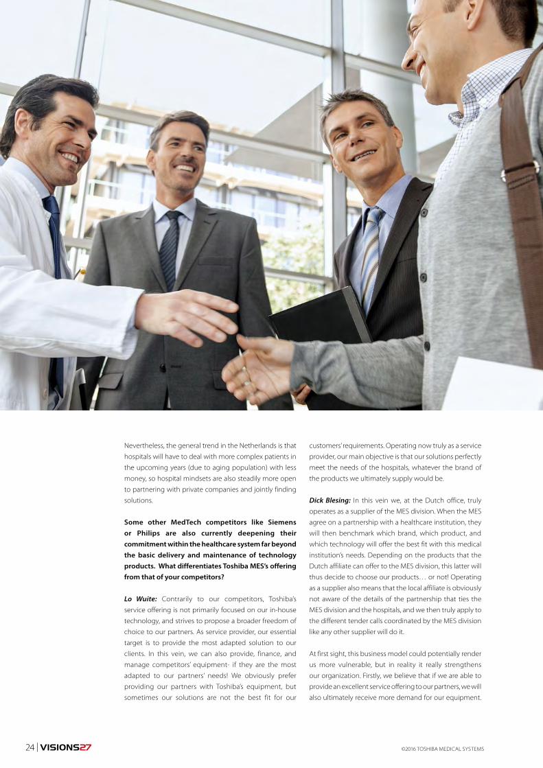
Some other MedTech competitors like Siemens or Philips are also currently deepening their commitment within the healthcare system far beyond the basic delivery and maintenance of technology products. What differentiates Toshiba MES’s offering from that of your competitors?
Lo Wuite: Contrarily to our competitors, Toshiba’s
service offering is not primarily focused on our in-house
technology, and strives to propose a broader freedom of
choice to our partners. As service provider, our essential
target is to provide the most adapted solution to our
clients. In this vein, we can also provide, finance, and
manage competitors’ equipment- if they are the most
adapted to our partners’ needs! We obviously prefer
providing our partners with Toshiba’s equipment, but
sometimes our solutions are not the best fit for our
customers’ requirements. Operating now truly as a service
provider, our main objective is that our solutions perfectly
meet the needs of the hospitals, whatever the brand of
the products we ultimately supply would be.
Dick Blesing: In this vein we, at the Dutch office, truly
operates as a supplier of the MES division. When the MES
agree on a partnership with a healthcare institution, they
will then benchmark which brand, which product, and
which technology will offer the best fit with this medical
institution’s needs. Depending on the products that the
Dutch affiliate can offer to the MES division, this latter will
thus decide to choose our products… or not! Operating
as a supplier also means that the local affiliate is obviously
not aware of the details of the partnership that ties the
MES division and the hospitals, and we then truly apply to
the different tender calls coordinated by the MES division
like any other supplier will do it.
At first sight, this business model could potentially render
us more vulnerable, but in reality it really strengthens
our organization. Firstly, we believe that if we are able to
provide an excellent service offering to our partners, we will
also ultimately receive more demand for our equipment.

VISIONS27 | 25
Secondly, creating this technology competition between
our products and our competitors’ is the best driver to
move us forward: we know that even if Toshiba has a
partnership with a hospital, we will nevertheless have to
offer the best products solution to ensure our products
are eventually chosen by the MES division.
How do hospitals and other business partners react when you announce that you are ready to finance competitors’ products if they are the most adapted for your patients ’ needs?
Lo Wuite: Hospital boards are sometimes doubtful
– particularly at the beginning. We are however able
to showcase many corporate case studies and real
situations that demonstrate this openness to truly act as
a service provider as well as our effort to not exclusively
give advantage to our products. For instance, in a mid-
size hospital in the UK, we financed, supplied and now
manage the entire equipment solutions of this hospital,
which represents more than 8,000 pieces of equipment.
In this hospital, only a minor share of the overall volume
equipment distributed are actually Toshiba products.
Dick Blesing: In a 10 or 15-year period, if we honestly
analyze the situation, it is really seldom that one single
company alone is able to offer the best or most suitable
solution to its partners for a given technology. Our
competitors use a less flexible business model, where
clinical freedom of choice is much more limited.
Lo Wuite: While our competitors’ service offering remains
mainly based on their products, our service offering is
primarily and strictly oriented toward the final quality of
service we want to deliver, even it implies distributing our
competitors’ products. We focus on the best solution for
our customers rather than the “brand” of the product.
Beside the possibility to distribute your competitors’ products if they offer the best fit with your partners’ needs, what other key features of the MES demonstrate its flexibility?
Lo Wuite: Technology is evolving very quickly and
products’ innovation cycles are shorter than the current
replacement cycles – usually between 10 and 15 years –
that most of the hospitals are financially able to sustain. As
a result, hospitals currently tend to ask for state-of-the-art
GNEU16016

©2016 TOSHIBA MEDICAL SYSTEMS26 | VISIONS27
products at each replacement, as they know the next ones
will only be acquired at least a decade later. As premium
equipment also obviously comes at the highest price,
hospitals over-invest at the beginning of their equipment
contracts despite already knowing this technology will
probably become technologically obsolete well within
the financial life cycle of the product
To tackle this non-sense, we implemented a “state-of-
need” approach in our MES partnership. “State-of-need”
can obviously sometimes be synonym of “state-of-the-art”
equipment, for spear point clinical and development areas
of the hospital for instance. Most of the hospitals however
don’t need such ultra-sophisticated equipment for all their
daily treatments.
With our “state-of-need” approach, the MES offering
provides hospitals with the technological level they truly
need for their patients, either with brand new or even
sometimes refurbished equipment. We indeed have
partnerships with University Medical centers, where state-
of-the art products are always replaced every two or three
years. Once it is removed from these hospitals, this kind of
equipment can perfectly suit smaller or peripheral medical
centers, which do not treat the most complex patients.
These centers can thus access high- quality products, which
are only two or three years old, for a very competitive price.
Beside financial and technological aspects, how are clinical and patient outcomes improved by your offering?
Dick Blesing: Firstly, the MES makes hospital staff ’s life
easier, as the MES manager operates towards the hospital
as a “one-stop shop” supply manager, while an average
European hospitals can deal with over a 100 different
suppliers only for imaging departments!
More about MES
Lo Wuite: Physicians and hospital managers should be able
to uniquely focus on treating their patients and improving
the clinical outcomes of hospitalization. Thanks to our
approach, the medical teams give us their requirements, and
we will prepare a benchmark of the best available options
to fulfill their needs. As this kind of partnership is gaining
in importance, we notice that physicians increasingly rely
and trust our propositions, while they particularly enjoy
not wasting time anymore in product negotiations with
different suppliers. Thanks to our partnerships, physicians
and medical staff simply provide care, and this is exactly
how the situation should be.
The MES business model is progressively being implemented in all European markets. Do you think that it is set to become the partnership of reference in the Netherlands in the upcoming years?
Dick Blesing: Without any hesitation: yes! This approach,
which is not centered nor conceived to exclusively sell
our products, is the service offering that displays the
better outcomes for healthcare partners, by providing
them with the solutions that will truly and perfectly meet
their needs.
Nevertheless, I call for a closer collaboration with
hospitals: we still do not perceive each other enough
as true partners; despite this being the only way
to strengthen an affordable and innovative Dutch
healthcare system. Companies and hospitals have their
own knowledge and expertise, but if we don’t manage
to combine these two expertises, we will never be able to
effectively improve the Dutch eco-system, which should
be our shared responsibility and duty. If the MES offering
indeed represents a new business model for Toshiba, it is
above all a true partnership that we want to propose to
our Dutch healthcare partners.
In a world of growing demands and limited budgets, managing the costs of healthcare is very challenging. A Toshiba Managed Equipment Service (MES) partnership is a valuable option for your facility to reduce the financial and operational risks of your medical equipment planning while delivering better patient care.
Covering everything from procurement and financing to maintenance, asset management, commissioning and decommissioning of equipment, staff training and a dedicated helpdesk, a Toshiba MES partnership is a
comprehensive solution for all your medical equipment needs. Together we will continuously strive to improve your workflows and processes so you can serve your patients better and more efficiently.
With more than ten years of experience in successfully managing MES partnerships in Europe and around the world we provide the expertise and trust you require. As a supplier with an ISO-certified quality system for MES partnerships we ensure fully transparent management and customer satisfaction.

VISIONS27 | 27
THINK SERVICES FOR ALL YOUR MEDICAL EQUIPMENT NEEDSPartnering with a leading healthcare provider such as
Toshiba under the umbrella of a Managed Equipment
Service partnership enables you to focus on delivering
the best patient care while we manage your equipment
with the highest standards of quality and efficiency at fully
projectable cost. Imagine the convenience of one reliable
partner covering all your medical equipment needs.
Managing your technologyFrom pro-active maintenance to servicing, from
procurement to decommissioning and replacement – as
a full-service provider we will manage all aspects of your
medical equipment under the MES agreement. A central
helpdesk dedicated to your facility will provide continuous
support and handle all service calls quickly and efficiently.
Maintenance and service from a single sourceMaintaining a large equipment park can be challenging.
In an MES partnership we take on this responsibility. We
guarantee uptime and service schedules under strict
Service Level Agreements and, in accordance with our
“Made for Life” promise, we make sure your equipment
is replaced timely to best meet your clinical needs at
all times.
Clinical freedom of choiceA Toshiba MES is vendor-independent. Although we offer a wide range of premium performance imaging equipment, we provide our clients the freedom of choice to ensure you will be working with the equipment that suits your clinical needs best. In case these needs change, you can amend your equipment plan at pre-agreed service fee adjustments.
MINIMIZE YOUR BUDGETARY RISKS WHILE IMPROVING YOUR PERFORMANCEA Toshiba MES partnership helps you to take the key risks and spikes out of your medical equipment capital spending.While we assume those risks and manage your assets, you can plan your operations with fully projectable and pre-determined monthly payments covering everything from equipment service and maintenance to new purchases and financing, as well as staff training.
Capital asset planningEach MES starts with developing a long-term investment plan – typically 10 to 15 years – based on your operational objectives, clinical needs and medical equipment requirements. This ensures clearly defined targets and fully transparent processes from day one of our partnership. Should your requirements change over time we will jointly adapt your technology plan to meet your needs.
Fixed monthly chargesAn MES partnership will convert your variable investments in capital assets and new technology into a fully projectable service charge with a pre-determined monthly fee. Under the agreement we will provide regular reports on equipment uptime, service and maintenance status and user support with clear, quantifiable scores to match or even exceed the agreed Service Level Agreements.
You remain always in controlWith our cloud-based AMP2HI software you have at all times full access to all relevant information with regard to your installed equipment, service schedules as well as contractual spendings. Should your clinical need change over time, the AMP2HI software allows you to simulate the impact of changes in equipment or services on your monthly charge.
SHARING BEST PRACTICE SOLUTIONS MEANS IMPROVING YOUR BUSINESSWith many years of experience in the widest variety of
hospital environments our teams of clinical, technical and
finance professionals provide expert advice helping you
to optimize diagnostic procedures, capital asset utilization
and financing strategies. Our six sigma-trained specialists
can assist in optimizing hospital processes and patient
pathways – all with the aim to improve efficiency while
delivering best clinical services.
Operational performance enhancementAs a major supplier to the medical imaging market we
provide a solid financial, clinical and technical foundation
to successfully manage your equipment needs. Getting
all services from one source means you can benefit from
Toshibaís significant procurement power and reduce the
complexity of your maintenance and service needs.
Capital asset utilizationRequirements can change. Therefore, a long-term
agreement needs flexibility. By regularly reviewing and
adjusting the investment plan against your actual and
projected needs we ensure effective utilization of assets,
for instance by re-scheduling equipment moves or
replacements. To bridge new installs or temporary high
workloads we can provide short-term rentals and mobile
solutions.
Clinical workflow optimizationSharing best practice experience is an important part of
our customer relationship. As a leading manufacturer of
medical imaging equipment our clinical experts can draw
on extensive know-how in the widest range of clinical
specialties. We work with leading institutions around the
globe on continuously improving diagnostic outcomes
and advancing clinical pathways.
GNEU16016

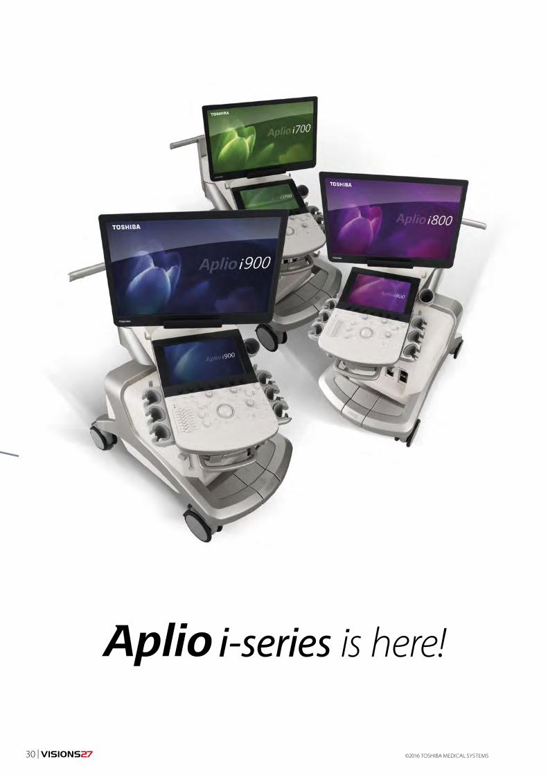
The era of i-series has begun.
The Future of Ultrasound - Shaped by Fifty Years of HeritageInnovation is deeply embedded in our history, as well as in our future. Half a century ago, Toshiba made its first steps into the world of Ultrasound, determined to improve the quality of life for all people around the globe.
Now, 50 years later we take another leap forward with the
introduction of the Aplio i-series, a premium diagnostic
ultrasound system that combines superior image quality
with the most advanced clinical applications in a highly
intuitive design.
Aplio i-series is the result of 50 years commitment to
Ultrasound, in which we have always stayed true to our
‘Made for Life’ philosophy. With the Aplio i-series, we are
writing a new chapter in Ultrasound history and provide
you with technology that is ready for the biggest clinical
challenges of today – and the future.
PRODUCT ULTRASOUND
Heritage, history, introduction
©2016 TOSHIBA MEDICAL SYSTEMS28 | VISIONS27

Link YouTube: http://tinyurl.com/jl5sjad
VISIONS27 | 29
2016
©2016 TOSHIBA MEDICAL SYSTEMS30 | VISIONS27

PRODUCT ULTRASOUND
Aplio i900
Fifty years of dedication to Ultrasound, and carefully
listening to our customers from around the globe,
have led to Toshiba’s newest premium Ultrasound
solution: The Aplio i-series.
This system combines industry-leading image
quality with the most advanced clinical applications
in a highly intuitive design. The Aplio i-series is
engineered to boost clinical confidence during
quick routine examinations, as well as the most
challenging cases. The Aplio i-series supports an
abundance of expert tools that help change patient
pathways and increase departmental productivity,
without compromising on clinical precision.
Intuitive. Intelligent. Innovative.Aplio i-series
INTUITIVE OPERATION
Onscreen navigation and a fully customizable touch command screen visually guide you through your examinations, making them easier and quicker.
INTELLIGENT IMAGE OPTIMIZATION
Simplified control panel together with a range of automated image optimization tools help you to increase efficiency, with less focus on the system and more on your patient.
POWERFUL PROCESSORS
Extraordinary computing power enables complex 4D visualizations, advanced flow imaging and real time quad-modes that help you to get the most reliable results in the shortest time.
INNOVATIVE TRANSDUCER TECHNOLOGY
All transducers are designed to do more with less, from ultra-wideband transducers that combine two probes in one, to 24 MHz probes that are capable of revealing extreme fine detail.
Advanced Wall Motion
Tracking
Automated Mitral Valve
Assessment
Visual assessment for better
surgical planning
PREMIUM CLASS ERGONOMICS
A premium Ultrasound system in a remarkably compact and light-weight design to optimize flexibility and ergonomic relief in every clinical situation.
IBEAM ARCHITECTURE
From the smallest to the toughest patients, iBeam-forming technology enhances clinical accuracy by delivering images with unprecedented clarity and penetration.
VISIONS27 | 31
©2016 TOSHIBA MEDICAL SYSTEMS32 | VISIONS27
1) Clinica Creu Blanca,
Barcelona, Spain
CASE STUDY MAGNETIC RESONANCE
Knee, sports medicine, MSK, cartilage
Hyaluronic acid protection of cartilage
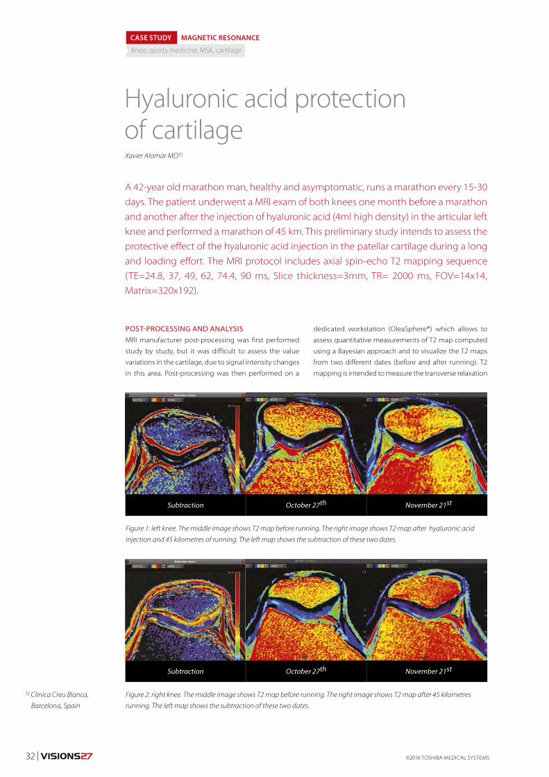
A 42-year old marathon man, healthy and asymptomatic, runs a marathon every 15-30 days. The patient underwent a MRI exam of both knees one month before a marathon and another after the injection of hyaluronic acid (4ml high density) in the articular left knee and performed a marathon of 45 km. This preliminary study intends to assess the protective effect of the hyaluronic acid injection in the patellar cartilage during a long and loading effort. The MRI protocol includes axial spin-echo T2 mapping sequence (TE=24.8, 37, 49, 62, 74.4, 90 ms, Slice thickness=3mm, TR= 2000 ms, FOV=14x14, Matrix=320x192).
Xavier Alomar MD1)
POST-PROCESSING AND ANALYSISMRI manufacturer post-processing was first performed
study by study, but it was difficult to assess the value
variations in the cartilage, due to signal intensity changes
in this area. Post-processing was then performed on a
dedicated workstation (OleaSphere®) which allows to
assess quantitative measurements of T2 map computed
using a Bayesian approach and to visualize the T2 maps
from two different dates (before and after running). T2
mapping is intended to measure the transverse relaxation
Figure 1: left knee. The middle image shows T2 map before running. The right image shows T2 map after hyaluronic acid
injection and 45 kilometres of running. The left map shows the subtraction of these two dates.
Figure 2: right knee. The middle image shows T2 map before running. The right image shows T2 map after 45 kilometres
running. The left map shows the subtraction of these two dates.
Subtraction
Subtraction
October 27th
October 27th
November 21st
November 21st

VISIONS27 | 33MREU150017
Figure 3: subtraction maps from T2 maps before and after running of right
and left knees with focal ROIs in the central patellar cartilage; axial T2 series
of right and left knees after running.
Figure 4: subtraction maps from T2 maps before and after running of right
and left knees with free hand ROIs surrounding the cartilage; axial T2 series of
right and left knees after running.
from a spin-echo sequence, and T2 parameter being very
sensitive to noise and sampling, the Bayesian probability
theory is used to estimate this parameter.
Automatic co-registration of both exams was applied
based on the femur localization. Since the patella moved
between the two exams, a manual adjustment was done
to match the cartilage zone.
Subtraction maps were computed to assess value
changes for both knees. Quantitative values allow to
confirm and quantify post-effort lesion.
IMAGE FINDINGSA dissection of medial patellar cartilage of the left knee
is observable, water was trapped in the crack and the
T2 maps values increase. The subtraction map shows no
significant changes on T2 values in the lateral patellar
cartilage and minimal changes in the crack. The right
knee did not undergo injection, the subtraction map
clearly shows higher T2 values than those of the left knee
that suggests an increase water proportion in the matrix.
The comparison of both subtraction maps from T2
maps before and after running confirms the separate
analysis done previously. Focal regions of interest (ROIs)
containing thirty five pixels (7mm2) were drawn in the
central patellar cartilage of the left (ROI1) and right
(ROI2) knees and a significant difference was remarkable
(ROI1=2.23; ROI2=7.38) (Figure 3).
In addition, free hand ROIs surrounding the cartilage were
drawn in the left (ROI3) and right (ROI4) knees and they
also show a major increase in values of the subtraction
map of the right knee, compared to those of the left knee
(ROI3 =1.95; ROI4=5.57) (Figure 4).
DISCUSSION The cartilage in joint areas helps to absorb the strengths
and share the loads supported by the joints. These
structures supporting repeated loads for many years can
be broken, but their degeneration always comes before.
The consistency of these structures changes but without
any modification of their morphology or their size.
T2 mapping sequences are commonly used to quantify
the grade of the edema and the alteration of connective
tissues, part of the cartilages in the human body.
Therefore, the degree of chondral degeneration can be
measured before its breakage.
In order to assess the efficiency of a treatment intended
to repair or protect the articular cartilage, it is essential
to undertake a longitudinal study, using images that
quantify the chondral damage prior and post-treatment.
Measuring the signal variations in the cartilage using ROIs
is very complicated. That is why subtracting images from
two different exams is very useful to assess the changes
in the cartilage composition. Such technique helps to
rapidly, simply and objectively quantify the effects of the
different chondral therapies. Figures and statistical works
allow to demonstrate their efficiency.

Vitrea® | Advanced Visualisation VitreaView® | Universal Viewer
Vitality SolutionsTM | Imaging Intelligence VioSuiteTM | Data Management and Interoperability
Join the conversation. www.vitalimages.com
Unleash the power of imaging by ensuring the right person has the right images at the right time. Vital’s enterprise imaging solutions facilitate streamlined access to images at relevant and meaningful
points of care, driving better patient outcomes and improved imaging operations.

VISIONS27 | 35
1) Head of Football
Medicine & Science
Manchester United,
United Kingdom.
TECHNOLOGY ULTRASOUND
SMI, sports medicine, neovessels
Superb Micro-Vascular Imaging –A new tool for the sports physician
In the past decade there has been an explosion in the use of diagnostic ultrasound by sports
medicine practitioners, predominantly physicians/surgeons but also the allied professions
such as physiotherapists. The ultrasound scanner has become a useful (and sometimes
invaluable) aid to the team physician when providing daily care to his/her athletes at the
clinic, sporting venues and when travelling.
Dr Steve McNally 1)
Grey-scale ultrasound provides anatomical and structural
information that can also be assessed under dynamic
functional stress and gives immediate information that
can influence diagnostic decisions, shape training and
rehabilitation programmes and provide visual feedback
and education to the athlete patient. Additional
functionality such as colour and power Doppler
enables the visualisation of vascularity and objective
measurement thereof within and around the joints and
soft tissues that form the vast majority of presentations
to a sports/musculoskeletal physician. Superb
Micro-Vascular Imaging (SMI) is the latest addition to the
armoury available to the sports physician/radiologist with
access to Toshiba’s diagnostic ultrasound hardware.
CLINICAL RELEVANCEWhy is visualisation of vascularity important when dealing
with sports medicine cases? There has been a shift of
thought regarding the presence of vascularity in or
around soft tissues, joints or bony entheses in recent years;
maybe the excitement of being able to detect neovessels
following the addition of colour and power Doppler
ULEU160050
Figure 1: cSMI of Achilles tendon showing subtle
neovascularisation in subacute tendinosis
Figure 2: Grey-scale and mSMI comparison
of subtle Achilles tendinosis

©2016 TOSHIBA MEDICAL SYSTEMS36 | VISIONS27
functionality on the early portable ultrasound scanners
around 15 years ago led the sports medicine practitioner
to an impression that this was a ‘bad’ pathological
entity. Moreover there was a tendency to associate such
vascularity with painful conditions or injuries and an
assumption that the associated neural proliferation was
the cause of pain and therefore a temptation to obliterate
the neovessels by various treatments. This was particularly
true in cases of tendinopathy; however further research
uncovered multiple potential pain generators including
chemical mediation and psychological factors that play
a significant part in the appreciation of tendon pain
irrespective of the degree of neovascularity. Vascularity in
tendons increases after exercise so the timing of the scan
is important and as the neovessels are easily compressed,
transducer pressure and positioning of the joint the
tendon crosses can heavily influence the appearances.
The author’s view from experience of scanning many
athletes is that neovascularity in an injured soft tissue
is a good thing as it suggests that a repair/regeneration
process is in progress; there is much more concern about
a non-painful weight-bearing tendon where an inert
hypoechoic lesion is detected on routine screening as
this implies a lack of healing response and potential loss
of tensile load-bearing capacity which translates to a
higher risk of rupture. In joints however neovascularity
tends to be associated with an unwanted chronic
inflammatory process (synovitis) which may be a result
of systemic disease, post-surgical or reactive to joint
instability or impingement.
Figure 4: cSMI of peritendinous/periosteal neovascularity
in healing navicular tuberosity fracture
Figure 6: cSMI of healing proximal
myotendinous tear in adductor longus muscle
Figure 5: mSMI Hold image of patellar tendon with
advanced proliferative proximal tendinosis
Figure 3: cSMI of periosteal neovascularity
in tibial sesamoiditis

VISIONS27 | 37ULEU160050
Detection of neovascularity by ultrasound modalities
must therefore be interpreted in context of the following:
DiagnosisUltrasound scanning is usually a confirmatory adjunct
when diagnosing an athletic injury suspected
from clinical history, clinical examination and sport
performance findings. However sometimes the presence
of subtle neovascularity can clinch a diagnosis that is
not completely evident from the history or examination
alone; this is often in the subacute phase where a
healing response is already in progress as the athlete
has continued training or competing with a niggling
injury leading to subjective or objective reduction in
performance. Examples that spring to mind include a
small intramuscular tear that might prevent an athlete
from sprinting at absolute top speed but causes no
impairment in all other respects or a very subtle Achilles
tendinosis that only causes pain on a change of surface/
pitch firmness.
StagingTendinosis has well recognised stages of progression
and the presence or otherwise of neovascularity is a
defining factor confirming that it has progressed from
the acute reactive phase into the proliferative phase;
this has important implications for clinical management
in terms of exercise load management and the use of
anti- inflammatory medication (the latter may be useful
in the early reactive phase but detrimental to collagen
formation in the proliferative phase).
Neovascularity following structural muscle injury
indicates an attempt to regenerate muscle tissue which
is a preferable outcome for an athlete when compared
to seroma formation or fibrosis; many treatments
used by sports physicians treating muscle injuries
specifically aim to stimulate production of Vascular
Endothelial Growth Factor (VEGF) whilst also inhibiting
Transforming Growth Factor (TGF) in order to promote
regeneration over repair. This can sometimes accelerate
recovery and return to athletic activities but more
importantly restores better functionality and reduces
risk of re-injury.
Healing responseThe majority of athletic muscle injuries are functional in
nature i.e. with no evidence of macroscopic structural
disruption on ultrasound or MRI even if oedema may
be present from lymphatic or perimuscular ‘congestion’.
In cases where there is fibre or bundle disruption to
an extent that can be detected within the resolution
capacity of imaging modalities the presence of
neovascularity indicates a favourable healing response.
If left to natural means vascularity encircling a muscle
tear is usually visible at around 14 days post-trauma.
Effectiveness of treatment/early rehabilitationMedical treatments that aim to promote neovascularity
in muscle injury such as High Power Laser Therapy,
Autologous Plasma injection, Platelet- Rich Plasma
injection or Actovegin can accelerate the production
of neovessel formation so monitoring via ultrasound is
a useful way of assessing the patient’s response to such
treatments; neovascularity can be observed around the
7 – 10 day stage if these treatments are effective.
Determination of readiness to load tissues Similarly, regression of the neovascularity and
replacement by new muscle, tendon or ligament fibres
indicates the onset of the remodelling phase and
readiness to load the injured tissue in a more functional
and challenging manner specific to the demands of the
sport.
Ongoing monitoringTendinopathy is often a chronic relapsing and remitting
process in athletes in line with the mantra ‘once a
tendinopath always a tendinopath’, however it can be
managed and does not necessarily lead to an inability
to compete at the athlete’s maximal performance
level. This of course requires judicious training regimes
and optimal preparation and recovery strategies but
monitoring of the tendon structure and vascularity
can play an important part in raising confidence
in the decision making by the athlete and his/her
coaches, trainers and therapists. This is perhaps the
greatest benefit of ultrasound scanning in general
as a modality in the hands of a sports physician as it
becomes an extension of the normal clinical and
functional assessment process and being very visual,
facilitates education and awareness of the athlete and
his/her support team with regard to the structure and
physiology of musculoskeletal tissues.
Comparison of Doppler and Superb Micro-Vascular Imaging (SMI) Power Doppler has generally been preferred to colour
Doppler as the sports physician is less interested in
direction of vascular flow and more interested in the
detection of small low flow neovessels and the pattern
thereof in injured tissues. Power Doppler leads to
unwanted ‘noise’ in the image and a very steady hand is
needed when scanning to avoid artefact when looking
for subtle neovascularity e.g. when assessing healing in
a minor muscular tear.
Superb Micro-Vascular Imaging provides a much
clearer and defined outline of the vascular pattern
with less movement-related artefact and the facility to
employ the ‘Hold’ function allows the vascular tree to be
constructed over several seconds whilst the operator
holds the transducer in a static position. The added

Figure 8: Power Doppler image of proximal
patellar tendinosisFigure 7: cSMI of near-complete regression of
neovascularity in a healing soleus muscle tear
Figure 9: cSMI of same patellar tendon highlighting the underlying
hypoechoic structural defect
attraction of both monochrome (mSMI) and colour
(cSMI) options makes this a more powerful visual tool
when discussing findings with the athlete. Building a
library of sequential scans is a useful record of progress
particularly when managing a chronic injury such as
tendinosis.
FUTURE DEVELOPMENTSWith the advent of sports science the modern athlete
and coach rely heavily on objective data and statistics
in order to complement their subjective opinions
and assessments; the addition of quantification of
microvascular blood flow could be a useful development
for the sports physician to further enhance the
advantages that Superb Micro-Vascular Imaging has
over power and colour Doppler. Athletes respond to
numbers and the addition of a quantifiable assessment
of blood flow in healing tissues would add another
level to the decision-making during rehabilitation or
when managing a chronic injury through a training
programme. In the meantime correlation of the image
appearances available via SMI against objective training
loads and injury presentations will provide a more
evidence-based approach to injury management.

VISIONS27 | 39
1) Children’s Health and
Exercise Research Centre,
University of Exeter, UK 2) Bristol Congenital Heart
Centre, The Bristol Heart
Institute, University
Hospitals, Bristol NHS
Foundation Trust, Bristol 3) National Institute for
Health Research (NIHR)
Cardiovascular Biomedical
Research Unit, Bristol Heart
Institute, University of Bristol 4) Toshiba Medical Systems
Europe, B.V.
SPORTS MEDICINE ULTRASOUND
2D WMT
Currently, echocardiography is however performed
only at rest and exercise performance, if assessed, uses
traditional cardio-pulmonary exercise testing (CPET).
A limitation of CPET is its inability to provide data on
myocardial functional responses to exercise, the domi-
nant process to increase cardiac output to enhance
oxygen delivery1 and it is therefore only an indirect
description of myocardial reserve. Data is particularly
sparse in children’s and adolescent’s athlete population
groups. But as sports professionalism and training levels
in elite youth athletes is reaching that of their adult peers,
the search for non- invasive, discriminative and pre dictive
imaging tools to assess the health of the young athlete,
for this rapidly increasing youth population, has gained
importance. Equally, the question about how to assess
exercise limitations is applicable and as important in many
paediatric disease groups e.g., congenital heart disease
(CHD) and novel methodologies and protocols to assess
cardiac function during exercise are needed.
In search for echocardiographic assessment tools
during exercise myocardial deformation imaging or
2D Wall Motion Tracking (WMT) has emerged as a
potentially suitable imaging modality during stress
echocardiography as it allows for direct assessment
of myocardial performance. Importantly, some 2D
strain parameters such as strain rate are relatively load
independent2 and correlate positively to invasive con-
tractility measurements3. 2D WMT is also more sensitive
to mild functional impairment than traditional echo-
cardiographic functional parameters and can assess
regional myocardial function. Image acquisition of 2D
Guido E. Pieles DPhil, MD, MRCPCH 2,3), Craig A. Williams, PhD, MSc 1), Carla Witkam – Van den Maegdenbergh 4)
Exercise Myocardial Performance in Adolescent Athletes
The assessment of cardiac function is essential in the athletic population not only as
part of the screening process for underlying cardiac disease, but also to longitudinally
assess performance and training adaptations.
A Novel Approach Using 2D WMT and Simultaneous Oxygen Consumption Analysis
ULEU160048
Figure 1: Set-up for simultaneous performance of exercise echocardiography and cardio-pulmonary exercise testing

©2016 TOSHIBA MEDICAL SYSTEMS40 | VISIONS27
strain data is time efficient and easy to obtain. Unlike
Tissue Doppler Imaging, 2D WMT also shows angle
independency, paramount when acquiring echocar-
diographic images during exercise. Echocardiographic
hardware and software however have to be robust and
able to allow tracking at high heart rates and challeng-
ing conditions during exercise and further development
work is required.
Combined, 2D WMT and CPET can provide a comprehen-
sive and direct description of cardio-pulmonary exercise
response and might also help differentiate between
normal and pathological myocardial function during
exercise. Here we present pilot data of a synergistic and
time efficient comprehensive assessment of cardio-
pulmonary response to incremental exercise, using
2D WMT to assess myocardial function in conjunction
with simultaneous assessment of metabolic gas ana lysis
by CPET, in adolescent elite athletes.
METHODS14 male adolescent professional football academy
players (mean age 15.4 ± 0.8 y) underwent echo -
cardiography at rest, during exercise and recovery while
completing an incremental CPET on a recumbent cycle
ergometer (Fig. 1). Echo c ardio-graphy at rest was per-
formed following England Football Association screen-
ing guidelines. LV myocardial performance was serially
assessed during exercise and recovery measuring LV peak
systolic longitudinal (LV Sl) and LV peak systolic circum-
ferential (LV Sc) 2D strain. The study was conducted
in collaboration with our research partners Toshiba
Medical Systems and Manchester United Football Club
Youth Academy, UK.
EXERCISE ECHOCARDIOGRAPHY AND 2D WMT Exercise echocardiography was performed using the
Artida for image acquisition and UltraExtend ACP soft-
ware for 2D WMT analysis. Analysis is also possible using
the AplioCV system and the on-board analysis software.
A LV focused 4-chamber view (Fig. 2) and a paraster-
nal short axis view (Fig. 3) were captured for 2D WMT
analysis at rest, at several exercise stages and
at recovery. Three cardiac cycles were acquired
at rates of 30–90 frames per second in raw
DICOM format and analysis was performed on
one manually selected cardiac cycle. The endo-
cardial borders were manually contoured at end-
diastole with the range of interest adjusted to
include the whole myocardium. Peak strain was defined
as the maximal deformation of a segment in systole
and represented as a percentage of the original size.
Standard nomenclature was used to describe the
LV segments and LV peak systolic longitudinal strain
recorded for the three lateral and three septal segments.
Circumferential peak systolic strain was measured at the
Figure 2: Representative 2D image sequence during exercise for LV Sl. A: rest at HR = 87 bpm; B: 50 W at HR = 90 bpm; C: 100 W at HR = 117 bpm
Figure 3: Representative 2D image sequence during exercise for LV Sc. A: rest at HR = 84 bpm; B: 50 W at HR = 96 bpm; C: 100 W at HR = 99 bpm

VISIONS27 | 41
base of the LV. Mean or global values for circumferential
and long itudinal strain were calculated for each level
only if good tracking was obtained in a minimum of four
segments. An incremental CPET on a recumbent cycle
ergometer to volitional exhaustion was performed by all
participants during image acquisition.
ASSESSMENT OF MYOCARDIAL PERFORMANCE BY 2D WMT DURING CARDIO-PULMONARY EXERCISE TESTINGWe tested if LV myocardial performance can be quantita-
tively assessed by 2D WMT during exercise and if 2D WMT
can describe myocardial performance during exercise.
Image acquisition for 2D WMT was robust up to a power
output of 150 W. The performance of 2D WMT during
exercise stages revealed several important relationships of
cardiac exercise adaptations. 2D WMT analyses showed a
linear increase in myocardial performance with increasing
power output and exercise stage (Fig. 4). LV peak systolic
global longitudinal (Sl) and LV peak systolic global cir-
cumferential (Sc) strain showed a linear relationship with
significant differences across increasing exercise
stages up to 150 W compared to rest (p < 0.01).
This indicates that, besides stroke volume and
HR increase, intrinsic increase of myocardial performance
is an important mechanism of cardiac output increase
during exercise.
FORCE-FREQUENCY RELATIONSHIP DURING EXERCISELV Sl peak also significantly correlated to HR max (r = 0.59,
p = 0.03) directly confirming the classic concept of a posi-
tive force-frequency relationship (FFR) – a key relation-
ship for the normal adaptation of cardiac function during
exercise (Fig. 5).
RELATIONSHIP BETWEEN CARDIAC FUNCTION DURING EXERCISE AND RECOVERYCardiac recovery response is an important marker
of fitness and we therefore also assessed 2D
strain at 2 min and 6 min of recovery and found
a significant correlation between LV Sl peak and LV
Sl rec (r = 0.57, p = 0.04) and LV Sc peak and LV Sc rec
(r = 0.56, p = 0.04). These findings point towards a direct
relationship between myocardial performance during
ULEU160048
Fig. 4: Left: LV Peak systolic global longitudinal (Sl) (left) and LV Peak systolic global circumferential (Sc) against power output,
with linear regression lines. Right: LV Peak systolic global longitudinal (Sl) and LV Peak systolic global circumferential (Sc)
against stage, with linear regression lines. Ten stages as follows: Rest, 0 W = unloaded pedalling, 25 W, 50 W, 75 W, 100 W, 125
W, 150 W, 2 min rec
Fig. 5: LV Peak systolic global longitudinal (Sl) (left) and LV Peak systolic global circumferential (Sc) strain (right) in relation to
HR with linear regression lines showing a positive force-frequency relationship over different exercise stages

©2016 TOSHIBA MEDICAL SYSTEMS42 | VISIONS27
exercise and recovery and this will need to be explored
further, as recovery 2D strain parameters could serve as
a useful tool in assessing cardiac function and reserve.
MYOCARDIAL PERFORMANCE METABOLIC RELATIONSHIPOur combined methodology also allows for assessment
of the relationship between myocardial performance
and exercise oxygen consumption. In our small cohort
we found only a weak correlation between LV Sl peak
and LV Sc peak to VO2max, (r = -0.20 – 0.40, p > 0.05) and
larger populations will need to be studied to assess the
relationship between myocardial exercise performance
and metabolic exercise parameters in more detail.
DISCUSSION2D WMT echocardiography during exercise is
feasible to describe myocardial performance and in com-
bination with simultaneous CPET can enhance our under-
standing and interpretation on the complex cardiac and
metabolic exercise adaptations during exercise and recov-
ery. To our knowledge this is the first time that the relation-
ship between myocardial performance as measured by 2D
WMT and the metabolic exercise parameters have been
assessed simultaneously in adolescent elite athletes.
We have determined that LV myocardial performance
increases significantly and incrementally through
different exercise stages without reaching a plateau.
We have also described an accentuated force-frequency
relationship during exercise. The exercise force-
frequency relationship has not been demonstrated
using 2D strain during exercise. Direct measurement of
the force-frequency relationship during exercise stress
could particularly be of importance to discover early
ventricular dysfunction in patients with near normal
resting function.
Overall, our data indicate that myocardial perfor-
mance assessment by 2D WMT is a sensitive and
responsive tool for the quantification of cardiac
adaptation during exercise and in recovery. The
advantage of our combined protocol compared
to other methods, e. g., inotropic stimulation or
pacing to increase myocardial performance, lies
in its non-invasiveness and more importantly has a
higher external validity, in that it mimics physical activity
and its effect on cardiac performance.
LIMITATIONSIt should be noted that 2D strain assesses only unidi-
rectional myocardial deformation forces and cannot
therefore capture the complex multi-dimensional and
directional cardiac myofibre deformation4. We have
attempted to address the multi-dimensional LV myocar-
dial deformation by analyzing the two most widely used
deformation planes, longitudinal and circumferential
strain analysis. The recent development of 3D WMT5 will
allow us to address this limitation in the future. Image
optimization during exercise to obtain adequate 2D WMT
data should include reduction of artefacts, noise and
image window focus with the view to obtain sufficient
frame rates.
CONCLUSIONDirect assessment of ventricular function parameters by
using 2D WMT during exercise can be utilized to directly
describe myocardial exercise performance and can
overcome the limited predictive value of exercise
capacity on myocardial function. In the clinical setting,
this protocol could serve as a tool to better quantify
myocardial reserve, which is an important concept in
patient risk stratification of ventricular dysfunction.
Our current study as introduced in this paper will use
2D WMT to compare myocardial performance in three
paediatric groups, non-trained but healthy children,
elite youth athletes and children with CHD to deter-
mine the mechanisms of exercise limitations and
cardiac dysfunction in children with CHD.
ACKNOWLEDGEMENTS This study is part of a research partnership between
Toshiba Medical Systems UK, Manchester United
Football Club and the Universities of Bristol and Exeter.
The research partnership between Toshiba and the
University of Bristol is a contractual research partner-
ship that determines the independence of the research
from either parties’ interests. The research is supported
by the University of Exeter and the University of Bristol
NIHR Biomedical Research Unit for Cardiovascular
Disease. GEP holds a National Institute for Health
Research (NIHR) Academic Clinical Lectureship. We
would like to thank the athletes and staff at Manchester
United Football Club Youth Academy.
CreditsMark Hitchman, Tim Palarm & Mair Howe
References1. Bassett, D. R., Jr. and E. T. Howley (2000). “Limiting factors for maximum oxygen uptake
and determinants of endurance performance.” Med Sci Sports Exerc 32(1): 70–84.2. Weidemann, F., F. Jamal, M. Kowalski, T. Kukulski, J. D’Hooge, B. Bijnens, L.
Hatle, I. De Scheerder and G. R. Sutherland (2002). “Can strain rate and strain quantify changes in regional systolic function during dobutamine infusion, B-blockade, and atrial pacing-implications for quantitative stress echo- cardiography.” J Am Soc Echocardiogr 15(5): 416–424.
3. Kovacs, A., A. Olah, A. Lux, C. Matyas, B. T. Nemeth, D. Kellermayer, M. Ruppert, M. Torok, L. Szabo, A. Meltzer, A. Assabiny, E. Birtalan, B. Merkely and T. Radovits (2015). “Strain and strain rate by speckle tracking echocardiography correlate with pressure-volume loop derived contractility indices in a rat model of athlete’s heart.” Am J Physiol Heart Circ Physiol: ajpheart 00828 02014.
4. Ferferieva, V., A. Van den Bergh, P. Claus, R. Jasaityte, P. Veulemans, M. Pellens, A. La Gerche, F. Rademakers, P. Herijgers and J. D’Hooge (2012). “The relative value of strain and strain rate for defining intrinsic myocardial function.” Am J Physiol Heart Circ Physiol 302(1): H188–195.
5. Seo, Y., T. Ishizu and K. Aonuma (2014). “Current status of 3-dimensional speckle tracking echo.

VISIONS27 | 43
1) Army Head Doctor,
Head Doctor and
Department Manager
of the Viii Radiology
Department, Armed
Forces Central Hospital,
Koblenz, Germany.
TECHNOLOGY COMPUTED TOMOGRAPHY
Dose, Metal artefact reduction
German Armed Forces and Patients Benefit from New Options for Low-Dose Volume CT
Clinical Added Value from New Technology
The Radiologists at the Bundeswehr Central Hospital in Koblenz (BWZK), Germany,
used the high-end volume CT - Toshiba’s Aquilion ONE™ / ViSION Edition - for several
months, along with the Vitrea Advanced web-based image-processing software.
The new, low-dose volume CT expanded clinical diagnostics and provided added value
for trauma- and routine CTs, as well as special examinations.
Dr. S. Waldeck 1)
Army Head Doctor, Dr. Stephan Waldeck (top left) and his Team.

©2016 TOSHIBA MEDICAL SYSTEMS44 | VISIONS27 CTEU160114
The Diagnostic and Interventional Radiology department
comprises of a team of 19 doctors and 24 technicians who
work under the direction of Head Doctor, Dr. Stephan
Waldeck. The new Volume CT scanner is predominantly
employed in the following areas: low dose scans of all
body regions and organs (e.g. for diagnosis of accident
victims and patients with multiple injuries); for complete
diagnosis of heart and brain disease; and in angiography.
It is also used for examinations of the face, paranasal
sinuses, upper and lower jaw, temporal bones and dental
CT. it is used in planning CT guided interventions, eg
stenting of thoracic, abdominal and carotid arteries and
neurological interventions; minimally invasive treatment
of tumors as well as targeted pain therapies in the spine.
With 16cm coverage and 640 slices per rotation, the CT
system has set a new technological standard, and has
created opportunities for a variety of new examinations.
Dr. Waldeck and his Team are excited both by the new
technology and the clinically advanced applications
options. Examinations can now be performed with the
Aquilion ONE / ViSION Edition, in which the balance
between low dose and excellent image quality is
standardized for all patients. The following examples
show the added value that volume CT offers routine
applications.
Case 1: 64-year-old patient with slightly progressive exertional dyspnea The patient presented with slightly progressive exertional
dyspnea, but otherwise unremarkable symptoms. With
no history of smoking, but a family history of coronary
disease, the patient was at a low risk for coronary artery
disease.
A cardiac CT scan was performed that demonstrated 2
plaques in the proximal LAD. Distal to the first diagonal
branch a calcified plaque was seen that was not causing
significant stenosis. More distally a second non-calcified
plaque was seen at the location of the second diagonal
branch. This plaque was determined to be causing a
significant stenosis (Fig. 1).
Due to the clear CT findings, a timely cardiac catheterization
examination was performed, which confirmed the high
grade LAD stenosis (Fig. 2). A stent was then placed and
the patient was symptom free after the revascularization.
In particular, the large detector width of the new CT,
which is 16 cm, enables high-resolution cardiac imaging,
while reducing the radiation exposure of the patient by
about 80% (actual dose in cardiac CT: 0.6 to 2 mSv, 1 mSv
on average). Artefact-free, diagnostic imaging of the heart
and the coronary arteries can be performed, even in the
case of cardiac arrhythmias.
After just a few months of using the new CT in the
Hospital, the system is widely accepted by clinicians and
cardiologists, who have expressed only positive feedback.
As part of the evaluation of chest pain, major causes can
be ruled out with CT using the ‘Triple Rule Out’ protocol.
In particular, in a hospital, like the BWZK, where the
emphasis is placed on great expertise in cardiology and
cardiac surgery, the long-awaited new CT has already met
the high expectations of the clinical colleagues.
Figure 1a: Stenosis in
the proximal
Figure 1b: Cath demonstrating
the LAD stenosis
Figure 1c: 3D of the
heart
Figure 1d: Stenosis
measurement

VISIONS27 | 45
Case 2: Subarachnoid hemorrhage with hemorrhagic bifurcation aneurysm and acom aneurysm A comatose patient was brought to the emergency room
by emergency services. In a third-party anamnesis, the
husband reported sudden extremely intense headaches
and increasing disorientation.
The initial Brain CT in the emergency room detected
a massive subarachnoid hemorrhage with ventricular
rupture. The subsequently performed CTA revealed a large
cerebral artery aneurysm in the area of the bifurcation,
which was confirmed by invasive angiography and treated
(Fig. 2).
The platinum coils used in the treatment of such aneurysms
are very susceptible to artefacts in conventional CTs due
to the quantity of constituent metal. In particular, an
assessment of the vessels and the immediate area, is thus,
considerably more difficult or impossible (see Fig.2: CCT
without SEMAR vs. CCT with SEMAR in Fig. 2).
The CTA scan was reconstructed with single energy metal
artefact reduction (SEMAR) to reduce the artefact from the
coils, enabling the contrast to be seen in the aneurysm
(Fig. 2).
The 16-cm detector width makes it possible, especially in
cases involving such complex issues, to acquire highly-
detailed vessel imaging and perfusion measurements in
the shortest possible time. The new SEMAR technology
also makes it possible to image vessels and cerebral
parenchyma elements immediately adjacent to the coiled
aneurysm in good diagnostic quality.
In particular, valid vessel imaging in cases involving
interventionally- or surgically treated aneurysms was
not previously possible with conventional devices due
to the pronounced metallic artefacts and could only be
performed by means of invasive catheter angiography. The
combination of whole-brain perfusion with whole-brain
angiography and optional artefact suppression (SEMAR),
thus, represents an enormous diagnostic advantage.
Case 3: Stroke The Emergency Room admitted a 53-year-old female
patient with acute right-side hemiparesis, which
occurred approximately two hours earlier. The patient
was immediately taken a CT evaluation for scute stroke
which was suspected.
Figure 2a: A CTA of the brain
demonstrating severe metallic artefact
Figure 2b: CTA with SEMAR shows
reduced artefact
Figure 2d: 3D of the CTA demonstrating the
aneurysms foliwong coiling
Figure 2e: Catheter angiogram showing
the aneurysm prior to coiling
Figure 2c: Coil placement during the
interventional procedure

©2016 TOSHIBA MEDICAL SYSTEMS46 | VISIONS27
A non contrast brain CT performed on the Aquilion ONE
excluded hemorrhag (Fig. 1).
A whole-brain perfusion examination was then carried
out with 50ml of contrast. The 4D DSA revealed a
occlusion of the left MCA, and analysis of the perfusion
examination detected a significant ‘mismatch’ between
the infarct core and penumbra in the right centrum
semiovale (Fig. 2).
Based on the CT findings and the patients symptoms,
subsequent to interdisciplinary clinical conferencing and
consultations on site, the patient was immediately taken
for catheter angiography, where the vessel occlusion was
confirmed, and could then be successfully treated by
stent retriever (Fig. 3).
CTEU160114
The Toshiba Aquilion ONE, which is capable of performing
whole brain perfusion examinations saves valuable time,
especially in such cases involving agitated stroke patients.
In no other disease is time of such critical importance, as
in the case of a stroke, in which the maxim ‘Time is Brain’
applies. Whole-brain perfusion patients can be selectively
filtered to determine those, in whom intervention will
be able to reperfuse brain tissues, since the still-to-be-
rescued but vulnerable brain (tissue-at-risk) can be
imaged in an extremely short time.
With regard to the envisaged stroke unit at BWZK, the
Aquilion ONE provides radiologists and clinicians with
an additional highly effective tool for diagnosing and
making treatment decisions in patients with acute stroke
symptoms. It has been welcomed into clinical routine by
the neurology and intensive care teams.
Figure1: Non contrast brain CT
Figure 3: Catheter angiogram
Figure 2: Selected axial sections of a colour-coded, whole-
brain perfusion CT, with axial perfusion maps showing A,
cerebral Blood Flow (CBF), B, Blood volume (CBV), C, Mean
Transit Time (MTT), D, Time to Peak (TTP).
Figure 4: Whole-brain perfusion and whole brain angiography in one examination

VISIONS27 | 47
Case 4: Hip Replacement with SEMARA 71-year-old woman with a dual right hip prosthesis
presented suffering increasing pain post-operatively.
Material rupture and significant loosening of the prosthesis
was ruled out by means of a conventional X-ray examination.
To detect any soft tissue abscess, a CT scan of the pelvis
was performed with IV contrast. the metal prosthesis
caused significant artefact preventing evaluation of the
surrounding tissues.
A reconstruction with SEMAR was then performed. This
reconstruction demonstrated a hypodense structure in the
right iliopsoas muscle, immediately anterior to the hip joint
with surrounding contrast enhancement. this structure was
determined to be an abscess (Fig. 1).
In this clinical situation, the SEMAR technology
demonstrated the diagnostic relevance of suppressing
metal artefacts due to implants, particularly in the
surrounding tissue.
Case 5: ArterioVenous Malformation in the Right lower legThis patient presented with a mass in the right lower leg.
A 4D CTA was performed to determine the cause of the
mass.
The arterial phase of contrast-enhanced examination
revealed a normal perfusion of the leg vessels.
The primary finding was an approximately 2.5 x 1.3 x 5 cm
large arteriovenous malformation posterior to the distal
tibia and almost 4 cm above the ankle joint. Considering
the inundation time, This corresponded to a low flow
malformation mainly supplied from two branches of the
fibular artery, as well as by a cranially extending branch
of the anterior tibial artery. There were, however, multiple
small feeders, particularly from the fibular artery.
Figure 4: The 4D CTA demonstrated the feeding arteries and
draining veins of this AVM of the leg.
Figure 1a: Plain X-ray of the hip
demonstrating no loosening of
the prosthesis
Figure 1b: CT scan demonstrating
significant artefact from the metal
prosthesis
Figure 1c: SEMAR reconstruction
demonstrating the abscess
anterior to the hip prosthesis

©2016 TOSHIBA MEDICAL SYSTEMS48 | VISIONS27
1) Cincinnati Children’s
Hospital Medical Center,
Cincinnati, United States
TECHNOLOGY ULTRASOUND
Micro-Vascular Imaging, Pediatric
SMI is ideal for Pediatric Radiology as the high frame
rates and reduced flash artifacts allow for easier acqui-
sitions in active, young children. Also, no intravenous
contrast agents are needed during an ultrasound exam
with SMI. SMI can serve as a new problem-solving
tool in Pediatric Radiology for diagnoses such as vesi-
coureteral reflux or testicular torsion.
In the latest version of the Aplio™ Platinum Series, Smart
3D is introduced and this further expands the clinical
utility of SMI. Smart 3D reconstructs 3D volumes from
2D SMI images using conventional 2D transducers. 3D
SMI volumes are easy to acquire by either pivoting a
convex transducer or sliding a linear transducer (Fig. 1),
without needing to switch to a 3D transducer during
the exam.
With only one button, 3D SMI images can be recon-
structed immediately. As a result, Smart 3D provides
a cost-effective, time-saving solution of creating an
entire, three-dimensional vasculature volume. 3D SMI
images provide volumes with high resolution of the
tissue vasculature with clear visualization of vessel
branching and networks.
Sara M. O'Hara, MD, FAAP 1)
Initial experiences of 3D SMI in Pediatric Radiology
Sara M. O'Hara, MD, FAAP
Doppler ultrasound is a non-invasive diagnostic tool to evaluate blood flow and
hemodynamics. Due to clutter noise and motion artifacts, conventional Doppler has
a limited ability to visualize low velocity blood flow. Therefore, Toshiba introduced
Superb Micro-Vascular Imaging (SMI) which overcomes the limitations of conventional
Doppler by enabling detection of low-velocity blood flow with high frame rates,
reduced motion artifacts and high resolution.
Figure 1: 3D SMI images constructed by moving a transducer on consecutive image frames

VISIONS27 | 49ULEU160049
Case 1: Teenage boy with suspicious shoulder massA 19 year-old patient presented with an enlarging, non-
tender mass on the anterior aspect of his right shoulder.
The patient denied trauma to the area but complained the
mass had limited his range of motion in the right shoulder.
X-ray (Fig. 2a) revealed soft tissue fullness but without
bony involvement or calcifications. MRI (Fig. 2b-2c)
showed a large, enhancing soft-tissue mass abutting
the shoulder joint and expanding adjacent muscle
bundles. The mass raised suspicions of sarcoma. PET
imaging (Fig. 2d) of the presumed malignancy showed
mild F18-FDG uptake in the mass but less uptake than
would be expected for an aggressive tumor. Strain
elastography showed a typical benign pattern (Fig. 2e)
and SMI nicely visualized the highly vascular nature of
the tumor (Fig. 2f-2g). The feeding vessels and drain-
ing veins are outlined in detail using 3D SMI (Fig. 2h),
allowing the interventional radiologist to safely plan a
percutaneous biopsy. The ultrasound-guided biopsy was
performed without any complications or embolization
needed and pathology revealed a benign intramuscular
hemangioma.
Ultrasound exam served as a useful clinical tool in biopsy
planning.
The following examples provide a sampling of clinical
cases that highlight the clinical performance of 3D SMI
on the Aplio Platinum Series.
Figure 2g: mSMI Figure 2h: 3D SMI
Figure 2d: PET Figure 2e: Strain Elastography Figure 2f: cSMI
Figure 2a: X-ray Figure 2b: MRI Figure 2c: MRI

©2016 TOSHIBA MEDICAL SYSTEMS50 | VISIONS27
Bring your brochures to life.
Download the Toshiba MedicalAR app for your smartphone or tablet. Scan any page containing the medicalAR icon and see it come to life as a movie on your device.
Case 2: 3 week-old infant with a suspected cystic kidneyA newborn infant was diagnosed with a cystic kidney
from early gestation. A follow-up ultrasound was per-
formed at 3 weeks of age to determine the status of
the kidneys. On the infant’s right side, ultrasound (Fig.
3a) showed no recognizable renal tissue and a normal-
appearing adrenal gland. On the infant’s left side, ultra-
sound (Fig. 3b) showed a normal kidney which was at
the upper range of normal in length. The right kidney
was presumably involuted during gestation due to high
grade obstruction or multicystic dysplasia.
3D SMI images of the solitary left kidney (Fig. 3c-3d)
were useful to confirm a healthy kidney and exclude
other commonly associated abnormalities, especially
ureteropelvic junction obstruction, which is often due
to a vessel crossing the renal pelvis. Other associated
abnormalities were clearly excluded by the ultrasound
exam with 3D SMI, which resulted in a more confident
diagnosis for this patient.
SMIToshiba’s innovative SMI technology expands the range of visible blood flow and provides visualization of low velocity microvascular flow unseen before with ultrasound. SMI’s level of vascular visualization, combined with high frame rates, advances diagnostic confidence when evaluating lesions, cysts and tumors, improving patient outcomes and experience.
Figure 3a: Right abdomen of 3 week-old infant
showing no recognizable renal tissue
Figure 3c: 3D SMI
Figure 3b: Solitary left kidney
Figure 3d: 3D SMI

VISIONS27 | 51
Case 3: Teenage boy with a history of carcinoid tumorA 17 year-old patient with a history of carcinoid tumor
found during appendectomy, presented for follow-up
to rule out recurrence or metastatic spread of carcinoid.
Although the vast majority of appendiceal carcinoids
behave in a benign fashion, they are considered malig-
nant because of their potential for invasion, metastasis
and production of physiologically-active substances.
At our institution tumor surveillance is initiated if
poor prognostic factors such as nodal metastasis,
CONCLUSION SMI provides an outstanding capability to detect low-
velocity minute vessels. With Smart 3D, 3D SMI images
can be reconstructed, providing a three-dimensional
view of the target vasculature. The combination of SMI
and Smart 3D allow a better visualization of vascular
anatomy for improved biopsy planning and diagnostic
confidence. 3D SMI has the potential to provide bet-
ter detection of the crossing vessels in ureteropelvic
lymphovascular invasion, mesoappendiceal invasion, or
mixed pathology are identified. This patient was scanned
with ultrasound and no suspicious areas were seen. SMI
was added to the ultrasound examination (Fig. 4a-4c)
to improve the detection sensitivity for isoechoic liver
metastases. Although the sensitivity of ultrasound in
detecting liver metastasis is less than CT with contrast,
the ionizing radiation and contrast aministration of CT
scans is a concern in young patients who may require
additional follow-up imaging. Neuroendocrine tumor
metastases to the liver can be hyper- or hypo-echoic
to surrounding liver tissue, but are nearly always hyper-
vascular. Ultrasound examination with 3D SMI improves
our diagnostic confidence without having to give intra-
venous contrast agents or expose patients to ionizing
radiation. Smart 3D with SMI enables a quick acquisition
from large regions of tissue, like the liver, to display three-
dimensional vascular details with high spatial resolution.
junction (UPJ) obstructions, better definition of hyper-
vascular tumors and better detection of isoechoic lesions
in solid organs such as liver, spleen, and kidney. In addi-
tion, 3D SMI may prove useful for assessing blood flow in
transplant organs. Smart 3D with SMI delivers an easy-to-
use, radiation-free and contrast-free method to improve
diagnostic confidence in Pediatric Radiology.
ULEU160049
Figure 4a: cSMI
Figure 4b: 3D SMI Figure 4c: 3D SMI

©2016 TOSHIBA MEDICAL SYSTEMS52 | VISIONS27
TAVI IS GAINING INCREASING IMPORTANCEClinically expressed aortic stenosis is an indication for
valve replacement. The background for this indication is
significantly reduced life expectancy without intervention.
Due to the generally advanced age at clinical presentation,
aortic stenosis often affects a group of patients with
various comorbidities. These comorbidities, which include
decreased ventricular ejection fraction, COPD (chronic
obstructive pulmonary disease) or significant renal failure,
but also the patient’s will, age and many other factors,
often mean that open surgical valve replacement is not a
viable option. Therefore, there is a large group of patients
who would benefit from an aortic valve prosthesis, but
who do not have access to the ‘classic’ therapy involving an
operation. TAVI is aimed at this group.
1) Charité University
Hospital, Berlin,
Germany
TECHNOLOGY
TAVI
Dr Stefan M. Niehues, DR Bernd Hamm 1)
Current Status of Computed Tomography for TAVI Planning
Dr Stefan M. Niehues
TAVI (Transcatheter Aortic Valve Implantation) is a pioneering medical technique that
has gained increasing significance in recent years. TAVI is used to treat aortic stenosis,
which is the most common heart valve disease in the Western world and especially
inflicts patients in the second half of life. Until recently, the primary treatment for aortic
stenosis was valve replacement by open surgery.
COMPUTED TOMOGRAPHY
The first description of TAVI in human subjects was
provided by Cribier in 20021. A multidisciplinary expert
paper produced by the American Heart Association, the
American Society of Echocardiography, the European
Association for Cardio Thoracic Surgery, the Heart
Failure Society of America, Mended Hearts, the Society
of Cardiovascular Anaesthesiologists, the Society of
Cardiovascular Computed Tomography and the Society
for Cardiovascular Magnetic Resonance, from 2012,
confirms TAVI as the treatment of aortic stenosis for
patients without the option of surgical therapy.
Clinical trials, such as the PARTNER study, have been able
to demonstrate that, for the above-described patient
group, TAVI is not inferior to open-surgical therapy
and exhibits a comparable two-year mortality. Similar

VISIONS27 | 53CTEU160113
kV mA Rotation time (s)Scan length (mm)
Scan time (s)Reconstruction (mm)
ECG gated scan of the chest
100 SUREExposure 0.35 100 8.5 0.5/0.3
Non-gated abdomen-pelvis
100 SUREExposure 0.35 450 8.5 0.5/0.3
numbers of patients are now being treated with TAVI
as with surgical valve prosthesis. Follow-up studies
have already been initiated to determine the treat-
ment’s effectiveness for younger patients with medium
operative risk. A significant increase in the use of TAVI, is
therefore, to be expected.
RESULTS OF TAVI IN RELATION TO COMPARABLE PROCEDURESA number of risks have been described with regard to
TAVI, but also operative valve replacement. The main
acute risks mostly concern the rupture of vessels, along
with associated hemorrhaging, embolic events and
arrhythmias. The implantation may cause thrombotic
or calcified material to be moved in front of, or into
the outflow of coronary vessels, so that there is a risk
of myocardial infarction. Consequently, there is a great
Figure 1: 3D imaging of the aorta
and its outflows in a patient for
TAVI planning, Scan with VHP and
80 ml Imeron® 400.
Figure 2: Valve plane from an angled view.
Triggering results in an artefact-free view of the
valves and their calcification. Due to the short
bolus, the right ventricle is unenhanced.
Figure 3: Curved reformatting of the aorta and the
right pelvic and leg vessels for the evaluation of length,
tortuosity and diameter. Automatic measurement of
diameter (top left) and tortuosity (bottom left). Total
DLP for the examination: 713 mGy*cm.
deal of debate around the question of whether TAVI
may only be performed at special centers, with the
involvement of cardiac and vascular surgeons. Unlike
in a surgical procedure, it is not possible to directly
examine the valve plane, operate and make any
required measurements of the vessel, during TAVI. After
intervention, the greatest risk involves paravalvular
leakage, mostly due to calcium deposits or a mismatch
between the aortic root diameter and diameter of the
implanted prosthesis.
EXACT TAVI PLANNING REQUIREDCareful examination of the feasibility and planning of the
intervention, along with selection of the correct valve
size is, therefore, necessary to reduce the chance of such
complications, especially in the case of TAVI. The number
and choice of prosthesis is currently still manageable,
PROTOCOLScan triggering with SUREStart in the ascending aorta.
Table 1

©2016 TOSHIBA MEDICAL SYSTEMS54 | VISIONS27
but third-generation TAVI systems are soon expected,
and they will address the problem of paravalvular leak-
age, for example. Depending on the center, different
planning modalities are used for this purpose. Since the
value and the precision of computed tomography (CT)
have now been identified in many TAVI studies, CT has
been given a significant role in the planning for TAVI in
the 2012 European Guidelines on the management of
valvular heart disease.
ACCORDING TO EUROPEAN GUIDELINES, CT IS EXTREMELY IMPORTANT FOR TAVI PLANNINGDepending on the valve manufacturer and applied implan-
tation technique, a series of measurements is required
for TAVI, which can be obtained and documented from a
single CT data set. Besides defining the valve plane, meas-
uring diameters and determining the surfaces of aortic
root and descending aorta, as well as the volume of the
position of the origins of the coronary arteries and other
measurements, the visualization of the pelvic and iliac
arteries is crucial. The measurement and visualization of
valve calcifications, possible vascular caliber variation and
thrombotic deposits help in the TAVI decision and in the
choice of a suitable valve type.
ELEGANT VHP SCANNING METHOD: VARIABLE HELICAL PITCH COMBINES TRIGGERED THORAX SCAN WITH UNTRIGGERED ABDOMEN CTThe combination of an ECG triggered thorax scan and a
untriggered abdominal CT (Variable Helical Pitch, VHP)
makes the acquisition of a TAVI-enabled CT dataset possi-
ble. This is an elegant compromise, providing an artefact-
free view of the valve plane and low dose images of the
aorta and iliac arteries (Fig. 1-3, acquisition by the Toshiba
160-slice CT Aquilion PRIME, evaluation and TAVI plan-
ning with Vital Vitrea Advanced®).
This technique also involves a one-time administration of
contrast media (CM), resulting in the creation of a short,
but highly concentrated contrast agent. A possible scan
protocol is shown in Table 1 below.
A CM BOLUS IS SUFFICIENTThe data thus obtained is loaded into the TAVI evaluation
program in the Vitrea software and can hence be system-
atically analysed in a short time at an ‘acceptable’ cost.
CONCLUSIONComputed tomography planning for a trans-vascular aor-
tic valve replacement is achieved with high precision and
contributes significantly to the success of this innovative
treatment method.
Reference1. Cribier A, Eltchaninoff H, Bash A, et al. “Percutaneous
transcatheter implantation of an aortic valve prosthesis for calcific aortic stenosis: first human case description.“ Circulation 2002; 106: 3006.-3008.

VISIONS magazine is a publication of Toshiba Medical Systems Europe (Toshiba) and is offered free of charge to medical and health professionals. The magazine is published twice a year. Registration to access full, previously published, digital editions can be done via the web site: www.toshiba-medical.eu/visions. Toshiba stores and uses personal data of the registration to send out the magazine and inform members about new developments. Members can customize preferences or opt-out, after registration, in the online VISIONS profile. VISIONS magazine is covering Toshiba’s European region and as such reflects products, technologies and services for this particular area. The mentioned products may not be available in other geographic regions. Please consult your Toshiba representative sales office in case of any questions. No part of this publication may be reproduced in whole or in part, stored in an automated storage and retrieval system or transmitted in any manner whatsoever without written permission of the publisher. VISIONS Magazine is not responsible for any inaccuracies in this publication.
News items and articles are announced firstly, as pre-publication, via the dedicated VISIONS LinkedIn Group: https://www.linkedin.com/groups/3698045. In this group you can actively participate in discussions about the content and future direction of the magazine. Follow us also on SlideShare: http://www.slideshare.net/toshibamedical.
PublisherTOSHIBA Medical Systems Europe B.V.Zilverstraat 1NL-2718 RP ZoetermeerTel.: +31 79 368 92 22Fax: +31 79 368 94 44Web: www.toshiba-medical.euEmail: [email protected]
Editor-in-chiefJack Hoogendoorn ([email protected])
Modality coordinators and reviewersCT: Roy Irwan, Chloe StevesonUL: Jeroen UijttenhoutMR: Dirk Berneking
Design & LayoutBoerma Reclame (www.boermareclame.com)
PhotographySteven Seet, Ralf Günther (Photopage)
PrintmanagementHet Staat Gedrukt (www.hetstaatgedrukt.nl)
Text contributions and editingThe Creative Practice (www.thecreativepractice.com)
Subscription Servicewww.toshiba-medical.eu/visions
© 2016 by TOSHIBA Medical Systems EuropeAll rights reserved
ISSN 1617-2876
27The ‘made for life’ philosophy prevails as Toshiba Medical’s ongoing commitment to humanity.Read more on page 18.
All articles should be double-spaced.
FULL LENGTH ARTICLES: Full length articles should generally include the following:- Author’s full name and highest academic degree, employer
medical institution- Author’s biography (150 words)- Author’s passport-size photograph (suitable for publication);
(image of 300 dpi)- 200-word abstract- Text including headline, sub-title, introduction and sections like:
materials & methods (which should include a full description of equipment used), results, discussion and references
- Text approx 4 to 5 pages or 12.000 to 14.000 characters (not including figures, tables and photographs)
- Correspondence address- Literature (no more than 10 references)- Separate, continuous numbered image- and table captions
SHORT CONTRIBUTIONS: Short contributions should generally include the following:- Author’s full name and highest academic degree- Author’s employer medical institution- Author’s biography (150 words)- Author’s passport-size photograph (suitable for publication);
(image of 300 dpi)- Text including headline, sub-title, introduction and full descrip-
tion of methods & materials/equipment used- Case Report or description of system improvements (Technical
Notes)- Correspondence address- Literature (no more than 10 references)- Separate, continuous numbered image- and table captions
TEXTThe article should be saved in Microsoft Word (PC format) if pos-sible, and, if not, in text only.
Please indicate the software program and version used (Microsoft Word 2007, etc.) and whether it is a PC or Macintosh formatted document. If e-mailing, make sure to send it as an attachment, rather than embedded in the e-mail message.
SYMBOLS, FORMULAS AND ABBREVIATIONSSymbols, Greek letters superscripts/Subscripts must to be identi-fied clearly. Furthermore, the figure 1 (one) and the letter l (el) as well as the capital letter 0 and the figure 0 (zero) should be easy to differentiate.
All abbreviations including units of measure, chemical names, technical or medical acronyms, names of organisations or institu-tions should be defined when they first appear in the text (e.g. congestive heart failure (CHF). Please refrain from using unfamiliar abbreviations, clinical slang or jargon.
IMAGES, ART AND TABLESCite all figures and tables in text, preferably in consecutive order. Please include a caption for each figure. All captions for each figure, should be separate from the text, at the end of the manu-script on a separate page. Captions should avoid duplication of text material. Credit lines for artwork can appear at the end of the corresponding caption by stating: (Provided by first initial, last name). Black out (or give clear instructions which parts should be blackened out) of the images to not violate any data protection regulations (e.g. patient data)
Do not embed figures, charts, or graphs into your document file. Please provide them as a separate file, as well as hard copy/correct .pdf file. Please use one the following formats: EPS , TIFF or JPEG.
Arrows stuck onto images for purposes of delineation should be clearly visible and reproducible.
Authors should indicate if they would like to have artwork returned.
Each table should have a title, and all abbreviations should be spelled out or explained in a footnote.
STYLETitle page should include full names, degrees and titles of authors, and affiliations (name of institution, city and state) for use in a by-line, as well as phone and fax numbers to facilitate sending edited copy back to author for approval.
Define all symbols, abbreviations and acronyms on first reference.
All manuscripts should be written in a third-person style, unless the article is specifically an editorial or first-hand review.
REFERENCESA maximum of 10 references is suggested. Complete references should be listed in order of citation in text, NOT alphabetically. Up to four authors will be listed; if there are five or more authors, only the first three will be listed, followed by et al. Within the text, reference numbers should appear as footnotes in parentheses or in superscript text at the end of each appropriate citation. Please do not use Microsoft Words endnote feature, as this causes major problems in the editing phase.
In addition, if the reference is not in English, please indicate the language of publication.
Journal example:Oberhaensli RD, Galloway GJ, Hilton-Jones D, et al. The study of human organs by phosphorus-31 topical magnetic resonance spectroscopy. Br J Radiol 1987;60:367-373
Book example:Welch KMA, Barkley GL. Biochemistry and pharmacology of cer-ebral ischemia. In: Barnett JHM, Stein BM, Mohr JP, Yatsu FM, eds. Stroke: pathophysiology, diagnosis and management. New York: Churchill Livingstone, 1986;75-90.
Works are generally classified into two categories: Full length articles (e.g. clinical added value of new/special applications & technologies) and Short contributions (e.g. system testimonials, case reports, technical notes).
General guidelines for authors
Copy
right
© 2
015
Soci
al M
edia
Exa
min
er®
V27_Authors Guidelines.indd 55 02-08-16 11:59

Infinix-iRite Edition
Exercise Myocardial Performance in Adolescent Athletes
Clinical Added Value of Volume CT
Everything we do is Made For life
VISIONSMagazine for Medical & Health Professionals I August 2016
14 I X-RAY
18 I CORPORATE CAMPAIGN
39 I ULTRASOUND
43 I COMPUTED TOMOGRAPHY 27