Virion-Independent Transfer of Replication-Competent ... · Ramakrishnaiah V, Thumann C, Fofana I,...
6
Virion-Independent Transfer of Replication-Competent Hepatitis C Virus RNA between Permissive Cells Andrea Longatti,* Bryan Boyd,* Francis V. Chisari Department of Immunology and Microbial Science, The Scripps Research Institute, La Jolla, California, USA In this study, we show that replication-competent subgenomic hepatitis C virus (HCV) RNA can be transferred to permissive Huh7 cells, leading to the establishment of viral RNA replication. Further, we show that these events are mediated by exosomes rather than infectious virus particles. If similar events occur in vivo, this could represent a novel, albeit inefficient, mechanism of viral spread and immune escape. H epatitis C virus is a positive-sense RNA virus that causes acute and chronic hepatitis, cirrhosis, and hepatocellular carci- noma. Infectious virions contain full-length HCV RNA encapsi- dated by the viral core protein and enveloped by the E1 and E2 viral glycoproteins. It was recently reported that HCV RNA can be transferred from HCV subgenomic replicon (SGR) cells (that lack the viral structural proteins and cannot produce vi- rus particles) to plasmacytoid dendritic cells (pDCs) in a viri- on-independent, cell contact-dependent manner, triggering the secretion of type 1 interferon (1), and that SGR-to-pDC cell-cell transfer is mediated by HCV RNA-containing exo- somes (2) produced by the SGR cells (3), in which HCV RNA is highly enriched relative to cellular (glyceraldehyde-3-phos- phate dehydrogenase [GAPDH]) mRNA (3). Extending these findings, Ramakrishnaiah and colleagues recently reported that exosome preparations from infected cells can “infect” naive Huh7.5.1 cells (4). However, because exosomes display the same size, density, and sedimentation characteristics as infectious HCV particles (5–9), exosome preparations unavoidably contain infectious virions, making it difficult to separate canonical virus infection from exosome- mediated HCV RNA transfer in those studies. To avoid that ambiguity, here and previously (1, 3) we used HCV SGR cells to investigate the role of exosome-mediated HCV RNA transfer under conditions in which the contribution of virions can be excluded. Replication-competent subgenomic HCV RNA is transferred from SGR cells to cocultured Huh7 cells. In these studies, Huh7 cells that contain (i) an HCV SGR that encodes the viral non- structural proteins and an antibiotic resistance protein (either hygromycin [hygro] or neomycin [G418] [neo] resistance) (10, 11) and (ii) retroviruses (12) that encode the reciprocal antibi- otic resistance genes (neomycin/G418 or hygromycin resis- tance) served as “donor” cell lines (Huh7-SGR neoR -RV hygroR and Huh7-SGR hygroR -RV neoR ) in which neomycin/G418 and hygromycin resistance were encoded either by cytosolic HCV RNA or chromosomal DNA (via retroviral insertion). We also engineered an Huh7-derived “recipient” cell line (Huh7- RV blastR-YFP ) by retrovirally inserting a blasticidin (blast) resis- tance gene and a yellow fluorescent protein (YFP) cassette. By applying dual-antibiotic selection with either neo and blast or hygro and blast (Fig. 1A and B), we could distinguish between cell-cell transfer of HCV SGR RNA to HCV-negative recipient cells by an RNA transfer mechanism and a DNA transfer mech- anism mediated by cell-cell fusion which would also produce HCV RNA-positive daughter cells via cytoplasmic mixing. For example, neo and blast double-resistant cell colonies de- rived after 19 days of antibiotic selection of cocultured Huh7- SGR neoR -RV hygroR donor cells and Huh7-RV blastR-YFP recipient cells would reflect HCV RNA transfer from donor to recipient cells, whereas hygro and blast double resistance in a parallel culture would reflect DNA transfer presumably due to cell-cell fusion. The reverse would be expected for Huh7-SGR hygroR -RV neoR donor cells cocul- tured with the Huh7-RV blastR-YFP recipient cells. As shown in Fig. 1A and B, approximately 55 resistant col- onies per well survived after a coculture containing 1E5 donor cells and 1E5 recipient cells was subjected to double selection for RNA transfer, suggesting that replication-competent HCV RNA can indeed be transmitted in a virion-independent man- ner to HCV-permissive uninfected cells. In contrast, fewer and smaller colonies were observed after double selection for DNA transfer, suggesting that cell fusion cannot account for more than a fraction of the HCV RNA-containing colonies after se- lection for RNA transfer. Cell mass measured by crystal violet staining confirmed the colony counts. Consistent with the need for direct SGR cell-pDC contact for exosome-mediated pDC activation by HCV SGR cells in previous experiments (3), no RNA or DNA transfer was observed when donor and recipient cells were separated in a Corning Transwell plate and subjected to double-resistant colony formation (not shown). HCV RNA transfer leads to HCV replication in recipient cells. To verify that drug resistance after double selection reflected Received 19 September 2014 Accepted 1 December 2014 Accepted manuscript posted online 10 December 2014 Citation Longatti A, Boyd B, Chisari FV. 2015. Virion-independent transfer of replication-competent hepatitis C virus RNA between permissive cells. J Virol 89:2956 –2961. doi:10.1128/JVI.02721-14. Editor: J.-H. J. Ou Address correspondence to Francis V. Chisari, [email protected], or Andrea Longatti, [email protected]. * Present address: Andrea Longatti, Medimmune, Granta Park, Cambridge, United Kingdom; Bryan Boyd, Abide Therapeutics, San Diego, California, USA. This is manuscript number 28049 from The Scripps Research Institute. Copyright © 2015, American Society for Microbiology. All Rights Reserved. doi:10.1128/JVI.02721-14 2956 jvi.asm.org March 2015 Volume 89 Number 5 Journal of Virology on September 12, 2018 by guest http://jvi.asm.org/ Downloaded from
Transcript of Virion-Independent Transfer of Replication-Competent ... · Ramakrishnaiah V, Thumann C, Fofana I,...

Virion-Independent Transfer of Replication-Competent Hepatitis CVirus RNA between Permissive Cells
Andrea Longatti,* Bryan Boyd,* Francis V. Chisari
Department of Immunology and Microbial Science, The Scripps Research Institute, La Jolla, California, USA
In this study, we show that replication-competent subgenomic hepatitis C virus (HCV) RNA can be transferred to permissiveHuh7 cells, leading to the establishment of viral RNA replication. Further, we show that these events are mediated by exosomesrather than infectious virus particles. If similar events occur in vivo, this could represent a novel, albeit inefficient, mechanism ofviral spread and immune escape.
Hepatitis C virus is a positive-sense RNA virus that causes acuteand chronic hepatitis, cirrhosis, and hepatocellular carci-
noma. Infectious virions contain full-length HCV RNA encapsi-dated by the viral core protein and enveloped by the E1 and E2viral glycoproteins. It was recently reported that HCV RNA canbe transferred from HCV subgenomic replicon (SGR) cells(that lack the viral structural proteins and cannot produce vi-rus particles) to plasmacytoid dendritic cells (pDCs) in a viri-on-independent, cell contact-dependent manner, triggeringthe secretion of type 1 interferon (1), and that SGR-to-pDCcell-cell transfer is mediated by HCV RNA-containing exo-somes (2) produced by the SGR cells (3), in which HCV RNA ishighly enriched relative to cellular (glyceraldehyde-3-phos-phate dehydrogenase [GAPDH]) mRNA (3).
Extending these findings, Ramakrishnaiah and colleaguesrecently reported that exosome preparations from infectedcells can “infect” naive Huh7.5.1 cells (4). However, becauseexosomes display the same size, density, and sedimentationcharacteristics as infectious HCV particles (5–9), exosomepreparations unavoidably contain infectious virions, making itdifficult to separate canonical virus infection from exosome-mediated HCV RNA transfer in those studies. To avoid thatambiguity, here and previously (1, 3) we used HCV SGR cells toinvestigate the role of exosome-mediated HCV RNA transferunder conditions in which the contribution of virions can beexcluded.
Replication-competent subgenomic HCV RNA is transferredfrom SGR cells to cocultured Huh7 cells. In these studies, Huh7cells that contain (i) an HCV SGR that encodes the viral non-structural proteins and an antibiotic resistance protein (eitherhygromycin [hygro] or neomycin [G418] [neo] resistance) (10,11) and (ii) retroviruses (12) that encode the reciprocal antibi-otic resistance genes (neomycin/G418 or hygromycin resis-tance) served as “donor” cell lines (Huh7-SGRneoR-RVhygroR
and Huh7-SGRhygroR-RVneoR) in which neomycin/G418 andhygromycin resistance were encoded either by cytosolic HCVRNA or chromosomal DNA (via retroviral insertion). We alsoengineered an Huh7-derived “recipient” cell line (Huh7-RVblastR-YFP) by retrovirally inserting a blasticidin (blast) resis-tance gene and a yellow fluorescent protein (YFP) cassette. Byapplying dual-antibiotic selection with either neo and blast orhygro and blast (Fig. 1A and B), we could distinguish betweencell-cell transfer of HCV SGR RNA to HCV-negative recipientcells by an RNA transfer mechanism and a DNA transfer mech-
anism mediated by cell-cell fusion which would also produceHCV RNA-positive daughter cells via cytoplasmic mixing.For example, neo and blast double-resistant cell colonies de-rived after 19 days of antibiotic selection of cocultured Huh7-SGRneoR-RVhygroR donor cells and Huh7-RVblastR-YFP recipientcells would reflect HCV RNA transfer from donor to recipient cells,whereas hygro and blast double resistance in a parallel culture wouldreflect DNA transfer presumably due to cell-cell fusion. The reversewould be expected for Huh7-SGRhygroR-RVneoR donor cells cocul-tured with the Huh7-RVblastR-YFP recipient cells.
As shown in Fig. 1A and B, approximately 55 resistant col-onies per well survived after a coculture containing 1E5 donorcells and 1E5 recipient cells was subjected to double selectionfor RNA transfer, suggesting that replication-competent HCVRNA can indeed be transmitted in a virion-independent man-ner to HCV-permissive uninfected cells. In contrast, fewer andsmaller colonies were observed after double selection for DNAtransfer, suggesting that cell fusion cannot account for morethan a fraction of the HCV RNA-containing colonies after se-lection for RNA transfer. Cell mass measured by crystal violetstaining confirmed the colony counts. Consistent with the needfor direct SGR cell-pDC contact for exosome-mediated pDCactivation by HCV SGR cells in previous experiments (3), noRNA or DNA transfer was observed when donor and recipientcells were separated in a Corning Transwell plate and subjectedto double-resistant colony formation (not shown).
HCV RNA transfer leads to HCV replication in recipient cells.To verify that drug resistance after double selection reflected
Received 19 September 2014 Accepted 1 December 2014
Accepted manuscript posted online 10 December 2014
Citation Longatti A, Boyd B, Chisari FV. 2015. Virion-independent transfer ofreplication-competent hepatitis C virus RNA between permissive cells. J Virol89:2956 –2961. doi:10.1128/JVI.02721-14.
Editor: J.-H. J. Ou
Address correspondence to Francis V. Chisari, [email protected], orAndrea Longatti, [email protected].
* Present address: Andrea Longatti, Medimmune, Granta Park, Cambridge, UnitedKingdom; Bryan Boyd, Abide Therapeutics, San Diego, California, USA.
This is manuscript number 28049 from The Scripps Research Institute.
Copyright © 2015, American Society for Microbiology. All Rights Reserved.
doi:10.1128/JVI.02721-14
2956 jvi.asm.org March 2015 Volume 89 Number 5Journal of Virology
on Septem
ber 12, 2018 by guesthttp://jvi.asm
.org/D
ownloaded from
HCV RNA transfer and subsequent RNA replication, we per-formed fluorescence in situ hybridization (FISH) analysis tovisualize HCV RNA (Fig. 2A) as well as real-time reverse tran-scription-quantitative PCR (RT-qPCR) to measure HCV RNAlevels in the resistant colonies (Fig. 2B) (3, 13). Indeed, HCVRNA was easily detected in recipient cells selected for RNAtransfer (Fig. 2A, lower left), although it replicates less effi-ciently in the resistant colonies than in the donor cells (Fig.2B). Cells selected for DNA transfer may contain less HCVRNA than cells selected for RNA transfer, suggesting that cyto-solic HCV RNA is transferred during fusion but may notbe able to replicate in all fused cells (Fig. 2A, lower right,and B).
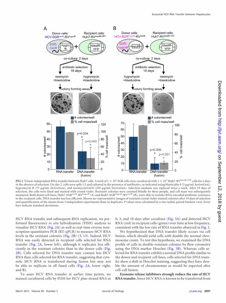
To asses HCV RNA transfer at earlier time points, westained cocultured cells by FISH for HCV plus-strand RNA at
0, 3, and 10 days after coculture (Fig. 3A) and detected HCVRNA (red) in recipient cells (green) over time at low frequency,consistent with the low rate of RNA transfer observed in Fig. 1.
We hypothesized that DNA transfer likely occurs via cellfusion, which should yield cells with double the normal chro-mosome count. To test this hypothesis, we examined the DNAprofile of cells in double-resistant colonies by flow cytometryusing the DNA marker Hoechst (Fig. 3B). Whereas cells se-lected for RNA transfer exhibit a normal DNA profile similar tothe donor and recipient cell lines, cells selected for DNA trans-fer show a shift in Hoechst staining, suggesting they have dou-ble the amount of chromosomes as would be expected aftercell-cell fusion.
Exosome release inhibitors strongly reduce the rate of HCVRNA transfer. Since HCV RNA is known to be transferred from
FIG 1 Virion-independent RNA transfer between Huh7 cells. A total of 1 � 105 SGR cells were cocultured with 1 � 105 Huh7-RVblastR-YFP cells for 2 daysin the absence of selection. On day 2, cells were split 1:2 and cultured in the presence of antibiotics, as indicated using blasticidin S (5 �g/ml; InvivoGen),hygromycin B (75 �g/ml; InvivoGen), and neomycin/G418 (250 �g/ml; InvivoGen). Selection medium was replaced twice a week. After 19 days ofselection, the cells were fixed and stained with crystal violet. Resistant colonies were counted blindly by three people, and cell mass was subsequentlymeasured. Both donor cell lines, Huh7-SGRneoR-RVhygroR (A) and Huh7-SGRhygroR-RVneoR (B), were able to transfer RNA-encoded antibiotic resistanceto the recipient cells. DNA transfer was less efficient. Shown are representative images of resistant crystal violet-stained colonies after 19 days of selectionand quantification of the means from 3 independent experiments done in duplicate. P values were calculated by a two-tailed, paired Student t test. Errorbars indicate standard deviations.
Exosomal HCV RNA Transfer between Hepatocytes
March 2015 Volume 89 Number 5 jvi.asm.org 2957Journal of Virology
on Septem
ber 12, 2018 by guesthttp://jvi.asm
.org/D
ownloaded from

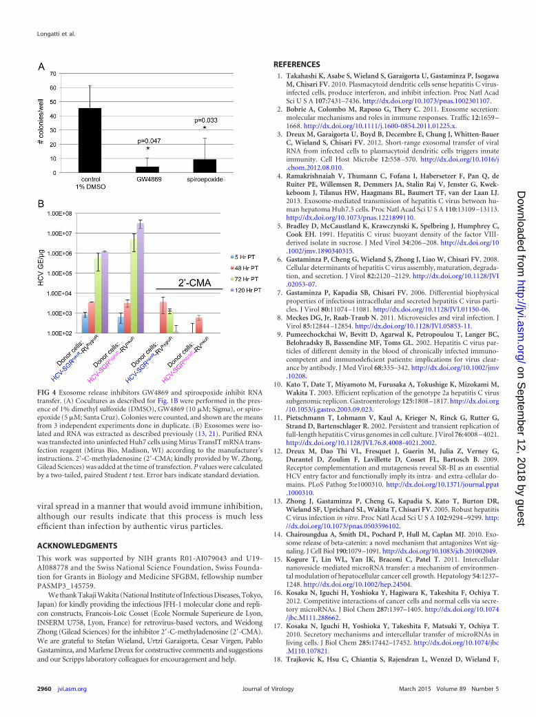
SGR cells to pDCs via exosomes (3), we hypothesized that exo-somes may be responsible for HCV RNA transfer from donorto recipient Huh7 cells. To test this hypothesis, we performedthe coculture experiments described in Fig. 1 in the presence oftwo different neutral sphingomyelinase inhibitors (GW4869and spiroepoxide), known to block exosome secretion (14–19).Both inhibitors severely reduced the level of RNA transfer (Fig.4A) without affecting cell viability or HCV RNA replication inthe donor cells (not shown). This suggests that exosomes arelikely the dominant mode of transmission of HCV RNA be-tween hepatocytes. In support of this, we also found that HCVRNA extracted from exosomes released by our donor cell line isreplication competent when transfected into Huh7 cells (Fig. 4B).
We are intrigued by the fact that although HCV RNA trans-fer is exosome mediated, it appears to require cell-cell contact
since coculture experiments done in Transwell plates did notlead to exosomal HCV RNA transfer, and exosomes concen-trated from SGR cells were not able to transmit HCV RNA totarget cells directly (data not shown). This suggests that, simi-lar to exosome-mediated transfer of HCV RNA from SGR cellsto plasmacytoid dendritic cells (3), SGR cells appear to secreteexosomes into the culture supernatant at concentrations thatare below a functional threshold that is easily reached in theintercellular space during cell-cell contact. Furthermore, in pi-lot studies (not shown), we found that only 0.1% of all secretedexosomes contain HCV RNA, and we were unable to isolatesufficient numbers of exosomes from SGR cell supernatants toachieve a high enough HCV RNA/cell ratio to initiate RNAreplication. These results contrast with previous reports (4, 20)in which the authors conclude that exosomes prepared from
FIG 2 Visualization of replication-competent HCV RNA transfer to recipient cells. (A) After coculture as described for Fig. 1A, donor SGR, recipient, andresistant colony cells were plated at equal densities and fixed 24 h later, and HCV RNA was labeled using FISH. HCV plus-strand RNA (HCV RNA�; red)and HCV minus-strand RNA (HCV RNA�; yellow) were detected using probe sets that target a region comprised within NS3 and NS4 proteins (referenceno. VF4-11069; Panomics/Affymetrix, Santa Clara, CA) according to the manufacturer’s instructions and as described previously (12). Nuclei werestained by Hoechst33342 dye (blue; Life Technologies product no. H1399). The picture on the bottom right shows the overlay. Images were acquired witha Zeiss LSM 710 laser scanning confocal microscope. Both positive- and negative-strand HCV RNA are readily detected in cells selected for RNA transferbut are found rarely in cells selected for DNA transfer. (B) Real-time RT-qPCR of the same cells as in panel A, detecting HCV RNA. Shown are the averagesfrom 3 independent experiments. Error bars indicate standard deviations. GE, genome equivalents; YFP, yellow fluorescent protein (green). P values werecalculated using a two-tailed, unpaired Student t test with GraphPad Prism software.
Longatti et al.
2958 jvi.asm.org March 2015 Volume 89 Number 5Journal of Virology
on Septem
ber 12, 2018 by guesthttp://jvi.asm
.org/D
ownloaded from
HCV-infected cell supernatants efficiently transfer infection torecipient cells. The interpretation of those results is compli-cated by the difficulty inherent in separating exosomes frominfectious HCV particles because of their similar size and den-sity characteristics, raising the possibility that infection wasmediated by virus particles in those experiments.
In summary, these data support the notion that replication-competent HCV RNA can be transferred in a virion-indepen-
dent manner between permissive Huh7 cells that support ge-nome expansion and expression. These events do not requireinfectious virus particles, because subgenomic replicon cellslacking viral structural genes were used in all experiments, andthey appear to be dependent on an exosome-mediated, cell-celltransfer mechanism. If these events occur in vivo, exosomescontaining replication-competent HCV RNA may be able to“infect” replication-permissive hepatocytes and contribute to
FIG 3 Chromosomal DNA transfer leads to polyploidy, whereas RNA transfer does not. (A) Donor and recipient cells were cocultured as described forFig. 1, and cocultured cells were labeled for plus-strand HCV RNA using FISH as described for Fig. 2 after 0, 3, and 10 days of coculture. In rare instances,HCV RNA can be detected over time in YFP-positive (green) recipient cells (B). After coculture as described for Fig. 1A, donor SGR, recipient, andresistant colony live cells were labeled with Hoechst33342 dye (Life Technologies product no. H1399) to detect DNA levels and analyzed by flowcytometry.
Exosomal HCV RNA Transfer between Hepatocytes
March 2015 Volume 89 Number 5 jvi.asm.org 2959Journal of Virology
on Septem
ber 12, 2018 by guesthttp://jvi.asm
.org/D
ownloaded from
viral spread in a manner that would avoid immune inhibition,although our results indicate that this process is much lessefficient than infection by authentic virus particles.
ACKNOWLEDGMENTS
This work was supported by NIH grants R01-AI079043 and U19-AI088778 and the Swiss National Science Foundation, Swiss Founda-tion for Grants in Biology and Medicine SFGBM, fellowship numberPASMP3_145759.
We thank Takaji Wakita (National Institute of Infectious Diseases, Tokyo,Japan) for kindly providing the infectious JFH-1 molecular clone and repli-con constructs, Francois-Loïc Cosset (Ecole Normale Superieure de Lyon,INSERM U758, Lyon, France) for retrovirus-based vectors, and WeidongZhong (Gilead Sciences) for the inhibitor 2=-C-methyladenosine (2=-CMA).We are grateful to Stefan Wieland, Urtzi Garaigorta, Cesar Virgen, PabloGastaminza, and Marlene Dreux for constructive comments and suggestionsand our Scripps laboratory colleagues for encouragement and help.
REFERENCES1. Takahashi K, Asabe S, Wieland S, Garaigorta U, Gastaminza P, Isogawa
M, Chisari FV. 2010. Plasmacytoid dendritic cells sense hepatitis C virus-infected cells, produce interferon, and inhibit infection. Proc Natl AcadSci U S A 107:7431–7436. http://dx.doi.org/10.1073/pnas.1002301107.
2. Bobrie A, Colombo M, Raposo G, Thery C. 2011. Exosome secretion:molecular mechanisms and roles in immune responses. Traffic 12:1659 –1668. http://dx.doi.org/10.1111/j.1600-0854.2011.01225.x.
3. Dreux M, Garaigorta U, Boyd B, Decembre E, Chung J, Whitten-BauerC, Wieland S, Chisari FV. 2012. Short-range exosomal transfer of viralRNA from infected cells to plasmacytoid dendritic cells triggers innateimmunity. Cell Host Microbe 12:558 –570. http://dx.doi.org/10.1016/j.chom.2012.08.010.
4. Ramakrishnaiah V, Thumann C, Fofana I, Habersetzer F, Pan Q, deRuiter PE, Willemsen R, Demmers JA, Stalin Raj V, Jenster G, Kwek-keboom J, Tilanus HW, Haagmans BL, Baumert TF, van der Laan LJ.2013. Exosome-mediated transmission of hepatitis C virus between hu-man hepatoma Huh7.5 cells. Proc Natl Acad Sci U S A 110:13109 –13113.http://dx.doi.org/10.1073/pnas.1221899110.
5. Bradley D, McCaustland K, Krawczynski K, Spelbring J, Humphrey C,Cook EH. 1991. Hepatitis C virus: buoyant density of the factor VIII-derived isolate in sucrose. J Med Virol 34:206 –208. http://dx.doi.org/10.1002/jmv.1890340315.
6. Gastaminza P, Cheng G, Wieland S, Zhong J, Liao W, Chisari FV. 2008.Cellular determinants of hepatitis C virus assembly, maturation, degrada-tion, and secretion. J Virol 82:2120 –2129. http://dx.doi.org/10.1128/JVI.02053-07.
7. Gastaminza P, Kapadia SB, Chisari FV. 2006. Differential biophysicalproperties of infectious intracellular and secreted hepatitis C virus parti-cles. J Virol 80:11074 –11081. http://dx.doi.org/10.1128/JVI.01150-06.
8. Meckes DG, Jr, Raab-Traub N. 2011. Microvesicles and viral infection. JVirol 85:12844 –12854. http://dx.doi.org/10.1128/JVI.05853-11.
9. Pumeechockchai W, Bevitt D, Agarwal K, Petropoulou T, Langer BC,Belohradsky B, Bassendine MF, Toms GL. 2002. Hepatitis C virus par-ticles of different density in the blood of chronically infected immuno-competent and immunodeficient patients: implications for virus clear-ance by antibody. J Med Virol 68:335–342. http://dx.doi.org/10.1002/jmv.10208.
10. Kato T, Date T, Miyamoto M, Furusaka A, Tokushige K, Mizokami M,Wakita T. 2003. Efficient replication of the genotype 2a hepatitis C virussubgenomic replicon. Gastroenterology 125:1808 –1817. http://dx.doi.org/10.1053/j.gastro.2003.09.023.
11. Pietschmann T, Lohmann V, Kaul A, Krieger N, Rinck G, Rutter G,Strand D, Bartenschlager R. 2002. Persistent and transient replication offull-length hepatitis C virus genomes in cell culture. J Virol 76:4008 – 4021.http://dx.doi.org/10.1128/JVI.76.8.4008-4021.2002.
12. Dreux M, Dao Thi VL, Fresquet J, Guerin M, Julia Z, Verney G,Durantel D, Zoulim F, Lavillette D, Cosset FL, Bartosch B. 2009.Receptor complementation and mutagenesis reveal SR-BI as an essentialHCV entry factor and functionally imply its intra- and extra-cellular do-mains. PLoS Pathog 5:e1000310. http://dx.doi.org/10.1371/journal.ppat.1000310.
13. Zhong J, Gastaminza P, Cheng G, Kapadia S, Kato T, Burton DR,Wieland SF, Uprichard SL, Wakita T, Chisari FV. 2005. Robust hepatitisC virus infection in vitro. Proc Natl Acad Sci U S A 102:9294 –9299. http://dx.doi.org/10.1073/pnas.0503596102.
14. Chairoungdua A, Smith DL, Pochard P, Hull M, Caplan MJ. 2010. Exo-some release of beta-catenin: a novel mechanism that antagonizes Wnt sig-naling. J Cell Biol 190:1079–1091. http://dx.doi.org/10.1083/jcb.201002049.
15. Kogure T, Lin WL, Yan IK, Braconi C, Patel T. 2011. Intercellularnanovesicle-mediated microRNA transfer: a mechanism of environmen-tal modulation of hepatocellular cancer cell growth. Hepatology 54:1237–1248. http://dx.doi.org/10.1002/hep.24504.
16. Kosaka N, Iguchi H, Yoshioka Y, Hagiwara K, Takeshita F, Ochiya T.2012. Competitive interactions of cancer cells and normal cells via secre-tory microRNAs. J Biol Chem 287:1397–1405. http://dx.doi.org/10.1074/jbc.M111.288662.
17. Kosaka N, Iguchi H, Yoshioka Y, Takeshita F, Matsuki Y, Ochiya T.2010. Secretory mechanisms and intercellular transfer of microRNAs inliving cells. J Biol Chem 285:17442–17452. http://dx.doi.org/10.1074/jbc.M110.107821.
18. Trajkovic K, Hsu C, Chiantia S, Rajendran L, Wenzel D, Wieland F,
FIG 4 Exosome release inhibitors GW4869 and spiroepoxide inhibit RNAtransfer. (A) Cocultures as described for Fig. 1B were performed in the pres-ence of 1% dimethyl sulfoxide (DMSO), GW4869 (10 �M; Sigma), or spiro-epoxide (5 �M; Santa Cruz). Colonies were counted, and shown are the meansfrom 3 independent experiments done in duplicate. (B) Exosomes were iso-lated and RNA was extracted as described previously (13, 21). Purified RNAwas transfected into uninfected Huh7 cells using Mirus TransIT mRNA trans-fection reagent (Mirus Bio, Madison, WI) according to the manufacturer’sinstructions. 2=-C-methyladenosine (2=-CMA; kindly provided by W. Zhong,Gilead Sciences) was added at the time of transfection. P values were calculatedby a two-tailed, paired Student t test. Error bars indicate standard deviation.
Longatti et al.
2960 jvi.asm.org March 2015 Volume 89 Number 5Journal of Virology
on Septem
ber 12, 2018 by guesthttp://jvi.asm
.org/D
ownloaded from

Schwille P, Brugger B, Simons M. 2008. Ceramide triggers budding ofexosome vesicles into multivesicular endosomes. Science 319:1244 –1247.http://dx.doi.org/10.1126/science.1153124.
19. Yuyama K, Sun H, Mitsutake S, Igarashi Y. 2012. Sphingolipid-modulatedexosome secretion promotes clearance of amyloid-beta by microglia. J BiolChem 287:10977–10989. http://dx.doi.org/10.1074/jbc.M111.324616.
20. Bukong TN, Momen-Heravi F, Kodys K, Bala S, Szabo G. 2014. Exosomes
from hepatitis C infected patients transmit HCV infection and contain repli-cation competent viral RNA in complex with Ago2-miR122-HSP90. PLoSPathog 10:e1004424. http://dx.doi.org/10.1371/journal.ppat.1004424.
21. Thery C, Amigorena S, Raposo G, Clayton A. 2006. Isolation andcharacterization of exosomes from cell culture supernatants and biologi-cal fluids. Curr Protoc Cell Biol Chapter 3:Unit 3.22. http://dx.doi.org/10.1002/0471143030.cb0322s30.
Exosomal HCV RNA Transfer between Hepatocytes
March 2015 Volume 89 Number 5 jvi.asm.org 2961Journal of Virology
on Septem
ber 12, 2018 by guesthttp://jvi.asm
.org/D
ownloaded from