IUI indications, Ovarian stimulation protocols CC, aromatase inhibitors, gonadotropins
PITUITARY TUMORS Onc26 (1)
Pituitary Tumors Last updated: April 12, 2020
Differential Diagnosis of Sellar and Parasellar Tumors ................................................................... 1
PITUITARY ADENOMAS ................................................................................................................... 1 PATHOPHYSIOLOGY, PATHOLOGY, ETIOLOGY ....................................................................................... 2
CLASSIFICATION .................................................................................................................................... 2 Size ........................................................................................................................................ 2
Hormonal secretion ............................................................................................................... 2 Histology ............................................................................................................................... 2
EPIDEMIOLOGY ...................................................................................................................................... 4 CLINICAL FEATURES .............................................................................................................................. 4
1. Hormonal function control ................................................................................................ 4
2. Mass effect ........................................................................................................................ 4 DIAGNOSIS ............................................................................................................................................. 5
Skull X-ray ............................................................................................................................ 5 CT .......................................................................................................................................... 5
MRI ....................................................................................................................................... 6
Radionuclide studies ............................................................................................................ 10 PET ...................................................................................................................................... 10
SPECT ................................................................................................................................. 10 Angiography ........................................................................................................................ 10
Neuro-ophthalmological evaluation .................................................................................... 10
Evaluation of pituitary function .......................................................................................... 10 Genetic testing ..................................................................................................................... 12
COMPLICATIONS .................................................................................................................................. 12 TREATMENT ......................................................................................................................................... 12
Different Strategies ........................................................................................................................ 12
Medical therapy .............................................................................................................................. 13 Surgery ........................................................................................................................................... 13
Postoperatively .................................................................................................................... 13 Endocrinological follow-up ................................................................................................. 13
Ophthalmological follow-up ............................................................................................... 14
Imaging follow-up ............................................................................................................... 14
Postop complications ........................................................................................................... 14
Radiotherapy .................................................................................................................................. 14 Types ................................................................................................................................... 14
Indications ........................................................................................................................... 14 Methodology (SRS) ............................................................................................................. 15 Preop .................................................................................................................................... 15 Outcomes ............................................................................................................................. 15 Complications ...................................................................................................................... 15
Chemotherapy ................................................................................................................................ 15 Algorithms according to Hormone ................................................................................................. 15
PROGNOSIS .......................................................................................................................................... 16 Natural history without treatment .................................................................................................. 16 Treatment of Recurrence / Residual Tumor ................................................................................... 16
PITUITARY CARCINOMAS ............................................................................................................. 17 EMPTY SELLA SYNDROME ........................................................................................................... 17
Etiology ............................................................................................................................... 17 Clinical Features .................................................................................................................. 17 Diagnosis ............................................................................................................................. 17
Treatment ............................................................................................................................. 17
PITUITARY APOPLEXY ................................................................................................................... 17 Clinical Features .................................................................................................................. 17 Diagnosis ............................................................................................................................. 17 Treatment ............................................................................................................................. 17
HYPOPHYSITIS ....................................................................................................................................... 17 CRANIOPHARYNGIOMA ................................................................................................................ 18
EPIDEMIOLOGY .................................................................................................................................... 18
PATHOLOGY ......................................................................................................................................... 18 Grossly ................................................................................................................................. 18 Histology ............................................................................................................................. 18
CLINICAL PRESENTATION .................................................................................................................... 20 DIAGNOSIS ........................................................................................................................................... 20 TREATMENT ......................................................................................................................................... 22
Surgery ........................................................................................................................................... 22 Radiotherapy .................................................................................................................................. 23
Chemotherapy ................................................................................................................................ 23 PROGNOSIS .......................................................................................................................................... 23
NEUROHYPOPHYSIS is rare site of neoplasia:
1) INFUNDIBULOMAS are rare variants of PILOCYTIC ASTROCYTOMAS.
2) GRANULAR CELL TUMORS (MYOBLASTOMAS, CHORISTOMAS) are rare tumors with uncertain
cell origin.
Most pituitary tumors are ADENOMAS!
DIFFERENTIAL DIAGNOSIS OF SELLAR AND PARASELLAR TUMORS
1. Tumors:
1) pituitary adenoma, pituitary carcinoma, craniopharyngioma
2) meningioma, metastatic tumors*
3) cranial nerves - optic glioma, CN5 schwannoma
4) bone - chordoma, chondrosarcoma
5) dermoid, epidermoid, teratoma, germ cell tumors (← treated with radiation)
*most commonly involve pituitary stalk
H: surgery with histological diagnosis.
2. Not tumors: compression of sella due to hemorrhage, carotid aneurysm, empty sella, Rathke’s
cleft cyst, tuber cinereum hamartoma, granulomas (e.g. tuberculosis, sarcoid), lymphocytic
hypophysitis?
H: neuroradiological imaging.
most common differential for nonsecreting adenoma is CRANIOPHARYNGIOMA and empty sella.
majority of (para)sellar tumors are benign.
BENIGN ≠ INNOCENT (optic apparatus, hypothalamus, hypophysis)
PITUITARY ADENOMAS
- neuroepithelial tumors of ADENOHYPOPHYSIS.

PITUITARY TUMORS Onc26 (2)
hormonal syndromes → see p. 2738 >>
PATHOPHYSIOLOGY, PATHOLOGY, ETIOLOGY
Guidelines on the Management of Patients with Nonfunctioning Pituitary Adenomas (CNS 2016):
molecular etiology and epidemiologic risk factors remain incompletely defined.
putative TUMOR SUPPRESSOR GENE alterations:
1) retinoblastoma gene
2) multiple endocrine neoplasia type I (MEN-I) gene 11q13 (found in 3-4 %) – inherited
pituitary adenoma!
3) p53 deletions correlates with aggressive behavior.
pituitary adenomas are not under hypothalamic control.
alternative hypothesis: overstimulation (or deranged signaling) from hypothalamus →
inappropriate pituitary growth.
adenomas grow slowly; initially confined to sella turcica → may grow out of sella and compress /
encase / destroy:
a) optic chiasm
b) cavernous sinus and internal carotids (lateral extension)
c) hypothalamus
d) surrounding bony structures (e.g. sphenoid sinus, clivus)
N.B. locally invasive adenomas nearly always are histologically benign! CNS metastases and,
rarely, distant metastases can occur!
often have small foci of hemorrhage or necrosis, but no mitotic activity.
N.B. pituitary adenomas never have calcifications!? (look at CT – if calcium is present,
it is craniopharyngioma)? Clinical characteristics of pituitary adenomas with radiological calcification. Toshihiro Ogiwara et al. Acta
Neurochirurgica 2017 August 19
Pituitary adenoma presenting with calcification is relatively rare (5.6%), but
should be kept in mind to avoid making a wrong preoperative diagnosis. As not
all pituitary adenomas with calcification are hard tumors, preoperative
radiological calcification should not affect decision-making regarding surgical
indications (tumor resection is usually possible without any complications).
adenomas lack discrete capsule, but presence of pseudocapsule facilitates surgical separation.
CLASSIFICATION
SIZE
< 1 cm in diameter – MICROADENOMAS
> 1 cm – MACROADENOMAS.
HORMONAL SECRETION
a) NONSECRETORS, S. NONFUNCTIONING PITUITARY ADENOMAS (most common pituitary tumors!) –
manifest when reach size of MACROADENOMA – mass effect (normal pituitary tissue destruction,
pressure on optic chiasm, etc).
some nonsecretors secrete α subunit of glycoprotein hormones (FSH, LH, TSH) – suggests
origin as gonadotrophs.
null cell adenomas demonstrate no evidence (clinical or immunohistochemical) of hormone
secretion.
b) HORMONE SECRETORS (frequency: prolactin > GH > ACTH > gonadotropins > TSH) – manifest
with specific endocrine syndromes. see p. 2738 >>
nonsecreting, prolactin-secreting in men*, gonadotropin-secreting, GH-secreting adenomas
manifest late (as MACROADENOMAS)!
*main symptom – impotence – men tend to present late for this symptom
other adenomas manifest early (still as MICROADENOMAS).
some tumors secrete multiple hormones (termed NULL TUMORS).
normally five pituitary cell types are regionally distributed:
lactotrophs and gonadotrophs – widely distributed;
somatotrophs – peripherally (two lateral wings of gland);
thyrotrophs – anteromedially;
corticotrophs – central median wedge.
HISTOLOGY
- on routine staining:
a) chromophilic cells (acidophilic or basophilic)
b) chromophobic cells.
N.B. routine staining is meaningless - tumor can be difficult to differentiate from normal tissue
or metastatic disease - immunohistochemical staining and electron microscopy are essential!
typical normal acinar structure is lost – adenomas may contain follicular, trabecular, or cystic
portions growing as diffuse sheet; cells are arranged in syncytial or sinusoidal pattern; monotonous
appearance.
nuclei with “salt and pepper” chromatin (s. endocrine chromatin).
differentiation of hyperplasia from adenoma may be difficult.
types of undifferentiated cell adenomas:
1) NONONCOCYTIC (NULL)

PITUITARY TUMORS Onc26 (3)
2) ONCOCYTOMA (s. OXYPHIL ADENOMA) - tumor contains buildup of mitochondria.
Adenoma - packeted arrangement of cells resembles that of anterior pituitary, together with prominent vascular network:
MICROADENOMA:
Source of picture: “WebPath - The Internet Pathology Laboratory for Medical Education” (by Edward C. Klatt, MD) >>
Photograph of MICROADENOMA (0.9 cm in largest diameter) - incidental null cell adenoma found postmortem; tumor is
well delineated and has compressed residual still functional adenohypophysis to crescent shape:
MACROADENOMA:

PITUITARY TUMORS Onc26 (4)
Source of picture: “WebPath - The Internet Pathology Laboratory for Medical Education” (by Edward C. Klatt, MD) >>
EPIDEMIOLOGY
The most common tumor in sella region (except CRANIOPHARYNGIOMAS in childhood)
Guidelines on the Management of Patients with Nonfunctioning Pituitary Adenomas (CNS 2016):
From cancer registries prevalence is:
19-28 cases per 100,000 people
Meta-analysis of autopsy data and radiologic studies in healthy volunteers - pituitary adenomas are
700 times more common than registry data suggests:
pituitary adenomas are found in 14% of autopsies
pituitary adenomas are found in 23% of CT/MRI studies
Mean prevalence of 17%
4-20 % of all intracranial tumors.
most occur in young adults (peak - 3rd-4th decades); children make 10% of all patients.
men = women (clinically evident more often in young women); symptomatic prolactinomas and
Cushing disease are found more frequently in women.
CLINICAL FEATURES
Most pituitary adenomas can be detected while relatively small (MICROADENOMAS) - located in
exquisitely sensitive area:
N.B. nonsecreting microadenomas are asymptomatic!
1. HORMONAL FUNCTION CONTROL
A) hormonal hypersecretion (most commonly prolactin!)
B) destruction of normal gland → hypopituitarism (partial in 37-85% patients with nonsecretory
tumors, pan in 6-29% patients with nonsecretory tumors)
N.B. all MACROADENOMAS eventually cause hypopituitarism.
if hypopituitarism occurs, hormone loss is sequential: GH → gonadotropins → ACTH → TSH.
primary pituitary tumors rarely* cause ADH deficiency (except when induced by
hypophysectomy); diabetes insipidus is more common in CRANIOPHARYNGIOMAS.
*7% of patients with NFPAs at the time of clinical presentation
2. MASS EFFECT
1) headache occurs in 20% (can be diffuse and nonpulsatile and may be mistaken for daily
headaches; more often in females) – due to stretching of diaphragma sellae and adjacent dural
structures; ICP is normal!
N.B. nonspecific headaches may be the only early symptoms, esp. in
nonsecreting adenomas!
– it is still debatable if pituitary tumors can cause / exacerbate headaches, but pituitary surgery is
associated with headache improvement or resolution in majority of patients (plus, pituitary
surgery was not found to cause or worsen headaches) Rizzoli P. et al “Headache in Patients With Pituitary Lesions: A Longitudinal Cohort Study” Neurosurgery: March
2016 - Volume 78 - Issue 3 - p 316–323
2) crossing fibers in optic chiasm (superior bitemporal quadrantanopia → full bitemporal
hemianopia - chief and earliest finding in most patients!)
relationship of pituitary and optic chiasm:
a) chiasm directly above pituitary (80%).
b) chiasm anteriorly to pituitary (9%)
c) chiasm behind pituitary (11%).
further expansion compromises noncrossing fibers - affects lower and finally upper
nasal quadrants.
any pattern of visual loss is possible, e.g.:
– asymmetrical loss results from chiasm ischemia produced by vessel occlusion.
– unilateral mass located anterior to postfixed chiasm may produce central scotoma in
one eye + upper outer quadrantanopia in contralateral eye (due to von Willebrand's

PITUITARY TUMORS Onc26 (5)
knee - looping of crossing fibers in proximal segment of optic nerve opposite side of
their retinal origin) – so called JUNCTIONAL SCOTOMA
Any form of temporal field defect, even if monocular,
can result from chiasmal compression
some tumors affect only macular fibers → central hemianopic scotomas - may be
missed on routine screening (so formal quantitative visual field testing is important in
all cases!!!).
other findings: optic disc atrophy (generally horizontal-oriented, i.e. bow-tie), dropout
of nerve fiber layer in nasal retina, loss of central visual acuity, loss of color vision,
visual field defects.
N.B. papilledema is exceptional (seen only in pituitary apoplexy).
3) lateral extension into cavernous sinus → diplopia, ophthalmoplegias, and postganglionic Horner
syndrome.
4) hypothalamic compression (e.g. hyperprolactinemia*, diabetes insipidus, alterations in
consciousness, memory, intake of food and water).
*if serum prolactin > 90-200 μg/L – prolactinoma is more likely!
5) extension into sphenoid sinus → CSF rhinorrhea (≈ 0.5% cases) - cortical bone separating sella
from sphenoid sinus is quite thin in normal individuals!; may occur as prolactinoma shrinks with
medical treatment.
6) compression of 3rd ventricle → obstructive hydrocephalus.
7) basal forebrain abnormalities (personality changes, dementia, anosmia).
8) temporal lobe seizures.
pituitary adenomas may enlarge during pregnancy (esp. prolactinomas); sudden hypotension during
delivery may cause ischemic stroke (apoplexy).
DIAGNOSIS
N.B. pituitary adenomas almost never have calcifications!?
Guidelines on the Management of Patients with Nonfunctioning Pituitary Adenomas (CNS 2016)
1) high resolution MRI (Level II) is recommended as the standard for preoperative assessment but
may be supplemented with CT* (Level III) and fluoroscopy (Level III).
*thin-cut CT for sphenoid septal anatomy; CTA for vascular anatomy; dual-energy CT
to discriminate between pituitary adenomas and meningiomas with a sensitivity of
90.9% and specificity of 100%
2) while there are promising results suggesting the utility of MR spectroscopy, MR perfusion, PET,
and SPECT to evaluate histology and characteristics, there is insufficient evidence to make
formal recommendations.
3) while promising results are available pertaining to high-resolution MR and proton density imaging
as tools of assessing cavernous sinus invasion, there is insufficient evidence to make a formal
recommendation.
4) while promising results are available pertaining to perfusion and gradient echo imaging as tools for
assessing tumor vascularity and hemorrhage, there is insufficient evidence to make a formal
recommendation.
SKULL X-RAY
- limited use.
MACROADENOMAS balloon pituitary fossa → asymmetrical floor of pituitary fossa:
frontal projection - one side of fossa is deeper than other;
lateral projection - two more or less parallel lines that create impression of “double floor”.
CT
(thin-section, direct coronal plane, with bone windows)

PITUITARY TUMORS Onc26 (6)
MACROADENOMAS are easily detected - hyperdense mass within enlarged pituitary fossa.
may miss small MICROADENOMA (appear as hypodense structures, vs. MACROADENOMAS).
Dual-energy CT (utilizes high-frequency cycling of high/low voltages to improve the quality of the
CT images) - can discriminates between pituitary adenomas and meningiomas with a sensitivity of
90.9% and specificity of 100%.
MRI
- gold standard, more sensitive method for tumor identification (esp. 1-mm cuts and magnified views
through sella – pituitary protocol) - investigation of choice for MICROADENOMA detection!!!
Normal NEUROHYPOPHYSIS on T1-MRI shows increased signal (representing neurosecretory granules
in ADH-containing axons).
Normal ADENOHYPOPHYSIS:
isointense with grey matter on all MR sequences.
circumventricular organ without an intact BBB - enhances homogeneously (punctate areas of
heterogeneity - local variations in vascularity, microcyst formation, or granularity), strongly and
rapidly (within 30 minutes of gadolinium infusion).
Normal pituitary gland size and configuration are highly variable (esp. in women of childbearing age
or pubertal girls – normal hypertrophy of gonadotrophs).
N.B. great care must be exercised in diagnosis of MICROADENOMAS on MRI
basis without associated evidence of hormonal abnormality.
– in neonatal period both anterior and posterior lobes are hyperintense and pituitary gland is
bulbous in shape.
– during adolescence and puberty there is significant physiological hypertrophy (in girls upper
surface is convex, giving gland almost spherical shape on sagittal views - do not mistake for
mass).
Schematic diagram of MRI OF NORMAL PITUITARY FOSSA: pituitary is bordered laterally by cavernous
sinus, which contains internal carotid artery and cranial nerves III, IV, V1 , V2 , and VI; optic chiasm
lies immediately above pituitary gland.
Source of picture: David C. Sabiston “Sabiston Textbook of Surgery: the Biological Basis of Modern Surgical
Practice”, 15th ed. (1997); W.B. Saunders Company; ISBN-13: 978-0721658872 >>
Guidelines on the Management of Patients with Nonfunctioning Pituitary Adenomas (CNS 2016):
discussions and recommendations(if any) are highlighted below.
MICROADENOMAS:
unenhanced MRI is not helpful - only some MICROADENOMAS have different signal intensity to
normal gland.
standard MRI protocol for investigation of microadenomas - 1-mm thick coronal T1 spin-echo
sequences through pituitary gland before and after IV GADOLINIUM;
– additional images in sagittal plane are performed in many centers;
– desirable to perform fat-saturated T1 sequence (fat-suppressed imaging) - eliminates high
signal from fat in clivus and clinoid processes (could be mistaken for enhancement).
adenomas always enhance less than normal pituitary gland (hypodense area also can represent
ischemic stroke in tumor).
normal pituitary gland is most often displaced superiorly and posteriorly by adenoma;
displacement of normal pituitary in other directions can suggest other pathology!
there is an association between the “bright stalk” and adenoma size:
– normal pituitary stalk is of relatively low intensity but can be bright when compressed by
tumor.
– stalk compression/deviation or bright stalk are not always associated with elevated
prolactin expected of stalk effect.
accuracy can be increased by dynamic pituitary scans (series of rapid images with 10–15 s time
intervals for about 3 min following gadolinium IV bolus) - differences in time course of
enhancement between adenoma and adjacent normal gland – very useful in detecting
microadenomas!
N.B. MICROADENOMAS enhance later and/or lesser than normal pituitary tissue!
other indirect MRI signs:
1) gland height↑ (normally < 10 mm)
2) gland upper margin contour alteration from concave or straight to convex
3) erosion of sella turcica floor adjacent to hypointensity area
PITUITARY TUMORS Onc26 (7)
4) displacement of pituitary stalk (normally midline) away from hypointensity area.
Most MACROADENOMAS enhance strongly and uniformly.
– surgeons can predict the consistency of adenoma from T2-MRI: the more hyperintense the
adenoma, the softer the tumor.
– low-density areas within mass may represent cysts/necrosis/stroke; tumor which enhances
only peripherally or not at all may be necrotic.
– if entire sellar contents are of low density, search for infundibulum - empty sella is much
more likely diagnosis than completely cystic pituitary macroadenoma!
– hemorrhages appear as high-intensity areas (best – gradient echo [GE] imaging).
suprasellar extension is easily demonstrated in both coronal and sagittal images.
– visual field loss is significantly correlated with the height of the chiasm and the tumor as
well as optic nerve hyperintensity on T2 images but not with optic tract edema.
it is more difficult to be sure about cavernous sinus invasion - no imaging technique can perfectly
visualize the medial cavernous sinus wall
– displacement of cavernous ICA segment may occur without tumor invasion into cavernous
sinus.
– abnormal signal intensity lateral to cavernous ICA segment, indicates invasion into
cavernous sinus.
– 3 T vs 1.5 T yields superior sensitivity (83% versus 67%, respectively) and specificity
(84% relative to 58%) in terms of correlation to surgical findings of cavernous sinus
invasion.
– volumetric interpolated breath-hold examination (VIBE) sequence may offer superior
image resolution of tumor invasion of the cavernous sinus (eliminates subtle patient motion
related to respiratory effort)
– proton density weighted MR is highly sensitive and specific for predicting tumor invasion
of the cavernous sinus.
– class III data suggests that multiple microcysts on T2, cavernous sinus invasion, lobulated
appearance, and size > 40 mm are associated with silent corticotroph adenomas.
– Knosp criteria - the extent of parasellar extension relative to inter-carotid lines drawn
through the intra-cavernous carotid on a coronal MRI; high Knosp grades are associated
with increased likelihood of sinus invasion.
– normal pituitary between adenoma and the cavernous sinus (“rim sign” or “peri-arterial
enhancement”) can exclude sinus invasion.
– asymmetric dural enhancement of tentorium along the posterior portion of the cavernous
sinus is associated with increased likelihood of sinus invasion and thought to be related to
venous congestion secondary to tumor mass.
bone invasion: magnetic susceptibility effects at the skull base (air vs bone) render normal fat-
suppression techniques less effective, making it more difficult to assess involvement of the bony
structures around the sella.
signal intensity on MRI CISS sequences is associated with the firmness of tumor; DWI may also
be helpful but results of studies are conflicting.
MR perfusion studies provide information regarding tumor vascularity.
DIFFERENTIAL DIAGNOSIS
MR spectroscopy
technically challenging within small volumes such as the sella and is very sensitive to magnetic
susceptibility effects due to the surrounding bone.
pituitary adenomas often show a choline peak.
both hypothalamic hamartomas and gliomas exhibit decreased N-acetyl aspartate (NAA),
however, hamartomas are characterized by increased myoinositol while gliomas show increased
choline accumulation.
craniopharyngiomas and germinomas both show dominant lipid peaks.
T2-MRI (left) shows microadenoma; contrast (right) normally enhances pituitary; adenoma appears lighter:
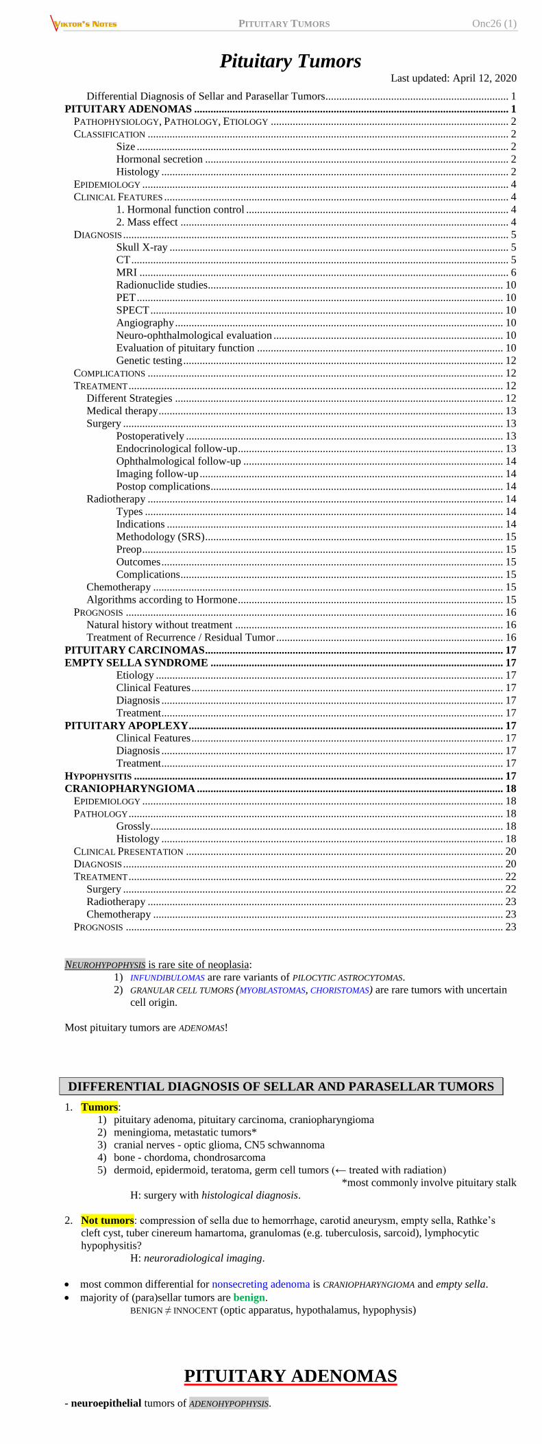
Contrast T1-MRI – MACROADENOMA: adenoma (A) enhances; tumor displaces carotid arteries laterally (black arrows); A1
segments of anterior cerebral arteries (white arrows) and chiasm (arrowheads) drape over mass.
Contrast MRI - MICROADENOMA (arrow):
Source of picture: Martin D. Abeloff “Clinical Oncology”, 2nd ed. (2000); Churchill Livingstone, Inc.; ISBN-13: 9780443075452 >>
Contrast MRI - MACROADENOMA (tumor extends out of sella into hypothalamus):

PITUITARY TUMORS Onc26 (8)
Source of picture: Martin D. Abeloff “Clinical Oncology”, 2nd ed. (2000); Churchill Livingstone, Inc.; ISBN-13: 9780443075452 >>
MICROADENOMA - hypodense (arrows) 9 mm in diameter involving right side of pituitary fossa displacing gland and stalk
to left:
Source of picture: Vincent T. DeVita Jr. “Cancer: Principles and Practice of Oncology”, 5th ed. (1997); Lippincott Williams & Wilkins;
ISBN-13: 978-0397584246 >>
MICROADENOMA (prolactinoma) - hypodense lesion (arrowhead); slight depression of sella floor under tumour:
Source of picture: Ronald G. Grainger, David J. Allison “Grainger & Allison’s Diagnostic Radiology: A Textbook of Medical Imaging”,
4th ed. (2001); Churchill Livingstone, Inc.; ISBN-13: 978-0443064326 >>
Dynamic coronal T1-MRI;
A) scan at 90 s following injection of gadolinium reveals microadenoma (arrowhead), which has enhanced to lesser
degree than surrounding normal pituitary tissue.
B) after 4 min enhancement is similar to rest of gland.
Source of picture: Ronald G. Grainger, David J. Allison “Grainger & Allison’s Diagnostic Radiology: A Textbook of Medical Imaging”,
4th ed. (2001); Churchill Livingstone, Inc.; ISBN-13: 978-0443064326 >> Contrast T1-MRI: > 1 cm intrasellar mass; note tumor expansion into sphenoid sinus, extension into suprasellar cistern
with partial compression of optic chiasm (arrows):
Source of picture: John H. Juhl “Paul and Juhl’s Essentials of Radiologic Imaging”, 7th ed. (1998); Lippincott Williams & Wilkins;
ISBN-10: 0-397-58421-0 >>
Prolactin-secreting MICROADENOMA: T1-MRI with contrast - hypodense lesion in left pituitary; upward convex margin of
left lobe of gland, indicating focal expansion (arrow):
Source of picture: John H. Juhl “Paul and Juhl’s Essentials of Radiologic Imaging”, 7th ed. (1998); Lippincott Williams & Wilkins;
ISBN-10: 0-397-58421-0 >>
MACROADENOMA (contrast T1-MRI) - invasion of left cavernous sinus - tumor (white arrow) surrounds left internal carotid
artery and sinus appears expanded; normal enhancement of uninvolved right cavernous sinus although tumour encroaches
under supraclinoid portion of right internal carotid artery (black arrowhead):

PITUITARY TUMORS Onc26 (9)
Source of picture: Ronald G. Grainger, David J. Allison “Grainger & Allison’s Diagnostic Radiology: A Textbook of Medical Imaging”,
4th ed. (2001); Churchill Livingstone, Inc.; ISBN-13: 978-0443064326 >>
HEMORRHAGIC MACROADENOMA (T1-MRI without contrast) - hyperintense intrasellar mass; fluid level within this lesion
(arrow); pituitary fossa has been expanded; surgery revealed hemorrhagic fluid within macroadenoma:
Source of picture: John H. Juhl “Paul and Juhl’s Essentials of Radiologic Imaging”, 7th ed. (1998); Lippincott Williams & Wilkins;
ISBN-10: 0-397-58421-0 >>
GH-secreting MACROADENOMA with left cavernous sinus invasion (T1-MRI without contrast) - convex outward margin of
left cavernous sinus (arrow); left internal carotid is elevated:
Source of picture: John H. Juhl “Paul and Juhl’s Essentials of Radiologic Imaging”, 7th ed. (1998); Lippincott Williams & Wilkins;
ISBN-10: 0-397-58421-0 >>
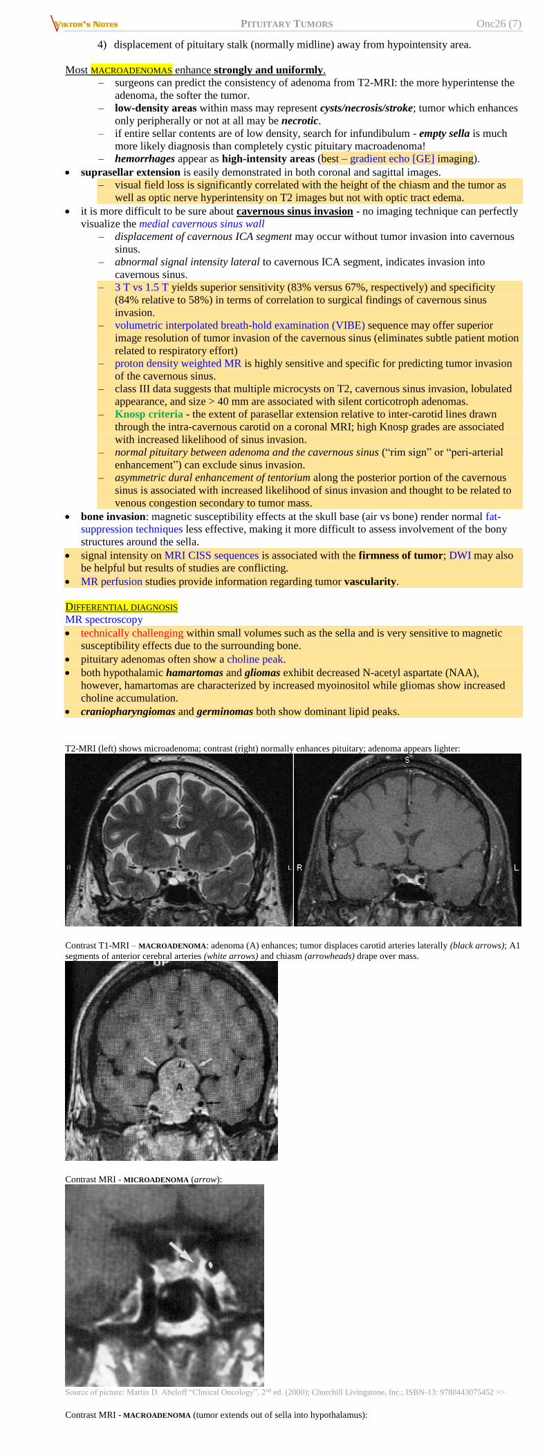
MRI - hemorrhage into tumor (apoplexy):
A) CT with contrast - MACROADENOMA with suprasellar and parasellar extension.
B) MRI with contrast following bromocriptine therapy shows marked decrease in tumor size such that infundibulum and
optic chiasm are decompressed (arrow).
A) MRI - MACROADENOMA filling sphenoid sinus and extending into 3rd ventricle floor.
B) suprasellar component compressing optic chiasm (arrow).
C) following gross total resection through extended frontal craniotomy - infundibulum is well decompressed (arrow).
D) no residual tumor, optic chiasm and portion of infundibulum can be clearly seen (arrow).
PITUITARY TUMORS Onc26 (10)
RADIONUCLIDE STUDIES
some GH-secreting adenomas (and some prolactinomas) express somatostatin receptors - 111In
octreotide uptake has place in:
a) evaluation of incomplete tumor resection due to involvement of adjacent
structures.
b) identification which patients may respond to OOCCTTRREEOOTTIIDDEE therapy.
PET
Guidelines on the Management of Patients with Nonfunctioning Pituitary Adenomas (CNS 2016):
utility is limited and is not routinely used in standard practice.
18(F)-FDG PET detects pituitary adenomas with a sensitivity of 94%-100% and a sensitivity of
88%-100%
[11C]-L-deprenyl PET may facilitate discrimination of meningiomas from adenomas.
SPECT
Guidelines on the Management of Patients with Nonfunctioning Pituitary Adenomas (CNS 2016):
diagnostic utility remains unclear.
SPECT using iodinated dopamine D2 antagonist S(-) iodobenzamide (IBZM) or similar
compounds demonstrated that D2 receptors in pituitary adenomas can be visualized using SPECT
Technetium-99m-hexakis-2-methyoxy-isobutyl-isonitrile SPECT can discriminate adenoma
from normal pituitary gland.
99mTc(V)-DMSA is actively taken up by adenomas relative to other sellar/suprasellar lesions.
radiolabeled somatostatin or dopamine can potentially differentiate hormone producing from
nonfunctioning pituitary adenomas and identify patients who would benefit from pharmacotherapy,
although the clinical feasibility of this is unclear.
ANGIOGRAPHY
1) to exclude aneurysm!!!! (lethal surgical cases described!!!)
2) surgical planning
NEURO-OPHTHALMOLOGICAL EVALUATION
- accurate mapping of visual disturbances (important for every patient prior to surgery).
in addition to formal ophthalmologic examination, tests of value include automated static perimetry
and optical coherence tomography (OCT).
often, patients with obvious chiasmal compression may not be aware of visual loss, discovered
only on quantitative ophthalmic assessment.
relative position of the chiasm may influence the incidence of visual field defects, with a decrease
frequency of visual deficits occurring in patients with an anatomically prefixed optic chiasm
Guidelines on the Management of Patients with Nonfunctioning Pituitary Adenomas (CNS 2016)
Level III Recommendation:
pretreatment evaluation by an ophthalmologist is recommended: asymptomatic visual deficits due
to the ophthalmologist’s ability to quantitate psychophysical (acuity and visual fields), functional
(quantitation of afferent pupillary defect and visual evoked potentials [VEP]), and anatomical (disc
appearance and ocular coherence tomography [OCT]) assessment. When paired with postoperative
evaluation, documents postoperative change.
automated static perimetry is recommended for early detection of visual field deficits.
visual evoked potentials may be used to assess the optic nerves in cases in which psychophysical
areas, such as acuity and visual fields, cannot be assessed.
older patients and patients with longer duration (over 4 months) of vision loss should be counseled
regarding the reduced chance of postoperative vision improvement
o formal ophthalmologic examination, looking for optic nerve atrophy or optical coherence
tomography (OCT) to measure both retinal nerve fiber layer (RNFL) thickness and the
presence of damage to the ganglion cell layer is recommended to assess chances of
postoperative vision improvement.
o anatomic assessment of the anterior visual pathways with optical coherence tomography
documents previous damage, showing evidence of nerve fiber bundle thinning and evidence of
ganglion cell dropout with segmentation analysis.
EVALUATION OF PITUITARY FUNCTION
(sensitive radioimmunoassays) in all patients!
N.B. guard against cortisol insufficiency postoperatively.
All endocrine axes + prolactin should be checked in every patient!
Hormone Laboratory Test
TSH TSH, free T4
LH/FSH Male: testosterone
Female: estradiol, progesterone
ACTH
Morning ACTH
Fasting AM cortisol
24-hour urine free cortisol
Dexamethasone suppression test
GH
Morning GH
Somatomedin-C
IGF-1 (reflects GH concentration
over the preceding 24 hours)
Prolactin Prolactin

PITUITARY TUMORS Onc26 (11)
High prevalence (37-85%) of hypopituitarism in patients with NFPAs.
inferior petrosal sinus sampling is used to localize tumors not seen radiographically (e.g. many
ACTH-secreting microadenomas are < 5 mm).
central hypothyroidism is typically confirmed by the thyrotropin releasing hormone stimulation
test, in which serum TSH is measured serially post-TRH at 20 and 60 minutes, with a normal
response defined as the 20- minute TSH value being higher than the 60-minute TSH value. A flat
response is seen in pituitary disease, and delayed response, with the 60-minute value higher than the
20-minute value, is seen in hypothalamic disease.
Guidelines on the Management of Patients with Nonfunctioning Pituitary Adenomas (CNS 2016):
NFPA may present with hypopituitarism (37-85%, esp. GH deficiency) or pituitary stalk
hyperprolactinemia (25-65%, with a mean level of 39 ng/mL and with a minority of patients
exceeding a serum prolactin level of 200 ng/mL).
level II recommendation: routine endocrine analysis of all anterior pituitary axes + prolactin to
assess for hypopituitarism (prolactin and IGF-1 are also valuable to assess for hypersecretion
states that might not be clinically suspected).
no evidence supporting routine biomarker testing (e.g., alpha-subunit or chromogranin A) was
available.
Although not widely used, chromogranin A (CGA) has also been assessed as a potential biomarker
for NFPAs. In a prospective case-control study by Gussi et al, 3 of 27 patients with NFPAs had
elevations of serum CGA at 576, 143, and 241 ng/mL, respectively. As the authors acknowledge, the
low prevalence of CGA elevations in the NFPA population makes its utility as a sensitive biomarker
less reliable.
Prolactin
serum prolactin level is perhaps the most important laboratory level that dictates a given patient’s
treatment course – the ability to distinguish between a prolactinoma (for which medical therapy
represents first-line therapy in most patients) and an NFPA with hyperprolactinemia caused by the
pituitary stalk effect (a surgically treated disease for most patients) is a critical one.
nonsecreting tumors are commonly associated with slight elevations of serum prolactin (< 150*)
– STALK SYNDROME (compression of pituitary stalk, interrupting dopaminergic fibers that inhibit
prolactin release) - must be distinguished from prolactin-secreting tumors because BBRROOMMOOCCRRIIPPTTIINNEE
has little or no effect on nonsecretory tumors.
*some studies indicate different thresholds beyond which stalk effect is unlikely:
> 94.3 ng/mL
> 85 ng/mL in the absence of renal failure or any prolactin-enhancing drugs +
prolactin increment less than 30% following thyrotropin-releasing hormone.
Be vigilant to prescription / recreational drugs that interfere with normal pituitary function!
hyperprolactinemia is seen in 25-65% of patients with histologically verified NFPAs, with a mean
level of 39 ng/mL and with a minority of patients exceeding a serum prolactin level of 200 ng/mL
be aware of hook effect (s. prozone effect) - type of interference which plagues certain
immunoassays and nephelometric assays, resulting in false negatives or inaccurately low results –
too much antigen (prolactin) interferes with results (H: diluting blood sample; modern labs do it
automatically):

PITUITARY TUMORS Onc26 (12)
GENETIC TESTING
Not indicated in sporadic cases.
in 2012, Cazabat et al published their results from a prospective single-center observational
study - 113 patients with presumed sporadic NFPAs underwent genetic screening for germline
mutations in the AIP gene - only 1 patient (0.9%) had evidence of an AIP mutation.
Guidelines on the Management of Patients with Nonfunctioning Pituitary Adenomas (CNS 2016):
no evidence supporting routine genetic testing was available.
COMPLICATIONS
1. Pituitary apoplexy - can be lethal!
2. Permanent visual loss, ophthalmoplegia, and other neurological complications.
3. CSF rhinorrhea – most commonly following favorable response of invasive prolactinomas to
initiation of dopamine agonist therapy.
possible mechanisms - decreased tumor volume (due to intrinsic infarction or hemorrhage),
ongoing invasion, ICP increases
treatment: surgical repair, preferentially via transsphenoidal approach
TREATMENT
Only surgical removal can produce cure!
DIFFERENT STRATEGIES
MACROADENOMAS are treated surgically (except maybe PROLACTINOMAS)
MICROADENOMAS:
a) prolactin-secreting - primary treatment is medical with dopamine agonists (role of
imaging in hyperprolactinemia is mainly to exclude MACROADENOMA; precise localization
of MICROADENOMA is therefore less important - in some centers, imaging is restricted to
unenhanced MRI).
b) other secreting adenomas are treated surgically - adenoma localization by other means
(petrosal venous sampling) is therefore important if MRI is unsuccessful.
c) incidental asymptomatic adenomas require no intervention but should be followed
periodically (endocrine examinations, visual field examinations, MRI) - onset of
symptoms or MRI documentation of growth are indications for treatment.
Goals of treatment differ according to tumor functional activity:
a) nonsecreting tumors → reduction of mass while maintaining pituitary function (although
complete surgical resection is desired, radiosensitivity of these tumors invites subtotal surgical
debulking followed by curative adjuvant radiation therapy).
b) secreting tumors → aggressive normalization of hypersecretion while preserving normal
pituitary function (usually by total surgical excision*, but some PROLACTINOMAS are better
controlled medically).
*response to radiation therapy is slow and less predictable.
Guidelines on the Management of Patients with Nonfunctioning Pituitary Adenomas (CNS 2016)
Level III Recommendation: Surgical resection is recommended as the primary treatment of
symptomatic* NFPAs.
There is insufficient evidence to make a recommendation for treatment vs. observation of
asymptomatic NFPAs.
Primary medical therapy showed inconsistent tumor response rates using somatostatin analogues (12-
40% response rate), dopamine agonist therapy (0-61% response rate), or combination therapy (60%
response rate); > 20% patients required surgery as a result of progressive clinical symptoms.
*visual field deficit or visual loss, ophthalmoplegia, compression of the optic apparatus on
MRI, endocrine dysfunction (incl. hypopituitarism or stalk effect causing hyperprolactinemia),
pituitary apoplexy, refractory headaches not attributable to other headache syndromes, or other
neurologic deficits related to compression from the tumor
NNaattuurraall hhiissttoorryy wwiitthh nnoo ttrreeaattmmeenntt ooff aassyymmppttoommaattiicc NNFFPPAAss Dekkers OM et al. The natural course of non-functioning pituitary macroadenomas. Eur. J. Endocrinol.
2007;156(2):217-224.
28 patients, mean follow-up was 118 months:
50% - radiologic evidence of tumor growth.
21% - required operation due to onset of visual field deficits.
29% - spontaneous reduction in tumor volume. Arita K et al. Natural course of incidentally found nonfunctioning pituitary adenoma, with special reference to
pituitary apoplexy during follow-up examination. J. Neurosurg. 2006;104(6):884-891
42 patients, F/U 4 years:
40% - tumor growth.
24% - became symptomatic (9.5% developed pituitary apoplexy over 5 years)
28.6% - underwent surgical intervention due to new symptoms or increasing
tumor size.

PITUITARY TUMORS Onc26 (13)
MEDICAL THERAPY
further see p. 2738 >>
A. Inhibition of hypersecretion:
Prolactin hypersecretion → dopamine agonists (e.g. BBRROOMMOOCCRRIIPPTTIINNEE, CCAABBEERRGGOOLLIINNEE).
N.B. increase cabergoline dosing incrementally to avoid too precipitous shrinkage of
mass → dura mater tear → CSF leak
ACTH hypersecretion → KKEETTOOCCOONNAAZZOOLLEE, PPAASSIIRREEOOTTIIDDEE, CCAABBEERRGGOOLLIINNEE
GH hypersecretion → OOCCTTRREEOOTTIIDDEE, dopamine agonists, PPEEGGVVIISSOOMMAANNTT (GH receptor
antagonist)
refinements in medical treatment may allow nonsurgical treatment for some MICROADENOMAS
(esp. prolactinomas!!!) throughout life.
N.B. GH hypersecretion and ACTH hypersecretion are clear indications for surgery,
even when mass is not important.
some antisecretory medications can lead tumors to be denser and more fibrotic - technically more
challenging to remove during microsurgery.
BBRROOMMOOCCRRIIPPTTIINNEE, OOCCTTRREEOOTTIIDDEE may confer relative radioresistance to tumors undergoing SRS.
B. Hormonal replacement most commonly includes thyroid and adrenal hormones.
SURGERY
- best way to definitive diagnosis and is usually curative. See p. Op305 >>
See also craniopharyngioma aspects >>
POSTOPERATIVELY
surgery often improves vision (over hours ÷ years), relieves headache, etc. see below >>
CSF leak prevention: HOB 30-45 all the time, no straws, no nose blowing, no straining, no
sneezing with closed mouth for 1-2 weeks.
nasal packs for 3 days; concomitantly abx (to prevent toxic shock syndrome – analogy with
vaginal tampons) - Ancef / Keflex / Clindamycin, saline nasal spray every 2-3 hours while awake,
phenylephrine nasal spray q6h PRN epistaxis
monitor for DI: strict Is and Os, BMP and urine spec gravity QID and PRN; patient must have easy
access to drinking water to auto-cope with high urinary output (if urinary output > 300 for 2
consecutive hours or Na persistently > 145 or patient cannot keep I&Os even H: DDAVP 1 mcg
q12h IV PRN as it may be transient; Dr. Sahni gives DDAVP liberally; if DDAVP required for > 5
days, transition to scheduled 0.5 mcg q12hr subQ and then intranasal if ENT clears for that).
HYDROCORTISONE taper per endocrinology recs / rapidly if BP is OK: 100 mg q8h → 50 mg q8h →
25 mg q12h → 15-20 mg + 5-10 mg (discharge on this dose)
if has lumbar drain – keep it clamped until nasal packs are out (if CSF leak - drain 10 cc/hr).
some experts prescribe AUGMENTIN for 14 days.
ENDOCRINOLOGICAL FOLLOW-UP
close monitoring of hormonal status (at least thyroid and adrenal function) at frequent intervals (at 3
and 6 mo, and yearly thereafter) - replacement hormonal therapy is usually required and
adjustments continue even years later.
MICROADENOMAS often can be removed without damage to normal pituitary tissue!
Dr. Broaddus prefers endocrinology consult postop; Dr. Holloway – only if patient was not
seen by endocrinologist preoperatively.
Guidelines on the Management of Patients with Nonfunctioning Pituitary Adenomas (CNS 2016)
Level III Recommendations
postoperative serum sodium levels on the first 2 days and on days 7-8 is recommended to prevent
symptomatic postoperative hyponatremia (insufficient evidence to make a recommendation on the
detection and treatment of postoperative diabetes insipidus).
evaluation of adrenal function on postop day 2, 6 weeks, and 12 months after surgery is
recommended.

PITUITARY TUMORS Onc26 (14)
perioperative corticosteroid supplementation is recommended for NFPA patients with preoperative
or immediate postoperative (day 2) hypocortisolemia.
postoperative endocrinologic follow-up in patients with normal pituitary function beyond 1 year is
not recommended.
indefinite endocrinologic follow-up is recommended in all patients after radiotherapy or with
abnormal pituitary function after surgery.
Level Inconclusive Recommendations
There is insufficient evidence to make a recommendation on the detection and treatment of
postoperative diabetes insipidus (DI).
There is insufficient evidence to make a recommendation regarding the frequency of endocrinologic
follow-up evaluation after surgery or radiation therapy.
OPHTHALMOLOGICAL FOLLOW-UP
Guidelines on the Management of Patients with Nonfunctioning Pituitary Adenomas (CNS 2016)
Level III Recommendations
ophthalmologic follow-up after surgical / radiation therapy for NFPAs is recommended
(insufficient evidence to make a recommendation on the length of time for this surveillance and the
frequency).
IMAGING FOLLOW-UP
MRI same night* and at 3 months; then annually for 10 years – so recurrence can be detected early
and, while small, can be treated with radiation, thus, avoiding redo surgery (Dr. Holloway).
*some surgeons (Dr. JRC, Holloway) skip immediate postop MRI (as it
does not change anything, plus, blood and grafts in sella mask picture)
but Dr. Broaddus does always want it
Guidelines on the Management of Patients with Nonfunctioning Pituitary Adenomas (CNS 2016)
Level III Recommendations
MRI with fat suppression* is recommended for follow-up after surgical or radiation treatment.
first radiologic study to evaluate the resection extent must be 3-4 months after surgery (insufficient
evidence to make a recommendation regarding the timing of initial radiologic follow-up after
radiation therapy); immediate postoperative radiographic studies may be misleading in determining
the amount of tumor residual.
radiologic surveillance has to be long-term (insufficient evidence to make a recommendation on the
length of time of surveillance and its frequency).
gross total resection of the NFPA requires radiologic surveillance less frequently than subtotal
resection.
*to distinguish hemorrhage, fat graft, and the posterior lobe of the
pituitary gland
Acromegaly
potential for the recurrence of high IGF-1 many years after achieving control, sometimes with
stable or absent tumor remnant, suggests the need for ongoing long-term monitoring of IGF-1
levels.
no patients with a normal IGF-1 index had evidence of tumor growth - the vast majority of patients
who have long-term normalization of IGF-1 and stable structural disease do not seem to require
routine pituitary imaging - pituitary MRI could be reserved for patients who exhibit new elevated
IGF-1 after some period of tumoral stability (prevents unnecessary exposure to gadolinium).
POSTOP COMPLICATIONS
CSF leak (4.7%), meningitis (2.0%), visual deterioration (2.0%)
Trans-sphenoidal approach
mortality < 1%
major complications (stroke, visual loss, meningitis, CSF leak, cranial palsy) < 3.5%.
permanent diabetes insipidus appears in 0.1% (microadenomas) or 1-5% (macroadenomas).
olfactory dysfunction – depends on approach. see p. Op305 >>
RADIOTHERAPY
TYPES
A. Radiosurgery – historically, only if distance from optic chiasm is > 10 mm; modern approach –
enough ≥ 1 mm from optic apparatus.
B. Stereotactic fractionated – can radiate even if tumor contacts chiasm (max fraction dose is 1.9
Gy); delivers radiographic and functional outcomes similar to those seen with SRS but latency is
longer with more frequent side effects (e.g. risk of hypopituitarism is significantly higher as
compared to SRS).
C. Conventional fractionated (45 Gy in 25 fractions of 1.8 Gy, calculated at 95% isodose line -
provides long-term control in 75-90% cases).
Studies of radiation therapy as a primary treatment method have not shown superiority or equivalence
to surgical resection of NFPAs.
General rule: radiotherapy is indicated when surgery is not an option.
INDICATIONS
(radiotherapy is normally adjunctive to surgery) – to control hypersecretion* and / or tumor mass:
a) residual tumor after subtotal resection (esp. widely invasive MACROADENOMAS) - single
session SRS provides growth control and long-term endocrine control that is superior to that
of repeat resective surgery.
b) cavernous sinus invasion!!!
c) recurrence (if previously received adjuvant radiotherapy → reoperate; if previously did not
receive radiotherapy → administer it now - single session SRS provides growth control and
long-term endocrine control that is superior to that of repeat resective surgery)
d) not surgical candidates (but histologic confirmation is generally desired!)
e) not benefited from / intolerant to postsurgical medical intervention.
*Gamma knife is less effective than conventional radiotherapy (higher
remission rates, no recurrences described) but Gamma knife carries
lesser chances of panhypopituitarism
Benign tumor as a target for SRS
1. Well circumscribed targets without infiltration
2. Easily visualized with sharp delineation
3. Slow growth rate makes high dose single fraction treatment desirable over fractionation (but
late complications have time for expression)

PITUITARY TUMORS Onc26 (15)
4. Goal of SRS: accurately deliver adequate radiation to the “target” with a minimal dose outside
the prescribed area (i.e. provide the highest potential for growth control and normalization of
hormone production + minimize the risk of cranial neuropathies)
METHODOLOGY (SRS)
Highly conformal dose plan is needed to spare the optic apparatus as well as any remaining
normal pituitary gland!
Optic considerations – see p. Rx11 >>
Pituitary considerations – see p. Rx11 >>
Tumor control
Minimal tumor margin dose 12-16 Gy for nonfunctioning, 30-35 Gy for functioning.
minimum margin dose of 12 Gy is generally considered a safe tumor control dose.
doses of at least 15 Gy to ensure reliable and early tumor growth control may be prescribed when
distance from the tumor margin to the optic apparatus allows.
N.B higher doses are needed for biochemical control (some investigators suggest up to 30–40
Gy to center, > 20 Gy to 50% margin isoline whenever possible for treating small volume
secretory pituitary adenomas); SRS has better chances of biochemical control than
fractionated XRT.
Cavernous sinus involvement
microsurgery and SRS are often utilized in a planned staged manner: initial first stage
extracavernous microsurgery to reduce the tumor volume and create space between the tumor and
the optic apparatus, thus allowing safe delivery of the highest dose of SRS possible.
PREOP
BBRROOMMOOCCRRIIPPTTIINNEE, OOCCTTRREEOOTTIIDDEE may confer relative radioresistance to tumors undergoing SRS -
many clinicians suggest stopping these agents 4-6 weeks prior to SRS and restart 1 week after SRS.
long acting drugs (e.g. SSLLOOWW RREELLEEAASSEE OOCCTTRREEOOTTIIDDEE) should be discontinued 3–4 months prior to
SRS.
OUTCOMES
Guidelines on the Management of Patients with Nonfunctioning Pituitary Adenomas (CNS 2016):
assessment of the efficacy of radiation therapy in the primary treatment of NFPAs is sparse (the risk of
tumor progression and radiation-induced hypopituitarism are major disincentives)
Gamma Knife results in published series:
decrease in tumor size 38-85% (at 10years)
stable tumor size 26.7-62%
increase in tumor size 13.3%
Unlike surgical resection, which eliminates the tumor on subsequent neuroimaging, the neoplastic goal
of SRS is PERMANENT TUMOR CONTROL - a tumor, which has been enlarging, is made incapable of
further tumor growth, and this control is confirmed through long-term neuroimaging follow-up.
while permanent stabilization of tumor size is the desired goal, the majority of tumors will
demonstrate varying degrees of tumor shrinkage over time.
tumor growth control success: 94-95% cases at 5 yrs, 76-85% at 10 yrs.
Radiation therapy is less effective in controlling endocrine HYPERSECRETION (although reported
success in 29-82% of cases with SRS vs. 31-80% with surgery)
normalization of hormone secretion requires time (median time to normal 1.09 yrs; cumulative
normal 86 % after 3.4 yrs)
normal vision can be achieved by irradiation alone in 2/3 patients (i.e. emergency radiotherapy is
an option even with visual changes if surgery is not feasible).
control rates: ACTH > GH > prolactin
GH levels decrease only at rate of 10-30% per year (several years may be required for levels to
normalize!).
Time to endocrinologic remission is 12-144 months
ideal situation - small target volume sufficiently far from optic chiasm (to avoid radiation-induced
optic neuropathy).
COMPLICATIONS
see also p. Rx11 >>
1) hypopituitarism (risk 12-100% for fractionated XRT; 0-39% for SRS) may develop after
years (largely correctable by hormone replacement therapy - patients treated for pituitary
adenomas should be observed by endocrinologist for remainder of their lives); safe dose to
gland is < 15 Gy, to stalk < 17 Gy.
2) optic chiasm radiation injury (risk 1-2% for fractionated XRT; especially sensitive in
acromegaly) → optic nerve neuropathy and ophthalmoplegia; optic structures should be
decompressed before radiation therapy!
3) temporal lobe injuries (infarctions, temporal epilepsy, cognitive dysfunctions) – due to
radiation shifted away from optic apparatus
4) radiation-induced brain tumor - risk is small (1.3% at 10 years and 1.9% at 20 years).
5) cerebrovascular injury.
2)-4) complications do not occur with Gamma knife;
with Gamma knife, only 38-60% tumors demonstrate shrinkage postop – SRS is not good for
decompression.
no dose limits to carotid artery (but avoid hotspots > 25 Gy on it).
CHEMOTHERAPY
invasive pituitary adenomas may respond to TTEEMMOOZZOOLLOOMMIIDDEE.
ALGORITHMS ACCORDING TO HORMONE

PITUITARY TUMORS Onc26 (16)
PROGNOSIS
very favorable prognosis (success of surgical intervention).
recurrence is possible only if resection is incomplete.
after surgery for NFPA: visual function improved in 75-91%, hypopituitarism improved in 35-50%,
new hypopituitarism developed in 12% of patients.
– Dekkers et al (2007) showed that visual acuity improved significantly within 3
months of transsphenoidal surgery; further improvement was seen 1 year
postoperatively (the beneficial effects of tumor decompression can be seen in a
delayed progressive fashion).
NATURAL HISTORY WITHOUT TREATMENT
See above >>
TREATMENT OF RECURRENCE / RESIDUAL TUMOR
recurrence after initial resection is 44-75% within 10-years.
OK to watch; if starts growing – linear growth – can calculate when will reach optic chiasm.
1. Surgery – 1st choice
2. Radiotherapy – fractionated (esp. if tumor touches optic chiasm) or SRS.
invasive or imaging-negative functioning adenoma following failed resection → whole-sellar SRS
can offer endocrine remission.
Guidelines on the Management of Patients with Nonfunctioning Pituitary Adenomas (CNS 2016) -
adult patients with recurrent or residual NFPAs (recurrence after initial resection has been noted to be
as high as 44%-75% within a 10-year period of time):

PITUITARY TUMORS Onc26 (17)
Level II Recommendations • SRS (12-20 Gy) and radiotherapy (fractionated 45-54 Gy) are recommended to lower the risk
of subsequent tumor progression (local tumor control ≥ 90% at 5 years).
• no or only small residual intrasellar tumor postoperatively - serial neuroimaging is
recommended.
Level III Recommendations • repeat resection is recommended for symptomatic recurrent / residual NFPAs; if repeat
resection is too risky → SRS or radiation therapy
• assessment of NFPA proliferative index and ACTH staining (to identify silent corticotrophic
adenomas) are recommended - risk of adenoma progression and the benefit of earlier adjuvant
radiation.
PITUITARY CARCINOMAS
- extremely rare!
despite highly invasive characteristics, rapid growth, and anaplastic features, histology is almost
indistinguishable from adenoma - diagnosis confirmation needs distant metastases.
EMPTY SELLA SYNDROME
– arachnoid herniation through incomplete diaphragma sellae → globular sella enlargement with no
discernible hypophysis (gland is flattened on sellar floor)
ETIOLOGY
1. Primary (congenitally incompetent sellar diaphragm)
2. Secondary – after:
1) trans-sphenoidal surgery
2) radiotherapy
3) pituitary apoplexy
4) involution of silent pituitary tumor
5) benign intracranial hypertension
CLINICAL FEATURES
no endocrine / visual / neurologic disturbances (but hypopituitarism may be present).
Chiasm herniation inside sella does not cause visual field defects!
typical patient - female (> 80%), obese (75%), hypertensive (30%) with benign intracranial
hypertension (10%) and CSF rhinorrhea (10%).
occasionally, patients have small coexisting secreting pituitary tumors.
DIAGNOSIS
MRI - enlarged pituitary fossa filled with CSF; infundibulum is seen extending down posteriorly to
lower part of fossa (thereby excluding cystic tumor).
on plain radiography, cannot be distinguished from sellar enlargement by tumor.
TREATMENT
- no specific therapy is needed for empty sella alone.
PITUITARY APOPLEXY
- hemorrhage into / acute ischemia of pituitary gland (esp. MACROADENOMAS - about 5% of their
presentations; rarely into normal hypophysis) → hypothalamic, chiasmal, cavernous sinus, brainstem
compression.
provoking factors:
1) reduced blood flow to gland (e.g. upper respiratory tract infection with frequent coughing
and sneezing).
2) sudden increment of blood flow
3) stimulation of gland by endocrine mechanisms
4) anticoagulation
5) trauma.
CLINICAL FEATURES
- sudden-onset:
1) meningeal irritation - severe headache (87%), nausea-vomiting, stiff neck, fever.
2) eye signs - partial ophthalmoplegia (45%), rapidly progressive visual loss (56%) in one or
other eye.
3) varying degrees of acute panhypopituitarism (73%) (e.g. vascular collapse ← deficient
ACTH)
4) altered consciousness (13%) because of hypothalamic compression.
May be fatal!
DIAGNOSIS
CSF – hemorrhagic.
CT / MRI will differentiate from SAH.
TREATMENT
IV fluids + IV high-dose steroid replacement!
Conservative treatment for stable cases.
Indications for emergency surgical trans-sphenoidal decompression:
a) rapidly deteriorating vision
b) progression to coma!!!
HYPOPHYSITIS
(s. autoimmune hypophysitis)
Two main forms:

PITUITARY TUMORS Onc26 (18)
1. Lymphocytic (adeno)hypophysitis (s. lymphoid adenohypophysitis): the more commonly
encountered form.
– autoimmune inflammation of the pituitary stalk with lymphocytic infiltrate; the antigens have
not been identified.
– primarily in late pregnancy or early postpartum period.
2. Granulomatous hypophysitis: more aggressive, no gender bias, no association with pregnancy.
May be autoimmune, but pathogenesis not definitely known.
often mimics a nonsecretory pituitary macroadenoma (enhancing sellar mass, with negative
endocrine tests) - often undergo surgical resection instead of what may be more appropriate
medical therapy (e.g. steroids, or discontinuing possible offending agents such as ipilimumab).
CRANIOPHARYNGIOMA
- slow-growing, extra-axial tumor.
EPIDEMIOLOGY
1-5% of all primary intracranial neoplasms
5-13% of all primary CNS tumors in children - 3rd most common tumor in childhood.
INCIDENCE ≈ 0.13-2.0 per 100,000 per year.
– bimodal age distribution – first peak is in children 5-15 yrs; second peak at 50-74 years.
– median age at diagnosis is 8 years.
– unusual before age 2 years.
male-to-female ratio is 1:1.
no known risk factors.
rare - 2%–5% of primary intracranial neoplasms (6-13% in children)
PATHOLOGY
Hypotheses of origin:
1. Embryogenetic theory - embryonic nests of squamous epithelium along involuted
HYPOPHYSIOPHARYNGEAL DUCT (i.e. congenital rests of Rathke's pouch stomodeal epithelium).
2. Metaplastic theory – metaplasia of residual mature squamous epithelium (derived from
stomodeum and normally part of adenohypophysis).
GROSSLY
- smooth, lobulated masses with solid and cystic components (90% are at least partially cystic).
suprasellar location (arises in pituitary stalk and projects into 3rd ventricle and hypothalamus). intrasellar + suprasellar (70%); only suprasellar (20%), purely intrasellar (10%).
80-90% are calcified (esp. in children).
cysts filled with turbid, brownish-yellow, proteinaceous material that glitters and sparkles because
of high content of floating cholesterol crystals (compared to machinery oil); cyst rupture into CSF
→ intense sterile chemical meningitis.
several cytokines are elevated in cyst fluid when compared with CSF:
IL-1α and TNF-alpha are elevated but lower than 10-fold.
IL-6 is > 50,000 times more concentrated in cystic fluid than CSF.
extend horizontally along path of least resistance in various directions - anteriorly into
prechiasmatic cistern and subfrontal spaces; posteriorly into prepontine and interpeduncular
cisterns, cerebellopontine angle, 3rd ventricle, posterior fossa, foramen magnum; laterally toward
subtemporal spaces (can even reach sylvian fissure).
N.B. do not expand sella (unless they become very large) - differentiating feature from
suprasellar pituitary macroadenomas!
vascular supply from anterior circulation.
HISTOLOGY
- well-differentiated tissue - two main histological types:
1) ADAMANTINOMATOUS form (in majority of children; embryogenetic origin) -
resembles enamel pulp of developing teeth, composed of interspersed fibrous and necrotic
tissue + multiloculated cysts;
– distinctive feature is peripheral palisading of basal epithelium layer, which
encloses inner epithelium.
– inner epithelium may undergo hydropic vacuolization (“stellate reticulum”).
– areas of compactly arranged squamous cells contain keratin nodules ("wet"
keratin* - hallmark of this tumor subtype).
*"wet" because of plump appearance of anuclear keratinocytes
(vs. flat, flaky keratin with interspersed cell nuclei seen in
epidermoid and dermoid cysts)
– "wet" keratin nodules frequently calcify.
– greater propensity to encase vessels and cranial nerves, invade brain and
recur after surgery!!!
2) SQUAMOUS PAPILLARY form (only in adults; metaplastic origin) – no complex
heterogeneous architecture: less cystic stratified squamous epithelium and fibrovascular
islands of connective tissue; does not form keratin nodules; does not calcify!
craniopharyngiomas stimulate significant glial response (with profuse numbers of eosinophilic
Rosenthal fibers* - densely compacted bundles of glial filaments in astrocytic cell processes) in
contact areas with nervous elements - thick glial layer may encase tumor (pseudocapsule)**, but
small epithelial “fingers” can extend into adjacent tissues through gliotic scar (“William Sweet
finding”) - tight adherence to surrounding tissue can make complete resection difficult and
hazardous; however, glial reaction is area to separate neoplasm from neural elements.
*Rosenthal fibers are characteristic feature of JUVENILE PILOCYTIC ASTROCYTOMAS - biopsy that
samples only surrounding neuropil of craniopharyngioma may yield erroneous diagnosis!

PITUITARY TUMORS Onc26 (19)
**absent in 3rd vetricular portion
N.B. although histologically benign (do not undergo malignant degeneration),
craniopharyngiomas may have malignant clinical course (location + adherence to critical
structures with difficult removal + ability to recur).
rarely undergo malignant degeneration
Adamantinomatous craniopharyngioma:
Peripheral palisading of epithelium:
Inner epithelium with hydropic
vacuolization (stellate reticulum):
"Wet" keratin nodule:
Calcified "wet" keratin nodule:
Papillary craniopharyngioma: Only simple squamous epithelium:
Rosenthal fibers in neuropils surrounding craniopharyngioma:

PITUITARY TUMORS Onc26 (20)
Typical epithelium showing basisquamous character with incarcerated keratin; note honeycombed character of epithelium
in areas:
Source of picture: Philip A. Pizzo, David G. Poplack “Principles and Practice of Pediatric Oncology”, 3rd ed. (1997); Lippincott-Raven
Publishers; ISBN 13: 9780397515615 >>
Basal cell layer (single rows of darker cuboidal cells) has given rise to larger squamous cell layer forming bulk of cellular
parts; focally cells have further matured to occasional "keratin pearl" (e.g. in right upper part); microcystic cavities are
scattered; macrocyst is seen in right upper corner, with "motor oil" content:
CLINICAL PRESENTATION
- resembles pituitary adenomas, but most become symptomatic only after tumor have attained diameter
of about 3 cm; symptom duration before diagnosis ≈ 1-2 yrs (i.e. chronic presentation).
1. Increased ICP (related to hydrocephalus) – headaches (55-86%), vomiting, etc. – most commonly
bring patient to clinical attention;
superior tumor extension (obstruction of 3rd ventricle and foramen of Monro) → hydrocephalus
in 50%.
because of slow growth, papilledema is less common than optic pallor.
2. Visual field defects (e.g. homonymous or bitemporal hemianopsia) of various degrees in 37-90%.
3. Neuroendocrine deficits (66-90%) esp. GH, TSH and ADH deficits
short stature and obesity are most common signs for pediatric endocrinological referral.
in contrast to pituitary adenomas, prolactin abnormalities are seen in only 20% cases.
88-90% men complain of impotence, while 82% women complain of amenorrhea.
DIAGNOSIS
Calcifications are present in the majority of pediatric tumors (up to 90%) and over half of adult
lesions!
1. Plain skull X-ray (valuable screening tool) – enlarged, distorted sella with suprasellar
calcification.
2. CT - partially cystic, low-density, contrast-enhancing (supra)sellar lesion with calcification.
adult craniopharyngiomas often do not have calcifications – without biopsy difficult to
differentiate from pituitary adenomas.
N.B. pituitary adenomas never have calcifications!
3. MRI (best visualization!) - (supra)sellar tumor with solid and cystic components.
cyst gives homogeneous high T2 signal and low T1 signal (cholesterol or blood products
within cyst may give rise to high signal).
solid portions and capsule show contrast enhancement.
CT is enough for diagnosis (calcifications), but tumor extension (e.g. hypothalamus invasion)
is evaluated by MRI.
4. Evaluation of hypothalamic-pituitary axis (esp. diabetes insipidus and hypoadrenalism –
minimum evaluation in emergency cases)
before surgery, repeated postoperatively and periodically thereafter for at least 1 year
(hormonal deficits often increase after surgery and may take several months to become fully
apparent!).
PITUITARY TUMORS Onc26 (21)
CT with contrast - partly calcified, partly cystic suprasellar lesion (note inhomogeneous enhancement of solid tumor
components):
Source of picture: Ronald G. Grainger, David J. Allison “Grainger & Allison’s Diagnostic Radiology: A Textbook of Medical Imaging”,
4th ed. (2001); Churchill Livingstone, Inc.; ISBN-13: 978-0443064326 >>
MRI - cystic contrast-enhancing suprasellar mass extending upward, compressing hypothalamus:
Source of picture: Christopher G. Goetz “Textbook of Clinical Neurology”, 1st ed. (1999); W.B. Saunders Company; ISBN 0-7216-6423-
7 >>
Gadolinium-enhanced MRI:
A) very large suprasellar mass extending to hypothalamus & thalamus (enhancement is confined to superior portion).
B) tumor extends bilaterally.
Source of picture: Martin D. Abeloff “Clinical Oncology”, 2nd ed. (2000); Churchill Livingstone, Inc.; ISBN-13: 9780443075452 >>
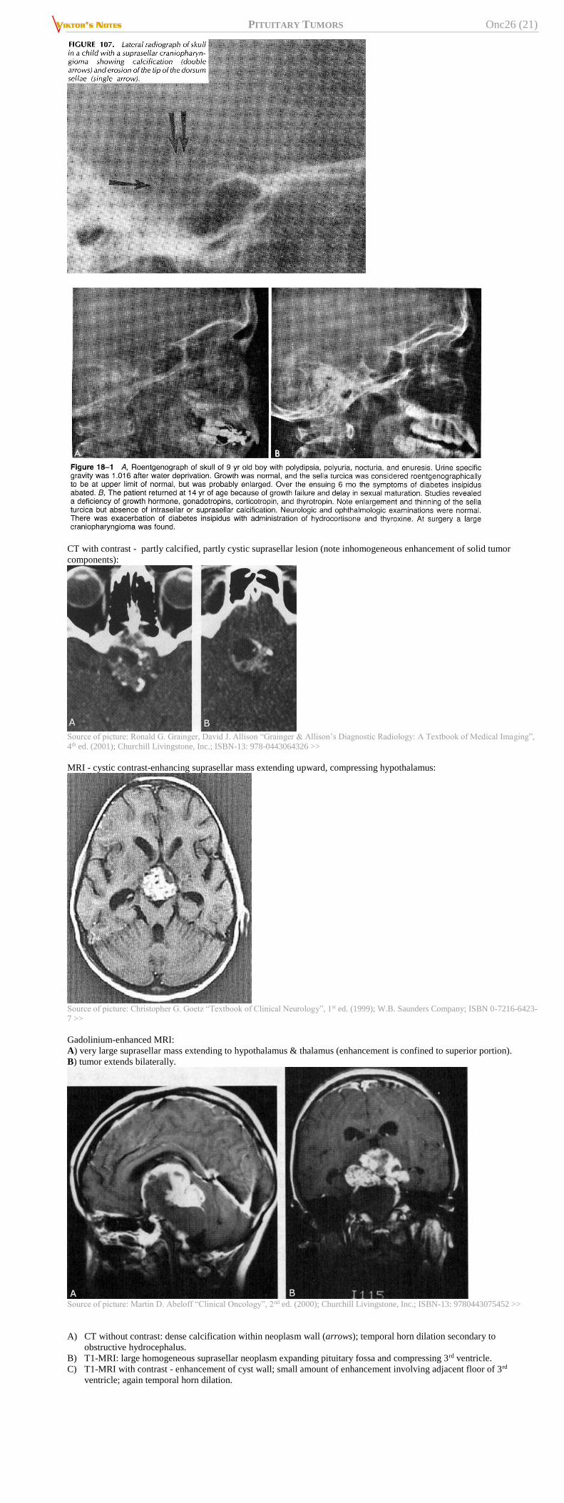
A) CT without contrast: dense calcification within neoplasm wall (arrows); temporal horn dilation secondary to
obstructive hydrocephalus.
B) T1-MRI: large homogeneous suprasellar neoplasm expanding pituitary fossa and compressing 3rd ventricle.
C) T1-MRI with contrast - enhancement of cyst wall; small amount of enhancement involving adjacent floor of 3rd
ventricle; again temporal horn dilation.
PITUITARY TUMORS Onc26 (22)
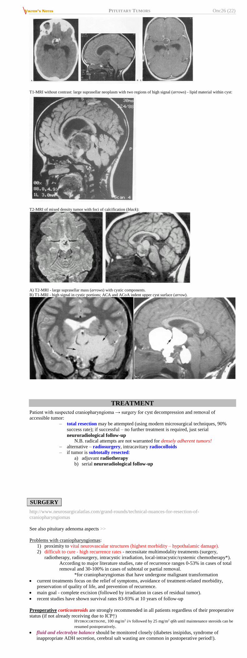
T1-MRI without contrast: large suprasellar neoplasm with two regions of high signal (arrows) - lipid material within cyst:
T2-MRI of mixed density tumor with foci of calcification (black):
A) T2-MRI - large suprasellar mass (arrows) with cystic components.
B) T1-MRI - high signal in cystic portions; ACA and ACoA indent upper cyst surface (arrow).
TREATMENT
Patient with suspected craniopharyngioma → surgery for cyst decompression and removal of
accessible tumor:
– total resection may be attempted (using modern microsurgical techniques, 90%
success rate); if successful – no further treatment is required, just serial
neuroradiological follow-up N.B. radical attempts are not warranted for densely adherent tumors!
– alternative – radiosurgery, intracavitary radiocolloids
– if tumor is subtotally resected:
a) adjuvant radiotherapy
b) serial neuroradiological follow-up
SURGERY
http://www.neurosurgicalatlas.com/grand-rounds/technical-nuances-for-resection-of-
craniopharyngiomas
See also pituitary adenoma aspects >>
Problems with craniopharyngiomas:
1) proximity to vital neurovascular structures (highest morbidity – hypothalamic damage).
2) difficult to cure - high recurrence rates - necessitate multimodality treatments (surgery,
radiotherapy, radiosurgery, intracystic irradiation, local-intracystic/systemic chemotherapy*).
According to major literature studies, rate of recurrence ranges 0-53% in cases of total
removal and 30-100% in cases of subtotal or partial removal.
*for craniopharyngiomas that have undergone malignant transformation
current treatments focus on the relief of symptoms, avoidance of treatment-related morbidity,
preservation of quality of life, and prevention of recurrence.
main goal - complete excision (followed by irradiation in cases of residual tumor).
recent studies have shown survival rates 83-93% at 10 years of follow-up
Preoperative corticosteroids are strongly recommended in all patients regardless of their preoperative
status (if not already receiving due to ICP↑) HYDROCORTISONE, 100 mg/m2 i/v followed by 25 mg/m2 q6h until maintenance steroids can be
resumed postoperatively.
fluid and electrolyte balance should be monitored closely (diabetes insipidus, syndrome of
inappropriate ADH secretion, cerebral salt wasting are common in postoperative period!).
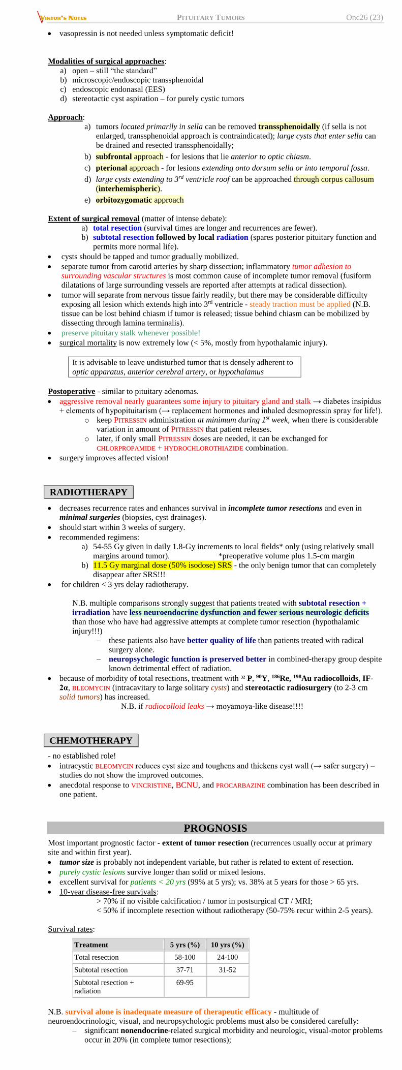
PITUITARY TUMORS Onc26 (23)
vasopressin is not needed unless symptomatic deficit!
Modalities of surgical approaches:
a) open – still “the standard”
b) microscopic/endoscopic transsphenoidal
c) endoscopic endonasal (EES)
d) stereotactic cyst aspiration – for purely cystic tumors
Approach:
a) tumors located primarily in sella can be removed transsphenoidally (if sella is not
enlarged, transsphenoidal approach is contraindicated); large cysts that enter sella can
be drained and resected transsphenoidally;
b) subfrontal approach - for lesions that lie anterior to optic chiasm.
c) pterional approach - for lesions extending onto dorsum sella or into temporal fossa.
d) large cysts extending to 3rd ventricle roof can be approached through corpus callosum
(interhemispheric).
e) orbitozygomatic approach
Extent of surgical removal (matter of intense debate):
a) total resection (survival times are longer and recurrences are fewer).
b) subtotal resection followed by local radiation (spares posterior pituitary function and
permits more normal life).
cysts should be tapped and tumor gradually mobilized.
separate tumor from carotid arteries by sharp dissection; inflammatory tumor adhesion to
surrounding vascular structures is most common cause of incomplete tumor removal (fusiform
dilatations of large surrounding vessels are reported after attempts at radical dissection).
tumor will separate from nervous tissue fairly readily, but there may be considerable difficulty
exposing all lesion which extends high into 3rd ventricle - steady traction must be applied (N.B.
tissue can be lost behind chiasm if tumor is released; tissue behind chiasm can be mobilized by
dissecting through lamina terminalis).
preserve pituitary stalk whenever possible!
surgical mortality is now extremely low (< 5%, mostly from hypothalamic injury).
It is advisable to leave undisturbed tumor that is densely adherent to
optic apparatus, anterior cerebral artery, or hypothalamus
Postoperative - similar to pituitary adenomas.
aggressive removal nearly guarantees some injury to pituitary gland and stalk → diabetes insipidus
+ elements of hypopituitarism (→ replacement hormones and inhaled desmopressin spray for life!).
o keep PPIITTRREESSSSIINN administration at minimum during 1st week, when there is considerable
variation in amount of PPIITTRREESSSSIINN that patient releases.
o later, if only small PPIITTRREESSSSIINN doses are needed, it can be exchanged for
CCHHLLOORRPPRROOPPAAMMIIDDEE + HHYYDDRROOCCHHLLOORROOTTHHIIAAZZIIDDEE combination.
surgery improves affected vision!
RADIOTHERAPY
decreases recurrence rates and enhances survival in incomplete tumor resections and even in
minimal surgeries (biopsies, cyst drainages).
should start within 3 weeks of surgery.
recommended regimens:
a) 54-55 Gy given in daily 1.8-Gy increments to local fields* only (using relatively small
margins around tumor). *preoperative volume plus 1.5-cm margin
b) 11.5 Gy marginal dose (50% isodose) SRS - the only benign tumor that can completely
disappear after SRS!!!
for children < 3 yrs delay radiotherapy.
N.B. multiple comparisons strongly suggest that patients treated with subtotal resection +
irradiation have less neuroendocrine dysfunction and fewer serious neurologic deficits
than those who have had aggressive attempts at complete tumor resection (hypothalamic
injury!!!)
– these patients also have better quality of life than patients treated with radical
surgery alone.
– neuropsychologic function is preserved better in combined-therapy group despite
known detrimental effect of radiation.
because of morbidity of total resections, treatment with 32 P, 90Y, 186Re, 198Au radiocolloids, IF-
2α, BBLLEEOOMMYYCCIINN (intracavitary to large solitary cysts) and stereotactic radiosurgery (to 2-3 cm
solid tumors) has increased.
N.B. if radiocolloid leaks → moyamoya-like disease!!!!
CHEMOTHERAPY
- no established role!
intracystic BBLLEEOOMMYYCCIINN reduces cyst size and toughens and thickens cyst wall (→ safer surgery) –
studies do not show the improved outcomes.
anecdotal response to VVIINNCCRRIISSTTIINNEE, BBCCNNUU, and PPRROOCCAARRBBAAZZIINNEE combination has been described in
one patient.
PROGNOSIS
Most important prognostic factor - extent of tumor resection (recurrences usually occur at primary
site and within first year).
tumor size is probably not independent variable, but rather is related to extent of resection.
purely cystic lesions survive longer than solid or mixed lesions.
excellent survival for patients < 20 yrs (99% at 5 yrs); vs. 38% at 5 years for those > 65 yrs.
10-year disease-free survivals:
> 70% if no visible calcification / tumor in postsurgical CT / MRI;
< 50% if incomplete resection without radiotherapy (50-75% recur within 2-5 years).
Survival rates:
Treatment 5 yrs (%) 10 yrs (%)
Total resection 58-100 24-100
Subtotal resection 37-71 31-52
Subtotal resection +
radiation
69-95
N.B. survival alone is inadequate measure of therapeutic efficacy - multitude of
neuroendocrinologic, visual, and neuropsychologic problems must also be considered carefully:
– significant nonendocrine-related surgical morbidity and neurologic, visual-motor problems
occur in 20% (in complete tumor resections);

PITUITARY TUMORS Onc26 (24)
– neuroendocrine deficit increase is common after aggressive surgery; permanent diabetes
insipidus occurs in 68-75% adults and 80-93% children; replacement of ≥ 2 anterior
pituitary hormones is necessary in 80-90% patients; obesity occurs in 50% patients. some craniopharyngiomas express IGF-1 and/or sex hormone receptors →
↑recurrence in patients receiving growth hormone and/or sex hormone replacement.
– postsurgical declines in neuropsychologic status are seen in many patients.
Neuropsychological deficits represent major limiting factor of independent social
functioning because 1) patients often can overcome minor neurological deficits and 2) hormone-replacing therapies are widely available. Psychosocial impairment
correlates directly with degree of hypothalamic injury sustained at time of surgery!
Female sex is independent predictor of increased cardiovascular, neurological, and psychosocial
morbidity!
BIBLIOGRAPHY for ch. “Neuro-Oncology” → follow this LINK >>
Viktor’s Notes℠ for the Neurosurgery Resident
Please visit website at www.NeurosurgeryResident.net