LNT From Your Backyard to Your Backcountry LNT Troop Training on.
DRAFT
(c) 2006 PCCRP
VII. Radiation Safety: LNT Model versus the Radiation Hormesis Model
RECOMMENDATION Radiography is a safe procedure. The ionizing radiation levels associated with spinal radiography is in the range associated with possible health benefits, termed Radiation Hormesis. Spinal radiography dose levels are in the low range where, health risk estimation can only be ‘qualitative accentuating a range of hypothetical health outcomes with an emphasis on the likely possibility of zero adverse health effects.’ The PCCRP panel concludes that the benefits of subluxation assessment and consequent treatment guidance that spinal radiographs provide outweighs the perceived and known health risks.
Supporting Evidence: Population Studies Class 1-4, Basic Science, and Validity. PCCRP Evidence Grade: Population, Basic Science, and Validity Studies = a.
Introduction
The purpose of this section is to elaborate a realistic understanding of the risks of medical/chiropractic x-rays for the general public. There are two models of radiation effects on organisms: Linear No-Threshold (LNT) model and the Radiation Hormesis model. Using the huge exposures during the atomic bombing of Japan in the 1940’s, the LNT model was derived by drawing a straight line down to zero exposure and claiming all radiation exposure causes a cancer risk. The LNT model continues to be used to estimate cancer risks from low doses of radiation, such as medical x-rays, without any conclusive supporting data. Proponents of the LNT model seem to omit Radiation Hormesis information from their commentaries, review articles, and government documents. Though many of the world’s experts on this subject matter seem to be more in favor of the Radiation Hormesis model, the LNT model continues to be discussed in hopes of better quality research which will allow scientists to logically refute it. Before this time, the LNT model continues to be considered as a means of exercising a more rigorous radiation safety precaution.88
There exists extremely strong evidence that Radiation Hormesis (health benefit) occurs in plants, microorganisms, invertebrates, and experimental animals. In fact, it was proven with statistically significant results from countless studies that benefits from low levels of radiation improved physiologic function from immunity and reproduction to growth and longevity. Ironically, much of this research came from studies evaluating ‘risks’ from radiation – so author bias is unlikely.
In this section, we will review both the LNT model and the Radiation Hormesis model and suggest that the health benefit from a screening protocol in medical/chiropractic x-rays likely outweighs any risk. According to some of the credible x-ray research out there, it is even possible there are actually health benefits from small amounts of x-ray exposure. Simplifying the Units of Radiation Exposure
There are several quantities/units used in articles on radiation doses. Some of these are abbreviated as Gy, rad, Sv, and rem. Most doctors in English speaking countries are familiar with the rad and rem abbreviations. This is because these are derived from the “English” system, of measuring units, such as the inch, the foot, the yard, etc. Most of the countries of our world have agreed to use the International System of Units (SI). This is where the abbreviations of Gy and Sv are defined.
DRAFT
(c) 2006 PCCRP
The unit of absorbed dose is the gray. One gray is defined as one joule of energy absorbed per kilogram of matter. This unit converts to the rad by:
1 Gy = 100 rad = 1 J/kg. The unit of dose equivalent is the sievert. The sievert is the product of the absorbed dose
and the quality factor for the irradiating beam. This sievert unit converts to the rem by: 1 Sv = 100 rem.
To determine equivalent dose (Sv), you multiply absorbed dose (Gy) by a quality factor
(Q) that is unique to the type of incident radiation. For Medical X-rays, generally it is assumed that Q = 1. However, the actual dose to different parts of the body from an X-ray procedure varies because different tissues and organs have varying sensitivity to radiation exposure. The term effective dose is used when referring to the dose averaged over the entire body. The effective dose accounts for the relative sensitivities of the different tissues exposed. Additionally, it allows for quantification of risk and comparison to more familiar sources of exposure that range from natural background radiation to radiographic medical procedures.
It is important to note that radiation exposure comes from the natural environment (the sun’s rays and radon gas from the ground found at some level in most homes), industrial processes and healthcare processes.
The US Environmental Protection Agency has a great annual dose calculator online (http://www.epa.gov/radiation/students/calculate.html).123 It should be noted that average annual radiation exposure is much higher in Denver Colorado than in the state of New Jersey. Looking at health statistics, people in Denver have on average, much better health than people in New Jersey. The National Council on Radiation Protection and Measurements (NCRP) is a federal organization who meets every 5 years to review health factors in ionizing radiation. The guidelines for radiation they establish are the gold standard in radiation exposure for the United States.88 These standards are often adopted by the International Council on Radiation Protection and Measurements (ICRP). It is important to note that in their most recent report, on page 97, the NCRP states the safe range of annual industrial exposure for workers is 5rem (or 5000mrem).88 This will be important information for discussion in the remainder of this section. Natural Background Radiation
We are exposed to radiation from natural sources every day. The average person in the U.S. receives an effective dose of approximately 300 mrem per year from naturally occurring radioactive materials and cosmic radiation from outer space. These natural "background" doses vary throughout the country. According to the United States EPA, influential factors can be from geographical elevation, geographical region, food, water and air ingestion, pacemakers, crowns in dental work, miles traveled on airplanes, time spent in airport security, gas lantern mantles used when camping, living in a concrete building, wearing a luminous wristwatch, watching television, using a computer, having a home smoke detector, living within 50 miles of a coal fired power plant and more!122 It is interesting that according to the EPA, nuclear power plants expose the public to less radiation than coal fired power plants do. The added dose from cosmic rays during a coast-to-coast round trip flight in a commercial airplane is about 3000 mrem. Altitude plays a big role. A large source of background radiation comes from radon gas in our homes; about 200 mrem per year.122 Like other sources of background radiation, exposure to radon varies widely from one part of the country to another.
DRAFT
(c) 2006 PCCRP
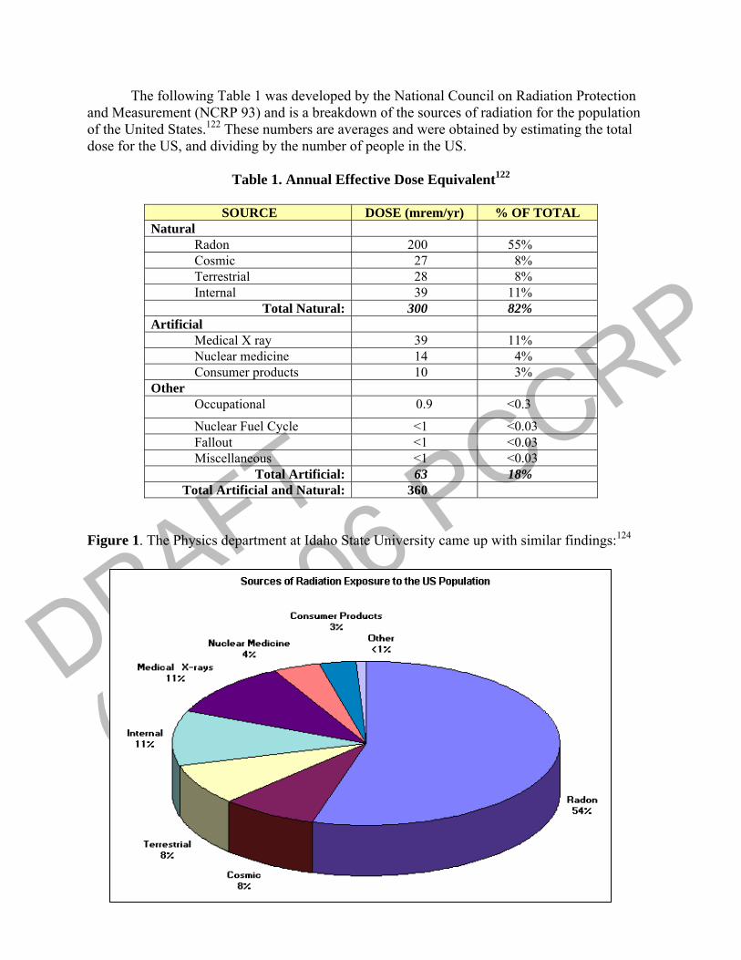
The following Table 1 was developed by the National Council on Radiation Protection and Measurement (NCRP 93) and is a breakdown of the sources of radiation for the population of the United States.122 These numbers are averages and were obtained by estimating the total dose for the US, and dividing by the number of people in the US.
Table 1. Annual Effective Dose Equivalent122
SOURCE DOSE (mrem/yr) % OF TOTAL
Natural Radon 200 55% Cosmic 27 8% Terrestrial 28 8% Internal 39 11%
Total Natural: 300 82% Artificial Medical X ray 39 11% Nuclear medicine 14 4% Consumer products 10 3% Other Occupational 0.9 <0.3
Nuclear Fuel Cycle <1 <0.03 Fallout <1 <0.03 Miscellaneous <1 <0.03
Total Artificial: 63 18% Total Artificial and Natural: 360
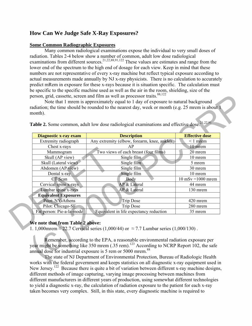
Figure 1. The Physics department at Idaho State University came up with similar findings:124
DRAFT
(c) 2006 PCCRP
How Can We Judge Safe X-Ray Exposures? Some Common Radiographic Exposures
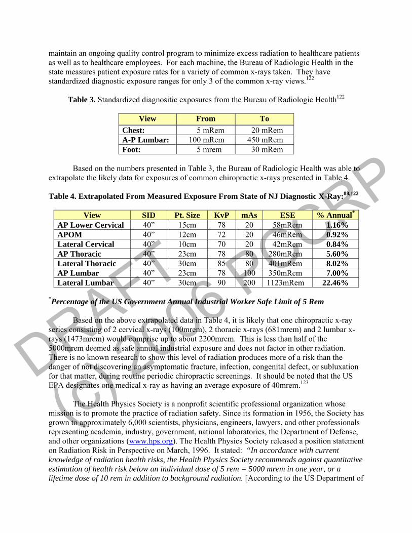
Many common radiological examinations expose the individual to very small doses of radiation. Tables 2-4 below show a number of common, adult low dose radiological examinations from different sources.21,22,88,91,122 These values are estimates and range from the lower end of the spectrum to the high end of dosage for each view. Keep in mind that these numbers are not representative of every x-ray machine but reflect typical exposure according to actual measurements made annually by NJ x-ray physicists. There is no calculation to accurately predict mRem in exposure for these x-rays because it is situation specific. The calculation must be specific to the specific machine used as well as the air in the room, shielding, size of the person, grid, cassette, screen and film as well as processor traits.88,122
Note that 1 mrem is approximately equal to 1 day of exposure to natural background radiation; the time should be rounded to the nearest day, week or month (e.g. 25 mrem is about 1 month). Table 2. Some common, adult low dose radiological examinations and effective dose.21,22,91
Diagnostic x-ray exam Description Effective dose Extremity radiograph Any extremity (elbow, forearm, knee, ankle) < 1 mrem
Chest x-rays AP 10 mrem Mammogram Two views of each breast (four films) 20 mrem
Skull (AP view) Single film 10 mrem Skull (Lateral view) Single film 5 mrem Abdomen (AP view) Single film 30 mrem
Dental x-ray Single film 10 mrem CT Scan Body 10 mSv =1000 mrem
Cervical spine x-rays AP & Lateral 44 mrem Lumbar spine x-rays AP & Lateral 130 mrem
Equivalent Exposures Pilot: NY-Athens Trip Dose 420 mrem Pilot: Chicago-SF Trip Dose 280 mrem
Fat person: Pie-a-la-mode Equivalent in life expectancy reduction 35 mrem We note that from Table 2 above: 1. 1,000mrem ≈ 22.7 Cervical series (1,000/44) or ≈ 7.7 Lumbar series (1,000/130) .
Remember, according to the EPA, a reasonable environmental radiation exposure per year might be something like 350 mrem (.35 rem).123 According to NCRP Report 102, the safe annual dose for industrial exposure is 5 rem or 5000 mrem.88
The state of NJ Department of Environmental Protection, Bureau of Radiologic Health works with the federal government and keeps statistics on all diagnostic x-ray equipment used in New Jersey.122 Because there is quite a bit of variation between different x-ray machine designs, different methods of image capturing, varying image processing between machines from different manufacturers in different years of production, using somewhat different technologies to yield a diagnostic x-ray, the calculation of radiation exposure to the patient for each x-ray taken becomes very complex. Still, in this state, every diagnostic machine is required to
DRAFT
(c) 2006 PCCRP
maintain an ongoing quality control program to minimize excess radiation to healthcare patients as well as to healthcare employees. For each machine, the Bureau of Radiologic Health in the state measures patient exposure rates for a variety of common x-rays taken. They have standardized diagnostic exposure ranges for only 3 of the common x-ray views.122
Table 3. Standardized diagnositic exposures from the Bureau of Radiologic Health122
View From To
Chest: 5 mRem 20 mRem A-P Lumbar: 100 mRem 450 mRem Foot: 5 mrem 30 mRem
Based on the numbers presented in Table 3, the Bureau of Radiologic Health was able to
extrapolate the likely data for exposures of common chiropractic x-rays presented in Table 4. Table 4. Extrapolated From Measured Exposure From State of NJ Diagnostic X-Ray:88,122
View SID Pt. Size KvP mAs ESE % Annual*
AP Lower Cervical 40” 15cm 78 20 58mRem 1.16% APOM 40” 12cm 72 20 46mRem 0.92% Lateral Cervical 40” 10cm 70 20 42mRem 0.84% AP Thoracic 40” 23cm 78 80 280mRem 5.60% Lateral Thoracic 40” 30cm 85 80 401mRem 8.02% AP Lumbar 40” 23cm 78 100 350mRem 7.00% Lateral Lumbar 40” 30cm 90 200 1123mRem 22.46%
*Percentage of the US Government Annual Industrial Worker Safe Limit of 5 Rem
Based on the above extrapolated data in Table 4, it is likely that one chiropractic x-ray series consisting of 2 cervical x-rays (100mrem), 2 thoracic x-rays (681mrem) and 2 lumbar x-rays (1473mrem) would comprise up to about 2200mrem. This is less than half of the 5000mrem deemed as safe annual industrial exposure and does not factor in other radiation. There is no known research to show this level of radiation produces more of a risk than the danger of not discovering an asymptomatic fracture, infection, congenital defect, or subluxation for that matter, during routine periodic chiropractic screenings. It should be noted that the US EPA designates one medical x-ray as having an average exposure of 40mrem.123
The Health Physics Society is a nonprofit scientific professional organization whose
mission is to promote the practice of radiation safety. Since its formation in 1956, the Society has grown to approximately 6,000 scientists, physicians, engineers, lawyers, and other professionals representing academia, industry, government, national laboratories, the Department of Defense, and other organizations (www.hps.org). The Health Physics Society released a position statement on Radiation Risk in Perspective on March, 1996. It stated: “In accordance with current knowledge of radiation health risks, the Health Physics Society recommends against quantitative estimation of health risk below an individual dose of 5 rem = 5000 mrem in one year, or a lifetime dose of 10 rem in addition to background radiation. [According to the US Department of
DRAFT
(c) 2006 PCCRP
Environmental protection, 5rem of exposure is equivalent to 125 medical x-rays.123] Risk estimation in this dose range should be strictly qualitative accentuating a range of hypothetical health outcomes with an emphasis on the likely possibility of zero adverse health effects. The current philosophy of radiation protection is based on the assumption that any radiation dose, no matter how small, may result in human health effects, such as cancer and hereditary genetic damage. There is substantial and convincing scientific evidence for health risks at high dose. Below 10 rem [10,000 mrem] (which includes occupational and environmental exposures) risks of health effects are either too small to be observed or are non-existent.”85
A. The LNT Model of Radiation Exposure
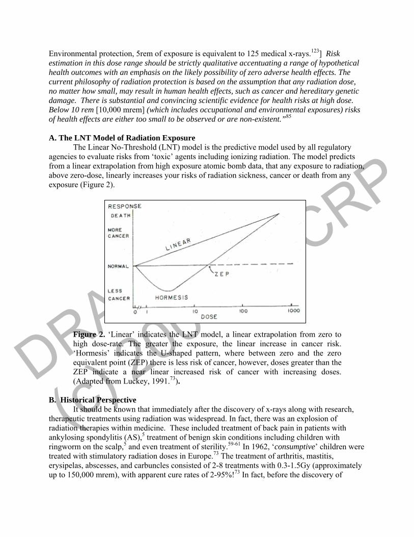
The Linear No-Threshold (LNT) model is the predictive model used by all regulatory agencies to evaluate risks from ‘toxic’ agents including ionizing radiation. The model predicts from a linear extrapolation from high exposure atomic bomb data, that any exposure to radiation, above zero-dose, linearly increases your risks of radiation sickness, cancer or death from any exposure (Figure 2).
Figure 2. ‘Linear’ indicates the LNT model, a linear extrapolation from zero to high dose-rate. The greater the exposure, the linear increase in cancer risk. ‘Hormesis’ indicates the U-shaped pattern, where between zero and the zero equivalent point (ZEP) there is less risk of cancer, however, doses greater than the ZEP indicate a near linear increased risk of cancer with increasing doses. (Adapted from Luckey, 1991.73).
B. Historical Perspective
It should be known that immediately after the discovery of x-rays along with research, therapeutic treatments using radiation was widespread. In fact, there was an explosion of radiation therapies within medicine. These included treatment of back pain in patients with ankylosing spondylitis (AS),5 treatment of benign skin conditions including children with ringworm on the scalp,5 and even treatment of sterility.59-61 In 1962, ‘consumptive’ children were treated with stimulatory radiation doses in Europe.73 The treatment of arthritis, mastitis, erysipelas, abscesses, and carbuncles consisted of 2-8 treatments with 0.3-1.5Gy (approximately up to 150,000 mrem), with apparent cure rates of 2-95%!73 In fact, before the discovery of
DRAFT
(c) 2006 PCCRP
antibiotics, countless minor diseases were treated by radiation therapy.119 Non-medical radiation therapy was also very common including removal of facial hair in ‘beauty clinics’ and even radium-containing drinks served at ‘health spas,’ and fluoroscopy in shoe-fitting.5 There was no legal control of who could use or not use x-ray machines in most countries.5
Although detrimental biological effects resulting from x-rays appeared in the literature as early as 1896,36,81,86 it was not until the 1920’s when the first radiation protection limits were set.62 In fact, throughout recent history the adoption of the LNT took place, in part for simplicity as well as the nuclear arms race threat. In the 1995 American Academy of Health Physics Radiology Centennial Hartman Oration, Kathren notes:
After World War II, the pathway takes a significant turn away from the tolerance dose directly towards the linear non-threshold dose-response model and becomes more of a two lane highway than the leisurely pleasant and relaxed country lane it once had been. In the late 1940’s, scientific interest in the applicability of the linear non-threshold model for somatic effects was kindled, and the model to be applied in radiation protection risk assessment methodology. Usage was gradually refined over a period of perhaps 15 or 20 years with special reference to the potential long term health effects from atmospheric nuclear weapons testing. Regrettably, political or ideological considerations were not absent in the choice and refinement of the low dose-risk assessment model for this and other purposes, and sometimes took precedence over strictly scientific considerations (emphasis ours).62
The original report from the United Nations Scientific Committee on the Effects of
Radiation (UNSCEAR, 1958)117 specifically states “present knowledge concerning long-term effects and their correlation with the amount of radiation received does not permit us to evaluate with any precision the possible consequence to man of exposure to low radiation levels.” This fact remains today.7,62 The report also states: “Many effects…cannot be distinguished from effects of other agents; many will only develop once a threshold dose has been exceeded some may be cumulative and others not…the possibility cannot be excluded that our present estimates exaggerate the hazards of chronic exposure to low levels of radiation.” Thus, there are several potential critical flaws to use of the LNT admitted in this report.
Despite the potential downfalls of using the LNT for radiation risk assessment, the following year the ICRP adopted this model. Ever since the 1950s, all official groups (i.e. UNSCEAR, ICRP, NCRP, and US National Academy of Sciences committees) have consistently used the LNT dose response model to estimate the risks of cancer induction from low-level radiation.16
Even if a report correctly interprets radiation data showing incongruence with LNT expectations (or even health benefits as the Hormesis model implies) from low-level radiation (i.e. levels many times above current ‘safe’ standards), it is never apart of its’ conclusions and recommendations. The BEIR V (1990) report, for example, states “studies of populations chronically exposed to low-level radiation, such as those residing in regions of elevated natural radiation, have not shown consistent or conclusive evidence of an association increase in the risk of cancer.”
To this day, including the most recent BEIR VII document (supporting LNT), little has changed in risk assessment. The LNT continues to prevail as the, ‘gold standard’, despite an incredible amount of evidence for the hormetic model or even the threshold model. This is done in spite of lack of strong evidence to support the LNT model simply because the LNT is a more exposure conservative model and final conclusive research simply has not been done at this time.
DRAFT
(c) 2006 PCCRP
In our opinion the LNT is a model of worst case scenario but by no means holds accuracy for actual health effects of low dose radiation exposure.
It is interesting to note the unanimous report by the French Academy of Sciences and National Academy of Medicine (2005) 1,85,113 states: “In conclusion, this report doubts the validity of using the LNT in the evaluation
of the carcinogenic risk of low doses (<10,000mrem) and even more for very low doses (<1000mrem). …the use of LNT in the low dose or dose rate range is not consistent with the current radiobiological knowledge; LNT cannot be used without challenge…for very low doses (<1000mrem)…. The eventual risks in the dose range of radiological examinations (10 to 500 mrem, up to 2000mrem for some examinations) must be estimated taking into account radiobiological and experimental data. An empirical relationship which is valid for doses higher than 20,000 mrem may lead to an overestimation of risk associated with doses one hundredfold lower and this overestimation could discourage patients from undergoing useful examinations and introduce a bias in radioprotection measures against very low doses (<1000 mrem).”1,113
C. Hormesis
Hormesis is the stimulatory effect of an agent at low exposure levels that is harmful at higher exposure levels. Radiation hormesis is the term used when this phenomenon results from radiation exposure.
Radiation hormesis is not new, it was first discovered by Professor W. Shrader over a century ago.109 Shrader found that guinea pigs being exposed to x-rays prior to being inoculated with the diphtheria bacillus actually lived while an unexposed comparison group died within 24 hours.109 Other medical research with radiation, outside the chiropractic profession, was being performed until about 1945 when the financial support shifted to studies evaluating harm from excess radiation.74
The chiropractic profession, however, continued to study subluxation detection by use of x-ray. In fact, by this time several techniques (and their x-ray analyses) were developed and many were to come.
A renewed interest into the stimulatory effects of radiation began in 1981, with the publication of the book by T.D. Luckey, titled ‘Hormesis with Ionizing Radiation.’75 This book, having 1200 references, validated radiation hormesis.75 It was determined that there existed incontrovertible evidence that radiation hormesis occurs in microorganisms, plants, invertebrates, and experimental animals.75 In fact, it was proven with statistically significant results from countless studies that benefits to low levels of radiation improved physiologic function from immunity and reproduction to growth and longevity. Ironically, much of this research came from studies evaluating ‘risks’ from radiation. Luckey is a controversial figure in the ionizing radiation world. One of the reasons for his controversial status is that no one has been able to disprove his theories and observations on hormesis.
Since that 1980 book by Luckey,75 he and several others have reviewed radiation hormesis.10,11,15,16,,39,52,53,58,64,66,73,76,77,87,91,93,94-99,101,102,113 The message is similar in all these reviews; that is, the LNT is invalid for estimating radiation risks from low dose exposures, and that these low dose exposures can improve physiologic function and health. In fact, it has been stated that 98% of the data in the low-dose region supports the hormesis concept.53
DRAFT
(c) 2006 PCCRP
Hiserodt53 sums up the ‘hormesis/LNT controversy nicely: “Those who advocate the Linear No-Threshold theory base their belief on the extrapolation of high-level exposure responses down to low levels. But when low-level data are available, they almost always show a bio-positive-or stimulatory-response.” This should not be a surprise given the dose response relationships from chemicals, pharmaceuticals, and physical agents, including ionizing radiation, “the hormetic model is not an exception to the rule – it is the rule.”10
“The evidence is incontrovertible. It is not challenged. It is ignored for whatever reason: ignorance, ideology, or indolence.”53 Luckey lists the main reasons why most epidemiologists and government agencies neglect the hormetic phenomenon apparent in most data that includes low doses: 74
1. Assume all radiation is harmful; 2. Include data from low-dose participants in their control cohort; 3. Have no low-dose groups in the protocol; 4. Do not use available low-dose data; 5. Do not report enough raw data to construct a dose-response curve at low doses; 6. Use a one-dimensional formula or statistic which does not allow expression of beneficial
effects; 7. Ignore data that does not fit the LNT dose-response curve; 8. Distort results by the use of median instead of mean or average value; 9. Interpolate between high doses and background levels to obtain fancied results to produce
and support unreasonable regulations; 10. Assume cell functions are not subject to whole body activities; 11. Ignore increased immune competence found in exposed organisms; and 12. Ignore increased health and average lifespan while emphasizing risks and death.
D. Human Evidence of Radiation Hormesis and Invalidation of the LNT
When reading the next few pages, it is important for the reader to keep in mind that there are a certain number of cancers and cancer deaths in the general population. Thus, whenever, a causation of a certain number of cancers is claimed, it must be compared to the percentage of such cancers in the general population, i.e., be above what is found in the general population.
The most important data for assessing risks of radiation exposures to humans began with the Japanese atomic bomb studies. Today, 60 years of follow-up data has been examined and reveals a very fascinating trend. Bomb survivors are outliving their unexposed peers! 63,66,84 In fact, exposures of up to 70rems (i.e. 70,000mrem) shows a death rate that is lower than the unexposed comparison population.84 Hiserodt noted that this Japanese data is from 40 years after the bomb and well after the established latency time for supposed cancer onset from the radiation.53 In numerical terms, for every 10,000 persons exposed to 1-1.9 cGy (1,000-1,900mrem) there were 50 fewer definitively diagnosed cancer deaths and 3 fewer leukemia deaths than in unexposed controls.108
Since the discovery of x-rays, those involved in taking images from patients have been exposed to decreasing exposures over the decades. Data from British radiologists have been studied over a 100-year period.3 It was determined that after 1920, British radiologists had lower cancer mortality than the average for the whole population of England and Wales. After 1954, radiologists had 29% lower standardized mortality ratio (SMR) from cancer, 32% lower SMR from all causes, and 36% lower SMR from non-cancer causes as compared to non-radiologists medical doctors. This data also invalidates the LNT. 11
DRAFT
(c) 2006 PCCRP
Prior to the antibiotic era, treatment for tuberculosis (TB) consisted of x-ray doses to the chest. Therefore, there was great concern about the possibility that examination and treatment may induce breast cancer in those with TB. The Canadian fluoroscopy study54,82 showed that among 31,710 females treated between the years 1930-1952, those females exposed to cumulative exposures ranging from 10-29cGy (i.e. 10,000-29,000mrem) had significantly lower than average rates of breast cancer. The hormetic pattern to the data63 demonstrates that if a million women were to receive a dose of 15cGy, it would relate to 7,000 fewer deaths from breast cancer.101
The ‘Nuclear Shipyard Workers Study’80 (NSWS) was published in 1991, took place in from 1980-1988, and cost $10 million dollars to perform by the School of Public Health at John Hopkins University, under contract of the US Department of Energy. It is the only study on radiation workers that has an age-matched and job-matched control groups.11 Three groups were included: 27,872 ‘exposed’ workers (>500mrem), 10,348 ‘minimally exposed’ workers (<500mrem), and 32,510 ‘unexposed’ controls (0rem). The ‘exposed’ workers death rate from cancer and all causes was four, and 16 standard deviations lower than the controls. “The probability of such a very low death rate from all causes being accidental is less than one in 10 million billion.”11
In Taiwan, during the 1980s, there were more than 180 buildings constructed with recycled steel contaminated with cobalt-60.69 About 10,000 people were affected and lived in these buildings for 9-20 years. The average exposure was 0.4Sv (i.e. 40,000mrem). The average cancer death rate in Taiwan over this 10-year period was 116 vs. 3.5 persons per 100,000 person years for the affected residents.15 Chen et al.15 stated that because many of the confounding factors apparent in other studies (i.e. A-bomb and Chernobyl studies) are not present in this data; they suggest that their data “should be one of the most important events on which to base radiation-protection standards.” They conclude that a “dose rate of the order of 50mSv per year (5,000 mrem) greatly reduces cancer mortality, which is a major cause of death in North America.”15
Radon gas inhalation, within the home, is feared to be a major contributor to lung cancer. After studying the effects of radon exposure to incidence of lung cancer for 90% of the US population, B.L. Cohen discovered that those residing in homes with greater radon levels had 40% lower mortality rate from radon-induced lung cancer.17 The data has been rigorously evaluated to eliminate more than 500 potential confounding factors.18,19 Radon exposure from health spas in Japan also demonstrate those exposed to radon have significantly lower cancer-induced mortality ratios than not exposed.66,101
Cancer rates for those exposed to radium has been investigated in ‘radium dial painters’ and others occupationally exposed.37,38 Those exposed to less than 1000cGy did not develop any cancers. A definite threshold phenomenon was demonstrated - invalidating the LNT. Studies of workers exposed to inhalation of Plutonium have demonstrated hormesis; that is, these workers have lower lung cancer mortality rates than controls.33,46,118
Natural background radiation vs. cancer mortality rate studies have demonstrated those living at higher altitudes (and getting greater natural background radiation exposures) have significantly less cancer mortality rates than those living in lower altitude states.43,57 In some countries exposure is greater along the coast. For example, a comprehensive survey of an Indian population exposed to high-level natural background radiation was reported by Nair et al.90 Approximately 25% (100,000 people) of the total population lived in areas with high natural radiation from the thorium deposits along coastal areas. In these regions, the environmental
DRAFT
(c) 2006 PCCRP
exposures are as high as 70 mGy/year and 7.5 times the level seen in interior areas. Using portable scintillometers, radiation levels in and outside more than 66,306 houses were measured. Confounding variables such as lifestyle, socio-demographic features, occupation, housing, residence history, tobacco and alcohol were obtained and analyzed.90 Nair et al.90 found no evidence that cancer occurrence is consistently higher due to increased levels of external gamma-radiation exposure. E. Cohen’s Outline The reader has noted that there is much evidence in the previous section that discredits the LNT model. However, there is much more to be learned. In preparation for this section of our document, we contacted one of the leading authorities on radiation exposure risks, Dr. B. L. Cohen,16-28 University of Pittsburgh, Pennsylvania, USA. We asked for a more comprehensive analysis of the LNT Model versus Radiation Hormesis. What follows is the outline and information that Dr. Cohen provided to us:28
1. Problems with the Basis for the Linear-No-Threshold Theory 2. Direct Experimental Challenges to the Basis for LNT 3. Effects of Low-Level Radiation on Biological Defense Mechanisms 4. Stimulation of the Immune System 5. Cancer Risks vs Dose in Animal Experiments 6. Cancer Risks vs Dose in Human Experiments
a. Critique of Data Frequently Cited Supportive of LNT b. Data Contradictory to LNT
E1. Problems with the Basis for LNT
The LNT model is theoretical and simple: A single particle of radiation hitting a single DNA molecule in a single cell nucleus of the human body can initiate cancer. Therefore cancer initiation probability is proportional to the number of events, which is proportional to the number of particles of radiation, which is proportional to the dose. Thus the LNT theory is “the risk is proportional to the dose”.28 The problem with this simple theory is that other factors affect cancer risk, i.e., human bodies have biological defense mechanisms that prevent the vast majority of radiation events from becoming a cancer.103
There are several defense mechanisms: (1) The most important cause of DNA injury is corrosive chemicals termed reactive oxygen species (ROS) and low-level radiation has been shown to stimulate the scavenging processes to eliminate these from cells;66 (2) There is abundant evidence that low-level radiation stimulates the immune system, while high levels/doses depress the immune response;39 (3) Radiation can alter cell timing, i.e., the time before the next cell division/mitosis and low-levels of radiation increase this time and allow for more possible DNA repair; (4) Low dose hypersensitivity and increased radiation radioresistance are affected by low-level radiation;8 and (5) It is now recognized that tissue response, whole organ response, and organism response, rather than just single cellular response, must be considered.1
There is another obvious failure of the original LNT model. The theory predicts that the number of initiating events is roughly proportional to the mass of the animal being irradiated. However, research has shown that the cancer risk for a given radiation field is similar for a 30 gram mouse and a 70,000 gram human.39
DRAFT
(c) 2006 PCCRP
Interestingly, validity of the LNT model is based on double strand breaks (DSB) in DNA molecules. However, Feinendegen39 estimated that ROS causes about 0.1 DSB per cell per day, whereas 100 mSv (10rem) of radiation causes about 4 DSB per cell. Using this information, a 100 mSv dose of radiation would increase the lifetime risk of cancer (28,000 days x 0.1 DSB/day) by only about 0.14% (4/28,000), but the LNT model predicts 7 times that much at 1%.
E2. Direct Experimental Challenges to the Basis for LNT A direct failure of the basis for the LNT model is derived from microarray studies, which determine what genes are up-regulated and what genes are down-regulated by radiation. It was discovered that generally different sets of genes are affected by low-level radiation as compared to high-level doses. In 2003, Yin et al.120 used doses of 0.1 Sv and 2.0 Sv applied to mouse brain. The 0.1 Sv dose induced expression of protective and repair genes, while the 2.0 Sv dose did not. A similar study on human fibroblast cells was conducted in 2002 by Golder-Novoselsky et al.47 Using doses of 0.02 Sv and 0.5 Sv, they discovered that the 0.02 Sv dose induced stress response genes, while the 0.5 Sv dose did not. Several other microarray studies have demonstrated that high radiation doses, which serve as the “calibration” for LNT, are not equivalent to adding an accumulation of low radiation doses.114 In fact, in 2001, Tanooka111 studied tumor induction by irradiating the skin of mice throughout their lifetimes. For irradiation rates of 1.5 Gy/week, 2.2 Gy/week, and 3 Gy/week, the percentage of mice that developed tumors was 0%, 35%, and 100%, respectively. This data111 demonstrated a clear threshold response directly in conflict with predictions of the LNT model. E3. Effects of Low-Level Radiation on Biological Defense Mechanisms
In 1994, the United Nations Scientific Committee on Effects of Atomic Radiation (UNSCEAR) report117 defined “adaptive response” as a type of biological defense mechanism that is characterized by sequent protection to stresses after an initial exposure of a stress (like radiation) to a cell. For radiation experiments, this is studied by exposing cells to low-doses to prime the adaptive response and then later exposing it to a high radiation “challenge dose” to see what happens. There have been several experiments in this topic,2,9,29,44,45,65,70,104,107,121 and we report on just a few of these.9,45,107
In 1990, Cai and Liu9 exposed mouse cells in 2 different ways: (1) a high dose of 65 cGy (65 rad), and (2) a low-dose of 0.2 cGy before the high-dose of 65 cGy. The number of chromosome aberrations reduced in the second group compared to the first group was 38% bone marrow cell aberrations reduced to 19.5% and 12.6% spermatocyte aberrations reduced to 8.4 %.
In 1992, Shadley and Dai107 irradiated human lymphocyte cells, some with high doses and some with a low-dose a few hours before a high-dose. The number of chromosome aberrations caused by a high-dose was substantially reduced when a preliminary low-dose was given first.
In 2001, Ghiassi-nejad et al.45 studied this effect in humans. In Iran, residents of a high background radiation area (1 cGy/year) were compared to residents in a normal background radiation area (0.1 cGy/yea). When lymphocytes, taken from these groups, were exposed to 1.5 Gy (150 rad), the percentages of aberrations were 0.098 for the high background area versus 0.176 (about double) for the low background area. The radiation in the high background area protected its residents from the 1.5 Gy dose.
DRAFT
(c) 2006 PCCRP
E4. Stimulation of the Immune System The effects of low -evel radiation on the immune system are important since the immune system is responsible for destroying cells with DNA damage. Low doses of radiation exposure cause stimulation of the immune system while high doses reduce immune activity.71,72,78
Contrary to expectations from the basic assumption of the LNT model (cancer risks depends only on total dose), effects on the immune system are quite different for the same total dose given at a low dose rate (summation of several small doses) versus one high dose rate, i.e., at low dose rates the immune system is stimulated, while at high doses, cancers are caused.49,55,56,79,106
E5. Cancer Risks vs Dose (Animal Experiments)
To test the validity of the LNT model, there have been numerous direct experiments of cancer risk versus dose, with animals exposed to various radiation doses. In 1979, Ullrich and Storer116 reported that exposed animals lived up to 40% longer than controls. In a series of animal studies in the 1950s and 1960s, review articles by Finkel and Biskis40-42 reported, with high statistical significance, that the LNT model over-estimated the cancer risks from low-level radiation exposures; they reported a threshold not a linear response.
In a 2001 review of over 100 animal radiation experiments, Duport34 reported on studies involving over 85,000 exposed animals and 45,000 controls, with a total of 60,000 cancers in exposed animals and 12,000 cancers in control animals. In cases where cancers were observed in controls receiving low doses, either no effect or an apparent reduction in cancer risk was observed in 40% of the data sets for neutron exposure, 50% of the data sets for x-ray exposure, 53% of the data sets for gamma rays exposure, and 61% of the data sets for alpha particles exposure. E6. Cancer Risks vs Dose (Human Experiments): A. Critique of Data Frequently Cited in Support of LNT The principle data, cited by those in influential positions, used to support the LNT model are those for solid tumors (all cancers except leukemia) in the survivors of the Japanese atomic bomb explosions. Pierce’s 1996 paper100 reported data from 1945-1990. By ignoring the error bars, supporters of the LNT model claim that the data suggests an approximate linear relationship with intercept near zero. But there is no data that gives statistical significant indication of excess cancers for radiation doses below 25 cSv.50 Leukemia data from Japanese A-bomb survivors strongly suggest a threshold above 20 cSv and the contradiction to the LNT model is recognized by the author.50 In 1998, Cohen27 used the three lowest dose points in the Japanese data (0-20 cSv) to show that the slope of the dose-response curve has a 20% probability of being negative (i.e., Hormesis = risk decreasing with increasing dose).
The next often cited evidence, by supporters of the LNT model, is the International Association for Research on Cancer (IARC) studies on monitored radiation workers. In 1995, Cardis et al.13 reported on 95,673 monitored radiation workers in 3 countries and in a follow-up study by the same authors in 2005,14 they reported on 407,000 monitored workers in 154 facilities in 15 countries. In the first study, for all cancers except leukemia (there were 3,830 deaths, but no excess over the number expected from the general population), the risks were reported as -0.07/Sv with 90% confidence limits of (-04,+0.3), i.e., there is NO support for LNT from this data! However, for leukemia (146 deaths), they reported a positive correlation, but their data had no indication of any excess cancers (risks) below 40 cSv. Most importantly, these
DRAFT
(c) 2006 PCCRP
authors manipulated their data by discarding 3/7 of their data points when observed/expected was less than unity. It is suspected that this same “manipulation of data” was performed by the same authors in the 2005 study. In fact, Cohen28 noted that (1) no information on such confounding factors as smoking was given, (2) if data from just one of the 15 countries was eliminated (Canada), the appearing excess is no longer statistically different from zero, (3) the authors did not consider non-occupational exposure (natural background radiation) and if they had, they would have noticed that their excess “signal” was much smaller than the “noise” from background radiation.
Often critics of Radiation Hormesis use the “Healthy Worker effect” to discredit what is found. When studying mortality rates for employed workers compared to the general population, it is found that workers have lower mortality rates. In Sweden in 1999, Gridley48 compared 545,000 employed women to 1,600,000 unemployed women. He reported that the cancer incidence rate was slightly higher for employed women (1.05 ± 0.01). This eliminated the claims of the “Healthy Worker effect”. For an example of improper use of this effect, in 2005 Rogel105 studied 22,000 monitored workers in the French nuclear power industry. The cancer mortality rate was only 58% of the general French population. Instead of concluding a Hormesis effect, Rogel claimed that this large difference was due to the “healthy worker effect”.105
Frequently, supporters of the LNT Model cite the recent BEIR Report30 as “proof” of the LNT theory. However, this report has been rebutted.1 Aurengo1 reported on two French groups who came to the opposite conclusions compared to the 2005 BEIR report.
Often in risk arguments, LNT supporters, eg Busseries et al.,6 passionately present Tables of the number of estimated cancer deaths per year as calculated from “known” x-ray usage in the Berrington de Gonzalez study.3 The reader should note that such tables of deaths have been ridiculed by Hiserodt,53 who asked “where are the bodies?” By this question, Hiserodt53 meant that these deaths are estimates from the LNT theory without any deaths to support it.
Besides the rebuttal by Oakley et al.93 to Busseries et al.6, we note that this Berrington de Gonzalez study3 relied solely on the LNT model and this study has been ridiculed by many for several reasons. First, as several critics noted32,51,89,115 and for which the authors3 admitted in their reply,4 they failed to weigh the benefits of diagnostic x-rays in their study, which only guarantees an overestimate of death calculations. Another criticism was their assumption of the LNT to make their cancer death estimations. Tubiana et al.115 pointed out the “speculative nature” of the LNT hypothesis, and along with Simmons,110 noted that the LNT is only compatible with exposures greater than 200mSv (drastically more radiation than any medical x-rays).
Another criticism is that the Japanese survival data has significant limitations to extrapolate its use for x-ray risk estimates from γ rays. The Japanese exposure was a one time high dose, which is entirely different from accumulated small dose rates. Herzog and Rieger51 note that this data will overestimate cancer risk because the Japanese were exposed to γ rays from bombs, a different energy spectrum than x-rays, but also the additional exposures of β radiation, radionuclides emitting β and high-energy α radiation from contaminated water, food, and dust.
Yet another criticism of the study was that there was no mention of the complexity and effectiveness of the human cell’s defenses against ionizing radiation. Tubiana et al.115 noted there are hundreds of enzymes devoted to protect a cell from these effects and that “there is no single defense mechanism but a variety,… an adaptive effect exists and a hormetic effect has even been
DRAFT
(c) 2006 PCCRP
seen in more than half of experimental studies after low or moderate doses.”10,35 “Extrapolation from high doses to low doses with LNT is unlikely to be able to assess the risks accurately.”115 B. Data Contradictory to LNT There is much data contradictory to the LNT model. There are multiple human studies which show a radiation Hormesis effect.15,17,26,31,46,54,67,68,106,112,118
For breast cancer in Canadian women, Miller82 reported a decrease risk with increasing dose up to 25 cSV. Howe54 (for lung cancer in Canadian women) and Davis31 (for 10,000 people in Massachusetts) separately reported a decrease in cancers in the low-dose region up to 100 cSv. There is a difference between lung cancer rates in Japanese A-bomb survivors and the data from Howe and Davis: the Japanese survivors show a much higher risk at all doses. This indicates that one must not accept A-bomb survivor data (one large dose) to predict risks from low-dose rates where low-level doses are summed. It is known that risks from summing low doses (such as spinal radiography use in chiropractic) does not equal the risks from one large dose (Tubiana).114
Kostyuchenko67 reported on a follow-up of 7,852 villagers exposed in the 1957 radioactive storage facility explosion in Russia. The cancer mortality rate was much lower in these villagers than in unexposed villagers in the same area supporting a hormetic effect. However, the exposure of the workers directly at the facility was quite high in one dose and these workers were found to have an increase in cancers indicating a dose threshold for increase cancers (Koshurnikova 2002).68 In 1997, Sakamoto106 reported on radiation treatments in non-Hodgkin’s lymphoma. Patient groups were randomly separated into radiation treatment and non-radiation treatment. After 9 years, 50% of the control group died but only 16% of the irradiated group died. The conclusion from Cohen’s outline28 is that the LNT theory fails badly in the low dose region. It grossly over-estimates the cancer risks from low-level radiation. The cancer risk from the vast majority of normally encountered radiation exposures (background radiation, medical x-rays, etc.) is much lower than estimates given by supporters of the LNT model, and it may well be zero or even negative. F. Implications for X-ray Guidelines In summary, it is noted that over the last 100 years, the public’s impression of the risks of medical x-rays has gone from “health benefits with insignificant risk”, to “benefits must be weighed against risks”. In the case of the Chiropractic profession, the situation is even worse; recently a subgroup of DACBRs have campaigned to attempt to force the profession to the position of only using x-rays when "Red Flags" are apparent in order to justify "benefits" over "potential" risk.
It is the conclusion of this section of this document that after reviewing the LNT Model and the Hormesis Model, it is the opinion of the PCCRP expert panel that a return to, “Benefits versus Risks”, for chiropractic x-rays is needed at present and that evidence has shown that the risks from x-rays are much less than the perception currently held by the general public. Additionally, it appears there may even be health benefits from low doses of radiation exposure from x-rays. We end this section on Radiation Exposure with a 2005 quote from Hiserodt,53 “The LNT theory has created irrational and unwarranted fears in citizens, causing them needless worry over their essential medical X-rays”53 “Sadly, an unwarranted fear of radiation causes many people who could be benefited by the use of X-ray diagnosis and therapy to shun such treatments-and thereby become subjected to unnecessary real dangers.” 53
DRAFT
(c) 2006 PCCRP
References 1. Aurengo, A. et al (2005) Dose-effect relationship and estimation of the carcinogenic effects of low
dose ionizing radiation, Available at http://cnts.wpi.edu/rsh/docs/FrenchAcads-EN-FINAL.pdf 2. Azzam EI, de Toledo SM, Raaphorst GP, Mitchel REJ. Low dose ionizing radiation decreases the
frequency of neoplastic transformation to a level below spontaneous rate in C3H 10T1/2 cells. Radiat Res 1996;146:369-373.
3. Berrington A, Darby SC, Weiss HA, Doll R: 100 years of observation on British radiologists: mortality from cancer and other causes 1897-1997. Br J Radiol 2001; 74:507-519.
4. Berrington de Gonzalez A, Darby S. Risk of cancer from diagnostic x-rays [Author's Reply]. Lancet 2004; 363(1910).
5. Berry RJ: The International Commission on Radiological Protection. A historical perspective. In: Jones,R.R., Southwood,R. Radiation and Health: the biological effects of low-level exposure to ionizing radiation; New York, John Wiley & Sons, 1987.
6. Bussieres AE, Ammendolia C, Peterson C, Taylor JAM. Ionizing radiation exposure - more good than harm? The preponderance of evidence does not support abandoning current standards and regulations. J Can Chiropr Assoc 2006; 50(2):103-106.
7. Bolus NE: Basic review of radiation and terminology. J Nucl Med Technol 2001; 29:67-73. 8. Bonner WN. Phenomena leading to cell survival values which deviate from linear-quadratic
models. Mutation Res 2004;568:33-39. 9. Cai L, Liu SZ. Induction of cytogenic adaptive response of somatic and germ cells in vivo and in
vitro by low dose X-irradiation. Int J Radiat Biol 1990;58:187-194 10. Calabrese EJ, Baldwin LA: Toxicology rethinks its central belief. Nature 2003;421:691-92. 11. Cameron JR: Longevity is the most appropriate measure of health effects of radiation. Radiol 2003;
229:14-15. 12. Cameron JR: Is radiation an essential trace energy? Physics and Society 2001;
Oct:http://www.aps.org/units/fps/newsletters/2001/october/a5oct01.html. 13. Cardis E, et al. Effects of low dose and low dose rates of external ionizing radiation: Cancer
mortality among nuclear industry workers in three countries. Radiat Res 1995;142:117-132. 14. Cardis E et al. Risk of cancer after low doses of ionizing radiation: retrospective cohort study in 15
countries. Brit Med J 2005;331:77-90. 15. Chen WL, Luan YC, Shieh MC, et al.: Is chronic radiation an effective prophylaxis against cancer?
J Am Phys Surg 2004; 9(1):6-10. 16. Cohen BL: Dose-response relationship for radiation carcinogenesis in the low-dose region. Int Arch
Occup Environ Health 1994; 66:71-75. 17. Cohen BL: Test of the linear-no threshold theory of radiation carcinogenesis for inhaled radon
decay products. Health Phys 1995; 68(2):157-174. 18. Cohen BL: Problems in the radon vs. lung cancer test of the linear no-threshold theory and a
procedure for resolving them. Health Phys 1997; 72:623-628. 19. Cohen BL: Updates and extensions to tests of the linear no-threshold theory. Technology 2000;
7:657-672. 20. Cohen B.L.: Limitations and problems in deriving risk estimates for low-level radiation exposures.
J Biol Med 1981; 54:329-338. 21. Cohen BL, Lee IS. A catalog of risks. Health Physics 1979;36:707-722. 22. Cohen BL. Catalog of risks extended and updated. Health Physics 1991; 61(3):317-335. 23. Cohen BL. How dangerous is low level radiation? Risk Anal 1995;15:645-653. 24. Cohen BL. The cancer risk from low level radiation: a review of recent evidence. Med Sent
2000;5:128-131. 25. Cohen BL. Cancer risk from low-level radiation. Am J Radiol 2002;179:1137-1143. 26. Cohen BL. Test of the linear-no-threshold theory: rationale for procedures. Nonlinearity in Biology,
Toxicology, and medicine 2005; 3:261-82. 27. Cohen BL. The cancer risk from low level radiation. Radiat Res 1998;149:525-526.
DRAFT
(c) 2006 PCCRP
28. Cohen BL. Outline of a Radiation Handbook. Private Communication, June 2006. 29. Coleman, M.A. et al. Low dose irradiation alters the transcript profiles of human lymphoblastoid
cells including genes associated with cytogenic radioadaptive response, Radiat Res 2005;164:369-382.
30. Committee to assess health risks from exposure to low levels of ionizing radiation. Health risks from exposure to low levels of ionizing radiation: BEIR VII Phase 2. National Research Council. Washington DC: National Academies Press, 2005.
31. Davis HG, Boice JD, Hrubec Z, Monson RR. Cancer mortality in a radiation-exposed cohort of Massachusetts tuberculosis patients. Cancer Res 1989;49:6130-6136.
32. Debnath D. Risk of cancer from diagnostic x-rays [Letter to Editor]. Lancet 2004; 363: 1909. 33. Drozhko EG: Multifactorial analysis of lung cancer dose-response relationships for workers at the
Mayak Nuclear Enterprise. Health Phys 1997; 73:899-905. 34. Duport P. A data base of cancer induction by low-dose radiation in mammals: overview and initial
observations, Second Conference of the World Council of Nuclear Workers (WUNOC), Dublin, 2001.
35. Duport P. A data base of cancer induction by low-dose radiation in mammals: overviews and initial observations. Int J Low Radiation 2003; 1:120-131.
36. Edison TA: Notes. Nature 1896; 53:421. 37. Evans RD: Radium in man. Health Phys 1974; 27:497-510. 38. Evans RD, Keane A, Shanahan MM: Radiological effects in man of long-term skeletal alpha-
irradiation. In: Stover,B.J., Lee,W.S.S. Radiobiology of plutonium; Salt Lake City, The J.W. Press, 1972, pp 431-468.
39. Feinendegen LE: Evidence for beneficial low level radiation effects and radiation hormesis. Br J Radiol 2005; 78:3-7.
40. Finkel MP, Biskis BO. Toxicity of Plutonium in mice. Health Phys1962; 8:565-579. 41. Finkel MP, Biskis BO. Prog Exp Tumor Res 1968;10: 72ff. 42. Finkel, MP, Biskis BO. Pathological consequences of radiostrontium administered to fetal and
infant dogs. Radiation biology of the fetal and juvenile mammal, AEC Symposium Series, vol. 17, Proceedings of the 9th Hanford Biology Symposium, 1969: pp. 543-565.
43. Frigerio NA, Eckerman KF, Stowe RS: Carcinogenic hazard from low-level, low-rate radiation, Part I. Rep ANL/ES-26 Argonne Nat Lab 1973.
44. Fritz-Niggli H and Schaeppi-Buechi C. Adaptive response to dominant lethality of mature and immature oocytes of D. Melanogaster to low doses of ionizing radiation: effects in repair-proficient and repair deficient strains. Int J Radiat Biol 1991;59:175-184.
45. Ghiassi-nejad M, Mortazavi SMJ, Beitollahi M, Cameron, JR, et al. Very high background radiation areas of Ramsar, Iran: preliminary biological studies and possible implications. Health Phys 2002;82:87-93.
46. Gilbert ES, Peterson GR, Buchanan JA: Mortality of workers at the Hanford site: 1945-1981. Health Phys 1989; 56:11-25.
47. Golder-Novoselsky, E., Ding, LH, Chen,F, and Chen,DJ. Radiation response in HSF cDNA microarray analysis, DOE Low dose radiation research program, Workshop III, U.S. Dept. of Energy, Washington, DC, 2002
48. Gridley G et al. Is there a healthy worker effect for cancer incidence among women in Sweden? Am J Ind Med 1999; 36:193-199.
49. Hashimoto S, Shirato H, Hosokawa M, et al. The suppression of metastases and the change in host immune response after low-dose total body irradiation in tumor bearing rats. Radiat Res 1999;151:717-724.
50. Heidenreich WF, Paretzke HG, Jacob B. No evidence for increased tumor risk below 200 mSv in the atomic bomb survivor data. Radiat Envoron Biophys 1997;36:205-207.
51. Herzog P, Rieger CT. Risk of cancer from diagnostic x-rays [Commentary]. Lancet 2004; 363:340-341.
DRAFT
(c) 2006 PCCRP
52. Hickey RJ, Bowers EJ, Clelland RC: Radiation hormesis, public health, and public policy: a commentary. Health Phys 1983; 44(3):207-219.
53. Hiserodt E: Underexposed: What if radiation is actually good for you? Little Rock, Arkansas, Laissez Faire Books, 2005.
54. Howe GR: Lung cancer mortality between 1950 and 1987 after exposure to fractionated moderate-dose-rate ionizing radiation in the Canadian flouroscopy cohort study and a comparison with lung cancer mortality in the atomic bomb survivors study. Radiat Res 1995; 142:295-304.
55. Ina Y, Sakai K. Activation of immunological network by chronic low dose rate irradiation in wild type mouse strains: analysis of immune cell populations and surface molecules, Int J Radiat Biol 2005;81:721-729.
56. Ina Y et al. Suppression of thymic lymphoma induction by life-long low dose rate irradiation accompanied by immune activation of C57BL/6 mice. Radiat Res 2005;163:153-158.
57. Jagger J: Natural background radiation and cancer death rate in Rocky Mountain and Gulf Coast States. Health Phys 1998; 75:428-434.
58. Kant K, Chauhan RP, Sharma GS, Chakarvarti SK: Hormesis in humans exposed to low-level ionising radiation. Int J Low Rad 2003; 1(1):76-87.
59. Kaplan II: Genetic effects in children and grandchildren of women treated for infertility and sterility by radiation therapy. Radiol 1959; 72:518.
60. Kaplan II: Clinical radiation therapy. New York, P.B. Hoeber, 1938. 61. Kaplan II: Clinical Radiation Therapy. New York, P.B. Hoeber, 1949. 62. Kathren RL: Pathway to a paradigm: the linear nonthreshold dose-response model in historical
context: the American Academy of Health Physics 1995 Radiology Centennial Hartman Oration. Health Phys 1996; 70(5):621-635.
63. Kato H, Schull WJ, Awa A, Akiyama M, Otake M: Dose-response analysis among bomb survivors exposed to low-level radiation. Health Phys 1987; 52(5):645-652.
64. Kauffman JM: Diagnostic radiation: are the risks exaggerated? J Am Phys Surg 2003; 8(2):54-55. 65. Kelsey KT, Memisoglu A, Frenkel A, and Liber HL. Human lymphocytes exposed to low doses of
X-rays are less susceptible to radiation induced mutagenesis. Mutat Res 1991;263:197-201. 66. Kondo S: Health effects of low-level radiation. Osaka, Japan/Madison, WI: Kinki University
Press/Medical Physics, 1993: pp 85-89 67. Kostyuchenko VA, Krestina LY. Long term irradiation effects in the population evacuated from
the East-Urals radioactive trace area. The Science of the Total Environment 1994;142:119-125. 68. Koshurnikova, NA et al. Studies of the Mayak nuclear workers: health effects, Radiat Environ
Biophys 2002;41:29-31. 69. Laun YC: Follow-up study of the incident of the Cobalt-60 radiation contaminated buildings in
Taiwan. The RBC Pollution Prevention Society of ROC, 1998. 70. Le XC, Xing JZ, Lee J, Leadon SA, Weinfeld M. Inducible repair of thymine glycol detected by
an ultrasensitive assay for DNA damage. Science 1998; 280:1066-1069. 71. Liu SJ. Multilevel mechanisms of stimulatory effect of low dose radiation on immunity. In:
Sugahara T, Sagan LA, Aoyama T, ed. Low Dose Irradiation and Biological Defense Mechanisms. Amsterdam, Elsevier Science, 1992: pp 225-232.
72. Liu S. Non-linear dose-response relationship in the immune system following exposure to ionizing radiation: mechanisms and implications, Nonlinearity in Biology, Toxicology, and Medicine 2003; 1:71-92.
73. Luckey TD: Radiation hormesis. Boston, Boca Raton: CRC Press, 1991. 74. Luckey TD: Radiation hormesis overview. RSO Magazine 2003; 8(4):19-36. 75. Luckey TD: Hormesis with Ionizing Radiation. CRC Press, Boca Raton, FL, 1980. 76. Luckey TD: Physiological benefits from low levels of ionizing radiation. Health Phys 1982;
43(6):771-789. 77. Luckey TD: Radiation hormesis in cancer mortality. Chi Med J 1994; 107(8):627-630.
DRAFT
(c) 2006 PCCRP
78. Makinodan T. Cellular and sub-cellular alteration in immune cells induced by chronic intermittent exposure in vivo to very low dose of ionizing radiation and its ameliorating effects on progression of autoimmune disease and mammary tumor growth. In: Sugahara T, Sagan LA, Aoyama T, ed. Low Dose Irradiation and Biological Defense Mechanisms, Amsterdam, Elsevier Science, 1992: pp. 233-237.
79. Makinodan T, James SJ. T cell potentiation by low dose ionizing radiation: possible mechanisms. Health Phys 1990;59:29-34
80. Matanoski GM. Health effect of low level radiation in shipyard workers: final report. report no. DOE DE-AC02-79 EV10095, 1991. Washington, DC, US Dept of Energy. Ref Type: Report.
81. Marcuse W: Nachtrag zu dem Fall von Dermatitis und Alopecie nach Durchleuchtungsversuchen mit Rontgenstrahlen [German]. Deutsche Med Wochschr 1896; 21:681.
82. Miller AB, Howe GR, Sherman GJ, et al.: Mortality from breast cancer after irradiation during flouroscopic examination in patients being treated for tuberculosis. N Engl J Med 1989; 321:1285-1289.
83. Miller RC, Randers-Pehrson G, Geard CR, Hall EJ, Brenner, DJ. The oncogenic transforming potential of the passage of single alpha particles through mammalian cell nuclei. Proc Natl Acad Sci 1999;96:19-22
84. Mine M, Okumura Y, Ichimaru M, Nakamura T, Kondo S: Apparently beneficial effect of low to intermediate doses of A-bomb radiation on human lifespan. Int J Radiat Biol 1990; 58: 1035-1043.
85. Mossman, Kenneth L; Goldman, Marvin; Masse, Frank; Mills, William A; Schiager, Keith J; Vetter, Richard L; Radiation Risk In Perspective: Health Physics Society Position Statement, March 1996; http://www.umich.edu/~radinfo/introduction/hprisk.htm : 1313 Dolley Madison Blvd. Suite 402; McLean, VA 22101; 703-790-1745 fax. 703-790-2672; email. [email protected].
86. Morton WJ: Notes. Nature 1896; 53:421. 87. Muckerheide J: Low-level radiation health effects: compiling the data. Needham, Radiation,
Science, and Health, Inc., 1999. 88. National Council on Radiation Protection and Measurements, “NCRP Report No. 102: Medical X-
Ray, Electron Beam and Gamma-Ray Protection for Energies up to 50MeV (Equipment Design, Performance and Use)” 7910 Woodmont Ave., Bethesda, MD 20814; Third Reprinting February 28, 1997. ISBN: 0-92600-03-7. Superseding Report No. 33 (NCRP 1968).
89. Nagataki S. Risk of cancer from diagnostic x-rays [Letter to Editor]. Lancet 2004; 363: 1909. 90. Nair MK, Nambi KS, Amma NS, Gangadharan P, Jayalekshmi P, Jayadevan S, Cherian V,
Reghuram KN. Population study in the high natural background radiation area in Kerala, India. Radiat Res. 1999 Dec;152(6 Suppl):S145-8.
91. Oakley PA, Harrison DD, Harrison DE, Haas JW: On "phantom risks" associated with diagnostic radiation: evidence in support of revising radiography standards and regulations in chiropractic. J Can Chiropr Assoc 2005; 49(4):264-269.
92. Oakley PA, Harrison DD, Harrison DE, Haas, JW. Evidence-Based Protocol for Structural Rehabilitation of the Spine and Posture: Review of Clinical Biomechanics of Posture (CBP®) Publications. J Canadian Chiro Assoc 2005; 49(4):268-294.
93. Oakley PA, Harrison DD, Harrison DE, Haas JW. A Rebuttal to Chiropractic Radiologists’ view of the 50-year old, Linear-No-Threshold Radiation Risk Model. J Can Chiropr Assoc 2006; 50(3): www.jcca.org.
94. Parsons PA: Low level exposure to ionizing radiation: do ecological and evolutionary considerations imply phantom risks? Perspectives Biol Med 1999; 43(1):57-68.
95. Parsons PA: Radiation hormesis: an ecological and energetic perspective. Med Hypoth 2000; 57(3):277-279.
96. Parsons PA: Hormesis: an adaptive expectation with emphasis on ionizing radiation. J Appl Tox 2000; 20:103-112.
DRAFT
(c) 2006 PCCRP
97. Parsons PA: Radiation hormesis: challenging LNT theory via ecological and evolutionary considerations. Health Phys 2002; 82(4):513-516.
98. Parsons PA: Energy, stress and the invalid linear no-threshold premise: a generalization illustrated by ionizing radiation. Biogerontology 2003; 4(4):227-231.
99. Parsons PA: Hormesis: an adaptive expectation with emphasis on ionizing radiation. J Appl Toxicol 2000; 20(2):103-112.
100. Pierce DA, Shimizu Y, Preston DL, Vaeth M, Mabuchi K. Studies of the mortality of atomic bomb survivors, Report 12, Part 1, Cancer: 1950-1990. Radiat Res 1996; 146:1-27.
101. Pollycove M: Nonlinearity of radiation health effects. Env Health Persp 1998; 106(Suppl 1):363-368.
102. Pollycove M: The issue of the decade: hormesis. Eur J Nucl Med 1995; 22:399-401. 103. Pollycove M and Feinendagen L. Biologic responses to low doses of ionizing radiation: detriment
vs hormesis; Part 1. J Nuclear Med 2001; 42:17N-27N. 104. Redpath JL, Antoniono RJ. Induction of a rapid response against spontaneous neoplastic
transformation in vitro by low dose gamma radiation, Rad Res 1998;149:517-520 105. Rogel A et al. Mortality of workers exposed to ionizing radiation at the French National
Electricity company. Am J Ind Med 2005;47:72-82.. 106. Sakamoto K, Myogin M, Hosoi Y, et al. Fundamental and clinical studied on cancer control with
total and upper half body irradiation. J Jpn Soc Ther Radiol Oncol 1997;9:161-175. 107. Shadley JD, Dai GQ. Cytogenic and survival adaptive responses in G-1 phase human
lymphocytes. Mutat Res 1992;265:273-281 108. Shimizu Y, Kato H, Schull WJ, Mabuchi K: Dose-response analyiss among atomic bomb
survivors exposed to low-dose irradiation; Low-dose irradiation and biological defense mechanisms. Amsterdam, Excerpta Medica, 1982, pp 71-74.
109. Shrader W: Experiments with X-rays upon germs. Electrical Engineering 1896; 22:170. 110. Simmons JA. Risk of cancer from diagnostic x-rays [Letter to Editor]. Lancet 2004; 363:1908-
1909. 111. Tanooka H. Threshold dose-response in radiation carcinogenesis: an approach from chronic beta-
irradiation experiments and a review of non-tumor doses. Int J Radiat Biol 2001;77:541-551. 112. Tokarskaya ZB, Okladlnikova ND, Belyaeva ZD, Drozhko EG. Multifactorial analysis of lung
cancer dose-response relationships for workers at the Mayak Nuclear Enterprise. Health Phys 1997;73:899-905.
113. Tubiana M: The carcinogenic effect of low doses: the validity of the linear no-threshold relationship. Int J Low Rad 2003; 1(1).
114. Tubiana M, Aurego A. Dose-effect relationship and estimation of the carcinogenic effects of low doses of radiation: the joint report of the Acadmie des Sciences (Paris) and of the Academie Nationale de Medecine. Int J Low Rad 2005;2:1-19.
115. Tubiana M, Aurengo A, Masse R, Valleron AJ. Risk of cancer from diagnostic x-rays [Letter to Editor]. Lancet 2004; 363:1908.
116. Ullrich RL, Storer JB. Influence of gamma radiation on the development of neoplastic diseases in mice: II solid tumors. Radiat Res 1979; 80:317-324.
117. UNSCEAR (United Nations Scientific Committee on Effects of Atomic Radiation). Report to the General Assembly, Annex B: Adaptive Response. United Nations, New York, 1994.
118. Voelz GL, Wilkinson CS, Acquavelle JF: An update of epidemiologic studies of plutonium workers. Health Phys 1983; 44[Suppl]:493-503.
119. Wachsmann F: Are small doses really so dangerous? Electromedia 1987; 55:86. 120. Yin, E. et al. Gene expression changes in mouse brain after exposure to low-dose ionizing
radiation, Int J Radiat Biol 2005; 79:759-775 121. Yukawa O et al. Induction of radical scavenging ability and suppression of lipid peroxidation in
rat liver microsomes following whole body low dose X-irradiation. J Radiat Biol 2005;81:681-688.
DRAFT
(c) 2006 PCCRP
122. http://www.nj.gov/dep/rpp/xrm/nextdata.htm : A Joint Survey by the State of NJ Department of Environmental Protection, Nationwide Evaluation of X-Ray Trends through the Conference of Radiation Control Program Directors in association with state radiation control agencies and the United States Food and Drug Administration Center for Devices and Radiological Health.
123. http://www.epa.gov/radiation/students/calculate.html : Student’s and Teacher’s Resource of the US Environmental Protection Agency; “Calculate Your Radiation Dose”. Average Annual Radiation Dose Calculator in mrem.
124. http://www.physics.isu.edu/radinf/popdose.htm : Idaho State University Department of Physics; Sources of Radiation Exposure to US Population. Adapted from NCRP Report 93.