View of the rat cradle and RF coil In vivo MRI of rodent heads Fits up to 350-g rat Animal is...
16
View of the rat cradle and RF coil View of the rat cradle and RF coil • In vivo MRI of rodent heads • Fits up to 350-g rat • Animal is stabilized with bite bar and body restraints • Gaseous anesthesia & vacuum exhaust • Precise climate control • Respiration monitor • Rectal thermometer • ECG monitor • Detachable probe heads • Single or double resonance 900-MHz MRI Probe for 900-MHz MRI Probe for In Vivo In Vivo Rats+: Rats+: flexibility flexibility Detachable RF probe Detachable RF probe heads heads safety belts respiratory monitor vacuum exhaust anesthesia Bite bar detachable RF coils detachable animal cradle
-
Upload
ashlynn-martina-williams -
Category
Documents
-
view
219 -
download
0
Transcript of View of the rat cradle and RF coil In vivo MRI of rodent heads Fits up to 350-g rat Animal is...
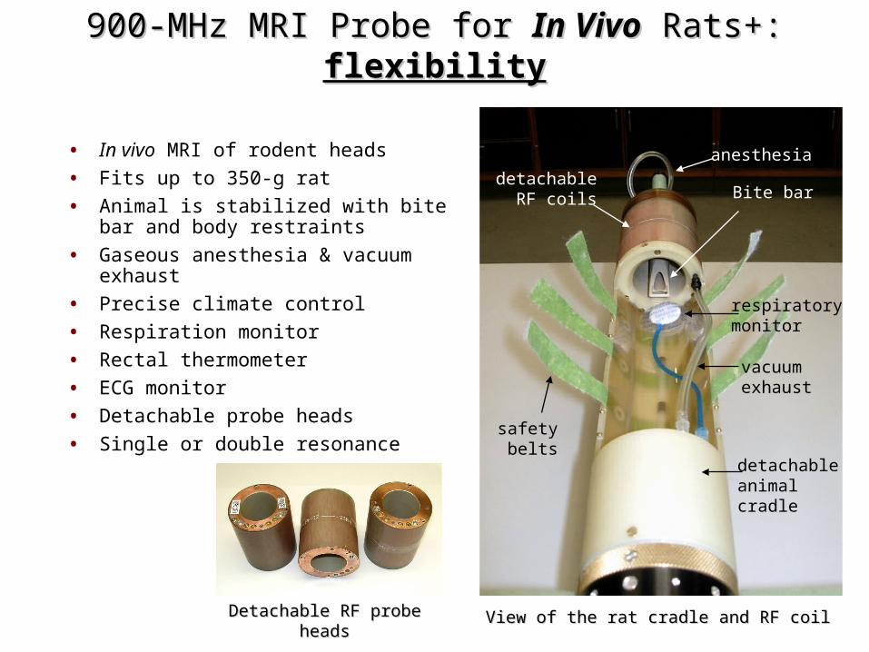
View of the rat cradle and RF coilView of the rat cradle and RF coil
• In vivo MRI of rodent heads
• Fits up to 350-g rat
• Animal is stabilized with bite bar and body restraints
• Gaseous anesthesia & vacuum exhaust
• Precise climate control
• Respiration monitor
• Rectal thermometer
• ECG monitor
• Detachable probe heads
• Single or double resonance
900-MHz MRI Probe for 900-MHz MRI Probe for In VivoIn Vivo Rats+: Rats+: flexibilityflexibility
Detachable RF probe headsDetachable RF probe heads
safetybelts
respiratorymonitor
vacuumexhaust
anesthesia
Bite bardetachable
RF coils
detachableanimalcradle
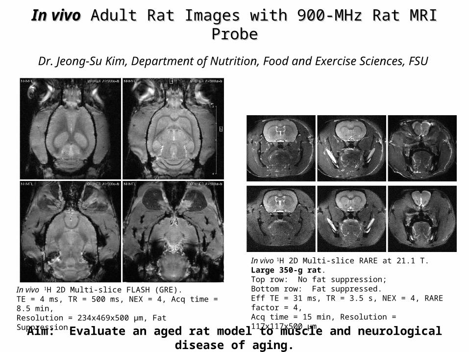
In vivoIn vivo Adult Rat Images with 900-MHz Rat MRI Probe Adult Rat Images with 900-MHz Rat MRI Probe
In vivo 1H 2D Multi-slice RARE at 21.1 T. Large 350-g rat.Top row: No fat suppression;Bottom row: Fat suppressed.Eff TE = 31 ms, TR = 3.5 s, NEX = 4, RARE factor = 4,Acq time = 15 min, Resolution = 117x117x500 μm.
In vivo 1H 2D Multi-slice FLASH (GRE).TE = 4 ms, TR = 500 ms, NEX = 4, Acq time = 8.5 min,Resolution = 234x469x500 μm, Fat Suppression.
Dr. Jeong-Su Kim, Department of Nutrition, Food and Exercise Sciences, FSU
Aim: Evaluate an aged rat model to muscle and neurological disease of aging.
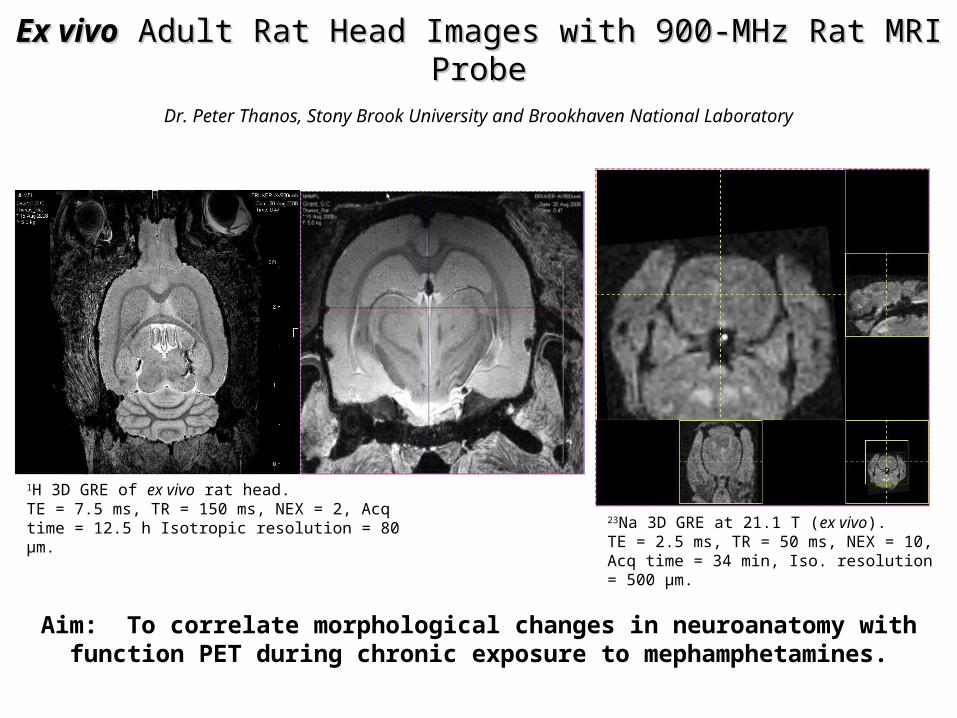
Ex vivoEx vivo Adult Rat Head Images with 900-MHz Rat MRI Probe Adult Rat Head Images with 900-MHz Rat MRI Probe
1H 3D GRE of ex vivo rat head.TE = 7.5 ms, TR = 150 ms, NEX = 2, Acq time = 12.5 h Isotropic resolution = 80 μm.
23Na 3D GRE at 21.1 T (ex vivo).TE = 2.5 ms, TR = 50 ms, NEX = 10,Acq time = 34 min, Iso. resolution = 500 μm.
Dr. Peter Thanos, Stony Brook University and Brookhaven National Laboratory
Aim: To correlate morphological changes in neuroanatomy with function PET during chronic exposure to mephamphetamines.

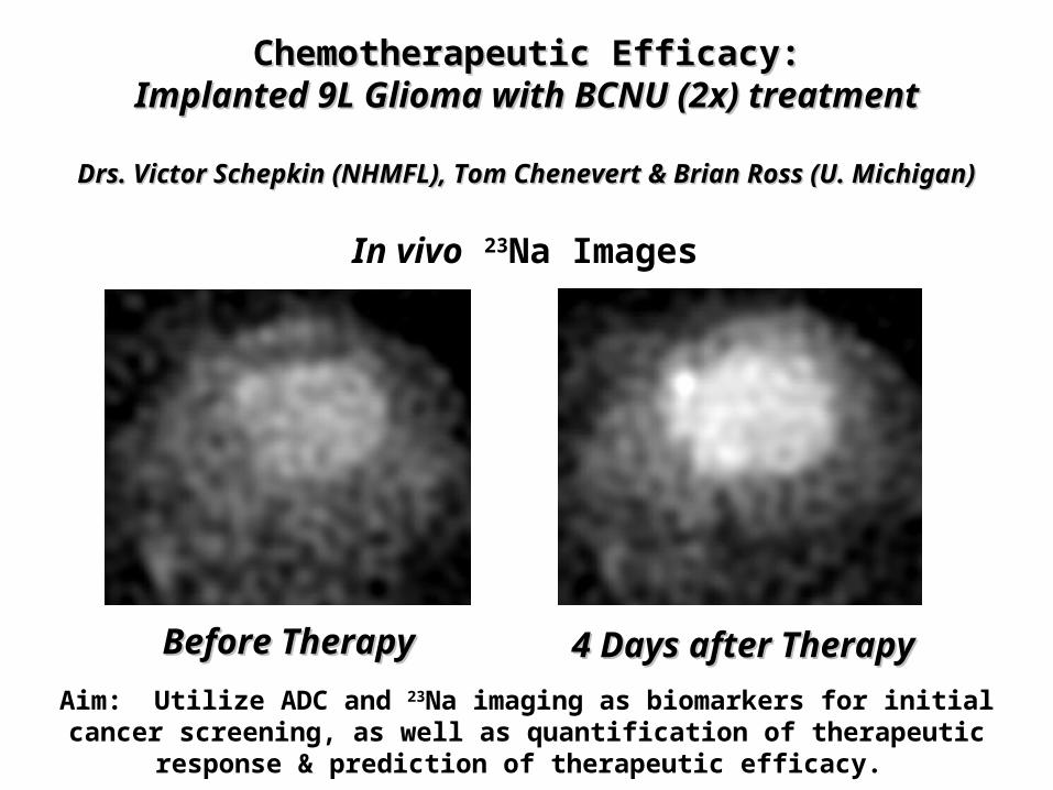
Chemotherapeutic Efficacy:Chemotherapeutic Efficacy:Implanted 9L Glioma with BCNU (2x) treatmentImplanted 9L Glioma with BCNU (2x) treatment
Drs. Victor Schepkin (NHMFL), Tom Chenevert & Brian Ross (U. Michigan)Drs. Victor Schepkin (NHMFL), Tom Chenevert & Brian Ross (U. Michigan)
Before TherapyBefore Therapy 4 Days after Therapy4 Days after Therapy
Aim: Utilize ADC and 23Na imaging as biomarkers for initial cancer screening, as well as quantification of therapeutic response & prediction of therapeutic efficacy.
In vivo ADC maps
Before TherapyBefore Therapy 4 Days after Therapy4 Days after Therapy
Chemotherapeutic Efficacy:Chemotherapeutic Efficacy:Implanted 9L Glioma with BCNU (2x) treatmentImplanted 9L Glioma with BCNU (2x) treatment
Drs. Victor Schepkin (NHMFL), Tom Chenevert & Brian Ross (U. Michigan)Drs. Victor Schepkin (NHMFL), Tom Chenevert & Brian Ross (U. Michigan)
Aim: Utilize ADC and 23Na imaging as biomarkers for initial cancer screening, as well as quantification of therapeutic response & prediction of therapeutic efficacy.
In vivo 23Na Images
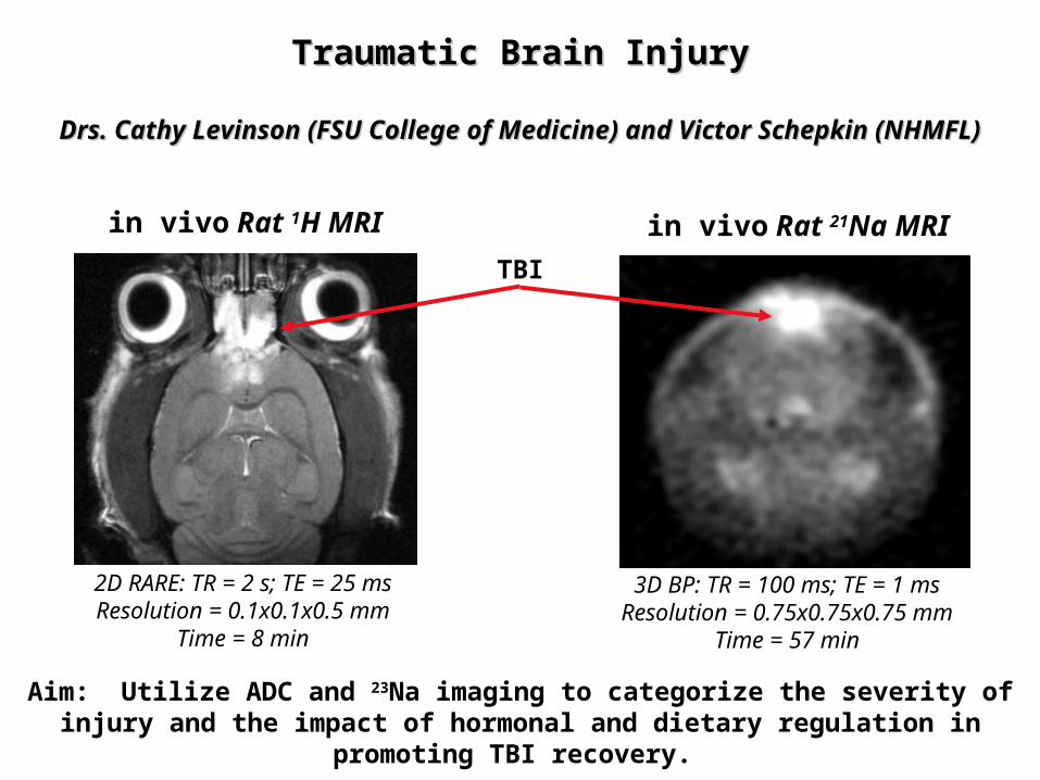
in vivo Rat 1H MRI
2D RARE: TR = 2 s; TE = 25 msResolution = 0.1x0.1x0.5 mm
Time = 8 min
Aim: Utilize ADC and 23Na imaging to categorize the severity of injury and the impact of hormonal and dietary regulation in promoting TBI recovery.
Traumatic Brain InjuryTraumatic Brain Injury
Drs. Cathy Levinson (FSU College of Medicine) and Victor Schepkin (NHMFL)Drs. Cathy Levinson (FSU College of Medicine) and Victor Schepkin (NHMFL)
TBI
in vivo Rat 21Na MRI
3D BP: TR = 100 ms; TE = 1 msResolution = 0.75x0.75x0.75 mm
Time = 57 min
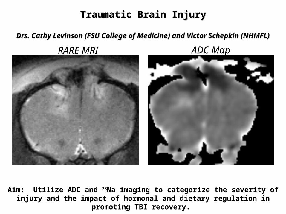
RARE MRI ADC Map
Traumatic Brain InjuryTraumatic Brain Injury
Drs. Cathy Levinson (FSU College of Medicine) and Victor Schepkin (NHMFL)Drs. Cathy Levinson (FSU College of Medicine) and Victor Schepkin (NHMFL)
Aim: Utilize ADC and 23Na imaging to categorize the severity of injury and the impact of hormonal and dietary regulation in promoting TBI recovery.
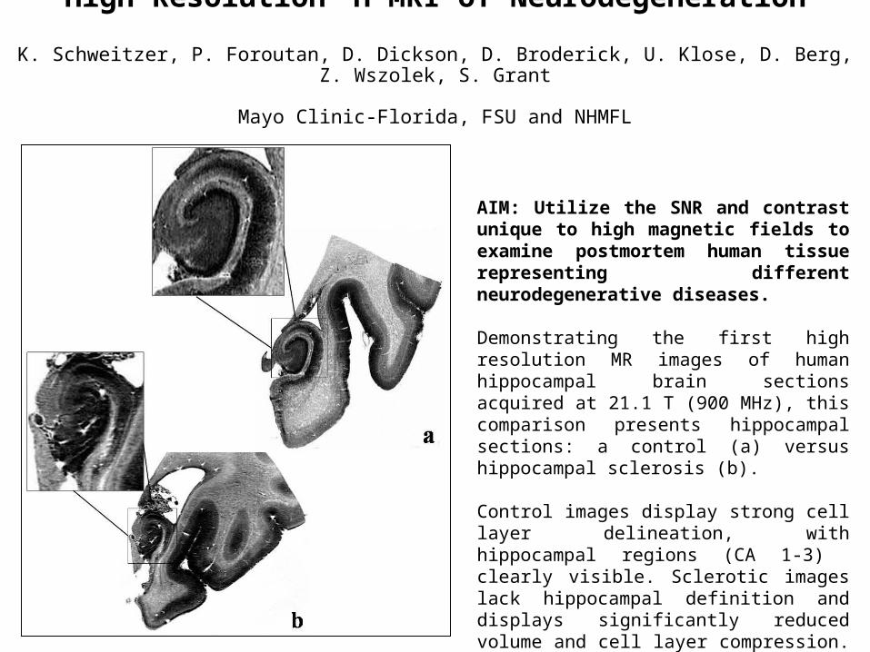
High Resolution 1H MRI of Neurodegeneration
K. Schweitzer, P. Foroutan, D. Dickson, D. Broderick, U. Klose, D. Berg, Z. Wszolek, S. Grant
Mayo Clinic-Florida, FSU and NHMFL
AIM: Utilize the SNR and contrast unique to high magnetic fields to examine postmortem human tissue representing different neurodegenerative diseases.
Demonstrating the first high resolution MR images of human hippocampal brain sections acquired at 21.1 T (900 MHz), this comparison presents hippocampal sections: a control (a) versus hippocampal sclerosis (b). Control images display strong cell layer delineation, with hippocampal regions (CA 1-3) clearly visible. Sclerotic images lack hippocampal definition and displays significantly reduced volume and cell layer compression.
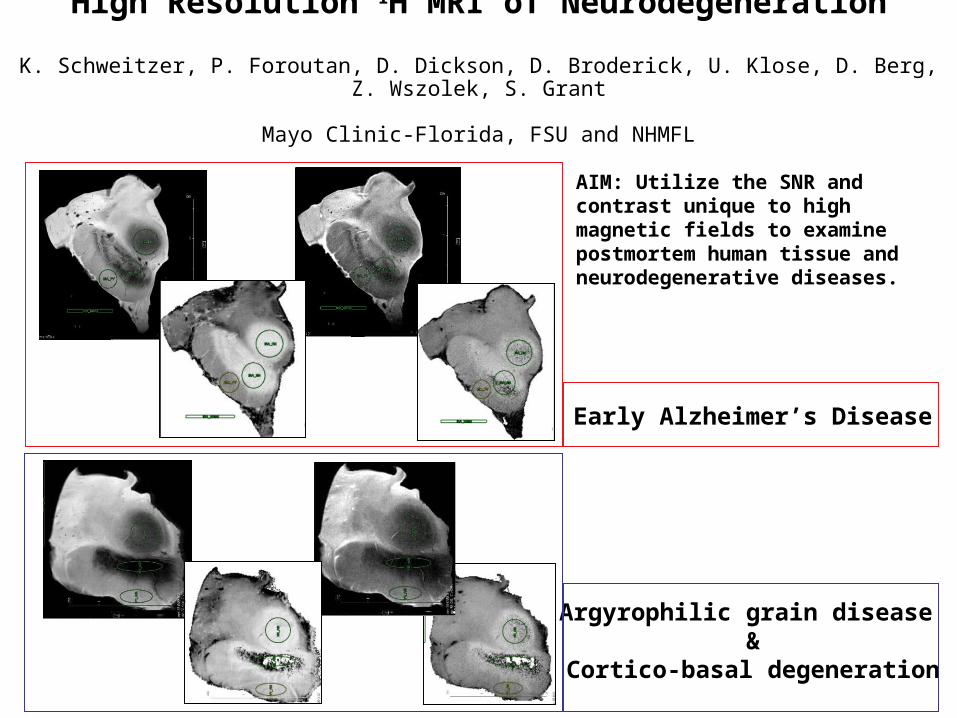
High Resolution 1H MRI of Neurodegeneration
K. Schweitzer, P. Foroutan, D. Dickson, D. Broderick, U. Klose, D. Berg, Z. Wszolek, S. Grant
Mayo Clinic-Florida, FSU and NHMFL
AIM: Utilize the SNR and contrast unique to high magnetic fields to examine postmortem human tissue and neurodegenerative diseases.
Argyrophilic grain disease &
Cortico-basal degeneration
Early Alzheimer’s Disease
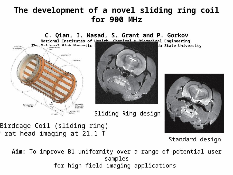
The development of a novel sliding ring coil for 900 MHz
C. Qian, I. Masad, S. Grant and P. GorkovNational Institutes of Health, Chemical & Biomedical Engineering,
The National High Magnetic Field Laboratory, The Florida State University
Aim: To improve B1 uniformity over a range of potential user samplesfor high field imaging applications
1H Birdcage Coil (sliding ring)for rat head imaging at 21.1 T
Sliding Ring design
Standard design
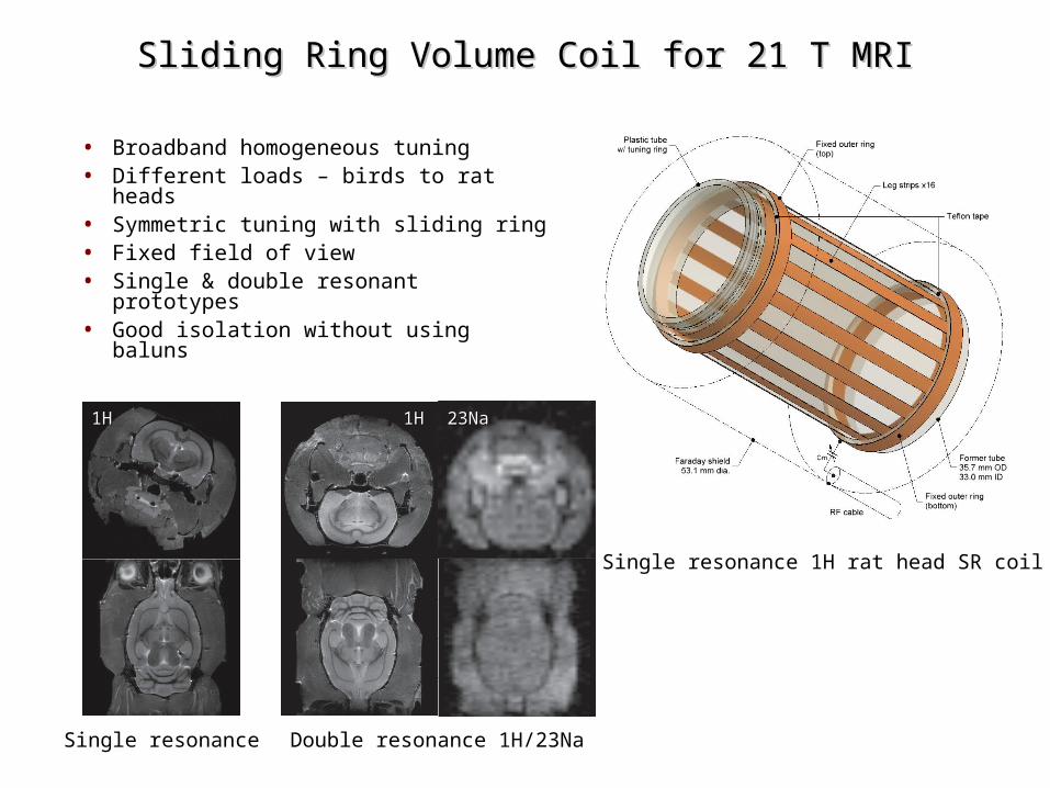
Single resonance 1H rat head SR coil
• Broadband homogeneous tuning• Different loads – birds to rat heads• Symmetric tuning with sliding ring • Fixed field of view• Single & double resonant prototypes• Good isolation without using baluns
Sliding Ring Volume Coil for 21 T MRISliding Ring Volume Coil for 21 T MRI
1H1H
Single resonance
1H1H 23Na23Na
Double resonance 1H/23Na
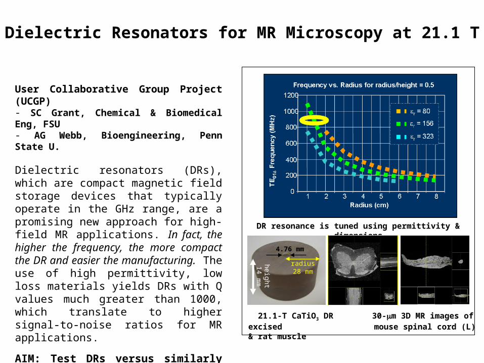
Dielectric Resonators for MR Microscopy at 21.1 T
User Collaborative Group Project (UCGP)- SC Grant, Chemical & Biomedical Eng, FSU- AG Webb, Bioengineering, Penn State U.
Dielectric resonators (DRs), which are compact magnetic field storage devices that typically operate in the GHz range, are a promising new approach for high-field MR applications. In fact, the higher the frequency, the more compact the DR and easier the manufacturing. The use of high permittivity, low loss materials yields DRs with Q values much greater than 1000, which translate to higher signal-to-noise ratios for MR applications.
AIM: Test DRs versus similarly sized copper coils for MR applications at 900 MHz (21.1 T) with respect to sensitivity & RF homogeneity.
4.76 mm
radius28 mm
heig
ht
14
mm
DR resonance is tuned using permittivity & dimensions
21.1-T CaTiO3 DR 30-m 3D MR images of excised mouse spinal cord (L) & rat muscle
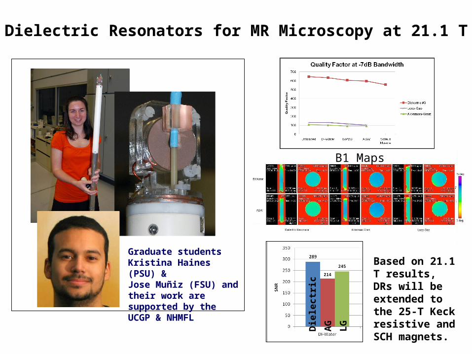
Dielectric Resonators for MR Microscopy at 21.1 T
Graduate studentsKristina Haines (PSU) &Jose Muñiz (FSU) and their work are supported by the UCGP & NHMFL
Based on 21.1 T results, DRs will be extended to the 25-T Keck resistive and SCH magnets.D
iele
ctr
ic
AG
LG
B1 Maps
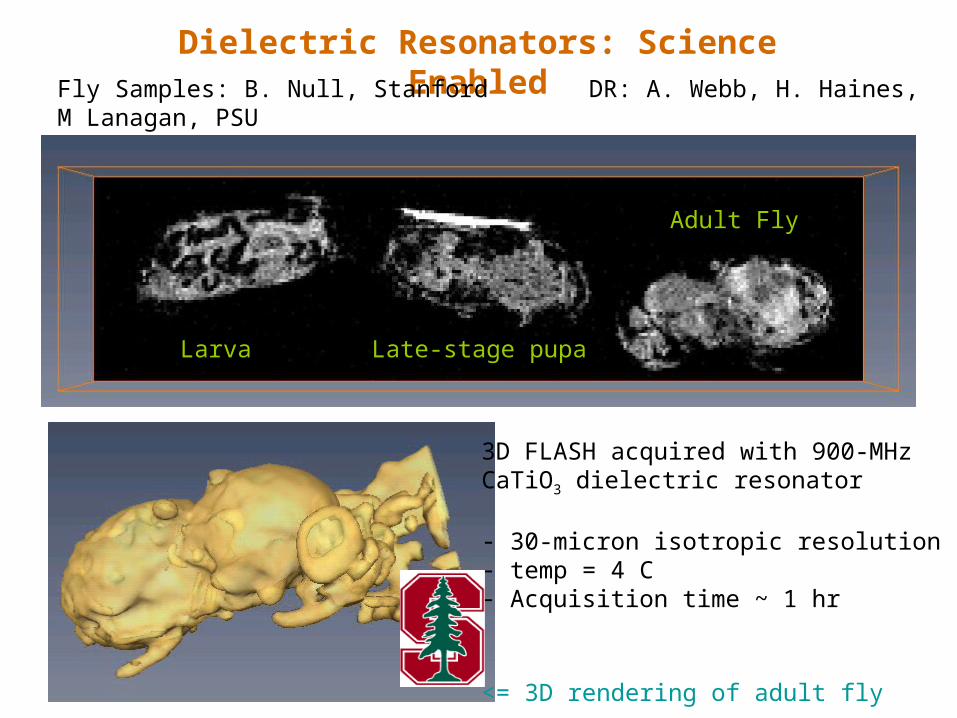
Dielectric Resonators: Science EnabledFly Samples: B. Null, Stanford DR: A. Webb, H. Haines, M Lanagan, PSU
J. Muniz, S. Grant, FSU
Larva Late-stage pupa
Adult Fly
3D FLASH acquired with 900-MHzCaTiO3 dielectric resonator
- 30-micron isotropic resolution- temp = 4 C- Acquisition time ~ 1 hr
<= 3D rendering of adult fly
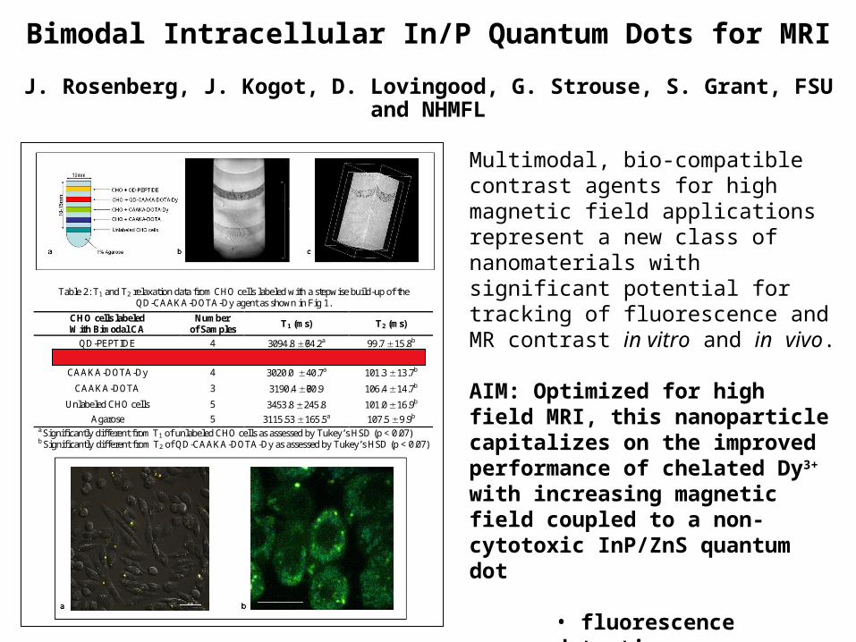
Bimodal Intracellular In/P Quantum Dots for MRI
J. Rosenberg, J. Kogot, D. Lovingood, G. Strouse, S. Grant, FSU and NHMFL
Multimodal, bio-compatible contrast agents for high magnetic field applications represent a new class of nanomaterials with significant potential for tracking of fluorescence and MR contrast in vitro and in vivo.
AIM: Optimized for high field MRI, this nanoparticle capitalizes on the improved performance of chelated Dy3+ with increasing magnetic field coupled to a non-cytotoxic InP/ZnS quantum dot
• fluorescence detection• MR responsiveness• payload delivery
Table 2: T1 and T2 relaxation data from CHO cells labeled with a stepwise build-up of the QD-CAAKA-DOTA-Dy agent as shown in Fig 1.
CHO cells labeled With Bimodal CA
Number of Samples
T1 (ms) T2 (ms)
QD-PEPTIDE 4 3094.8 ?64.2a 99.7 15.8b
QD-CAAKA-DOTA-Dy 5 3022.7 52.3a 72.2 10.3
CAAKA-DOTA-Dy 4 3020.0 40.7a 101.3 13.7b
CAAKA-DOTA 3 3190.4 ??60.9 106.4 14.7b
Unlabeled CHO cells 5 3453.8 245.8 101.0 16.9b
Agarose 5 3115.53 165.5a 107.5 9.9b a Significantly different from T1 of unlabeled CHO cells as assessed by Tukey’s HSD (p < 0.07) b Significantly different from T2 of QD-CAAKA-DOTA-Dy as assessed by Tukey’s HSD (p < 0.07)
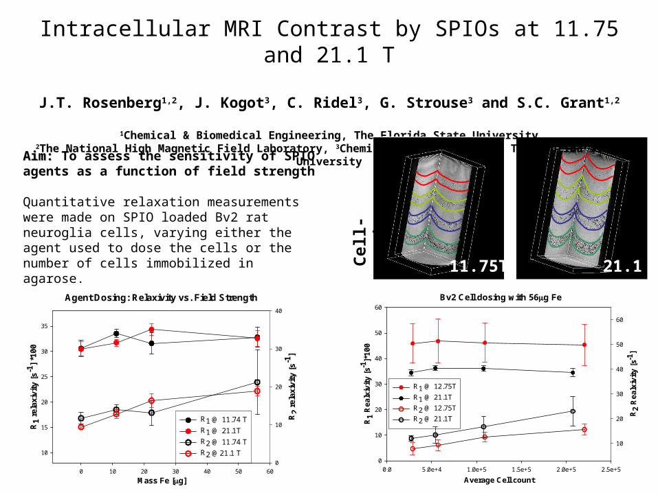
Intracellular MRI Contrast by SPIOs at 11.75 and 21.1 T
J.T. Rosenberg1,2, J. Kogot3, C. Ridel3, G. Strouse3 and S.C. Grant1,2
1Chemical & Biomedical Engineering, The Florida State University2The National High Magnetic Field Laboratory, 3Chemistry & Biochemistry, The Florida State University
Aim: Aim: To assess the sensitivity of SPIO agents as a function of field strength
Quantitative relaxation measurements were made on SPIO loaded Bv2 rat neuroglia cells, varying either the agent used to dose the cells or the number of cells immobilized in agarose.
Agent Dosing: Relaxivity vs. Field Strength
Mass Fe [g]0 10 20 30 40 50 60
R1
rela
xivi
ty [
s-1]
*100
10
15
20
25
30
35
R2
rela
xivi
ty [
s-1]
0
10
20
30
40
R1 @ 11.74 T
R1 @ 21.1T
R2 @ 11.74 T
R2 @21.1 T
Bv2 Cell dosing with 56g Fe
Average Cell count
0.0 5.0e+4 1.0e+5 1.5e+5 2.0e+5 2.5e+5
R1
Realx
ivit
y [
s-1]*
10
0
0
10
20
30
40
50
60
R2
Realx
ivit
y [
s-1]
10
20
30
40
50
60
R1 @ 12.75T
R1 @ 21.1T
R2 @ 12.75T
R2 @ 21.1T
Cel
l-D
osi
ng
21.1T11.75T