Vibronic coupling in the superoxide anion: The vibrational dependence ... · Vibronic coupling in...
10
Vibronic coupling in the superoxide anion: The vibrational dependence of the photoelectron angular distribution Matthew Van Duzor, Foster Mbaiwa, Jie Wei, Tulsi Singh, Richard Mabbs, Andrei Sanov, Steven J. Cavanagh, Stephen T. Gibson, Brenton R. Lewis, and Jason R. Gascooke Citation: The Journal of Chemical Physics 133, 174311 (2010); doi: 10.1063/1.3493349 View online: http://dx.doi.org/10.1063/1.3493349 View Table of Contents: http://scitation.aip.org/content/aip/journal/jcp/133/17?ver=pdfcov Published by the AIP Publishing Articles you may be interested in High-resolution photoelectron imaging of cold C 60 − anions and accurate determination of the electron affinity of C60 J. Chem. Phys. 140, 224315 (2014); 10.1063/1.4881421 Slow photoelectron velocity-map imaging spectroscopy of the C9H7 (indenyl) and C13H9 (fluorenyl) anions J. Chem. Phys. 139, 104301 (2013); 10.1063/1.4820138 Low-lying electronic states of CH 3 NO 2 via photoelectron imaging of the nitromethane anion J. Chem. Phys. 131, 164308 (2009); 10.1063/1.3256233 Slow electron velocity-map imaging photoelectron spectra of the methoxide anion J. Chem. Phys. 125, 014306 (2006); 10.1063/1.2212411 Spectroscopic characterization of the ground and low-lying electronic states of Ga 2 N via anion photoelectron spectroscopy J. Chem. Phys. 124, 064303 (2006); 10.1063/1.2159492 This article is copyrighted as indicated in the article. Reuse of AIP content is subject to the terms at: http://scitation.aip.org/termsconditions. Downloaded to IP: 130.56.106.27 On: Thu, 10 Dec 2015 02:36:24
Transcript of Vibronic coupling in the superoxide anion: The vibrational dependence ... · Vibronic coupling in...
Vibronic coupling in the superoxide anion: The vibrational dependence of thephotoelectron angular distributionMatthew Van Duzor, Foster Mbaiwa, Jie Wei, Tulsi Singh, Richard Mabbs, Andrei Sanov, Steven J. Cavanagh,Stephen T. Gibson, Brenton R. Lewis, and Jason R. Gascooke Citation: The Journal of Chemical Physics 133, 174311 (2010); doi: 10.1063/1.3493349 View online: http://dx.doi.org/10.1063/1.3493349 View Table of Contents: http://scitation.aip.org/content/aip/journal/jcp/133/17?ver=pdfcov Published by the AIP Publishing Articles you may be interested in High-resolution photoelectron imaging of cold C 60 − anions and accurate determination of the electron affinity ofC60 J. Chem. Phys. 140, 224315 (2014); 10.1063/1.4881421 Slow photoelectron velocity-map imaging spectroscopy of the C9H7 (indenyl) and C13H9 (fluorenyl) anions J. Chem. Phys. 139, 104301 (2013); 10.1063/1.4820138 Low-lying electronic states of CH 3 NO 2 via photoelectron imaging of the nitromethane anion J. Chem. Phys. 131, 164308 (2009); 10.1063/1.3256233 Slow electron velocity-map imaging photoelectron spectra of the methoxide anion J. Chem. Phys. 125, 014306 (2006); 10.1063/1.2212411 Spectroscopic characterization of the ground and low-lying electronic states of Ga 2 N via anion photoelectronspectroscopy J. Chem. Phys. 124, 064303 (2006); 10.1063/1.2159492
This article is copyrighted as indicated in the article. Reuse of AIP content is subject to the terms at: http://scitation.aip.org/termsconditions. Downloaded to IP:
130.56.106.27 On: Thu, 10 Dec 2015 02:36:24
Vibronic coupling in the superoxide anion: The vibrational dependenceof the photoelectron angular distribution
Matthew Van Duzor,1 Foster Mbaiwa,1 Jie Wei,1 Tulsi Singh,1 Richard Mabbs,1,a�
Andrei Sanov,2 Steven J. Cavanagh,3 Stephen T. Gibson,3 Brenton R. Lewis,3 andJason R. Gascooke4
1Department of Chemistry, Washington University, One Brookings Dr., Campus Box 1134 Saint Louis,Missouri 63130, USA2Department of Chemistry and Biochemistry, University of Arizona, Tucson, Arizona 85721-0041, USA3Research School of Physics and Engineering, The Australian National University, Canberra,Australian Capital Territory 0200, Australia4School of Chemical and Physical Sciences, Flinders University, G.P.O. Box 2100, Adelaide,South Australia 5001, Australia
�Received 20 April 2010; accepted 3 September 2010; published online 3 November 2010�
We present a comprehensive photoelectron imaging study of the O2�X 3�g− ,v�=0–6�
←O2−�X 2�g ,v�=0� and O2�a 1�g ,v�=0–4�←O2
−�X 2�g ,v�=0� photodetachment bands atwavelengths between 900 and 455 nm, examining the effect of vibronic coupling on thephotoelectron angular distribution �PAD�. This work extends the v�=1–4 data for detachment intothe ground electronic state, presented in a recent communication �R. Mabbs, F. Mbaiwa, J. Wei, M.Van Duzor, S. T. Gibson, S. J. Cavanagh, and B. R. Lewis, Phys. Rev. A 82, 011401�R� �2010��.Measured vibronic intensities are compared to Franck–Condon predictions and used as supportingevidence of vibronic coupling. The results are analyzed within the context of the one-electron, zerocore contribution �ZCC� model �R. M. Stehman and S. B. Woo, Phys. Rev. A 23, 2866 �1981��. Forboth bands, the photoelectron anisotropy parameter variation with electron kinetic energy, ��E�,displays the characteristics of photodetachment from a d-like orbital, consistent with the �g
� 2phighest occupied molecular orbital of O2
−. However, differences exist between the ��E� trends fordetachment into different vibrational levels of the X 3�g
− and a 1�g electronic states of O2. The ZCCmodel invokes vibrational channel specific “detachment orbitals” and attributes this behavior tocoupling of the electronic and nuclear motion in the parent anion. The spatial extent of the modeldetachment orbital is dependent on the final state of O2: the higher the neutral vibrational excitation,the larger the electron binding energy. Although vibronic coupling is ignored in most theoreticaltreatments of PADs in the direct photodetachment of molecular anions, the present findings clearlyshow that it can be important. These results represent a benchmark data set for a relatively simplesystem, upon which to base rigorous tests of more sophisticated models. © 2010 American Instituteof Physics. �doi:10.1063/1.3493349�
I. INTRODUCTION
The coupling between electronic and vibrational degreesof freedom often affects the spectroscopic and structuralproperties of molecules. However, experimental evidence ofvibrational influence on the photoelectron angular distribu-tions �PADs� in negative-ion photodetachment has so farbeen sparse, and little attention has been paid to such effectsin theoretical treatments of direct detachment processes. In arecent communication,1 we reported a strong dependence ofthe PADs in O2
−�X 2�g ,v�=0� photodetachment on the finalvibrational state of the neutral, O2�X 3�g
− ,v�=1–4�. Theseresults provided experimental evidence of vibronic couplingin the anion ground electronic state, supplying essential datafor the evaluation and refinement of existing theoreticalmodels.
In the present work, we extend the above measurementsto include the additional v�=0, 5, and 6 transitions of the
O2�X 3�g−�←O2
−�X 2�g� band and the v�=0–4 transitions ofthe excited-state O2�a 1�g�←O2
−�X 2�g� band. We present anew, detailed discussion of these results in the context of thezero core contribution �ZCC� model,2 complementing ourprevious analysis.1 In addition, the observed trend in vi-bronic transition intensities in the O2�X 3�g
−�←O2−�X 2�g�
band is evaluated.The dominant long range interaction in anion photode-
tachment is associated with the centrifugal term in the effec-tive potential.3 The nature of the long range potential signifi-cantly affects the energy dependence of the total4 anddifferential detachment cross sections.5 In fact, the applica-tion of orbital angular momentum conservation and consid-eration of the influence of the centrifugal barrier in principleallows the characterization of the parent orbital from thephotoelectron angular distribution.5–7 Within the one-electronand electric-dipole approximations, the PAD of an atomicanion detachment can be thought of as a signature of theparent orbital.8a�Electronic mail: [email protected].
THE JOURNAL OF CHEMICAL PHYSICS 133, 174311 �2010�
0021-9606/2010/133�17�/174311/9/$30.00 © 2010 American Institute of Physics133, 174311-1
This article is copyrighted as indicated in the article. Reuse of AIP content is subject to the terms at: http://scitation.aip.org/termsconditions. Downloaded to IP:
130.56.106.27 On: Thu, 10 Dec 2015 02:36:24
The relationship between the parent orbital and the PADis in principle more complex for molecular anions.9,10 Orbitalangular momentum ��� is no longer a good quantum number,and the vibrational excitation often accompanies the changein electronic state. The application of symmetry argumentscan still act as a guide to the nature of the parent molecularorbital.11–18 More subtle effects, such as the influence of thefinal, neutral molecular state are often difficult to extract.There are, however, a few small molecular anions that mightallow detailed experimental studies into these effects.
In the case of superoxide, O2−, the highest occupied mo-
lecular orbital ��g� has a strong similarity to an atomic dorbital. There is only one vibrational degree of freedom, andphotoelectron-spectroscopic techniques are capable of re-solving the vibrational structure in the low electron kineticenergy region of the spectrum. A comparison of O2
− photode-tachment results with atomic anion model predictions yieldsconsiderable insight into the effect of vibrational excitationon the photodetachment properties, in particular the PAD.1
Several detachment studies of this species have probedthe photon energy dependence of the total detachment crosssection19–26 or reported the photoelectron spectrum ofO2
−.1,27–38 Until very recently, much less data were reportedregarding the PAD.1,28,35 Among other anions, the prominentdependence of photoelectron anisotropy on the final vibra-tional state of the neutral was reported in the photodetach-ment of NO−.39 In the case of neutral diatomic molecules,dramatic changes in the PAD have been observed betweendifferent vibronic photoionization bands of N2, CO, andO2.40–42 Such effects are associated with strong coupling ofthe electronic and vibrational motion and usually a break-down of the Born–Oppenheimer approximation as a conse-quence of excitation of intermediate, autoionizing Rydbergstates or shape resonances.42
It is well known that the PAD is dependent on electronkinetic energy �E�.43 To separate the effect of vibrationalexcitation from the E dependence, one must, ideally, com-pare transitions that terminate in different vibrational statesbut correspond to the same E. This approach requires experi-ments at multiple, carefully selected photoexcitation wave-lengths. Studies of this type are rarely performed, particu-larly for molecular anions. Despite the work reported in Ref.44 for photoionization, the prevalent view seems to havebeen that in the absence of vibrationally resolved data indirect photodetachment processes, vibrational effects on thePAD can be ignored.
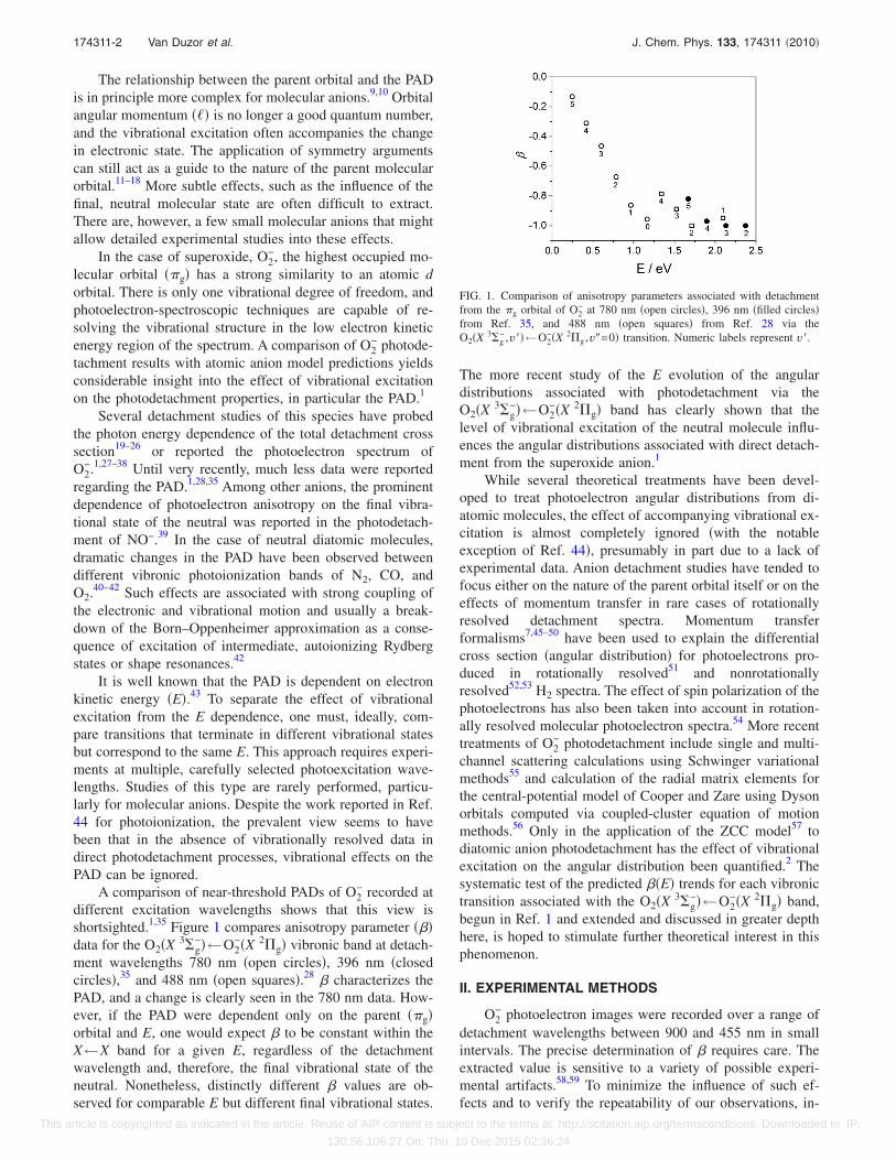
A comparison of near-threshold PADs of O2− recorded at
different excitation wavelengths shows that this view isshortsighted.1,35 Figure 1 compares anisotropy parameter ���data for the O2�X 3�g
−�←O2−�X 2�g� vibronic band at detach-
ment wavelengths 780 nm �open circles�, 396 nm �closedcircles�,35 and 488 nm �open squares�.28 � characterizes thePAD, and a change is clearly seen in the 780 nm data. How-ever, if the PAD were dependent only on the parent ��g�orbital and E, one would expect � to be constant within theX←X band for a given E, regardless of the detachmentwavelength and, therefore, the final vibrational state of theneutral. Nonetheless, distinctly different � values are ob-served for comparable E but different final vibrational states.
The more recent study of the E evolution of the angulardistributions associated with photodetachment via theO2�X 3�g
−�←O2−�X 2�g� band has clearly shown that the
level of vibrational excitation of the neutral molecule influ-ences the angular distributions associated with direct detach-ment from the superoxide anion.1
While several theoretical treatments have been devel-oped to treat photoelectron angular distributions from di-atomic molecules, the effect of accompanying vibrational ex-citation is almost completely ignored �with the notableexception of Ref. 44�, presumably in part due to a lack ofexperimental data. Anion detachment studies have tended tofocus either on the nature of the parent orbital itself or on theeffects of momentum transfer in rare cases of rotationallyresolved detachment spectra. Momentum transferformalisms7,45–50 have been used to explain the differentialcross section �angular distribution� for photoelectrons pro-duced in rotationally resolved51 and nonrotationallyresolved52,53 H2 spectra. The effect of spin polarization of thephotoelectrons has also been taken into account in rotation-ally resolved molecular photoelectron spectra.54 More recenttreatments of O2
− photodetachment include single and multi-channel scattering calculations using Schwinger variationalmethods55 and calculation of the radial matrix elements forthe central-potential model of Cooper and Zare using Dysonorbitals computed via coupled-cluster equation of motionmethods.56 Only in the application of the ZCC model57 todiatomic anion photodetachment has the effect of vibrationalexcitation on the angular distribution been quantified.2 Thesystematic test of the predicted ��E� trends for each vibronictransition associated with the O2�X 3�g
−�←O2−�X 2�g� band,
begun in Ref. 1 and extended and discussed in greater depthhere, is hoped to stimulate further theoretical interest in thisphenomenon.
II. EXPERIMENTAL METHODS
O2− photoelectron images were recorded over a range of
detachment wavelengths between 900 and 455 nm in smallintervals. The precise determination of � requires care. Theextracted value is sensitive to a variety of possible experi-mental artifacts.58,59 To minimize the influence of such ef-fects and to verify the repeatability of our observations, in-
FIG. 1. Comparison of anisotropy parameters associated with detachmentfrom the �g orbital of O2
− at 780 nm �open circles�, 396 nm �filled circles�from Ref. 35, and 488 nm �open squares� from Ref. 28 via theO2�X 3�g
− ,v��←O2−�X 2�g ,v�=0� transition. Numeric labels represent v�.
174311-2 Van Duzor et al. J. Chem. Phys. 133, 174311 �2010�
This article is copyrighted as indicated in the article. Reuse of AIP content is subject to the terms at: http://scitation.aip.org/termsconditions. Downloaded to IP:
130.56.106.27 On: Thu, 10 Dec 2015 02:36:24
dependent measurements were recently reported from twodifferent laboratories in St. Louis �USA� and Canberra�Australia�.1
The results presented here were recorded on theCanberra instrument and extend the data set to theO2�X 3�g
− ,v��←O2−�X 2�g ,v�=0�, v�=0,5 ,6 transitions,
and O2�a 1�g ,v��←O2−�X 2�g ,v�=0�, v�=0–4 band. The
instrumentation has been described previously,58 and onlybrief details will be provided. Essentially, the apparatus pro-duces a collimated beam of mass-selected negative ions,which can then be interrogated by a laser beam, subsequentlyproducing photoelectrons. The photodetached electrons areimaged onto a multichannel-plate �MCP� phosphor detectorusing a modified velocity-map imaging lens.
Molecular oxygen anions are produced by passing pureoxygen gas through a pulsed nozzle �General Valve Series 9�at a stagnation pressure of 2.7 atm and then supersonicallyexpanding it through a pulsed discharge.58 All negative ionsare extracted, accelerated to 500 eV, focused into an ion-gating, bunching, and potential rereferencing unit,60 and al-lowed to propagate along a ground-referenced 2 m time-of-flight �TOF� tube. The ion-beam and imaging lens axes arearranged coaxially. A fast potential switch rereferences theion packet to the imaging assembly repeller-plate potential.A potential barrier is placed between the second potentialswitch and the repeller plate of the imaging lens. This acts,when switched together with the potential switch, as a massdiscriminator, allowing only the mass of interest to enter theimaging lens.
The mass-selected ion packet intersects the detachmentlaser beam, generated by a Continuum Sunlite EX opticalparametric oscillator, which is pumped by a Powerlite 9010Nd:yttrium aluminum garnet laser, operated at its third har-monic, 355 nm. The laser operates between 1 and 3 mJ perpulse and at 10 Hz. To ensure a very high degree of polar-ization parallel to the MCP detector face, the laser beam waspassed through a 1/2-waveplate and a high-quality broadbandwidth Glan-Laser polarizer. A Galilean telescope ar-rangement is used to produce a parallel beam of 2 mm di-ameter, reduced from the laser diameter of 8 mm. The wave-length of the laser is measured using a high-qualitywavemeter �High Finesse WS7 UV�.
The high resolution velocity-map imaging lens is a sub-stantially modified version of Eppink and Parker’sarrangement.61 It was designed to accommodate fast ion-beam energies of 102–103 eV and an increased interactionregion volume size �at least 2 mm3�, achieving electron ki-netic energy resolutions of �E /E�0.3% or better. Photo-electrons are imaged onto a position sensitive detector con-sisting of a pair of high dynamic range, imaging quality10 �m-pore MCPs, a P47 phosphor screen, and a 2048�2048-pixel monochrome charge coupled device camera�PCO2000�. The imaging detector is situated at the end ofthe TOF tube, 800 mm from the interaction region, and isshielded from magnetic fields through a combination of�-metal and three orthogonal Helmholtz coils. The MCPgain is gated using a purpose-built fast high-voltage pulser tocoincide with the arrival of the photoelectrons. This ensuresthat unwanted events from ions or neutral species are not
detected. Each camera frame is transferred to a PC at the 10Hz repetition rate and is processed in real time to identifyevents, centroiding to subpixel accuracy, with the x-,y-coordinates written to a file, for subsequent analysis.
III. RESULTS AND ANALYSIS
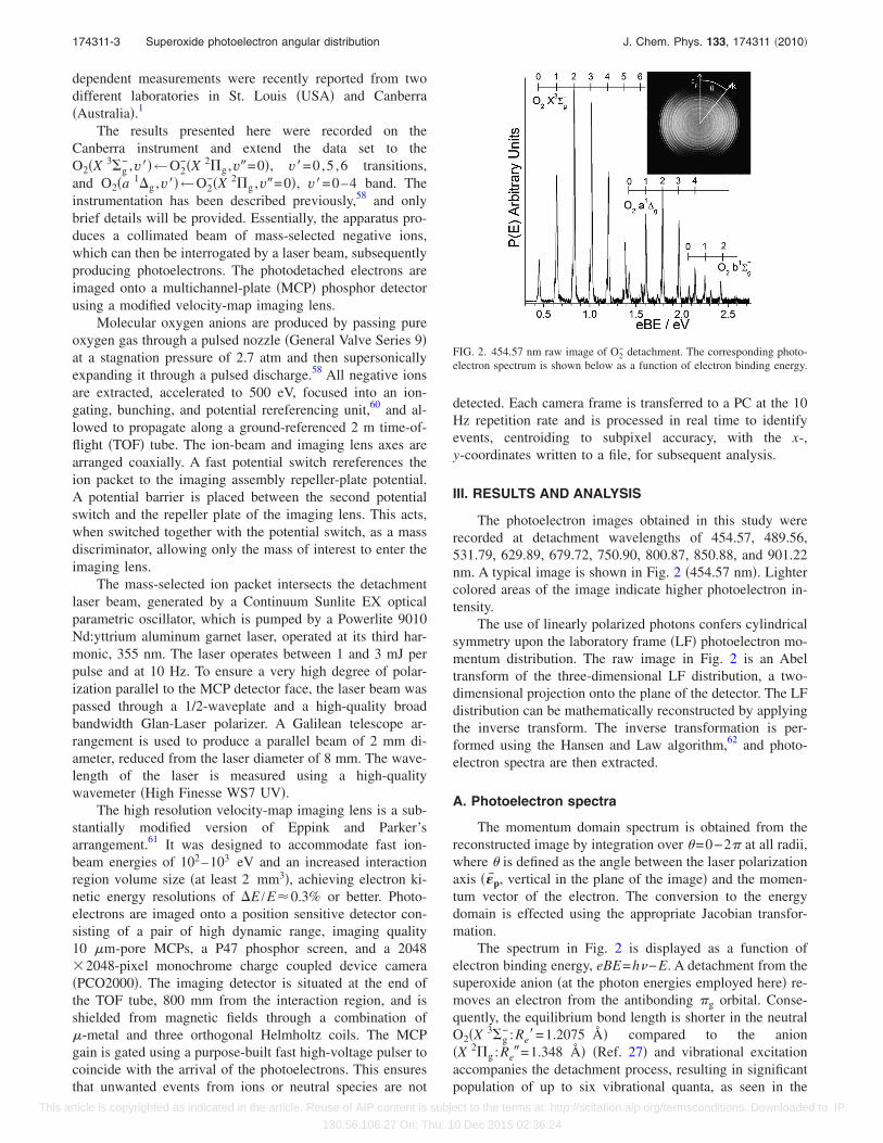
The photoelectron images obtained in this study wererecorded at detachment wavelengths of 454.57, 489.56,531.79, 629.89, 679.72, 750.90, 800.87, 850.88, and 901.22nm. A typical image is shown in Fig. 2 �454.57 nm�. Lightercolored areas of the image indicate higher photoelectron in-tensity.
The use of linearly polarized photons confers cylindricalsymmetry upon the laboratory frame �LF� photoelectron mo-mentum distribution. The raw image in Fig. 2 is an Abeltransform of the three-dimensional LF distribution, a two-dimensional projection onto the plane of the detector. The LFdistribution can be mathematically reconstructed by applyingthe inverse transform. The inverse transformation is per-formed using the Hansen and Law algorithm,62 and photo-electron spectra are then extracted.
A. Photoelectron spectra
The momentum domain spectrum is obtained from thereconstructed image by integration over =0−2� at all radii,where is defined as the angle between the laser polarizationaxis ���p, vertical in the plane of the image� and the momen-tum vector of the electron. The conversion to the energydomain is effected using the appropriate Jacobian transfor-mation.
The spectrum in Fig. 2 is displayed as a function ofelectron binding energy, eBE=h−E. A detachment from thesuperoxide anion �at the photon energies employed here� re-moves an electron from the antibonding �g orbital. Conse-quently, the equilibrium bond length is shorter in the neutralO2�X 3�g
− :Re�=1.2075 Å� compared to the anion�X 2�g :Re�=1.348 Å� �Ref. 27� and vibrational excitationaccompanies the detachment process, resulting in significantpopulation of up to six vibrational quanta, as seen in the
FIG. 2. 454.57 nm raw image of O2− detachment. The corresponding photo-
electron spectrum is shown below as a function of electron binding energy.
174311-3 Superoxide photoelectron angular distribution J. Chem. Phys. 133, 174311 �2010�
This article is copyrighted as indicated in the article. Reuse of AIP content is subject to the terms at: http://scitation.aip.org/termsconditions. Downloaded to IP:
130.56.106.27 On: Thu, 10 Dec 2015 02:36:24
vibrational progression in the O2�X 3�g−�←O2
−�X 2�g ,v�=0� band. The vibronic transitions are clearly seen in theimage as a series of concentric rings, with the larger ringscorresponding to lower levels of internal excitation in theneutral O2. These features are clear in the spectrum, whichalso resolves transitions from the two spin-orbit states�2�g,�=3/2,1/2� of the anion.
The spectral intensities are normalized relative to theO2�X 3�g
− ,v�=2�←O2−�X 2�g ,v�=0� transition. Three vi-
bronic bands are visible in the portion of the spectrum shownin Fig. 2, which correspond to detachment via the ground�X 3�g
−� and first two excited �a 1�g ,b 1�g+� electronic states
of O2.
B. The photoelectron angular distributions
It is clear in the image in Fig. 2 that the photoelectronintensity is not necessarily constant about a ring of givenradius. This is particularly obvious in the outer rings in theimage in Fig. 2, where the intensity is greatest at =� /2.The intensity distribution, I�E ,� is a measure of the differ-ential cross section for detachment and is described by
I�E,� =��E�4�
�1 + ��E�P2�cos �� , �1�
where � is the integrated cross section and P2 is the secondLegendre polynomial. The anisotropy parameter � is deter-mined by the details of the detachment process and charac-terizes the angular distribution. Any effects of the detach-ment process on the angular distribution can therefore beconveniently discussed in terms of changes in the anisotropyparameter.
In this work, we focus on the PADs accompanying theO2�X 3�g
−�←O2−�X 2�g� vibronic transitions of O2
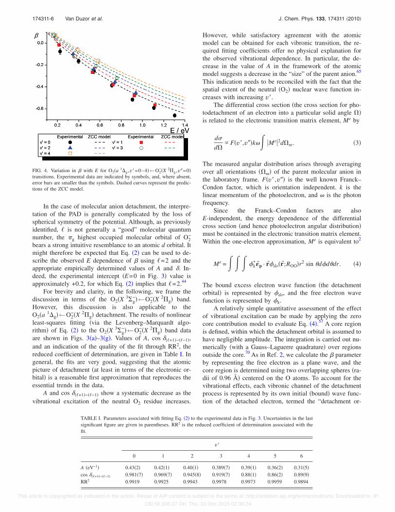
− detach-ment. However, Fig. 4 shows that the O2�a 1�g�←O2
−�X 2�g� vibronic transitions display a similar behavior.The apparent changes in ��E� trends have significant impli-cations for theoretical treatments of the detachment process.The anisotropy parameter is extracted by plotting I�E ,�against P2�cos � at a given E. We note that the � valuesreported in this work differ slightly from those previouslyreported.28,35 This is not too surprising, considering that �values are very sensitive to image distortion �imparted byexternal fields or imperfect focusing� and detector saturation.In the present work, extreme care has been applied to mini-mize such adverse effects. The images are circularized toeliminate distortion effects.58 Additionally, saturation effectsare easily identified as deviations from linearity at the ex-
tremes of the angular range in a plot of I�E ,� versusP2�cos �, and we note that none of the images reported heredisplay any evidence of saturation. Furthermore, the resultshave been verified independently in the different laboratoriesof the authors, with each set of measurements being in ex-cellent agreement.1
For the remainder of this work, we focus on the relation-ship between � and E and in particular the effect that thelevel of vibrational excitation of the neutral residue seems toexert. In Fig. 3, we show the variation of � with E for theO2�X 3�g
−�←O2−�X 2�g� band. All data shown are for transi-
tions originating in the O2−�X 2�g,�=3/2�v�=0 spin-orbit state.
The neutral O2 vibrational levels accessed are �a� v�=0, �b�v�=1, �c� v�=2, �d� v�=3, �e� v�=4, �f� v�=5, and �g� v�=6. A change in � with E is not unexpected. More surpris-ing, at least in the light of the expectation that the PADdepends on the parent �g orbital only, is the difference in �seen in Fig. 3�h� for different vibronic transitions at compa-rable E. Figure 4 shows similar behavior in theO2�a 1�g ,v��←O2
−�X 2�g ,v�=0� band for v�=0–4.There is an initial shift toward increasingly negative val-
ues until a minimum is reached �at least for the v�=0 andv�=1 series� before becoming less negative. However, therate of change of � in each case is different. For E between0 and 1.5 eV, lower v� correspond to more negative �.Clearly, accessing different terminal neutral states has a pro-found influence on the photoelectron angular distribution.
IV. DISCUSSION
The E dependence of � when � 0 has been previouslydemonstrated in atomic anion detachmentexperiments.58,63–68 In these cases, the change in � can beexplained through consideration of the dominant long rangeterm in the potential, the centrifugal term. The free electronwave can be represented as a superposition of partial angularmomentum waves for which we will use the symbol ���=��1� to allow distinction from the parent orbital angularmomentum quantum number �. The centrifugal barrier in-creases with �� and decreases with distance. In theasymptotic limit, the barrier is therefore zero, but in the near-field limit, the barrier leads to different contributions of thepartial waves to the overall superposition. According to theWigner law,4 at the detachment threshold the cross section ofeach partial wave varies as ����E���+1/2�. The angular distri-bution, and hence �, is the result of interference betweenthese partial waves, and so changing the composition of thesuperposition changes the angular distribution. For atomicanions, this behavior is encapsulated in the equation
��E� =��� − 1� + �� + 1��� + 2�A2 · E2 − 6��� + 1�A · E cos ���+1�−��−1�
�2� + 1��� + �� + 1�A2 · E2�. �2�
174311-4 Van Duzor et al. J. Chem. Phys. 133, 174311 �2010�
This article is copyrighted as indicated in the article. Reuse of AIP content is subject to the terms at: http://scitation.aip.org/termsconditions. Downloaded to IP:
130.56.106.27 On: Thu, 10 Dec 2015 02:36:24
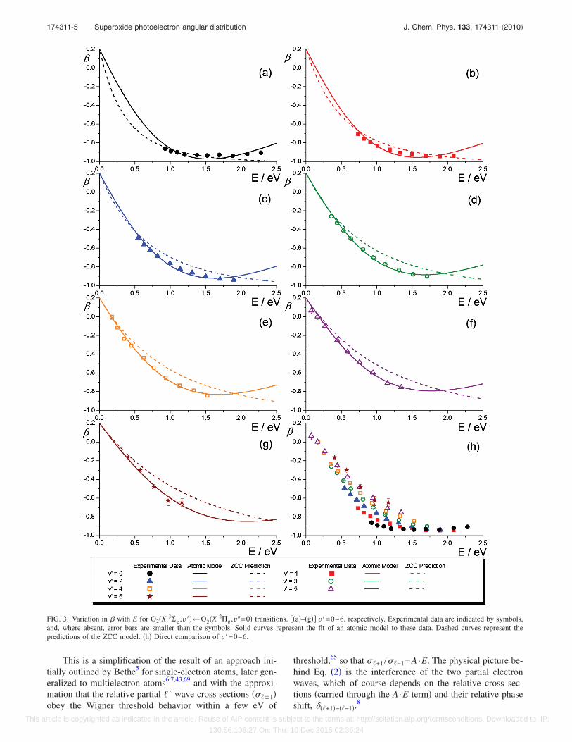
This is a simplification of the result of an approach ini-tially outlined by Bethe5 for single-electron atoms, later gen-eralized to multielectron atoms6,7,43,69 and with the approxi-mation that the relative partial �� wave cross sections ����1�obey the Wigner threshold behavior within a few eV of
threshold,65 so that ��+1 /��−1=A ·E. The physical picture be-hind Eq. �2� is the interference of the two partial electronwaves, which of course depends on the relative cross sec-tions �carried through the A ·E term� and their relative phaseshift, ���+1�−��−1�.
8
FIG. 3. Variation in � with E for O2�X 3�g− ,v��←O2
−�X 2�g ,v�=0� transitions. ��a�–�g�� v�=0–6, respectively. Experimental data are indicated by symbols,and, where absent, error bars are smaller than the symbols. Solid curves represent the fit of an atomic model to these data. Dashed curves represent thepredictions of the ZCC model. �h� Direct comparison of v�=0–6.
174311-5 Superoxide photoelectron angular distribution J. Chem. Phys. 133, 174311 �2010�
This article is copyrighted as indicated in the article. Reuse of AIP content is subject to the terms at: http://scitation.aip.org/termsconditions. Downloaded to IP:
130.56.106.27 On: Thu, 10 Dec 2015 02:36:24
In the case of molecular anion detachment, the interpre-tation of the PAD is generally complicated by the loss ofspherical symmetry of the potential. Although, as previouslyidentified, � is not generally a “good” molecular quantumnumber, the �g highest occupied molecular orbital of O2
−
bears a strong intuitive resemblance to an atomic d orbital. Itmight therefore be expected that Eq. �2� can be used to de-scribe the observed E dependence of � using �=2 and theappropriate empirically determined values of A and �. In-deed, the experimental intercept �E=0 in Fig. 3� value isapproximately +0.2, for which Eq. �2� implies that �=2.44
For brevity and clarity, in the following, we frame thediscussion in terms of the O2�X 3�g
−�←O2−�X 2�g� band.
However, this discussion is also applicable to theO2�a 1�g�←O2
−�X 2�g� detachment. The results of nonlinearleast-squares fitting �via the Levenberg–Marquardt algo-rithm� of Eq. �2� to the O2�X 3�g
−�←O2−�X 2�g� band data
are shown in Figs. 3�a�–3�g�. Values of A, cos ���+1�−��−1�,and an indication of the quality of the fit through RR2, thereduced coefficient of determination, are given in Table I. Ingeneral, the fits are very good, suggesting that the atomicpicture of detachment �at least in terms of the electronic or-bital� is a reasonable first approximation that reproduces theessential trends in the data.
A and cos ���+1�−��−1� show a systematic decrease as thevibrational excitation of the neutral O2 residue increases.
However, while satisfactory agreement with the atomicmodel can be obtained for each vibronic transition, the re-quired fitting coefficients offer no physical explanation forthe observed vibrational dependence. In particular, the de-crease in the value of A in the framework of the atomicmodel suggests a decrease in the “size” of the parent anion.65
This indication needs to be reconciled with the fact that thespatial extent of the neutral �O2� nuclear wave function in-creases with increasing v�.
The differential cross section �the cross section for pho-todetachment of an electron into a particular solid angle ��is related to the electronic transition matrix element, Me by
d�
d�� F�v�,v��k�� �Me�2d�m. �3�
The measured angular distribution arises through averagingover all orientations ��m� of the parent molecular anion inthe laboratory frame. F�v� ,v�� is the well known Franck–Condon factor, which is orientation independent. k is thelinear momentum of the photoelectron, and � is the photonfrequency.
Since the Franck–Condon factors are alsoE-independent, the energy dependence of the differentialcross section �and hence photoelectron angular distribution�must be contained in the electronic transition matrix element.Within the one-electron approximation, Me is equivalent to2
Me =� � � �k���p · r��do�r� ;ROO�r2 sin d�ddr . �4�
The bound excess electron wave function �the detachmentorbital� is represented by �do, and the free electron wavefunction is represented by �k.
A relatively simple quantitative assessment of the effectof vibrational excitation can be made by applying the zerocore contribution model to evaluate Eq. �4�.57 A core regionis defined, within which the detachment orbital is assumed tohave negligible amplitude. The integration is carried out nu-merically �with a Gauss–Laguerre quadrature� over regionsoutside the core.70 As in Ref. 2, we calculate the � parameterby representing the free electron as a plane wave, and thecore region is determined using two overlapping spheres �ra-dii of 0.96 Å� centered on the O atoms. To account for thevibrational effects, each vibronic channel of the detachmentprocess is represented by its own initial �bound� wave func-tion of the detached electron, termed the “detachment or-
FIG. 4. Variation in � with E for O2�a 1�g ,v�=0–4�←O2−�X 2�g ,v�=0�
transitions. Experimental data are indicated by symbols, and, where absent,error bars are smaller than the symbols. Dashed curves represent the predic-tions of the ZCC model.
TABLE I. Parameters associated with fitting Eq. �2� to the experimental data in Fig. 3. Uncertainties in the lastsignificant figure are given in parentheses. RR2 is the reduced coefficient of determination associated with thefit.
v�
0 1 2 3 4 5 6
A �eV−1� 0.43�2� 0.42�1� 0.40�1� 0.389�7� 0.39�1� 0.36�2� 0.31�5�cos ���+1�−��−1� 0.981�7� 0.969�7� 0.945�8� 0.919�7� 0.88�1� 0.86�2� 0.89�9�RR2 0.9919 0.9925 0.9943 0.9978 0.9973 0.9959 0.9894
174311-6 Van Duzor et al. J. Chem. Phys. 133, 174311 �2010�
This article is copyrighted as indicated in the article. Reuse of AIP content is subject to the terms at: http://scitation.aip.org/termsconditions. Downloaded to IP:
130.56.106.27 On: Thu, 10 Dec 2015 02:36:24
bital.” We approximate the detachment orbital using theunited-atom limit and employing an atomic d orbitalfunction,2
�do = 1 +3
�r+
3
�2r2 e−�r
rsin cos , �5�
where ���eBE incorporates the final vibrational state de-pendence, while r is the distance from the center of mass ofthe molecule.
The ��E� values calculated using the above approach areshown as the dashed curves in Figs. 3 and 4.71 The calculated��E� trends clearly vary with the vibrational quantum num-ber of the neutral O2. The ZCC model is the first treatmentthat attempts to quantitatively describe this behavior for mo-lecular anion detachment.
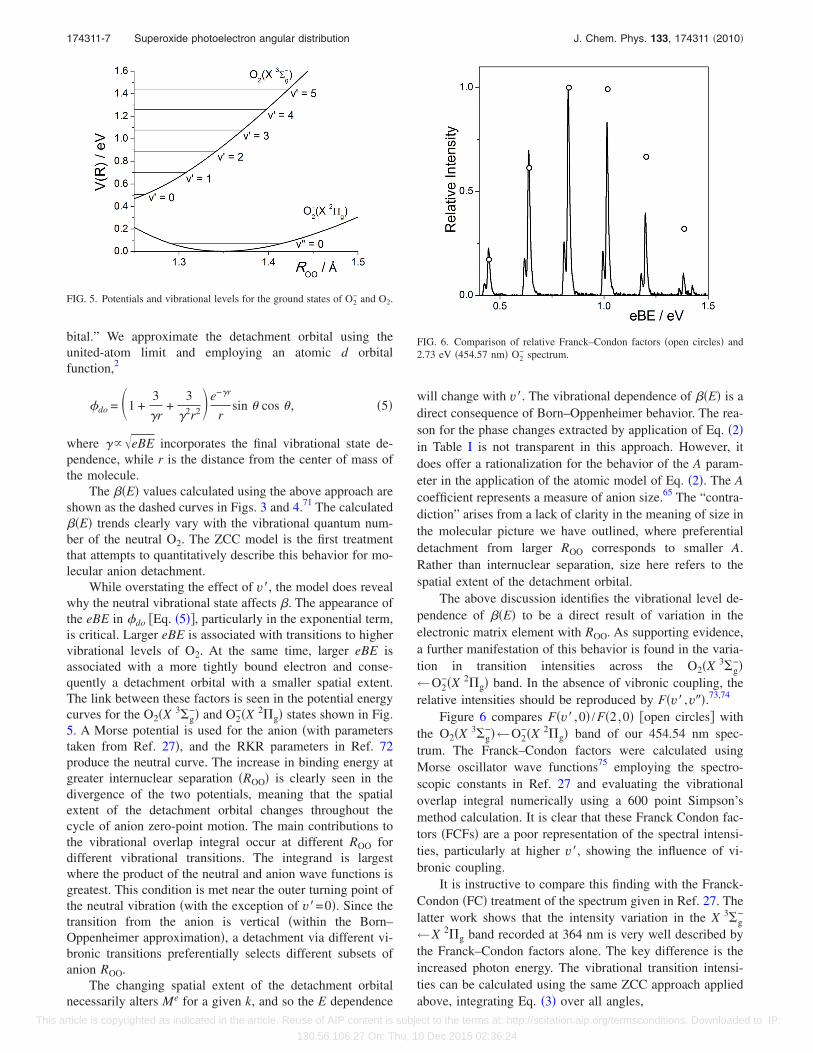
While overstating the effect of v�, the model does revealwhy the neutral vibrational state affects �. The appearance ofthe eBE in �do �Eq. �5��, particularly in the exponential term,is critical. Larger eBE is associated with transitions to highervibrational levels of O2. At the same time, larger eBE isassociated with a more tightly bound electron and conse-quently a detachment orbital with a smaller spatial extent.The link between these factors is seen in the potential energycurves for the O2�X 3�g
−� and O2−�X 2�g� states shown in Fig.
5. A Morse potential is used for the anion �with parameterstaken from Ref. 27�, and the RKR parameters in Ref. 72produce the neutral curve. The increase in binding energy atgreater internuclear separation �ROO� is clearly seen in thedivergence of the two potentials, meaning that the spatialextent of the detachment orbital changes throughout thecycle of anion zero-point motion. The main contributions tothe vibrational overlap integral occur at different ROO fordifferent vibrational transitions. The integrand is largestwhere the product of the neutral and anion wave functions isgreatest. This condition is met near the outer turning point ofthe neutral vibration �with the exception of v�=0�. Since thetransition from the anion is vertical �within the Born–Oppenheimer approximation�, a detachment via different vi-bronic transitions preferentially selects different subsets ofanion ROO.
The changing spatial extent of the detachment orbitalnecessarily alters Me for a given k, and so the E dependence
will change with v�. The vibrational dependence of ��E� is adirect consequence of Born–Oppenheimer behavior. The rea-son for the phase changes extracted by application of Eq. �2�in Table I is not transparent in this approach. However, itdoes offer a rationalization for the behavior of the A param-eter in the application of the atomic model of Eq. �2�. The Acoefficient represents a measure of anion size.65 The “contra-diction” arises from a lack of clarity in the meaning of size inthe molecular picture we have outlined, where preferentialdetachment from larger ROO corresponds to smaller A.Rather than internuclear separation, size here refers to thespatial extent of the detachment orbital.
The above discussion identifies the vibrational level de-pendence of ��E� to be a direct result of variation in theelectronic matrix element with ROO. As supporting evidence,a further manifestation of this behavior is found in the varia-tion in transition intensities across the O2�X 3�g
−�←O2
−�X 2�g� band. In the absence of vibronic coupling, therelative intensities should be reproduced by F�v� ,v��.73,74
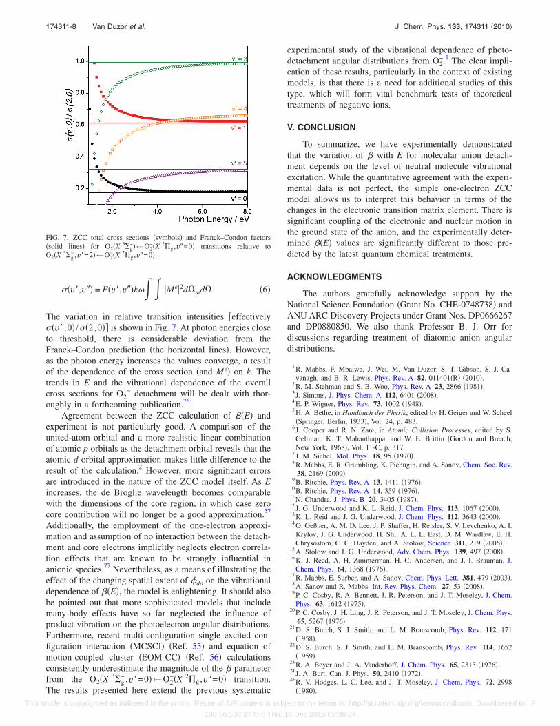
Figure 6 compares F�v� ,0� /F�2,0� �open circles� withthe O2�X 3�g
−�←O2−�X 2�g� band of our 454.54 nm spec-
trum. The Franck–Condon factors were calculated usingMorse oscillator wave functions75 employing the spectro-scopic constants in Ref. 27 and evaluating the vibrationaloverlap integral numerically using a 600 point Simpson’smethod calculation. It is clear that these Franck Condon fac-tors �FCFs� are a poor representation of the spectral intensi-ties, particularly at higher v�, showing the influence of vi-bronic coupling.
It is instructive to compare this finding with the Franck-Condon �FC� treatment of the spectrum given in Ref. 27. Thelatter work shows that the intensity variation in the X 3�g
−
←X 2�g band recorded at 364 nm is very well described bythe Franck–Condon factors alone. The key difference is theincreased photon energy. The vibrational transition intensi-ties can be calculated using the same ZCC approach appliedabove, integrating Eq. �3� over all angles,
FIG. 5. Potentials and vibrational levels for the ground states of O2− and O2.
FIG. 6. Comparison of relative Franck–Condon factors �open circles� and2.73 eV �454.57 nm� O2
− spectrum.
174311-7 Superoxide photoelectron angular distribution J. Chem. Phys. 133, 174311 �2010�
This article is copyrighted as indicated in the article. Reuse of AIP content is subject to the terms at: http://scitation.aip.org/termsconditions. Downloaded to IP:
130.56.106.27 On: Thu, 10 Dec 2015 02:36:24
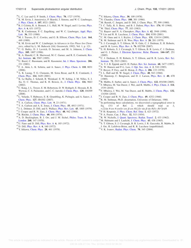
��v�,v�� = F�v�,v��k�� � �Me�2d�md� . �6�
The variation in relative transition intensities �effectively��v� ,0� /��2,0�� is shown in Fig. 7. At photon energies closeto threshold, there is considerable deviation from theFranck–Condon prediction �the horizontal lines�. However,as the photon energy increases the values converge, a resultof the dependence of the cross section �and Me� on k. Thetrends in E and the vibrational dependence of the overallcross sections for O2
− detachment will be dealt with thor-oughly in a forthcoming publication.76
Agreement between the ZCC calculation of ��E� andexperiment is not particularly good. A comparison of theunited-atom orbital and a more realistic linear combinationof atomic p orbitals as the detachment orbital reveals that theatomic d orbital approximation makes little difference to theresult of the calculation.2 However, more significant errorsare introduced in the nature of the ZCC model itself. As Eincreases, the de Broglie wavelength becomes comparablewith the dimensions of the core region, in which case zerocore contribution will no longer be a good approximation.57
Additionally, the employment of the one-electron approxi-mation and assumption of no interaction between the detach-ment and core electrons implicitly neglects electron correla-tion effects that are known to be strongly influential inanionic species.77 Nevertheless, as a means of illustrating theeffect of the changing spatial extent of �do on the vibrationaldependence of ��E�, the model is enlightening. It should alsobe pointed out that more sophisticated models that includemany-body effects have so far neglected the influence ofproduct vibration on the photoelectron angular distributions.Furthermore, recent multi-configuration single excited con-figuration interaction �MCSCI� �Ref. 55� and equation ofmotion-coupled cluster �EOM-CC� �Ref. 56� calculationsconsistently underestimate the magnitude of the � parameterfrom the O2�X 3�g
− ,v�=0�←O2−�X 2�g ,v�=0� transition.
The results presented here extend the previous systematic
experimental study of the vibrational dependence of photo-detachment angular distributions from O2
−.1 The clear impli-cation of these results, particularly in the context of existingmodels, is that there is a need for additional studies of thistype, which will form vital benchmark tests of theoreticaltreatments of negative ions.
V. CONCLUSION
To summarize, we have experimentally demonstratedthat the variation of � with E for molecular anion detach-ment depends on the level of neutral molecule vibrationalexcitation. While the quantitative agreement with the experi-mental data is not perfect, the simple one-electron ZCCmodel allows us to interpret this behavior in terms of thechanges in the electronic transition matrix element. There issignificant coupling of the electronic and nuclear motion inthe ground state of the anion, and the experimentally deter-mined ��E� values are significantly different to those pre-dicted by the latest quantum chemical treatments.
ACKNOWLEDGMENTS
The authors gratefully acknowledge support by theNational Science Foundation �Grant No. CHE-0748738� andANU ARC Discovery Projects under Grant Nos. DP0666267and DP0880850. We also thank Professor B. J. Orr fordiscussions regarding treatment of diatomic anion angulardistributions.
1 R. Mabbs, F. Mbaiwa, J. Wei, M. Van Duzor, S. T. Gibson, S. J. Ca-vanagh, and B. R. Lewis, Phys. Rev. A 82, 011401�R� �2010�.
2 R. M. Stehman and S. B. Woo, Phys. Rev. A 23, 2866 �1981�.3 J. Simons, J. Phys. Chem. A 112, 6401 �2008�.4 E. P. Wigner, Phys. Rev. 73, 1002 �1948�.5 H. A. Bethe, in Handbuch der Physik, edited by H. Geiger and W. Scheel�Springer, Berlin, 1933�, Vol. 24, p. 483.
6 J. Cooper and R. N. Zare, in Atomic Collision Processes, edited by S.Geltman, K. T. Mahanthappa, and W. E. Brittin �Gordon and Breach,New York, 1968�, Vol. 11-C, p. 317.
7 J. M. Sichel, Mol. Phys. 18, 95 �1970�.8 R. Mabbs, E. R. Grumbling, K. Pichugin, and A. Sanov, Chem. Soc. Rev.
38, 2169 �2009�.9 B. Ritchie, Phys. Rev. A 13, 1411 �1976�.
10 B. Ritchie, Phys. Rev. A 14, 359 �1976�.11 N. Chandra, J. Phys. B 20, 3405 �1987�.12 J. G. Underwood and K. L. Reid, J. Chem. Phys. 113, 1067 �2000�.13 K. L. Reid and J. G. Underwood, J. Chem. Phys. 112, 3643 �2000�.14 O. Geßner, A. M. D. Lee, J. P. Shaffer, H. Reisler, S. V. Levchenko, A. I.
Krylov, J. G. Underwood, H. Shi, A. L. L. East, D. M. Wardlaw, E. H.Chrysostom, C. C. Hayden, and A. Stolow, Science 311, 219 �2006�.
15 A. Stolow and J. G. Underwood, Adv. Chem. Phys. 139, 497 �2008�.16 K. J. Reed, A. H. Zimmerman, H. C. Andersen, and J. I. Brauman, J.
Chem. Phys. 64, 1368 �1976�.17 R. Mabbs, E. Surber, and A. Sanov, Chem. Phys. Lett. 381, 479 �2003�.18 A. Sanov and R. Mabbs, Int. Rev. Phys. Chem. 27, 53 �2008�.19 P. C. Cosby, R. A. Bennett, J. R. Peterson, and J. T. Moseley, J. Chem.
Phys. 63, 1612 �1975�.20 P. C. Cosby, J. H. Ling, J. R. Peterson, and J. T. Moseley, J. Chem. Phys.
65, 5267 �1976�.21 D. S. Burch, S. J. Smith, and L. M. Branscomb, Phys. Rev. 112, 171
�1958�.22 D. S. Burch, S. J. Smith, and L. M. Branscomb, Phys. Rev. 114, 1652
�1959�.23 R. A. Beyer and J. A. Vanderhoff, J. Chem. Phys. 65, 2313 �1976�.24 J. A. Burt, Can. J. Phys. 50, 2410 �1972�.25 R. V. Hodges, L. C. Lee, and J. T. Moseley, J. Chem. Phys. 72, 2998
�1980�.
FIG. 7. ZCC total cross sections �symbols� and Franck–Condon factors�solid lines� for O2�X 3�g
−�←O2−�X 2�g ,v�=0� transitions relative to
O2�X 3�g− ,v�=2�←O2
−�X 2�g ,v�=0�.
174311-8 Van Duzor et al. J. Chem. Phys. 133, 174311 �2010�
This article is copyrighted as indicated in the article. Reuse of AIP content is subject to the terms at: http://scitation.aip.org/termsconditions. Downloaded to IP:
130.56.106.27 On: Thu, 10 Dec 2015 02:36:24
26 L. C. Lee and G. P. Smith, J. Chem. Phys. 70, 1727 �1979�.27 K. M. Ervin, I. Anusiewicz, P. Skurski, J. Simons, and W. C. Lineberger,
J. Phys. Chem. A 107, 8521 �2003�.28 R. J. Celotta, R. A. Bennett, J. L. Hall, M. W. Siegel, and J. Levine, Phys.
Rev. A 6, 631 �1972�.29 R. R. Corderman, P. C. Engelking, and W. C. Lineberger, Appl. Phys.
Lett. 36, 533 �1980�.30 M. J. Travers, D. C. Cowles, and G. B. Ellison, Chem. Phys. Lett. 164,
449 �1989�.31 K. M. Ervin and W. C. Lineberger, in Advances in Gas Phase Ion Chem-
istry, edited by L. M. Babcock �JAI, Greenwich, 1992�, Vol. 1, p. 121.32 C. G. Bailey, D. J. Lavrich, D. Serxner, and M. A. Johnson, J. Chem.
Phys. 105, 1807 �1996�.33 K. A. Hanold, C. R. Sherwood, M. C. Garner, and R. E. Continetti, Rev.
Sci. Instrum. 66, 5507 �1995�.34 U. Boesl, C. Bassmann, and R. Kassmeier, Int. J. Mass. Spectrom. 206,
231 �2001�.35 F. A. Akin, L. K. Schirra, and A. Sanov, J. Phys. Chem. A 110, 8031
�2006�.36 A. K. Luong, T. G. Clements, M. Sowa Resat, and R. E. Continetti, J.
Chem. Phys. 114, 3449 �2001�.37 K. Le Barbu, J. Scheidt, R. Weinkauf, E. W. Schlag, J. M. Nilles, S. J.
Xu, O. C. Thomas, and K. H. Bowen, Jr., J. Chem. Phys. 116, 9663�2002�.
38 C. Kang, J. L. Troyer, E. M. Robertson, D. W. Rothgeb, E. Hossain, R. B.Wyrwas, C. S. Parmenter, and C. C. Jarrold, J. Chem. Phys. 128, 104309�2008�.
39 L. Velarde, T. Habteyes, E. R. Grumbling, K. Pichugin, and A. Sanov, J.Chem. Phys. 127, 084302 �2007�.
40 T. A. Carlson, Chem. Phys. Lett. 9, 23 �1971�.41 T. A. Carlson and A. E. Jonas, J. Chem. Phys. 55, 4913 �1971�.42 J. L. Dehmer, D. Dill, and S. Wallace, Phys. Rev. Lett. 43, 1005 �1979�.43 J. Cooper and R. N. Zare, J. Chem. Phys. 48, 942 �1968�.44 B. Ritchie, J. Chem. Phys. 60, 898 �1974�.45 A. D. Buckingham, B. J. Orr, and J. M. Sichel, Philos. Trans. R. Soc.
London 268, 147 �1970�.46 U. Fano and D. Dill, Phys. Rev. A 6, 185 �1972�.47 D. Dill, Phys. Rev. A 6, 160 �1972�.48 Y. Itikawa, Chem. Phys. 28, 461 �1978�.
49 Y. Itikawa, Chem. Phys. 30, 109 �1978�.50 N. Chandra, Chem. Phys. 108, 301 �1986�.51 M. Raoult, C. Jungen, and D. Dill, J. Chem. Phys. 77, 599 �1980�.52 J. C. Tully, R. S. Berry, and B. J. Dalton, Phys. Rev. 176, 95 �1968�.53 W. Thiel, Chem. Phys. 77, 103 �1983�.54 G. Raşeev and N. A. Cherepkov, Phys. Rev. A 42, 3948 �1990�.55 P. Lin and R. R. Lucchese, J. Chem. Phys. 114, 9350 �2001�.56 C. M. Oana and A. I. Krylov, J. Chem. Phys. 131, 124114 �2009�.57 R. M. Stehman and S. B. Woo, Phys. Rev. A 20, 281 �1979�.58 S. J. Cavanagh, S. T. Gibson, M. N. Gale, C. J. Dedman, E. H. Roberts,
and B. R. Lewis, Phys. Rev. A 76, 052708 �2007�.59 E. H. Roberts, S. J. Cavanagh, S. T. Gibson, B. R. Lewis, C. J. Dedman,
and G. J. Picker, J. Electron Spectrosc. Relat. Phenom. 144–147, 251�2005�.
60 C. J. Dedman, E. H. Roberts, S. T. Gibson, and B. R. Lewis, Rev. Sci.Instrum. 72, 2915 �2001�.
61 A. T. J. B. Eppink and D. H. Parker, Rev. Sci. Instrum. 68, 3477 �1997�.62 E. W. Hansen and P.-L. Law, J. Opt. Soc. Am. A 2, 510 �1985�.63 F. Breyer, P. Frey, and H. Hotop, Z. Phys. A 286, 133 �1978�.64 J. L. Hall and M. W. Siegel, J. Chem. Phys. 48, 943 �1968�.65 D. Hanstorp, C. Bengtsson, and D. J. Larson, Phys. Rev. A 40, 670
�1989�.66 R. Mabbs, E. Surber, and A. Sanov, J. Chem. Phys. 122, 054308 �2005�.67 F. Mbaiwa, M. Van Duzor, J. Wei, and R. Mabbs, J. Phys. Chem. A 114,
1539 �2010�.68 F. Mbaiwa, J. Wei, M. Van Duzor, and R. Mabbs, J. Chem. Phys. 132,
134304 �2010�.69 J. Cooper and R. N. Zare, J. Chem. Phys. 49, 4252 �1968�.70 R. M. Stehman, Ph.D. dissertation, University of Delaware, 1980.71 In performing these calculations, we discovered a typographical error in
Eq. �31� of Ref. 2, which should read as I1
=��sin cos cos�kr cos � cos �J1�kr sin � sin �f�r ,�r3drd.72 P. H. Krupenie, J. Phys. Chem. Ref. Data 1, 423 �1972�.73 P. A. Fraser, Can. J. Phys. 32, 515 �1954�.74 R. W. Nicholls, J. Quant. Spectrosc. Radiat. Transf. 2, 433 �1962�.75 M. Halmann and I. Laulicht, J. Chem. Phys. 43, 438 �1965�.76 S. T. Gibson, S. J. Cavanagh, B. R. Lewis, J. R. Gascooke, R. Mabbs, B.
J. Orr, H. Lefebvre-Brion, and R. R. Lucchese �unpublished�.77 V. K. Ivanov, Radiat. Phys. Chem. 70, 345 �2004�.
174311-9 Superoxide photoelectron angular distribution J. Chem. Phys. 133, 174311 �2010�
This article is copyrighted as indicated in the article. Reuse of AIP content is subject to the terms at: http://scitation.aip.org/termsconditions. Downloaded to IP:
130.56.106.27 On: Thu, 10 Dec 2015 02:36:24