
Vibrational spectroscopy of clusters with a Free Electron ... · Vibrational spectroscopy of...
60
Vibrational spectroscopy of clusters with a Vibrational spectroscopy of clusters with a Free Electron Laser Free Electron Laser André Fielicke Fritz-Haber-Institut der Max-Planck-Gesellschaft, Berlin Abteilung Molekülphysik http://www.fhi-berlin.mpg.de/mp/fielicke/
Transcript of Vibrational spectroscopy of clusters with a Free Electron ... · Vibrational spectroscopy of...

Vibrational spectroscopy of clusters with a Vibrational spectroscopy of clusters with a Free Electron LaserFree Electron Laser
André FielickeFritz-Haber-Institut der Max-Planck-Gesellschaft, Berlin
Abteilung Molekülphysik
http://www.fhi-berlin.mpg.de/mp/fielicke/

Now: Free Electron Laser for Infrared eXperiments (F
Nieuwegein, NL

FHI-FEL (under construction)
far-IRFEL
mid-IRFEL
to user lab
linearaccelerator
…first light end of 2011
F
D
C
A
B

Vibrational spectroscopy of clusters with a Free Electron Laser
• “Action spectroscopy”
• Multiple photon absorption
• Free Electron Lasers (how it works)
• Examples of cluster spectroscopy

Infrared spectroscopy of metal cluster complexes
ligand modes500-3500 cm-1 (0.06-0.43 eV)
internal cluster modes< 500 cm-1 (0.06 eV)
Structure of “bare”metal clusters
Exploring the cluster’s surface chemistry

IR spectroscopy of clusters in molecular beams
Not sensitive enough (low particle density)Not species specific (cluster distribution)
Direct measurement of absorption
More sensitive and selective:Mass spectrometric detectionof absorption via “Action Spectroscopy”
Changes of the charge state (ionization)Changes of particle mass (dissociation)
An intense and tunable IR source is needed for the excitation

Cluster dissociation; ”Depletion” spectroscopy
Xn+
hνXn
+* fragments
H. Haberland, Clusters of Atoms and Molecules, Springer-Verlag, Berlin, 1995.
Xn+
hν
MS
i) Ions
geometry
)exp(-σφ=oII
Lambert Beer “absorption law”

IR spectra of clusters via messenger technique
clusterdistribution
massselection
Laser
2nd mass filter forselecting fragments
Example: IR photodissociation of mass selected VxOy+·He
Absorption spectrum is obtained from the depletion of the He complexintensity vs. Laser frequency
pare
nt io
n de
plet
ion
K.R. Asmis, et al., J. Chem. Phys. 2004, 120, 6461.
AB+·M (AB+·M)* AB+ + M
chromophore spectator
14 Kion trap

“fingerprint” region
ν(M-M)
IR photon energy vs. bond dissociation energies
Chemisorption energies: 1-3 eVBinding energies in transition metal clusters 3-6 eVPhysisorption energies <0.2 eV
Photodissociation of most systems requires absorption of multiple IR photons
ν(X-H)ν(C=O)ν(MO)
1 eV = 96.5 kJ mol-1

Resonant multiple photon absorption
resonant absorption fast intramolecular vibrational redistribution (IVR), tIVR << 1 nsabsorption of the next photonetc.
harmonicoscillator
)(h 21+ν= vEvib
hνhνhνhνhνE
d
anharmonicoscillator
L++ν−
+ν=2
21
21
)(h)(h
vxvEee
evib
hν
E
d
hνd
hνhν
E
IVR
Cluster with manyvibrational modes

“fingerprint” region
ν(M-M)
IR sources for Multiple photon excitation
ν(X-H)ν(C=O)ν(MO)
Free Electron Laser
ns pulsed DFM, OPO/OPACO2 laser

Infrared multiple photon dissociation spectroscopy
CO2 laser: line tunable around 10 µm (1000±100 cm-1 )
Later: Knickelbein, Rayner, Walther
TodayIR-OPO, DFM: down to ~16 µm
(600 cm-1)but: rel. low power

General properties of Free Electron Lasers
accelerator based light source
intense, high brightness
(usually) wavelength tunable
monochromatic
pulsed or continuous (less common)

IR-FELs worldwide (not complete)
n.c.
n.c.
n.c.
n.c.
n.c.
s.c. cw
El.stat.
n.c.
n.c.
s.c. cw
n.c.
Surgery2 – 9 μm25-45 MeVVanderbilt (US)High power1.5 – 14 μm80-200MeVJLAB (US)Solid state 30 – 2500 μm2-6 MeVUCSB (US)High magn. field100–1500 μm10-15 MeVNijFEL (NL)
Solid state1 – 100 μm20-75 MeViFEL (JP)Solid state, biochem.4 – 13 μm25 MeVKU-FEL (JP)
Mat. science5 – 25 μm15-30 MeVBFEL (China)
Clusters, isotope separation
5 – 16 μm300–1000 μm
30-40 MeV10-20 MeV
FEL-TUS (JP)
Solid state, gas phase3 – 150 μm10-50 MeVCLIO (F)Solid state4 – 280 μm17-38 MeVFELBE (D)Mol. spectroscopy3 – 250 μm15-50 MeVFELIX (NL)
Mol. & cluster Spec., surface science
5 – 300 μm20-50 MeVn.c.FHI-FEL

There are FELs for all possible wavelengths!
X-rays
0.1 nm10000 eV
Microwaves
10 mm10-4 eV

Most recent family members: X-ray FELs
FLASH, DESY (Hamburg, Germany), SC Linac, 6.5-45 nm
XFEL, 0.1–6 nm, first light planned for 2014
LCLS, Stanford (USA), SC Linac, 0.15–1.5 nm (0.8-8 keV)working since April 2009

The Laser
resonator
gain medium
Energy source
Light Amplification by Stimulated Emission of Radiation

Processes in a light field (2-level system)
stimulatedabsorption
stim.emission
spontan.emission
Rates for induced processes depend on the spectral energy density ρ(ν)
Amplification

Free Electron Laser (FEL)
Medium and energy source:High energy (relativistic) electron beam
Interaction of the light field with the medium ?Amplification mechanism
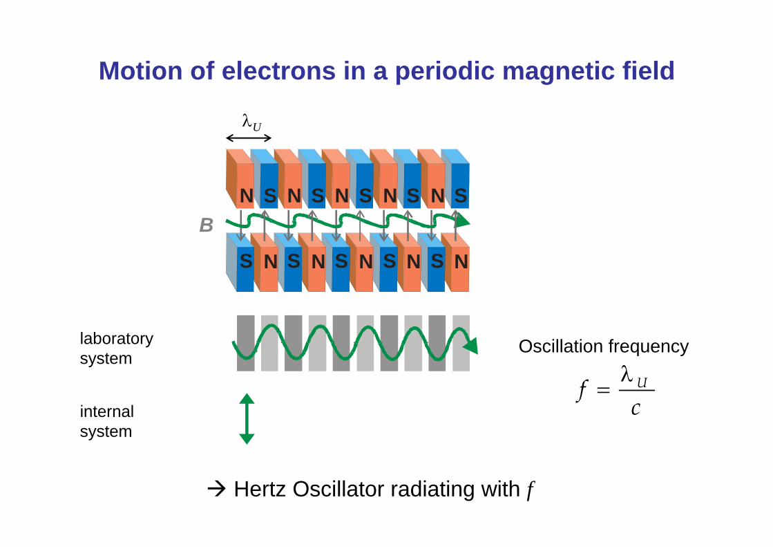
Motion of electrons in a periodic magnetic field
N S N S N S N S NS
N S N S N S N S N SB
laboratorysystem
internalsystem
Oscillation frequency
cf Uλ
=
Hertz Oscillator radiating with f
λU

Relativistic electron beams
cve ≈ 1. radiation characteristics
at rest relativistic
( )2,1 cv ze−≈ϑΔ

Relativistic electron beams
2. Lorentz contraction cve ≈
γλ
=λ′ UU
( )220 ,1
1
cv
kin
zecmE
−==γ
λU
MeV5.020 ≈cm 40 MeV beam γ ≈ 80

Relativistic electrons in the undulator I
γλ
=λ′ UU
λγ
≈λ
λ+
−=λ
211
1,
,
obs
cv
cv
obsze
ze
Oscillation (=radiation) frequency γ⋅
λ=c
f U
Corresponding wavelength (internal, moving system)
Wavelength in the laboratory system is detected with a strong Doppler shift
2221
γλ
=γγ
λ=λ UU

Relativistic electrons in the undulator II
But: electrons do not move straight along the z-axis in the undulator
Wiggling motion reduces speed in forward (z) direction Effective γ is reduced
2221
z
U
zz
U
γλ
=γγ
λ=λ
20cmEkin
z =γ<γ
Reduction depends on the deflection and thereby the magnetic field strength B )1( 2K
z+
γ=γ ( )B∝K
( )22 1
2K
z
U +γλ
=λSpontaneously emitted radiation from electrons in a periodic magnetic field

Setting the wavelength
( )22 1
2K
z
U +γλ
=λ
FHIFEL:Ekin= 50 MeVmid-IR Undulator: λU=0.04 mWavelength for K=1 ?
K: typically 0.5…3
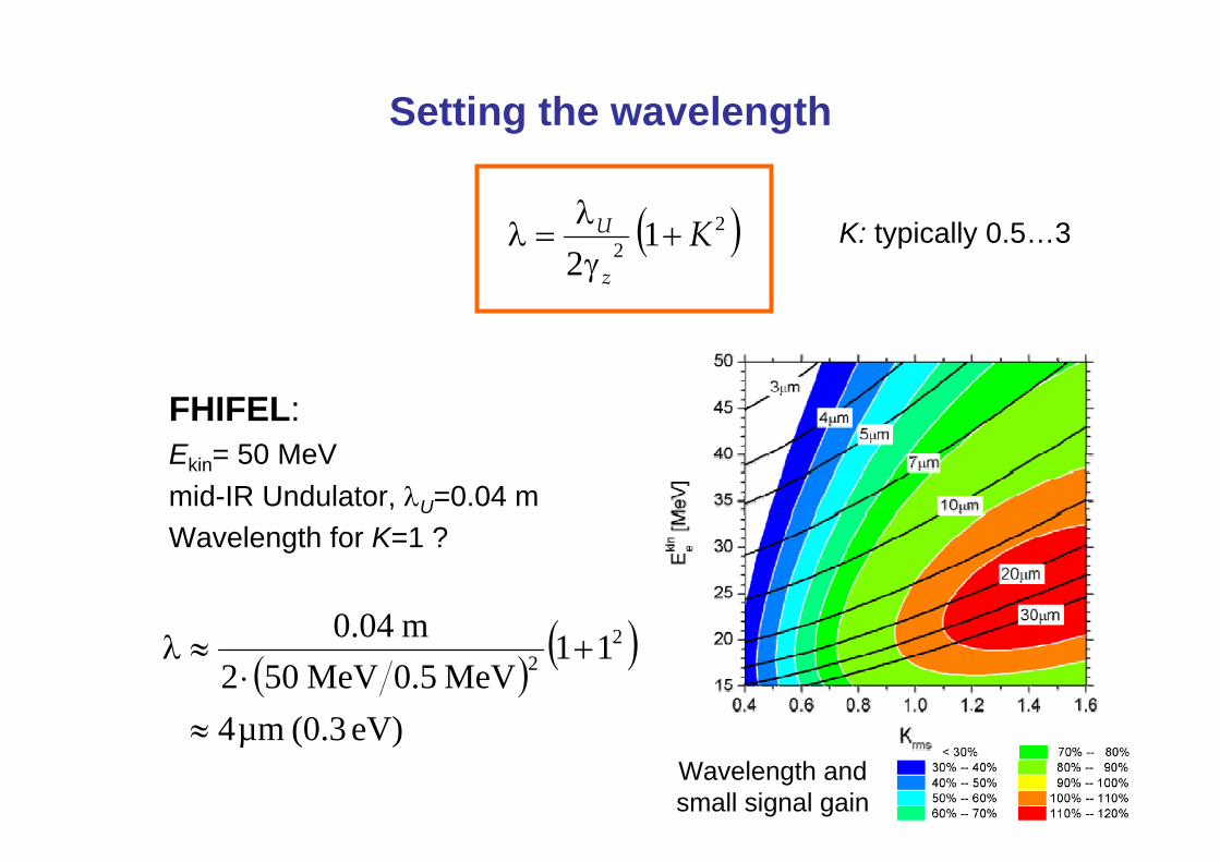
Setting the wavelength
( )22 1
2K
z
U +γλ
=λ K: typically 0.5…3
( )( )
eV) (0.3 µm4
11MeV5.0MeV502
m04.0 22
≈
+⋅
≈λ
Wavelength and small signal gain
FHIFEL:Ekin= 50 MeVmid-IR Undulator, λU=0.04 mWavelength for K=1 ?

Setting the wavelength
( )22 1
2K
z
U +γλ
=λ
BESSY II: Ekin=1.7 GeVUndulator UE56
K: typically 0.5…3
( )( )
eV) (250 nm5
11MeV5.0GeV7.12
m056.0 22
≈
+⋅
≈λ

Ways for tuning the wavelength
( )22 1
2K
z
U +γλ
=λ magnet field BBeam energy Ekin
Adjusting theundulator gap d
d
Changing accelerator parameters and electron optics

Interaction of the electrons with the light field
Ponderomotive force acts on the electrons depending on relative phase Acceleration or slowing down
Initial situation: incoherent spontaneously emitted radiation captured in the resonator

Interaction of the electrons with the light field
Microbunching of the electron packets through momentum transfer between light field and electrons
Result: electrons move in phaseRadiations from single bunches can interfere constructively

Oszillator FEL
Stored light field increases with timeNew electron bunches traverse the undulatorMore efficient bunching enhances energy transfer into the light field
exponential rise until saturation

X-rays can not be stored…
“Self amplified stimulated emission”(SASE)<
No effective mirror materials
Amplification in a single pass along the undulator
FLASH, DESY (Hamburg, Germany)

A train of short light pulses
Electron beam determines temporal characteristics of emitted light:Short pulses of a few fs – ps length
Transform limited bandwidth (FELIX: 0.4-7 %)
Repetition within a macropulse (also pulsed cw possible)
Pulse scheme of FELIX

Applications of Free Electron Lasers
high intensity
Weak absorbers(low density and/orlow cross sections)
Multiple photon processes
(ultra-) short pulses
Time resolution
Relaxation processes
To take snapshots

The Free Electron Laser for Infrared eXperiments (FELIX)FOM Institute for Plasma Physics “Rijnhuizen”, Nieuwegein, The Netherlands
tunable between 40-2400 cm-1
(up to ~3700 cm-1 on 3rd harmonic)
up to 100 mJ per macropulse(1010 W/cm2 in a micropulse)
bandwidth typically 0.5-2 %of the central wavelength

cluster source
IR Multiple Photon Dissociation (IR-MPD) Spectroscopy of neutral and charged clusters
reactive gas
gas pulse
UV laser(F2, 7.9 eV)
metal rod
Nd:YAGlaser
Reflectron time-of-flightmass spectrometer
Ion Detector
FELIX beam
FELIX4.92 μm
500 550 600 650 700m / z (amu)
Rh6CO+Rh5CO+

Probing the surface chemistry of the ligand
character of CO bindingdissociative molecular
Surf. Sci. 603 (2009) 1427.

CO at Rhn+: Size dependence of the binding site
• Observation of CO bound in 3-fold face capping (µ3), 2-fold bridging (µ2), and linear (µ1) geometries
• CO binding depends on cluster size
• mainly µ1 ligands
• Isomers, e.g. for n = 7
JACS 125 (2003) 15716.J. Phys. Chem. B 108 (2004) 14591.

Comparison of neutral, cationic and anionic RhnCO+/-
cations anionsneutrals

Effect of charge: example of Rh8CO+/0/-
C OCO
p
s p
ssσ(3σ)
pσ(4σ)
pπ(1π)
sσ∗(5σ)
pπ∗(2π)
pσ∗(6σ)
M(σ) CO(5σ)σ donation
M(δ) CO(2π)π back donation
CM O
CM O

Saturated Rhodium Cluster Carbonyl Cations
6*9 + 16*2 -1 = 85Rh6(CO)16+
5*9 + 14*2 -1 = 72Rh5(CO)14+
4*9 + 12*2 -1 = 59Rh4(CO)12+
3*9 + 9*2 -1 = 44Rh3(CO)9+
2*9 + 8*2 -1 = 33Rh2(CO)8+
9 + 5*2 -1 = 18Rh(CO)5+
CVEsSaturation composition
(18-3)*4 = 60
(18-3)*2+(18-2)*2 = 62
Inferring structures from counting electrons
18 electron rule, Rh: 4d85s1
“The Bonding Capabilities of Transition Metal Clusters”J. W. Lauher, J. Am. Chem. Soc. 100 (1978) 5305

Saturated Rhodium Cluster Carbonyl Cations
J. Am. Chem. Soc. 130 (2008) 2126

Rhodium cluster carbonyls: Cations vs. Neutrals
neutralcation
SAME
DIFFERENT
DIFFERENT
No bridging CO in the cations of Rh2(CO)8 and Rh4(CO)12
Neutrals in Xe, Allian et al., Vib. Spec. 41 (2006) 101

Rh
C
O
-10 -8 -6 -4 -2 0 2 4 6 8 10
x 10 -3
Destabilization of µ2-CO upon ionization comes from removal of electron density out of orbitals with Rh-µ2-CO binding character
Rhodium cluster carbonyls: Cations vs. Neutrals

Far-IR multiple photon dissociation spectroscopy of metal cluster rare-gas complexes
resonant absorption Fragmentation of the complexdepletion spectrum
wavelength (µm)
inte
nsity
(%)
frequency (cm-1)
IR absorption spectrum
cros
sse
ctio
n

Obtaining the structural information
Density Functional TheoryPredict stable isomers (may include a global optimization)Compute infrared spectrumEffects of rare gas atoms?Accounting for anharmonicity, fluctional behavior via MD simulations
exp. spectrum calculated IR spectra for different structures
structure assigment
frequency (cm-1)
rel.
cros
sse
ctio
n
Au7

Analyzing the structure vs. understanding the IR spectraExperiment: Au7Kr at ~100 K
PBE+D
i) Structural isomers ii) Effect of Kr atoms iii) Dynamics at 80 K
L. Ghiringhelli, M. SchefflerFHI Theory Dep. (DFT+D)

IR Spectrum and Structure of Neutral Nb8
1.4294.4302.9a10.0276.5284.4a14.1270.0277.8b10.0249.5256.7b11.3242.7249.7a10.4236.5243.3b20.3233.8240.5b20.5228.2234.8a11E-5187.8193.3b21.8185.3190.6a10184.1189.4a20.7183.4188.7b10181.7186.9a20.2144.8148.9a10.3140.5144.6b20.3134.7138.6b10.0117.1120.5a10111.5114.7a2
IntScaledcm-1
Calccm-1
Sym.
neutral: ν=180 cm-1
anion: ν=165 cm-1
Calc. spectrum (DFT)
T. P. Marcy and D. G. Leopold, Int. J. Mass Spectr. 196, 653 (2000).

Magnetism in small rhodium clusters
Y.-C. Bae et al. Phys. Rev. B 72 (2005) 125427.
108 9
► Cubic growth can explain magnetic properties
► Eight-center bonding through d orbitals
1312
A.J. Cox et al. Phys. Rev. B 49 (1994) 12295.

The (predicted) cubic structures of rhodium clusters
Rh8 cube, Oh symmetry1 IR active mode (t1u)
108 9 1312

0
0.5
1.0re
l. en
ergy
(eV
)
cube
anticube
bicap-ohdiamond-p
bicap-tp
Predicted isomers of Rh8+
(basin hopping DFT)
PBE PBE-hybrid
Dan Harding, Tiff Walsh, U Warwick, UKStuart Mackenzie, U Oxford, UK
cube
anticube
bicap-oh
bicap-tp
J.P. Perdew, K. Burke, M. Ernzerhof, Phys. Rev. Lett. 77, (1996) 3865 J.P. Perdew, M. Ernzerhof, K. Burke, J. Chem. Phys. 105, (1996) 9982

e
b2
bicapped octahedralstructure as identified also for other transition metals
0
+0.18 eV
+0.56 eV
+0.92 eV
Assignment of the structure of Rh8+
J. Chem. Phys. 132 (2010) 011101.

Structures of small transition metal clusters
Nbneutral
cation
Rh cation
n = 5 6 7 8 9
Polytetrahedral packings

Cooling processes following IR multiple photon excitation
IR multiple photon dissociation(IR-MPD)
IR resonance enhanced multiplephoton ionization (IR-MPD)
For most systems: BDE < IP
Exceptions: refractory materialsmetals (W, Mo, Nb…)
oxides (MgO, Al2O3, TiO2)carbides (TiC, NbC)fullerenes C60

IR Resonance Enhanced Multiple Photon Ionization(IR-REMPI)
Ionization of neutral NbCclusters with IR light of ~500 cm-1 (0.06 eV)NbC: cubic bulk structure(NaCl) geometric shell closings at
Nb14C13 (3x3x3 atoms)Nb18C18 (3x3x4 atoms)Nb24C24 (3x4x4 atoms)Nb32C32 (4x4x4 atoms)
Bulk niobium carbide: metallic, interstitiell C in octahedral holes of Nb fcc lattice
ionization energies ≈ 4 eV (67 photons@500 cm-1)binding energy per atom: > 6 eV

IR-REMPI spectra of niobium carbide clusters
Bands are observed close to the surface phonon modes:
S4 “Lucas“ mode at 640 cm-1
C atoms move within the surface plane
S2 “Wallis“ mode at 490 cm-1
C atoms move perpendicular to the surface plane
Small clusters (upto Nb9C9) show only bands around ~670 cm-1
C. Oshima et al. Phys. Rev. Lett. 56 (1986) 240

From FELIX to FELICE
100 mJ(1 %)
FELIX: Intensity still insufficient for many multiple photon excitation experiments
FELICE:Free Electron Laser for IntraCavity Experiments
10 J

FELICE: Free Electron Laser for IntraCavity Experiments
wavelength range 3-100 µmbeam energy 20-60 MeVmicropulse rep. rate 1 GHzmicropulse energy ≤1 mJmacropulse energy some 10J

Electron detachment and ionization by IR multiple photon excitation
Nb13 Nb13+
Nb13Ar Nb13+Ar
IR-REMPI of neutral niobium clusters
Ionization (IP=5.5 eV)
Dissociation (BDE<0.1 eV)
Vibrationally mediated electron detachment from anionic clusters
Nb13
Ta4C- Ta4C (ca. 1-2 eV)
(with V. Lapoutre, J. Bakker, FOM Rijnhuizen)

Techniques: Raman (matrix)Anion photoelectron spectroscopyPhotodissociation, IR-MPDIR-REMPI
Vibrational spectroscopy is a suitable tool for obtaining structural information for metal (compound) clusters, especially with complementary theoretical studies (DFT).
Surface chemistry and binding geometries of adsorbed molecules can be identified.
Summary