Vibrational Spectroscopy (Infrared, IR-Spect.)sci.tanta.edu.eg/files/IR spectroscopy...
42
Vibrational Spectroscopy (Infrared, IR-Spect.) Prof. Tarek A. Fayed
Transcript of Vibrational Spectroscopy (Infrared, IR-Spect.)sci.tanta.edu.eg/files/IR spectroscopy...
Vibrational Spectroscopy
(Infrared, IR-Spect.)
Prof. Tarek A. Fayed
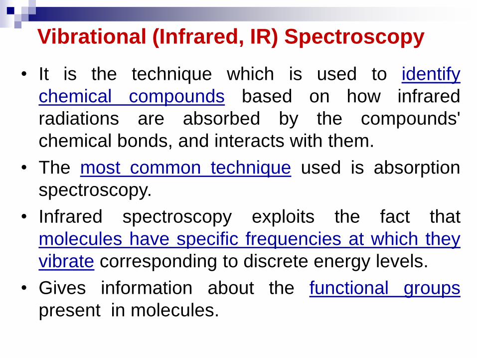
Vibrational (Infrared, IR) Spectroscopy
• It is the technique which is used to identify
chemical compounds based on how infrared
radiations are absorbed by the compounds'
chemical bonds, and interacts with them.
• The most common technique used is absorption
spectroscopy.
• Infrared spectroscopy exploits the fact that
molecules have specific frequencies at which they
vibrate corresponding to discrete energy levels.
• Gives information about the functional groups
present in molecules.

IR region of electromagnetic spectrum:
λ : 780 nm – 1000 μm
Wavenumber : 12,800 – 10 cm-1
IR region is subdivided into 3 sub-regions:
1. Near IR region (Nearest to the visible)
- 780 nm to 2.5 μm (12,800 - 4000 cm-1)
2. Mid IR region
- 2.5 to 50 μm (4000 – 200 cm-1)
3. Far IR region
- 50 to 1000 μm (200 – 10 cm-1)
infr
ared
N
E
A
R
M
I
D
F
A
R
3

What happens when absorption of IR occurs?
1. Changes in the shape of molecules such as stretching
of bonds, bending of bonds, or internal rotation around
single bonds.
2. IR absorption only occurs when IR radiation interacts
with a molecule undergoing a change in dipole moment
as it vibrates or rotates.
3. Infrared absorption only occurs when the incoming IR
photon has sufficient energy for transition to the next
allowed vibrational state to take place (E = h).
Note: If the rules 2 and 3, above are not met, no
absorption can occur.
SO, NOT ALL bonds in a molecule are
capable of absorbing IR- energy.

Infrared Absorption
For a molecule to show infrared absorptions it must possess a
specific feature: an electric dipole moment which must change
during the vibration.
A dipole moment, µ is defined as the charge value (q) multiplied
by the separation distance (d) between the positive and negative
charges.
µ = qd (C.m)
In hetero-nuclear diatomic molecules, due to the difference in
electronegativities of the two atoms, one atom acquires a small
positive charge (+), the other a negative charge ( -).
O
H H
δ-
δ+ δ+

A molecule is IR active if it has a permanent
dipole moment; HCl is active while N2 is inactive.
IR spectrum represents the rotation-vibration
spectrum of the molecule.
In solution, the rotation of molecules is strongly
hindered, bands are strongly broadened and the
maxima of these bands correspond to the
vibrational spectrum.
In the solid state, the rotations are effectively
frozen so that the spectrum appears as relatively
sharp bands, which corresponds to the normal
vibrations.
In gases, the rotation-vibration spectrum can be
also observed.

Modes of Vibration
The interaction of infrared radiations with matter
can be understood in terms of changes in
molecular dipoles associated with vibrations.
Vibrations can involve either changes in bond
length (stretching) or bond angle (bending).
Some bonds can stretch in-plane (symmetric
stretching) or out-of-plane (asymmetric stretching).
Bending vibrations can be either in-plane (as;
scissoring, rocking) or out-of-plane (as; wagging,
twisting) bending vibrations.
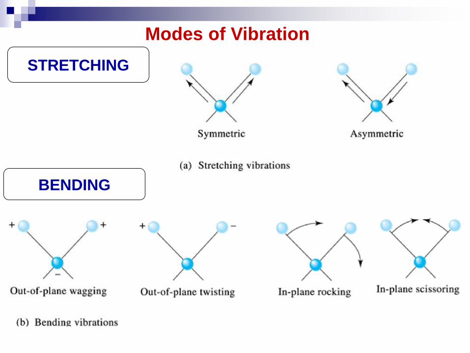
STRETCHING
Modes of Vibration
BENDING

Molecular vibration
divided
into
stretching bending
back & forth
movement
involves
change in
bond angles
symmetrical asymmetrical
scissoring
rocking twisting
wagging
in-plane
vibration
out of plane
vibration

Total number of modes of vibrations (normal modes)
The number of coordinates required to specify the
position of all atoms in a molecule is called the number
of degree of freedom, thus, for a molecule with N-
atoms, it has 3N degree of freedom.
So, the degree of vibrational freedom (Total number of
modes of vibrations) for polyatomic molecules
containing (N) atoms is given by;
1. 3N – 5 (For linear molecules)
2. 3N – 6 (For non-linear molecules)
Two other concepts are also used to explain the
frequency of vibrational modes:
(1) The stiffness of the bond expressed as the force
constant (called; k or F) and,
(2) The masses of the atoms at each end of the bond ( ).

TRANSLATIONAL ROTATIONAL VIBRATIONAL
For linear
molecule 3 2 3N-5
For nonlinear
molecule
3 3 3N-6
When the thermal energy is absorbed by molecules, it
is stored in molecules in the form of;
(1) Transitional movement of the molecule. There are 3-
transitional degrees of freedom along X, Y and Z-axes,
so the remaining (3N-3) co-ordinates represent the
internal degree of freedom. These are sub-divided into;
Rotational degree of freedom
Vibrational degree of freedom
(2) Internal motion of atoms present in the molecule (i. e.
rotational and vibrational motions).
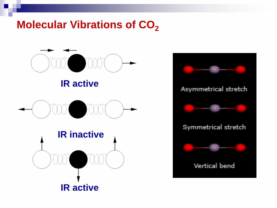
Molecular Vibrations of CO2
IR inactive
IR active
IR active

Vibrational Modes for Water

Fundamental IR Bands for Water

4000 3000 2000 1000
WAVENUMBER (cm-1)
Basic Functional Groups
C-H
O-H
Ch
C
C=C
alkenes
aromatic
C=O C-O
C-H
O-H
bendin
g
str
etc
hin
g
C-C
400
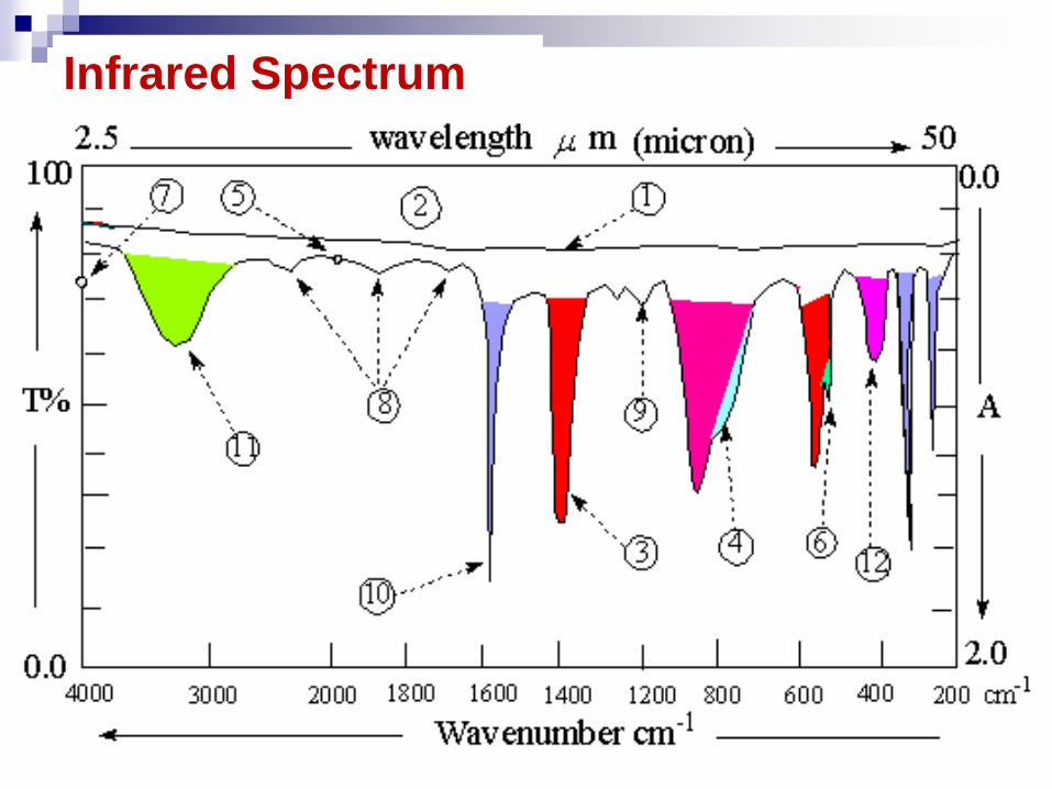
Infrared Spectrum
IR Spectrum of Complex Molecules
• There are many possible vibrational modes giving
rise to complicated spectra with many peaks.
• IR spectra are mainly used to identify unknown
compounds
• Peak positions can demonstrate what functional
groups are present in the molecule. Each
functional group gives rise to an absorption peak
at a characteristic frequency, no matter what the
rest of the molecule contains.
• The peak positions and intensities of an unknown
can be compared with the spectrum of known
suspects in the same manner that police use
fingerprints.

Approximate
frequency
cm-1
Group
Approximate
frequency
cm-1
group
2580 -SH (free) 3600 - OH (free)
2250 - C N 3400 - NH2 (free)
2220 - C C - 3300 CH
1750-1600 C = O 3060 Ph-H
1650 C = C 3030 = CH2
1200-1000 C-C, C-N,C-O 2970 asym.
2870 sym.
1460 asym.
1375 sym.
CH3
1100 C = S
1050 C-F
725 C-Cl
650 C-Br 2930 asym. -CH2-
550 C-I 2860 sym
X
m
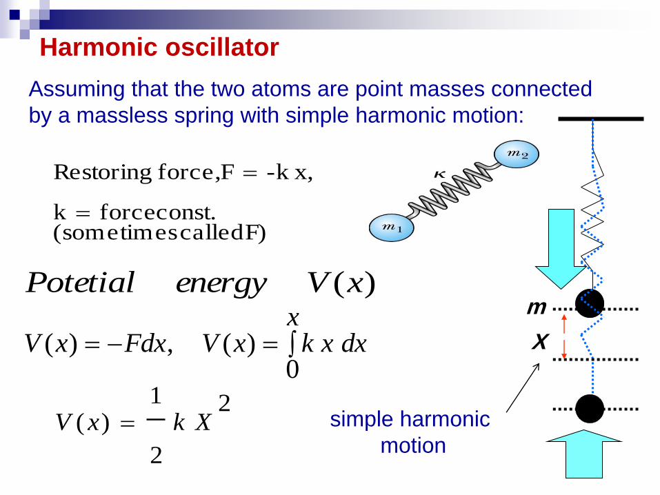
F) called times(some const. forcek
x,-kF force, Restoring
)(xVenergyPotetial
x
dxxkxVFdxxV0
)(,)(
2
2
1)( XkxV
Harmonic oscillator
Assuming that the two atoms are point masses connected
by a massless spring with simple harmonic motion:
simple harmonic
motion

For stretching of the bond A-B, the frequency of
oscillation as harmonic oscillator is given by the
relation;
k
2
1
k
2
1
c
k (single bond) = 5x10⁵ dyne/cm.,
k (double bond) = 10x105 dyne/cm.,
k (triple bond) = 15x105 dyne/cm.
Is the “reduced mass” where m1,
m2 are the masses on either side of
vibration
k is the “force constant”, like the Hooke’s Law restoring force
for a spring. Known and tabulated for different vibrations.
BA
BA
mm
mm
or in cm-1

re
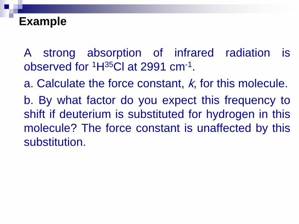
A strong absorption of infrared radiation is
observed for 1H35Cl at 2991 cm-1.
a. Calculate the force constant, k, for this molecule.
b. By what factor do you expect this frequency to
shift if deuterium is substituted for hydrogen in this
molecule? The force constant is unaffected by this
substitution.
Example

a. We first write .
Solving for k,
b. The vibrational frequency for DCl is lower
by a substantial amount.
1.0078 36.983
0.7172.0140 35.977
HCl
DCl
H cl D C
D cl H C
m m m m
m m m m
mN
ck
khhchvE
/3.5162710661.1977.35
969.34008.121002991810998.22
2
2 44
2
and
// khchvE
Solution

From the Schrödinger equation;
,so;
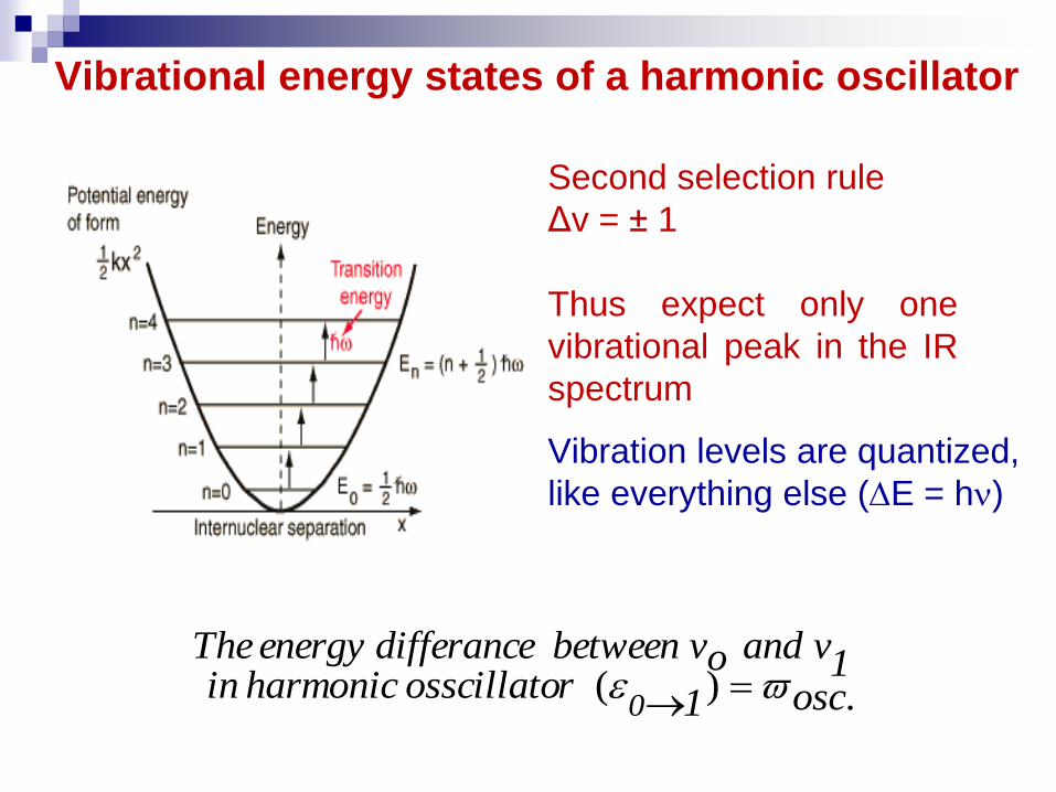
Vibrational energy states of a harmonic oscillator
Second selection rule
Δv = ± 1
Thus expect only one
vibrational peak in the IR
spectrum
Vibration levels are quantized,
like everything else (E = h)
.)( osc1rosscillato harmonic in1v andov between differanceenergy The
0

Consequences of the harmonic approximation
At room T, molecules are mainly in their vibrational ground state. Hence, in
IR absorption spectroscopy, the molecules are excited from the ground state
to the first excited state: 0→1, since the selection rule is = ±1 the IR
spectrum should contain only one line for diatomic molecule.
At higher T, other transitions can occur: 2 → 3 or 3 → 4, but all of them
need the absorption of a photon with the same energy, i.e. the absorption
lines appear at the same frequency, because the energy between two states
is constant.
Although the main features are there, it is not exactly what shows the
actual absorption spectra…. The potential is not harmonic.
According to the harmonic oscillator, a chemical bond cannot break.
Classical explanation of molecular vibrations:
If the oscillation frequency of the electric field of a radiation is similar to the
frequency =/2 of one vibrational motion in a molecule (which involves a
variation of the charge distribution), then the molecule can absorb the energy
h of one photon from the radiation. Intuitively, we can see this absorption of
energy like the resonance phenomenon in classical mechanics.

Real molecules do not obey exactly the laws of simple
harmonic motion. If the bond between atoms is
stretched, there comes a point at which the bond will
break i. e. the molecule dissociates into atoms.
A parabola cannot be correct at all extensions
because it does not allow the bond to dissociate. So,
a parabolic approximation is not the actual potential
energy curve, then the motion becomes anharmonic
and represented by the Morse potential energy curve.
Anharmonic Oscillator

Therefore, the Morse potential energy is the most
closely to the true potential energy curve.
Potential energy curve for anharmonic oscillator
Do= Dissociation energy
Deq is the depth of the
potential energy minimum
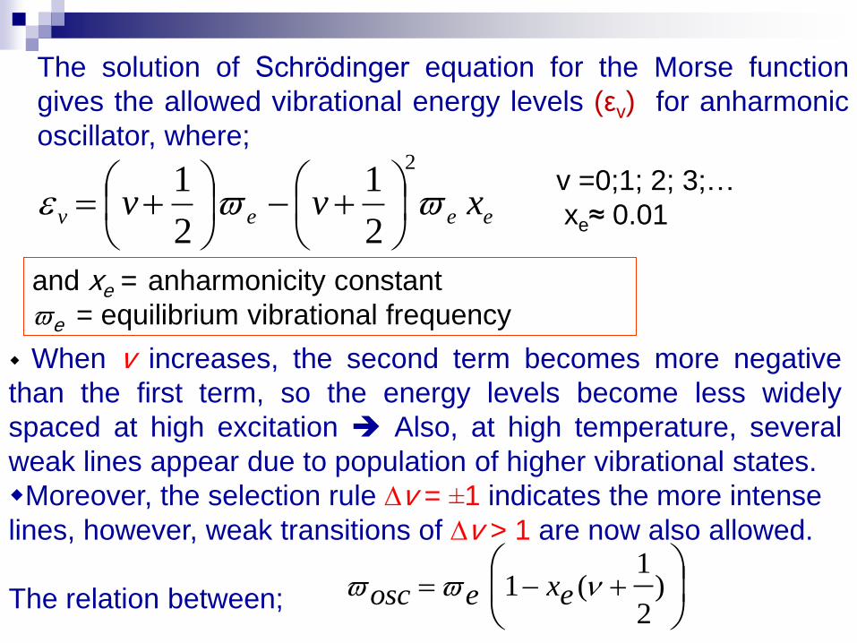
The solution of Schrödinger equation for the Morse function
gives the allowed vibrational energy levels (εv) for anharmonic
oscillator, where;
and xe = anharmonicity constant
e = equilibrium vibrational frequency
eeev xvv
2
2
1
2
1
v =0;1; 2; 3;…
xe≈ 0.01
When v increases, the second term becomes more negative
than the first term, so the energy levels become less widely
spaced at high excitation Also, at high temperature, several
weak lines appear due to population of higher vibrational states.
Moreover, the selection rule v = ±1 indicates the more intense
lines, however, weak transitions of v > 1 are now also allowed.
The relation between;
)
2
1(1 exeosc


•Example
•The fundamental and first overtone of 1H35Cl
are observed at 2886 cm-1 and 2668 cm-1,
respectively. Calculate:
1- Equilibrium vibration frequency.
2- Anharmonity constant.
3- Exact zero-potential frequency.
4- The force constant of the molecule.
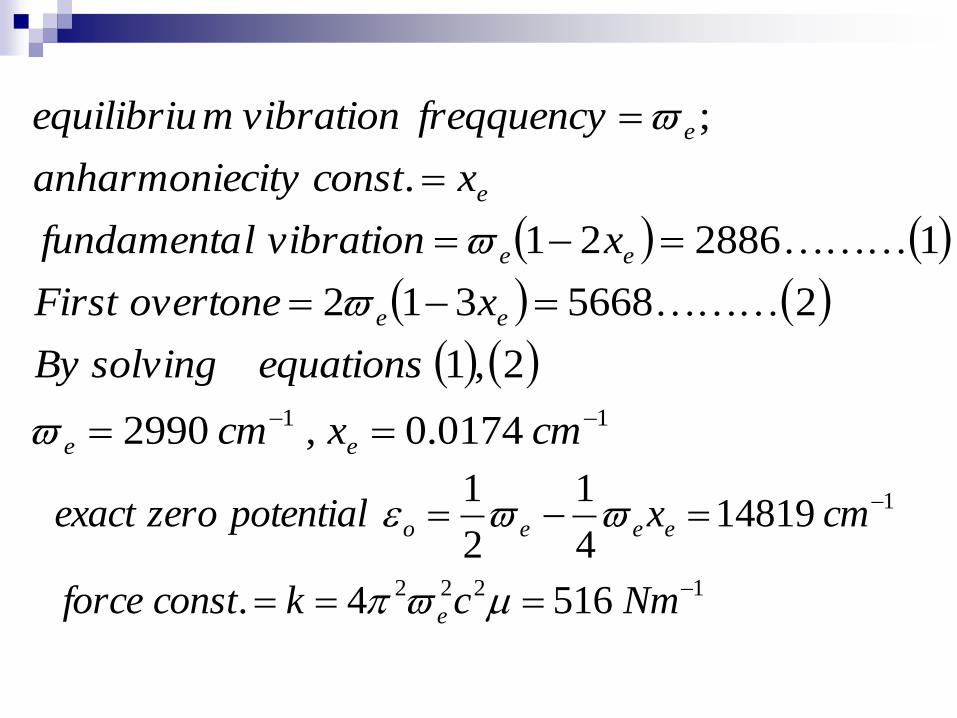
11 0174.0,2990
2,1
25668312
1288621
.
;
cmxcm
equationssolvingBy
xovertoneFirst
xvibrationlfundamenta
xconstcityanharmonie
freqquencyvibrationmequilibriu
ee
ee
ee
e
e
1222
1
5164.
148194
1
2
1
Nmckconstforce
cmxpotentialzeroexact
e
eeeo

Typical IR-Spectrophotometer
The layout of a typical dispersive Infra-red absorption spectrometer
From the comparison between the reference beam and the one
passing through the sample, we can deduce the frequencies
absorbed by the excitation of molecules in their vibrational
energy levels. So, the IR spectrum is recorded.

1- Gaseous samples; require little preparation beyond
purification, but a sample cell with a long path-length
(typically 5-10 cm) is normally needed. The walls are of
glass or brass.
Sample preparation
2- Liquid samples; use solution cells. Two types of solution
cells – permanent and demountable. Permanent cell is
difficult to clean and can be damaged by water.
Demountable cell is easy to maintain as it can be readily
dismantled and cleaned and the windows can be
repolished.
3- Solid samples; can be prepared in two ways:
1. Crush the sample with a mulling agent (as
paraffin oil) in a marble or agate mortar, with a
pestle. A thin film of the mull is applied onto
salt plates and measured.
2. Grind a quantity of the sample with a specially
purified salt (usually potassium bromide) finely
(to remove scattering effects from large
crystals). This powder mixture is then crushed
in a mechanical die press to form a translucent
pellet through which the beam of the
spectrometer can pass.

Fourier Transform FT-IR Spectrometer
A spectrometer is an optical instrument used to measure properties of light over a specific portion of the electromagnetic spectrum, 5 microns to 20 microns.
FTIR (Fourier Transform Infrared) spectrometer obtains an infrared spectra by first collecting an interferogram of a sample signal using an interferometer, then performs a Fourier Transform on the interferogram to obtain the spectrum.
An interferometer is an instrument that uses the technique of superimposing (interfering) two or more waves, to detect differences between them. The FTIR spectrometer uses a Michelson interferometer.

Interferometer
Special instrument which can read IR frequencies
simultaneously.
faster method than dispersive instrument.
interferograms are transformed into frequency
spectrums by using mathematical technique
called Fourier Transformation
FT
Calculations
interferograms IR spectrum
37

Components of Fourier Transform Instrument
1
3
2
4
5
6
38

Theory and Instrumentation (contd.)
The light originates from the He-Ne laser
Half of the light is reflected 90 degrees and hits
a fixed mirror, while the other half passes
through the beam splitter and hits the moving
mirror
The split beams are recombined, but having
traveled different distances, they exhibit an
interference pattern with each other
As they pass through the sample, the detector
collects the interfering signals and returns a
plot of response vs. mirror displacement
known as an interferogram

Advantages of FT-IR (over dispersive instrument)
high sensitivity
high resolution
speed of data acquisition ( data for an entire
spectrum can be obtained in 1 s or less)
40

Applications of IR and FT-IR spectroscopy
Identification of inorganic and organic compounds.
Identification of components of an unknown mixture.
Analysis of solids, liquids, and gasses.
In remote sensing.
In measurement and analysis of atmospheric spectra.
Can also be used on satellites to probe the space.
In Forensic labs. to identify chemicals in samples such as; paints, polymers, coatings, drugs, contaminants, explosive residues.
Analysis of aircraft exhausts and measurement of toxic gas in fuels, as well as in oil industry.

Vibrational frequency: (C-C) [700-1200cm-1] < (C=C) [1620-1680cm-1] < (C≡C) [2100-2260cm-1]
Bond lengths: Re(C-C) [1.54Å] > Re(C=C) [1.35Å] > Re(C ≡ C) [1.20Å]
Bond dissociation energy: D0(C-C) [368 kJ/mol] < D0(C=C) [720 kJ/mol] <
D0(C ≡ C) [962 kJ/mol]
R
E
k(C-C) < k(C=C)< k(C ≡ C)