Veterinary Oncology Roundtable - Zoetis · David M. Vail, DVM, DACVIM (Oncology) School of...
21
PART 1 1 PALLADIA ROUNDTABLE | PART 1 Mast cell tumors (MCT) are the most common cutaneous tumors in the dog (London & Thamm, 2013). A majority of cutaneous canine MCTs can be treated successfully with surgery, but locally recurrent, large or infiltrative tumors, and those in locations not amenable to wide surgical excision can present a therapeutic challenge. Other treatment modalities include radiation therapy, cytotoxic chemotherapy, and most recently, targeted therapy with drugs in the tyrosine kinase inhibitor (TKI) class. Toceranib phosphate (Palladia®, Zoetis) was approved by the FDA in 2009 as the first cancer drug specifically for dogs for treatment of cutaneous MCTs. Palladia is a tyrosine kinase inhibitor with both antitumor and antiangiogenic activity through the inhibition of Kit, vascular endothelial growth factor 2 (VEGF2), and platelet-derived growth factor receptor beta (PDGFRß). Virtually all canine MCTs express the KIT receptor tyrosine kinase (RTK), and 20% to 40% have a mutation in the c-kit gene that activates the Kit protein (London & Thamm, 2013; Downing et al, 2002; London et al, 1999; Zemke et al, 2002). In a clinical field trial of 145 dogs with Grade II (80%) or Grade III (20%), Palladia was shown to provide an overall response rate of 37.2% (London et al, 2009). The response rate in tumors positive for the c-kit mutation, however, was 60.0%, compared with 31.3% for tumors nega-tive for the c-kit mutation (London et al, 2009). Administered as an oral tablet, Palladia can be given in com- bination with radiation therapy and/or chemotherapy (Carlsten et al, 2012; Pellin et al, 2016; Burton et al, 2015; Pan et al, 2016, Mitchell et al, 2012), and presents an attractive option for treatment of cutaneous canine MCTs that cannot be treated successfully with surgery. IMPORTANT SAFETY INFORMATION: During clinical studies, the most common adverse events associated with PALLADIA included: diarrhea, anorexia (including decreased appetite), lethargy, neutropenia, emesis, lameness, weight loss, muscu- loskeletal disorder, and blood in stool/GI bleed/hemorrhagic diarrhea. PALLADIA may cause vascular dysfunction, which can lead to edema and thromboembolism, including pulmonary thromboembolism. Serious and sometime fatal GI com- plications, including GI perforation, have occurred rarely in dogs treated with PALLADIA. If GI ulceration is suspected stop drug administration and treat appropriately. Children should not come in contact with PALLADIA. In addition, all individuals, including children and pregnant women, should avoid direct contact with broken or partially dissolved PALLADIA tablets or biological waste from dogs treated with PALLADIA. To report a suspected adverse reaction call Zoetis at 1-888-963-8471. See full Prescribing Information. Veterinary Oncology Roundtable The Role of Prognostic Factors in Patient Selection for Canine Mast Cell Tumor Therapy with Palladia® Participants Craig Clifford, DVM, MS, DACVIM (Oncology) Hope Veterinary Specialists, Malvern, PA Laura D. Garrett, DVM, DACVIM (Oncology) College of Veterinary Medicine, University of Illinois, Urbana-Champaign, IL Carolyn Henry, DVM, MS, DACVIM (Oncology) School of Veterinary Medicine, University of Missouri, Columbia, MO Ann E. Hohenhaus, DVM, DACVIM (SAIM, Oncology) The Animal Medical Center, New York, NY Chad Johannes, DVM, DACVIM (SAIM, Oncology) College of Veterinary Medicine, Iowa State University, Ames, IA Pamela D. Jones, DVM, DACVIM (Oncology) and DACVR (Radiation Oncology) Consultant, Houston, TX M.K. Klein, DVM, MS, DACVIM (Oncology) and DACVR (Radiation Oncology) Clinical Lecturer, University of Arizona Cancer Center; and Veterinary Oncologist, Southwest Veterinary Oncology, Tucson, AZ Cheryl A. London, DVM, PhD, DACVIM (Oncology) College of Veterinary Medicine, The Ohio State University, Columbus, OH Kathy Mitchener, DVM, CVMA Angel Care Cancer Clinic for Animals, Memphis, TN Douglas H. Thamm, VMD, DACVIM (Oncology) Flint Animal Cancer Center, Colorado State University, Fort Collins, CO David M. Vail, DVM, DACVIM (Oncology) School of Veterinary Medicine, University of Wisconsin, Madison, WI Katherine S. Gloyd, DVM (Moderator) Elevate DVM, Wilmington, DE
Transcript of Veterinary Oncology Roundtable - Zoetis · David M. Vail, DVM, DACVIM (Oncology) School of...

P A R T 1
1PA L L A D I A R O U N DTA B L E | PA R T 1
Mast cell tumors (MCT) are the most common cutaneous tumors in the dog (London & Thamm, 2013). A majority of cutaneous canine MCTs can be treated successfully with surgery, but locally recurrent, large or infiltrative tumors, and those in locations not amenable to wide surgical excision can present a therapeutic challenge. Other treatment modalities include radiation therapy, cytotoxic chemotherapy, and most recently, targeted therapy with drugs in the tyrosine kinase inhibitor (TKI) class.
Toceranib phosphate (Palladia®, Zoetis) was approved by the FDA in 2009 as the first cancer drug specifically for dogs for treatment of cutaneous MCTs. Palladia is a tyrosine kinase inhibitor with both antitumor and antiangiogenic activity through the inhibition of Kit, vascular endothelial growth factor 2 (VEGF2), and platelet-derived growth factor receptor beta (PDGFRß). Virtually all canine MCTs express the KIT receptor tyrosine kinase (RTK), and 20% to 40% have a mutation in the c-kit gene that activates the Kit protein (London & Thamm, 2013; Downing et al, 2002; London et al, 1999; Zemke et al, 2002). In a clinical field trial of 145 dogs with Grade II (80%) or Grade III (20%), Palladia was shown to provide an overall response rate of 37.2% (London et al, 2009). The response rate in tumors positive for the c-kit mutation, however, was 60.0%, compared with 31.3% for tumors nega-tive for the c-kit mutation (London et al, 2009). Administered as an oral tablet, Palladia can be given in com-bination with radiation therapy and/or chemotherapy (Carlsten et al, 2012; Pellin et al, 2016; Burton et al, 2015; Pan et al, 2016, Mitchell et al, 2012), and presents an attractive option for treatment of cutaneous canine MCTs that cannot be treated successfully with surgery.
IMPORTANT SAFETY INFORMATION: During clinical studies, the most common adverse events associated with PALLADIA included: diarrhea, anorexia (including decreased appetite), lethargy, neutropenia, emesis, lameness, weight loss, muscu-loskeletal disorder, and blood in stool/GI bleed/hemorrhagic diarrhea. PALLADIA may cause vascular dysfunction, which can lead to edema and thromboembolism, including pulmonary thromboembolism. Serious and sometime fatal GI com-plications, including GI perforation, have occurred rarely in dogs treated with PALLADIA. If GI ulceration is suspected stop drug administration and treat appropriately. Children should not come in contact with PALLADIA. In addition, all individuals, including children and pregnant women, should avoid direct contact with broken or partially dissolved PALLADIA tablets or biological waste from dogs treated with PALLADIA. To report a suspected adverse reaction call Zoetis at 1-888-963-8471. See full Prescribing Information.
Veterinary Oncology RoundtableThe Role of Prognostic Factors in Patient Selection for Canine Mast Cell Tumor Therapy with Palladia®
Participants
Craig Clifford, DVM, MS, DACVIM (Oncology) Hope Veterinary Specialists, Malvern, PA
Laura D. Garrett, DVM, DACVIM (Oncology) College of Veterinary Medicine, University of Illinois, Urbana-Champaign, IL
Carolyn Henry, DVM, MS, DACVIM (Oncology) School of Veterinary Medicine, University of Missouri, Columbia, MO
Ann E. Hohenhaus, DVM, DACVIM (SAIM, Oncology) The Animal Medical Center, New York, NY
Chad Johannes, DVM, DACVIM (SAIM, Oncology) College of Veterinary Medicine, Iowa State University, Ames, IA
Pamela D. Jones, DVM, DACVIM (Oncology) and DACVR (Radiation Oncology) Consultant, Houston, TX
M.K. Klein, DVM, MS, DACVIM (Oncology) and DACVR (Radiation Oncology) Clinical Lecturer, University of Arizona Cancer Center; and Veterinary Oncologist, Southwest Veterinary Oncology, Tucson, AZ
Cheryl A. London, DVM, PhD, DACVIM (Oncology) College of Veterinary Medicine, The Ohio State University, Columbus, OH
Kathy Mitchener, DVM, CVMA Angel Care Cancer Clinic for Animals, Memphis, TN
Douglas H. Thamm, VMD, DACVIM (Oncology) Flint Animal Cancer Center, Colorado State University, Fort Collins, CO
David M. Vail, DVM, DACVIM (Oncology) School of Veterinary Medicine, University of Wisconsin, Madison, WI
Katherine S. Gloyd, DVM (Moderator) Elevate DVM, Wilmington, DE

P A R T 1
PA L L A D I A R O U N DTA B L E | PA R T 1 2
To provide an overview on Palladia and increase the knowledge base for internists and veterinarians in general practice who will be diagnosing and referring cancer patients to specialists, and in many cases, will also be providing follow-up care for their patients.
To provide veterinary oncologists with the most current information on best practices.
The goals of this roundtable are twofold:
All participants in this discussion are residency trained in oncology and/or Diplomates of the Ameri-can College of Veterinary Internal Medicine (ACVIM) certified in the Specialty of Oncology and members of the PACE Oncology Advisory Board.Many of the participants were also involved in the original clinical trials of Palladia leading to its approval (London et al, 2009) as well as current studies in progress.
About the Participants
2
1
Part I of this two-part article covers prognostic factors, diagnostics, and criteria for selection of canine MCT patients for treatment with Palladia.
In Part 2, treatment protocols in combination with other modalities, adverse events and quality of life issues, and the roles of the oncologist and referring veterinarian in monitoring patients treated with Palladia will be covered.
P A R T 1
1PA L L A D I A R O U N DTA B L E | PA R T 1
Mast cell tumors (MCT) are the most common cutaneous tumors in the dog (London & Thamm, 2013). A majority of cutaneous canine MCTs can be treated successfully with surgery, but locally recurrent, large or infiltrative tumors, and those in locations not amenable to wide surgical excision can present a therapeutic challenge. Other treatment modalities include radiation therapy, cytotoxic chemotherapy, and most recently, targeted therapy with drugs in the tyrosine kinase inhibitor (TKI) class.
Toceranib phosphate (Palladia®, Zoetis) was approved by the FDA in 2009 as the first cancer drug specifically for dogs for treatment of cutaneous MCTs. Palladia is a tyrosine kinase inhibitor with both antitumor and antiangiogenic activity through the inhibition of Kit, vascular endothelial growth factor 2 (VEGF2), and platelet-derived growth factor receptor beta (PDGFRß). Virtually all canine MCTs express the KIT receptor tyrosine kinase (RTK), and 20% to 40% have a mutation in the c-kit gene that activates the Kit protein (London & Thamm, 2013; Downing et al, 2002; London et al, 1999; Zemke et al, 2002). In a clinical field trial of 145 dogs with Grade II (80%) or Grade III (20%), Palladia was shown to provide an overall response rate of 37.2% (London et al, 2009). The response rate in tumors positive for the c-kit mutation, however, was 60.0%, compared with 31.3% for tumors nega-tive for the c-kit mutation (London et al, 2009). Administered as an oral tablet, Palladia can be given in com-bination with radiation therapy and/or chemotherapy (Carlsten et al, 2012; Pellin et al, 2016; Burton et al, 2015; Pan et al, 2016, Mitchell et al, 2012), and presents an attractive option for treatment of cutaneous canine MCTs that cannot be treated successfully with surgery.
IMPORTANT SAFETY INFORMATION: During clinical studies, the most common adverse events associated with PALLADIA included: diarrhea, anorexia (including decreased appetite), lethargy, neutropenia, emesis, lameness, weight loss, muscu-loskeletal disorder, and blood in stool/GI bleed/hemorrhagic diarrhea. PALLADIA may cause vascular dysfunction, which can lead to edema and thromboembolism, including pulmonary thromboembolism. Serious and sometime fatal GI com-plications, including GI perforation, have occurred rarely in dogs treated with PALLADIA. If GI ulceration is suspected stop drug administration and treat appropriately. Children should not come in contact with PALLADIA. In addition, all individuals, including children and pregnant women, should avoid direct contact with broken or partially dissolved PALLADIA tablets or biological waste from dogs treated with PALLADIA. To report a suspected adverse reaction call Zoetis at 1-888-963-8471. See full Prescribing Information.
Veterinary Oncology RoundtableThe Role of Prognostic Factors in Patient Selection for Canine Mast Cell Tumor Therapy with Palladia®
Participants
Craig Clifford, DVM, MS, DACVIM (Oncology) Hope Veterinary Specialists, Malvern, PA
Laura D. Garrett, DVM, DACVIM (Oncology) College of Veterinary Medicine, University of Illinois, Urbana-Champaign, IL
Carolyn Henry, DVM, MS, DACVIM (Oncology) School of Veterinary Medicine, University of Missouri, Columbia, MO
Ann E. Hohenhaus, DVM, DACVIM (SAIM, Oncology) The Animal Medical Center, New York, NY
Chad Johannes, DVM, DACVIM (SAIM, Oncology) College of Veterinary Medicine, Iowa State University, Ames, IA
Pamela D. Jones, DVM, DACVIM (Oncology) and DACVR (Radiation Oncology) Consultant, Houston, TX
M.K. Klein, DVM, MS, DACVIM (Oncology) and DACVR (Radiation Oncology) Clinical Lecturer, University of Arizona Cancer Center; and Veterinary Oncologist, Southwest Veterinary Oncology, Tucson, AZ
Cheryl A. London, DVM, PhD, DACVIM (Oncology) College of Veterinary Medicine, The Ohio State University, Columbus, OH
Kathy Mitchener, DVM, CVMA Angel Care Cancer Clinic for Animals, Memphis, TN
Douglas H. Thamm, VMD, DACVIM (Oncology) Flint Animal Cancer Center, Colorado State University, Fort Collins, CO
David M. Vail, DVM, DACVIM (Oncology) School of Veterinary Medicine, University of Wisconsin, Madison, WI
Katherine S. Gloyd, DVM (Moderator) Elevate DVM, Wilmington, DE
P A R T 2
PA L L A D I A R O U N DTA B L E | PA R T 2 1
While the majority of cutaneous canine mast cell tumors (MCTs) can be treated successfully with surgery, locally recurrent, large or infiltrative tumors and those in locations not amenable to wide surgical excision can present a therapeutic challenge. Part I of this two-part article covered prog-nostic factors, diagnostics, and criteria for selection of canine MCT patients for treatment with toceranib phosphate (Palladia®). Treatment protocols in combination with other modalities, adverse events and quality of life issues, and the roles of the oncologist and referring veterinarian in moni-toring patients treated with Palladia are covered here in Part 2.
Treatment Protocols
A complete review of the treatment of MCTs is beyond the scope of this discussion, which focuses on the use of Palladia in treatment protocols for dogs with Grade III MCTs and any Grade II MCTs with negative prognostic indicators. In these cases, Palladia preferably is not used as a single agent but rather as part of a protocol that can include surgery, radiation therapy, chemotherapy, and ancillary medications (see sidebar for summary of treatment options).
IMPORTANT SAFETY INFORMATION: During clinical studies, the most common adverse events associated with PALLADIA included: diarrhea, anorexia (including decreased appetite), lethargy, neutropenia, emesis, lameness, weight loss, musculoskeletal disorder, and blood in stool/GI bleed/hemorrhagic diarrhea. PALLADIA may cause vascular dysfunction, which can lead to edema and thromboembolism, including pulmonary thromboembolism. Serious and sometime fatal GI complications, including GI perforation, have occurred rarely in dogs treated with PALLADIA. If GI ulceration is suspected stop drug administration and treat appropriately. Children should not come in contact with PALLADIA. In addition, all individuals, including children and pregnant women, should avoid direct contact with broken or partially dissolved PALLADIA tablets or biological waste from dogs treated with PALLADIA. To report a suspected adverse reaction call Zoetis at 1-888-963-8471. See full Prescribing Information.
Veterinary Oncology RoundtablePalladia® Treatment Protocols and Consensus on Current Best Practices
Participants
Craig Clifford, DVM, MS, DACVIM (Oncology) Hope Veterinary Specialists, Malvern, PA
Laura D. Garrett, DVM, DACVIM (Oncology) College of Veterinary Medicine, University of Illinois, Urbana-Champaign, IL
Carolyn Henry, DVM, MS, DACVIM (Oncology) School of Veterinary Medicine, University of Missouri, Columbia, MO
Ann E. Hohenhaus, DVM, DACVIM (SAIM, Oncology) The Animal Medical Center, New York, NY
Chad Johannes, DVM, DACVIM (SAIM, Oncology) College of Veterinary Medicine, Iowa State University, Ames, IA
Pamela D. Jones, DVM, DACVIM (Oncology) and DACVR (Radiation Oncology) Consultant, Houston, TX
M.K. Klein, DVM, MS, DACVIM (Oncology) and DACVR (Radiation Oncology) Clinical Lecturer, University of Arizona Cancer Center; and Veterinary Oncologist, Southwest Veterinary Oncology, Tucson, AZ
Cheryl A. London, DVM, PhD, DACVIM (Oncology) College of Veterinary Medicine, The Ohio State University, Columbus, OH
Kathy Mitchener, DVM, CVMA (Oncology) Angel Care Cancer Clinic for Animals, Memphis, TN
Douglas H. Thamm, VMD, DACVIM (Oncology) Flint Animal Cancer Center, Colorado State University, Fort Collins, CO
David M. Vail, DVM, DACVIM (Oncology) School of Veterinary Medicine, University of Wisconsin, Madison, WI
Katherine S. Gloyd, DVM (Moderator) Elevate DVM, Wilmington, DE
P A R T 1
PA L L A D I A R O U N DTA B L E | PA R T 1 3
Patient selection for treatment with Palladia —prognostic factors and diagnostics
Canine cutaneous MCTs are not uniform in their response to the various treatment mo-dalities available. It is important to base treatment decisions on the presence or absence of certain prognostic factors to ensure the best clinical outcome. Palladia provides an excellent option for those cases in which complete surgical excision of the tumor is not possible (eg, due to location) or for treatment or prevention of systemic disease in dogs with negative prognostic factors.
From the Label: Palladia is indicated for the treatment of Patnaik grade II or III, recurrent, cutaneous mast cell tumors with or without regional lymph node involvement in dogs.
Prognostic factors for canine cutaneous MCTs are listed in Table 1 (London & Thamm, 2013). Histologic grade remains the gold standard for predicting the biologic behavior of canine cutaneous MCTs (Table 2) (Patnaik et al, 1984). A two-tier system of histologic grading (high grade vs low grade) is currently proposed and may be more accurate in the histologic assessment and biological behavior of these tumors (Kiupel et al, 2011; Stefanello et al, 2015; Sabatini et al, 2015). While clinical stage is also predictive of prognosis (Table 3) (Ayl et al, 1992; Turrel et al, 1988; Krick et al, 2009), there is controversy regarding the impact of multiple tumors and the effect of lymph node metastasis on clinical staging and outcome (London & Thamm, 2013). This staging system may also be flawed in that stage II carries a worse prognosis than stage III (multiple tumors) (Murphy et al, 2004; Mullins et al, 2006; O’Connell & Thomson, 2013).

P A R T 1
PA L L A D I A R O U N DTA B L E | PA R T 1 4
Strongly predictive of outcome. Dogs with undifferentiated tumors typically die of their disease following local therapy alone, whereas those with well-differentiated tumors are usually cured with appropriate local therapy.
Stages 0 and I, confined to the skin without local lymph node or distant metastasis, have a better prognosis than higher-stage disease
Subungual, oral, and other mucous membrane sites are associated with more high-grade tumors and worse prognosis. Preputial and scrotal tumors are also associated with a worse prognosis. Subcutaneous tumors may have a better prognosis. Visceral or bone marrow disease usually carries a grave prognosis.
MI, relative frequency of AgNORs, percentage of PCNA, or Ki67 immunopositivity are predictive of postsurgical outcome.
MCTs that remain localized and are present for prolonged periods of time (months or years) without significant change usually behave less aggressively.
There is a trend toward shorter survival times and higher-stage disease in dogs with aneuploid tumors.
Increased microvessel density is associated with higher grade, a higher degree of invasiveness, and a worse prognosis.
Local recurrence following surgical excision may carry a more guarded prognosis.
The presence of systemic illness (eg, anorexia, vomiting, melena, GI ulceration) may be associated with higher-stage disease.
Older dogs may have shorter median DFIs when treated with RT than younger dogs.
MCTs in Boxers (and potentially other brachycephalic breeds) tend to be low or intermediate grade and are thus associated with a better prognosis.
Male dogs may have a shorter survival time than female dogs when treated with chemotherapy in some studies.
Large tumors may be associated with a worse prognosis following surgical removal and/or RT.
The presence of an activating mutation in the c-kit gene is associated with a worse prognosis.
Adapted from London C, Thamm DH. Mast cell tumors. In Withrow SJ, Vail DM, Page RL (eds). Withrow and MacEwen’s Small Animal Clinical Oncology. 5th ed. St Louis: Elsevier, 2013, Chapter 20, page 337.
Abbreviations: MI, Mitotic index; AgNORs, argyrophilic nucleolar organizer regions; PCNA, proliferating cell nuclear antigen; MCTs, mast cell tumors; GI, gastrointestinal; DFIs, disease-free intervals; RT, radiation therapy
TABLE 1 | Prognostic Factors for Canine MCTs*
CommentFactor
Histologic grade
Clinical stage
Location
Cell proliferation rate
Growth rate
DNA ploidy
Microvessel density
Recurrence
Systematic signs
Age
Breed
Sex
Tumor size
c-kit mutation
P A R T 1
PA L L A D I A R O U N DTA B L E | PA R T 1 5
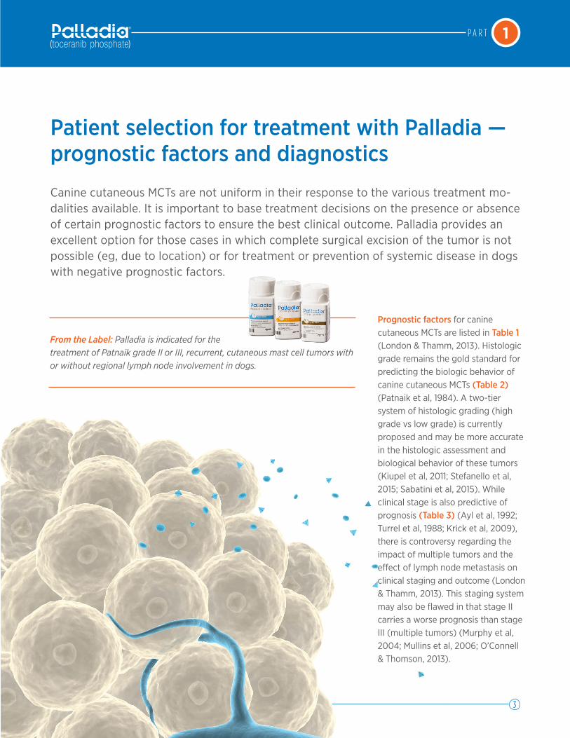
* From Patnaik AK, Ehler WJ, MacEwen EG: Canine cutaneous mast cell tumor: morphologic grading and survival time in 83 dogs. Vet Pathol. 1984;21:469-474.† A two-tier system of histologic grading is currently proposed and may be more accurate in the histologic assessment and biological behavior of these tumors
(Kiupel et al, 2011).
TABLE 2 | Histologic Classification of Cutaneous MCTs from Surgical Biopsy Samples*, †
TABLE 3 | World Health Organization Clinical Staging System for MCTs
One tumor completely excised from the dermis, identified histologically, without regional lymph node involvement
1. Without systemic signs 2. With systemic signs
One tumor, confined to the dermis, without regional lymph node involvement
1. Without systemic signs 2. With systemic signs
One tumor confined to the dermis, with regional lymph node involvement
1. Without systemic signs 2. With systemic signs
Multiple dermal tumors; large infiltrating tumors with or without regional lymph node involvement
1. Without systemic signs 2. With systemic signs
Any tumor with distant metastases, including blood or bone marrow involvement
Description
Low grade (well-differentiated)
High grade (anaplastic, undifferentiated)
Intermediate grade
Clearly defined cytoplasmic boundaries with regular, spheric, or ovoid nuclei; mitoses rare or absent; cytoplasmic granules large, deep staining, and abundant
Cells closely packed with indistinct cytoplasmic boundaries, nucleus-to-cytoplasmic ratio lower than anaplastic, infrequent mitoses, more granules than anaplastic
Highly cellular, undifferentiated cytoplasmic boundaries, irregular size and shape of nuclei, frequent mitoses, sparse cytoplasmic granules
Microscopic Description GradePatnaik Grading
I
II
III
Stage
0
I
II
III
IV

P A R T 1
PA L L A D I A R O U N DTA B L E | PA R T 1 6
Dr. Gloyd: In this discussion we’ll focus on those dogs that are determined to have MCTs that are not amenable to wide surgical excision. What are the lessons learned since Palladia has been available in terms of diagnostics, workup, and patient selection for treatment?
Dr. Thamm: Many of the changes over the past seven years have involved what we do before surgery — how much or how little we do. Eighty percent of dogs with MCT do not need medical therapy — they are cured with surgery with adequate margins. We’re talking about the very rarified population of dogs that either need further treatment after surgery or can’t have surgery. Those are the cases in which we are po-tentially going to be reaching for some type of drug, whether it’s a cytotoxic or a kinase inhibitor.
Abdominal Ultrasound and Aspiration Biopsy of the Spleen and Draining Node
Dr. Garrett: For staging, we always attempt to aspirate the locoregional lymph node for cyto-logic assessment. A large number of dogs do not have any negative prognostic factors in their history or physical appearance; these
dogs go directly to surgery and do not get staged with abdominal ultrasound. As far as abdominal evaluation for cases with negative prognostic factors, in the past, we would just do ultrasounds, and only if the scans were suspicious would we aspirate the spleen.
Two recent studies showed, however, that the sonographic appearance of the spleen does not correlate with whether there is mast cell infiltration (Book et al, 2011; Stefanello et al, 2009). The danger of splenic aspiration is that mast cells are in the spleen
Discussion
The initial diagnosis is based on fine-needle aspiration (FNA) of the mass and regional lymph nodes, surgery with wide (3 cm) margins), and biopsy with histopathologic testing.
If the tumor is in a location amenable to surgery and no negative prognostic indicators (Table 1) are present, no further tests are necessary prior to wide surgical excision other than:
•Minimumdatabase(completebloodcount,serum biochemistry profile)
•FNAcytologyofthemassand(ifpossible)theregional (draining) lymph node
•Histopathologicassessmentofthebiopsysampleofthemass to determine tumor grade and mitotic index (MI)
If the tumor location is not amenable to surgery, or if negative prog-nostic factors are noted on physical examination or in the history, or if surgery is attempted but clean margins are not attained, further tests are indicated to assess prognosis and guide treatment decisions.
Some tests that historically have been included in complete staging of MCTs are now considered by many veterinary oncologists to be unnecessary in dogs with MCTs that do not have negative prognostic factors (see Discussion).
Abdominal ultrasound with cytologic assessment of the spleen or liver if warranted
Bone marrow aspiration cytology Buffy coat smear Thoracic radiographs
Tissue samples also can be submitted to a veterinary diagnostic labo-ratory, such as the Molecular Pathology Laboratory at Colorado State University or the Diagnostic Center for Population and Animal Health at Michigan State University for a panel of prognostic tests.
Diagnosis of Cutaneous Canine MCTs
P A R T 1
PA L L A D I A R O U N DTA B L E | PA R T 1 7
normally, which can be over-inter-preted as metastasis. These recent studies developed cytologic cri-teria to classify whether mast cell involvement represented metas-tasis or merely a normal resident population. The patients that were classified as positive for malignant mast cell involvement did signifi-cantly worse on follow-up in both of those studies. So, as a result, we now routinely aspirate spleens if we are going to do an abdominal ultrasound when staging these dogs. I think if you are going to bother to ultrasound, you should bother to do cytology.
Dr. Gloyd: You don’t get an abdominal ultrasound on all dogs that present with MCTs?
Dr. Garrett: No. If the tumor is located somewhere where we can aspirate an external lymph node, we will just do that. Otherwise, unless there is something in the history or physical exam or lymph node cytology that carries neg-ative prognostic information, we perform wide surgical excision, wait on histologic and surgical margin assessment, and then eval-uate further if warranted.
Dr. Vail: I agree. If the tumors are in a location that can be cut easily and we don’t have negative prog-nostic indices, other than
aspirating a lymph node, we don’t do much until we have the histol-ogy in hand.
Dr. Jones: I perform abdominal ultrasound on a case by case basis. If a tumor is histologically high grade, has a poor prognostic factor, positive lymph node on cytology, or if I am going to per-form a large invasive surgery with radiation therapy, then I follow with abdominal ultrasound and cy-tology of liver and spleen because positive liver or spleen has been shown to carry a poor prognosis with short survival time.
Dr. Gloyd: What advice do you have for general practitioners when they aspirate a lump or get a biopsy for histopathology? How do you want that sample to be handled so that you get the maxi-mum amount of information?
Dr. London: First, at the most basic level, they need to perform a fine needle aspiration before to confirm that it is a MCT before they take it off. I think that’s the biggest barrier for us — getting practitioners to get a diagnosis before they take a lump off so that the surgery is done correctly the first time. Second, I am always try-ing to get them to at least ink the deep margin because the whole anatomy often falls apart once they place the mass in formalin. That way, when the pathologist cuts in the sample, it is done correctly.
Dr. Hohenhaus: We want general practitioners to send the entire mass — everything they cut off — to the lab. Unfortunately, veteri-nary reference labs provide them with a small jar and they often can’t fit the entire mass in it, which is a huge barrier for general prac-titioners. I would also want the mi-croscopic description, not just the histology or pathologic diagnosis. Furthermore, if you know it’s a MCT and it’s in an anatomically bad location, don’t attempt the surgery yourself. Refer the patient to a board-certified surgeon to ensure that adequate margins are attained.
It is not necessary to routinely perform abdominal ultrasonography on every dog with a MCT.
CONSENSUSP INT
P A R T 1
PA L L A D I A R O U N DTA B L E | PA R T 1 8
Dr. London: Yes, plan to resect it appropriately the first time so that you don’t end up having to recut it or have recurrent disease. I think that the challenge is that often these are after-the-fact di-agnoses and that makes it harder to manage.
Dr. Gloyd: What’s the next step?
Dr. London: We have become much more aggressive about find-ing the draining lymph node, and if necessary, we will use ultrasound to find it and aspirate it. If there is any potential metastatic disease there, we usually try to remove the lymph node at the time of surgery to confirm metastasis because it may change how we proceed.
Dr. Garrett: If the draining node is a sub-lumbar node, for example, then we will do abdominal ultra-sound, and while we’re doing it, we will aspirate the spleen be-cause we are there.
Dr. London: In human medicine the paradigm for any disease that has the potential to be metastatic is to always examine the draining node. I don’t think we have been appropriately aggressive enough in veterinary medicine in following that dogma. I think it has made a difference in human cancer therapy, so it’s something that we always encourage our referring veterinarians and our clients to consider pursuing.
Dr. Vail: There is a large dataset coming out of the UK showing that if a draining node is negative you will not find spread to distant sites (eg, spleen and other abdom-inal viscera) (Warland et al, 2014). If I have what I feel quite certain is the draining node, and it isn’t positive, I don’t spend the client’s money on an abdominal exam and aspiration of the spleen.
Dr. Clifford: If the draining node is positive then we will certainly pursue further staging prior to surgery, including abdominal ultra-sound, to evaluate for any involve-ment of the spleen or liver. If it is not, the patient will go to surgery
and the tumor and lymph node is still excised.
Dr. Vail: I am unconvinced of the need to aspirate the normal- anatomy spleen and liver in these cases, especially if the node is clean.
Dr. London: I agree; if the node is clean I usually would not aspirate the spleen. But sometimes you can’t find the node, and in those cases, if the patient has a negative prognostic indicator (eg, the mass has exhibited recent rapid growth, or the mass is really ulcerated, or the dog is sick), then I will perform a fine needle aspiration of the spleen.
Dr. Henry: We are moving more towards using PET scans, too. We are finding that what we thought was the draining node is not necessarily the draining node, and what we thought we should be aspirating probably wasn’t getting us anywhere.
Dr. Clifford: The problem we have in regards to staging is the financial limitations of our clients. For a potential “garden variety” mast cell case, an ultrasound costs ~$400, an aspirate is ~$90, cytol-ogy is $160, and that adds up.
A suspected MCT should always be aspirated to confirm the diagnosis before surgery.
CONSENSUSP INT
P A R T 1
PA L L A D I A R O U N DTA B L E | PA R T 1 9
So it would be a challenge for us to be able to stage every MCT that way. I offer it to owners but do not require.
Dr. Garrett: Even if they have negative prognostic factors?
Dr. Clifford: If it is a positive node, yes, then without question. But for the garden variety MCT, I don’t perform an ultrasound before surgical removal.
Tumor Location Not Amenable to Surgery
Dr. Henry: Tumors in some loca-tions are not necessarily going to be resectable — an eyelid for ex-ample — which is going to change the approach. Many tumors that are in unresectable locations also are not going to be great can-didates for radiation therapy. In these cases, the question is do I perform a minimal debulking (cytoreductive) surgery and follow up with chemotherapy? Or do I not touch it at all with surgery or with radiation therapy? The dog we had on Palladia the longest was one that had an eyelid MCT to start with and it was just way too huge to consider surgery.
Dr. Garrett: If you are going to treat systemically anyway then looking for metastasis becomes less relevant; and it is expensive.
Dr. Vail: Even in resectable cases, if it is going to be a difficult resec-tion, if it’s going to be aggressive, or if your margins are not clean, the likelihood increases that you may want to follow up with some other treatment. In those cases where the owner is going to spend a lot of money dealing with the primary tumor, we will offer staging because it’s kind of
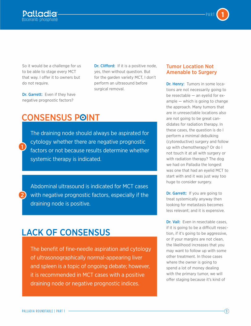
The draining node should always be aspirated for cytology whether there are negative prognostic factors or not because results determine whether systemic therapy is indicated.
CONSENSUS P INT
The benefit of fine-needle aspiration and cytology of ultrasonographically normal-appearing liver and spleen is a topic of ongoing debate; however, it is recommended in MCT cases with a positive draining node or negative prognostic indices.
LACK OF CONSENSUS
1
Abdominal ultrasound is indicated for MCT cases with negative prognostic factors, especially if the draining node is positive.
22

P A R T 1
PA L L A D I A R O U N DTA B L E | PA R T 1 10
like an insurance policy. But if we find something, we may not get as aggressive on the primary tumor.
Dr. Clifford: Yes, especially in a case that we are going to irradiate, we will always more fully stage the patient.
Dr. Vail: We now know that unre-sectable MCTs can also be treated with a combination of hypofrac-tionated (once weekly) radiation therapy in combination with Palladia (Carlsten et al, 2012).
Dr. Thamm: Back to the challeng-ing issue about doing minimal debulking versus not, I think we are all probably in agreement that if there’s an opportunity to at least get the case down to microscopic and achieve primary closure, that is always preferable to trying to treat a bulky MCT with chemother-apy or Palladia up front.
Dr. Garrett: It depends, however, on the aggressiveness of the surgery required — for example, a mandibulectomy, which is the case I saw recently.
Dr. Thamm: Yes, or if you have to do a hemipelvectomy to get dirty margins, then that would be a different case.
Grading and Mast Cell Tumor Panels
Dr. Clifford: The tumor grade is another important factor that is going to play a role in which treat-ment we select in an individual case. We are going to approach a tumor that is incompletely excised and has a 2.0 mitotic index (MI) differently than a grade III tumor with a MI of 18.
Dr. Klein: The problem is every-thing in between. No matter which grading system you use, 10% to 15% of those grade II MCTs are going to behave badly and the rest are going to respond. How do I identify that small minority of patients that need the drugs? That is always going to be a challenge until we get better biomarkers, whether PCR or mutations in c-kit. Whatever the marker, there is not 100% certainty that the patient will or will not respond. I think the
biggest challenge with grade II MCTs, which the vast majority of these cases are, is to try to pick out the small percentage that really needs the drugs before you treat them.
Dr. Gloyd: What information do you want from the referring veteri-narian (rDVM)?
Dr. Jones: The first thing I would want is the description of any pathology. The problem is that rDVMs typically do two things: First, they get only the mini histo-pathologic report that gives only the diagnosis, so they don’t get the mitotic index or a description of the pathology from the patholo-gist. Second, they will get the MCT panel that the labs recommend. The panel can run $600, and when
In dogs with bulky MCTs, it is always preferable to downstage the tumor with cytoreductive surgery prior to starting treatment with Palladia. If the tumor is unresectable, chemotherapy or radiation are options for cytoreduction before Palladia treatment.
CONSENSUS P INT

P A R T 1
PA L L A D I A R O U N DTA B L E | PA R T 1 11
I can’t interpret it because they didn’t get the full report, I can’t tell the rDVM that it was worth the money their client spent.
Dr. Hohenhaus: I think we’d all agree that we would tell rDVMs not to get the mini biopsy — get the full biopsy report. Get all the work that the pathologist wants to run.
Dr. Thamm: That brings up one of the questions that will probably be a subject of some debate. How
many people are routinely doing MCT prognostic panels on every dog with a resected MCT that walks in the door?
Dr. London: I only set up for the c-kit exon 8 and exon 11 mutation status. I think the c-kit mutation testing has helped when I am try-ing to decide which therapy may be most appropriate. This pref-erence is not based on anything we’ve published yet, but on the human experience.
Dr. Klein: I agree; if I struggle trying to decide, that’s when I’ll do the PCRs and see if the c-kit mutations are present. With that information, I know whether I have a decent chance of the tumor responding if I’m going to choose Palladia.
Dr. Clifford: We looked at a large subset of dogs with grade II MCTs and followed them with the com-plete MCT panel (unpublished). It was challenging to be able to draw any conclusions because an individual case might have a high PCNA but a low AgNOR, and how do you interpret that? As a result, for the most part, if the tumor is a grade III or has a high mitotic index, I now will send off for a PCR on it for mutation status in order to tailor the use of Palladia. On a very, very basic level, it represents personalized medicine.
Dr. Thamm: Colorado State University is one of the sites that offers the MCT panel and they get about 30 or 40 cases a week sent in from elsewhere. At the CSU Cancer Center, however, we rarely use the MCT panel.
Dr. Klein: Michigan State Universi-ty’s Diagnostic Lab website has a flow chart for making therapeutic decisions based on prognostic parameters.
Dr. Garrett: Veterinarians call me with MCT panel results and want me to interpret them. I tell them that I don’t actually run these pan-els. Just tell me the mitotic index (along with the grade).
Dr. Clifford: I will usually tell the rDVM when we get the panels they ordered that I don’t necessarily find them all that useful so they won’t make the mistake of order-ing these expensive panels for all cases in the future. The times I discuss the use of a panel include incomplete resection of a low or moderate grade tumor (Smith et al, 2015) in which low Ki67 index & AgNOR x Ki67 (Ag67) values were unlikely to recur; if the biologic behavior does not fit histopa-thology; the tumor is located at a “hot” anatomic sites (eg, muzzle,
Histologic grading should be performed on all surgical biopsy samples. Veterinar-ians should order the full biopsy report with mitotic index, grade, and microscopic description of the pathology.
CONSENSUSP INT
P A R T 1
PA L L A D I A R O U N DTA B L E | PA R T 1 12
mucocutaneous), or the owner has a low risk tolerance.
Dr. Garrett: I don’t order mutation analysis very often. The majority of MCT cases do not get mutation analysis because you are going to cure the large majority of cases with surgery alone. If you decide later that you want to find out about the mutation status, then you can send a sample from the biopsy.
Dr. Gloyd: Is the consensus that you don’t run the MCT prognostic panels?
Dr. Garrett: Yes! There have been no published studies showing how these panels may provide additional benefit over the MI and grade for prediction of tumor behavior.
Dr. Thamm: There are some exceptional circumstances. One example is a grade 2 tumor with a mitotic index that is borderline, say between 4 and 6, and I don’t know what that really means. Sometimes I think doing some of these more sensitive proliferation markers can help be a tie-breaker. But those are only about 1% of cases.
Dr. Hohenhaus: We used to do MCT panels in-house at The Animal Medical Center, but we have since changed labs and don’t do
it anymore. The cost increased quite a bit so we stopped doing them as often, and I don’t think my cases are doing any better or any worse for lack of that information. Sometimes these MCT cases do very poorly and sometimes they do much better than you thought and you’re still not sure why that happened.
Dr. Clifford: All this information probably led to us over-treating cases for a while. In addition, it’s very rare that all the results in the panel point the same way. You may have to pick from three or
Veterinary oncolo-gists do not rou-tinely use the full MCT prognostic panels and, in general, do not want veterinarians to ask for them.
CONSENSUSP INT
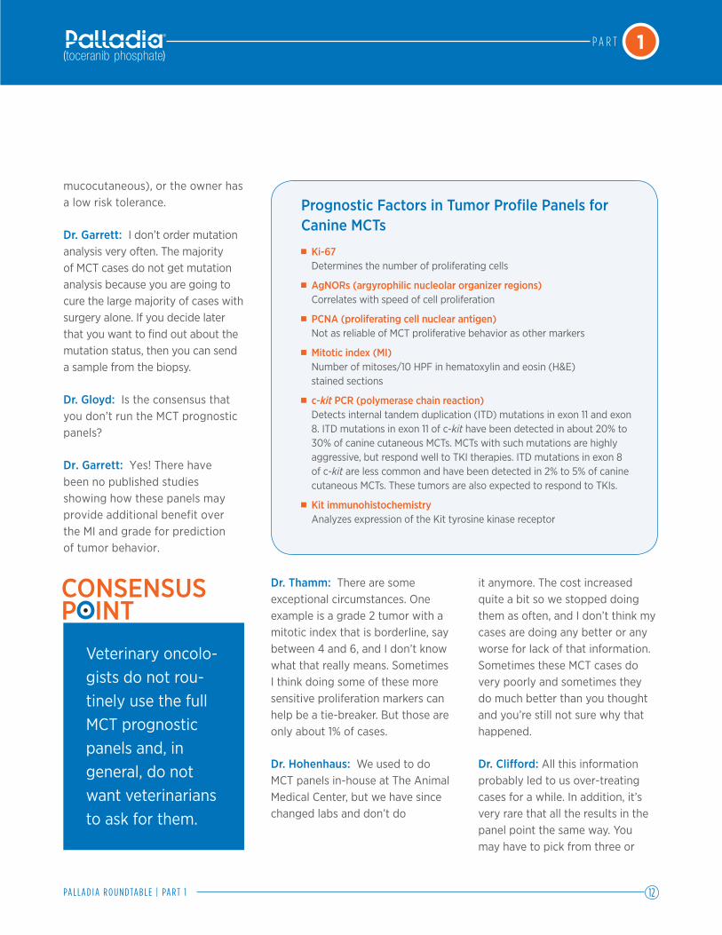
Prognostic Factors in Tumor Profile Panels for Canine MCTs Ki-67 Determines the number of proliferating cells
AgNORs (argyrophilic nucleolar organizer regions) Correlates with speed of cell proliferation
PCNA (proliferating cell nuclear antigen) Not as reliable of MCT proliferative behavior as other markers
Mitotic index (MI) Number of mitoses/10 HPF in hematoxylin and eosin (H&E) stained sections
c-kit PCR (polymerase chain reaction) Detects internal tandem duplication (ITD) mutations in exon 11 and exon 8. ITD mutations in exon 11 of c-kit have been detected in about 20% to30% of canine cutaneous MCTs. MCTs with such mutations are highly aggressive, but respond well to TKI therapies. ITD mutations in exon 8 of c-kit are less common and have been detected in 2% to 5% of canine cutaneous MCTs. These tumors are also expected to respond to TKIs.
Kit immunohistochemistry Analyzes expression of the Kit tyrosine kinase receptor
P A R T 1
PA L L A D I A R O U N DTA B L E | PA R T 1 13
four results. Which one is the most important?
Dr. Hohenhaus: If there are two or three indicators in agreement, I’ll treat. But then it’s totally empirical.
Dr. Henry: I think there are a lot of misconceptions about what Kit staining means — when are you looking at a mutation and when you are not. I don’t do Kit staining because I personally find it uninterpretable. It’s the same thing when you get a panel and you have a bad AgNOR plus Ki-67 number and it has Kit stain pattern one; I don’t know what any of this means, so I tend not to do them. But Dr. Thamm is in the middle of a clinical trial that will hopefully help clear up some of these questions. I think that if there’s something that comes out of that study that shows there is a subset of staining that seems to be correlated with response to Palladia, especially durable response, that would be important to know.
Dr. Thamm: We are conducting a randomized study comparing cyto-toxic chemotherapy with vinblas-tine or therapy with Palladia, and the randomization is based on the results of both Kit staining at exons 8 and 11 and c-kit mutation testing. Because of the study design we
had to pick a relatively short-term endpoint, response at 5 weeks in gross disease. That doesn’t answer the question about whether what we see in 5 weeks translates into a long-term survival advantage and it also doesn’t say anything about how this might influence the choice of adjuvant therapy.
Dr. London: Dr. Thamm brings up an important point that we haven’t addressed yet — in human as well as in veterinary medicine — and that is the use of these drugs in the adjuvant setting, which ideally is where you want to use them. We haven’t done those studies but there’s such a big difference in what you are looking at with respect to endpoint in the gross disease study versus the micro-scopic disease study — that is, to using Palladia in the adjuvant setting after a gross macroscopic tumor has been down-staged to microscopic disease.
Dr. Vail: I think we all agree that in gross disease, you will see response to Palladia but dura-bility is generally low. There are exceptions to every rule, but that just tells us that we are using the drug as a Band-Aid method right now without data on whether we should be moving beyond that.
Dr. Thamm: Clinical response equated with living longer, and that’s an indicator that the therapy
is having an effect on longevity and quality of life. In at least some of the investigational studies we have done with Palladia, response to therapy definitely correlated with survival. So, if the drug works you live longer, which implies that — depending on your definition — it is more than a Band-Aid. Does it mean that we are curing them? No.
Dr. Thamm: The question if you look at the statistics is: does over-all survival increase if the patient is a responder versus if they are not, and I think the answer is yes.
Dr. London: There was no change, however, in overall survival with c-kit mutation status in the pivotal study (London et al, 2009). Dogs with tumor mutations in the c-kit gene were more than twice
In gross disease, a response to Palladia will be seen, but the durability of response is generally low, about 6 months.
CONSENSUSP INT
P A R T 1
PA L L A D I A R O U N DTA B L E | PA R T 1 14
as likely to respond to Palladia as those without the mutation (60.0% vs 31.3%). In the phase I study response was close to 90%. At that time we weren’t testing for the exon 8 mutation, so it’s entirely possible that some of the responders actually had exon 8 mutations. If you look at the data on single-agent drugs, Palladia is the most effective single agent other than prednisone.
Dr. Jones: There are studies that have concluded the presence of c-kit mutations is associated with high histologic grade and are associated with a shorter pro-gression-free survival and overall survival (Zemke D et al, 2002; Takeuchi et al, 2013). At this time, c-kit hasn’t been firmly estab-lished as an independent prognos-tic factor although I use it to help guide treatment decisions.
Dr. Klein: I will use mutation status sometimes if a client is really struggling with making a decision. If we decide that Palladia is indicated but the tumor muta-tion status is negative, then I will tell them that their dog has a third rather than a two thirds chance of responding. That can make a dif-ference as to whether they decide to go with Palladia because it is a big financial commitment.
Dr. Thamm: In the radiation study (Carlsten et al, 2012) a large majority of the tumors were tested for mutations. Of 14 dogs tested, 8 dogs had no mutation identified, 1 had an exon 8 mutation, and 5 had exon 11 mutations. The presence of the c-kit mutation was a negative prognostic factor for long-term outcome. At 1 year, 66.7% of dogs with c-kit mutant MCT and 100% of dogs with c-kit wild-type MCT were alive. In the pulse-Palladia plus lomustine study (Burton et al,
2015), c-kit mutation status had no effect on outcome.
Dr. Thamm: However, the effect of mutation status on outcome is context-dependent and may be different when Palladia is used in combination with other treatments.
Dr. London: One of the huge challenges in our profession is that we are taking a spectrum of disease and trying to lump it into one thing, and it’s not. It is clear that in humans, breast cancer is not just breast cancer; there are several different subtypes. So, we’re talking about mast cell dis-ease in the same manner, and this underrepresents that complexity of the cancer. It is very hard to apply a single paradigm with re-spect to prognosis onto a disease that exhibits a range of biologic behaviors.
c-kit mutation status, if known, can be a factor in making treatment decisions. Dogs with tumor mutations in the c-kit gene were more than twice as likely to respond to Palladia as those without the mutation (60.0% vs. 31.3%).
CONSENSUSP INT
P A R T 1
PA L L A D I A R O U N DTA B L E | PA R T 1 15
Ayl RD, Couto GC, Hammer AS, et al. Correlation of DNA ploidy to tumor histologic grade, clinical variables, and survival in dogs with mast cell tumors. Vet Pathol. 1992;29:386-390.
Book AP, Fidel J, Willis T, et al. Correlation of ultrasound findings, liver and spleen cytology, and prognosis in the clinical staging of high metastatic risk canine mast cell tumors. Vet Radiol Ultrasound. 2011;52(5):548-554.
Burton JH, Venable RO, Vail DM, et al. Pulse-administered toceranib phosphate plus lomustine for treatment of unresectable mast cell tumors in dogs. J Vet Intern Med. 2015 Jul-Aug;29(4):1098-104.
Carlsten KS, London CA, Haney S, et al. Multicenter prospective trial of hypofraction-ated radiation treatment, toceranib, and prednisone for measurable canine mast cell tumors. J Vet Intern Med. 2012;26:135–141.
Downing S, Chien MB, Kass PH, et al. Prevalence and importance of internal tandem duplications in exons 11 and 12 of c-kit in mast cell tumors of dogs. Am J Vet Res. 2002;63(12):1718-1723.
Finora K, Leibman NF, Fettman MJ, et al. Cytological comparison of fine-needle aspi-rates of liver and spleen of normal dogs and of dogs with cutaneous mast cell tumors and an ultrasonographically normal appearing liver and spleen. Vet Comp Oncol. 2006;4(3):178–183.
Kiupel, M, Webster JD, et al. Proposal of a 2-tier grading system for canine cutaneous mast cell tumors to more accurately predict biologic behavior. Vet Pathol. 2011;48:147.
Krick EL, Billings AP, Shofer PS, et al. Cytological lymph node evaluation in dogs with mast cell tumors: association with grade and survival. Vet Comp Oncol. 2009;7:130-138.
London C, Thamm DH. Mast cell tumors. In Withrow SJ, Vail DM, Page RL (eds). Withrow and MacEwen’s Small Animal Clinical Oncology. 5th ed. St Louis: Elsevier, 2013, Chapter 20, pages 335-355.
London CA, Galli SJ, Yuuki T, et al. Spontaneous canine mast cell tumors express tandem duplications in the proto-oncogene c-kit. Exp Hematol. 1999;27(4)689-697.
London CA, Malpas PB, Wood-Follis SL, et al. Multi-center placebo-controlled, dou-ble-blind, randomized study of oral toceranib phosphate (SU11654), a receptor tyrosine kinase inhibitor, for the treatment of dogs with recurrent (either local or distant) mast cell tumor following surgical excision. Clin Cancer Res. 2009;15(11):3856-3865.
Mitchell L, Thamm DH, Biller BJ. Clinical and immunomodulatory effects of toceranib combined with low-dose cyclophosphamide in dogs with cancer. J Vet Intern Med. 2012 Mar-Apr;26(2):355-362.
Mullins MN, Dernell WS, Withrow SJ, et al. Evaluation of prognostic factors associated with outcome in dogs with multiple cutaneous mast cell tumors treated with surgery with and without adjuvant treatment: 54 cases (1998-2004). J Am Vet Med Assoc. 2006;228(1):91-95.
References
P A R T 1
PA L L A D I A R O U N DTA B L E | PA R T 1 16
Murphy S, Sparkes AH, Smith KC, et al. Relationships between the histological grade of cutaneous mast cell tumours in dogs, their survival and the efficacy of surgical resection. Vet Rec. 2004;154:743–746.
O’Connell K, Thomson M. Evaluation of prognostic indicators in dogs with multiple, simultaneously occurring cutaneous mast cell tumours: 63 cases. Vet Comp Oncol. 2013; 11(1):51-62.
Pan X, Tsimbas K, Kurzman ID, Vail DM. Safety evaluation of combination CCNU and continuous toceranib phosphate (Palladia(®)in tumour-bearing dogs: a phase I dose-finding study. Vet Comp Oncol. 2016 Jun;14(2):202-209.
Patnaik AL. Ehler WJ, MacEwen EG. Canine cutaneous mast cell tumor: Morphologic grading and survival time in 83 dogs. Vet Pathol. 1984;21:469-464
Pellin MA, Wouda RM, Robinson K, et al. Safety evaluation of combination doxorubicin and toceranib phosphate (Palladia®) in tumour bearing dogs: a phase I dose-finding study. Vet Comp Oncol. 2016 May 5. doi: 10.1111/vco.12232. [Epub ahead of print]
Sabattini et al. Histologic grading of canine mast cell tumor: Is 2 Better Than 3? Vet Pathol. 2015.
Smith J, Kiupel M, et al. Recurrence rates and clinical outcome for dogs with grade II mast cell tumours with a low AgNOR count and Ki67 index treated with surgery alone. Vet Comp. Oncol. 2015;15(1): 36-45.
Stefanello D, Buracco P, et al. Comparison of 2- and 3-category histologic grading systems for predicting the presence of metastasis at the time of initial evaluation in dogs with cutaneous mast cell tumors: 386 cases (2009–2014). J Am Vet Med Assoc. 2015;246:765-769.
Stefanello D, Valenti P, Faverzani S, et al. Ultrasound-guided cytology of spleen and liver: a prognostic tool in canine cutaneous mast cell tumor. J Vet Intern Med. 2009;23(5):1051–1057.
Takeuchi et al. Validation of the prognostic value of histopathological grading or c-kit mutation in canine cutaneous mast cell tumours: A retrospective cohort study. Vet J. 2013.
Turrel JM, Kitchell BE, Miller LM, et al. Prognostic factors for radiation treatment of mast cell tumors in 85 dogs. J Am Vet Med Assoc. 1988;193:936-940.
Warland J, Amores-Fuster I, Newbury W, et al. The utility of staging in canine mast cell tumors. Vet Comp Oncol. 2014;12:287-298.
Zemke D, Yamini B, Yuzbasiyan-Gurkan V. Mutations in the juxtamembrane domain of c-kit are associated with higher grade mast cell tumors in dogs. Vet Pathol. 2002;39(5):529-535.

(toceranib phosphate) Tablets®
AntineoplasticFor oral use in dogs only
Caution: Federal (USA) law restricts this drug to use by or on the order of a licensed veterinarian.
Description: PALLADIA, a multi-kinase inhibitor targeting several receptor tyrosine kinases (RTK), is the phosphate salt of toceranib. The empirical formula is C22H25FN4O2H3O4P and the molecular weight is 494.46. The chemical name is (Z)-5-[(5-Fluoro-2-oxo-1,2-dihydro-3H-indol-3-ylidene)methyl]-2,4-dimethyl-N-(2-pyrrolidin-1-ylethyl)-1Hpyrrole-3-carbox-amide phosphate. Toceranib phosphate is a small molecule with an indolinone chemical structure.The chemical structure of toceranib phosphate is
Indications: PALLADIA tablets are indicated for the treatment of Patnaik grade II or III, recurrent, cutaneous mast cell tumors with or without regional lymph node involvement in dogs.
Dosage and Administration: Always provide Client Information Sheet with prescription. Administer an initial dosage of 3.25 mg/kg (1.48 mg/lb) body weight, orally every other day (see Table 1). Dose reductions of 0.5 mg/kg (to a minimum dose of 2.2 mg/kg (1.0 mg/lb) every other day) and dose interruptions (cessation of PALLADIA for up to two weeks) may be utilized, if needed, to manage adverse reactions (see Table 2 as well as Warnings and Precautions). Adjust dose based on approximately weekly veterinary assessments for the first 6 weeks and approximately every 6 weeks, thereafter. PALLADIA may be administered with or without food. Do not split tablets.
Table 1. 3.25 mg/kg Dose Chart
Dog Body Weight Number of Tablets Pounds Kilograms Dose 10 mg 15 mg 50 mg
11.0 – 11.8 5.0 - 5.3 15 mg 111.9 – 15.2 5.4 - 6.9 20 mg 215.3 – 18.5 7.0 - 8.4 25 mg 1 118.6 – 22.0 8.5 - 10.0 30 mg 222.1 – 25.4 10.1 - 11.5 35 mg 2 125.5 – 28.7 11.6 - 13.0 40 mg 1 228.8 – 32.2 13.1 - 14.6 45 mg 332.3 – 35.5 14.7 - 16.1 50 mg 135.6 – 38.8 16.2 - 17.6 55 mg 1 338.9 – 42.3 17.7 - 19.2 60 mg 1 142.4 – 45.6 19.3 - 20.7 65 mg 1 145.7 – 50.7 20.8 - 23.0 70 mg 2 150.8 – 59.3 23.1 - 26.9 80 mg 2 159.4 – 65.9 27.0 - 29.9 95 mg 3 166.0 – 71.2 30.0 - 32.3 100 mg 271.3 – 76.3 32.4 - 34.6 110 mg 1 276.4 – 79.6 34.7 - 36.1 115 mg 1 279.7 – 84.7 36.2 - 38.4 120 mg 2 284.8 – 94.8 38.5 - 43.0 130 mg 2 294.9 – 105.0 43.1 - 47.6 150 mg 3
105.1 – 110.0 47.7 - 49.9 160 mg 1 3110.1 – 113.5 50.0 - 51.5 165 mg 1 3113.6 – 118.6 51.6 - 53.8 170 mg 2 3118.7 – 128.8 53.9 - 58.4 180 mg 2 3128.9 – 138.9 58.5 - 63.0 200 mg 4139.0 – 144.0 63.1 - 65.3 210 mg 1 4144.1 – 157.6 65.4 - 71.5 215 mg 1 4157.7 – 173.1 71.6 - 78.5 250 mg 5173.2 – 177.9 78.6 - 80.7 260 mg 1 5178.0 – 191.6 80.8 - 86.9 265 mg 1 5191.7 – 220.5 87.0 - 100.0 300 mg 6
.H3PO4
H3C
CH3
NH
N
O
NH
NH
O
F
Table 2. Dose Modification Based on Toxicity Observed
Toxicity Dose AdjustmentNeutropenia>1000/μL Maintain dose level≤1000/μL or neutropenic fever or infection
Stop drug until >1000/μL and clinical signs normal; then decrease dose by 0.5 mg/kg
Renal Toxicities (Creatinine)<2.0 mg/dL Maintain dose level≥2.0 mg/dL Stop drug until <2.0 mg/dL then decrease
dose by 0.5 mg/kgAlbumin<1.5 g/dL Stop drug until >2.5 g/dL then
decrease dose by 0.5 mg/kgHematocrit<26% Stop drug until >30% then decrease
dose by 0.5 mg/kgDiarrhea<4 watery stools/day for less than 2 days Maintain dose level and institute
supportive care≥4 watery stools/day or ≥ 2 days Stop drug until formed stools and
institute supportive care. When dosingis resumed, decrease dose by 0.5 mg/kg
GI BleedingFresh blood in stool or black tarry stool for > 2 days or frank hemorrhage or blood clots in stool.
Stop drug and institute supportive care until resolution of all clinical signs of blood in stool, then decrease dose by 0.5 mg/kg.
Contraindications:Do not use in dogs used for breeding, or for pregnant or lactating bitches (see Clinical Pharmacology).
Warnings:PALLADIA may cause vascular dysfunction which can lead to edema and thromboembolism, including pulmonary thromboembolism. Discontinue drug until clinical signs and clinical pathology have normalized. To assure vasculature homeostasis, wait at least 3 days after stopping drug before performing surgery (see Adverse Reactions).Serious and sometimes fatal gastrointestinal complications including gastrointestinal perforation have occurred rarely in dogs treated with PALLADIA (see Adverse Reactions). If gastrointestinal ulceration is suspected, stop drug administration and treat appropriately.
Human Warnings:NOT FOR USE IN HUMANS. KEEP THIS AND ALL MEDICATIONS OUT OF THE REACH OF CHILDREN. Children should not come in contact with PALLADIA. Keep children away from feces, urine, or vomit of treated dogs.To avoid exposure to drug, wash hands with soap and water after administering PALLADIA and wear protective gloves to prevent direct contact with feces, urine, vomit, and broken or moistened PALLADIA tablets. Place all waste materials in a plastic bag and seal before general disposal. If eyes are accidentally exposed to the drug, rinse eyes with water imme-diately. In case of accidental ingestion by a person, seek medical advice immediately, show the package insert or label to the physician. Gastrointestinal discomfort such as vomiting or diarrhea may occur if this drug is accidentally ingested.Pregnant women, women who may become pregnant, or nursing mothers should pay special attention to these handling precautions. (See handling instructions above.) PALLADIA, like other drugs in its class, prevents the formation of new blood vessels in tumors. In a similar manner, PALLADIA may affect blood vessel formation in the developing fetus and may harm an unborn baby (cause birth defects). For pregnant women, accidental ingestion of PALLADIA may have adverse effects on pregnancy.
Precautions:Temporarily discontinue the use of PALLADIA if anemia, azotemia, hypoalbuminemia, and hyperphosphatemia occur simultaneously. Resume treatment at a dose reduction of 0.5 mg/kg after 1 to 2 weeks when values have improved and albumin is >2.5 g/dL. Temporary treatment interruptions may be needed if any one of these occurs alone: hematocrit <26%, creatinine ≥2.0 mg/dL or albumin <1.5 g/dL. Then resume treatment at a dose reduction of 0.5 mg/kg once the hematocrit is >30%, the creatinine is <2.0 mg/dL, and the albumin is >2.5 g/dL.
Temporarily discontinue the use of PALLADIA if neutrophil count is ≤1000/μL. Resume treatment after 1 to 2 weeks at a dose reduction of 0.5 mg/kg, when neutrophil count has returned to >1000/μL. Further dose reductions may be needed if severe neutropenia reoccurs.

The presence of systemic mast cell tumor prior to treatment may predispose a dog to clinically significant mast cell degranulation with possible severe systemic adverse reactions when treated with PALLADIA. Attempts should be made to rule out systemic mastocytosis prior to initiation of treatment with PALLADIA.
PALLADIA has been associated with severe diarrhea or GI bleeding that requires prompt treatment. Dose interruptions and dose reductions may be needed depending upon the severity of clinical signs. (See Table 2 in Dosage and Administration.)
Use non-steroidal anti-inflammatory drugs with caution in conjunction with PALLADIA due to an increased risk of gastrointestinal ulceration or perforation.
PALLADIA is metabolized in the liver. Co-administration of PALLADIA with strong inhibitors of the CYP3A4 family may increase PALLADIA concentrations. The effect of concomitant medications that may inhibit the metabolism of PALLADIA has not been evaluated. Drug compatibility should be monitored in patients requiring concomitant medications.
The safe use of PALLADIA has not been evaluated in dogs less than 24 months of age or weighing less than 5 kg.
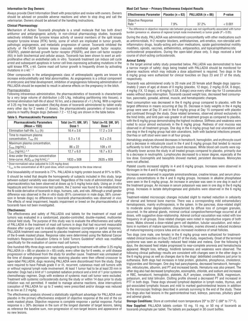
Adverse Reactions:A US clinical field study comprised of a 6-week masked phase, followed by an open-label phase, evaluated the safety and effectiveness of PALLADIA in 151 client-owned dogs that had Patnaik grade II or III, recurrent, cutaneous mast cell tumors with or without regional lymph node involvement. The most common adverse reactions reported during the masked phase are summarized in Table 3; those reported during the entire study (maskedphase combined with the open-label phase) are summarized in Table 4.
Table 3. Summary of the most common adverse reactions during the masked phasea
Placebo (n = 64) PALLADIA (n = 87)Adverse Reaction Any Gradeb Grade 3 or 4b Any Gradeb Grade 3 or 4b
Diarrhea 26.6% 3.1% 46.0% 6.9%Anorexia 31.3% 6.3% 39.1% 6.9%Lethargy 29.7% 3.1% 35.6% 4.6%Vomiting 32.8% 6.3% 32.2% 9.2%Lameness 9.4% 0.0% 17.2% 0.0%Weight loss 3.1% 0.0% 14.9% 1.1%Blood in stool/GI bleed/ hemorrhagic diarrhea 3.1% 0.0% 12.6% 2.3%Musculoskeletal disorder 6.3% 0.0% 11.5% 1.1%Dehydration 4.7% 0.0% 9.2% 2.3%Dermatitis 9.4% 1.6% 9.2% 0.0%Pruritus 4.7% 0.0% 9.2% 0.0%Tachypnea 4.7% 0.0% 8.0% 1.1%Localized pain 4.7% 0.0% 8.0% 0.0%Nausea 3.1% 0.0% 8.0% 1.1%General pain 4.7% 1.6% 6.9% 0.0%Polydipsia 7.8% 0.0% 6.9% 0.0%Pyrexia 3.1% 0.0% 5.7% 2.3%Flatulence 3.1% 0.0% 5.7% 0.0%Pigmentation disorder 1.6% 0.0% 5.7% 0.0%Laboratory Abnormality Any Gradec Grade 3 or 4c Any Gradec Grade 3 or 4c
Neutropenia 6.3% 0.0% 46.0% 0.0%Thrombocytopenia 20.3% 0.0% 24.1% 0.0%Increased alanineaminotransferase 21.9% 4.7% 24.1% 1.1%Hypoalbuminemia 7.8% 0.0% 12.6% 0.0%Decreased hematocrit 7.8% 0.0% 5.7% 3.4%Hyperbilirubinemia 1.6% 1.6% 5.7% 0.0%Increased creatinine 4.7% 0.0% 5.7% 0.0%Urinary tract infection 1.6% 0.0% 5.7% 0.0%
a The mean time on study during the masked phase was 37.0 days for PALLADIA-treated dogs (median, 42.0 days) and 27.6 days for placebo-treated dogs (median, 21.0 days); no adjustments were made in the statistical comparisons for this disparity.b Investigators assigned severity grade of 1, 2, 3 or 4 (1 - least severe; 4 - most severe).c Grading of laboratory abnormalities was based on the National Cancer Institute’s Common Toxicity Criteria guideline adapted for canines (1 - least severe; 4 - most severe).
Table 4. Summary of the most common adverse reactions during the study (masked phase combined with the open-label phase)a
PALLADIA (n = 145) a
Adverse Reactions Any Gradeb Grade 3 or 4b
Diarrhea 58.6% 8.3%Anorexia 49.7% 8.3%Vomiting 47.6% 9.7%Lethargy 39.3% 4.1%Lameness 22.8% 0.0%Weight loss 21.4% 2.8%Blood in stool/GI bleed/hemorrhagic diarrhea 18.6% 2.8%Dehydration 15.2% 2.1%Pruritus 12.4% 0.0%Pigmentation disorder 11.7% 0.0%Dermatitis 11.0% 0.0%Musculoskeletal disorder 11.0% 0.0%General pain 8.3% 0.0%Otitis externa 8.3% 0.0%Tachypnea 8.3% 0.0%Nausea 7.6% 1.4%Polydipsia 7.6% 0.0%Pyrexia 6.9% 2.8%Arthritis 6.2% 0.0%Localized edema 6.2% 0.0%Bacterial skin infection 5.5% 0.0%Conjunctivitis 5.5% 0.0%Laboratory Abnormality Any Gradec Grade 3 or 4c
Neutropenia 44.8% 1.4%Hypoalbuminemia 28.3% 1.4%Thrombocytopenia 28.3% 2.1%Increased alanine aminotransferase 27.6% 4.1%Decreased hematocrit 11.0% 2.8%Increased creatinine 13.8% 1.4%Hyperbilirubinemia 6.9% 0.0%Urinary tract infection 7.6% 0.0%
a The duration of treatment with PALLADIA ranged from 2 to 812 days (mean, 144 days; median, 68 days). All dogs received at least 1 dose of PALLADIA.b Investigators assigned severity grade of 1, 2, 3 or 4 (1 – least severe; 4 – most severe).c Grading of laboratory abnormalities was based on the National Cancer Institute’s Common Toxicity Criteria guideline adapted for canines (1 – least severe; 4 – most severe).Other adverse events were reported but occurred in < 5% of dogs.Any individual dog may have had multiple adverse events.
There were 5 deaths during this study that were possibly drug related. Pathology findings generally revealed evidence of vascular dysfunction including pulmonary thromboembolism (post-operative); multi-organ failure associated with vasculitis and thrombosis; vascular thrombosis with disseminated intravascular coagulopathy (DIC) and pancreatitis; and vasculitis with DIC. One dog died secondary to gastric perforation; the duration of treatment with PALLADIA was 221 days and there was no evidence of mast cell tumor at necropsy. These deaths occurred in the presence or absence of gross-disease; treatment durations ranged from 18 to 221 days.The relationship of the following deaths to drug are unknown. One dog, first treated for 3 weeks with a placebo, died of unknown cause 7 days after initiation of PALLADIA therapy. Another dog died of unknown cause 92 days after initiation of PALLADIA therapy. No necropsy was conducted in either dog.Twenty seven dogs developed some form of gastrointestinal bleeding with 2.8% of dogs having severe bleeding. One dog developed gastric ulceration which was possibly drug related. Three dogs died from gastric (1 dog) or duodenal (2 dogs) perforations during the study. One dog with a duodenal perforation received only 1 dose of study drug and, therefore, was not considered drug related.Seven dogs developed nasal depigmentation within the first few weeks of treatment. Eleven dogs developed coat color or skin changes during the study. Two of these dogs had complete coat color changes from fawn to white and from deep red to blonde. Seven dogs experienced alopecia.There is a drug related effect on body weight: 20.0% of dogs had >13% weight loss in the masked plus open-label phase attributable to drug. Of these, 5 dogs had >25% weight loss. Three dogs had seizure-like activity while on study drug. It can not be determined if these were drug related.Two dogs developed epistaxis that was not associated with thrombocytopenia. Another dog developed epistaxis with concurrent disseminated intravascular coagulopathy.For a copy of the Safety Data Sheet (SDS) or to report adverse events call Zoetis at 1-888-963-8471.
Information for Dog Owners:Always provide Client Information Sheet with prescription and review with owners. Owners should be advised on possible adverse reactions and when to stop drug and call the veterinarian. Owners should be advised of the handling instructions.
Clinical Pharmacology:Mechanism of Action: Toceranib phosphate is a small molecule that has both direct antitumor and antiangiogenic activity. In non-clinical pharmacology studies, toceranib selectively inhibited the tyrosine kinase activity of several members of the split kinase receptor tyrosine kinase (RTK) family, some of which are implicated in tumor growth, pathologic angiogenesis, and metastatic progression of cancer. Toceranib inhibited the activity of Flk-1/KDR tyrosine kinase (vascular endothelial growth factor receptor, VEGFR2), platelet-derived growth factor receptor (PDGFR), and stem cell factor receptor (Kit) in both biochemical and cellular assays. Toceranib has been shown to exert an anti-proliferative effect on endothelial cells in vitro. Toceranib treatment can induce cell cycle arrest and subsequent apoptosis in tumor cell lines expressing activating mutations in the split kinase RTK, c-kit. Canine mast cell tumor growth is frequently driven by activating mutations in c-kit.1, 2
Other compounds in the antiangiogenesis class of antineoplastic agents are known to increase embryolethality and fetal abnormalities. As angiogenesis is a critical component of embryonic and fetal development, inhibition of angiogenesis following administration of PALLADIA should be expected to result in adverse effects on the pregnancy in the bitch.PharmacokineticsFollowing intravenous administration, the pharmacokinetics of toceranib is characterized by a very large volume of distribution (>20 L/kg, indicating partitioning into tissues), a terminal elimination half-life of about 16 hrs, and a clearance of >1 L/hr/kg. With a regimen of 3.25 mg free base equivalent (fbe)/kg doses of toceranib administered by tablet orally every other day for 2 weeks (7 doses), the pharmacokinetic parameters of toceranib in plasma in healthy Beagle dogs (between 7.2 – 12.5 kg) are shown in the table below.
Table 5. Pharmacokinetic Parameters
Pharmacokinetic Parameters (Mean + 1SD)
Total (n=11; 6M, 5F) Dose 1
Total (n=10; 5M, 5F) Dose 7
Elimination half-life, t1/2 (h) 16.4 ± 3.6 17.2 ± 3.9Time to maximum plasmaconcentration, Tmax (h) 5.3 ± 1.6 6.2 ± 2.6Maximum plasma concentration, Cmax (ng/mL) 86 ± 22 109 ± 41Cmin (ng/mL) a, b 12.7 ± 6.0 18.7 ± 8.3Area under the plasma concentration time-curve, AUC0-48 (ng·h/mL) a 1833 ± 508 2635 ± 939
a Dose-normalized value (adjusted to 3.25 mg/kg dose)b Cmin is the concentration at 48 h post-dose, which corresponds to the dose interval.
Oral bioavailability of toceranib is 77%. PALLADIA is highly protein bound at 91% to 93%.It should be noted that despite the homogeneity of subjects included in this study, large between-subject variability was observed. Regardless of the route of administration, linearpharmacokinetics has been observed at doses up to 5 mg/kg twice daily. Using an in vitro hepatocyte and liver microsome test system, the Z isomer was found to be metabolized to the N-oxide derivative of toceranib in dogs, humans, cats, and rats. Although a small gender difference was observed in the in vitro study (81% conversion in male dogs, 56% conver-sion in female dogs) no differences in toceranib pharmacokinetics was observed in vivo. The effects of renal impairment, hepatic impairment or breed on the pharmacokinetics of toceranib have not been investigated.
Effectiveness:The effectiveness and safety of PALLADIA oral tablets for the treatment of mast cell tumors was evaluated in a randomized, placebo-controlled, double-masked, multicenter clinical field study. The purpose of this study was to evaluate the effectiveness and safety of PALLADIA in the treatment of mast cell tumors in dogs that had recurrent measurable disease after surgery and to evaluate objective response (complete or partial response). PALLADIA treatment was compared to placebo treatment using response rates at the end of the 6-week masked phase. Response rates were determined using the National Cancer Institute’s Response Evaluation Criteria in Solid Tumors Guideline3 which was modified specifically for the evaluation of canine mast cell tumors.One-hundred-fifty-three dogs were randomly assigned to treatment with either 3.25 mg/kg PALLADIA (n = 88) or placebo (n = 65) orally, every other day for 6 weeks, or until disease progression or withdrawal from the study for another cause. Treatment was unmasked at the time of disease progression: dogs receiving placebo were then offered crossover to open-label PALLADIA; dogs receiving PALLADIA were discontinued from the study. Dogs were required to have Patnaik grade II or III, recurrent, cutaneous mast cell tumors with or without regional lymph node involvement. At least 1 tumor had to be at least 20 mm in diameter. Dogs had a limit of 1 completed radiation protocol and a limit of 1 prior systemic chemotherapy regimen. Dogs with evidence of systemic mast cell tumor were excluded. Treatment with systemic corticosteroids during the study or within 14 days prior to study initiation was not permitted. If needed to manage adverse reactions, dose interruptions (cessation of PALLADIA for up to 2 weeks) were prescribed and/or dosage was reduced to as low as 2.2 mg/kg.The effectiveness analysis showed a statistically significant advantage for PALLADIA over placebo in the primary effectiveness endpoint of objective response at the end of the six week masked phase. Objective response is complete response + partial response. Partial response is ≥ 30% decrease in the sum of the longest diameter of target lesions, taking as reference the baseline sum, non-progression of non-target lesions and appearance of no new lesions.
Mast Cell Tumor – Primary Effectiveness Endpoint Results
Effectiveness Parameter Placebo (n = 63) PALLADIA (n = 86) P-value
Objective ResponseRate * 7.9% 37.2% < 0.001
* The difference in objective response rate between groups was not significantly associated with tumor burden (presence vs. absence of regional lymph node involvement) or tumor grade (P > 0.05).
During the study, PALLADIA was administered concomitantly with other medications such as antimicrobials, H-2 receptor blockers, antihistamines, anti-emetics, non-steroidal anti-inflammatory drugs, locally-acting anti-ulcer medications, opiate gastrointestinal motility modifiers, opioids, vaccines, anthelmintics, antiparasitics, and topical/ophthalmic/oticcorticosteroid preparations. During the open-label phase only, 5 dogs received a brief course of short-acting corticosteroids.
Animal Safety:In the target animal safety study presented below, PALLADIA was demonstrated to have a narrow margin of safety; dogs being treated with PALLADIA should be monitored for adverse reactions which may indicate a dose adjustment is required. Two dogs in the 6 mg/kg group were euthanized for clinical toxicities on Days 23 and 27 of the study, respectively.Toceranib was administered orally to 20 male and 20 female adult Beagle dogs (approx-imately 2 years of age) at doses of 0 mg/kg (placebo, 12 dogs), 2 mg/kg (0.5X, 8 dogs), 4 mg/kg (1X, 12 dogs), or 6 mg/kg (1.5X, 8 dogs) once every other day for 13 consecutive weeks without dose interruption. Toceranib caused weight loss, decreased feed consump-tion, pancreatic, gonadal, adrenal, muscle, and hematopoietic changes.Feed consumption was decreased in the 6 mg/kg group compared to placebo, with the largest difference in means occurring at Day 35. Decrease in body weights in the 4 mg/kg group were seen at Day 31 and in the 6 mg/kg group at Day 15 compared with placebo and continued through the study. Dose related lameness, observed almost exclusively in the hind limbs, and limb pain was greater in all treatment groups as compared to placebo, with the 6 mg/kg group demonstrating the highest incidence. Stiffness and weakness were noted to occur almost exclusively in the 6 mg/kg group. Redness of oral mucosa was observed in all treatment groups. One dog in the 4 mg/kg group had oral ulcerations and one dog in the 6 mg/kg group had skin ulcerations, both with bacterial infections present. Diarrhea or soft stool were seen in all four groups.Hematology analyses showed decreases in hematocrit, hemoglobin, and erythrocyte count and a decrease in reticulocyte count in the 4 and 6 mg/kg groups that tended to recover sufficiently to limit further erythrocyte count decreases. White blood cell counts were sig-nificantly lower across the study in all treated groups compared to placebo, primarily due to a decrease in neutrophils. Lymphocytes decreased to a lesser degree, especially at the low dose. Eosinophils and basophils showed marked, persistent decreases. Monocytes were not affected.Platelet counts increased slightly in 4 and 6 mg/kg groups. Increases were observed in fibrinogen in the 4 and 6 mg/kg group.Increases were observed in aspartate aminotransferase, creatine kinase, and serum phos-phorus concentrations in the 4 and 6 mg/kg groups. Increases in alkaline phosphatase were seen in the 6 mg/kg group. An increase in amylase was seen in one dog in each of the treatment groups. An increase in serum potassium was seen in one dog in the 6 mg/kg group. Increases in lactate dehydrogenase and globulins were observed in the 6 mg/kg group.Treatment-related microscopic changes included slight to marked reduction in cellularity of sternal and femoral bone marrow. There was a corresponding mild extramedullary hematopoiesis, mainly erythropoiesis, in the spleen. In the pancreas, dose-related slight to moderate acinar degranulation, characterized by diffuse loss of zymogen granules, occurred. In the adrenal glands, minimal cortical congestion/hemorrhage occurred at all doses, with suggestive dose-relationship. Adrenal cortical vacuolation was noted with low frequency in all groups. Dose related changes were noted in reproductive organs of both sexes. Males showed a dose-related germ cell depletion, tubular vacuolation, and reduc-tions in numbers of mature spermatozoa. In females, ovaries showed a reduced incidence of mature/regressing corpora lutea and an increased incidence of small follicles.Two dogs (one male, one female) in the 6 mg/kg group were euthanized for treatment- related clinical toxicities on Days 23 and 27 of the study, respectively. Onset of the terminal syndrome was seen as markedly reduced feed intake and melena. Over the following 9 days, the decreased feed intake progressed to near-complete anorexia and hematochezia appeared. Weight loss, lethargy, hindlimb lameness and weakness were observed. The following clinical pathology results are consistent with changes seen in the other dogs in the 6 mg/kg group as well as changes due to the dogs’ debilitated conditions just prior to euthanasia. Both dogs had increases in total protein, globulins, phosphorus, cholesterol, triglycerides and fibrinogen. One dog had pancytopenia, decreased hematocrit, hemoglo-bin, reticulocytes, albumin, and PT and increased bands. Hematuria was also present. The other dog also had decreased lymphocytes, eosinophils, chloride, and sodium and increases in RBC, hematocrit, hemoglobin, platelets, ALP, amylase, creatinine, BUN, magnesium, potassium, and total bilirubin. Clotting profile showed a decreased PT and increased in PTT in both dogs. These dogs showed lymphoid depletion in lymph nodes, thymus, and gut-associated lymphatic tissues and mild to marked gastrointestinal lesions in addition to the microscopic findings described in animals surviving to the end of the study. These two dogs also had lesions in the gastrointestinal tract, kidneys, pancreas, pituitary gland and adrenal glands.Storage Conditions: Store at controlled room temperature 20° to 25° C (68° to 77° F).How Supplied: PALLADIA tablets contain 10 mg, 15 mg, or 50 mg of toceranib as toceranib phosphate per tablet. The tablets are packaged in 30 count bottles.
References: uLondon CA, Hannah AL, Zadovoskaya R, et al. Phase I Dose-Escalating Study of SU11654,
a Small Molecule Receptor Tyrosine Kinase Inhibitor, in Dogs with Spontaneous Malignancies. Clinical Cancer Research 9(7):2755-2768; 2003
vPryer NK, Lee LB, Zadovoskaya R, et al. Proof of Target for SU11654: Inhibition of KIT phosphorylation in Canine Mast Cell Tumors. Clinical Cancer Research 9(15): 5729-5734; 2003
whttp://ctep.cancer.gov/protocolDevelopment/
NADA #141-295, Approved by FDA
Manufactured by: Pfizer Inc, Ascoli, ItalyDistributed by:Zoetis Inc., Kalamazoo, MI 49007
Revised: August 2015
Client InformationSheet
This summary contains important information about PALLADIA. You should read this information before you start giving your dog PALLADIA and review it each time the prescription is refilled as there may be new information. This sheet is provided only as a summary and does not take the place of instructions from your veterinarian. Talk with your veterinarian if you do not understand any of this information or if you want to know more about PALLADIA.What is PALLADIA?PALLADIA, a tyrosine kinase inhibitor, is a drug used to treat mast cell tumors,
a common form of cancer that affects dogs.PALLADIA works in two ways: • By killing tumor cells. • By cutting off the blood supply to the tumor.Your veterinarian has decided to include PALLADIA as a part of your dog’s
treatment plan for mast cell tumor. Other types of treatment, such as surgery, drug treatment and/or radiation may be included in the plan. Be sure to speak with your veterinarian about all parts of your dog’s treatment plan.
What do I need to tell my veterinarian about my dog before administering PALLADIA?Tell your veterinarian about all other medications your pet is taking, including:
prescription drugs; over the counter drugs; heartworm, flea & tick medica-tions; vitamins and supplements, including herbal medications.
Tell your veterinarian if your dog is pregnant, nursing puppies, or is intended for breeding purposes.
How do I give PALLADIA to my dog?PALLADIA should be given to your dog by mouth (orally).PALLADIA may be hidden inside a treat; be certain your dog swallows the entire
tablet(s).Follow your veterinarian’s instructions for how much and how often to give
PALLADIA.See the Handling Instructions section below in order to administer PALLADIA
safely to your dog.How will PALLADIA affect my dog?PALLADIA may help shrink your dog’s tumor. Like other cancer treatments,
it can be difficult to predict whether your dog’s tumor will respond to PALLADIA, and if it does respond, how long it will remain responsive to PALLADIA. Regular check ups by your veterinarian are necessary to deter-mine whether your dog is responding as expected, and to decide whether your dog should continue to receive PALLADIA.
What are some possible side effects of PALLADIA?Like all drugs, PALLADIA may cause side effects, even at the prescribed dose.
Serious side effects can occur, with or without warning, and may in some situations result in death.
• The most common side effects which may occur with PALLADIA include diarrhea, decreased/loss of appetite, lameness, weight loss and blood in the stool.
Stop PALLADIA immediately and contact your veterinarian if you notice any of the following changes in your dog:
• Refusal to eat • Vomiting or watery stools (diarrhea), especially if more frequent than twice
in 24 hours • Black tarry stools • Bright red blood in vomit or stools • Unexplained bruising or bleeding • Or if your dog experiences other changes that concern youThere are other side effects which may occur. For a more complete list, ask
your veterinarian.
(toceranib phosphate) Tablets®
Handling InstructionsWhat do I need to know to handle PALLADIA safely?Because PALLADIA is an anti-cancer drug, extra care must be taken when handling the tablets, giving the drug to your dog, and cleaning up after your dog.PALLADIA is not for use in humans.You should keep PALLADIA in a secure storage area out of the reach of
children.Children should not come in contact with PALLADIA. Keep children away
from feces, urine, or vomit of treated dogs.If you are pregnant, a nursing mother, or may become pregnant and you
choose to administer PALLADIA to your dog, you should be particularly careful and follow the handling procedures described below.
PALLADIA prevents the formation of new blood vessels in tumors. In a similar manner, PALLADIA may affect blood vessel formation in the developing fetus and may harm an unborn baby (cause birth defects). For pregnant women, accidental ingestion of PALLADIA may have adverse effects on pregnancy.
If PALLADIA is accidentally ingested by you or a family member, seek medical advice immediately. It is important to show the treating physician a copy of the package insert or label. In cases of accidental human ingestion of PALLADIA, you may experience gastrointestinal discomfort, including vomiting or diarrhea.
The following handling procedures will help to minimize exposure to the active ingredient in PALLADIA for you and other members of your household:Anyone who administers PALLADIA to your dog should wash their hands
after handling tablets.When you or others are handling the tablets:
• Do not split or break the tablets to avoid disrupting the protective film coating.
• PALLADIA tablets should be administered to your dog immediately after they are removed from the bottle.
• Protective gloves should be worn if handling broken or moistened tablets. If your dog spits out the PALLADIA tablet, the tablet will be moistened and should be handled with protective gloves.
• If the PALLADIA tablet is “hidden” in food, make sure that your dog has eaten the entire dose. This will minimize the potential for exposure to children or other household members to PALLADIA.
Cleaning up after your dog:• Because PALLADIA is present in the stool, urine and vomit of dogs under
treatment, you must wear protective gloves to clean up after your treated dog.
• While your dog receives PALLADIA, place the stool, feces or vomit, and any disposable towels used to clean up in a plastic bag which should be sealed for general household disposal. This will minimize the potential for exposure to people in contact with the trash.
• You should not wash any items soiled with stool, urine or vomit from your dog with other laundry.
This client information sheet gives the most important information about PALLADIA. For more information about PALLADIA, talk with your veterinarian.
To report a suspected adverse reaction call Zoetis at 1-888-963-8471.
Manufactured by: Pfizer Inc, Ascoli, ItalyDistributed by:Zoetis Inc., Kalamazoo, MI 49007
Revised: August 2015
4304302.03A&P

P A R T 1
All trademarks are the property of Zoetis Services LLC or a related company or a licensor unless otherwise noted. ©2017 Zoetis Services LLC. All rights reserved. PAL-00053