VETERINARY DIAGNOSTIC SERVICES VIROLOGY AND … · VETERINARY DIAGNOSTIC SERVICES VIROLOGY AND...
15
1 VETERINARY DIAGNOSTIC SERVICES VIROLOGY AND MOLECULAR DIAGNOSTICS Updated: October 1, 2014 _____________________________________________________________________ The Virology and Molecular Diagnostics section uses polymerase chain reaction (PCR) assays to detect pathogens (viruses as well as certain bacteria and protozoa) and serological tests to detect antibody to pathogens. PCR TESTING PCR is a method for amplifying a portion of DNA using a set of two short synthetic oligonucleotides (primers). Detection of the amplified product (amplicon) indicates the presence of target pathogen in the sample. In conventional PCR assays, gel elecrophoresis is used to compare amplicons with positive controls. Real-time PCR assays use oligonucleotide probes with fluorescent signals to detect the amplicon during the PCR. Real-time PCR is semiquantitative, in that a higher amount of target DNA in the sample will correlate with detection after fewer PCR cycles. To detect RNA viruses, a reverse transcriptase enzyme is used to make a DNA copy of the RNA target. PCR is then applied to the copy DNA. PCR tests are much faster and generally more sensitive than virus isolation regardless of whether samples are obtained from tissues/lesions, blood during viremia or secretions during a period of subclinical shedding. However, PCR does not discriminate between infectious and non-infectious virus particles. A positive PCR result only indicates the presence of viral nucleic acid and not necessarily the presence of infectious virus. PCR Test Turnaround Times Turnaround time (TAT) is based on the time at which samples and complete paperwork are received by VDS. The TAT may increase by up to one day for serum samples that require centrifugation. Incomplete or erroneous submission forms will delay sample processing. For sets of more than 5 samples with unique identification (e.g., animal I.D., pen number, etc.), clients must email the downloadable sample I.D. sheet in addition to the submission form. 1. PRRS virus PCR: same day result for serum, blood swab and oral fluid samples if samples and forms are received by 9:30 a.m. 2. All other PCR tests: 1-3 business days. Longer turnaround times may result when large numbers of samples are received. Testing related to disease outbreaks is always given priority. During critical situations, other testing may be delayed, and RUSH requests may not be accommodated.
Transcript of VETERINARY DIAGNOSTIC SERVICES VIROLOGY AND … · VETERINARY DIAGNOSTIC SERVICES VIROLOGY AND...
1
VETERINARY DIAGNOSTIC SERVICES VIROLOGY AND MOLECULAR DIAGNOSTICS Updated: October 1, 2014 _____________________________________________________________________ The Virology and Molecular Diagnostics section uses polymerase chain reaction (PCR) assays to detect pathogens (viruses as well as certain bacteria and protozoa) and serological tests to detect antibody to pathogens.
PCR TESTING PCR is a method for amplifying a portion of DNA using a set of two short synthetic oligonucleotides (primers). Detection of the amplified product (amplicon) indicates the presence of target pathogen in the sample. In conventional PCR assays, gel elecrophoresis is used to compare amplicons with positive controls. Real-time PCR assays use oligonucleotide probes with fluorescent signals to detect the amplicon during the PCR. Real-time PCR is semiquantitative, in that a higher amount of target DNA in the sample will correlate with detection after fewer PCR cycles. To detect RNA viruses, a reverse transcriptase enzyme is used to make a DNA copy of the RNA target. PCR is then applied to the copy DNA. PCR tests are much faster and generally more sensitive than virus isolation regardless of whether samples are obtained from tissues/lesions, blood during viremia or secretions during a period of subclinical shedding. However, PCR does not discriminate between infectious and non-infectious virus particles. A positive PCR result only indicates the presence of viral nucleic acid and not necessarily the presence of infectious virus. PCR Test Turnaround Times Turnaround time (TAT) is based on the time at which samples and complete paperwork are received by VDS. The TAT may increase by up to one day for serum samples that require centrifugation. Incomplete or erroneous submission forms will delay sample processing. For sets of more than 5 samples with unique identification (e.g., animal I.D., pen number, etc.), clients must email the downloadable sample I.D. sheet in addition to the submission form.
1. PRRS virus PCR: same day result for serum, blood swab and oral fluid samples if samples and forms are received by 9:30 a.m.
2. All other PCR tests: 1-3 business days. Longer turnaround times may result when large numbers of samples are received.
Testing related to disease outbreaks is always given priority. During critical situations, other testing may be delayed, and RUSH requests may not be accommodated.
2
Specimen Selection and Collection for PCR Assays Refer to the PCR Assay List for available tests and appropriate specimens. The success any virus detection method depends on proper collection and handling of clinical specimens. Since peak virus titers are usually present at the onset of clinical signs, collection of diagnostic specimens should occur immediately after the animal first develops clinical signs. Collection of samples during the acute phase of viral infection usually provides a sufficient amount of virus for detection by various assays. Samples collected later in the course of infection may lead to negative results. After collection, a proper transport system must be used to protect the virus in clinical specimens from environmental damage. Improper packaging, storage and transport conditions can adversely affect virus detection techniques. In addition, incorrect collection, inappropriate specimen submission, and/or inadequate sample volumes will reduce the chances of detection of viruses in the diagnostic laboratory. Clinical specimens must be aseptically collected, stored at 4ºC and transported immediately to the laboratory to obtain best results. Serum samples should be submitted off the clot. Swabs should be submitted in virus transport medium to maintain the infectivity of viruses and prevent bacterial and fungal growth.
All samples must be in leak proof containers. All paperwork and container exteriors must be clean and dry. This will help VDS to maintain quality of diagnostic tests and minimise health and safety risks to the laboratory staff. Whole blood: Use tubes containing anti-coagulants citrate (blue stopper), EDTA (purple stopper) or heparin (green stopper) and submit a minimum of 2-3 mL. Feces: Submit approximately 2 mL in a 10 mL red-top serum tube or up to 50 mL in a 100 mL urine container (or equivalent container with a screw cap). Sample containers must be securely closed. Outer surfaces must be clean and dry. Do not submit feces in plastic bags or gloves. Semen: Submit 5 mL individual samples in plain red top serum tubes for PRRSV PCR. Do not submit semen in extender. Serum:
Refer to the serology section.
3
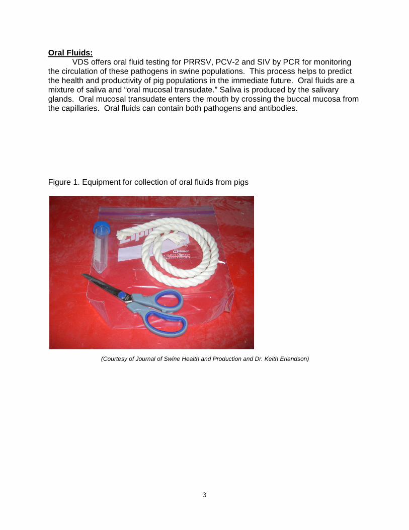
Oral Fluids: VDS offers oral fluid testing for PRRSV, PCV-2 and SIV by PCR for monitoring the circulation of these pathogens in swine populations. This process helps to predict the health and productivity of pig populations in the immediate future. Oral fluids are a mixture of saliva and “oral mucosal transudate.” Saliva is produced by the salivary glands. Oral mucosal transudate enters the mouth by crossing the buccal mucosa from the capillaries. Oral fluids can contain both pathogens and antibodies. Figure 1. Equipment for collection of oral fluids from pigs
(Courtesy of Journal of Swine Health and Production and Dr. Keith Erlandson)
4
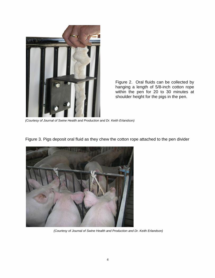
Figure 3. Pigs deposit oral fluid as they chew the cotton rope attached to the pen divider
(Courtesy of Journal of Swine Health and Production and Dr. Keith Erlandson)
(Courtesy of Journal of Swine Health and Production and Dr. Keith Erlandson)
Figure 2. Oral fluids can be collected by hanging a length of 5/8-inch cotton rope within the pen for 20 to 30 minutes at shoulder height for the pigs in the pen.
5
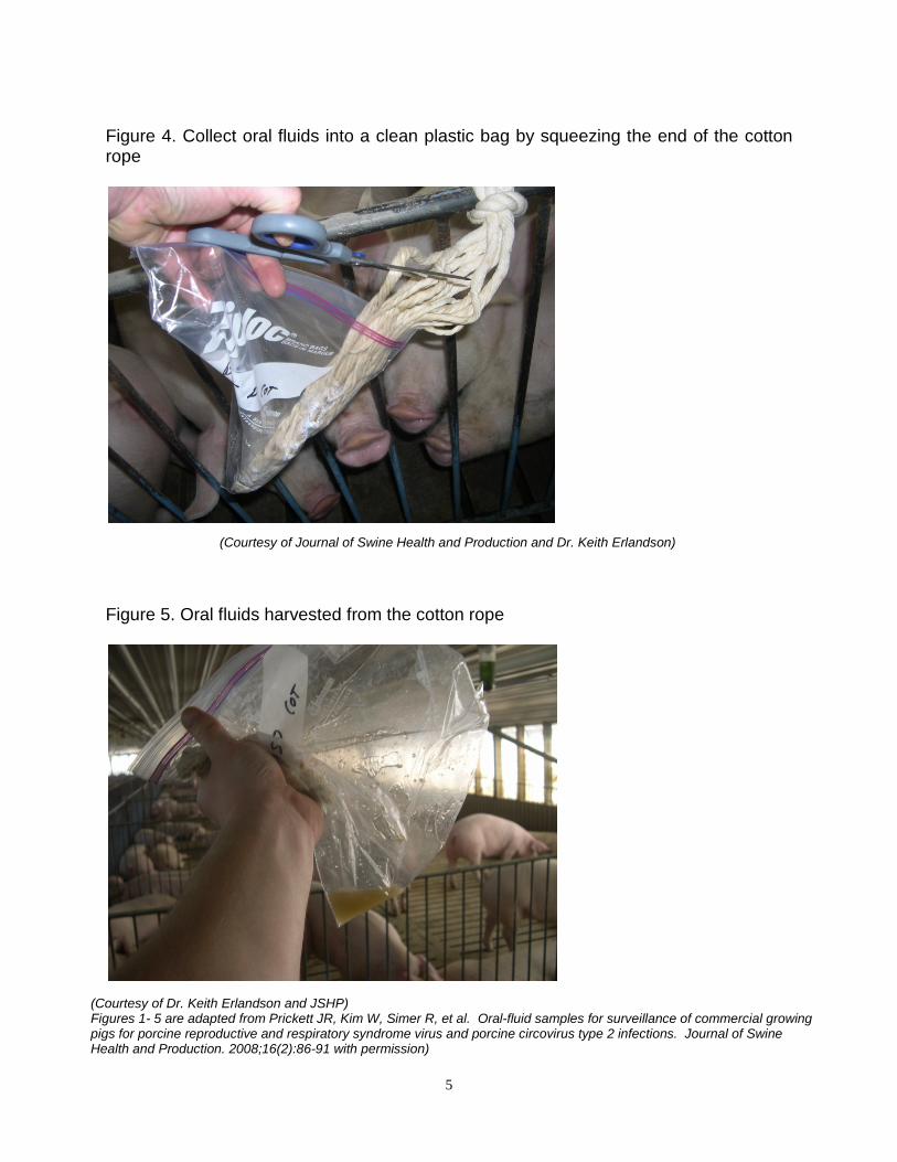
Figure 4. Collect oral fluids into a clean plastic bag by squeezing the end of the cotton rope
Figure 5. Oral fluids harvested from the cotton rope
(Courtesy of Dr. Keith Erlandson and JSHP) Figures 1- 5 are adapted from Prickett JR, Kim W, Simer R, et al. Oral-fluid samples for surveillance of commercial growing pigs for porcine reproductive and respiratory syndrome virus and porcine circovirus type 2 infections. Journal of Swine Health and Production. 2008;16(2):86-91 with permission)
(Courtesy of Journal of Swine Health and Production and Dr. Keith Erlandson)
6
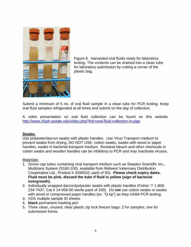
Submit a minimum of 5 mL of oral fluid sample in a clean tube for PCR testing. Keep oral fluid samples refrigerated at all times and submit on the day of collection. A video presentation on oral fluid collection can be found on this website. http://www.cfsph.iastate.edu/video.php?link=oral-fluid-collection-in-pigs Swabs: Use polyester/dacron swabs with plastic handles. Use Virus Transport medium to prevent swabs from drying. DO NOT USE: cotton swabs, swabs with wood or paper handles, swabs in bacterial transport medium. Residual bleach and other chemicals in cotton swabs and wooden handles can be inhibitory to PCR and may inactivate viruses. Materials: 1. Screw cap tubes containing viral transport medium such as Starplex Scientific Inc.,
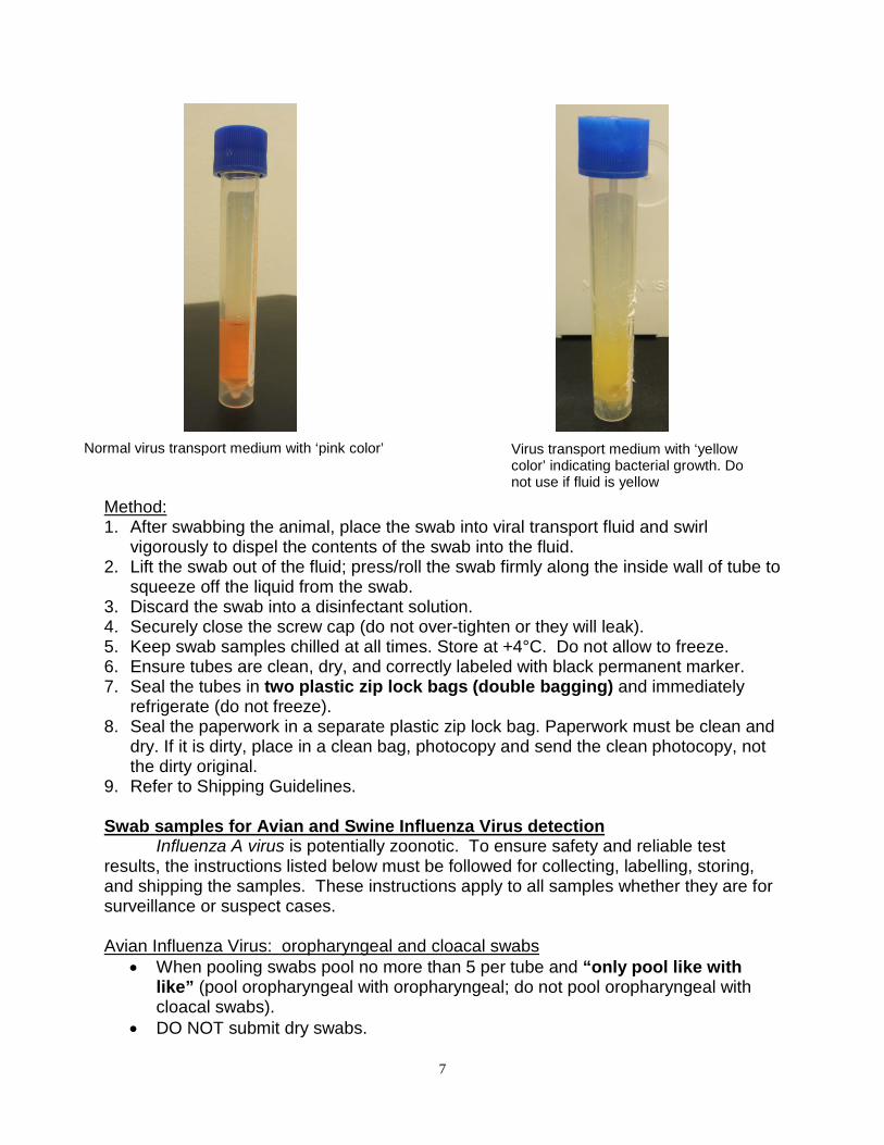
Multitrans System (S160-100), available from Midwest Veterinary Distribution Cooperative Ltd., Product # 2090010, pack of 50). Please check expiry dates. Fluid must be pink, discard the tube if fluid is yellow (sign of bacterial overgrowth).
2. Individually wrapped dacron/polyester swabs with plastic handles (Fisher: T 1-800-234-7437, Cat.# 14-959-90 sterile pack of 100). Do not use cotton swabs or swabs with wood or compressed paper handles (ex. “Q-tip”) as they inhibit PCR testing.
3. VDS multiple sample ID sheets. 4. black permanent marking pen 5. Three clean, unused, clear plastic zip lock freezer bags: 2 for samples; one for
submission forms.
Figure 6. Harvested oral fluids ready for laboratory testing. The contents can be drained into a clean tube for laboratory submission by cutting a corner of the plastic bag.
7
Method: 1. After swabbing the animal, place the swab into viral transport fluid and swirl
vigorously to dispel the contents of the swab into the fluid. 2. Lift the swab out of the fluid; press/roll the swab firmly along the inside wall of tube to
squeeze off the liquid from the swab. 3. Discard the swab into a disinfectant solution. 4. Securely close the screw cap (do not over-tighten or they will leak). 5. Keep swab samples chilled at all times. Store at +4°C. Do not allow to freeze. 6. Ensure tubes are clean, dry, and correctly labeled with black permanent marker. 7. Seal the tubes in two plastic zip lock bags (double bagging) and immediately
refrigerate (do not freeze). 8. Seal the paperwork in a separate plastic zip lock bag. Paperwork must be clean and
dry. If it is dirty, place in a clean bag, photocopy and send the clean photocopy, not the dirty original.
9. Refer to Shipping Guidelines. Swab samples for Avian and Swine Influenza Virus detection Influenza A virus is potentially zoonotic. To ensure safety and reliable test results, the instructions listed below must be followed for collecting, labelling, storing, and shipping the samples. These instructions apply to all samples whether they are for surveillance or suspect cases. Avian Influenza Virus: oropharyngeal and cloacal swabs
• When pooling swabs pool no more than 5 per tube and “only pool like with like” (pool oropharyngeal with oropharyngeal; do not pool oropharyngeal with cloacal swabs).
• DO NOT submit dry swabs.
Normal virus transport medium with ‘pink color’ Virus transport medium with ‘yellow color’ indicating bacterial growth. Do not use if fluid is yellow
8
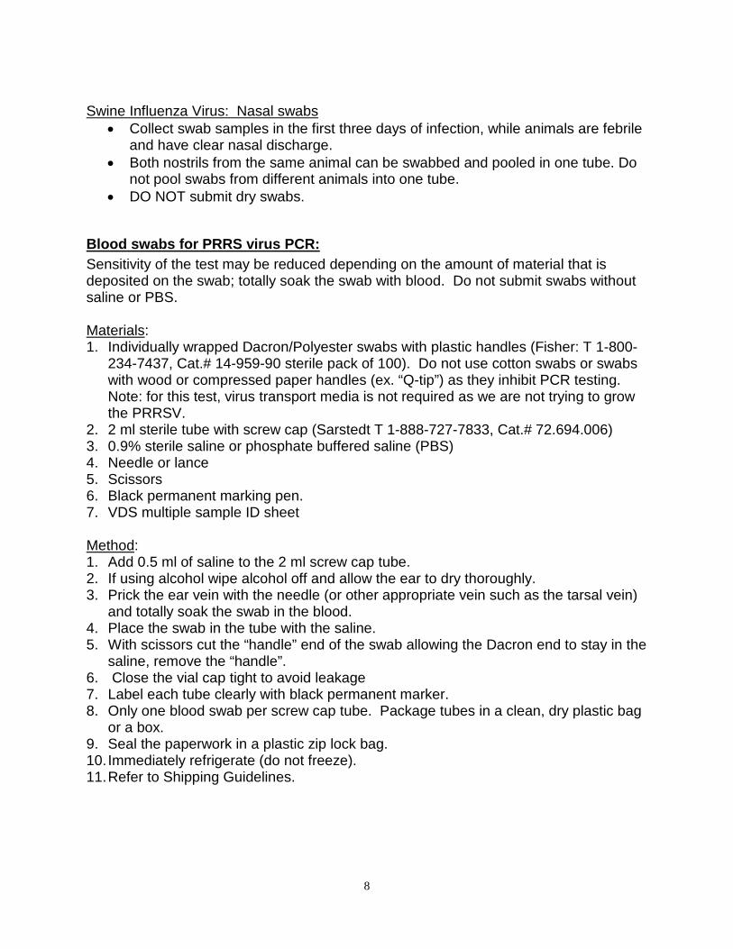
Swine Influenza Virus: Nasal swabs
• Collect swab samples in the first three days of infection, while animals are febrile and have clear nasal discharge.
• Both nostrils from the same animal can be swabbed and pooled in one tube. Do not pool swabs from different animals into one tube.
• DO NOT submit dry swabs.
Blood swabs for PRRS virus PCR: Sensitivity of the test may be reduced depending on the amount of material that is deposited on the swab; totally soak the swab with blood. Do not submit swabs without saline or PBS. Materials: 1. Individually wrapped Dacron/Polyester swabs with plastic handles (Fisher: T 1-800-
234-7437, Cat.# 14-959-90 sterile pack of 100). Do not use cotton swabs or swabs with wood or compressed paper handles (ex. “Q-tip”) as they inhibit PCR testing. Note: for this test, virus transport media is not required as we are not trying to grow the PRRSV.
2. 2 ml sterile tube with screw cap (Sarstedt T 1-888-727-7833, Cat.# 72.694.006) 3. 0.9% sterile saline or phosphate buffered saline (PBS) 4. Needle or lance 5. Scissors 6. Black permanent marking pen. 7. VDS multiple sample ID sheet Method: 1. Add 0.5 ml of saline to the 2 ml screw cap tube. 2. If using alcohol wipe alcohol off and allow the ear to dry thoroughly. 3. Prick the ear vein with the needle (or other appropriate vein such as the tarsal vein)
and totally soak the swab in the blood. 4. Place the swab in the tube with the saline. 5. With scissors cut the “handle” end of the swab allowing the Dacron end to stay in the
saline, remove the “handle”. 6. Close the vial cap tight to avoid leakage 7. Label each tube clearly with black permanent marker. 8. Only one blood swab per screw cap tube. Package tubes in a clean, dry plastic bag
or a box. 9. Seal the paperwork in a plastic zip lock bag. 10. Immediately refrigerate (do not freeze). 11. Refer to Shipping Guidelines.
9
Virus Isolation Many of the samples used for PCR can be used for virus isolation as well if collected and stored properly. VDS will forward samples to referral laboratories for virus isolation upon request. Virus isolation is a nonspecific assay in which infectious virus is grown in appropriate cell lines or embryonated eggs. If virus isolation is anticipated, the clinic must instruct VDS in writing to store the samples at -70°C immediately after the samples have been processed. VDS routinely stores DNA/RNA extracts for 12 months at -70°C; tissues, nasal swabs and joint swabs for 3 months at -70°C; blood, feces, semen, serum and blood swabs for 3 months at +4°C. DNA Sequencing
DNA sequencing is a molecular tool used to characterize the genome of a
microorganism. DNA sequencing identifies which strain of virus is present in the clinical sample and in some cases helps to differentiate field and vaccine strains of viruses. Only samples testing strong positive by conventional or real-time PCR can be used for DNA sequencing. The clinic must provide in writing (by email) the following information: 1. VDS Case number 2. Sequencing test to be performed 3. Correct identity of sample(s) to be tested by the reference lab 4. The reference lab to which samples should be sent
10
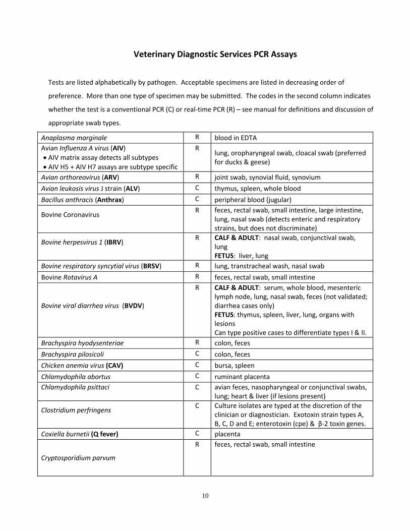
Veterinary Diagnostic Services PCR Assays
Tests are listed alphabetically by pathogen. Acceptable specimens are listed in decreasing order of
preference. More than one type of specimen may be submitted. The codes in the second column indicates
whether the test is a conventional PCR (C) or real-time PCR (R) – see manual for definitions and discussion of
appropriate swab types.
Anaplasma marginale R blood in EDTA Avian Influenza A virus (AIV) • AIV matrix assay detects all subtypes • AIV H5 + AIV H7 assays are subtype specific
R lung, oropharyngeal swab, cloacal swab (preferred for ducks & geese)
Avian orthoreovirus (ARV) R joint swab, synovial fluid, synovium Avian leukosis virus J strain (ALV) C thymus, spleen, whole blood
Bacillus anthracis (Anthrax) C peripheral blood (jugular)
Bovine Coronavirus
R feces, rectal swab, small intestine, large intestine, lung, nasal swab (detects enteric and respiratory strains, but does not discriminate)
Bovine herpesvirus 1 (IBRV)
R CALF & ADULT: nasal swab, conjunctival swab, lung FETUS: liver, lung
Bovine respiratory syncytial virus (BRSV) R lung, transtracheal wash, nasal swab
Bovine Rotavirus A R feces, rectal swab, small intestine
Bovine viral diarrhea virus (BVDV)
R CALF & ADULT: serum, whole blood, mesenteric lymph node, lung, nasal swab, feces (not validated; diarrhea cases only) FETUS: thymus, spleen, liver, lung, organs with lesions Can type positive cases to differentiate types I & II.
Brachyspira hyodysenteriae R colon, feces Brachyspira pilosicoli C colon, feces Chicken anemia virus (CAV) C bursa, spleen Chlamydophila abortus C ruminant placenta Chlamydophila psittaci
C avian feces, nasopharyngeal or conjunctival swabs, lung; heart & liver (if lesions present)
Clostridium perfringens
C Culture isolates are typed at the discretion of the clinician or diagnostician. Exotoxin strain types A, B, C, D and E; enterotoxin (cpe) & β-2 toxin genes.
Coxiella burnetii (Q fever) C placenta
Cryptosporidium parvum
R feces, rectal swab, small intestine
11
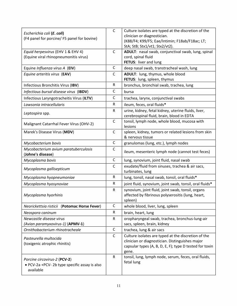
Escherichia coli (E. coli) (F4 panel for porcine/ F5 panel for bovine)
C Culture isolates are typed at the discretion of the clinician or diagnostician. (K88/F4; K99/F5; Eae/Intimin; F18ab/F18ac; LT; StA; StB; Stx1/vt1; Stx2/vt2).
Equid herpesvirus (EHV 1 & EHV 4) (Equine viral rhinopneumonitis virus)
C ADULT: nasal swab, conjunctival swab, lung, spinal cord, spinal fluid FETUS: liver and lung
Equine Influenza virus A (EIV) C deep nasal swab, transtracheal wash, lung Equine arteritis virus (EAV)
C ADULT: lung, thymus, whole blood
FETUS: lung, spleen, thymus Infectious Bronchitis Virus (IBV) R bronchus, bronchial swab, trachea, lung Infectious bursal disease virus (IBDV) C bursa Infectious Laryngotracheitis Virus (ILTV) C trachea, larynx, conjunctival swabs Lawsonia intracellularis R ileum, feces, oral fluids*
Leptospira spp. R urine, kidney, fetal kidney, uterine fluids, liver, cerebrospinal fluid, brain, blood in EDTA
Malignant Catarrhal Fever Virus (OHV-2) C tonsil, lymph node, whole blood, mucosa with lesions
Marek's Disease Virus (MDV)
C spleen, kidney, tumors or related lesions from skin & nervous tissue
Mycobacterium bovis C granulomas (lung, etc.), lymph nodes Mycobacterium avium paratuberculosis (Johne's disease)
C ileum, mesenteric lymph node (cannot test feces)
Mycoplasma bovis C lung, synovium, joint fluid, nasal swab
Mycoplasma gallisepticum C exudate/fluid from sinuses, trachea & air sacs, turbinates, lung
Mycoplasma hyopneumoniae R lung, tonsil, nasal swab, tonsil, oral fluids*
Mycoplasma hyosynoviae R joint fluid, synovium, joint swab, tonsil, oral fluids*
Mycoplasma hyorhinis R synovium, joint fluid, joint swab, tonsil, organs
affected by fibrinous polyserositis (lung, heart, spleen)
Neorickettsia risticii (Potomac Horse Fever) C whole blood, liver, lung, spleen Neospora caninum R brain, heart, lung Newcastle disease virus (Avian paramyxovirus-1) (APMV-1)
R oropharyngeal swab, trachea, bronchus-lung-air sacs, spleen, brain, kidney
Ornithobacterium rhinotracheale C trachea, lung & air sacs
Pasteurella multocida (toxigenic atrophic rhinitis)
C Culture isolates are typed at the discretion of the clinician or diagnostician. Distinguishes major capsular types (A, B, D, E, F); type D tested for toxin gene.
Porcine circovirus-2 (PCV-2) • PCV-2a +PCV- 2b type specific assay is also
available
R tonsil, lung, lymph node, serum, feces, oral fluids, fetal lung
12
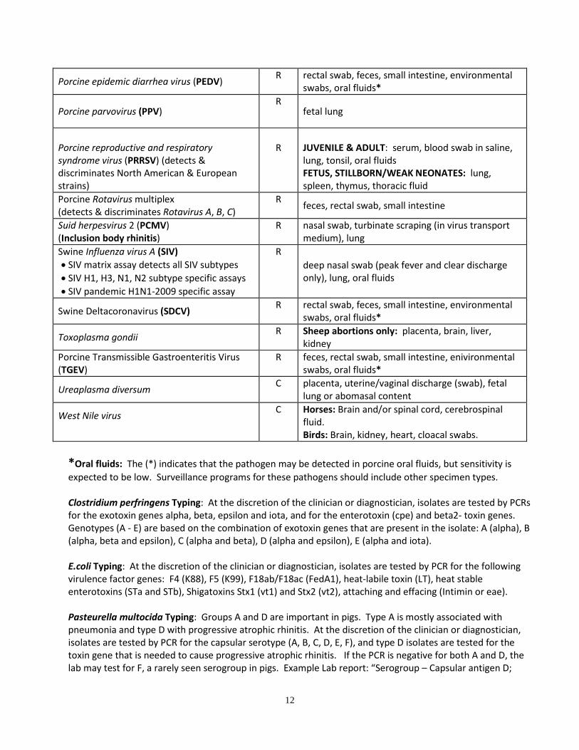
Porcine epidemic diarrhea virus (PEDV) R rectal swab, feces, small intestine, environmental swabs, oral fluids*
Porcine parvovirus (PPV) R
fetal lung
Porcine reproductive and respiratory syndrome virus (PRRSV) (detects & discriminates North American & European strains)
R
JUVENILE & ADULT: serum, blood swab in saline, lung, tonsil, oral fluids FETUS, STILLBORN/WEAK NEONATES: lung, spleen, thymus, thoracic fluid
Porcine Rotavirus multiplex (detects & discriminates Rotavirus A, B, C)
R feces, rectal swab, small intestine
Suid herpesvirus 2 (PCMV) (Inclusion body rhinitis)
R nasal swab, turbinate scraping (in virus transport medium), lung
Swine Influenza virus A (SIV) • SIV matrix assay detects all SIV subtypes • SIV H1, H3, N1, N2 subtype specific assays • SIV pandemic H1N1-2009 specific assay
R deep nasal swab (peak fever and clear discharge only), lung, oral fluids
Swine Deltacoronavirus (SDCV) R rectal swab, feces, small intestine, environmental swabs, oral fluids*
Toxoplasma gondii R Sheep abortions only: placenta, brain, liver, kidney
Porcine Transmissible Gastroenteritis Virus (TGEV)
R feces, rectal swab, small intestine, enivironmental swabs, oral fluids*
Ureaplasma diversum C placenta, uterine/vaginal discharge (swab), fetal lung or abomasal content
West Nile virus
C Horses: Brain and/or spinal cord, cerebrospinal fluid. Birds: Brain, kidney, heart, cloacal swabs.
*Oral fluids: The (*) indicates that the pathogen may be detected in porcine oral fluids, but sensitivity is expected to be low. Surveillance programs for these pathogens should include other specimen types. Clostridium perfringens Typing: At the discretion of the clinician or diagnostician, isolates are tested by PCRs for the exotoxin genes alpha, beta, epsilon and iota, and for the enterotoxin (cpe) and beta2- toxin genes. Genotypes (A - E) are based on the combination of exotoxin genes that are present in the isolate: A (alpha), B (alpha, beta and epsilon), C (alpha and beta), D (alpha and epsilon), E (alpha and iota). E.coli Typing: At the discretion of the clinician or diagnostician, isolates are tested by PCR for the following virulence factor genes: F4 (K88), F5 (K99), F18ab/F18ac (FedA1), heat-labile toxin (LT), heat stable enterotoxins (STa and STb), Shigatoxins Stx1 (vt1) and Stx2 (vt2), attaching and effacing (Intimin or eae). Pasteurella multocida Typing: Groups A and D are important in pigs. Type A is mostly associated with pneumonia and type D with progressive atrophic rhinitis. At the discretion of the clinician or diagnostician, isolates are tested by PCR for the capsular serotype (A, B, C, D, E, F), and type D isolates are tested for the toxin gene that is needed to cause progressive atrophic rhinitis. If the PCR is negative for both A and D, the lab may test for F, a rarely seen serogroup in pigs. Example Lab report: “Serogroup – Capsular antigen D;
13
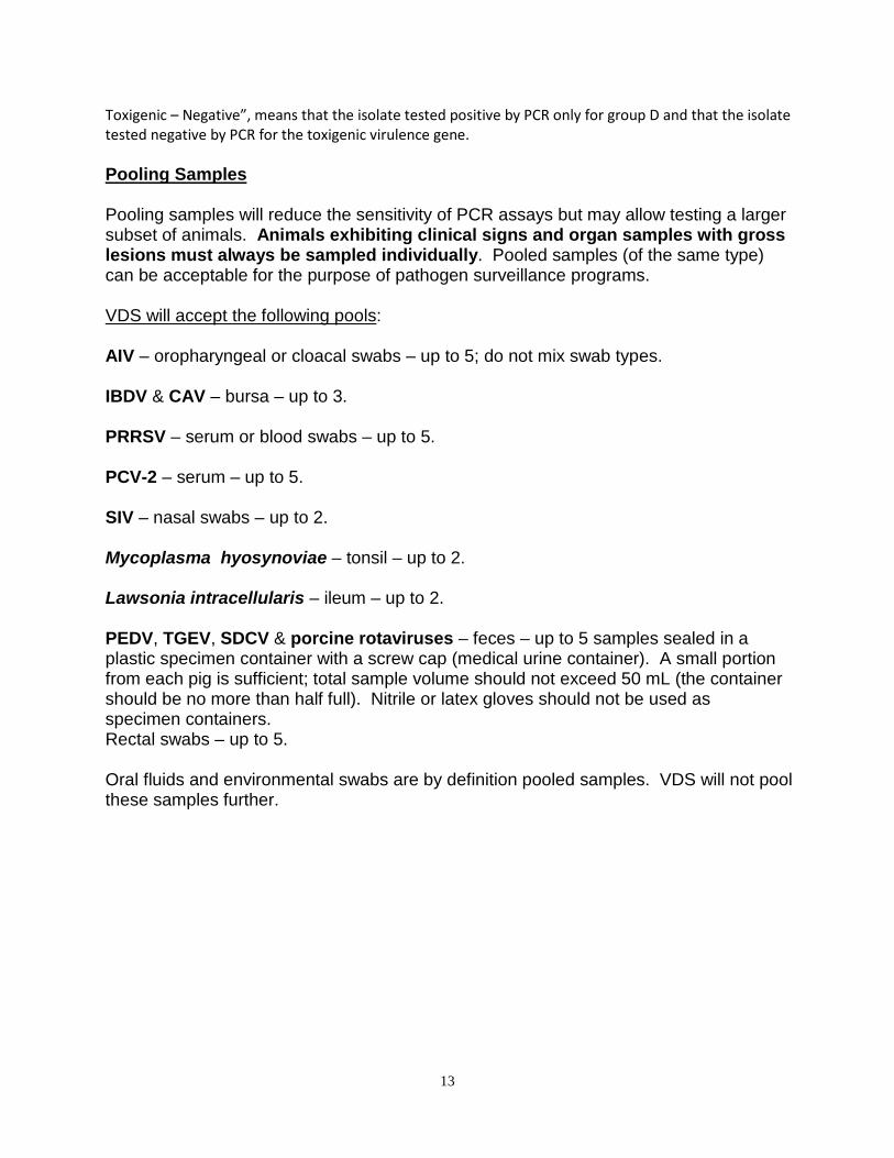
Toxigenic – Negative”, means that the isolate tested positive by PCR only for group D and that the isolate tested negative by PCR for the toxigenic virulence gene. Pooling Samples Pooling samples will reduce the sensitivity of PCR assays but may allow testing a larger subset of animals. Animals exhibiting clinical signs and organ samples with gross lesions must always be sampled individually. Pooled samples (of the same type) can be acceptable for the purpose of pathogen surveillance programs. VDS will accept the following pools: AIV – oropharyngeal or cloacal swabs – up to 5; do not mix swab types. IBDV & CAV – bursa – up to 3. PRRSV – serum or blood swabs – up to 5. PCV-2 – serum – up to 5. SIV – nasal swabs – up to 2. Mycoplasma hyosynoviae – tonsil – up to 2. Lawsonia intracellularis – ileum – up to 2. PEDV, TGEV, SDCV & porcine rotaviruses – feces – up to 5 samples sealed in a plastic specimen container with a screw cap (medical urine container). A small portion from each pig is sufficient; total sample volume should not exceed 50 mL (the container should be no more than half full). Nitrile or latex gloves should not be used as specimen containers. Rectal swabs – up to 5. Oral fluids and environmental swabs are by definition pooled samples. VDS will not pool these samples further.
14
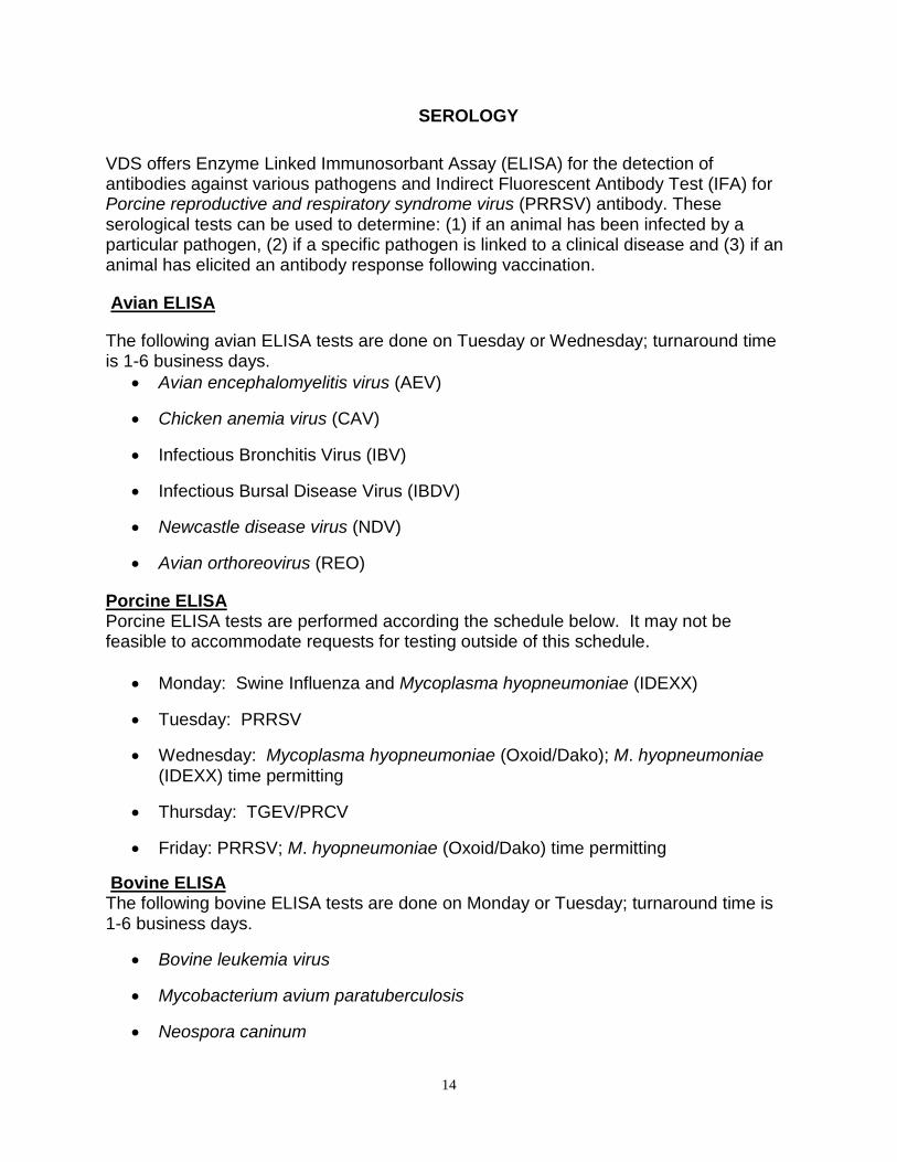
SEROLOGY VDS offers Enzyme Linked Immunosorbant Assay (ELISA) for the detection of antibodies against various pathogens and Indirect Fluorescent Antibody Test (IFA) for Porcine reproductive and respiratory syndrome virus (PRRSV) antibody. These serological tests can be used to determine: (1) if an animal has been infected by a particular pathogen, (2) if a specific pathogen is linked to a clinical disease and (3) if an animal has elicited an antibody response following vaccination.
Avian ELISA
The following avian ELISA tests are done on Tuesday or Wednesday; turnaround time is 1-6 business days.
• Avian encephalomyelitis virus (AEV)
• Chicken anemia virus (CAV)
• Infectious Bronchitis Virus (IBV)
• Infectious Bursal Disease Virus (IBDV)
• Newcastle disease virus (NDV)
• Avian orthoreovirus (REO)
Porcine ELISA Porcine ELISA tests are performed according the schedule below. It may not be feasible to accommodate requests for testing outside of this schedule.
• Monday: Swine Influenza and Mycoplasma hyopneumoniae (IDEXX)
• Tuesday: PRRSV
• Wednesday: Mycoplasma hyopneumoniae (Oxoid/Dako); M. hyopneumoniae (IDEXX) time permitting
• Thursday: TGEV/PRCV
• Friday: PRRSV; M. hyopneumoniae (Oxoid/Dako) time permitting
Bovine ELISA The following bovine ELISA tests are done on Monday or Tuesday; turnaround time is 1-6 business days.
• Bovine leukemia virus
• Mycobacterium avium paratuberculosis
• Neospora caninum
15
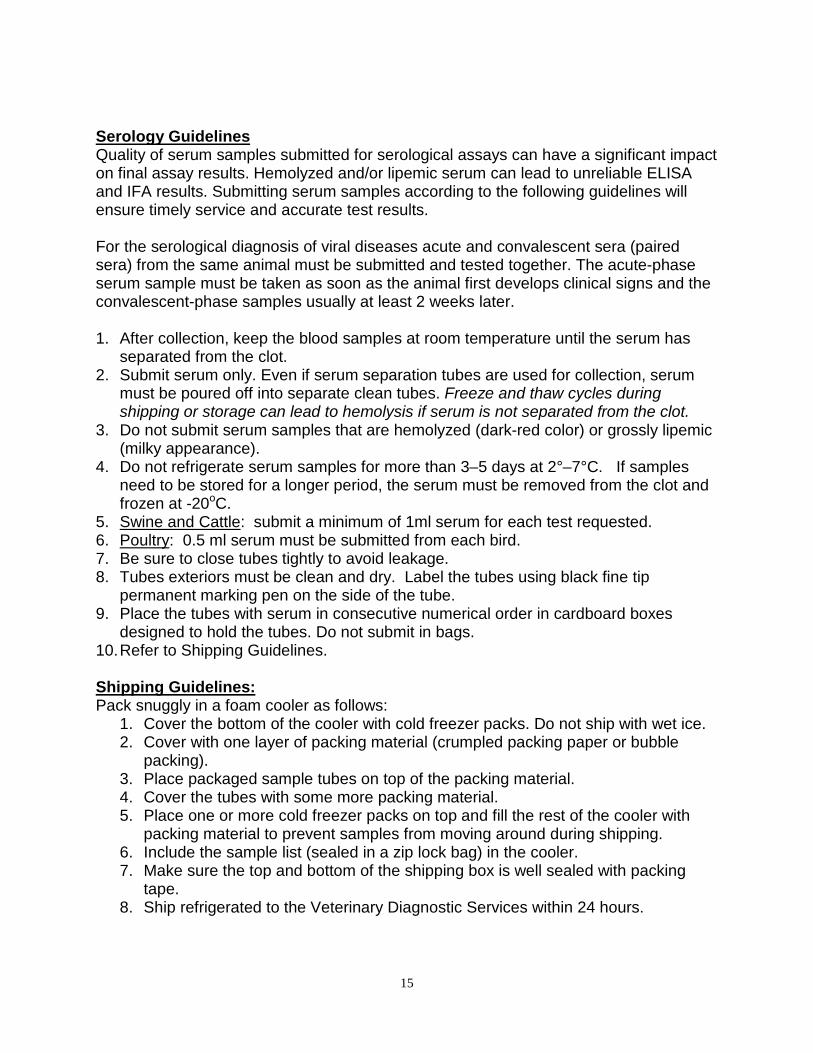
Serology Guidelines Quality of serum samples submitted for serological assays can have a significant impact on final assay results. Hemolyzed and/or lipemic serum can lead to unreliable ELISA and IFA results. Submitting serum samples according to the following guidelines will ensure timely service and accurate test results. For the serological diagnosis of viral diseases acute and convalescent sera (paired sera) from the same animal must be submitted and tested together. The acute-phase serum sample must be taken as soon as the animal first develops clinical signs and the convalescent-phase samples usually at least 2 weeks later. 1. After collection, keep the blood samples at room temperature until the serum has
separated from the clot. 2. Submit serum only. Even if serum separation tubes are used for collection, serum
must be poured off into separate clean tubes. Freeze and thaw cycles during shipping or storage can lead to hemolysis if serum is not separated from the clot.
3. Do not submit serum samples that are hemolyzed (dark-red color) or grossly lipemic (milky appearance).
4. Do not refrigerate serum samples for more than 3–5 days at 2°–7°C. If samples need to be stored for a longer period, the serum must be removed from the clot and frozen at -20oC.
5. Swine and Cattle: submit a minimum of 1ml serum for each test requested. 6. Poultry: 0.5 ml serum must be submitted from each bird. 7. Be sure to close tubes tightly to avoid leakage. 8. Tubes exteriors must be clean and dry. Label the tubes using black fine tip
permanent marking pen on the side of the tube. 9. Place the tubes with serum in consecutive numerical order in cardboard boxes
designed to hold the tubes. Do not submit in bags. 10. Refer to Shipping Guidelines. Shipping Guidelines: Pack snuggly in a foam cooler as follows:
1. Cover the bottom of the cooler with cold freezer packs. Do not ship with wet ice. 2. Cover with one layer of packing material (crumpled packing paper or bubble
packing). 3. Place packaged sample tubes on top of the packing material. 4. Cover the tubes with some more packing material. 5. Place one or more cold freezer packs on top and fill the rest of the cooler with
packing material to prevent samples from moving around during shipping. 6. Include the sample list (sealed in a zip lock bag) in the cooler. 7. Make sure the top and bottom of the shipping box is well sealed with packing
tape. 8. Ship refrigerated to the Veterinary Diagnostic Services within 24 hours.