Veterinary Dentistry: Growing your Practice with Pathology ...
26
1 Veterinary Dentistry: Growing your Practice with Pathology Recognition, Preventive Dental Care, and Value Marketing© Kevin S. Stepaniuk, DVM, Fellow AVD, Diplomate AVDC Columbia River Veterinary Specialists, Vancouver WA Adjunct Assistant Professor University of Minnesota College of Veterinary Medicine CVMA - Society of BC Veterinarians Chapter Fall Conference and Trade Show – November 9, 2014 INTRODUCTION Developing and growing a successful veterinary dentistry service in a general practice relies on several key factors. But what do I know? My background has allowed me the opportunity to build a general practice, a private referral dentistry practice, develop programs in academia, and develop a specialty practice within a multiple discipline specialty hospital. I do not have a business degree or a corporate leadership position or an entrenched academic ivory tower position. I have, and continue to, work on the front line in the trenches. However, my collective experiences, clinical training, leadership and business training, board positions, and opportunity to see success and failure from both sides of the referral fence, provide me with a unique perspective on veterinary dentistry and oral surgery in veterinary practice; where it has come from, where it is at, where it is going, and where can we direct it to go. I believe there are many missed opportunities in veterinary dentistry, patient avocation, and patient care due to seven (7) common short comings in practical veterinary dentistry©: 1) Lack of collective veterinary dental education leading to many myths and misconceptions within the profession and our client base 2) lack of acceptance regarding the morbidity, pathology associated with odontogenic infections, and acknowledging the importance and role dental infection and pain play in quality of life and systemic related issues 3) lack of pathology recognition of the most common and prevalent diseases in your practice 4) lack support for yourself, colleagues, and staff in your practice to develop the opportunities to learn and practice the skills necessary to build your practice 5) lack of utilizing intraoral radiology 6) lack of correct surgical dentistry equipment and 7) lack of multimodal anesthesia and analgesia. The following will address just a few of these seven (7) categories: I) The prevention of periodontal disease with periodontal cleanings and the prevention of fractured teeth with client education, and how quality practice can lead to business growth. Historically veterinary medicine, surgery, and dentistry have, and continue to, focus on treatment rather than prevention. II) How to provide value to your services will be presented. III) In addition, a short section on how complications related to dental and surgical procedures can be prevented and managed. As with most subjects, it is impossible to completely cover every facet and detail necessary in a subject area in only a few hours. Further marketing and scientific information can found at the Annual Veterinary Dental Forum, local, regional, national, and international continuing education events. Kevin S. Stepaniuk, DVM, FAVD, DAVDC Veterinary Dentistry: Growing your Practice with Pathology Recognition, Preventive Dental Care, and Value Marketing©2014 [email protected]
Transcript of Veterinary Dentistry: Growing your Practice with Pathology ...

1
Veterinary Dentistry: Growing your Practice with Pathology Recognition, Preventive Dental Care, and Value Marketing©
Kevin S. Stepaniuk, DVM, Fellow AVD, Diplomate AVDCColumbia River Veterinary Specialists, Vancouver WAAdjunct Assistant Professor University of Minnesota College of Veterinary Medicine
CVMA - Society of BC Veterinarians Chapter Fall Conference and Trade Show – November 9, 2014
INTRODUCTION
Developing and growing a successful veterinary dentistry service in a general practice relies on several key factors. But what do I know? My background has allowed me the opportunity to build a general practice, a private referral dentistry practice, develop programs in academia, anddevelop a specialty practice within a multiple discipline specialty hospital. I do not have a business degree or a corporate leadership position or an entrenched academic ivory tower position. I have, and continue to, work on the front line in the trenches. However, my collective experiences, clinical training, leadership and business training, board positions, and opportunity to see success and failure from both sides of the referral fence, provide me with a unique perspective on veterinary dentistry and oral surgery in veterinary practice; where it has come from, where it is at, where it is going, and where can we direct it to go.
I believe there are many missed opportunities in veterinary dentistry, patient avocation, and patient care due to seven (7) common short comings in practical veterinary dentistry©: 1) Lack of collective veterinary dental education leading to many myths and misconceptions within the profession and our client base 2) lack of acceptance regarding the morbidity, pathology associated with odontogenic infections, and acknowledging the importance and role dental infection and pain play in quality of life and systemic related issues 3) lack of pathology recognition of the most common and prevalent diseases in your practice 4) lack support for yourself, colleagues, and staff in your practice to develop the opportunities to learn and practice the skills necessary to build your practice 5) lack of utilizing intraoral radiology 6) lack of correctsurgical dentistry equipment and 7) lack of multimodal anesthesia and analgesia.
The following will address just a few of these seven (7) categories:
I) The prevention of periodontal disease with periodontal cleanings and the prevention of fractured teeth with client education, and how quality practice can lead to businessgrowth. Historically veterinary medicine, surgery, and dentistry have, and continue to,focus on treatment rather than prevention.
II) How to provide value to your services will be presented. III) In addition, a short section on how complications related to dental and surgical
procedures can be prevented and managed.
As with most subjects, it is impossible to completely cover every facet and detail necessary in a subject area in only a few hours. Further marketing and scientific information can found at the Annual Veterinary Dental Forum, local, regional, national, and international continuing educationevents.
Kevin S. Stepaniuk, DVM, FAVD, DAVDCVeterinary Dentistry: Growing your Practice with Pathology Recognition, Preventive Dental Care, and Value Marketing©[email protected]

2
I. PERIODONTAL DISEASE RECOGNIZING THE MOST PREVELANT HIDDEN DISEASE IN YOUR PRACTICE
IntroductionThe tooth is anchored in the jaws by the periodontium. The incisive bones, maxillary bone, and mandibular bone anchor the teeth. The periodontium consists of the 1) gingiva, 2) alveolar bone, 3) periodontal ligament, and 4) cementum.
Periodontal disease is the loss of the periodontal attachment apparatus (periodontal ligament, alveolar bone, cementum and gingiva). Since 75% of these structures are identified below the soft tissues of the oral cavity (gingiva, alveolar mucosa, and palatal mucosa), a thorough clinicalsubgingival evaluation and intraoral radiographs are required to assess, diagnose and treat periodontal disease. Therefore, general anesthesia is required.
Pathophysiology Of Periodontal DiseasePeriodontitis is active inflammation of the periodontium. It begins with the accumulation of the dental pellicle (e.g., salivary glycoproteins and enzymes) that occurs within seconds of a tooth being cleaned. Within hours, first colonizing oral bacterial colonize the pellicle and the plaque biofilm is formed. The plaque biofilm matures within days. Mineralization of the plaque biofilm results in calculus (tarter). Periodontal disease is caused by the bacterial biofilm (plaque) and the associated inflammatory response. Significant periodontal disease can be present without calculus. Calculus is not the cause of periodontal disease.
As the plaque biofilm matures, early bacterial colonizers, gram-positive aerobic cocci, become less predominant as the biofilm transforms to more prominent gram-negative anaerobes and spirochetes located more apical in the periodontal pockets. Bacterial products such as ammonia, volatile sulfur compounds, and proteolytic enzymes contribute to the destruction of the periodontium. The host inflammatory response, matrix metalloproteinases that degrade collagen of the periodontal ligament, elastase (break down collagen and elastin), and prostaglandins (PGE2) are directly responsible for tissue damage and/or stimulate osteoclastic bone resorption (PGE2, IL-1β, TNF-α). These inflammatory mediators also enter the systemic circulation. The calcium carbonate in the saliva of cats and dogs combines with the plaque to form calculus. Calculus increases surface area for bacterial attachment and can mechanically disrupt and damage the gingiva.
Periodontal disease may be potentiated by, but not limited to, malocclusions, crowding and rotation of teeth, systemic disease, nutritional status, individual patient susceptibility, genetics, trauma, and increased tooth to jaw size ratios.
Clinical Signs Of Periodontal DiseaseThe clinical signs of periodontal disease are often hidden and insidious. Halitosis, gingivitis, supragingival plaque and calculus, reluctance to chew, head shyness, pawing at the mouth, dropping food, sneezing, nasal discharge, are clinical signs. Unfortunately, many of those clinical signs require astute client observation and/or careful questioning from the clinician. Mostcommonly, there may be no obvious clinical signs to the owner and untrained veterinarian. Almost all the patients are still eating. American Animal Hospital Association Dental Guidelines and Canine and Feline Life Stages Guidelines recommend annual evaluations of the oral cavity. The recommended time to start professional evaluations and cleanings, in order to prevent disease, is in the 1st-2nd year of life.
Kevin S. Stepaniuk, DVM, FAVD, DAVDCVeterinary Dentistry: Growing your Practice with Pathology Recognition, Preventive Dental Care, and Value Marketing©[email protected]
3
Diagnosis Of Periodontal DiseaseGeneral anesthesia, professional examination, periodontal probing, charting, and intraoral radiographs are all required to successfully diagnose and treat periodontal disease.
Conscious Oral ExaminationPeriodontal assessment begins in the examination room with the client and the conscious patient. A complete medical and oral history, general physical exam, and conscious oral examination are necessary. Questions such as, but not limited to, onset, duration, environment, chew toys, oral health care, current medications, diet, eating patterns, past illness, past anesthetic episodes, behavioral changes, etc. are explored. Many patients with oral disease do not have obvious clinical signs.
The maxillofacial skeletal is palpated and the eyes retropulsed. The three basic skull types are brachycephalic (e.g., Pugs, Persian Cats), mesocephalic (e.g., Labrador, DSH), and dolichocephalic (e.g., Sight hounds). The regional lymph nodes and salivary glands are palpated. Facial symmetry and occlusion are noted. The range of motion of the temporomandibular joints should be palpated and the patient observed for pain and/or difficulty in opening and closing the mouth. The lips and mucocutaneous junctions should be observed for ulcerations that might indicate an autoimmune disease or pyoderma. Finally, the dentition is evaluated and the teeth counted to determine if all teeth are present. Discolored teeth, persistent deciduous teeth, root and furcation exposure, oral mucosal lesions, sinus tracts, tongue abnormalities, oral masses, plaque and calculus are noted and require further diagnostics and treatment.
Anesthetized ExaminationWhile the patient is under general anesthesia a full oral examination and dental charting is performed. For the purpose of this lecture, periodontal indices are discussed (nomenclature www.avdc.org). During the periodontal examination, crowded teeth, missing teeth, rotated teeth,mobile teeth, teeth with furcation exposures, gingival recession (root exposure), sinus tracts, gingival enlargements, and periodontal probing depths are noted (The normal gingival sulcus depth in a dog is 0-3 mm and less than 0.5 to 1.0 mm in a cat).
Gingival indice is assessed:
Gingival indice of 1 – inflammation and swelling of the gingiva with no bleeding during periodontal probingGingival indice of 2 – inflammation and swelling of the gingiva with bleeding during periodontal probing Gingival indice of 3 – inflammation and swelling of the gingiva with spontaneously bleeding of the inflamed gingiva prior to periodontal probing
Furcation exposure (involvement) occurs when a periodontal probe can extend between the roots, under the crown, of multi-rooted teeth as a result of attachment loss.
Stage 1 furcation - the probe extends less than half wayStage 2 furcation - the probe extends greater than halfwayStage 3 furcation - the probe extends from one side to the other, through and through
Kevin S. Stepaniuk, DVM, FAVD, DAVDCVeterinary Dentistry: Growing your Practice with Pathology Recognition, Preventive Dental Care, and Value Marketing©[email protected]

4
Gingival recession (root exposure) is measured from the location of the cementoenamel junctionto the free gingival margin. Any probing depths, whether normal or not, are recorded because and additional probing depth is additive to periodontal attachment loss. For example, if there is 3mm of gingival recession and a 2 mm probing depth, there is a total of 5 mm of periodontal attachment loss.
Periodontal pockets are clinical periodontal probing measurements greater than the normal sulcus. The normal gingival sulcus depth of the dog is 0-3 mm. The normal gingival sulcus in a cat is less than 0.5 to 1.0 mm. The periodontal probe is gently walked 360° around each tooth. Aminimum of 6 locations are measured. It is important to recognize a 6 mm pocket on the buccal aspect of a Labrador retriever maxillary canine tooth is a mild periodontal pocket that requires a periodontal cleaning where as a similar measurement on a Maltese may indicate end stage periodontal disease and time for a surgical extraction with the final decision based on the intraoral radiographic findings.
Periodontal pockets can be a combination of various types of pockets created by periodontal bone loss and gingival enlargements. Periodontal pockets are a haven for gram-negative anaerobic bacteria and spirochetes in the subgingival plaque biofilm and planktonic bacteria in the pocket gingival crevicular fluid. There are often combinations of periodontal pocket types as they are not mutually exclusive. However, they may be simplified for clinical practice:
Pseudopockets are created when the gingiva enlarges (often gingival hyperplasia) and the marginal bone remains at the appropriate level. Breeds such as Boxers and Collies have a genetic predilection for gingival hyperplasia. Common veterinary medications such as cyclosporine and amlodipine may cause gingival enlargement.
Suprabony pockets occur when marginal bone loss exceeds gingival recession (the marginal bone is lost horizontally below the tissue).
Intra(Infra)bony pockets occur when bone is lost vertically around a tooth. Infrabony pockets canbe classified as one-wall, two-wall, three-wall, and four-walled (cup or crater) defects.
Common locations for intrabony pockets in dog patients include the distal aspect of the mandibular 1st molars, the furcation of the mesial roots of the maxillary 4th premolars, the mesial aspects of the mandibular canine teeth, particularly after the 3rd incisors are lost or are extractedwithout proper technique, and the palatal aspect of the maxillary canine teeth. Surgical and medial treatment beyond a periodontal cleaning can be found in veterinary dentistry textbooks and other proceedings.
A key point: The different types of periodontal pockets have different types of treatment recommendations and the lack of diagnosis of the type of periodontal pocket can lead to inappropriate treatment and loss of the tooth resulting in a frustrated client and a pet that continued to suffer with chronic inflammation, infection, and pain.
Kevin S. Stepaniuk, DVM, FAVD, DAVDCVeterinary Dentistry: Growing your Practice with Pathology Recognition, Preventive Dental Care, and Value Marketing©[email protected]

5
Periodontal StagesTreatment plans can be designed based on the individual tooth stage as well as the overall periodontal stage of the oral cavity. There are 42 or 30 individual patients (teeth) to diagnose and treat in the dog and cat, respectively.
Stage 0 (PD0) - clinically normal oral cavity with no gingival inflammation and periodontitis Stage 1 (PD1) - Gingivitis without attachment loss (normal height and architecture of alveolar marginStage 2 (PD2) - periodontitis with less than 25% attachment loss and/or a stage-1 furcation in multi-rooted teethStage 3 (PD3) - 25-50% attachment loss and/or stage-2 furcation in multi-rooted teethStage 4 (PD4) - greater than 50% bone loss and/or stage-3 furcation in multi-rooted teeth
Clinical Findings Of Hidden Disease
A parulis is a raised nodule at the opening of a draining sinus tract. If the parulis is located apical to the mucogingival junction it is often associated with endodontic disease. If the parulis islocated near the mucogingival junction, it is often associated with periodontal disease.
Maxillary draining tracts should be investigated for odontogenic infections such as periodontal disease or endodontic disease prior to extensive dermatological or neoplastic work ups including advanced imaging and biopsy. Teeth should be the primary differential for the maxillofacial swellings and draining tracts. If an odontogenic infection is not the cause, then evaluation for neoplasia, etc. can be pursued. Often if it is neoplasia, a tooth is involved and surgical extraction and deep biopsy via the extraction site will provide a histological diagnosis.
The maxillary 1st and 2nd molars in dogs may have minimal clinical probing depths but be mobile during clinical examination. The intraoral radiographs may identify a very wide or absent periodontal ligament space/large palatal root periapical lucency. This is consistent with severe periodontal disease and surgical extraction and closure of the extraction site is required.
Large periodontal probing depths may be identified mesial or distal to the mandibular 1st molars in dogs with minimally associated gingival inflammation. Intraoral radiographs will identify large intrabony pockets. Treatment may include selective extraction of non-strategic teeth, open root planning and bone augmentation or guided tissue regeneration. If the mandibular molar cannot be saved, then surgical extraction is necessary.
When probing the teeth always probe between them mesial buccal and mesial palatal roots of the maxillary 4th premolars. This is a common place for a hidden intrabony pocket that is not easily identified with intraoral radiographs due to summation and superimposition of radiopaque dental structures and bone. Deep intrabony pockets will require guided tissue regeneration or the tooth will require surgical extraction.
Brachycephalic cats often have a scissors bite or level occlusion of the incisors. However, the mandibles have bowed laterally during growth. As a result the central cusp of the maxillary 4th premolars contacts the mesial-buccal tooth and periodontium of the mandibular 1st molars resulting in periodontal dehiscence and disease. Likewise, the veterinarian may extract the lower 1st molar and identify the site is not healing and/or identify a mass pre or post extraction Kevin S. Stepaniuk, DVM, FAVD, DAVDCVeterinary Dentistry: Growing your Practice with Pathology Recognition, Preventive Dental Care, and Value Marketing©[email protected]

6
that has a histological description such as pyogenic granuloma, lymphoplasmacytic gingivitis, etc. secondary to the trauma created by the maxillary 4th premolars. Surgical extraction of a periodontally expired mandibular 1st molar is necessary. The maxillary 4th premolar requires surgical extraction or appropriate crown reduction and endodontic and restorative treatment to remove the offending cusp(s).
ConclusionPeriodontal disease is an insidious disease that requires general anesthesia, a complete oral examination with periodontal probing, and intraoral radiographs in order to identify, diagnosis, and create a treatment plan.
Treatments For Periodontal Disease And Periodontal Pockets In Your Practice
To fully understand professional treatment options and home care products understanding periodontal disease reduces to two topics 1) plaque biofilm and 2) periodontal pockets.
Treatment of periodontal disease is not a once in a lifetime event for the patient but rather an ongoing treatment program throughout continued life stages of the patient. Gingivitis is reversible. However, once periodontium attachment destruction occurs, the process is not reversible. The goal with periodontitis is to stop the disease, minimize further attachment loss, and treat compromised teeth (e.g. periodontal surgery, guided tissue regeneration, and extraction as indicated). Therefore, education and prevention of disease (daily brushing, dentifrices, and frequent professional dental care) are the best defenses.
A professional dental cleaning, to return the tooth to a clean surface, followed by daily home care, to remove the plaque biofilm, is the gold standard to prevent and control periodontal disease. If pockets are eliminated and the plaque biofilm removed on a daily basis, then the maturation of plaque and further pocket formation can be controlled and minimized.
Prevention and Treatment of Early Periodontal Disease Veterinary patients should be scheduled for a periodontal cleaning when there is gingivitis and before irreversible periodontal disease and attachment loss has occurred. Supragingival scaling and subgingival scaling is performed. Subgingival scaling separates a professional dental cleaning from a purely cosmetic procedure. Correct subgingival cleaning is impossible in the non-anesthetized patient. Depending on patient signalment, AAHA guidelines recommend a professional dental cleaning in the 1st-2nd year of a patient’s life.
A professional dental (periodontal) cleaning takes time to assess the oral cavity, obtain intraoral radiographs, and professionally clean the oral cavity. Additional periodontal treatments, periodontal surgery and extractions, as indicated, can easily double the treatment time. Therefore, appropriate time must be scheduled in the surgical schedule to allow unrushed assessment and execution of treatment plans
Periodontal Cleaning Equipment And InstrumentsEquipment necessary for a complete, professional periodontal cleaning includes, but is not limited to, ultrasonic scalers [piezoelectric and magnetostrictive (ferromagnetic stacks and ferriterods)], hand scalers, universal curettes, gracey curettes (only one working surface offset 70°), slow speed handpiece for polishing, irrigation, dental probes and explorers, and dental charts.
Kevin S. Stepaniuk, DVM, FAVD, DAVDCVeterinary Dentistry: Growing your Practice with Pathology Recognition, Preventive Dental Care, and Value Marketing©[email protected]
7
Periodontal Cleaning StepsClient consent is required prior to the initiation of treatment (be prepared to find more disease then you would expect and prepare the client). Masks, caps, gloves and protective eyewear are worn. General anesthesia is required. The oral cavity is rinsed with a 0.12% chlorhexidine gluconate oral rinse to decrease aerosolization of bacteria. Supragingival scaling involves removing the calculus and plaque from above the gumline (hand scalers and water cooled ultrasonic scalers – no more than 5-7 seconds per tooth to prevent thermal and concussive injury). Subgingival scaling (root planing and subgingival curettage) is crucial for the treatment and prevention of periodontal disease. Hand curettes and some water cooled ultrasonic scalers,with approved periodontal or universal tips, are used to clean subgingivally. Polishing involves using a pumice (fine) to smooth out roughness created in the enamel during the periodontal cleaning. Polishing should be minimized to less than 3 seconds per tooth. The polishing cup should flare 1 to 2 mm subgingivally to polish the subgingival tooth surface cleaned during the subgingival scaling. The air-water syringe is used to irrigate the sulcus and remove debris, plaque, and polishing paste. Intraoral radiographs are obtained.
The periodontal cleaning is not complete until client education is presented. If the procedure was a periodontal cleaning without surgery, then the client should be educated on home care at discharge. If surgery was performed, education may be delayed until the recheck appointment to verify the surgical sites are healed (10-14 days) prior to instituting a plaque control home careprogram. A recall for the next periodontal cleaning and oral exam is set for 6-12 months depending on the stage of periodontal disease, client commitment to home care, and signalment of the patient.
Periodontal SurgeryPeriodontal surgery occurs with, and after, the oral cavity has had a thorough assessment, intraoral radiographs, and professional periodontal cleaning. Often, it is best to stage the procedures so that the periodontal surgery is performed several weeks after a periodontal cleaning if periodontal flaps or guided tissue regeneration are being utilized. Soft tissue resection and some osseous subtractive surgeries may be performed during the periodontal cleaning.
When patients have teeth in stage 2 or 3 periodontal disease and/or periodontal pockets, periodontal surgery may be necessary to return periodontal anatomy to a manageable level. Once returned to a manageable status, frequent periodontal cleanings and home care programscan maintain and stabilize the periodontium. Stage 4 periodontal disease is best treated by exodontics depending on the tooth and signalment of the patient.
Periodontal pockets greater than 5 mm, periodontal probing depths beyond the mucogingival junction (whether 5 mm or not), stage 2 and 3 furcation exposures, intrabony pockets, gingival clefts, mobile incisors, loss of gingiva, and periodontal trauma require periodontal surgery.
Periodontal Surgery is not covered in this seminar or proceedings.
Periodontal Disease And The Menu Of Home Care ProductsPeriodontal home care is crucial for prevention of periodontal disease. A periodontal cleaning and/or a true dental prophylaxis are not complete unless home care education is discussed withthe client. A brief presentation of home care product CATEGORIES is presented in the seminar.
Kevin S. Stepaniuk, DVM, FAVD, DAVDCVeterinary Dentistry: Growing your Practice with Pathology Recognition, Preventive Dental Care, and Value Marketing©[email protected]
8
DISCLAIMERThe following is a list of periodontal home care product categories. It is not a comprehensive listor a list to recommend one product over the other. Each dentifrice category of product has pros and cons. The individual practitioner must read the product claims and published research in order to choose an appropriate combination of home care products and plans for each individual patient based on the clients willingness to commit, the patients overall health status and medical restrictions, and the patient’s and client’s compliance with home care.
IntroductionNo home care product is a monotherapy to treat periodontal disease caused by the plaque biofilm. Home care products are not a substitution for a professional periodontal cleaning. General anesthesia, complete oral examination and assessment, and professional periodontal cleaning are necessary to treat the oral cavity. Home care products help prevent and slow the return of plaque and calculus. The plaque biofilm and the host inflammatory response are the cause of periodontal disease. Even with meticulous home care, anesthesia for complete oral examinations and subgingival scaling is necessary throughout the life of the patient.
The list of home care products and over the counter products is extensive. Some products makeunsubstantiated claims and exacerbate clients’ fears of general anesthesia that is required for dentistry. Many products simply control/mask halitosis and do not address the cause of periodontal disease – the subgingival plaque biofilm. All dental and oral surgical patients can be safely anesthetized with proper pre-anesthetic planning, multimodal anesthesia, patient monitoring and support, and/or referral to a veterinary dentist and veterinary anesthesiologist when indicated to establish a clean tooth surface.
A way to determine if a product or diet meets its label claims is to look for the veterinary oral health council (VOHC) seal. To learn more about the VOHC and how it evaluates veterinary dental products visit www.avdc.org. The VOHC does not conduct testing. The VOHC reviews data voluntarily submitted by the manufacturer.
Starting Home CareHome care should be started prior to the establishment of periodontal disease. Home care is best started in the puppy and kitten in order to train them to accept oral care. However, many animals can be trained to accept home care after oral infection and pain has been treated. If periodontal disease is present, it is necessary to first anesthetize the patient, assess the oral cavity with periodontal probing and intraoral radiographs, treat disease (extractions, periodontal surgery, etc.), and establish the oral cavity to a new normal baseline. Then, a home care plan can be developed for the individual patient.
BrushingDaily brushing with a soft-bristled nylon toothbrush is the most effective method of plaque control. The soft bristles mechanically remove the plaque biofilm. However, it should be noted it is often difficult for owners to reach all areas of the mouth. Particularly, the distal maxillary and mandibular molars and the lingual/palatal sides of the dentition may be missed. The bristles may help remove plaque 1-2 mm into the gingival sulcus with appropriate techniques. Therefore, active periodontal disease and periodontal pockets require professional treatment to eliminate periodontal pockets and re-establish normal sulcular measurements. Additives to pet toothpastes are used to increase palatability, augment the normal salivary protective systems, and provide chemical control of plaque.
Kevin S. Stepaniuk, DVM, FAVD, DAVDCVeterinary Dentistry: Growing your Practice with Pathology Recognition, Preventive Dental Care, and Value Marketing©[email protected]

9
Mechanical Cleansing (e.g., Diets, Chews)There are a variety of dental chews that help control plaque and calculus. They are often designed to encourage chewing so the tooth can be mechanically scrubbed. Additionally, some products have the addition of different chemical anti-plaque compounds and chemicals to bind the volatile compounds causing halitosis. Extremely hard chew toys (e.g. cow hooves, nylon style bones, butcher bones, ice cubes) commonly fracture teeth leading to endodontic disease and hidden periapical infections.
Some veterinary prescription dental diets control plaque through fiber arrangement. The tooth ismechanically cleaned as the pet chews through the kibble. If chewing teeth are absent, occluding teeth are absent, or the pet does not chew the food, then diets or chews will not be effective dentifrices.
There are diets and chews with the addition of polyphosphates that control calculus accumulation by binding the salivary calcium carbonate in the saliva and thereby prevent some mineralized deposits (calculus) on the teeth.
Hard dental chews can cause fractured teeth. It is hard to predict which pet will fracture teeth, as it is a combination of chewing behavior, age of the tooth, hardness of the chew, and additional dental and oral pathology that is preexisting. Therefore, it is recommended to avoid hard chews (e.g., antlers, bones, hard nylon style bones, cow hooves)
Chemical Antiplaque TreatmentsThere are 0.1% chlorhexidine acetate and 0.12% chlorhexidine gluconate oral rinses that can be used on a daily basis. Chlorhexidine is a cationic bis-biguanide that disrupts bacterial cell wall lipoproteins and precipitates the bacterial cytoplasm. It can bind to the pellicle and have a prolonged effect. It can be inactivated when interacting with other oral product compounds. These agents help control plaque but do little to slow the accumulation of calculus.
Zinc containing products (zinc ascorbate, zinc gluconate) may help control plaque. Their mechanism is an antibacterial effect. They may also bind volatile sulfur compounds that cause halitosis.
Xylitol is a sugar alcohol incorporated in many human dental products and gum for its anti-caries effects. It is been incorporated into various drinking water additives for pets. It should be noted that acute life threatening hypoglycemic episodes and hepatic necrosis has been reportedin dogs consuming human products containing xylitol. Careful review of manufacturer safety studies, peer reviewed literature, efficacy studies, poison control center data, and individual patient susceptibility should be investigated prior to using these products.
Other products include cetylpyridium chloride, thymol, sodium fluoride, triclosan, quaternary ammonium, phenol, sodium lauryl sulfate, sanguinaria, povidone-iodine, herbal compounds, thymol, eucalyptol, methanol, eugenol, etc. have been used in humans. These compounds are discussed in the human literature.
Enzyme SystemsEnzyme systems are often added to pet toothpastes and dental products. Common enzymes are glucose oxidase and lactoperoxidase that react with oxygen and water in the oral cavity to form hypothiocyanite (an endogenous salivary product shown to have antibacterial effects).
Kevin S. Stepaniuk, DVM, FAVD, DAVDCVeterinary Dentistry: Growing your Practice with Pathology Recognition, Preventive Dental Care, and Value Marketing©[email protected]
10
Dental Surface Barrier Sealants/TreatmentsInert polymer sealants applied to teeth following a periodontal cleaning, and at home are, designed to form an electrostatic bond to the tooth enamel. Once bound, they are designed to provide a hydrophobic barrier that diminishes attachment of plaque and stain. Other products are designed to be applied following a professional cleaning and provide a 6-month barrier.
Water AdditivesBoth chemical and natural water additives are available. The veterinarian and client should readall labels and instructions to be certain products are used correctly. A natural water additive for dogs to help control plaque received VOHC approval. A combination of natural antibacterial products, antioxidants, and natural preservatives are included in the formulation.
Host ModulationHost modulation is an emerging treatment for human periodontal disease. Once again, it is NOTa substitute for professional subgingival periodontal cleanings and treatment. Rather, it helps address the inflammatory response to the plaque biofilm.
Antibiotics – Not A Home Care TreatmentAntibiotics are NOT needed in most professional periodontal cleanings involving gingivitis or mild periodontal disease. If the supportive periodontal structures are beyond repair, periodontal infection is treated by eliminating the cause of periodontal disease (i.e. plaque biofilm) or extracting the tooth. The use of antibiotics of itself is not a treatment. Perioperative antibiotic recommendations in more moderate and severe periodontal infections, immunocompromised individuals, patients with implants (e.g. pacemakers, total hip replacements), and uncontrolled systemic disease should be considered when indicated. The selection of antibiotics should be chosen based on pathogens causing disease (gram-negative anaerobes). Therefore, clindamycin, amoxicillin/clavulanic acid, and tetracyclines/doxycyclines (in appropriate aged animals and certain conditions) are good choices. Remember, oral bacteria are constantly released into the blood stream during chewing and eating and the normal reticuloendothelial system manages the bacteremia. Overuse of antibiotics in pets and people leads to bacterial resistance and we have a professional responsibility to use antibiotics correctly and judiciously.
ConclusionPeriodontal disease is in your practice every single day. Our veterinary patients are suffering silently from chronic infection and pain. Implementing a home care plan in veterinary patients begins prior to the establishment of periodontal disease in young patients or after a professionalperiodontal cleaning and examination in patients with established periodontal disease has been treated. Each patient is an individual, and different patients will require different treatment plans depending on behavior, remaining teeth, underlying medical conditions, compliance of owner, compliance of pet, etc. Daily tooth brushing is the gold standard. However, even with meticuloushome care, anesthetized comprehensive oral examinations, intraoral radiographs, and periodontal cleanings will be necessary throughout the patient’s life.
Fractured And Non-vital Teeth
Introduction
Approximately 25% of dogs will have one or more fractured teeth and 10% will have a fractured tooth with pulp exposure on examination in your office each day. Those published numbers mayKevin S. Stepaniuk, DVM, FAVD, DAVDCVeterinary Dentistry: Growing your Practice with Pathology Recognition, Preventive Dental Care, and Value Marketing©[email protected]

11
be lower than the true higher prevalence. Intraoral dental radiographs for assessment and treatment are required. All fractured teeth with pulp exposure require endodontic treatment or extraction. Many teeth with uncomplicated crown fractures and enamel fractures may also have endodontic disease requiring treatment that can only be found via intraoral radiographs.
Classification of tooth fractures can be found at www.avdc.org (nomenclature). Enamel infraction (an incomplete fracture of the enamel without loss of tooth substance), enamel fracture (a fracture with loss of crown substance confined to the enamel), uncomplicated crown fracture (a fracture of the crown that does not expose the pulp), complicated crown fracture (a fracture of the crown that does expose the pulp), uncomplicated crown-root fracture (a fracture of the crown and root that does not expose the pulp), complicated crown root-fracture (a fractureof the crown and root that does expose the pulp), and a root fracture (a fracture involving the root). Uncomplicated crown fractures may lead to the death of the tooth by translocation of bacteria and toxins across exposed dentin tubules or the force that fractured the tooth (concussive pulpitis). Complicated and uncomplicated crown root fractures may lead to periodontal disease since the normal anatomical structures of the subgingival periodontium are altered.
Localized intrinsic staining is consistent with a non-vital tooth. Total or partial pulp necrosis was found in 92.2% of intrinsically stained teeth. Radiographic signs consistent with endodontic disease were absent in 42.9% of the teeth. The intrinsic stain is the result of pulpitis and pulp hemorrhage resulting in hemoglobin and the subsequent breakdown products in the dentin tubules.
Often the patient suffers quietly in silence with only subtle clinical signs of chronic pain being noticed by an astute owner. Clients often remark the improved change in behavior following treatment of a non-vital tooth.
Extraction
Endodontically infected teeth require exodontic treatment or endodontic treatment. If the client does not elect endodontic treatment, the patient signalment and concurrent disease processes preclude endodontic treatment, or the tooth cannot be maintained with endodontic treatment, extraction is indicated.
Endodontic and restorative treatment other than normograde endodontics
In some cases of fractured teeth vital pulp therapy and dental sealants may be indicated. Indications and techniques are discussed elsewhere.
Total pulpectomy (root canal treatment) – normograde endodontics
Teeth with complicated crown or crown-root fractures require endodontic treatment if not extracted. Often root canal treatment is chosen for strategic teeth (mandibular and maxillary canine teeth, maxillary 4th premolars, mandibular 1st molars) and esthetic teeth (incisors). However, root canal treatment may be elected for any tooth depending on the purpose of the patient and the client’s desires.
Dental Sealants/ResinsThere is some discussion and controversy when to “seal” uncomplicated crown fractures and enamel fractures. The vital tooth (odontoblasts) can occlude the dentin tubules with mineral Kevin S. Stepaniuk, DVM, FAVD, DAVDCVeterinary Dentistry: Growing your Practice with Pathology Recognition, Preventive Dental Care, and Value Marketing©[email protected]
12
during reversible pulpitis. As with all assessments, intraoral radiographs are required. Dental sealants are recommended with known acute uncomplicated fractures exposing the dentin tubules in young animals (<18-24 months of age). By sealing the dentin tubules, the dental pain from the exposed odontoblastic processes can be controlled and the translocation of bacteria and oral solutions resulting in pulp necrosis can be prevented. The sealing of older uncomplicated crown fractures in older teeth, and animals with no known recent fractures remains controversial and lacks good scientific evidence to justify the treatment and associated expense. Dental sealants may be unfilled resins, resins with filler particles, glass ionomers, and/or combinations with or without fluoride used mostly as pit and fissure sealants for human teeth.
II. PROVIDING VALUE FOR SERVICES
IntroductionHistorically, with only recent improvements, the collective veterinary profession has not been well educated in dentistry and oral pathology. Therefore, collectively, we have not educated our clients regarding the serious quality of life issues and health issues related to odontogenic infections. When a dog comes into the clinic limping and they improve with surgery or medical management the veterinarian and client see the immediate results. When the patient is vomitingand responds to surgical or medical treatment the veterinarian and client see the immediate results. When the dog presents with a “cherry eye” and is corrected with surgery, the veterinarian and client see immediate results. Although clients report to boarded veterinary dentists on a daily basis how their pets “act younger”, are “more playful”, are “doing things they have not done in years”, we do not have scientific information to “prove” this. Also, if clients have had bad outcomes during dental procedures with their pets while visiting their veterinarian,it becomes even more difficult to make appropriate recommendations and have compliance. “My other dog died after dental”. “She took a day to wake up after a dental”. “My pet was no better after a dental”. Anesthesia and incomplete dental treatment are almost always the cause of these statements. What is becoming even more challenging, “Anesthesia Free” dentistry performed by lay individuals are feeding on the fears and filling the vacuum created by the collective veterinary profession from the previous decades of veterinary dental health care neglect.
Multimodal anesthesia and analgesia are not discussed in this seminar or proceedings.
Additionally, unlike a cruciate ligament repair surgery or exploratory laparotomy and gastrotomy to remove a gastric foreign body, dental pathology can be hidden and many unexpected diseased teeth can be found. There are 42 and 30 individual patients in the dog and cat mouth, respectively. Therefore, a broad estimate may be necessary.
Collective Veterinary Profession refers to veterinary colleges, veterinary medical college administrators and educators, various specialty colleges, and veterinarians, including myself.
Kevin S. Stepaniuk, DVM, FAVD, DAVDCVeterinary Dentistry: Growing your Practice with Pathology Recognition, Preventive Dental Care, and Value Marketing©[email protected]
13
Veterinary Oath (American Veterinary Medical Association, https://www.avma.org/KB/Policies/Pages/veterinarians-oath.aspx, accessed September 8, 2014) Emphasis added.
“Being admitted to the profession of veterinary medicine, I solemnly swear to use my scientific knowledge and skills for the benefit of society through the protection of animal health and welfare, the prevention and relief of animal suffering, the conservation of animal resources, the promotion of public health, and the advancement of medical knowledge.
I will practice my profession conscientiously, with dignity, and in keeping with the principles of veterinary medical ethics.
I accept as a lifelong obligation the continual improvement of my professional knowledgeand competence.”
As with everything in medicine in science, what we know today may be untrue or outdated tomorrow. It is our responsibility to grow, change, and continually improve ourselves. It is also our responsibility to advocate for the health and well being of the animals that are presented to us for care. By no means do I believe in “throwing stones in a glass house.” After my internship, I started in the trenches of general practice and emergency practice. I have many faults, missedrecognizing pathology, and I reflect on my work historically and daily in order to learn and grow. The aforementioned is not judgment, rather reflection of where we have come from, including me, so we can use that information to move forward and improve our profession and care.
Key Point: The following are commensal recommendations, as talk and action are necessary tobe successful. Integrity in assessment of individual skill and medical recommendations are necessary when advocating for the care of individual patients.
Talking the TalkAs veterinarians you have earned the right and privilege as well as invested and sacrificed substantial financial resources and personal time to be able to care for animals. It is important not to devalue your knowledge, skill, and privilege.
Avoid using words and phrases such as “dental”. “Dental” is an adjective NOT a verb or noun. Hence, do not say “we did a dental”, we need to do a “dental”. Likewise do not use “prophy” by itself. It does not exist. A prophylaxis refers to a measure used to preserve health or prevent disease. In veterinary medicine we are waiting too long to truly be performing a dental prophylaxis (Periodontal Stage 0 or 1).
Instead use words with your staff and clients such as:i. Professional periodontal cleaning
ii. Periodontal cleaningiii. Dental cleaningiv. Professional dental cleaningv. Periodontal treatment
vi. Professional periodontal treatmentvii. And oral examination
Do you “pull teeth”?
Instead use words such as:Kevin S. Stepaniuk, DVM, FAVD, DAVDCVeterinary Dentistry: Growing your Practice with Pathology Recognition, Preventive Dental Care, and Value Marketing©[email protected]
14
i. Extract teethii. Surgically extract teeth
iii. Oral Surgery
Are you a passive pet advocate?
i. “We should clean the teeth next year”ii. “He is still eating so the fractured tooth is okay” – not true, there is pain
iii. “The crowns are relatively clean so things are okay” – not true, see periodontal section above
Be an Advocatei. Collect pre-anesthetic blood work, prepare a treatment plan and schedule the
appointment that day for that patientii. Call them one or two days prior to the procedure to make sure the pet is prepared for
anesthesia and the client is aware of the time for patient admission to the hospital for the anesthetized professional periodontal cleaning and extractions; NOT “Drop Off”.
iii. If scheduling not completed, then follow up with a reminder system to have staff call and schedule the procedure
iv. Set an automated reminder system to follow up
For example: “Mrs. Smith we need collect some pre-anesthetic blood work today. Can we borrow Fluffy to get the blood? I will put together a treatment plan to review the anticipated costsand have the front office team member schedule you for the procedure on Wednesday.” Then SILENCE. Listen to your client’s feedback. Now be flexible on trying to accommodate the procedure on a different day based on your schedule and the client’s schedule.
Multiple intraoral radiology publications, American Animal Hospital Association Guideline Publications, and the veterinary dentistry literature (e.g. Journal of Veterinary Dentistry) have documented over the last 15 years the prevalence of oral pathology, the subgingival nature of odontogenic infection, and the need for general anesthesia and intraoral radiology to assess and treat various oral and dental pathologies.
Outcomes of Dentistry ProceduresAAHA guidelines recommend the first procedure be recommended and performed in the first 1-2years of life, depending on signalment, and then semi-annually or annually there after. It is mucheasier and better to prevent disease rather than react and treat disease after the patient has been suffering with infection, inflammation, and pain for years prior to the first recommended treatment.
Multimodal anesthesia and analgesia is necessary to have successful same day procedure outcomes where the pet is discharged from the hospital alert, awake, and eating. This is even possible with multiple surgical extractions when regional nerve blocks (analgesia) are administered, and neuroleptanaglesia for balanced anesthesia is utilized bases on each individual’s signalment, concurrent medical problems, and anticipated procedure and anticipated pain. With quality anesthetic outcomes the client is more likely to return for recommended dental procedures for that pet as well as other pets. Furthermore, they will share both their positive or negative stories with friends and people who they meet at the dog park. With good medicine and surgery and appropriate charges associated revenue will follow.
Kevin S. Stepaniuk, DVM, FAVD, DAVDCVeterinary Dentistry: Growing your Practice with Pathology Recognition, Preventive Dental Care, and Value Marketing©[email protected]
15
Additionally, leaving untreated pathology behind, such as teeth with complicated crown fractures, supragingival tooth resorption, stage 3-4 periodontal disease, etc. results in continuedoral pain and infection. Only cleaning the crowns and extracting the “mobile” teeth leaves pathology behind and gives the client a false idea that all the disease is treated. Which would bean incorrect assumption by the client. The pet may not feel much better or any better after the procedure and therefore the client may not have perceived any value in the treatment they paid for.
Intraoral Radiology – Absolutely Necessary And Now Being Required By Law In Some Jurisdictions
Intraoral dental radiographs are required in order to assess, diagnose, and treat all dental related pathology. The majority of dental pathology occurs subgingivally. The cost of a dental radiograph generator, image capture equipment and software is a cost effective, and a quality medical decision to include in all general practices.
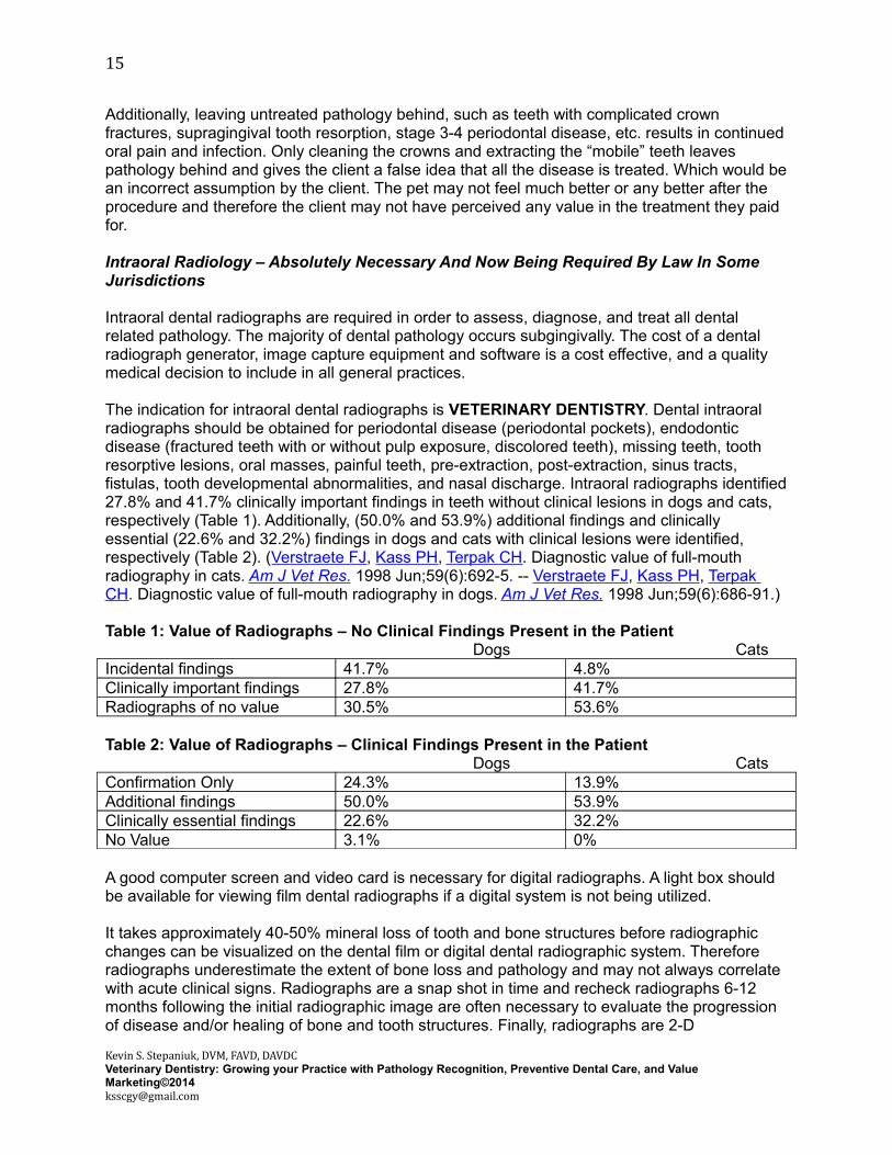
The indication for intraoral dental radiographs is VETERINARY DENTISTRY. Dental intraoral radiographs should be obtained for periodontal disease (periodontal pockets), endodontic disease (fractured teeth with or without pulp exposure, discolored teeth), missing teeth, tooth resorptive lesions, oral masses, painful teeth, pre-extraction, post-extraction, sinus tracts, fistulas, tooth developmental abnormalities, and nasal discharge. Intraoral radiographs identified27.8% and 41.7% clinically important findings in teeth without clinical lesions in dogs and cats, respectively (Table 1). Additionally, (50.0% and 53.9%) additional findings and clinically essential (22.6% and 32.2%) findings in dogs and cats with clinical lesions were identified, respectively (Table 2). (Verstraete FJ, Kass PH, Terpak CH. Diagnostic value of full-mouth radiography in cats. Am J Vet Res . 1998 Jun;59(6):692-5. -- Verstraete FJ, Kass PH, Terpak CH. Diagnostic value of full-mouth radiography in dogs. Am J Vet Res. 1998 Jun;59(6):686-91.)
Table 1: Value of Radiographs – No Clinical Findings Present in the PatientDogs Cats
Incidental findings 41.7% 4.8%Clinically important findings 27.8% 41.7%Radiographs of no value 30.5% 53.6%
Table 2: Value of Radiographs – Clinical Findings Present in the PatientDogs Cats
Confirmation Only 24.3% 13.9%Additional findings 50.0% 53.9%Clinically essential findings 22.6% 32.2%No Value 3.1% 0%
A good computer screen and video card is necessary for digital radiographs. A light box should be available for viewing film dental radiographs if a digital system is not being utilized.
It takes approximately 40-50% mineral loss of tooth and bone structures before radiographic changes can be visualized on the dental film or digital dental radiographic system. Therefore radiographs underestimate the extent of bone loss and pathology and may not always correlate with acute clinical signs. Radiographs are a snap shot in time and recheck radiographs 6-12 months following the initial radiographic image are often necessary to evaluate the progression of disease and/or healing of bone and tooth structures. Finally, radiographs are 2-D
Kevin S. Stepaniuk, DVM, FAVD, DAVDCVeterinary Dentistry: Growing your Practice with Pathology Recognition, Preventive Dental Care, and Value Marketing©[email protected]

16
representations of 3-D structures. Therefore, overlying structures causing summation and superimposition frequently create artifacts.
Selected References:1-11
1. Bannon KM. Clinical canine dental radiography. Vet Clin North Am Small Anim Pract 2013;43(3):507-532. 2. Coffman CR, Brigden GM. Oral and dental imaging equipment and techniques for small animals. Vet Clin North Am Small Anim Pract 2013;43(3):489-506. 3. Lemmons M. Clinical feline dental radiography. Vet Clin North Am Small Anim Pract 2013;43(3):533-554. 4. Lommer MJ, Verstraete FJ. Radiographic patterns of periodontitis in cats: 147 cases (1998-1999). J Am Vet Med Assoc 2001;218(2):230-234. 5. Lommer MJ, Verstraete FJ. Prevalence of odontoclastic resorption lesions and periapical radiographic lucencies in cats: 265 cases (1995-1998). J Am Vet Med Assoc 2000;217(12):1866-1869. 6. Peralta S, Verstraete FJ, Kass PH. Radiographic evaluation of the classification of the extent of tooth resorption in dogs. Am J Vet Res 2010;71(7):794-798. 7. Peralta S, Verstraete FJ, Kass PH. Radiographic evaluation of the types of tooth resorption indogs. Am J Vet Res 2010;71(7):784-793. 8. Tsugawa AJ, Verstraete FJ, Kass PH, et al. Diagnostic value of the use of lateral and occlusalradiographic views in comparison with periodontal probing for the assessment of periodontal attachment of the canine teeth in dogs. Am J Vet Res 2003;64(3):255-261. 9. Verstraete FJ, Kass PH, Terpak CH. Diagnostic value of full-mouth radiography in cats. Am J Vet Res 1998;59(6):692-695. 10. Verstraete FJ, Kass PH, Terpak CH. Diagnostic value of full-mouth radiography in dogs. Am J Vet Res 1998;59(6):686-691. 11. Kim CG, Lee SY, Kim JW, et al. Assessment of dental abnormalities by full-mouth radiography in small breed dogs. J Am Anim Hosp Assoc 2013;49(1):23-30.
Not only are dental radiographs necessary for veterinary dentistry, they are one of the best cost effective diagnostic modalities in a veterinary practice. The cost of a dental radiographic generator, indirect or direct image capture device, and software can be recovered in less than 6-12 months in most general practices.
Is Your Eye A Microscope?I ask myself this question every day. Each week, I am surprised by a histological diagnosis of a biopsy sample or an intraoral radiograph of a tooth/bone despite viewing many normal reports and radiographs, many common diseases and radiographs, and many of the rare and bizarre pathologies as a specialist and as a tertiary referral specialist (during my academic position).
Full mouth intraoral radiographs are always recommended. Histopathology of all oral masses, non-healing extraction sites, and any unusual or uncommon roentgen signs of bone or teeth should be submitted. Unfortunately, not all pathologists are well versed in subtle differentiation and interpretation of oral pathology. Oral pathology is its own specialty within human pathology. Just as in veterinary pathology where certain pathologists are better skilled in dermatopathology, the same is true for oral pathology.
Kevin S. Stepaniuk, DVM, FAVD, DAVDCVeterinary Dentistry: Growing your Practice with Pathology Recognition, Preventive Dental Care, and Value Marketing©[email protected]
17
For example, I have seen severe feline tooth resorption and the associated radiographic descriptions submitted with small inadequate biopsies misdiagnosed as a suspect oral squamous cell carcinoma (SCC). This could be a “death sentence” in cat! Where as, second opinion, revealed a stage TR4c type 2 tooth resorption with an epithelial down growth, that is normal, into the tooth resorption after the crown had broken off. Histologically it appears as islands of epithelial cells, but will be lacking mitotic figures and have relatively uniform epithelial cell populations without cellular atypia, interspersed in the replacing tooth structure (dentin) and new bone replacing the tooth. This interpreted by itself or with a description of a resorbing root or misinterpreted as a “lysis” of a root may lead a pathologist to make a presumptive or suggestive histological diagnosis of SCC. Always, interpret histological findings in light of clinicalfindings and presentation, signalment, and radiographic findings and seek the opinion of a Diplomate of the American Veterinary College or a veterinary college that works or consults withif you have questions about a diagnosis or you get a presumptive or suggestive diagnosis from a pathologist. Similar examples can be said about “epulides”.
EquipmentIt is necessary to invest in appropriate hand instruments (e.g. curettes, scalers), a high-speed dental unit, and intraoral radiology. Individual recommendations are beyond the scope of this seminar and presentation.
Continuing EducationIt is necessary to invest in continuing education of yourself in regards to pathology recognition, treatment options, periodontal treatments, and surgical extraction skills. Likewise, investment in your technicians skills and knowledge to leverage them as part of the dental team for procedures they are licensed to perform in your province or state, and to assist in patient discharge and client education.
Alternative TreatmentsUnderstand that sometimes there are no treatment or medical alternatives. I veterinarian should be able and skilled to extract every tooth. Think of surgical extraction as an amputation or enucleation. The tooth may be saved with a root canal treatment or periodontal surgery but if the client is not willing or it is too late for a stage 4 periodontal diseased tooth, then extraction is necessary. Just as a hind limb amputation is necessary for a severely comminuted open femur fracture the client is not willing or able to have repaired due to financial constraints or availabilityfor specialty repair.
Antibiotics are not a monotherapy for odontogenic infections. The pathophysiology of both endodontic disease and periodontal disease preclude the use of antimicrobials as a monotherapy resulting in definitive treatment for odontogenic infections. If there were a magic bullet, spray, ointment, wipe, etc., would human dentistry not be utilizing these treatments? Dogs have been used as models for human dentistry for decades since the pathophysiology is similar. Yet, there is no magic bullet that replaces professional dental cleanings, treatment, and daily tooth brushing. The right and privilege you earned with your doctor of veterinary medicine degree.
Dental and Oral Pathology RecognitionIt is necessary to recognize and correctly identify oral and dental pathology. Some supragingivallesions may be only millimeters in size but have significant underlying pathology (e.g., parulides,complicated crown fractures, tooth resorption). Being able to identify these in the conscious and anesthetized patient is important. Understanding the pain, inflammation, infection, and associated morbidity they cause is necessary to advocate for the patient and gain pet parent Kevin S. Stepaniuk, DVM, FAVD, DAVDCVeterinary Dentistry: Growing your Practice with Pathology Recognition, Preventive Dental Care, and Value Marketing©[email protected]
18
compliance. Keep meticulous dental records and notes so that pathology can be monitored and evaluated each year during recommended annual wellness examinations.
All dental and oral pathology is beyond the scope of this seminar and speaker notes.
Pre-treatment Education and CounselingThe majority of periodontal pathology is hidden subgingivally. Likewise, endodontic pathology is first identified radiographically around the apex. Therefore, general anesthesia and intraoral radiographs are necessary for assessment, diagnosis, and treatment planning. With 42 individual patients in the dog mouth and 30 individual patients in the cat mouth it is very common to identify unexpected disease in the oral cavity.
There are different approaches to handle the scenario.
1. You can call the owner during the anesthetic episode to gain permission to treat and/or extract additional teeth. In my humble opinion (IMHO) this prolongs general anesthesia in the patient and at the same time increases the cost of anesthesia to the client while you are talking on the phone. That is assuming you can actually reach the client on the phone to gain permission. Finally, IMHO, it feels like high-pressure sales because their pet is under anesthesia and the pet parent needs to quickly make a decision.
2. You can complete the treatment plan as agreed to and recover the patient from anesthesia. Review the findings on the dental chart, photographs, and radiographs with the client and plan a second procedure. Sometimes this is necessary, even with planningas recommended below, when significant disease and pathology is identified. IMHO some clients can be frustrated they need to come back for a second procedure but if time is taken to review the findings, most clients will understand.
3. IMHO the preferred method is to create a broad treatment plan with a range of anticipated costs based on signalment, past dental history, conscious oral examination findings, and owner’s commitment to home care. This will create a wide range in anticipated costs so it is CRUCIAL that time is taken to educate the client regarding subgingival disease, the need for anesthesia, the need for intraoral radiographs, the timeit takes for proper surgical extractions with surgical flap closure, etc. Significant more time and effort is necessary prior to the procedure. However, most clients are more satisfied with honesty and integrity when you come in under the estimated costs rather than either adding on additional costs or your business having to consume the costs when additional treatments are given away to help the patient. The downside is this method does not lend itself to price shopping clients. However, at this time in veterinary dentistry you are not comparing apples to apples when price shopping veterinary dentistry. All that you can do is educate that client about the services you provide and why items like intravenous catheters, preoperative blood work, patient monitoring, patient thermoregulation, intraoral radiographs, dedicated staff member to patient monitoring, etc. are necessary. Give them the information and let them ask and decide if they wish to price shop. IMHO you are trying to build life long clients and provide quality patient care. I have learned that I cannot be everything to everybody.
Patient DischargeThis is a critical time of the day in potentially the busiest time of the day. To provide value to yourservices it is necessary to take the time as the doctor or with a leveraged trained, knowledgeable technician to review the findings, discharge instructions, post-operative medications, post-operative expectations, and recommendations in TYPE WRITTEN DISCHARGES. Kevin S. Stepaniuk, DVM, FAVD, DAVDCVeterinary Dentistry: Growing your Practice with Pathology Recognition, Preventive Dental Care, and Value Marketing©[email protected]
19
The client should be placed in private exam room. The intraoral radiographs and any photographs of pathology are reviewed. Written discharge instructions with appropriate intraoral radiographs and photographs inserted into the discharges are reviewed. Medications are reviewed. This is done PRIOR to payment of the invoice and WITHOUT the pet present in the room. The client is then escorted to the front team and payment is collected. After payment is collected, the patient is reunited with their pet parent. Be certain the patient is clean, the intravenous catheter is removed, and is ready to go home.
If the pet is returned to the client before discharges are reviewed the pet parents are often too distracted to really focus on the medical aspects and recommendations for their pet. We want what is best for the pet so it is important the pet parent knows what needs to be done and what is expected when the pet is discharged. Likewise, by reviewing the black and white radiographs it is literally and figuratively black and white where bone is missing in relation to infection. This would provide increased value if done prior to collection of payment.
The Commensal Team©A minimum of two individuals is necessary for veterinary dentistry. A dedicated staff member to monitor anesthesia (e.g. licensed and trained veterinary technician) and a veterinarian are necessary. Ideally, a 3rd staff member, such as a trained assistant, can help facilitate the day.
Dentistry procedures should be scheduled similar to any other surgical procedures. It is surgery!It is impossible for any staff member to safely induce, monitor anesthesia, and work in the oral cavity by himself/herself.
The commensal dental organism© works together. After the licensed technician induces anesthesia the veterinarian can examine the mouth. Then the technician can clean the teeth and obtain intraoral radiographs while the assistant, where legal, can monitor anesthesia. The doctor is viewing the radiographs and making a treatment plan, in conjunction with the oral exam findings, as the images are obtained. The veterinarian can prescribe post-operative medications such as analgesics and antibiotics as indicated. As the veterinarian extracts the teeth the assistant or technician monitor anesthesia and assist. While the other team member starts the typed discharge instructions. At this time, premedication can be administered to the next patient, as indicated. After the first patient is extubated and stable, the doctor calls the client to discuss the findings and schedule a discharge time. Meanwhile, the licensed technicianand assistant may be inducing the next patient for a procedure, obtaining intraoral radiographs, and starting the periodontal cleaning as licensed in each state or provinces individual practice act. With the team approach the patient can receive more efficient care and the clients can receive more efficient service.
There will be variations of the aforementioned scenario that may work differently in different states and provinces based on various delegations of duties in each individual practice act. However, the DOCTOR needs to be available for the entire procedure in the treatment area andnot trying to multitask appointments at the same time. In every practice act that I am aware of and reviewed, ONLY the DOCTOR can make a diagnosis and make a treatment recommendation/plan. Likewise, DOCTORS are the only individuals that can perform SURGERY in every practice act I have reviewed. Surgery is commonly defined as the branch of medicine that involves cutting, abrading, and changing body tissues and organs. Gingiva is a tissue. Mucosa is a tissue. Bone is a tissue. A tooth is a tissue. Only a doctor of veterinary medicine can perform an ovariohysterectomy on a cat. I would argue that is less technical compared to surgically extracting a mandibular canine tooth or a maxillary 4th premolar. Kevin S. Stepaniuk, DVM, FAVD, DAVDCVeterinary Dentistry: Growing your Practice with Pathology Recognition, Preventive Dental Care, and Value Marketing©[email protected]

20
How to Make it HappenChanges will not occur overnight. Depending where you are at in your current practice, it will depend on where you need to start.
What is crucial is that the leaders and decision makers have true “buy in” with a comprehensive dentistry program. Lip service without action may lead to frustration. Is every one willing to walk the walk? If you or the leaders and decision makers are not willing to fully commit financially and intellectually to a short and long-term plan to improve the quality care and service, it will be constant struggle for the non-decision maker to execute change. Believe me, I have been there. It may be necessary to invest in new equipment and radiographs. It will be necessary to invest in continuing education of the medical staff (e.g., doctors and technicians). Itmay not be necessary to invest in everyone. If you have a passionate doctor and technician, thismay be the starting point. If you are really lucky and have a collaborative hospital, most the dental procedures could be directed to that doctor and their team.
Recall the majority of us received little or no dental education, yet there are literally textbooks dedicated to just periodontal disease, endodontic disease, intraoral radiology, etc. Continuing education on pathology recognition, intraoral radiographic interpretation, and treatment recommendations is crucial. Knowing how to successfully extract teeth and prevent complications is also very important.
Internal MarketingProviding quality care to your staff and staff pets. Remember the front team has the first and lastcontact with the client. If they have seen great changes in their pets following successful dental procedures, they may provide unsolicited testimonials. In house training in dentistry topics such as analgesia, anesthesia, periodontal cleanings, instrument and equipment care, intraoral radiology positioning, and other topics should be considered. There are Diplomates such as myself that offer these types of services throughout North America.
External MarketingIt is important to educate your clients about different types of dental disease (e.g. periodontal disease, fractured teeth, tooth resorption in cats)
1. Client Education• Models, Brochures, Websites• Comparative Aspects to Human Dental Disease• White boards• Chalk Boards
2. Examination• Show client’s the pathology in their pets mouth during the conscious
examination, if possible• Use Appropriate Professional Language
3. Utilize Digital Technology to Review Dental Radiographs and Photographs• LED TV• Ipad or Tablet
4. Review discharges and information in a quiet and private examination room5. Written Discharges
• Provide medical information and diagnosis other than “dental disease”• Periodontal Stages
Kevin S. Stepaniuk, DVM, FAVD, DAVDCVeterinary Dentistry: Growing your Practice with Pathology Recognition, Preventive Dental Care, and Value Marketing©[email protected]

21
• Fractured Teeth• Etc.
• Provide medication instructions• Provide post-operative instructions
III. WHEN THINGS GO WRONG DURING EXTRACTIONS
Introduction“Extractions” are surgical procedures requiring significant training and skill. Surgery is defined as the treatment of disease, injury, or deformity by manual or instrumental operations, as the removal of diseased parts or tissue by cutting. Extraction is defined as the process or act of pulling or drawing out. In order to correctly “extract” teeth in veterinary patients, surgery must beperformed. “Extractions” are oral surgery and should be treated as such. Veterinarians should be performing “extractions”.
Armamentarium
As with all extractions the following list of equipment, in addition to intraoral radiographs and a high-speed dental unit, is necessary to be successful and professionally satisfied: 1) Protective masks, glasses, and gloves 2) Good lighting 3) An ergonomic work environment 4) Magnification (if possible) 5) Scalpel Blades (#15, #15c) 6) Scalpel Handle 7) Water Cooled High Speed Hand Piece 8) Dental Burs (Such as #330, #331, #1/2, #1, #2, #4, #701L, or a bur of your choice and a medium or fine diamond bur) 9) Dental Periosteal Elevators 10) Dental Luxators 11) Dental Elevators 12) Extraction Forceps 13) Root Tip Picks 14) Excavators/Curettes (to clean out the alveolus) 15) Tissue Forceps 16) Needle Holders 17) Absorbable Suture (4-0 or 5-0 poliglecaprone 25, 4-0 or 5-0 chromic gut, 4-0 or 5-0 polyglactin 910 (Vicryl-Rapid™) on a P-3 needle 18) Tongue depressor 19) Minnesota retractor 20) Suction 21) All instrumentation used delicately and with control – A finger stop should be near the tip of the working instrument to stop the instrument if it slips and 22) PATIENCE!
It is better to prevent iatrogenic complications with proper training, equipment, education, techniques, and patience compared to managing them after they occurred. However, understanding how to manage them if they do occur is important. Also, referral, as indicated for prevention and treatment should be considered in the best interest of the patient.
Iatrogenic Complications:
1. Fractured Mandible and Maxilla 2. Retained tooth roots3. Intrusion of tooth roots into mandibular canal, nasal cavity, infraorbital canal, and
maxillary recess4. Ocular penetration5. Maxillary lip entrapment6. Alveolar osteitis, osteomyelitis, osteonecrosis
Kevin S. Stepaniuk, DVM, FAVD, DAVDCVeterinary Dentistry: Growing your Practice with Pathology Recognition, Preventive Dental Care, and Value Marketing©[email protected]
22
7. Non-healing extraction site8. Hemorrhage?
Fractured Mandible and MaxillaCommon locations for iatrogenic fractures include the body of the mandible while extraction the mandibular first molar in the dog and the rostral mandible (with or without a mandibular symphyseal separation) during extraction of the mandibular canine teeth in the dog and cat. For endodontic disease (fractured tooth) root canal treatment is the best option compared to extraction in some patients. Intraoral radiographs prior to surgical extraction help identify significant bone loss secondary to odontogenic infection, tooth root resorption and ankylosis, and minimal ventral cortical bone (i.e. 1-2 mm normal) in small breed dogs.
Mandibular body fractures generally require interfragmentary wires and noninvasive maxillofacial fracture repair (e.g. interdental wiring and composite splint). Temporary stabilization with a tape muzzle is beneficial for the patient. Rostral mandibular canine tooth alveolar fractures may require noninvasive fracture repair. However, if there is 1) minimal fracture and mandibular displacement and 2) a normal occlusion exists, then closing the soft tissue and feeding soft food for 4-6 weeks may be sufficient to allow healing. These must be differentiated from symphyseal separations that are disruption of the fibrous symphysis betweenthe two mandibles. Incorrect repair of mandibular fractures and separations can make healing worse and result in nonunions and malunions requiring additional surgery and associated morbidity and expense.
Fractured Tooth RootsDuring extractions roots will break creating root fragments. Retained tooth roots (RTR) and retained root tips (RRT) should not be left behind in most circumstances. The majority of the time the teeth were being extracted to due significant periodontal disease, endodontic disease, or tooth resorption and leaving a root fragment results in leaving disease within the maxilla and maxilla. This will result in chronic hidden pain, inflammation, infection, and in some cases non-healing or delayed healing extraction sites. In some cases, a liability issue may result.
Fractured roots may occur secondary to variation in anatomy or pre-existing pathological damage to the root structure. Pre-operative intraoral radiographs help identify and plan the surgical approach for these teeth. Additionally, overzealous and impatient extraction techniques delivering force to quickly will fracture roots and even the maxillofacial bones. The goal is to remove the entire tooth from the alveolus with minimal trauma to the surrounding bone and tissue. The periodontal ligament attaches to the tooth and the alveolar bone in order to hold the tooth in the mouth. The ligament is designed to withstand short bursts of pressures/forces. The ligament must be fatigued in order to remove the tooth. Extracting the tooth is about patience and finesse not brute force. Finally, many teeth are not simply conical in shape and contain grooves that interlock with the bone via the periodontal ligament.
If the fracture of the root is located in the coronal half of the root, removal tends to have an easier approach. If necessary, a small amount of buccal bone can be removed. In some cases no additional bone will require removal because the root fragment is visible. In these cases, choose a smaller luxator or elevator and carefully continue the luxation/elevation similar to extraction of the tooth. An appropriate sized dental elevator/luxator (fits the curvature of the tooth root) is used with axial pressure to cut the periodontal ligament fibers. A major pitfall is not placing the instrument in the periodontal ligament space that results in crushing of alveolar bone. This delays extraction and results in unnecessary surgical trauma. Once the elevator is in position, rotation will cause the periodontal ligaments to stretch. Hold the position and wait (15-Kevin S. Stepaniuk, DVM, FAVD, DAVDCVeterinary Dentistry: Growing your Practice with Pathology Recognition, Preventive Dental Care, and Value Marketing©[email protected]

23
30 seconds). Bleeding (periodontal ligament is being damaged) should be noted. Repeat the procedure and use a smaller elevator/luxator that fits the curvature of the root as you work apically. Repeat again as needed. Take time to fatigue the ligament and damage the periodontalligament fibers. Once the root is sufficiently mobile, and only when it is mobile, do you reach for the extraction forceps. The tooth then can be grasped with extraction forceps and rotated on thelong axis. The whole tooth root should be present.
If the fracture of the root occurs in the apical 3rd additional techniques are often necessary. If a mucoperiosteal flap had not already been created for the surgical extraction, then creating a flapwill be necessary to remove additional buccal bone. Teeth with dilacerated roots or hypercementosis, identified on pre-operative radiographs, will often require additional bone removal and the following techniques.
Visualization of the root fragment will be necessary and a surgical assistant is needed to assist with suction and operation of the air-water syringe while the veterinarian localizes the root fragment in the alveolus. A root tip pick or a small 1-2 mm luxator, as indicated, is used to cut the periodontal ligament space around the root fragment. It is absolutely necessary to visualize the process so that excessive force does not intrude the root fragment into the nasal cavity or mandibular canal further complicating retrieval. Time and patience are necessary to visualize the root fragment.
In addition to root tip picks, the periodontal ligament space can be opened wider with the carefuland precise use of a high-speed bur. Generally a small round bur less than size #2. Preferably, a #1 or #1/2 round bur on a surgical length FG shaft is needed. With careful visualization the periodontal ligament space can be cut around the root fragment. Once a space is created, then small root tip picks and luxators can be used to remove the fragment.
“Burring out” or “atomizing” retained roots is not recommended and must not be done because damage to surrounding bone and neurovascular structures can occur. The retained root fragments can be retrieved with the use of root tip picks. The “whole tooth, and nothing but the tooth”, is removed.
The alveolus is cleaned with a curette, to remove granulation tissue, purulent debris, and bone fragments, and then lavaged with an oral 0.12% chlorhexidine gluconate solution. The margins of the alveolus and cortical bone are carefully smoothed with a medium diamond football shaped bur in order to remove rough spicules of bone.
Surgical margins are freshened so healthy, non-epithelialized margins are opposed. Extraction sites are sutured closed. The use of 4-0 or 5-0 poliglecaprone-25, chromic gut, or polyglactin 910 on a P-3 needle is used to suture the extraction site closed with a simple interrupted pattern.
Intentionally leaving root fragments is NEVER a first recommendation. Likewise, if there is any associated periapical lucency or other radiographic evidence of infection in the surrounding bone (vertical bone loss from periodontal disease), etc. the root fragments MUST be extracted. However, in RARE circumstances when the patient is unstable during the anesthetic episode, there is no radiographic evidence of infection (e.g., periapical lucency, vertical bone loss), the risk of removal may damage adjacent neurovascular structures (e.g., mandibular neurovascular bundle, maxillary artery/vein/nerve and ocular structures), and the root fragment is estimated to be less than 3 mm, then intentional root retention may be considered. In these cases 1) the thought process for retention of the root must be well documented and described in the medical Kevin S. Stepaniuk, DVM, FAVD, DAVDCVeterinary Dentistry: Growing your Practice with Pathology Recognition, Preventive Dental Care, and Value Marketing©[email protected]

24
record 2) a post-operative radiograph documenting the intentionally retained root must be obtained and documented in the patient record 3) the client must be informed verbally and documented on the client discharge of the intentionally retained root and the judgment to leave the retained root was made in order to “First do no harm” or additional harm that may have resulted with additional surgery and/or anesthesia time 4) the client needs to be advised if the surgical site does not heal or additional swellings or draining tracts are noted, that the veterinarian should be contacted immediately and 5) the patient should be scheduled in 6 months and then regular intervals to document for any radiographic evidence of infection/inflammation, if at those times infection is noted, the tooth fragment should be surgically removed, if the tooth fragment is being replaced by bone and there is no radiographic evidence of infection then subsequent radiographic monitoring is recommended.
Appropriate multimodal post-operative pain management is prescribed for 5-7 days. The patient should be fed soft food for 7-10 days. An oral 0.12% chlorhexidine gluconate rinse PO BID for 7-10 days should be prescribed or until brushing. Appropriate antibiotics (amoxicillin-clavulanic acid or clindamycin) are prescribed for 7-10 days as indicated. A recheck of the surgical site in 10-14 days is recommended.
Intrusion of Roots and Root TipsIntrusion of root tips into the mandibular canal, infraorbital canal, maxillary recess, and nasal cavity should be avoided. If intrusion occurs and the root tip cannot be safely retrieved, referral to a veterinary dentist is recommended for removal of the root tips as soon as possible. Fibrosis with delayed referral can make the removal even harder.
As soon as the intrusion occurs, STOP. The first instinct is to try to grab the root tip which generally results in pushing the tooth fragment rostral or caudal and making it more difficult to retrieve and/or further damaging neurovascular bundles. OBTAIN an intraoral radiograph to document and verify the location of the tooth root. Use suction with a Frazier suction tip visualize the region. Create a larger opening in the bone where the tip was intruded and use thesuction and delicate tissue forceps to gently remove the fragment. A larger surgical window will need to be created if the fragment has moved rostral or caudal. Obtain a post-surgical radiograph documenting the removal of the root fragment. First DO NOT HARM. Refer when indicated.
Ocular Penetration
Slipping of surgical instruments and orbital penetration should not occur with proper surgical technique but has been reported. Worse, intracranial penetration can occur. If orbital penetrationoccurs immediately treat the patient with appropriate systemic antibiotics, appropriate systemic non-steroidal anti-inflammatory medications and facilitate and emergency referral to an ophthalmologist within hours in order to have a chance to save the eye. If intracranial penetration occurs, emergency referral to a neurologist is necessary.
Maxillary Lip EntrapmentMaxillary lip entrapment by the mandibular canine teeth following mandibular canine tooth extraction can occur in cats and dogs. Excessive buccal bone removal, tension on the flap, and conformation potentiate the problem. If non-healing maxillary dermal abrasions occur, crown reduction and vital pulpotomy or extraction of the offending mandibular canine tooth/teeth is necessary to resolve the complication. Prevention with appropriate tension free surgical flaps is necessary.
Kevin S. Stepaniuk, DVM, FAVD, DAVDCVeterinary Dentistry: Growing your Practice with Pathology Recognition, Preventive Dental Care, and Value Marketing©[email protected]

25
Alveolar osteitis, osteomyelitis, osteonecrosisThere are many myths and misconceptions that have been, and continue to be, propagated in veterinary dentistry and oral surgery due to general, collective, historical lack of veterinary dental education within veterinary colleges. One of these is that extraction sites need to be “left open to drain”. This is a fallacy as removal of the infected tooth, essentially a foreign body, and alveolar wound care prior to surgical closure results in primary healing that rapidly returns the patient to function. Additionally, dogs and cats are not human and the human oral surgeons mayleave extraction sites open as they process large number of patients through there operatory each day but rely on the patient following strict post-operative care guidelines. Even with strict guidelines and compliance, human extractions sites may take 4 +/- weeks to heal and re-epithelize depending on the extent of oral surgery and individual factors. Frequently, human patients may require revision surgery or periodontal surgery to repair defects later in life (e.g., distal aspect of the mandibular 2nd molar following “wisdom” tooth extraction). It is impossible, tohave the strict diet and home care compliance necessary in a dog and cat.
The majority of veterinary extractions are surgical and dog and cat anatomy often requires mucoperiosteal flaps with significant exposed bone and soft tissue incisions in comparison to human extractions. The post-op oral hygiene and re-injury cannot be controlled in dogs and catsas it can in a human patient. Disruption and loss of the fibrin clot disruption will lead to a second costly anesthesia and surgery or worse, necrosis of maxillary and mandibular bone in some cases. Patient pain and comfort is improved with stabilization of fibrin clot and more rapid primary intention healing vs. second intention wound healing. Disastrous periodontal defects (e.g., mesial to the mandibular canine tooth when the mandibular 3rd incisor is extracted without closure) are prevented. Finally, dogs and cats ARE NOT HUMANS!
Exposed alveoli can result in very painful and potentially life threatening systemic conditions. Alveolar osteitis (inflammation of the bone), osteomyelitis (inflammation and infection of the bone) and/or osteonecrosis (devitalized and death of the bone) can occur. All require additional surgery, surgical debridement, closure with an appropriate tension free mucoperiosteal flap, andmedical treatment with antibiotics as indicated.
Non-healing Extraction SiteA non-healing extraction site can occur secondary to non-closure or incomplete closure of the extraction site, infected retained tooth roots, tooth fragments from resorbing and replacing roots protruding above the marginal bone, bone sequestrum from damaged bone during extraction (i.e. traumatic, thermal, chemical, pharmaceutical, biomaterials), or neoplasia. Intraoral radiographs of the site are obtained. If a there is remaining tooth structure or bone interfering with healing, it is removed. If there were products placed in the alveolus, they must be removed.The region should be biopsied to rule out cancer. Key Point: Be certain the biopsy site is representative! Small superficial biopsies will often be read as variants of lymphoplasmacytic and/or neutrophilic gingivitis/stomatitis with the deeper neoplastic region being missed.
HemorrhageMajor neurovascular structures are in immediate proximity to, adjacent to, and/or encircling the roots of many teeth (infraorbital, palatine, mandibular, and mental neurovascular bundles). Extensive hemorrhage is possible if the maxillary, infraorbital, palatine, or mandibular arteries are damaged during extraction. Additionally, neuropathies are possible if the nerves are damaged. However, if you hit a major vessel, blood will squirt or well up very rapidly, and this is not common. More common, is oozing from the oral tissues or nasal turbinates, when compromised.
Kevin S. Stepaniuk, DVM, FAVD, DAVDCVeterinary Dentistry: Growing your Practice with Pathology Recognition, Preventive Dental Care, and Value Marketing©[email protected]
26
The mouth and nasal turbinates have a tremendous vascular supply. Chronic inflammation and inflammatory tissue bleeds. Work diligently, purposefully, effectively, and efficiently when surgical extracting teeth, debriding and excising inflammatory tissue, creating the mucoperiosteal flap, and closing the surgical site. Allow the normal primary and secondary hemostatic systems to do the job and create a FIBRIN clot. The only time I require assistance inhemostasis is if there is an actual artery (i.e. palatal or infraorbital) squirting blood that requires ligation. I do not use or recommend any marketed product to help with hemostasis in the oral cavity. Ask the question is it necessary? Will closure of the site resolve the issue?
Kevin S. Stepaniuk, DVM, FAVD, DAVDCVeterinary Dentistry: Growing your Practice with Pathology Recognition, Preventive Dental Care, and Value Marketing©[email protected]