
Vertically and horizontally-transmitted memories –...
12
HYPOTHESIS Vertically and horizontally-transmitted memories – the fading boundaries between regeneration and inheritance in planaria Moran Neuhof 1, *, Michael Levin 2, * and Oded Rechavi 1,2,3, * ABSTRACT The Weismann barrier postulates that genetic information passes only from the germline to the soma and not in reverse, thus providing an obstacle to the inheritance of acquired traits. Certain organisms such as planaria – flatworms that can reproduce through asymmetric fission – avoid the limitations of this barrier, thus blurring the distinction between the processes of inheritance and development. In this paper, we re-evaluate canonical ideas about the interaction between developmental, genetic and evolutionary processes through the lens of planaria. Biased distribution of epigenetic effects in asymmetrically produced parts of a regenerating organism could increase variation and therefore affect the species’ evolution. The maintenance and fixing of somatic experiences, encoded via stable biochemical or physiological states, may contribute to evolutionary processes in the absence of classically defined generations. We discuss different mechanisms that could induce asymmetry between the two organisms that eventually develop from the regenerating parts, including one particularly fascinating source – the potential capacity of the brain to produce long-lasting epigenetic changes. KEY WORDS: Planaria, Regeneration, Memory, Inheritance, Epigenetics, Evolution, Generations, Transgenerational, Small RNAs, Chromatin Introduction Most models of evolution, which are based on Mendelian genetics, depend conceptually on the existence of a distinct separation between generations across an ancestry. This distinction between parents and children is supposedly enforced by Weismann’s barrier, which in theory precludes information transfer from the soma to the germline, and thus prevents inheritance of parentally-acquired traits (Poulton et al., 1889; Sabour and Schöler, 2012). The germline, according to this framework, is conceived as a ‘bottleneck’, which filters out epigenetic responses. In other words, all the changes that affect somatic cells, whether epigenetic or genetic (e.g. mutations, transpositions), are erased in the next generation. Lamarck’s discarded theory of evolution, according to which somatic responses (and acquired traits) are carried over to the progeny, assumed a continuation between the generations, and until recently was considered to be entirely incorrect (Jablonka and Lamb, 2015). New discoveries in the field of epigenetics, some of which will be discussed here, suggest the need for reexamination of these original ideas in a new light. As unicellular organisms have been shown to preserve cellular states over generations (Zacharioudakis et al., 2007), Weismann’s barrier as originally suggested is relevant to organisms that have a well-defined and segregated germline (namely, only specific, designated cells will become germ cells). However, do similar restrictions on the process of evolution apply to plants (where the germline is not segregated), or to the many phyla of animals that can reproduce asexually without going through a germline bottleneck? Even in metazoans, which segregate their germline and for which Weismann’s barrier is supposedly relevant, different mechanisms are used to specify the primordial germ cells (Extavour, 2003). These different mechanisms allow different degrees of communication between the parent’s environment and the germline. Recent evidence suggests that the variance between germline specification mechanisms could influence the process of evolution, and specifically, that a continuity with the previous generation could accelerate evolution. For example, it was shown that genes evolve faster in amphibians that define their germline by using maternally inherited determinants (‘preformation’), in comparison to the rates of gene evolution seen in related organisms that define their germline by inductive signals (‘epigenesis’), without inheriting ‘germplasm’ (which should be affected by the environment) from the mother (Evans et al., 2014). One asexually reproducing animal, on which we will focus in this paper, which presents an interesting challenge to the Weismann barrier, is planaria. Planarians are an order of free-living flatworms which are complex bilaterians possessing a wide range of cell types, a true centralized brain, and a complex repertoire of behavioral responses (Saló et al., 2009). Planaria have advanced mechanisms of regeneration, and are able to coordinate their resident population of stem cells to recreate any portion of the animal that is surgically removed, including their brain, throughout adulthood (Roberts- Galbraith and Newmark, 2015). These attributes have made it a popular model system for studies of stem cell regulation, morphogenesis, behavioral plasticity, and physiological signaling (Gentile et al., 2011; Nicolas et al., 2008; Shomrat and Levin, 2013). While many of the common planarian species (which are grown in the lab and are considered model organisms for regeneration) can reproduce sexually (Cardona et al., 2006), they most frequently reproduce asexually through fission followed by regeneration. Upon bisection (whether externally induced or self-initiated), a structure called the ‘blastema’ forms in each fragment (Birnbaum and Sánchez Alvarado, 2008). The blastema gives rise to new tissues, and a process of remodeling then scales both new and existing structures appropriately (Beane et al., 2013). When a head fragment regenerates its missing tail, or when a tail fragment regenerates a missing head, new cells differentiate from pluripotent stem cells Received 15 June 2016; Accepted 6 July 2016 1 Department of Neurobiology, Wise Faculty of Life Sciences, Tel Aviv University, Tel Aviv 69978, Israel. 2 Allen Discovery Center, Tufts University, 200 Boston Avenue, Suite 4600, Medford, MA 02155, USA. 3 Sagol School of Neuroscience, Tel Aviv University, Tel Aviv 69978, Israel. *Authors for correspondence ([email protected]; [email protected]; [email protected]) This is an Open Access article distributed under the terms of the Creative Commons Attribution License (http://creativecommons.org/licenses/by/3.0), which permits unrestricted use, distribution and reproduction in any medium provided that the original work is properly attributed. 1 © 2016. Published by The Company of Biologists Ltd | Biology Open (2016) 0, 1-12 doi:10.1242/bio.020149 Biology Open • Advance article by guest on June 30, 2018 http://bio.biologists.org/ Downloaded from
Transcript of Vertically and horizontally-transmitted memories –...

HYPOTHESIS
Vertically and horizontally-transmitted memories – the fadingboundaries between regeneration and inheritance in planariaMoran Neuhof1,*, Michael Levin2,* and Oded Rechavi1,2,3,*
ABSTRACTThe Weismann barrier postulates that genetic information passesonly from the germline to the soma and not in reverse, thus providingan obstacle to the inheritance of acquired traits. Certain organismssuch as planaria – flatworms that can reproduce through asymmetricfission – avoid the limitations of this barrier, thus blurring thedistinction between the processes of inheritance and development.In this paper, we re-evaluate canonical ideas about the interactionbetween developmental, genetic and evolutionary processes throughthe lens of planaria. Biased distribution of epigenetic effects inasymmetrically produced parts of a regenerating organism couldincrease variation and therefore affect the species’ evolution. Themaintenance and fixing of somatic experiences, encoded via stablebiochemical or physiological states, may contribute to evolutionaryprocesses in the absence of classically defined generations. Wediscuss different mechanisms that could induce asymmetry betweenthe two organisms that eventually develop from the regeneratingparts, including one particularly fascinating source – the potentialcapacity of the brain to produce long-lasting epigenetic changes.
KEY WORDS: Planaria, Regeneration, Memory, Inheritance,Epigenetics, Evolution, Generations, Transgenerational, SmallRNAs, Chromatin
IntroductionMost models of evolution, which are based on Mendelian genetics,depend conceptually on the existence of a distinct separationbetween generations across an ancestry. This distinction betweenparents and children is supposedly enforced byWeismann’s barrier,which in theory precludes information transfer from the soma to thegermline, and thus prevents inheritance of parentally-acquired traits(Poulton et al., 1889; Sabour and Schöler, 2012). The germline,according to this framework, is conceived as a ‘bottleneck’, whichfilters out epigenetic responses. In other words, all the changes thataffect somatic cells, whether epigenetic or genetic (e.g. mutations,transpositions), are erased in the next generation. Lamarck’sdiscarded theory of evolution, according to which somaticresponses (and acquired traits) are carried over to the progeny,assumed a continuation between the generations, and until recentlywas considered to be entirely incorrect (Jablonka and Lamb, 2015).
New discoveries in the field of epigenetics, some of which will bediscussed here, suggest the need for reexamination of these originalideas in a new light.
As unicellular organisms have been shown to preserve cellularstates over generations (Zacharioudakis et al., 2007), Weismann’sbarrier as originally suggested is relevant to organisms that have awell-defined and segregated germline (namely, only specific,designated cells will become germ cells). However, do similarrestrictions on the process of evolution apply to plants (where thegermline is not segregated), or to the many phyla of animals that canreproduce asexually without going through a germline bottleneck?
Even in metazoans, which segregate their germline and forwhich Weismann’s barrier is supposedly relevant, differentmechanisms are used to specify the primordial germ cells(Extavour, 2003). These different mechanisms allow differentdegrees of communication between the parent’s environment andthe germline. Recent evidence suggests that the variance betweengermline specification mechanisms could influence the process ofevolution, and specifically, that a continuity with the previousgeneration could accelerate evolution. For example, it was shownthat genes evolve faster in amphibians that define their germline byusing maternally inherited determinants (‘preformation’), incomparison to the rates of gene evolution seen in relatedorganisms that define their germline by inductive signals(‘epigenesis’), without inheriting ‘germplasm’ (which should beaffected by the environment) from the mother (Evans et al., 2014).
One asexually reproducing animal, on which wewill focus in thispaper, which presents an interesting challenge to the Weismannbarrier, is planaria. Planarians are an order of free-living flatwormswhich are complex bilaterians possessing a wide range of cell types,a true centralized brain, and a complex repertoire of behavioralresponses (Saló et al., 2009). Planaria have advanced mechanismsof regeneration, and are able to coordinate their resident populationof stem cells to recreate any portion of the animal that is surgicallyremoved, including their brain, throughout adulthood (Roberts-Galbraith and Newmark, 2015). These attributes have made it apopular model system for studies of stem cell regulation,morphogenesis, behavioral plasticity, and physiological signaling(Gentile et al., 2011; Nicolas et al., 2008; Shomrat and Levin,2013).
While many of the common planarian species (which are grownin the lab and are considered model organisms for regeneration) canreproduce sexually (Cardona et al., 2006), they most frequentlyreproduce asexually through fission followed by regeneration. Uponbisection (whether externally induced or self-initiated), a structurecalled the ‘blastema’ forms in each fragment (Birnbaum andSánchez Alvarado, 2008). The blastema gives rise to new tissues,and a process of remodeling then scales both new and existingstructures appropriately (Beane et al., 2013). When a head fragmentregenerates its missing tail, or when a tail fragment regenerates amissing head, new cells differentiate from pluripotent stem cellsReceived 15 June 2016; Accepted 6 July 2016
1Department of Neurobiology, Wise Faculty of Life Sciences, Tel Aviv University,Tel Aviv 69978, Israel. 2Allen Discovery Center, Tufts University, 200 BostonAvenue, Suite 4600, Medford, MA 02155, USA. 3Sagol School of Neuroscience,Tel Aviv University, Tel Aviv 69978, Israel.
*Authors for correspondence ([email protected]; [email protected];[email protected])
This is an Open Access article distributed under the terms of the Creative Commons AttributionLicense (http://creativecommons.org/licenses/by/3.0), which permits unrestricted use,distribution and reproduction in any medium provided that the original work is properly attributed.
1
© 2016. Published by The Company of Biologists Ltd | Biology Open (2016) 0, 1-12 doi:10.1242/bio.020149
BiologyOpen
•Adva
nce
article
by guest on June 30, 2018http://bio.biologists.org/Downloaded from
known as ‘neoblasts’. These unique cells are required forregeneration, and also for the continuous remodeling andmorphological rescaling observed in intact worms during growthand starvation (Oviedo et al., 2003). The neoblasts are instructedboth by intrinsic state (cell-autonomous pathways) and informationfrom surrounding cells (Oviedo and Levin, 2007; Oviedo et al.,2010; Wagner et al., 2011; Witchley et al., 2013).Here, we explore a number of scenarios that could potentially
defy classical models of evolution. Specifically, we ask whether inplanaria and other organisms that reproduce by fission, differenttypes of epigenetic information are asymmetrically passed acrossgenerations. Such stored information, which can be regarded asmemory (see more below and in the glossary), could play manycrucial roles in regulating behavioral and developmental patterns. Inthis manuscript wewill discuss different types of memories that maypersist upon regeneration/inheritance; memories of gene activity,memories which are encoded in the connectivity of neuronalcircuits, and memories of non-neural physiological states.In the broadest sense of the word, memory is what enables
altering of future responses based on history. Biological memory isencoded at many levels: metabolic differences (Cameron et al.,2012; Ros et al., 2006), epigenetic factors (e.g. small RNAs, histonemarks, DNA methylation and prions) (Bird, 2002; D’Urso andBrickner, 2014; Iwasaki and Paszkowski, 2014), stable bioelectricalcircuit modes (Cervera et al., 2014; Law and Levin, 2015), orneuronally-encoded memories (Axmacher et al., 2006; Daoudal andDebanne, 2003; Herry and Johansen, 2014; Maren and Quirk, 2004;Zhang and Linden, 2003). A myriad of mechanisms exist to allowmolecules, molecular pathways, cells, and cellular networks totransduce physiological or behavioral inputs (experiences) intostable state changes that guide future activities. In this sense,processes that ensure the persistence of different developmentalfates or trajectories are also forms of memory.The Weismann barrier is relevant to asexual organisms as well,
because the issue is not only which cells will contribute to the next
generation, but whether and how the life history of the body getspermanently encoded in cells so as to significantly alter theoffspring in a stable manner. Indeed, the potential breaching of theWeismann barrier in planaria has previously been considered, in thecontext of tracking the source of the cellular contents of neoblaststhat form a new organism (Solana, 2013). However, couldparentally-produced alterations that encode biological memorybreach Weismann’s barrier and persist across generations? Even ifinformation could travel from somatic tissues to the germline,several rounds of reprogramming events (in the germline and in theembryo) were previously thought to prevent the inheritance ofepigenetic memory in animals (Mann and Bartolomei, 2002;Messerschmidt et al., 2014; Morgan et al., 2005; Vucetic et al.,2010). Nevertheless, in recent years it has become clear thatcomplex and still poorly understood regulatory processes determinewhich epigenetic memories would persist, and which would beerased across generations. The removal of DNA cytosinemethylation and histone marks during embryogenesis was thoughtto ‘clean’ the embryo of epigenetic modifications that were presenton its parents’ genome. The addition of de novo chromatinmodifications in the next generation was similarly thought todepend solely on the current environmental conditions, and thedictation of the hard-wired, genomically-encoded developmentalprogram. However, reprogramming is not complete and a fewparental marks escape removal (Hackett et al., 2013).
How widespread are heritable memories and what types ofmemories avoid reprogramming? We will explore these questionsthrough planaria, by focusing on the events that take place whenanimals reproduce by fission.
HypothesisWe hypothesize that the asymmetric fission of planaria, and similarorganisms, and the resulting genetic and epigenetic differences inthe individuals that regenerate from the different fragments, cancreate stable variation and therefore participate in the process ofevolution.
Reproduction as regenerationA generation can be defined as ‘a single step in natural descent’(http://www.dictionary.com/, accessed 2015). In planarian asexualreproduction, this definition does not necessarily apply, since afterfission the relationship between the two resulting individuals doesnot display a clear hierarchy – which half is the ‘parent’ and whichhalf is the ‘child’? Is one half ‘older’ than the other? Despite theseambiguities, we suggest that parentally-acquired information (theresult of the parent’s life experiences) could be transmitted from theworm that underwent splitting to the two organisms that form uponregeneration, and therefore the term ‘inheritance’ is relevant whendiscussing fission. The term ‘genetics’ could also be relevant in thisregard, although, as will be elaborated below, the information that isinherited from the parent might not be restricted to changes in genes.
Fission and regeneration in planaria involve long-rangeinstructive communication among cells (a signaling mode that canfacilitate breaches of Weismann’s barrier). When a worm isbisected, cells on the anterior- and posterior-facing sides of thecut must form a tail and head, respectively; the cut plane separatescells that were adjacent neighbors, and therefore had essentiallythe same positional information, yet these generate completelydifferent anatomical structures. Thus, cell position (the localmicroenvironment) does not uniquely dictate the appropriatemorphological outcome; instead, cells must communicate withother remaining tissues in order to determine which structures each
GlossaryMemory: retention of information about a state of affairs for some timeperiod; the ability of a system to specifically alter some aspect of a labilemedium in response to stimuli, such that future responses to stimuli arealtered. Memory requires latency between stimulus and salientresponse.Epigenetic modifications: defined here as factors that alter thephenotype that are not stored in the genetic code, including but notlimited to DNA methylation, histone modifications and small RNAs.Bioelectric network/circuit: a group of cells, not restricted to neurons/muscle, often connected by gap junctions, which communicate via slowchanges in resting potential and endogenous electric fields, whichregulates cell state and large-scale morphogenesis.Maternal effects: factors that alter the phenotype of the progeny thatdepend on the maternal environment, including genetic, epigenetic andphysiological effects.Epimutations: as opposed to DNA mutation, an epimutation is amolecular alteration to the DNA that does not alter the DNA sequencethat can be stably transmitted across generations. Most commonly refersto differences in cytosine methylations between certain alleles.Epimutations can be segregated with the chromosomes in accordancewith Mendel’s roles.Plant embryo: a phylogenetically conserved structure that developsfrom the zygote containing the shoot and root apical meristems, and theprimordial tissues that will differentiate into tissues of the mature plant.Meristem: in plant biology, meristems are self-maintaining structures ofundifferentiated cells from which plant organs develop.
2
HYPOTHESIS Biology Open (2016) 0, 1-12 doi:10.1242/bio.020149
BiologyOpen
•Adva
nce
article
by guest on June 30, 2018http://bio.biologists.org/Downloaded from
blastema needs to build (Nogi and Levin, 2005; Oviedo et al., 2010;Reddien and Sánchez Alvarado, 2004). A similar long-range, highlyintegrated pattern control is seen in amphibians, where tailstransplanted to the side of a salamander eventually remodel tolimbs (including the transformation of the tail tip into fingers, whichreveals that tissues can change their morphological structure inresponse to global patterning cues) (Farinella-Ferruzza, 1956).The process of regeneration is essentially one of cell networks
processing information about large-scale growth and form. A focuson information reveals an interesting analogy between generationaldescent and regeneration; that of space versus time. With classicalgenerational inheritance, patterning information is passed ontemporally from parent to offspring via the genome, conservedwith high fidelity and yet susceptible to environmental influence. Inregeneration, in addition to its temporal progression, instructiveinformation is also propagated spatially, from the rest of the body toa wound region and thus to new tissues; planarian regeneration is aremarkable example of how these two distinct but highly parallelpattern control processes converge. It should be noted that while wefocus on planaria as a uniquely tractable model for these studies,stable modifications to regenerative pattern occur also in mammals(trophic memory in deer antlers) and other invertebrate systems suchas crab limbs (reviewed in Lobo et al., 2014).The parallelism between development and regeneration is also
seen at the cellular level, as manifested in the similarities betweengerm cells and the stem cells that enable regeneration in planaria(Solana, 2013). In asexual reproduction, both tail and headfragments regenerate their missing tissues through theproliferation and differentiation of neoblasts. Thus, whenplanarians reproduce asexually, the new generation does notoriginate from one cell, but from a ‘community of cells’(generation/regeneration of a worm from a single neoblast withouta surrounding mature body has never been shown). Becausegenomic changes arise during cell division, and as a result of DNAdamage of different sorts, this ‘cell community’ is expected to becomposed of a mixture of different neoblasts, and also fromgenomically-different surrounding cells, which are not totipotent. Itwas recently demonstrated that the different neoblasts are notcompletely genetically identical – even in the same individual, alarge number of mutations and SNPs differentiate between neoblasts(Nishimura et al., 2015). Moreover, it is not clear that theinformation that is required for regeneration (where, when, andhow much to make of the new cell types, how to arrange those newtissues in correct geometric patterns, and crucially, when to stopgrowing) is present in the neoblasts; thus, the genetic variance in thesurrounding cells could also be crucial, and differentiate theorganisms that grow from the two regenerating halves. The barriersto the interaction between the surrounding cells and the neoblastsare also analogical to the Weismann barrier, between somatic cellsand germ cells.Similarly to the germ cells of other animals, planarian neoblasts
(unlike other cell types) express PIWI homologues (Friedländeret al., 2009). In other organisms, PIWI proteins, and PIWI-associated small RNAs, or piRNAs, are important for maintainingthe immortality of the germline (Meister, 2013), and their role insomatic tissues in less clear (Rajasethupathy et al., 2012). InCaenorhabditis elegans, for example, animals without a germlineare virtually devoid of piRNAs (Bagijn et al., 2012). PIWI proteinsand piRNAs play a critical role in the silencing of transposons andenable distinction between ‘self’ and ‘foreign’ genes, and thereforepreserve the progeny’s genome (Rechavi, 2014). The heritablesmall RNA pool, which includes piRNAs and other types of small
RNAs (e.g. endo-siRNAs in C. elegans) (Claycomb, 2014; Gentet al., 2010; Rechavi et al., 2014; Vasale et al., 2010), and tRNA-fragments in mice (Chen et al., 2016; Liao et al., 2014; Peng et al.,2012; Sharma and Rando, 2014), constitutes a germline ‘memorybank’ of sequences that were found in past generations to be‘dangerous’ (mobile parasitic DNA elements) or ‘safe’ (genes thatneed to be expressed in the germline). Transmission of piRNAs toprogeny ensures that transposons will not jump, thus preventingdisruption of the germline’s genome, and ensuring error-prooftransgenerational information transfer (Malone and Hannon, 2009).Neoblasts, which grant planarians their powerful ability toregenerate endlessly, express PIWI proteins and piRNAs (Reddienet al., 2005), and were recently shown, like germ cells, to usepiRNAs to preserve the integrity of their genomic heritage (Zhouet al., 2015).
Asymmetry and memoryAsymmetric retention or erasure of cellular memory, after celldivision, is an important and well-studied mechanism indevelopment, crucial both for renewal of pluripotency/proliferation, and for differentiation and establishment of cell fate(Armakolas et al., 2010; Di Laurenzio et al., 1996; Jan and Jan,1998; Klar, 1987). Asymmetric cell division (in neurons and othercell types) is also used as a mechanism for preventing aggregated,damaged or misfolded proteins from being inherited to the cellprogeny by confining them to only one daughter-cell (Ogrodniket al., 2014). A similar phenomenon is familiar in budding yeast,where asymmetric division results in two daughter-cells; one ofthem contains large amounts of unfolded and aggregated proteins,usually associated with aging, while the other remains ‘young’(Spokoini et al., 2012).
Similarly, asymmetric fission of an entire multicellular organism,such as planaria, could result in asymmetric inheritance of cellswhich, in theory, could have distinct expression patterns maintainedby cell-specific epigenetic states. Could the uneven inheritance ofepigenetic effects make the organisms that develop from the twoseparate fragments phenotypically unequal?
Indeed, planarian ‘clones’ that regenerate from fragments of asingle animal and that live in the same container, can show variableresponses to an external perturbation such as a pharmacologicalcompound (Beane et al., 2011; Chan et al., 2014; Oviedo et al.,2010). At the molecular level, fission and the ensuing recreation of anew individual in planarians may not necessarily entail complete‘resetting’ of modifications (such as histone marks, RNA contentand synaptic connection strengths) that were acquired by theprevious ‘generation’. Asymmetric fission could therefore be amechanism that enables retention of life history memories; someepigenetic changes, specific to the tail or head sections, may persist,at least in the tissues that were not regenerated anew. As a result ofthese retained memories of the ancestor’s gene activity, the resultingindividuals might respond differentially to changes in theenvironment in the future. If indeed epigenetic marks areasymmetrically distributed, whether through a passive/randomprocess, or via active mechanisms (similarly to the mechanismsthat asymmetrically distribute aggregated proteins in dividingneurons or yeast, that were described above), then we suggest thatthe clonality of the resulting individuals should be questioned, andthat the evolution of the species could be affected.
Which memories might survive fission?Therefore, are all clones created equal, or could epigeneticinformation survive splitting? The answer depends on the
3
HYPOTHESIS Biology Open (2016) 0, 1-12 doi:10.1242/bio.020149
BiologyOpen
•Adva
nce
article
by guest on June 30, 2018http://bio.biologists.org/Downloaded from
capacity of asymmetric fission to maintain long-term variability –the ability of each cloning product (each ‘individual’) to holdmemories acquired by their ancestral body (or the relevant partthereof ) in its lifetime. A few different mechanisms, which are notmutually exclusive, and could operate in tandem, could in theoryestablish asymmetry following planarian fission.
Genetic diversity in the progenitor cell populationSince the new individual is regenerated from a ‘community of cells’and not from one unique cell, asymmetric fission could non-randomly distribute genetically distinct neoblasts to the twofragments. The asymmetry in this regard may not be entirelyrandom; genetic variability could be caused by differential mutationrates in different tissues of the body; it was suggested that neurons,for example, display more genetic variability (Muotri and Gage,2006).In theory, since the different genomes are packed into different
cells, which do not fuse, the genetics of planaria that reproduce byfission could be dictated by the frequencies of multiple non-recombining alleles that are present within a single organism. Thispossible mosaicism also has practical considerations for planariageneticists. Since each worm is created from multiple ‘germline-like’ neoblasts, genetic editing of an entire worm’s genome (byCRISPR for instance) would require manipulation of all theneoblasts’ genomes, or highly efficient selection of those neoblastswhich were successfully edited; otherwise, only a mosaic animalwould be achieved. Indeed, a recent study reveals that geneticmosaicism in planarian cells can create genetic diversity in apopulation of asexually reproducing animals (Nishimura et al.,2015).
Biochemical gradientsFollowing splitting, each fragment obtains a different compositionof molecules (e.g. proteins, RNA molecules, gradients ofmorphogens) (Adell et al., 2010), which influence and guide itssubsequent physiology and regeneration. The existence of suchgradients and local environments in the worm may contribute to theinitial state of the newly-formed fragments. In addition to short-termimmediately derived ‘maternal effects’, long-term effects, amplifiedby positive feedback processes, could perpetuate after ‘maternal’factors are diluted. It must be noted that such gradients have to beself-scaling, to maintain their instructive pattern within the resultingsmall fragments (Ben-Zvi and Shilo, 2011; Werner et al., 2015).
Epigenetic mechanisms in planariaEpigenetic mechanisms in planarian neoblasts are currently beingexplored (Duncan et al., 2015; Hubert et al., 2013; Robb andSánchez Alvarado, 2014; Rouhana et al., 2014). If differentenvironmental events affect small RNA pools (microRNAs andpiRNAs have been described in planaria) or chromatinmodifications in a spatially restricted manner, then suchepigenetic processes, which in a number of organisms perpetuatetransgenerational gene regulation, could mediate asymmetryfollowing fission. Interestingly, as is the case in C. elegansnematodes and in plants, RNA interference (RNAi) workssystemically in planaria (Rouhana et al., 2013). Thus, in theory,small RNAs could allow both spatial and temporal spreading ofepigenetic memory in planaria.
Somatic effects on neoblastsAs neoblasts are influenced by information received from othersomatic cells around them (Oviedo and Levin, 2007), the practical
meaning is that in planaria a breaching of Weismann’s barrier couldtake place. While neoblasts are thought to drive regeneration, theanatomical outcomes they implement are regulated by gapjunctional coupling and neural inputs from other cells (Oviedoet al., 2010); however, it is unclear precisely which elements ofpatterning information are intrinsic to the stem cell and which arecomputed by interactions with surrounding cells and theenvironment. If the neoblasts are indeed influenced by somaticcells while regenerating, somatic cells may be involved indetermining the phenotype of the new individual.
Communication of somatic cells with neoblasts could bemediated by multiple mediators (e.g. hormones, small RNAs,ionic signaling). One common solution for coordinating the activityof cell networks is the use of gap junctions (electrical synapses thatunderlie plasticity in networks, both neural and non-neural)(Palacios-Prado and Bukauskas, 2009; Pereda et al., 2013). Suchchannels were directly shown to be required for neoblast function(Oviedo and Levin, 2007). Gap junctions are critical for cell-cellcommunication in embryogenesis (reviewed in Mathews and Levin,2016) and in patterning disruptions such as cancer (Mesnil et al.,2005; Trosko, 2007; Yamasaki et al., 1999); this is well-conserved,from invertebrates through man, including the regulation of stemcell activity by gap junction-dependent signals (Jäderstad et al.,2010; Todorova et al., 2008; Wolvetang et al., 2007; Wong et al.,2008). Because they determine a cell’s resting potential (byallowing electrical inputs from neighboring cells) but arethemselves voltage-gated, they implement positive feedback loopsthat are an ideal mechanism for stabilizing physiological signals asstable memories (Levin, 2014b; Palacios-Prado and Bukauskas,2009). It is thus no accident that brains capitalize extensively on gapjunction-mediated plasticity for learning and memory in the CNS(Allen et al., 2011; Maciunas et al., 2016; Wang and Belousov,2011; Wu et al., 2011).
Bioelectric circuits and somatic pattern memoryRecent work has begun to reveal that patterns of resting potentialdifferences across cell groups in vivo specify aspects of large-scalepattern formation during development and regeneration (reviewedin Levin, 2012, 2014b). Memory in the CNS is thought to involvesynaptic plasticity implemented by neurotransmitters, ion channelsand gap junctions (electric synapses) (Bailey and Kandel, 2008; Heet al., 2014; Pereda et al., 2013). However not only neurons andmuscle cells possess these proteins and the ability to communicateelectrically (Bates, 2015; Funk, 2013; Sundelacruz et al., 2009).Slow changes in resting potential (not millisecond-rate spiking)regulate proliferation, differentiation, apoptosis and migration in arange of somatic and stem cells (reviewed in Blackiston et al., 2009;Funk, 2015; Sundelacruz et al., 2009). The dynamics of thesebioelectric circuits implement signals that trigger or suppressregeneration (Adams et al., 2007; Jenkins et al., 1996; Tseng et al.,2010). In both embryogenesis and regeneration, endogenous spatialgradients of these potentials across tissues and anatomical axescoordinate aspects of large-sale patterning, including stem celldifferentiation (Sundelacruz et al., 2008, 2013), size control (Beaneet al., 2013; Perathoner et al., 2014), polarity of the left-right (Levinet al., 2002), dorso-ventral (Stern, 1987), and anterior-posterior(Beane et al., 2011) axes, and induction of organs such as eyes (Paiet al., 2012), limbs (Altizer et al., 2001), and brains (Pai et al., 2015),in a range of species from planaria to mammals.
Thus, many tissues (not only the brain) can keep a record ofphysiological experience in stable modifications of bioelectriccircuits that impinge on form and function of the animal. Indeed,
4
HYPOTHESIS Biology Open (2016) 0, 1-12 doi:10.1242/bio.020149
BiologyOpen
•Adva
nce
article
by guest on June 30, 2018http://bio.biologists.org/Downloaded from
physiological circuits consisting of ion channels and electricalsynapses have now been shown to underlie long-term cardiacmemory, where stable changes of heart beat rhythm to a differentpattern can be induced by transient physiological effects(Chakravarthy and Ghosh, 1997; Zoghi, 2004), changes ofpancreas response due to patterns of physiological stimuli in typeII diabetes (Goel andMehta, 2013), and bone, where osteogenesis isinduced as a long-lasting effect of use-dependent potentiation(Spencer and Genever, 2003; Turner et al., 2002). Even single cellscan stably store bioelectric state (induced changes in their restingpotential) as intrinsic plasticity commonly studied in neurons(Cervera et al., 2014; Law and Levin, 2015; Levin, 2014a; Williamset al., 2002). However, far more complex memory can beimplemented in networks of electrically-active cells by synapticplasticity; experience-dependent changes in the electricalconnectivity (topology) of a tissue and resulting reverberatingloops. In many tissues (including the brain), this is in part mediatedby gap junctions; electrical synapses that are themselves voltage-sensitive, allowing physiological history to shape future cellinteractions (Palacios-Prado and Bukauskas, 2012; Pereda et al.,2013).We recently tested the ability of gap junctional communication in
somatic cell networks to implement somatic memory in planaria(reviewed in Durant et al., 2016) by transiently reducing gapjunctional connectivity among cells. This can be accomplished byRNAi targeting 3 distinct Innexin proteins (Oviedo et al., 2010),which resulted in a bipolar two-headed planarian; posterior woundsof middle fragments grew heads instead of tails. The same result canbe achieved by a transient (2-day) inhibition of gap junctioncommunication using a blocker such as octanol (Nogi and Levin,2005). The benefit of this approach is that unlike RNAi, whichpersists in tissues for long periods of time, octanol leaves planariantissues within 24 h (as shown by HPLC) (Oviedo et al., 2010).Remarkably, two-headed worms derived from a brief exposure to
octanol immediately after cutting, continue to regenerate as two-headed in future rounds of amputation without the presence ofoctanol. This may be a result of gap junction connections beingstably altered through conventional synaptic plasticity, or whethergap junction connectivity is restored to a normal state after the effectis canalized into another medium (e.g. chromatin modification), orboth.The ability of a transient physiological modulator to stably
change the target morphology (the shape to which planarianfragments regenerate upon damage) suggests that at least someaspect of pattern memory is encoded in physiological networks andcan be re-written by life events. Related phenotypes have also beenproduced by altering neurotransmitter pathways (Chan et al., 2014)and voltage-mediated circuits in planaria (Beane et al., 2011; Nogiet al., 2009; Zhang et al., 2011), consistent with a conserved role forbioelectric modules exploited for adaptive, plastic control of cellbehavior in the body and organism behavior in the brain (Pezzuloand Levin, 2015).
Neuronally-encoded memoriesThe planarian brain can form complex associations, such as learningand utilizing a set of context-specific behaviors (Best andRubinstein, 1962; Halas et al., 1962; Thompson and McConnell,1955; Umesono and Agata, 2009). Planaria have a true centralizedbrain (Nakazawa et al., 2003; Sarnat and Netsky, 1985), andbrainless fragments exhibit no internally-motivated behavior orcomplex responses. Long-term memory in a number of vertebrateand invertebrate species has been shown to survive massive brain
remodeling and regeneration (reviewed in Blackiston et al., 2015).Controversial experiments conducted with planaria in the 60s and70s, but also modern experiments that were properly controlled andconducted using fully-automated training and tracking (Shomratand Levin, 2013), suggest the possibility that some type ofneuronally produced memories (an association between food andthe haptic characteristic of the plate) can survive decapitation.Specifically, the data show that tail fragments of trained worms canretain information acquired during learning phases of the worm’slife (Corning, 1966; McConnell et al., 1959; Shomrat and Levin,2013). While the mechanisms by which information is encoded,stored, and imprinted on the newly regenerating brain remain to beunderstood, these data show modifications induced in adulthoodcan propagate via this animal’s most frequent mode of reproduction.
These experiments raise the intriguing possibility thatexperience-dependent modifications are not limited to the head,but present throughout the animal. As the CNS is known to controlnot only behavior but also pattern regulation (Kiortsis andMoraitou,1965; Mondia et al., 2011; Singer, 1952), including in planaria(Oviedo et al., 2010), fragments that inherit distinct portions of thenervous system could exhibit not only varied behavior butpotentially different anatomical structure. In some species ofplanaria, transient changes of bioelectric connectivity, in theabsence of mutation or introduction of foreign genes (i.e. despitea constant, normal genomic sequence), induce the formation of headmorphology, brain shape and neoblast distribution typical to otherextant species of planaria (Emmons-Bell et al., 2015). These datasuggest that species-specific anatomical pattern upon regeneration isa function not only of the organism’s genomic sequence but also ofphysiological events impinging on the body. These changes, unlikethe induction of the two-head phenotype using gap junctioninhibitors, are transient, again supporting the hypothesis that theeffects are mediated by epigenetic mechanisms.
Asymmetric retention of neuronally encoded memoryThe provocative idea, which demands additional study, that certainmemories in planaria survive decapitation, presents a usefulopportunity for debate. We present a few hypothetical scenarios,not mutually exclusive, that will allow us to ask whether uponfission a planarian that is derived from the head fragment canconsider the regenerated fragment that arises from its cut-off tailfragment as ‘my twin’, ‘my sibling’, ‘my child’ or ‘myself’ (Fig. 1).
Case 1: If upon fission and regeneration the two resultingfragments are identical in every aspect (genetically, epigenetically),and if experiential brain-encoded memory is erased (Rilling, 1996),then the two individuals can be considered clones or truly ‘identicaltwins’.
Case 2: If asymmetric fission non-homogenously establishesepigenetic differences, including in the process of braindevelopment, so that the two planarians have different startingconditions to life, then the two individuals are ‘siblings’, notidentical twins.
Case 3: If a memory is specifically acquired in the brain, and ifupon beheading the worm that regenerated the tail retains thememory, while the worm which regenerated a new brain does not,then perhaps the birth of the new naïve tissue (e.g. a new brain) is thebirth of a new generation. The naïve fragment is the ‘child’ in thiscase, and the experienced fragment is the ‘parent’.
Case 4: If some neuronally acquired memories can still bemaintained in a new worm regenerated from the tail piece of theoriginal worm (Corning, 1966; Shomrat and Levin, 2013), then theunderlying mechanisms for transgenerational transmission of
5
HYPOTHESIS Biology Open (2016) 0, 1-12 doi:10.1242/bio.020149
BiologyOpen
•Adva
nce
article
by guest on June 30, 2018http://bio.biologists.org/Downloaded from
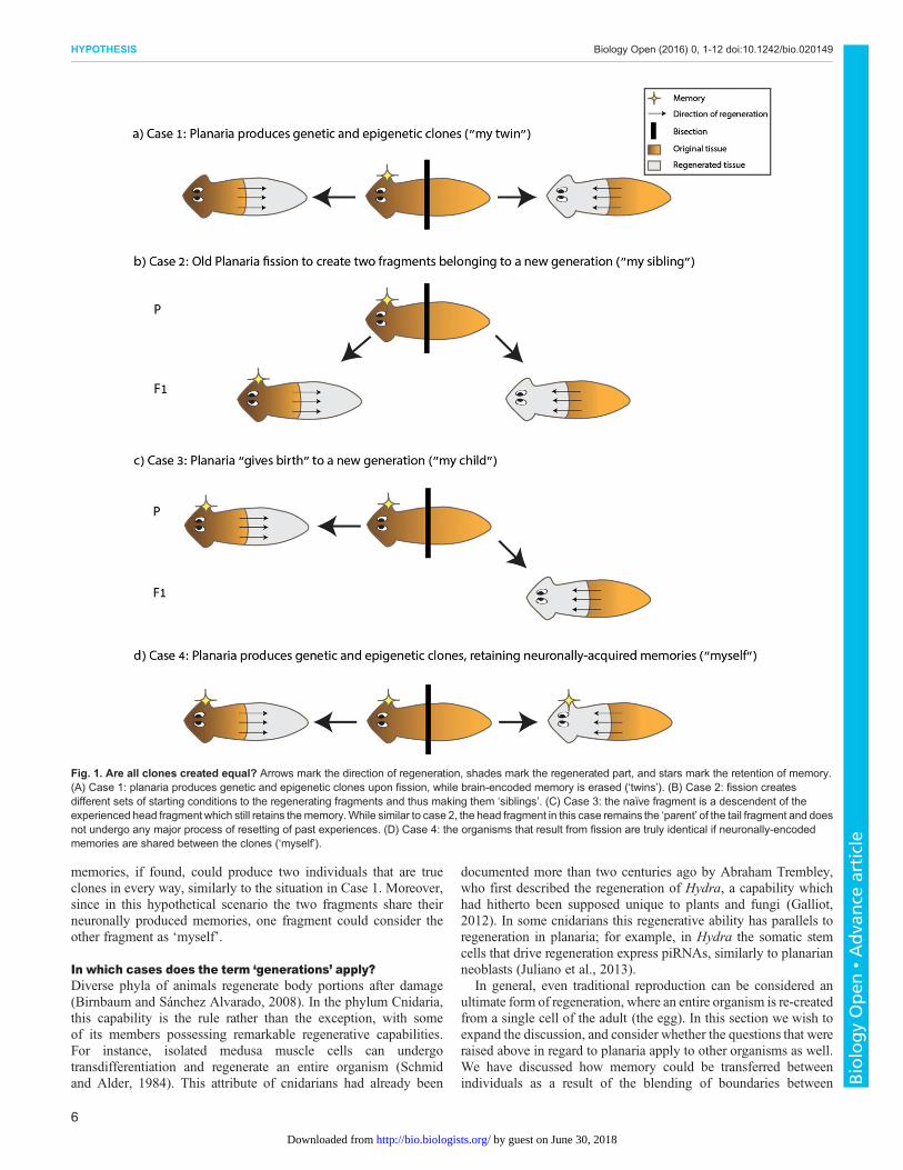
memories, if found, could produce two individuals that are trueclones in every way, similarly to the situation in Case 1. Moreover,since in this hypothetical scenario the two fragments share theirneuronally produced memories, one fragment could consider theother fragment as ‘myself’.
In which cases does the term ‘generations’ apply?Diverse phyla of animals regenerate body portions after damage(Birnbaum and Sánchez Alvarado, 2008). In the phylum Cnidaria,this capability is the rule rather than the exception, with someof its members possessing remarkable regenerative capabilities.For instance, isolated medusa muscle cells can undergotransdifferentiation and regenerate an entire organism (Schmidand Alder, 1984). This attribute of cnidarians had already been
documented more than two centuries ago by Abraham Trembley,who first described the regeneration of Hydra, a capability whichhad hitherto been supposed unique to plants and fungi (Galliot,2012). In some cnidarians this regenerative ability has parallels toregeneration in planaria; for example, in Hydra the somatic stemcells that drive regeneration express piRNAs, similarly to planarianneoblasts (Juliano et al., 2013).
In general, even traditional reproduction can be considered anultimate form of regeneration, where an entire organism is re-createdfrom a single cell of the adult (the egg). In this section we wish toexpand the discussion, and consider whether the questions that wereraised above in regard to planaria apply to other organisms as well.We have discussed how memory could be transferred betweenindividuals as a result of the blending of boundaries between
Fig. 1. Are all clones created equal? Arrows mark the direction of regeneration, shades mark the regenerated part, and stars mark the retention of memory.(A) Case 1: planaria produces genetic and epigenetic clones upon fission, while brain-encoded memory is erased (‘twins’). (B) Case 2: fission createsdifferent sets of starting conditions to the regenerating fragments and thus making them ‘siblings’. (C) Case 3: the naïve fragment is a descendent of theexperienced head fragment which still retains thememory.While similar to case 2, the head fragment in this case remains the ‘parent’ of the tail fragment and doesnot undergo any major process of resetting of past experiences. (D) Case 4: the organisms that result from fission are truly identical if neuronally-encodedmemories are shared between the clones (‘myself’).
6
HYPOTHESIS Biology Open (2016) 0, 1-12 doi:10.1242/bio.020149
BiologyOpen
•Adva
nce
article
by guest on June 30, 2018http://bio.biologists.org/Downloaded from
development and inheritance in asymmetrically dividing animals.Since diverse organisms use different mechanisms to procreate andto store information, it is worthwhile to reflect on the broaderdefinition of the terms ‘generations’ and ‘memory’, and theinteraction between these processes.
‘Generations’ of dividing cellsWhen cells are grown in culture in the laboratory, the ‘generationtime’ of the culture is frequently tracked and different ‘generations’display different phenotypes, which often accumulate in later‘generations’ (Merrill, 1998; Niida et al., 1998). In addition toamassing damage (e.g. shortening of telomeres, mutations), whencells divide, whether in a multicellular organism or in unicellularorganisms, certain memories can be inherited through mitosis;daughter cells can stably maintain the memory of different cellularactivities initiated in the parental cell when the cytoplasm is split intwo, through different feedback mechanisms (Campos et al., 2014;Wang et al., 2013). The ability to maintain expression patterns of theparental cells in the daughter cells is a key to development anddifferentiation (Hobert, 2011). Not all the information is preserved;for example, DNA replication and the ensuing dilution of thehistones present a challenge for preservation of chromatin marks(which epigenetic marks are maintained after S phase is still an openquestion in the field) (Budhavarapu et al., 2013; Lanzuolo et al.,2011; Probst et al., 2009). Histone variants are being removed offreplicating DNA, and the new histones are being deposited on thenewly synthesized DNA as the replication fork progresses. Whichmolecules or information enable, in cases when this type of memoryis indeed preserved (Gaydos et al., 2014), to decorate the histones ofthe daughter strands with the same post-translational modificationsthat were present on the histones of the template DNA? This is avery active field of investigation and there are currently no definitiveanswers (Campos et al., 2014). In contrast, re-establishment of DNAmethylation patterns on the newly synthesized DNA is fairly wellunderstood (the process depends on the maintenance activity of theDNA methyltransferase, DNMT1) (Kar et al., 2012). Despite themechanistic ambiguity, it is clear that certain environmentalchanges can elicit responses that are memorized over celldivision; maintenance of acquired properties in a bacterial andyeast population, such as fast responses to different environmentalconditions or nutrients, was shown to persist over long periods oftime (and thus through generations) (Lambert and Kussell, 2014).
Generations in plantsPlants provide a striking example of evolution of organisms whichlack a designated population of stem cells that will become germcells. One of the aspects of plant cell biology that distinguishesbetween regeneration in plants and planaria is the ability of certainplant cells to dedifferentiate or transdifferentiate in response toenvironmental cues. There is no single source of cells for newtissues in plants, as apart from meristems (structures consisting ofpluripotent stem cells) there are various undifferentiated cellpopulations in the plant that can propagate and differentiate(Aichinger et al., 2012). Additionally, certain somatic cells maytransdifferentiate to grow various plant tissues (Sugimoto et al.,2011). The ability of plant cells to dedifferentiate is under tightregulation of cell-specific gene expression, in the absence of whichflower meristems and even embryos may develop spontaneouslyfrom somatic tissues (Bowman et al., 1992; Horst et al., 2016;Ikeuchi et al., 2015).The same processes that regulate the dedifferentiation of somatic
cells are part of normal plant growth and development. For instance,
the presence of an apical meristem inhibits the development ofaxillary meristems (Leyser, 2003). The absence of a nearbymeristem, either caused by its removal or by the growth of theplant, will reduce this inhibition and allow the development ofdormant meristems, or the development of undifferentiated cellsinto meristems or the formation of new meristems fromdedifferentiated somatic tissue (Leyser, 2003).
Whether through a natural or artificial process, cloning can resultfrom injury or detachment of a portion of the plant. However,formally, the definition of ‘generations’ in plants refers to thecompletion of a ‘life-cycle’, from embryo to adult (Bai et al., 2000;Harada et al., 2001). Since in vegetative reproduction there is nopassing through an embryonic stage, does the individual whichgrows out of the severed part constitutes a new generation? Inaddition to vegetative reproduction, some plant species reproducethrough the formation of plantlets on somatic tissues (Kulka, 2006).This process is defined as asexual reproduction, due to the formationof an embryo. Although the ‘progeny’ is a clone, and there is nogermline involved, the embryo can mark a border betweengenerations, due to its position in the plant’s life cycle (Bai,2015). Also in the case of clonal reproduction in plants, clones maydiffer depending on the fragmented tissue from which it was grown.This type of variation is termed somaclonal variation, and may becaused by genetic or epigenetic differences in the cells from whichthe clone develops (Wang et al., 2013). The notion that mosaicismcan give rise to differences between regenerated parts isschematically described in Fig. 2.
Though it is also possible to clone plants without the mediation ofan embryo using its regenerative properties, and despite the fact thatthis action yields two individuals, this form of cloning is notcommonly referred to as asexual reproduction. Plants display a fullarsenal of epigenetic mechanisms, including the ones described inrelation to planaria, such as histone modifications, DNAmethylation, and small RNA-induced RNAi (Dunoyer et al.,2010; Habu et al., 2001; Kaeppler et al., 2000). Moreover, plantshave the ability to amplify heritable small RNAs that are used forgene silencing using RNA-dependent RNA polymerases (similarlytoC. elegans nematodes) (Rechavi et al., 2011) and small RNAs canalso direct DNA methylation in the nucleus. These mechanismsenable preservation of transgenerational epigenetic memory, inaddition to maintenance of epigenetic memory after cell division(Castel and Martienssen, 2013). Additionally, as dedicatedstructures such as plasmodesmata connect different plant cells,diffusible epigenetic markers in somatic cells may affect the stemcells that regenerate, produce embryos or germ cells. It is possiblethat the mechanisms, which may create variability in planarianclones, could contribute to somaclonal variation. Indeed, in additionto prevalent genetic mosaicism (Gill et al., 1995), some‘epimutations’ that originate in plant ancestors can become stableover hundreds of generations (Ong-Abdullah et al., 2015).
Since different reproduction processes in plants, as describedabove, do not require passage through an embryonic step (thatdefines which individual is the ‘parent’ and which is the ‘child’), therelationship between the two resulting individuals is somewhatambiguous, and bears many similarities to the relationship betweentwo regenerated planarian fragments.
Generations in sexually reproducing animalsIn sexually reproducing animals, the ‘clear’ conceptualclassification of individuals along a lineage to distinct generationsis allowed due to the discrete steps of meiosis and fertilization. Inmice and humans, extensive erasure of epigenetic information that
7
HYPOTHESIS Biology Open (2016) 0, 1-12 doi:10.1242/bio.020149
BiologyOpen
•Adva
nce
article
by guest on June 30, 2018http://bio.biologists.org/Downloaded from

originated in the parent by germline and embryo ‘reprograming’takes place (Hajkova, 2011). Reprogramming of DNAmethylations, for example, has been shown to be critical fortotipotency (Messerschmidt et al., 2014; Surani, 2001). Sincereprogramming entails the erasure of ancestral ‘memories’, it mightbe suggested that reprogramming could serve to define ‘time zero’,when the separation of the new generation from the parent takesplace. However, in sexually reproducing animals, for exampleC. elegans, it is not clear to what degree epigenetic marks undergo‘reprograming’ (Anava et al., 2014); C. elegans do not methylatecytosines, however some ancestral small RNAs and chromatinmodification were explicitly shown to persist in the progeny, formultiple generations (Gaydos et al., 2014; Rechavi et al., 2011,2014).It is not yet known which type of memories/reactions can persist
across generations in sexually reproducing animals (not even inorganisms where this is an intensely studied question, such asC. elegans). Thus, it is not clear in what sense animal pedigreescould be considered to form an epigenetic ‘continuum’ whichstretches over time, and to what extent each member in a lineage is a‘true epigenetic individual’.It is probable that the degree of ‘epigenetic continuity’ between
generations of different animals differ, since different animalsappear to diverge in the mechanisms that are at their disposal for
maintaining epigenetic memory across generations. For example, nomammals are currently known to share the ability of C. elegans toamplify heritable small RNAs using RNA-dependent small RNAs(Rechavi et al., 2011). The notion of a clear-cut generation is anabstract concept, however, in sexually reproducing animals a newgeneration can be identified solely based on meiosis andfertilization – the definition should not be based on epigeneticresetting.
Suggested experimentsWe proposed that asymmetric fission might encourage variationbetween the individuals that regenerate from the fragments. Here wedetail experiments that could add support to this hypothesis.
Maintenance of epigenetic markers derived from fragmenttissueEach fragment has a gene expression pattern that is specific to itsmorphology; however, when it is removed from an intact worm andforced to regenerate new structures, it must remodel these gene-regulatory events on top of new anatomy (i.e. a trunk fragmentcontaining largely intestine must generate new positionalinformation to specify head and tail regions). The incompletereprogramming of these markers may lead to their maintenancethroughout the complete animal (Thomas and Schötz, 2011). In
Fig. 2. The effects of cellular mosaicism on regenerated tissues in planaria and plants. (I) After fission or bisection, each neoblast in the formed blastemamay differ in its genetic and epigenetic content, and contribute to the variation in the regenerated tissue, resulting in difference between andwithin the regeneratedfragments. (II) After a break in the plant tissue, various somatic cells may regenerate plant tissue. The newly grown tissuemay differ genetically and epigeneticallydue to environmental effects on its originating somatic cells.
8
HYPOTHESIS Biology Open (2016) 0, 1-12 doi:10.1242/bio.020149
BiologyOpen
•Adva
nce
article
by guest on June 30, 2018http://bio.biologists.org/Downloaded from
other words, an organism that regenerates from a tail may be more‘tail-like’ than one that regenerates from a head. This can beassessed after a single fission event by comparing the geneexpression of the resulting whole organisms and those of thespecific tissue of the fragment. While the continued success ofregeneration over millions of fission events through the history ofplanaria suggests that such history or enrichment cannot accumulateindefinitely, it is possible that some limited amount of ‘recent’history of spatial origin is kept. It will be especially interesting toidentify persistent molecular or biophysical markers of anatomical(positional) history (Carlson, 1983; Chang et al., 2002) infragments that originate in different regions of one-headed versuspermanently two-headed worms, to decipher the algorithm bywhich blastema cells of any fragment type decide which structuresto generate at each wound surface.
Maintenance of bioelectric gradients derived from fragmenttissueThe main open questions concern what changes (transcriptional,chromatin-level, or bioelectrical) distinguish a trunk fragment fromawild-type worm (destined to make one head) and an anatomically-normal trunk fragment from a two-headed worm (which will maketwo heads). Examination of bioelectric state (using fluorescentreporters of voltage distributions) (Adams and Levin, 2012; Oviedoet al., 2008), transcriptional profiling, and chromatin state analysismust be used to understand what is different about these fragments.Quantitative models must be developed to explain how stable statescan be stored, and edited, in physiological circuits (Cervera et al.,2015; Law and Levin, 2015; Levin, 2014a).
Maintenance of behavioral memories across regenerativereproductionTo determine how and where memory may be stored outside thebrain during head regeneration, it would be necessary to firstoptimize training protocols (Abbott and Wong, 2008; Blackistonet al., 2010; Inoue et al., 2015; Nicolas et al., 2008; Pagán et al.,2012), capitalizing on more ecologically-salient stimuli andlearning paradigms to achieve high-throughput induction of robustlearning. The key experiments would be to assess the persistence ofmemory in fragments of different sizes, anatomical locations andbody compositions. A variety of molecular and biophysical toolsnow exist to establish suppression screens targeting variouspathways, to begin to probe the mechanisms necessary forimprinting of the memory upon a newly-regenerating brain (Aokiet al., 2009; Gentile et al., 2011; Sheıman and Kreshchenko, 2015).
ConclusionsIn planaria, and other organisms that reproduce by fission,producing and maintaining variation between fragments afterasymmetric division may be adaptive (much like the beneficialincrease in variation following sexual reproduction andrecombination). Therefore, the theoretical ability of asymmetricdivision to create variability in an otherwise isogenic populationcould be considered as a tool for producing evolutionary progress.Thus, asymmetric fission is a mechanism that challenges our currentview of what defines the temporal axis of evolution, since epigeneticprocesses, environmental cues, biochemical gradients andgeneration of a complete individual from a community of cellscan generate natural variation, without requiring so called ‘distinct’generations. It is likely that we have only begun to glimpse theprevalence and variety of long-term memory in somatic tissuesduring lifespan and across reproduction throughout phyla. The
continued future analysis of such instructive interactions is likely tohave profound implications for understanding evolution. Moreover,a mature understanding of these fascinating processes will drivenumerous applications in regenerative medicine and bioengineeringthat exploit the rich informational plasticity of tissues for the rationalcontrol of form and function.
AcknowledgementsWe are grateful to Fallon Durant, Maya Emmons-Bell, and Jennifer Hammelman forhelpful comments on the draft. We especially thank Keith Harris for his greatcontribution to the writing of the paper and for helping with the development of theideas that constitute this work. We thank all members of the Rechavi and Levin labfor fruitful discussions and advice. We are grateful to Eva Jablonka, Yehu Moran,and Nir Ohad for reading the manuscript and for their helpful comments.
Competing interestsThe authors declare no competing or financial interests.
Author contributionsConceptualization, M.N, M.L and O.R; Writing - Original Draft, M.N, M.L and O.R;Writing - Review & Editing, M.N, M.L and O.R.
FundingM.N. has been supported in part by the Naomi Prawer Kadar Foundation through theTel Aviv University GRTF Program. O.R. has been supported in part by the JohnTempleton Foundation and Israel Science Foundation. M.L. gratefully acknowledgesan Allen Discovery Center award from The Paul G. Allen Frontiers Group, andsupport of the G Harold and Leila Y. Mathers Foundation.
ReferencesAbbott, S. M. and Wong, G. K. (2008). The conditioning and memory retention of
planaria (Dugesia tigrina) for directional preferences. Bios 79, 160-170.Adams, D. S. and Levin, M. (2012). General principles for measuring resting
membrane potential and ion concentration using fluorescent bioelectricityreporters. Cold Spring Harb. Protoc. 2012, 385-397.
Adams, D. S., Masi, A. and Levin, M. (2007). H+ pump-dependent changes inmembrane voltage are an early mechanism necessary and sufficient to induceXenopus tail regeneration. Development 134, 1323-1335.
Adell, T., Cebria, F. and Salo, E. (2010). Gradients in planarian regeneration andhomeostasis. Cold Spring Harb. Perspect. Biol. 2, a000505.
Aichinger, E., Kornet, N., Friedrich, T. and Laux, T. (2012). Plant stem cell niches.Annu. Rev. Plant Biol. 63, 615-636.
Allen, K., Fuchs, E. C., Jaschonek, H., Bannerman, D. M. andMonyer, H. (2011).Gap junctions between interneurons are required for normal spatial coding in thehippocampus and short-term spatial memory. J. Neurosci. 31, 6542-6552.
Altizer, A. M., Moriarty, L. J., Bell, S. M., Schreiner, C. M., Scott, W. J. andBorgens, R. B. (2001). Endogenous electric current is associated with normaldevelopment of the vertebrate limb. Dev. Dyn. 221, 391-401.
Anava, S., Posner, R. and Rechavi, O. (2014). The soft genome. Worm 3,e989798.
Aoki, R., Wake, H., Sasaki, H. and Agata, K. (2009). Recording and spectrumanalysis of the planarian electroencephalogram. Neuroscience 159, 908-914.
Armakolas, A., Koutsilieris, M. and Klar, A. J. S. (2010). Discovery of the mitoticselective chromatid segregation phenomenon and its implications for vertebratedevelopment. Curr. Opin. Cell Biol. 22, 81-87.
Axmacher, N., Mormann, F., Fernandez, G., Elger, C. E. and Fell, J. (2006).Memory formation by neuronal synchronization. Brain Res. Rev. 52, 170-182.
Bagijn, M. P., Goldstein, L. D., Sapetschnig, A., Weick, E.-M., Bouasker, S.,Lehrbach, N. J., Simard, M. J. and Miska, E. A. (2012). Function, targets, andevolution of Caenorhabditis elegans piRNAs. Science 337, 574-578.
Bai, S.-N. (2015). The concept of the sexual reproduction cycle and its evolutionarysignificance. Front. Plant Sci. 6, 11.
Bai, S., Chen, L., Yund, M. A. and Sung, Z. R. (2000). Mechanisms of plant embryodevelopment. Curr. Top. Dev. Biol. 50, 61-88.
Bailey, C. H. and Kandel, E. R. (2008). Synaptic remodeling, synaptic growth andthe storage of long-term memory in Aplysia. Prog. Brain Res. 169, 179-198.
Bates, E. (2015). Ion channels in development and cancer. Annu. Rev. Cell Dev.Biol. 31, 231-247.
Beane, W. S., Morokuma, J., Adams, D. S. and Levin, M. (2011). A chemicalgenetics approach reveals H,K-ATPase-mediated membrane voltage is requiredfor planarian head regeneration. Chem. Biol. 18, 77-89.
Beane, W. S., Morokuma, J., Lemire, J. M. and Levin, M. (2013). Bioelectricsignaling regulates head and organ size during planarian regeneration.Development 140, 313-322.
Ben-Zvi, D. and Shilo, B.-Z. (2011). Scaling of morphogen gradients. Curr. Opin.Genet. Dev. 21, 704-710.
9
HYPOTHESIS Biology Open (2016) 0, 1-12 doi:10.1242/bio.020149
BiologyOpen
•Adva
nce
article
by guest on June 30, 2018http://bio.biologists.org/Downloaded from
Best, J. B. and Rubinstein, I. (1962). Environmental familiarity and feeding in aplanarian. Science 135, 916-918.
Bird, A. (2002). DNA methylation patterns and epigenetic memory. Genes Dev. 16,6-21.
Birnbaum, K. D. and Sanchez Alvarado, A. (2008). Slicing across kingdoms:regeneration in plants and animals. Cell 132, 697-710.
Blackiston, D. J., McLaughlin, K. A. and Levin, M. (2009). Bioelectric controls ofcell proliferation: ion channels, membrane voltage and the cell cycle.Cell Cycle 8,3527-3536.
Blackiston, D., Shomrat, T., Nicolas, C. L., Granata, C. and Levin, M. (2010). Asecond-generation device for automated training and quantitative behavioranalyses of molecularly-tractable model organisms. PLoS ONE 5, e14370.
Blackiston, D. J., Shomrat, T. and Levin, M. (2015). The stability of memoriesduring brain remodeling: a perspective. Commun. Integr. Biol. 8, e1073424.
Bowman, J. L., Sakai, H., Jack, T., Weigel, D., Mayer, U. and Meyerowitz, E. M.(1992). SUPERMAN, a regulator of floral homeotic genes in Arabidopsis.Development 114, 599-615.
Budhavarapu, V. N., Chavez, M. and Tyler, J. K. (2013). How is epigeneticinformation maintained through DNA replication? Epigenet. Chromatin 6, 32.
Cameron, K. M., Miwa, S., Walker, C. and von Zglinicki, T. (2012). Male miceretain a metabolic memory of improved glucose tolerance induced during adultonset, short-term dietary restriction. Longev. Heal. 1, 3.
Campos, E. I., Stafford, J. M. and Reinberg, D. (2014). Epigenetic inheritance:histone bookmarks across generations. Trends Cell Biol. 24, 664-674.
Cardona, A., Hartenstein, V. and Romero, R. (2006). Early embryogenesis ofplanaria: a cryptic larva feeding on maternal resources. Dev. Genes Evol. 216,667-681.
Carlson, B. M. (1983). Positional memory in vertebrate limb development andregeneration. Prog. Clin. Biol. Res. 110, 433-443.
Castel, S. E. andMartienssen, R. A. (2013). RNA interference in the nucleus: rolesfor small RNAs in transcription, epigenetics and beyond. Nat. Rev. Genet. 14,100-112.
Cervera, J., Alcaraz, A. and Mafe, S. (2014). Membrane potential bistability innonexcitable cells as described by inward and outward voltage-gated ionchannels. J. Phys. Chem. B 118, 12444-12450.
Cervera, J., Manzanares, J. A. and Mafe, S. (2015). Electrical coupling inensembles of nonexcitable cells: modeling the spatial map of single cellpotentials. J. Phys. Chem. B 119, 2968-2978.
Chakravarthy, S. V. and Ghosh, J. (1997). On Hebbian-like adaptation in heartmuscle: a proposal for “cardiac memory”. Biol. Cybern. 76, 207-215.
Chan, J. D., Agbedanu, P. N., Zamanian, M., Gruba, S. M., Haynes, C. L., Day,T. A. and Marchant, J. S. (2014). “Death and axes”: unexpected Ca2+ entryphenologs predict new anti-schistosomal agents. PLoS Pathog. 10, e1003942.
Chang, H. Y., Chi, J.-T., Dudoit, S., Bondre, C., van de Rijn, M., Botstein, D. andBrown, P. O. (2002). Diversity, topographic differentiation, and positional memoryin human fibroblasts. Proc. Natl. Acad. Sci. USA 99, 12877-12882.
Chen, Q., Yan, M., Cao, Z., Li, X., Zhang, Y., Shi, J., Feng, G.-H., Peng, H., Zhang,X., Zhang, Y. et al. (2016). Sperm tsRNAs contribute to intergenerationalinheritance of an acquired metabolic disorder. Science 351, 397-400.
Claycomb, J. M. (2014). Ancient endo-siRNA pathways reveal new tricks. Curr.Biol. 24, R703-R715.
Corning, W. C. (1966). Retention of a position discrimination after regeneration inplanarians. Psychon. Sci. 5, 17-18.
Daoudal, G. and Debanne, D. (2003). Long-term plasticity of intrinsic excitability:learning rules and mechanisms. Learn. Mem. 10, 456-465.
Di Laurenzio, L., Wysocka-Diller, J., Malamy, J. E., Pysh, L., Helariutta, Y.,Freshour, G., Hahn, M. G., Feldmann, K. A. and Benfey, P. N. (1996). TheSCARECROW gene regulates an asymmetric cell division that is essential forgenerating the radial organization of the arabidopsis root. Cell 86, 423-433.
Duncan, E. M., Chitsazan, A. D., Seidel, C. W. and Sanchez Alvarado, A. (2015).Set1 and MLL1/2 target distinct sets of functionally different genomic loci in vivo.Cell Rep. 13, 2741-2755.
Dunoyer, P., Schott, G., Himber, C., Meyer, D., Takeda, A., Carrington, J. C. andVoinnet, O. (2010). Small RNA duplexes function as mobile silencing signalsbetween plant cells. Science 328, 912-916.
Durant, F., Lobo, D., Hammelman, J. and Levin, M. (2016). Physiological controlsof large-scale patterning in planarian regeneration: amolecular and computationalperspective on growth and form. Regeneration 3, 78-102.
D’Urso, A. and Brickner, J. H. (2014). Mechanisms of epigenetic memory. TrendsGenet. 30, 230-236.
Emmons-Bell, M., Durant, F., Hammelman, J., Bessonov, N., Volpert, V.,Morokuma, J., Pinet, K., Adams, D. S., Pietak, A., Lobo, D. et al. (2015). Gapjunctional blockade stochastically induces different species-specific headanatomies in genetically wild-type Girardia dorotocephala flatworms. Int. J. Mol.Sci. 16, 27865-27896.
Evans, T., Wade, C. M., Chapman, F. A., Johnson, A. D. and Loose, M. (2014).Acquisition of germ plasm accelerates vertebrate evolution. Science 344,200-203.
Extavour, C. G. (2003). Mechanisms of germ cell specification across themetazoans: epigenesis and preformation. Development 130, 5869-5884.
Farinella-Ferruzza, N. (1956). The transformation of a tail into limb after xenoplastictransplantation. Experientia 12, 304-305.
Friedlander, M. R., Adamidi, C., Han, T., Lebedeva, S., Isenbarger, T. A., Hirst,M., Marra, M., Nusbaum, C., Lee, W. L., Jenkin, J. C. et al. (2009). High-resolution profiling and discovery of planarian small RNAs. Proc. Natl. Acad. Sci.USA 106, 11546-11551.
Funk, R. H. W. (2013). Ion gradients in tissue and organ biology. Biol. Syst. OpenAccess 2, 2.
Funk, R. H. W. (2015). Endogenous electric fields as guiding cue for cell migration.Front. Physiol. 6, 143.
Galliot, B. (2012). Hydra, a fruitful model system for 270 years. Int. J. Dev. Biol. 56,411-423.
Gaydos, L. J., Wang, W. and Strome, S. (2014). H3K27me and PRC2 transmit amemory of repression across generations and during development. Science 345,1515-1518.
Gent, J. I., Lamm, A. T., Pavelec, D. M., Maniar, J. M., Parameswaran, P., Tao, L.,Kennedy, S. and Fire, A. Z. (2010). Distinct phases of siRNA synthesis in anendogenous RNAi pathway in C. elegans soma. Mol. Cell 37, 679-689.
Gentile, L., Cebria, F. and Bartscherer, K. (2011). The planarian flatworm: an invivo model for stem cell biology and nervous system regeneration. Dis. Model.Mech. 4, 12-19.
Gill, D. E., Chao, L., Perkins, S. L. and Wolf, J. B. (1995). Genetic mosaicism inplants and clonal animals. Annu. Rev. Ecol. Syst. 26, 423-444.
Goel, P. and Mehta, A. (2013). Learning theories reveal loss of pancreatic electricalconnectivity in diabetes as an adaptive response. PLoS ONE 8, e70366.
Habu, Y., Kakutani, T. and Paszkowski, J. (2001). Epigenetic developmentalmechanisms in plants: molecules and targets of plant epigenetic regulation. Curr.Opin. Genet. Dev. 11, 215-220.
Hackett, J. A., Sengupta, R., Zylicz, J. J., Murakami, K., Lee, C., Down, T. A. andSurani, M. A. (2013). Germline DNA demethylation dynamics and imprint erasurethrough 5-hydroxymethylcytosine. Science 339, 448-452.
Hajkova, P. (2011). Epigenetic reprogramming in the germline: towards the groundstate of the epigenome. Philos. Trans. R. Soc. Lond. B. Biol. Sci. 366, 2266-2273.
Halas, E. S., James, R. L. and Knutson, C. S. (1962). An attempt at classicalconditioning in the planarian. J. Comp. Physiol. Psychol. 55, 969-971.
Harada, J. J., Belmonte, M. F. and Kwong, R. W. (2001). Plant Embryogenesis(Zygotic and Somatic). In eLS, Chichester: John Wiley and Sons, Ltd.
He, Y., Kulasiri, D. and Samarasinghe, S. (2014). Systems biology of synapticplasticity: a review on N-methyl-D-aspartate receptor mediated biochemicalpathways and related mathematical models. Biosystems 122, 7-18.
Herry, C. and Johansen, J. P. (2014). Encoding of fear learning and memory indistributed neuronal circuits. Nat. Neurosci. 17, 1644-1654.
Hobert, O. (2011). Maintaining a memory by transcriptional autoregulation. Curr.Biol. 21, R146-R147.
Horst, N. A., Katz, A., Pereman, I., Decker, E. L., Ohad, N. and Reski, R. (2016). Asingle homeobox gene triggers phase transition, embryogenesis and asexualreproduction. Nat. Plants 2, 15209.
Hubert, A., Henderson, J. M., Ross, K. G., Cowles, M. W., Torres, J. and Zayas,R. M. (2013). Epigenetic regulation of planarian stem cells by the SET1/MLLfamily of histone methyltransferases. Epigenetics 8, 79-91.
Ikeuchi, M., Iwase, A., Rymen, B., Harashima, H., Shibata, M., Ohnuma, M.,Breuer, C., Morao, A. K., de Lucas, M., De Veylder, L. et al. (2015). PRC2represses dedifferentiation of mature somatic cells in Arabidopsis. Nat. Plants 1,15089.
Inoue, T., Hoshino, H., Yamashita, T., Shimoyama, S. and Agata, K. (2015).Planarian shows decision-making behavior in response to multiple stimuli byintegrative brain function. Zool. Lett. 1, 7.
Iwasaki, M. and Paszkowski, J. (2014). Epigenetic memory in plants. EMBO J. 33,1987-1998.
Jablonka, E. and Lamb, M. J. (2015). The inheritance of acquired epigeneticvariations. Int. J. Epidemiol. 44, 1094-1103.
Jaderstad, J., Jaderstad, L. M., Li, J., Chintawar, S., Salto, C., Pandolfo, M.,Ourednik, V., Teng, Y. D., Sidman, R. L., Arenas, E. et al. (2010).Communication via gap junctions underlies early functional and beneficialinteractions between grafted neural stem cells and the host. Proc. Natl. Acad.Sci. USA 107, 5184-5189.
Jan, Y. N. and Jan, L. Y. (1998). Asymmetric cell division. Nature 392, 775-778.Jenkins, L. S., Duerstock, B. S. and Borgens, R. B. (1996). Reduction of the
current of injury leaving the amputation inhibits limb regeneration in the redspotted newt. Dev. Biol. 178, 251-262.
Juliano, C. E., Reich, A., Liu, N., Gotzfried, J., Zhong, M., Uman, S., Reenan,R. A., Wessel, G. M., Steele, R. E. and Lin, H. (2013). PIWI proteins and PIWI-interacting RNAs function in Hydra somatic stem cells. Proc. Natl. Acad. Sci. USA111, 337-342.
Kaeppler, S. M., Kaeppler, H. F. and Rhee, Y. (2000). Epigenetic aspects ofsomaclonal variation in plants. Plant Mol. Biol. 43, 179-188.
Kar, S., Deb, M., Sengupta, D., Shilpi, A., Parbin, S., Torrisani, J., Pradhan, S.and Patra, S. K. (2012). An insight into the various regulatory mechanismsmodulating human DNA methyltransferase 1 stability and function. Epigenetics 7,994-1007.
10
HYPOTHESIS Biology Open (2016) 0, 1-12 doi:10.1242/bio.020149
BiologyOpen
•Adva
nce
article
by guest on June 30, 2018http://bio.biologists.org/Downloaded from
Kiortsis, V. and Moraitou, M. (1965) . Factors of regeneration in Spirographisspallanzanii. In:Regeneration in animals and related problems (ed. V. Kiortsis andH. A. L. Trampusch), pp. 250-261. New York: North Holland.
Klar, A. J. S. (1987). Differentiated parental DNA strands confer developmentalasymmetry on daughter cells in fission yeast. Nature 326, 466-470.
Kulka, R. G. (2006). Cytokinins inhibit epiphyllous plantlet development on leaves ofBryophyllum (Kalanchoe) marnierianum. J. Exp. Bot. 57, 4089-4098.
Lambert, G. and Kussell, E. (2014). Memory and fitness optimization of bacteriaunder fluctuating environments. PLoS Genet. 10, e1004556.
Lanzuolo, C., Lo Sardo, F., Diamantini, A. and Orlando, V. (2011). PcGcomplexes set the stage for epigenetic inheritance of gene silencing in early Sphase before replication. PLoS Genet. 7, e1002370.
Law, R. and Levin, M. (2015). Bioelectric memory: modeling resting potentialbistability in amphibian embryos and mammalian cells. Theor. Biol. Med. Model.12, 22.
Levin, M. (2012). Molecular bioelectricity in developmental biology: new tools andrecent discoveries: control of cell behavior and pattern formation bytransmembrane potential gradients. Bioessays 34, 205-217.
Levin, M. (2014a). Endogenous bioelectrical networks store non-genetic patterninginformation during development and regeneration. J. Physiol. 592, 2295-2305.
Levin, M. (2014b). Molecular bioelectricity: how endogenous voltage potentialscontrol cell behavior and instruct pattern regulation in vivo. Mol. Biol. Cell 25,3835-3850.
Levin, M., Thorlin, T., Robinson, K. R., Nogi, T. and Mercola, M. (2002).Asymmetries in H+/K+-ATPase and cell membrane potentials comprise a veryearly step in left-right patterning. Cell 111, 77-89.
Leyser, O. (2003). Regulation of shoot branching by auxin. Trends Plant Sci. 8,541-545.
Liao, J.-Y., Guo, Y.-H., Zheng, L.-L., Li, Y., Xu,W.-L., Zhang, Y.-C., Zhou, H., Lun,Z.-R., Ayala, F. J. and Qu, L.-H. (2014). Both endo-siRNAs and tRNA-derivedsmall RNAs are involved in the differentiation of primitive eukaryote Giardialamblia. Proc. Natl. Acad. Sci. USA 111, 14159-14164.
Lobo, D., Solano, M., Bubenik, G. A. and Levin, M. (2014). A linear-encodingmodel explains the variability of the target morphology in regeneration. J. R. Soc.Interface 11, 20130918.
Maciunas, K., Snipas, M., Paulauskas, N. and Bukauskas, F. F. (2016).Reverberation of excitation in neuronal networks interconnected throughvoltage-gated gap junction channels. J. Gen. Physiol. 147, 273-288.
Malone, C. D. and Hannon, G. J. (2009). Small RNAs as guardians of the genome.Cell 136, 656-668.
Mann, M. R. W. and Bartolomei, M. S. (2002). Epigenetic reprogramming in themammalian embryo: struggle of the clones. Genome Biol. 3, reviews1003.
Maren, S. and Quirk, G. J. (2004). Neuronal signalling of fear memory. Nat. Rev.Neurosci. 5, 844-852.
Mathews, J. and Levin, M. (2016). Gap Junctional Signaling in Pattern Regulation:physiological network connectivity instructs growth and form. DevelopmentalNeurobiology.
McConnell, J. V., Jacobson, A. L. and Kimble, D. P. (1959). The effects ofregeneration upon retention of a conditioned response in the planarian. J. Comp.Physiol. Psychol. 52, 1-5.
Meister, G. (2013). Argonaute proteins: functional insights and emerging roles. Nat.Rev. Genet. 14, 447-459.
Merrill, G. F. (1998). Cell synchronization. Methods Cell Biol. 57, 229-249.Mesnil, M., Crespin, S., Avanzo, J.-L. and Zaidan-Dagli, M.-L. (2005). Defectivegap junctional intercellular communication in the carcinogenic process. Biochim.Biophys. Acta 1719, 125-145.
Messerschmidt, D. M., Knowles, B. B. and Solter, D. (2014). DNA methylationdynamics during epigenetic reprogramming in the germline and preimplantationembryos. Genes Dev. 28, 812-828.
Mondia, J. P., Levin, M., Omenetto, F. G., Orendorff, R. D., Branch, M. R. andAdams, D. S. (2011). Long-distance signals are required for morphogenesis ofthe regenerating Xenopus tadpole tail, as shown by femtosecond-laser ablation.PLoS ONE 6, e24953.
Morgan, H. D., Santos, F., Green, K., Dean, W. and Reik, W. (2005). Epigeneticreprogramming in mammals. Hum. Mol. Genet. 14 Suppl. 1, R47-R58.
Muotri, A. R. and Gage, F. H. (2006). Generation of neuronal variability andcomplexity. Nature 441, 1087-1093.
Nakazawa, M., Cebria, F., Mineta, K., Ikeo, K., Agata, K. and Gojobori, T. (2003).Search for the evolutionary origin of a brain: planarian brain characterized bymicroarray. Mol. Biol. Evol. 20, 784-791.
Nicolas, C. L., Abramson, C. I. and Levin, M. (2008). Analysis of Behavior in thePlanarian Model. In Planaria: A Model for Drug Action and Abuse (ed. R. B. Raffaand S. M. Rawls), pp. 83-94. Austin: RG Landes Co.
Niida, H., Matsumoto, T., Satoh, H., Shiwa, M., Tokutake, Y., Furuichi, Y. andShinkai, Y. (1998). Severe growth defect in mouse cells lacking the telomeraseRNA component. Nat. Genet. 19, 203-206.
Nishimura, O., Hosoda, K., Kawaguchi, E., Yazawa, S., Hayashi, T., Inoue, T.,Umesono, Y. and Agata, K. (2015). Unusually large number of mutations inasexually reproducing clonal planarian Dugesia japonica. PLoS ONE 10,e0143525.
Nogi, T. and Levin, M. (2005). Characterization of innexin gene expression andfunctional roles of gap-junctional communication in planarian regeneration. Dev.Biol. 287, 314-335.
Nogi, T., Zhang, D., Chan, J. D. and Marchant, J. S. (2009). A novel biologicalactivity of praziquantel requiring voltage-operated Ca2+ channel beta subunits:subversion of flatworm regenerative polarity. PLoS Negl. Trop. Dis. 3, e464.
Ogrodnik, M., Salmonowicz, H., Brown, R., Turkowska, J., Sredniawa, W.,Pattabiraman, S., Amen, T., Abraham, A.-C., Eichler, N., Lyakhovetsky, R.et al. (2014). Dynamic JUNQ inclusion bodies are asymmetrically inherited inmammalian cell lines through the asymmetric partitioning of vimentin. Proc. Natl.Acad. Sci. USA 111, 8049-8054.
Ong-Abdullah, M., Ordway, J. M., Jiang, N., Ooi, S.-E., Kok, S.-Y., Sarpan, N.,Azimi, N., Hashim, A. T., Ishak, Z., Rosli, S. K. et al. (2015). Loss of Karmatransposon methylation underlies the mantled somaclonal variant of oil palm.Nature 525, 533-537.
Oviedo, N. J. and Levin, M. (2007). smedinx-11 is a planarian stem cell gapjunction gene required for regeneration and homeostasis. Development 134,3121-3131.
Oviedo, N. J., Newmark, P. A. and Sanchez Alvarado, A. (2003). Allometricscaling and proportion regulation in the freshwater planarian Schmidteamediterranea. Dev. Dyn. 226, 326-333.
Oviedo, N. J., Nicolas, C. L., Adams, D. S. and Levin, M. (2008). Live imaging ofplanarian membrane potential using DiBAC4(3). CSH Protoc. 2008, pdb.prot5055.
Oviedo, N. J., Morokuma, J., Walentek, P., Kema, I. P., Gu, M. B., Ahn, J.-M.,Hwang, J. S., Gojobori, T. and Levin, M. (2010). Long-range neural and gapjunction protein-mediated cues control polarity during planarian regeneration.Dev. Biol. 339, 188-199.
Pagan, O. R., Baker, D., Deats, S., Montgomery, E., Tenaglia, M., Randolph, C.,Kotturu, D., Tallarida, C., Bach, D., Wilk, G. et al. (2012). Planarians inpharmacology: parthenolide is a specific behavioral antagonist of cocaine in theplanarian Girardia tigrina. Int. J. Dev. Biol. 56, 193-196.
Pai, V. P., Aw, S., Shomrat, T., Lemire, J. M. and Levin, M. (2012).Transmembrane voltage potential controls embryonic eye patterning inXenopus laevis. Development 139, 313-323.
Pai, V. P., Lemire, J. M., Pare, J.-F., Lin, G., Chen, Y. and Levin, M. (2015).Endogenous gradients of resting potential instructively pattern embryonic neuraltissue via Notch signaling and regulation of proliferation. J. Neurosci. 35,4366-4385.
Palacios-Prado, N. and Bukauskas, F. F. (2009). Heterotypic gap junctionchannels as voltage-sensitive valves for intercellular signaling. Proc. Natl. Acad.Sci. USA 106, 14855-14860.
Palacios-Prado, N. and Bukauskas, F. F. (2012). Modulation of metaboliccommunication through gap junction channels by transjunctional voltage;synergistic and antagonistic effects of gating and ionophoresis. Biochim.Biophys. Acta 1818, 1884-1894.
Peng, H., Shi, J., Zhang, Y., Zhang, H., Liao, S., Li, W., Lei, L., Han, C., Ning, L.,Cao, Y. et al. (2012). A novel class of tRNA-derived small RNAs extremelyenriched in mature mouse sperm. Cell Res. 22, 1609-1612.
Perathoner, S., Daane, J. M., Henrion, U., Seebohm, G., Higdon, C. W.,Johnson, S. L., Nusslein-Volhard, C. and Harris, M. P. (2014). Bioelectricsignaling regulates size in zebrafish fins. PLoS Genet. 10, e1004080.
Pereda, A. E., Curti, S., Hoge, G., Cachope, R., Flores, C. E. and Rash, J. E.(2013). Gap junction-mediated electrical transmission: regulatory mechanismsand plasticity. Biochim. Biophys. Acta 1828, 134-146.
Pezzulo, G. and Levin, M. (2015). Re-membering the body: applications ofcomputational neuroscience to the top-down control of regeneration of limbs andother complex organs. Integr. Biol. 7, 1487-1517.
Poulton, E. B., Schonland, S., Shipley, A. E. and Weismann, A. (1889). Essaysupon Heredity and Kindred Biological Problems. Authorised Translation (ed. E. B.Poulton, S. Schonland and A. E. Shipley), pp. 48-411. Oxford: Clarendon Press.
Probst, A. V., Dunleavy, E. and Almouzni, G. (2009). Epigenetic inheritanceduring the cell cycle. Nat. Rev. Mol. Cell Biol. 10, 192-206.
Rajasethupathy, P., Antonov, I., Sheridan, R., Frey, S., Sander, C., Tuschl, T.and Kandel, E. R. (2012). A role for neuronal piRNAs in the epigenetic control ofmemory-related synaptic plasticity. Cell 149, 693-707.
Rechavi, O. (2014). Guest list or black list: heritable small RNAs as immunogenicmemories. Trends Cell Biol. 24, 212-220.
Rechavi, O., Minevich, G. and Hobert, O. (2011). Transgenerational inheritance ofan acquired small RNA-based antiviral response in C. elegans. Cell 147,1248-1256.
Rechavi, O., Houri-Ze’evi, L., Anava, S., Goh, W. S. S., Kerk, S. Y., Hannon, G. J.and Hobert, O. (2014). Starvation-induced transgenerational inheritance of smallRNAs in C. elegans. Cell 158, 277-287.
Reddien, P. W. and Sanchez Alvarado, A. (2004). Fundamentals of planarianregeneration. Annu. Rev. Cell Dev. Biol. 20, 725-757.
Reddien, P. W., Oviedo, N. J., Jennings, J. R., Jenkin, J. C. and SanchezAlvarado, A. (2005). SMEDWI-2 is a PIWI-like protein that regulates planarianstem cells. Science 310, 1327-1330.
11
HYPOTHESIS Biology Open (2016) 0, 1-12 doi:10.1242/bio.020149
BiologyOpen
•Adva
nce
article
by guest on June 30, 2018http://bio.biologists.org/Downloaded from
Rilling, M. (1996). The mystery of the vanished citations: James McConnell’sforgotten 1960s quest for planarian learning, a biochemical engram, and celebrity.Am. Psychol. 51, 589-598.
Robb, S. M. C. and Sanchez Alvarado, A. (2014). Histone modifications andregeneration in the planarian Schmidtea mediterranea. Curr. Top. Dev. Biol. 108,71-93.
Roberts-Galbraith, R. H. and Newmark, P. A. (2015). On the organ trail: insightsinto organ regeneration in the planarian. Curr. Opin. Genet. Dev. 32, 37-46.
Ros, J., Pellerin, L., Magara, F., Dauguet, J., Schenk, F. and Magistretti, P. J.(2006). Metabolic activation pattern of distinct hippocampal subregions duringspatial learning and memory retrieval. J. Cereb. Blood Flow Metab. 26, 468-477.
Rouhana, L., Weiss, J. A., Forsthoefel, D. J., Lee, H., King, R. S., Inoue, T.,Shibata, N., Agata, K. and Newmark, P. A. (2013). RNA interference by feedingin vitro-synthesized double-stranded RNA to planarians: methodology anddynamics. Dev. Dyn. 242, 718-730.
Rouhana, L., Weiss, J. A., King, R. S. and Newmark, P. A. (2014). PIWI homologsmediate histone H4 mRNA localization to planarian chromatoid bodies.Development 141, 2592-2601.
Sabour, D. and Scholer, H. R. (2012). Reprogramming and the mammaliangermline: the Weismann barrier revisited. Curr. Opin. Cell Biol. 24, 716-723.
Salo, E., Abril, J. F., Adell, T., Cebria, F., Eckelt, K., Fernandez-Taboada, E.,Handberg-Thorsager, M., Iglesias, M., Molina, M. D. and Rodrıguez-Esteban,G. (2009). Planarian regeneration: achievements and future directions after 20years of research. Int. J. Dev. Biol. 53, 1317-1327.
Sarnat, H. B. andNetsky, M. G. (1985). The brain of the planarian as the ancestor ofthe human brain. Can. J. Neurol. Sci. 12, 296-302.
Schmid, V. and Alder, H. (1984). Isolated, mononucleated, striated muscle canundergo pluripotent transdifferentiation and form a complex regenerate. Cell 38,801-809.
Sharma, U. and Rando, O. J. (2014). Father-son chats: inheriting stress throughsperm RNA. Cell Metab. 19, 894-895.
Sheıman, I. M. and Kreshchenko, N. D. (2015). [Regeneration of planarians:experimental object]. Ontogenez 46, 1-9.
Shomrat, T. and Levin, M. (2013). An automated training paradigm reveals long-termmemory in planarians and its persistence through head regeneration. J. Exp.Biol. 216, 3799-3810.
Singer, M. (1952). The influence of the nerve in regeneration of the amphibianextremity. Q. Rev. Biol. 27, 169-200.
Solana, J. (2013). Closing the circle of germline and stem cells: the Primordial StemCell hypothesis. Evodevo 4, 2.
Spencer, G. J. andGenever, P. G. (2003). Long-term potentiation in bone–a role forglutamate in strain-induced cellular memory? BMC Cell Biol. 4, 9.
Spokoini, R., Moldavski, O., Nahmias, Y., England, J. L., Schuldiner, M. andKaganovich, D. (2012). Confinement to organelle-associated inclusionstructures mediates asymmetric inheritance of aggregated protein in buddingyeast. Cell Rep. 2, 738-747.
Stern, C. D. (1987). Control of epithelial polarity and induction in the early chickembryo. InMesenchymal-Epithelial Interactions in Neural Development (ed. J. R.Wolff, J. Sievers and M. Berry), pp. 91-100. Berlin: Springer-Verlag.
Sugimoto, K., Gordon, S. P. andMeyerowitz, E. M. (2011). Regeneration in plantsand animals: dedifferentiation, transdifferentiation, or just differentiation? TrendsCell Biol. 21, 212-218.
Sundelacruz, S., Levin, M. and Kaplan, D. L. (2008). Membrane potential controlsadipogenic and osteogenic differentiation of mesenchymal stem cells. PLoS ONE3, e3737.
Sundelacruz, S., Levin, M. and Kaplan, D. L. (2009). Role of membrane potentialin the regulation of cell proliferation and differentiation. Stem Cell Rev. 5, 231-246.
Sundelacruz, S., Levin, M. and Kaplan, D. L. (2013). Depolarization altersphenotype, maintains plasticity of predifferentiated mesenchymal stem cells.Tissue Eng. Part A 19, 1889-1908.
Surani, M. A. (2001). Reprogramming of genome function through epigeneticinheritance. Nature 414, 122-128.
Thomas, M. A. and Schotz, E.-M. (2011). SAPling: a Scan-Add-Print barcodingdatabase system to label and track asexual organisms. J. Exp. Biol. 214, 3518-3523.
Thompson, R. and McConnell, J. (1955). Classical conditioning in the planarian,Dugesia dorotocephala. J. Comp. Physiol. Psychol. 48, 65-68.
Todorova, M. G., Soria, B. and Quesada, I. (2008). Gap junctional intercellularcommunication is required to maintain embryonic stem cells in a non-differentiated and proliferative state. J. Cell. Physiol. 214, 354-362.
Trosko, J. E. (2007). Gap junctional intercellular communication as a biological“Rosetta stone” in understanding, in a systems biological manner, stem cellbehavior, mechanisms of epigenetic toxicology, chemoprevention andchemotherapy. J. Membr. Biol. 218, 93-100.
Tseng, A.-S., Beane,W. S., Lemire, J. M., Masi, A. and Levin, M. (2010). Inductionof vertebrate regeneration by a transient sodium current. J. Neurosci. 30,13192-13200.
Turner, C. H., Robling, A. G., Duncan, R. L. and Burr, D. B. (2002). Do bone cellsbehave like a neuronal network? Calcif. Tissue Int. 70, 435-442.
Umesono, Y. and Agata, K. (2009). Evolution and regeneration of the planariancentral nervous system. Dev. Growth Differ. 51, 185-195.
Vasale, J. J., Gu, W., Thivierge, C., Batista, P. J., Claycomb, J. M., Youngman,E. M., Duchaine, T. F., Mello, C. C. and Conte, D. (2010). Sequential rounds ofRNA-dependent RNA transcription drive endogenous small-RNA biogenesis inthe ERGO-1/Argonaute pathway. Proc. Natl. Acad. Sci. USA 107, 3582-3587.
Vucetic, Z., Kimmel, J., Totoki, K., Hollenbeck, E. and Reyes, T. M. (2010).Maternal high-fat diet alters methylation and gene expression of dopamine andopioid-related genes. Endocrinology 151, 4756-4764.
Wagner, D. E., Wang, I. E. and Reddien, P. W. (2011). Clonogenic neoblasts arepluripotent adult stem cells that underlie planarian regeneration. Science 332,811-816.
Wang, Y. and Belousov, A. B. (2011). Deletion of neuronal gap junction proteinconnexin 36 impairs hippocampal LTP. Neurosci. Lett. 502, 30-32.
Wang, X., Wu, R., Lin, X., Bai, Y., Song, C., Yu, X., Xu, C., Zhao, N., Dong, Y. andLiu, B. (2013). Tissue culture-induced genetic and epigenetic alterations in ricepure-lines, F1 hybrids and polyploids. BMC Plant Biol. 13, 77.
Werner, S., Stuckemann, T., Beiran Amigo, M., Rink, J. C., Julicher, F. andFriedrich, B. M. (2015). Scaling and regeneration of self-organized patterns.Phys. Rev. Lett. 114, 138101.
Williams, S. R., Christensen, S. R., Stuart, G. J. and Hausser, M. (2002).Membrane potential bistability is controlled by the hyperpolarization-activatedcurrent I(H) in rat cerebellar Purkinje neurons in vitro. J. Physiol. 539, 469-483.
Witchley, J. N., Mayer, M., Wagner, D. E., Owen, J. H. and Reddien, P. W. (2013).Muscle cells provide instructions for planarian regeneration. Cell Rep. 4, 633-641.
Wolvetang, E. J., Pera, M. F. and Zuckerman, K. S. (2007). Gap junction mediatedtransport of shRNA between human embryonic stem cells. Biochem. Biophys.Res. Commun. 363, 610-615.
Wong, R. C. B., Pera, M. F. and Pebay, A. (2008). Role of gap junctions inembryonic and somatic stem cells. Stem Cell Rev. 4, 283-292.
Wu, C.-L., Shih, M.-F. M., Lai, J. S.-Y., Yang, H.-T., Turner, G. C., Chen, L. andChiang, A.-S. (2011). Heterotypic gap junctions between two neurons in thedrosophila brain are critical for memory. Curr. Biol. 21, 848-854.
Yamasaki, H., Krutovskikh, V., Mesnil, M., Tanaka, T., Zaidan-Dagli, M. L. andOmori, Y. (1999). Role of connexin (gap junction) genes in cell growth control andcarcinogenesis. C. R. Acad. Sci. III 322, 151-159.
Zacharioudakis, I., Gligoris, T. and Tzamarias, D. (2007). A yeast catabolicenzyme controls transcriptional memory. Curr. Biol. 17, 2041-2046.
Zhang, W. and Linden, D. J. (2003). The other side of the engram: experience-driven changes in neuronal intrinsic excitability. Nat. Rev. Neurosci. 4, 885-900.
Zhang, D., Chan, J. D., Nogi, T. and Marchant, J. S. (2011). Opposing roles ofvoltage-gated Ca2+ channels in neuronal control of regenerative patterning.J. Neurosci. 31, 15983-15995.
Zhou, X., Battistoni, G., El Demerdash, O., Gurtowski, J., Wunderer, J.,Falciatori, I., Ladurner, P., Schatz, M. C., Hannon, G. J. and Wasik, K. A.(2015). Dual functions of Macpiwi1 in transposon silencing and stem cellmaintenance in the flatworm Macrostomum lignano. RNA 21, 1885-1897.
Zoghi, M. (2004). Cardiac memory: do the heart and the brain remember the same?J. Interv. Card. Electrophysiol. 11, 177-182.
12
HYPOTHESIS Biology Open (2016) 0, 1-12 doi:10.1242/bio.020149
BiologyOpen
•Adva
nce
article
by guest on June 30, 2018http://bio.biologists.org/Downloaded from


![Untitled-2 [vigoursgroup.com]vigoursgroup.com/wp-content/themes/vigours/Vigours-Brochure.pdf · The company has been persistently expanding vertically and horizontally ... profile](https://static.fdocuments.in/doc/165x107/5f0804da7e708231d41fec54/untitled-2-the-company-has-been-persistently-expanding-vertically-and-horizontally.jpg)