Vertebral Column Lecture - جامعة الرازيalraziuni.edu.ye/book1/natural therapy/vertebral...
34
FUNCTIONS: 1) Support weight - transmits weight to pelvis and lower limbs 2) Houses and protects spinal cord - spinal nerves leave cord between vertebrae 3) Permits movements - *clinical problems 4) Provides for muscle attachments - muscles of back; also muscles of head, neck, upper extremity, thorax VERTEBRAL COLUMN VERTEBRAL COLUMN
Transcript of Vertebral Column Lecture - جامعة الرازيalraziuni.edu.ye/book1/natural therapy/vertebral...
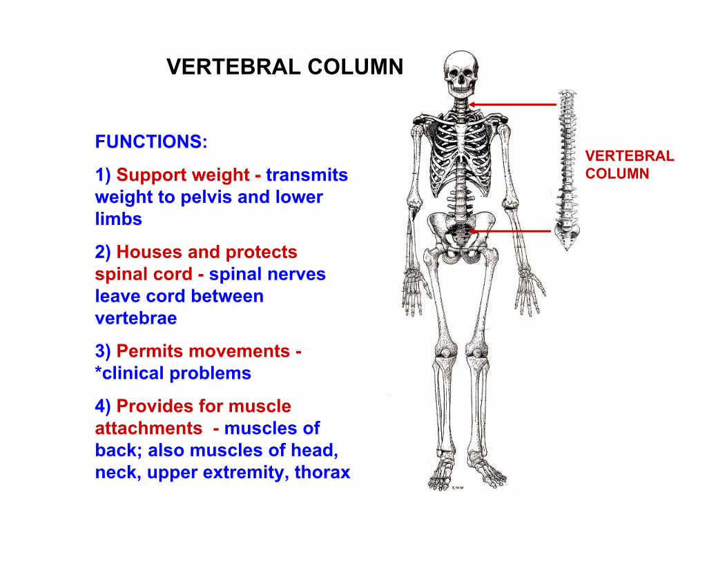
FUNCTIONS:
1) Support weight - transmits weight to pelvis and lower limbs
2) Houses and protects spinal cord - spinal nerves leave cord between vertebrae
3) Permits movements -*clinical problems
4) Provides for muscle attachments - muscles of back; also muscles of head, neck, upper extremity, thorax
VERTEBRAL COLUMN
VERTEBRAL COLUMN
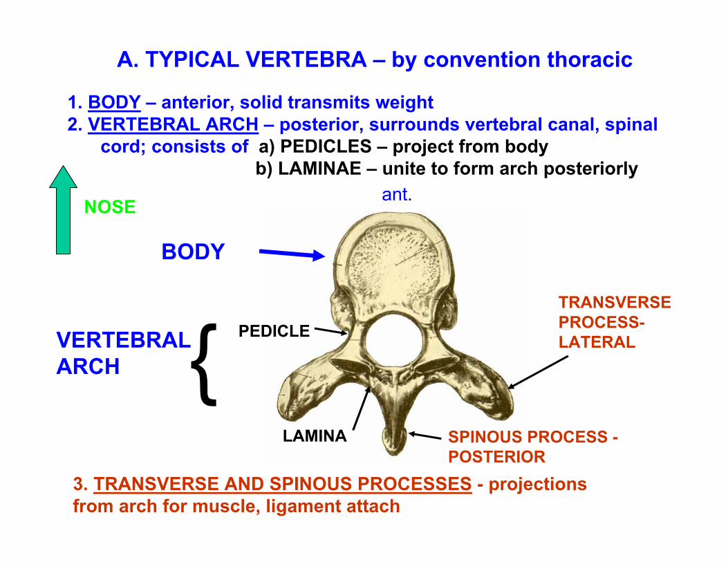
1. BODY – anterior, solid transmits weight2. VERTEBRAL ARCH – posterior, surrounds vertebral canal, spinal
cord; consists of a) PEDICLES – project from body b) LAMINAE – unite to form arch posteriorly
BODY
VERTEBRAL ARCH
PEDICLETRANSVERSE PROCESS-LATERAL
LAMINA
3. TRANSVERSE AND SPINOUS PROCESSES - projections from arch for muscle, ligament attach
A. TYPICAL VERTEBRA – by convention thoracic
SPINOUS PROCESS -POSTERIOR
{
ant.NOSE
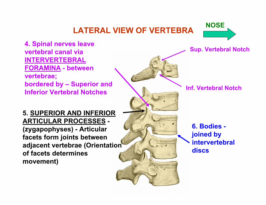
LATERAL VIEW OF VERTEBRA4. Spinal nerves leave vertebral canal via INTERVERTEBRAL FORAMINA - between vertebrae;bordered by – Superior and Inferior Vertebral Notches
Sup. Vertebral Notch
Inf. Vertebral Notch
5. SUPERIOR AND INFERIOR ARTICULAR PROCESSES -(zygapophyses) - Articular facets form joints between adjacent vertebrae (Orientation of facets determines movement)
6. Bodies -joined by intervertebral discs
NOSE
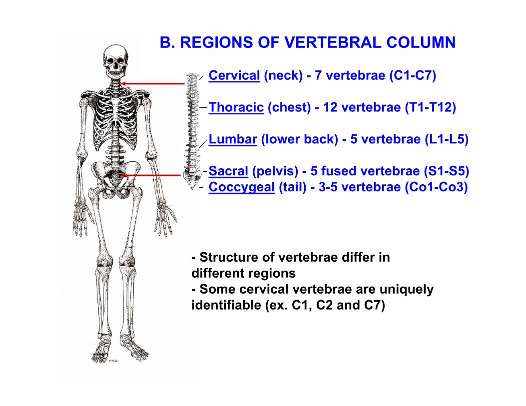
Cervical (neck) - 7 vertebrae (C1-C7)
Thoracic (chest) - 12 vertebrae (T1-T12)
Lumbar (lower back) - 5 vertebrae (L1-L5)
Sacral (pelvis) - 5 fused vertebrae (S1-S5)Coccygeal (tail) - 3-5 vertebrae (Co1-Co3)
B. REGIONS OF VERTEBRAL COLUMN
- Structure of vertebrae differ in different regions - Some cervical vertebrae are uniquely identifiable (ex. C1, C2 and C7)
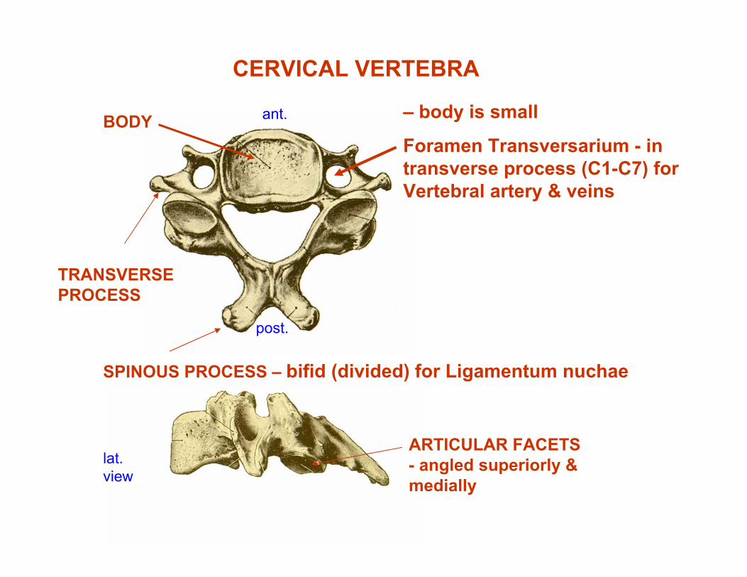
– body is small
Foramen Transversarium - in transverse process (C1-C7) for Vertebral artery & veins
ARTICULAR FACETS - angled superiorly & medially
SPINOUS PROCESS – bifid (divided) for Ligamentum nuchae
TRANSVERSE PROCESS
BODY
CERVICAL VERTEBRA
ant.
post.
lat.view
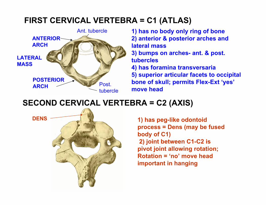
1) has no body only ring of bone2) anterior & posterior arches and lateral mass3) bumps on arches- ant. & post. tubercles 4) has foramina transversaria 5) superior articular facets to occipital bone of skull; permits Flex-Ext ‘yes’ move head
1) has peg-like odontoid process = Dens (may be fused body of C1)2) joint between C1-C2 is pivot joint allowing rotation; Rotation = ‘no’ move head important in hanging
Ant. tubercle
FIRST CERVICAL VERTEBRA = C1 (ATLAS)
SECOND CERVICAL VERTEBRA = C2 (AXIS)
ANTERIORARCH
POSTERIORARCH
DENS
LATERALMASS
Post. tubercle
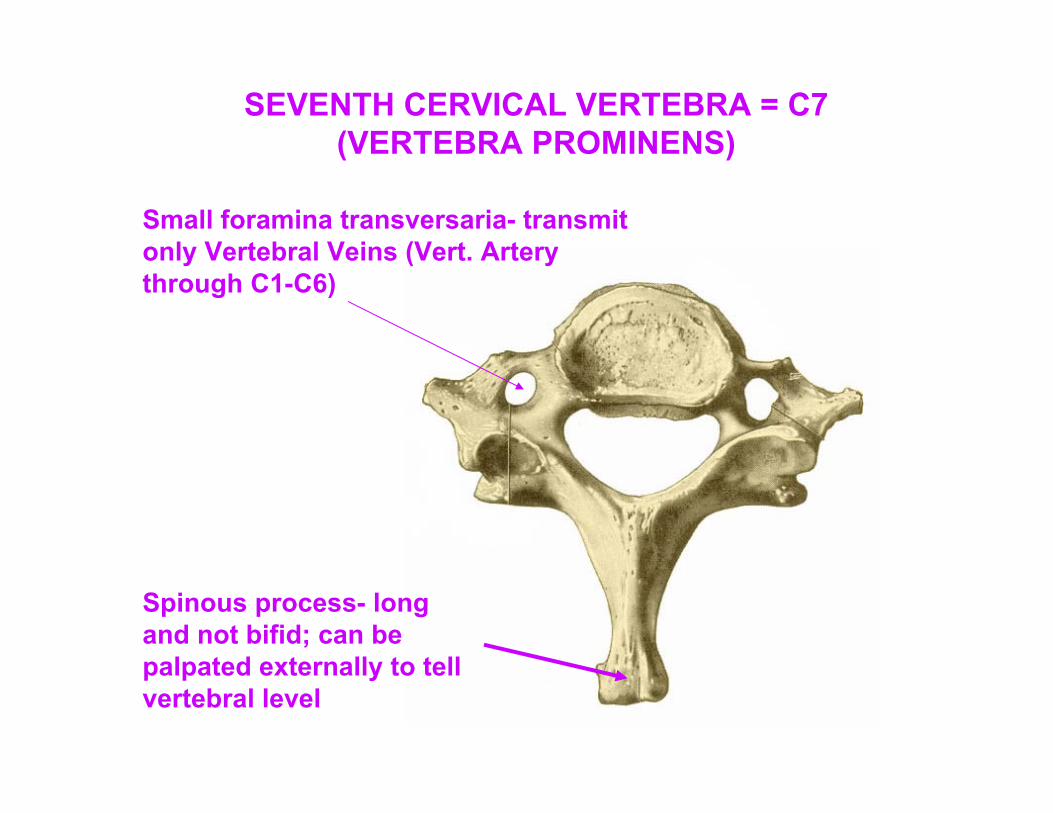
Small foramina transversaria- transmit only Vertebral Veins (Vert. Artery through C1-C6)
Spinous process- long and not bifid; can be palpated externally to tell vertebral level
SEVENTH CERVICAL VERTEBRA = C7 (VERTEBRA PROMINENS)
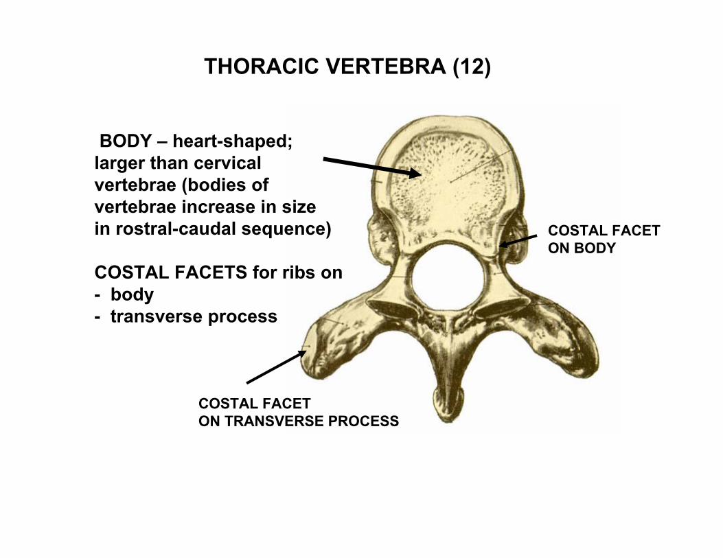
THORACIC VERTEBRA (12)
BODY – heart-shaped; larger than cervical vertebrae (bodies of vertebrae increase in sizein rostral-caudal sequence)
COSTAL FACETS for ribs on- body- transverse process
COSTAL FACETON BODY
COSTAL FACETON TRANSVERSE PROCESS
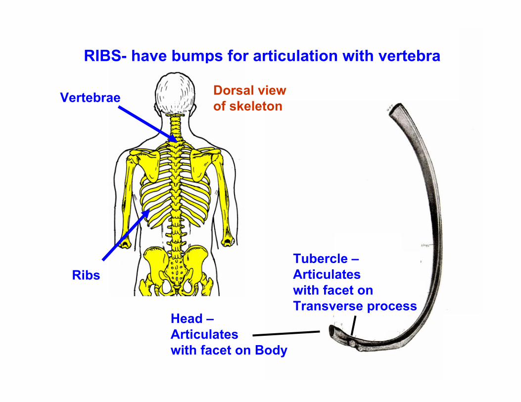
RIBS- have bumps for articulation with vertebra
Head –Articulateswith facet on Body
Tubercle –Articulateswith facet onTransverse process
Vertebrae
Ribs
Dorsal viewof skeleton
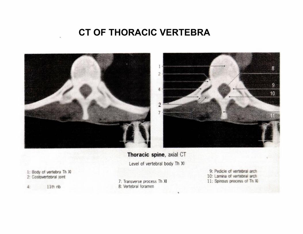
CT OF THORACIC VERTEBRA
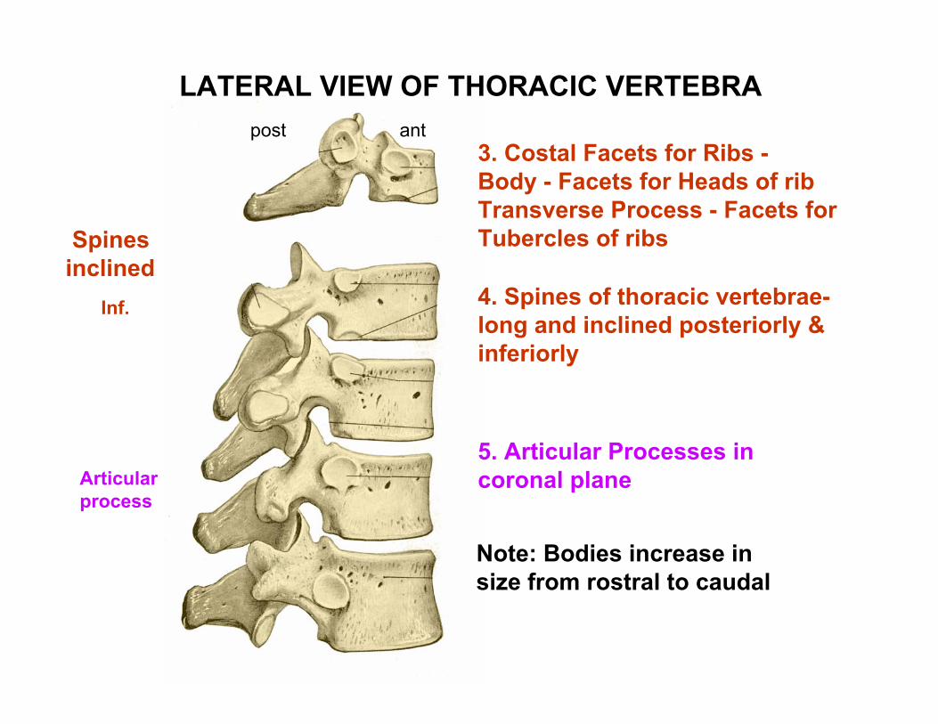
3. Costal Facets for Ribs -Body - Facets for Heads of ribTransverse Process - Facets for Tubercles of ribs
4. Spines of thoracic vertebrae-long and inclined posteriorly & inferiorly
5. Articular Processes in coronal plane
Spines inclined
Inf.
Articular process
antpost
LATERAL VIEW OF THORACIC VERTEBRA
Note: Bodies increase in size from rostral to caudal
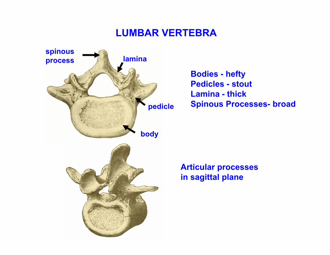
Bodies - hefty Pedicles - stout Lamina - thick Spinous Processes- broad
Articular processes in sagittal plane
spinous process lamina
pedicle
body
LUMBAR VERTEBRA
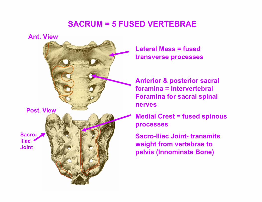
Lateral Mass = fused transverse processes
Anterior & posterior sacral foramina = Intervertebral Foramina for sacral spinal nerves
Medial Crest = fused spinous processes
Sacro-Iliac Joint- transmits weight from vertebrae to pelvis (Innominate Bone)
Ant. View
Post. View
Sacro-Iliac Joint
SACRUM = 5 FUSED VERTEBRAE
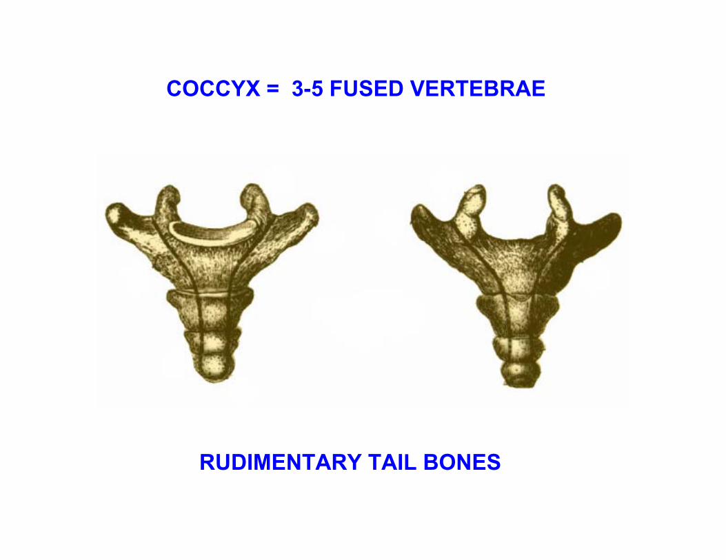
COCCYX = 3-5 FUSED VERTEBRAE
RUDIMENTARY TAIL BONES
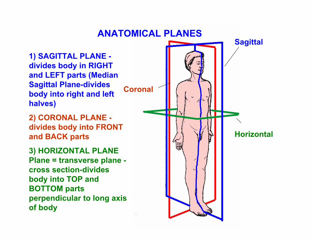
1) SAGITTAL PLANE -divides body in RIGHT and LEFT parts (Median Sagittal Plane-divides body into right and left halves)
2) CORONAL PLANE -divides body into FRONT and BACK parts
3) HORIZONTAL PLANE Plane = transverse plane -cross section-divides body into TOP and BOTTOM parts perpendicular to long axis of body
ANATOMICAL PLANESSagittal
Coronal
Horizontal
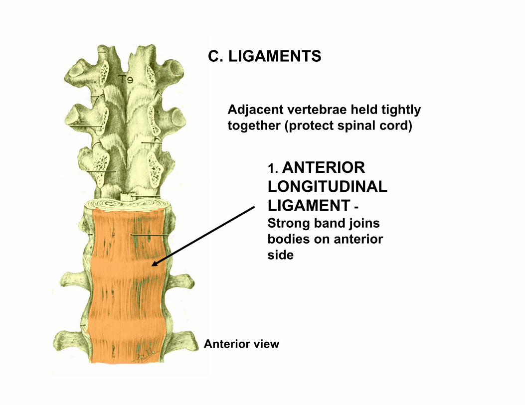
Adjacent vertebrae held tightly together (protect spinal cord)
1. ANTERIOR LONGITUDINAL LIGAMENT -Strong band joins bodies on anterior side
Anterior view
C. LIGAMENTS
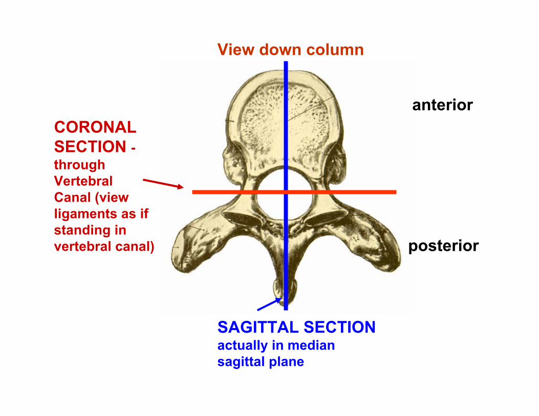
CORONAL SECTION -through Vertebral Canal (view ligaments as if standing in vertebral canal)
SAGITTAL SECTIONactually in median sagittal plane
View down column
anterior
posterior
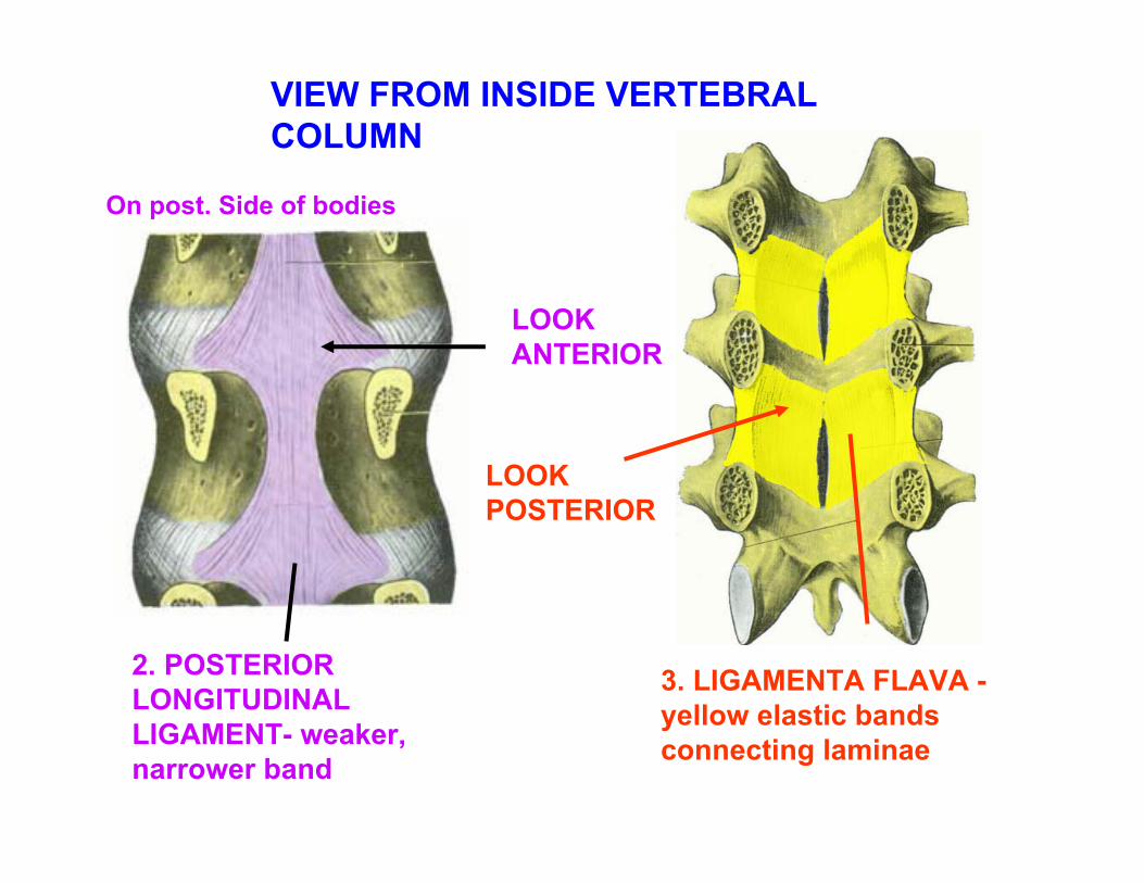
VIEW FROM INSIDE VERTEBRAL COLUMN
2. POSTERIOR LONGITUDINAL LIGAMENT- weaker, narrower band
On post. Side of bodies
3. LIGAMENTA FLAVA -yellow elastic bands connecting laminae
LOOKANTERIOR
LOOKPOSTERIOR
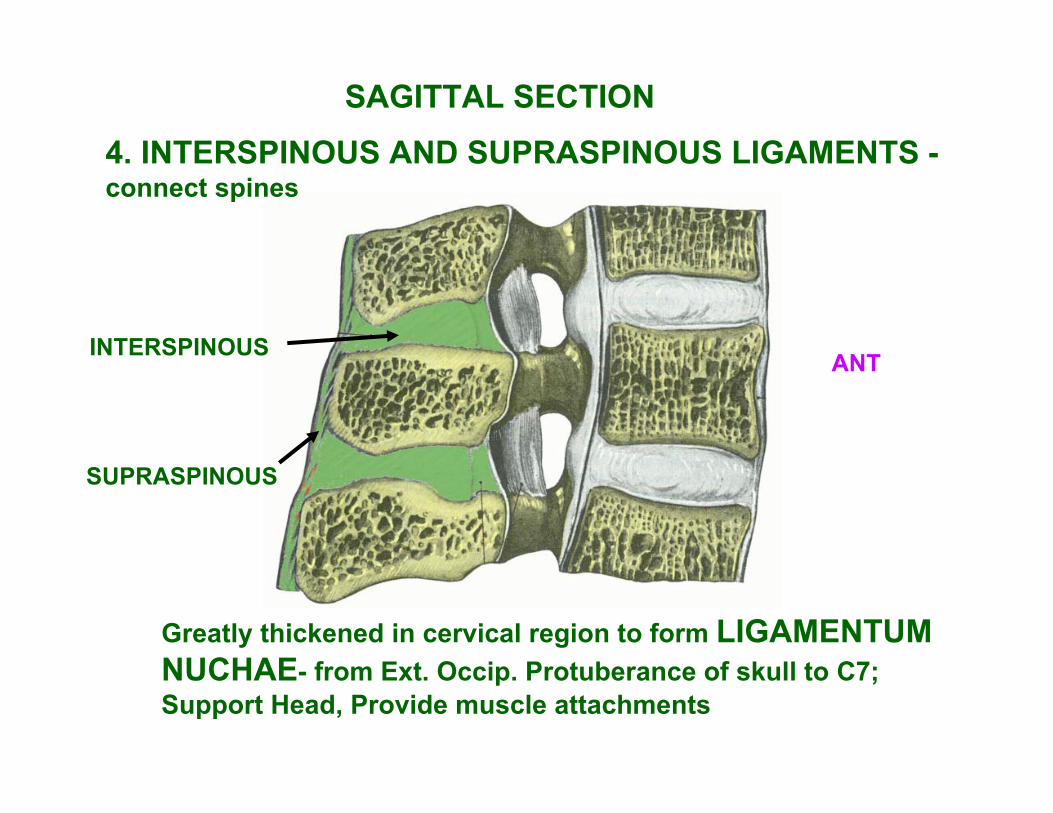
SAGITTAL SECTION4. INTERSPINOUS AND SUPRASPINOUS LIGAMENTS -connect spines
Greatly thickened in cervical region to form LIGAMENTUM NUCHAE- from Ext. Occip. Protuberance of skull to C7; Support Head, Provide muscle attachments
ANT
SUPRASPINOUS
INTERSPINOUS
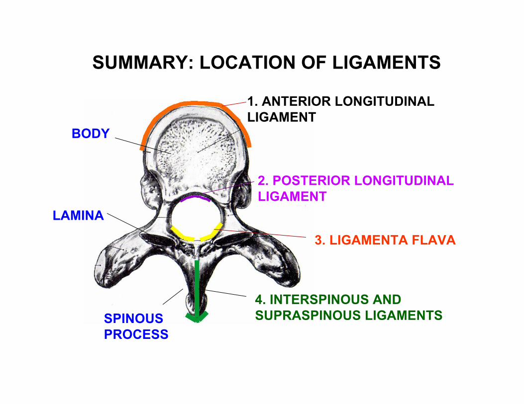
1. ANTERIOR LONGITUDINAL LIGAMENT
2. POSTERIOR LONGITUDINAL LIGAMENT
3. LIGAMENTA FLAVA
4. INTERSPINOUS AND SUPRASPINOUS LIGAMENTS
BODY
LAMINA
SPINOUSPROCESS
SUMMARY: LOCATION OF LIGAMENTS
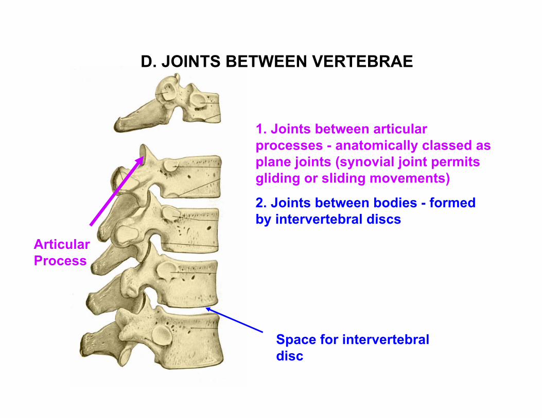
1. Joints between articular processes - anatomically classed as plane joints (synovial joint permits gliding or sliding movements)
2. Joints between bodies - formed by intervertebral discs
Space for intervertebral disc
Articular Process
D. JOINTS BETWEEN VERTEBRAE
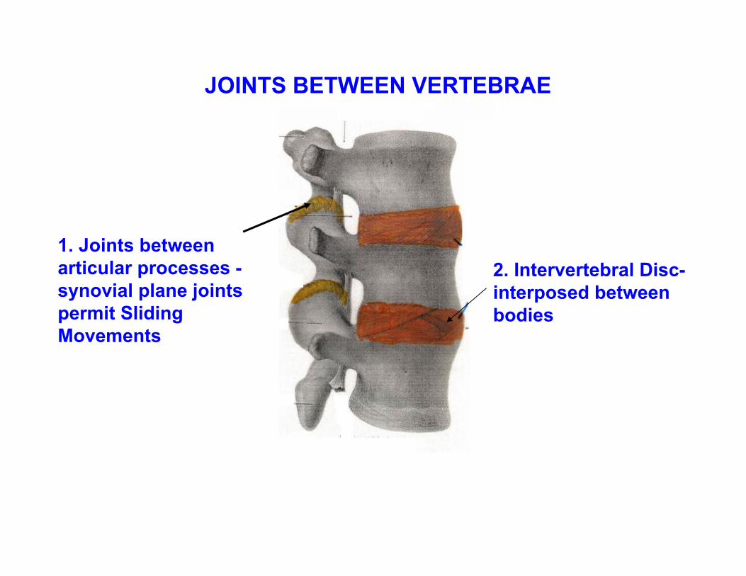
JOINTS BETWEEN VERTEBRAE
1. Joints between articular processes -synovial plane joints permit Sliding Movements
2. Intervertebral Disc-interposed between bodies
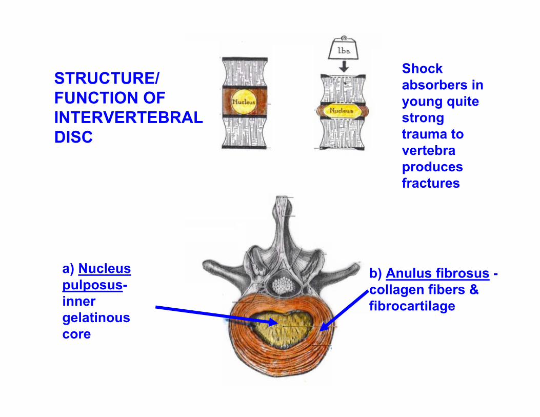
Shock absorbers in young quite strong trauma to vertebra produces fractures
STRUCTURE/FUNCTION OF INTERVERTEBRAL DISC
a) Nucleus pulposus-inner gelatinous core
b) Anulus fibrosus -collagen fibers & fibrocartilage
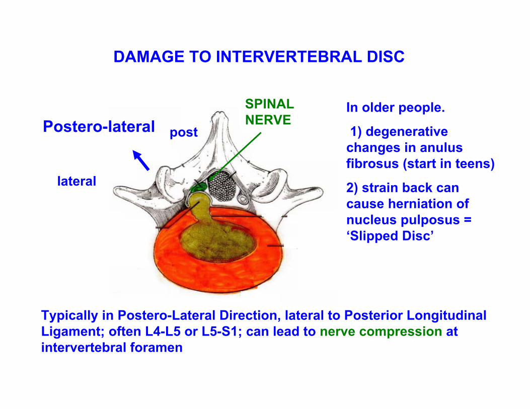
In older people.
1) degenerative changes in anulus fibrosus (start in teens)
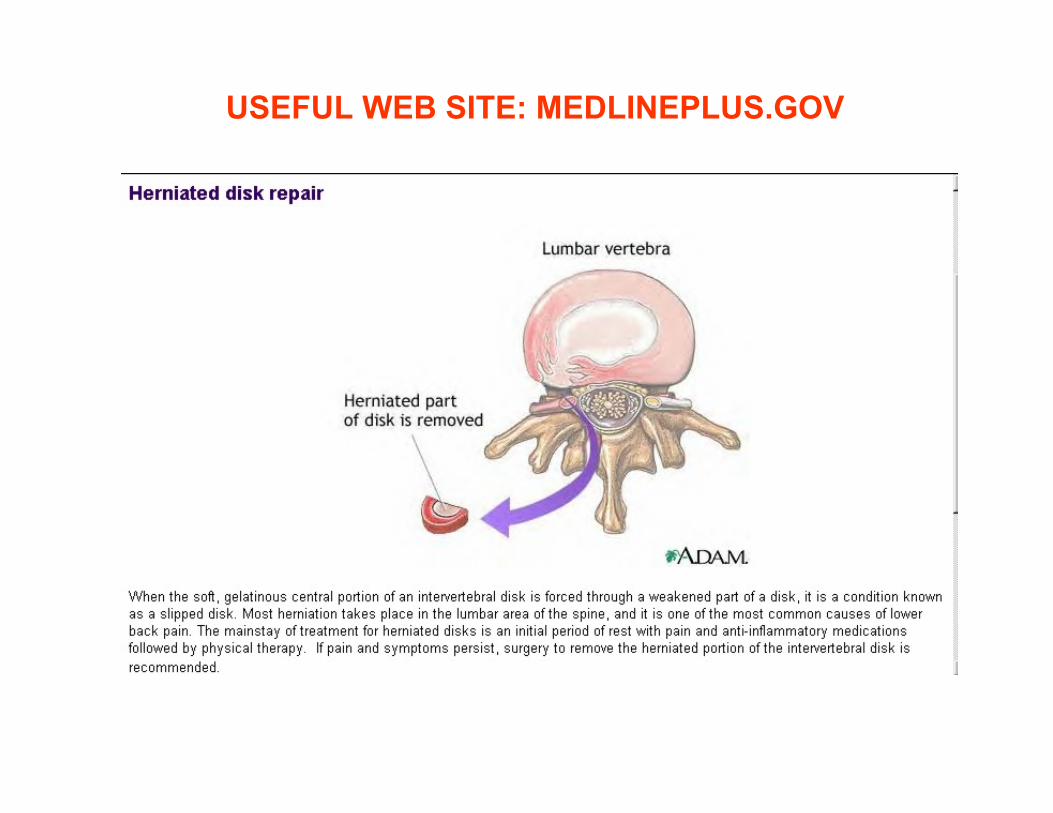
2) strain back can cause herniation of nucleus pulposus = ‘Slipped Disc’
Typically in Postero-Lateral Direction, lateral to Posterior Longitudinal Ligament; often L4-L5 or L5-S1; can lead to nerve compression at intervertebral foramen
Postero-lateral
lateral
post
DAMAGE TO INTERVERTEBRAL DISC
SPINALNERVE
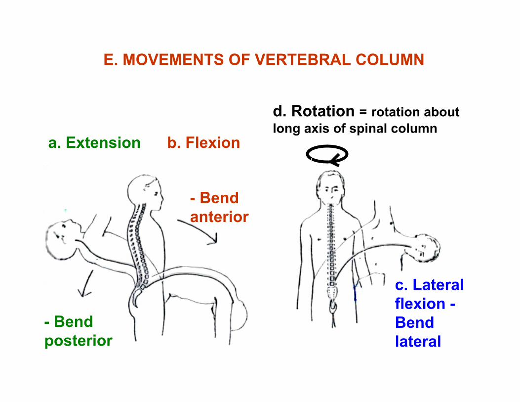
E. MOVEMENTS OF VERTEBRAL COLUMN
d. Rotation = rotation about long axis of spinal column
c. Lateral flexion -Bend lateral
a. Extension b. Flexion
- Bend posterior
- Bend anterior
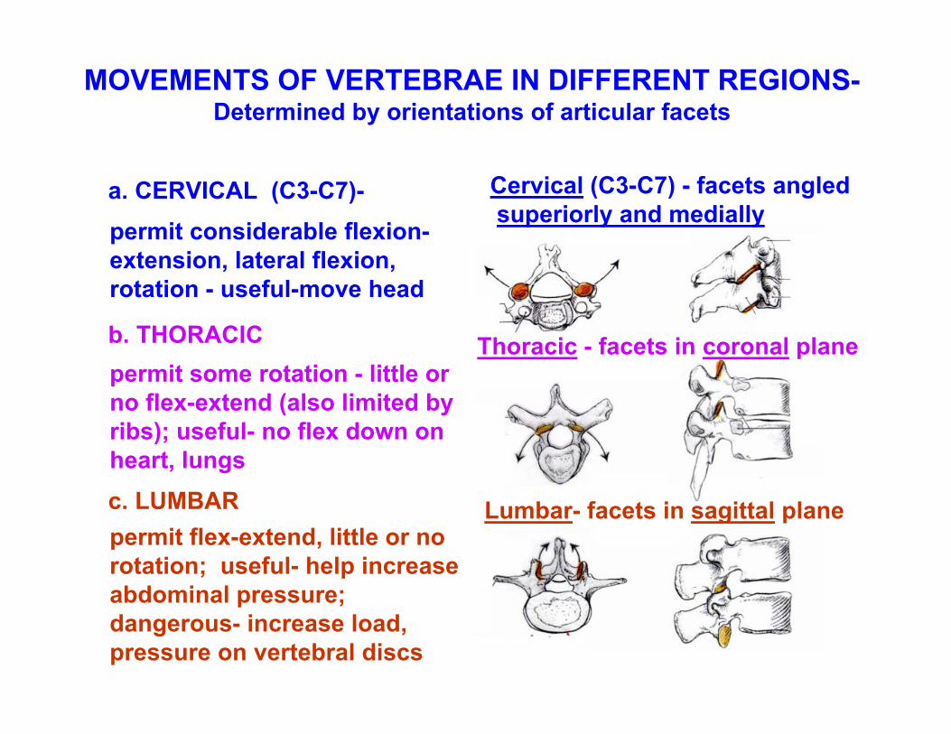
MOVEMENTS OF VERTEBRAE IN DIFFERENT REGIONS-Determined by orientations of articular facets
permit considerable flexion-extension, lateral flexion, rotation - useful-move head
permit some rotation - little or no flex-extend (also limited by ribs); useful- no flex down on heart, lungs
Lumbar- facets in sagittal plane
Thoracic - facets in coronal plane
permit flex-extend, little or no rotation; useful- help increase abdominal pressure; dangerous- increase load, pressure on vertebral discs
Cervical (C3-C7) - facets angledsuperiorly and medially
a. CERVICAL (C3-C7)-
b. THORACIC
c. LUMBAR
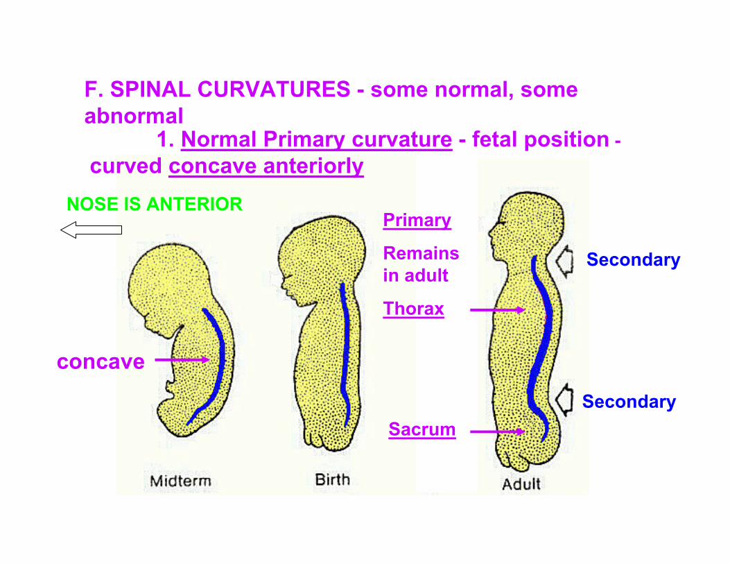
F. SPINAL CURVATURES - some normal, some abnormal
1. Normal Primary curvature - fetal position -curved concave anteriorly
Primary
Remains in adult
Thorax
Sacrum
concave
Secondary
Secondary
NOSE IS ANTERIOR
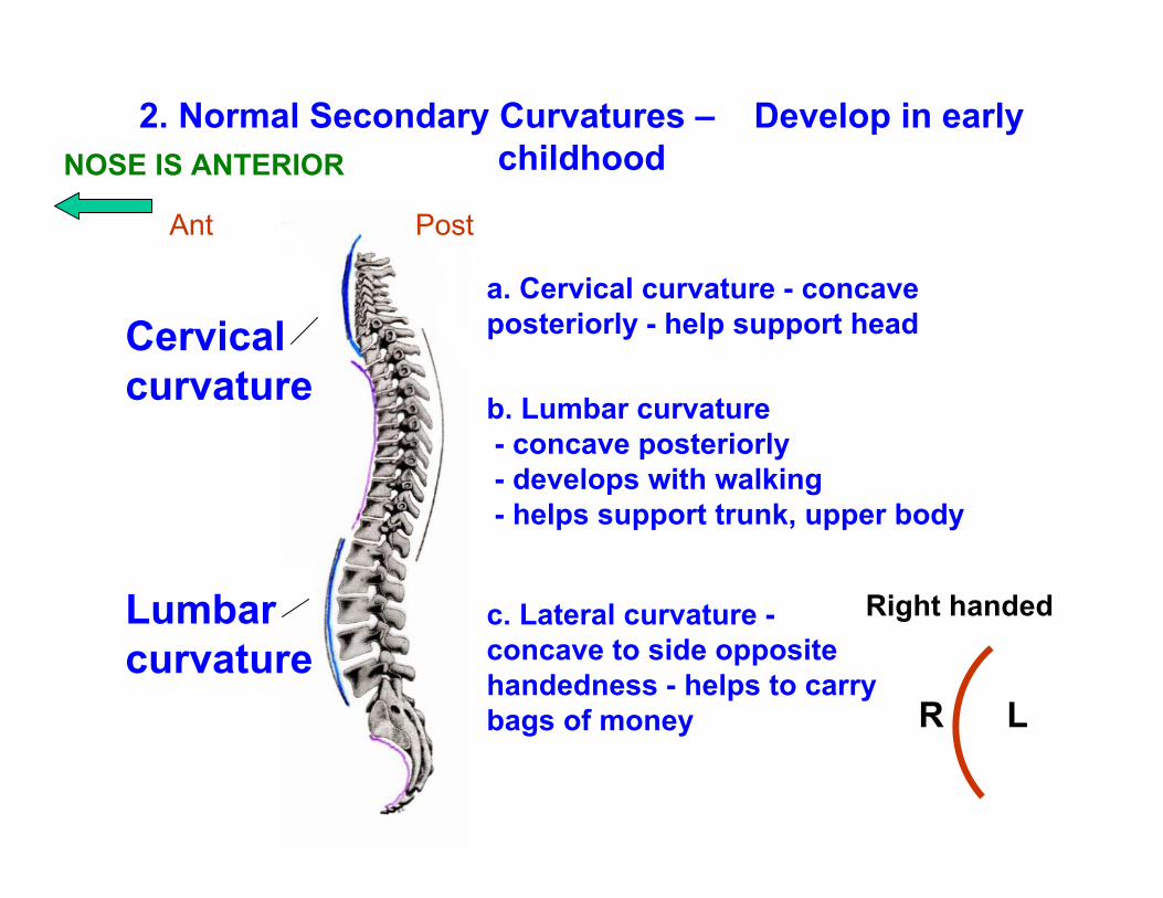
Cervical curvature
Lumbar curvature
2. Normal Secondary Curvatures – Develop in early childhood
a. Cervical curvature - concave posteriorly - help support head
b. Lumbar curvature- concave posteriorly - develops with walking - helps support trunk, upper body
c. Lateral curvature -concave to side opposite handedness - helps to carry bags of money
Ant Post
Right handed
R L
NOSE IS ANTERIOR
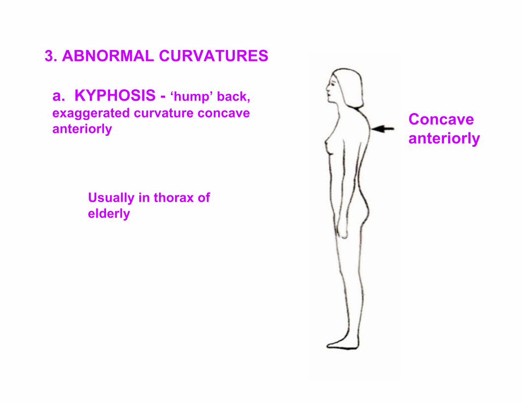
3. ABNORMAL CURVATURES
a. KYPHOSIS - ‘hump’ back, exaggerated curvature concave anteriorly Concave
anteriorly
Usually in thorax of elderly
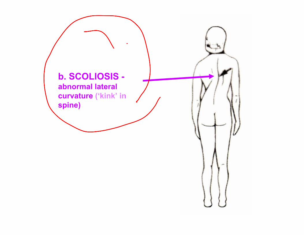
b. SCOLIOSIS -abnormal lateral curvature (‘kink’ in spine)
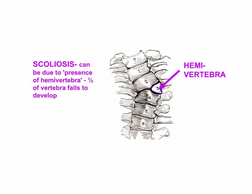
SCOLIOSIS- can be due to 'presence of hemivertebra' - ½ of vertebra fails to develop
HEMI-VERTEBRA
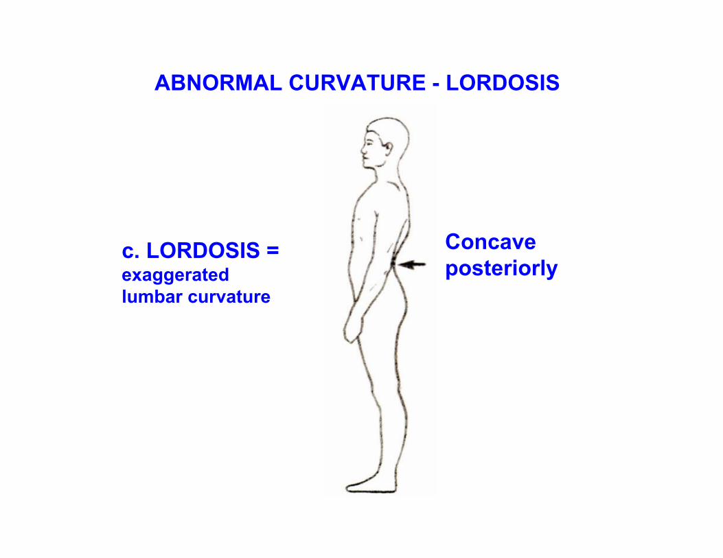
ABNORMAL CURVATURE - LORDOSIS
c. LORDOSIS = exaggerated lumbar curvature
Concave posteriorly
USEFUL WEB SITE: MEDLINEPLUS.GOV
- THROUGHNIHNATIONALLIBRARY OFMEDICINE;CAN DOSEARCHESFOR DEFINITIONSOF TERMS,CLINICALCORRELATIONS,TREATMENTS,ETC.
USEFUL WEB SITE: MEDLINEPLUS.GOV