Ventricular tachycardia associated with a left ventricular - Deep Blue
3
334 Brief Communications =ebruary 1983 American Heart .Journai Table I. Hemodynamic data JVCJ therap) Pulmonary artery (mm Hg) Systolic Diastolic Mean Pulmonary capillary wedge pressure (mm Hg) Mean Systemic arterial pressure (mm Hg) Systolic Hiastolic Mean Resistances (dynes-set-cm-’ ) Pulmonary arteriolar Total systemic (Cardiac output (I,/min) Heart rate (bpm) beneficial effects of diltiazem were followed by an associ- ated subjective improvement over an 11-month period.’ In this report we describe a late-stage patient who sustained a significant improvement with nifedipine over a 6-month period. This improvement was associated with a significant change in hemodynamic data measured repeatedly. A white female patient (age 27 years) was admitted for evaluation of progressive dyspnea and synco- pe on exertion. At the age of 20 years she was known to have cardiomegaly. Her blood pressure was 115/75 mm Hg. There was clinical evidence of right ventricular hyper- trophy. On auscultation a loud pulmonic closure sound was followed by a long early diastolic murmur of pulmo- nary regurgitation. Routine laboratory examinations were normal. The ECG showed right axis deviation and right ventricular hypertrophy. The chest x-ray showed cardio- megaly and central pulmonary arterial dilatation with distal vessel narrowing. A pulmonary angiogram revealed no signs of pulmonary emboli. The most important hemodynamic data are shown in Table I. The pulmonary capillary wedge pressure was normal. After treatment with nifedipine, 20 mg six times a day, significant improvement of hemodynamic data was seen after 3 days. During the follow-up period, symptoms markedly improved. No side effects occurred. Repeated catheterization after 6 months of continued oral therapy showed a sustained beneficial effect (Table I). The results in our patient clearly indicate that further evaluation of nifedipine use in primary pulmonary hypertension is warranted. REFERENCES I. Rraunwald E: Heart disease: A textbook of cardiovascular medicine. Philadelphia, 1980, WB Saunders Co, p 1636. 2. McMurtry IF, Davidson AB, Reeves JT, Grover RF: Inhibi- tion of hypoxic pulmonary vasoconstriction by calcium antag- onists in isolated rat lungs. Circ Res 38:99, 1976. 110 6.5 SO 857 1428 4.2 80 3. Camerini F, Alberti E, Klugman S, Salvi A: Primary pulmo- nary hypertension: Effects of nifedipine. Br Heart cJ 44:352. 1980. 4. Kambara H, Fujimoto K, Wakabayashi A, Kawai C: Primary pulmonary hypertension: Beneficial therapy with diltiazem. AM HEART J 101:230. 1981. Ventricular tachycardia associated with a left ventricular apex sump aneurysm in an adolescent Kenneth M. Weesner, M.D., Craig J. Byrum, M.D., Macdonald Dick, M.D., Albert P. Rocchini, M.D., Douglas M. Behrendt, M.D., and Paul Hees, B.S. Ann Arbor, Mich. Recently we reported a group of children who were surgically treated for congenital heart disease and who developed left ventricular (LV) apical aneurysms after venting of the left ventricle at the time of cardiopulmo- nary bypass.’ We speculated that the aneurysms, although small, might provide a focus for ventricular arrhythmias. This report describes a case of potentially life-threatening ventricular arrhythmia developing postoperatively in a patient with residual subaortic stenosis. Location of ven- E’rom the Sections of Pediatric Cardiology and Thoracic Surgery. C. S. Mott Children’s Hospital. and the Departments of Pediatrics and Surgery. I’niversity of Michigan Medical Center. Received for publication June 26. 1981: accepted July 13. 1981. Heprint requests: Macdonald Dick, M.D.. Pediatric Cardiology, FlI15. C. S. Mott Children’s Hospital, LXversity of Michigan Medical Center. Ann Arbor, MI 48109. OOOZ-8703/83/02O:i:34 + 03$0030/O 1983 The C. V. Mosby Co.
Transcript of Ventricular tachycardia associated with a left ventricular - Deep Blue

334 Brief Communications =ebruary 1983
American Heart .Journai
Table I. Hemodynamic data
JVCJ therap)
Pulmonary artery (mm Hg) Systolic Diastolic Mean
Pulmonary capillary wedge pressure (mm Hg) Mean
Systemic arterial pressure (mm Hg) Systolic Hiastolic Mean
Resistances (dynes-set-cm-’ ) Pulmonary arteriolar Total systemic
(Cardiac output (I,/min) Heart rate (bpm)
beneficial effects of diltiazem were followed by an associ- ated subjective improvement over an 11-month period.’
In this report we describe a late-stage patient who sustained a significant improvement with nifedipine over a 6-month period. This improvement was associated with a significant change in hemodynamic data measured repeatedly. A white female patient (age 27 years) was admitted for evaluation of progressive dyspnea and synco- pe on exertion. At the age of 20 years she was known to have cardiomegaly. Her blood pressure was 115/75 mm Hg. There was clinical evidence of right ventricular hyper- trophy. On auscultation a loud pulmonic closure sound was followed by a long early diastolic murmur of pulmo- nary regurgitation. Routine laboratory examinations were normal. The ECG showed right axis deviation and right ventricular hypertrophy. The chest x-ray showed cardio- megaly and central pulmonary arterial dilatation with distal vessel narrowing. A pulmonary angiogram revealed no signs of pulmonary emboli.
The most important hemodynamic data are shown in Table I. The pulmonary capillary wedge pressure was normal. After treatment with nifedipine, 20 mg six times a day, significant improvement of hemodynamic data was seen after 3 days. During the follow-up period, symptoms markedly improved. No side effects occurred. Repeated catheterization after 6 months of continued oral therapy showed a sustained beneficial effect (Table I). The results in our patient clearly indicate that further evaluation of nifedipine use in primary pulmonary hypertension is warranted.
REFERENCES
I. Rraunwald E: Heart disease: A textbook of cardiovascular medicine. Philadelphia, 1980, WB Saunders Co, p 1636.
2. McMurtry IF, Davidson AB, Reeves JT, Grover RF: Inhibi- tion of hypoxic pulmonary vasoconstriction by calcium antag- onists in isolated rat lungs. Circ Res 38:99, 1976.
110 6.5 SO
857 1428
4.2
80
3. Camerini F, Alberti E, Klugman S, Salvi A: Primary pulmo- nary hypertension: Effects of nifedipine. Br Heart cJ 44:352. 1980.
4. Kambara H, Fujimoto K, Wakabayashi A, Kawai C: Primary pulmonary hypertension: Beneficial therapy with diltiazem. AM HEART J 101:230. 1981.
Ventricular tachycardia associated with a left ventricular apex sump aneurysm in an adolescent
Kenneth M. Weesner, M.D., Craig J. Byrum, M.D., Macdonald Dick, M.D., Albert P. Rocchini, M.D., Douglas M. Behrendt, M.D., and Paul Hees, B.S. Ann Arbor, Mich.
Recently we reported a group of children who were surgically treated for congenital heart disease and who developed left ventricular (LV) apical aneurysms after venting of the left ventricle at the time of cardiopulmo- nary bypass.’ We speculated that the aneurysms, although small, might provide a focus for ventricular arrhythmias. This report describes a case of potentially life-threatening ventricular arrhythmia developing postoperatively in a patient with residual subaortic stenosis. Location of ven-
E’rom the Sections of Pediatric Cardiology and Thoracic Surgery. C. S. Mott Children’s Hospital. and the Departments of Pediatrics and Surgery. I’niversity of Michigan Medical Center. Received for publication June 26. 1981: accepted July 13. 1981.
Heprint requests: Macdonald Dick, M.D.. Pediatric Cardiology, FlI15. C. S. Mott Children’s Hospital, LXversity of Michigan Medical Center. Ann Arbor, MI 48109.
OOOZ-8703/83/02O:i:34 + 03$0030/O 1983 The C. V. Mosby Co.
Volume 105
Number 2 Brief Communications 335
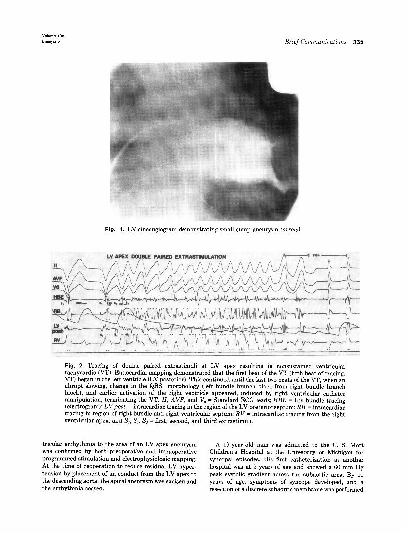
Fig. 1. LV cineangiogram demonstrating small sump aneurysm (arrow).
Fig. 2. Tracing of double paired extrastimuli at LV apex resulting in nonsustained ventricular tachycardia (VT). Endocardial mapping demonstrated that the first beat of the VT (fifth beat of tracing, VT) began in the left ventricle (LV posterior). This continued until the last two beats of the VT, when an abrupt slowing, change in the QRS morphology (left bundle branch block from right bundle branch block), and earlier activation of the right ventricle appeared, induced by right ventricular catheter manipulation, terminating the VT. ZZ, AVF, and V, = Standard ECG leads; HBE = His bundle tracing (electrogram); LV post = intracardiac tracing in the region of the LV posterior septum; RB = intracardiac tracing in region of right bundle and right ventricular septum; RV = intracardiac tracing from the right ventricular apex; and S,, Sp, S, = first, second, and third extrastimuli.
tricular arrhythmia to the area of an LV apex aneurysm A 19-year-old man was admitted to the C. S. Mott was confirmed by both preoperative and intraoperative Children’s Hospital at the University of Michigan for programmed stimulation and electrophysiologic mapping. syncopal episodes. His first catheterization at another At the time of reoperation to reduce residual LV hyper- hospital was at 5 years of age and showed a 60 mm Hg tension by placement of an conduct from the LV apex to peak systolic gradient across the subaortic area. By 10 the descending aorta, the apical aneurysm was excised and years of age, symptoms of syncope developed, and a the arrhythmia ceased. resection of a discrete subaortic membrane was performed

336 h'rief C’ommunications
at our institution. The patient did well postoperatively until 14 1 2 years of age, when he again had two episodes of syncope accompanied by decreased exercise tolerance. A second catheterization at that time revealed LV pressure of 208/18 mm Hg and aortic pressure of 140/90 mm Hg. One month later, he underwent resection of subaortic membrane and a hypertrophic muscle band. He was improved following this second operation, and at 171’: years of age he underwent a postoperative catheterization that demonstrated a 50 mm Hg residual LV outflow gradient, trivial aortic insufficiency, and a small apical aneurysm (Fig. 1). He remained without symptoms until 1 month prior to admission, when he had three syncopal episodes, two during light exertion and one while reading. He denied other cardiovascular symptoms. Pertinent physical findings included a thrill, a harsh grade 416 systolic ejection murmur at the right upper sternal border radiating to the carotid arteries, and a grade l/4 diastolic decrescendo murmur at the left lower sternal border. A resting ECG showed LV hypertrophy with nonspecific ST segment and T wave changes in the lateral precordial leads. A graded treadmill exercise test using the Bruce protocol showed no ischemic changes; Lown’s grade 1B ventricular ectopy was present in the early stages of exercise.:’ A 24.hour Holter monitor demonstrated one short burst of ventricular tachycardia at 172 beats per minute, as well as frequent unifocal ventricular premature beats (VPBs) in all recorded hours. Cardiac catheteriza- tion demonstrated a 55 mm Hg residual gradient across the LV outflow at rest and a 110 mm Hg gradient during supine bicycle exercise. LV end-diastolic pressure was 20 mm Hg at rest and increased to 25 mm Hg with exercise. No ectopy was noted during exercise, but a salvo of ventricular tachycardia was seen during the recovery phase.
An electrophysiologic study was conducted with a hexa- polar catheter placed transseptally across the mitral valve and with bipolar electrode pairs at the LV apex and LV midposterior septum.’ Electrograms were also recorded from the high right atrium, low right atrium, His bundle region, right septal region, and right ventricular apex. Programmed extrastimulation of the right ventricular apex, right ventricular outflow, and LV midposterior septum failed to evoke repetitive beats. During LV apex pacing (S,S,) at a basic drive rate of 100 bpm (C, = 600 msec), single paired (S,S, = 230 msec) extrastimuli consis. tently resulted in repetitive beats originating near the apex. Double paired extrastimuli at the LV apex also resulted in single repetitive beats originating at the apex. At an S,-S, interval of 225 msec and an S,-S:; interval of 185 msec, ventricular tachycardia at 275 bpm, accompa- nied by an acute blood pressure decline, was initiated (Fig. 2). Catheter-induced VPBs converted the tachycardia to supraventricular; spontaneous conversion to normal sinus rhythm then followed. One week later the patient under- went construction of a conduit between the LV apex and the descending aorta, and a 20 mm Hancock valved prosthesis was inserted.J Intraoperative endocardial map-
ping confirmed ectopic beats originating on tht posteriol septal side of the sump aneurysm. Programmed single extrastimulation at this site resulted in three repetitive beats.,’ This area was resected during conduit placement. Programmed extrastimulation at this site failed to evoke repetitive beats following excision. Post,operative Halter monitoring (48 hours) revealed no ventricular ect.opy. and the patient has remained asymptomatic since his opera- tion.
At our institution during a I-year period, 50 children underwent routine cardiac catheterization following open heart surgery in which the LV sump venting technique was used; 32”, had small and usually intramural LV apical aneurysms because of intraoperative venting of the LV apex.’ We were concerned that this aneurysm might become a source of ventricular arrhythmias, particularly with advancing age. This patient represents the first such case. Using programmed stimulation techniques, we were able to locate the site of the origin of ventricular ectopy to the intramural defect caused by the aneurysm. The poten- tial for hemodynamically unstable ventricular arrhythmia was clearly demonstrated, probably correlating with the reported symptoms. Although the precise mechanism of the ventricular tachycardia could not be absolutely deter mined,’ the response t,o extrastimuli suggested that the most likely mechanism was reentry in the area of the aneurysm, similar to the mechanism of ventricular tachy- cardia seen in adults with ischemic heart disease and ventricular aneurysm.i ” Fortunately the point of origin was in an area that had to be resected for insert.ion of the conduit. between the LV apex and the descending aorta. We conclude that the LV apex may be a potentially dangerous site for intraoperative venting and should be avoided whenever possibIe. Children who undergo surgery for congenital heart disease and who have life-threatening arrhythmia should also have preoperative electrophysio logic evaluatiun. since surgical therapy for the arrhythmia may also be accomplished at the t.ime of operation.
REFERENCES
Weesner KM, Ryrum C, Rosenthal A: Left ventricular aneu- rysms associated with intraoperative venting of the cardiac apex in children. AM HEART d 101~622, 1981. idown R: Sudden cardiac death-- 1978. Circulation 60:159:1,
1979. Josephson MU. Harken AH, Horowitz LN: Endocardial excision: A new surgical technique for the treatment of recurrent \zentricular t,achycardia. Circulation 60:1430, 1979. Norman JI). Cooley DA, Hallman GL, Nihill MR: Let’t ventricular apical-abdominal aortic conduits for left ventric- ular outflow tract obstruction. Circulation 56:(suppl II):62. 1977. Rosen MR. Keder RF: Does triggered activity have a role in the genesis 01’ cardiac arrhythmias? Ann Intern Med 94:794, 1981. -Josephson ME, Horowitz LN, Farshidi A, Kastor J: Recur- rent sustained ventricular tachycardia. I. Mechanisms. Circu- lation 57:431. 197%


![Ventricular Tachycardia in the Ischemic Heart[1]](https://static.fdocuments.in/doc/165x107/55283aae49795921048b4677/ventricular-tachycardia-in-the-ischemic-heart1.jpg)