Ventilation
27
VENTILATOR Soumya Ranjan Parida Basic B.Sc. Nursing 4 th year Sum Nursing College
-
Upload
soumya-ranjan-parida -
Category
Healthcare
-
view
60 -
download
1
Transcript of Ventilation

VENTILATOR
Soumya Ranjan ParidaBasic B.Sc. Nursing 4th year
Sum Nursing College

MECHANICAL VENTILATION
Ist Introduced in 1950’s during polio epidemic in the west. Normal gas exchange is dependent on two processes.
Oxygenation – deals with the process of gas exchange with in the lung at alveolar level (external) or at the tissue level (internal).
Ventilation – deals with the movement of air in and out of the lungs.

GOALS OF MECHANICAL VENTILATION
1. Improve ventilation 2. Reduce the ventilation perfusion mismatch. 3. Reexpand the atelectatic or collapsed lung
segment. 4. Reduce the work of breathing.

COMMON INDICATIONS FOR MECHANICAL VENTILATION
1. Respiratory failure
Inadequate oxygenation – PaO2 < 60 mmHg
CO2 Retention (ventilation abnormalities) PaCO2 50 mmHg or more
2. Cardiovascular- Myocardial failure
3. Neurological disorder- decreased ventilatory drive, increased ICP
4. Respiratory paralysis - Neuromuscular disorder, LGB syndrome, snake bite,poisoning

BASIC TERMINOLOGY
Compliance – Stiffness of chest. Change in volume per unit change in pressure.
Expressed in lit/cm of H2O.
Resistance – Pressure difference between mouth and alveoli. Most important factor – Radius of the airway.
Functional Residual capacity – Volume of gas that is present in the lungs at the end of expiration. Normal FRC 30ml/kg.
Closing capacity – Refers to volume of gas present in the lung at which small conducting airways begin to collapse.
Tidal volume – Volume of gas that flows in and out during quite breathing. 6-8 ml/kg.
Cont……

Minute ventilation – Tidal volume X rate.
I.E. Ratio- Normal 1:2 to 1:3
PIP- Highest pressure during the Inspiratory time.
Positive end expiratory pressure – (PEEP)- Minimum positive pressure in airways during expiration. It keep alveoli open at the end of expiration.
Normal PEEP ( Physiological PEEP) is 2-3 cmH2O
FIO2 (Fractional inspired oxygen) concentration - 0.21-1.00
Mean Airway pressure (MAP) – Average positive pressure generated in thelungs throughout the respiratory cycle.
K(Ti x PIP)
MAP = + PEEP
Ti + Te

Classification of mechanical ventilator breathsBreath type
Phase
Trigger
variable
Cycle Limit
Mandatory Machine Machine Machine
Assisted Patient Machine Machine
Supported Patient Patient Machine
Spontaneous
Patient Patient Patient

MODES OF VENTILATION
Spontaneous Mode – Patients is breathing on his own- CPAP (continuous positive airway pressure) PSU (pressure support ventilation).
Controlled mechanical ventilation (CMV)- ventilation controls all the ventilation while patient has minimal or no respiratory efforts. It delivers pre selected ventilatory rate, tidal volume and inspiratory flow rate.
Indication – Apnea, depression of CNS, neuromuscular paralysis significant fatigue of the ventilatory muscles.
Continuous positive pressure ventilation (CPPV) -It delivers a positive pressure breathing. Airway pressure never return to zero.
Cont….

Assisted mechanical ventilation It is patients triggered positive pressure ventilation, used during weaning of patients from CMV.
Assist control ventilation Assisted Mechanical ventilation + CMV The ventilator may be triggered by the patients, spontaneous inspiratory effort or by a timing devise whichever comes first.
Intermittent Mandatory ventilation (IMV) - delivers a preset number of controlled breaths at preset intervals. In between these breaths patients is allowed to breath spontaneously. Pressure controlled Volume controlled
Synchronized Intermittent mandatory Ventilation (SIMU)- Pressure support - Volume support - The ventilation delivered mandatory breath is synchronized to begin with patients own breathing efforts.

INITIAL VENTILAOR SETTINGS
1. 1. Rate – Physiological norm for age
2. Select VT~6-8ml/kg or PIP~15 CM H2O (Normal lungs)
20-25 cmH2O (Moderate disorder)
25-30 cm H2O (Severe disease)
3. I:E ratio – Begin with 1:2. Initial T1 may be 0.4-0.5 sec in a young infant
&higher value is chosen for older children.4. In obstructive disease use longer Te and avoid prolonged Ti.
5. Assess chest expansion and breath sounds to determine adequacy of ventilation.
6. Measure PaCO2 – Adjust VT/PIP or rate to maintain PaCO2 between 35-45
mmHg. 1

CARE OF PATIENT ON VENTILATOR
1. Repositioning of patient 2-4 hrly.
2. Change of ventilator tubing periodically. 3. Filling up of humidifier with sterile water daily.
4. Wash compressor filter daily.
5. Culture of respiratory endotracheal secretions daily to monitor the microbial flora.
6. Majority of ventilated children may be fed enterally via a nasogastric tube.
Cont….

7. Sedation and muscle relaxant – Particular attention is needed in following situations
-Requirement for high airway pressure -Critically inadequate oxygenation (ARDS)
-Ventilator intolerance in anxious patient.
-Need for controlled hyperventilation .
Before switching to muscle relaxant (Pancuronium,vecuronium) adequate sedation and analgesia should be achieved with help of diazepam ( 0.2 mg/kg i.v.) and morphine (0.1-0.2 mg/kg i.v.).

Monitoring
Physical examination:-Breathing rate,Signs of resp.distress,B.P.,HR.Skin color,Chest movement,
Invasive monitoring:-ABG analysis Non invasive monitoring:-Chest
radiography,ECG.Pulse oxymetry,Capnography.

MONITORING
1. ABGs & DO2 calculation within 20-30 min of
initiation and after a change in setting and every 6 hourly.
2. Chest x-ray – Once daily or in 2 days.
3. I/O measurements – 8 hourly.
4. Electrolytes, urea, coagulation parameters
5. Nutritional support – Enteral and parenteral
6. Nutritional status and wt.

CLINICAL AND LABORATORY INDICES OF ADEQUATE VENTILATION
Clinical Parameters
- Pink color
- Adequate chest expansion
- Absence of retractions
- CFT < 2 sec
- Normal blood pressure
Pulse Oxymetry
- O2 saturation 90-95%
Blood gases
- PaO2 - 50-80 mmHg
- PaCO2 - 40-50 mmHg
- pH - 7.35-7.45
-

Blood gas abnormalities and changes in the ventilator settings to correct them
S.no Blood gas abnormality
Corrective measures
FiO2 Rate PIP PEEP Ti
1. Hypercapnea (PaCO2
> 50mm Hg)
- Increased Increased - -
2 Hypocapnea (PaCO2
< 35mm Hg)
- Decreased Decreased - -
3 Hyperoxia (PaO2
> 100mm Hg)
Decreased - Decreased Decreased Decreased
4 Hypoxia (PaO2
<50mmHg)
Increased - Increased Increased Increased

WEANING FROM VENTILATOR
Criteria for weaning
Clinical –
1. Stable pulmonary status without evidence of pneumonia, severe infection or bronchospasm.
2. Satisfactory muscle power.
3. Cardiovascular stability – No need for vasoactive drugs.
4. Afebrile.
Ventilation and oxygenation parameters
1. Tidal volume > 5 ml/kg
2. Vital capacity > 10 ml/kg
3. PaO2 > 60 mmHg at FiO2 < 0.4
4. Maximum voluntary ventilation > 2 x minute volume
5. PaO2 / FiO > 200
6. Shunt fraction < 15%
7. VD/VT ratio < 0.6
8. FRC > 50% predicted
9. Minute ventilation > 180 ml/kg/min for PaCO2 of 40 mmHg

VENTILATOR EMERGENCY
D – Displacement of the tube
O – Obstruction of the tube
P – Pneumothorax
E – Equipment failure
Sudden fall of SPO2,Cyanosis,Pallor, Fall in BP. CFT,Bradycardia.
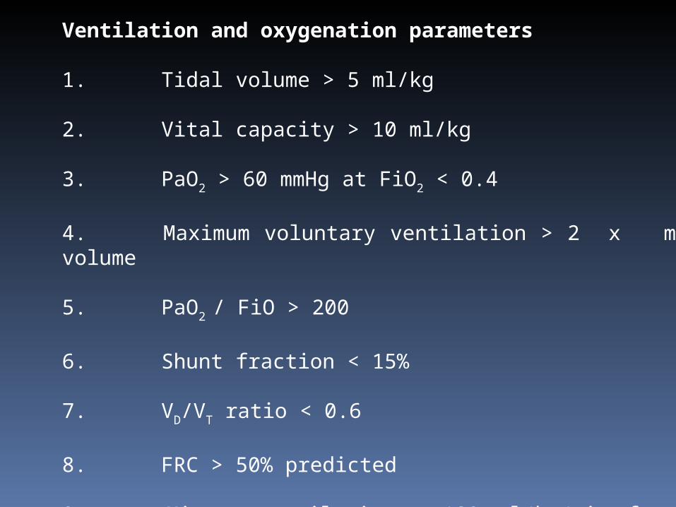
COMPLICATIONS OF VENTILATOR
Related to airway pressure and lung volume –
A.Barotrauma/Volutrauma – Pneumonia, Pneumothorax, pneumopericardium, subcutaneous emphysema. B.Decreased cardiac fillingC.Pulmonary parenchymal damage D.Acid base disturbances 2. Related to Endotracheal Tube –
A.Tracheal, laryngeal & pharyngeal mucosal damage especially with intubation > 3 weeks B.Sinusitis C.Laryngeal edema, subglottic stenosis
Cont….

3. Nosocomial infection
A. Ventilator associated pneumonia
B. Sepsis
4. Mechanical operational failure
A. Mechanical failure
B. Alarm failure
C. Inadequate humidification

DISEASE SPECIFIC INITIAL VENTILATOR SETTINGS
PNEUMONIA VT - 6-8 ml/kg
PIP - 20-25 cm H2O
PEEP - 4-5 cm H2O
Rate - 40-50/min (Infant)- 30-40 / min (older children)
I:E ratio - 1:2
ASTHMA/BRONCHIOLITIS Permissive hypercapnea PEEP is kept low to prevent air trapping
VT - 6-8 ml/kg
PIP - 20-25 cm H2O
PEEP - 3-4 cm H2O
Rate 30/min (Infant)20 / min (older children)
I:E ratio - 1:2 to 1:3 Cont….

ARDS Oxygenation is maintained using high PEEP
VT - 4-6 ml/kg
PEEP - 5-10cm H2O
Rate 40/min (Infant)20-30 / min (older children)
I:E ratio - 1:2 to inverse ratio
NORMAL LUNG AS IN SHOCK,.FLACCID PARALYSIS IMV mode In septic shock use of higher FiO2 upto 1 initially
VT - 6-8 ml/kg
PEEP - 3-4 cm H2O
Rate 40/min (Infant)20-30 / min (older children)
I:E ratio - 1:2 to 1:3

NEWER MODALITIES
HIGH FREQUENCY VENTILATION (HFV)
This is mode of assisted ventilation, which employs small tidal volume (1-3ml/kg body weight) and much higher frequencies. It has adequate minute ventilation with relatively low mean airway pressure.
Uses – 1. RDS with PPHN2. RDS, HIE where conventional ventilation failed.3. Pulmonary air leaks.
Complications of HFV – 1. Inadvertent PEEP2. ICH 3. Necrotising tracheobronchitis

ISOLATED LUNG VENTILATION Single lung ventilation has been increasingly used in
video assisted ,thoracoscopic intervention as well as open thoracic surgical procedure.
HELIOX VENTILATION- HELIUM+OXYGEN Has reduced density and hence lower resistance
in turbulent flow and more favorable diffusion.
PARTIAL LIQUID VENTILATION Perflubron a highly oxygen soluble emulsion It is heavy distribution to dependent lung regions after instillation.This open and stabilizes collapsed alveolar units.It also functions as a surfactant.
Permissive hypercapnia :-mantaining a high level of PaCO2(45-55 mmHg),safe in RDS,Status asthmaticus & Neonatal resp.failure
Supportive adjuncts
Prone positioning Dexamethasone Epinephrine Nitric oxide