VENTANA PD-L1 (SP263) Assay · presenting cells and activated T cells. PD -L1 binding to CD80 on T...
62
2017-04-19 1 / 7 1014737US Rev A VENTANA PD-L1 (SP263) Assay 740-4907 07208162001 50 Rx Only INTENDED USE VENTANA PD-L1 (SP263) Assay is a qualitative immunohistochemical assay using rabbit monoclonal anti-PD-L1 clone SP263 intended for use in the assessment of the PD-L1 protein in formalin-fixed, paraffin-embedded (FFPE) urothelial carcinoma tissue stained with OptiView DAB IHC Detection Kit on a VENTANA BenchMark ULTRA instrument. PD-L1 status is determined by the percentage of tumor cells with any membrane staining above background or by the percentage of tumor- associated immune cells with staining (IC+) at any intensity above background. The percent of tumor area occupied by any tumor-associated immune cells (Immune Cells Present, ICP) is used to determine IC+, which is the percent area of ICP exhibiting PD-L1 positive immune cell staining. PD-L1 status is considered High if any of the following are met: • ≥ 25% of tumor cells exhibit membrane staining; or, • ICP > 1% and IC+ ≥ 25%; or, • ICP = 1% and IC+ = 100%. PD-L1 High status as determined by VENTANA PD-L1 (SP263) Assay was associated with increased objective response rate (ORR) in a single arm study of IMFINZI™ (durvalumab). This product is intended for in vitro diagnostic (IVD) use. SUMMARY AND EXPLANATION VENTANA PD-L1 (SP263) Assay is an immunohistochemical assay utilizing an anti-PD-L1 rabbit monoclonal primary antibody (VENTANA PD-L1 (SP263)) to recognize the programmed death ligand 1 (PD-L1). It recognizes a transmembrane bound glycoprotein that has a molecular mass of 45-55 kDa. This antibody produces membranous and/or cytoplasmic staining. Urothelial carcinoma (also known as urothelial cell carcinoma, transitional cell carcinoma of the urinary tract, or urothelial bladder cancer) is the most common cancer of the urinary system worldwide. The majority of urothelial tumors arise in the bladder with the remainder originating in the renal pelvis, urethra, or ureter. Transitional cell carcinoma (TCC) is the most common histologic subtype associated with bladder cancer and accounts for greater than 90% of all urothelial carcinoma cases in the industrialized world; non-urothelial subtypes (e.g., squamous cell carcinoma, adenocarcinoma, small cell carcinoma) are more frequent in other areas of the world. 1 Globally, there were an estimated 429,793 new cases of bladder cancer and 165,084 deaths in 2012. 2 In Europe alone, for 2012, there were an estimated 151,297 new cases of bladder cancer and 52,411 deaths. In 2015, it was estimated that there would be 74,000 new cases of bladder cancer and 16,000 deaths in the United States. 3 Urothelial carcinoma presents as non-muscle-invasive, muscle-invasive, or metastatic disease. The overall 5-year survival rate for metastatic urothelial carcinoma (mUC) is approximately 5.4%. 4 PD-L1 is a transmembrane protein that downregulates immune responses through binding to its two receptors programmed death-1 (PD-1) and B7-1 (CD80). 5 PD-1 is an inhibitory receptor expressed on T cells following T-cell activation, which is sustained in states of chronic stimulation such as in chronic infection or cancer. 6 Binding of PD-L1 with PD-1 inhibits T cell proliferation, cytokine production, and cytolytic activity, leading to the functional inactivation or exhaustion of T cells. CD80 is a molecule expressed on antigen presenting cells and activated T cells. PD-L1 binding to CD80 on T cells and antigen presenting cells can mediate downregulation of immune responses, including inhibition of T-cell activation and cytokine production. 7 PD-L1 expression has been observed in immune cells and tumor cells. 8,9 Aberrant expression of PD-L1 on tumor cells and tumor associated immune cells has been reported to impede anti-tumor immunity, resulting in immune evasion. 6,9 Therefore, interruption of the PD-L1/PD-1 pathway represents an attractive strategy to reinvigorate tumor-specific T cell immunity suppressed by the expression of PD-L1 in the tumor microenvironment. PD-L1 is expressed in a broad range of cancers including lung, melanoma, urothelial, ovarian, and colorectal cancer. Prevalence of PD-L1 expression has been reported from 12% to 100% depending on the tumor type, anti PD-L1 clone and cutoff for positivity. 10 IMFINZI (durvalumab) is a human immunoglobulin G1 (IgG1) kappa mAb with immune checkpoint inhibitory and antineoplastic activities that blocks the interaction of PD-L1 (but not programmed cell death ligand-2) with PD-1 on T cells and CD80 on immune cells (IC). IMFINZI (durvalumab) is engineered to reduce antibody dependent cell mediated cytotoxicity and has a calculated molar mass of 146.3 kg/mol. PRINCIPLE OF THE PROCEDURE VENTANA PD-L1 (SP263) is a rabbit monoclonal primary antibody which binds to PD-L1 in paraffin-embedded tissue sections. The specific antibody can be localized using a haptenated secondary antibody followed by a multimer anti-hapten-HRP conjugate (OptiView DAB IHC Detection Kit, Cat. No. 760-700 / 06396500001). The specific antibody-enzyme complex is then visualized with a precipitating enzyme reaction product. Each step is incubated for a precise time and temperature. At the end of each incubation step, the VENTANA BenchMark instrument washes the sections to stop the reaction and to remove unbound material that would hinder the desired reaction in subsequent steps. It also applies ULTRA LCS (Cat. No. 650-210 / 05424534001), which minimizes evaporation of the aqueous reagents from the specimen slide. In addition to staining with VENTANA PD-L1 (SP263), a second slide should be stained with Rabbit Monoclonal Negative Control Ig (Cat. No. 790-4795 / 06683380001). The negative reagent control is used to assess background staining. REAGENT PROVIDED VENTANA PD-L1 (SP263) contains sufficient reagent for 50 tests. One 5 mL dispenser of VENTANA PD-L1 (SP263) contains approximately 8.05 μg of a rabbit monoclonal antibody. The antibody is diluted in 0.05 M Tris-HCI with 1% carrier protein, and 0.10% ProClin 300, a preservative. Total protein concentration of the reagent is approximately 10 mg/mL. Specific antibody concentration is approximately 1.61 μg/mL. There is no known non-specific antibody reactivity observed in this product. VENTANA PD-L1 (SP263) is a recombinant rabbit monoclonal antibody produced as purified cell culture supernatant. Refer to the appropriate VENTANA detection kit package insert for detailed descriptions of: (1) Principles of the Procedure, (2) Materials and Reagents Needed but Not Provided, (3) Specimen Collection and Preparation for Analysis, (4) Quality Control Procedures, (5) Troubleshooting, (6) Interpretation of Results, and (7) General Limitations. MATERIALS REQUIRED BUT NOT PROVIDED Staining reagents, such as VENTANA detection kits and ancillary components, including negative and positive tissue control slides, are not provided. The following reagents and materials may be required for staining: 1. Rabbit Monoclonal Negative Control Ig (Cat. No. 790-4795 / 06683380001) 2. Microscope slides, positively charged 3. Bar code labels (appropriate for negative reagent control and primary antibody being tested) 4. Xylene (Histological grade) 5. Ethanol or reagent alcohol (Histological grade) • 100% solution: Undiluted ethanol or reagent alcohol • 95% solution: Mix 95 parts of ethanol or reagent alcohol with 5 parts of deionized water • 80% solution: Mix 80 parts of ethanol or reagent alcohol with 20 parts of deionized water Figure 1. PD-L1 expression in urothelial carcinoma.
Transcript of VENTANA PD-L1 (SP263) Assay · presenting cells and activated T cells. PD -L1 binding to CD80 on T...

2017-04-19 1 / 7 1014737US Rev A
VENTANA PD-L1 (SP263) Assay
740-4907 07208162001
50 Rx Only
INTENDED USE VENTANA PD-L1 (SP263) Assay is a qualitative immunohistochemical assay using rabbit monoclonal anti-PD-L1 clone SP263 intended for use in the assessment of the PD-L1 protein in formalin-fixed, paraffin-embedded (FFPE) urothelial carcinoma tissue stained with OptiView DAB IHC Detection Kit on a VENTANA BenchMark ULTRA instrument. PD-L1 status is determined by the percentage of tumor cells with any membrane staining above background or by the percentage of tumor-
associated immune cells with staining (IC+) at any intensity above background. The percent of tumor area occupied by any tumor-associated immune cells (Immune Cells Present, ICP) is used to determine IC+, which is the percent area of ICP exhibiting PD-L1 positive immune cell staining. PD-L1 status is considered High if any of the following are met: • ≥ 25% of tumor cells exhibit membrane staining; or, • ICP > 1% and IC+ ≥ 25%; or, • ICP = 1% and IC+ = 100%. PD-L1 High status as determined by VENTANA PD-L1 (SP263) Assay was associated with increased objective response rate (ORR) in a single arm study of IMFINZI™ (durvalumab). This product is intended for in vitro diagnostic (IVD) use.
SUMMARY AND EXPLANATION VENTANA PD-L1 (SP263) Assay is an immunohistochemical assay utilizing an anti-PD-L1 rabbit monoclonal primary antibody (VENTANA PD-L1 (SP263)) to recognize the programmed death ligand 1 (PD-L1). It recognizes a transmembrane bound glycoprotein that has a molecular mass of 45-55 kDa. This antibody produces membranous and/or cytoplasmic staining. Urothelial carcinoma (also known as urothelial cell carcinoma, transitional cell carcinoma of the urinary tract, or urothelial bladder cancer) is the most common cancer of the urinary system worldwide. The majority of urothelial tumors arise in the bladder with the remainder originating in the renal pelvis, urethra, or ureter. Transitional cell carcinoma (TCC) is the most common histologic subtype associated with bladder cancer and accounts for greater than 90% of all urothelial carcinoma cases in the industrialized world; non-urothelial subtypes (e.g., squamous cell carcinoma, adenocarcinoma, small cell carcinoma) are more frequent in other areas of the world.1 Globally, there were an estimated 429,793 new cases of bladder cancer and 165,084 deaths in 2012.2 In Europe alone, for 2012, there were an estimated 151,297 new cases of bladder cancer and 52,411 deaths. In 2015, it was estimated that there would be 74,000 new cases of bladder cancer and 16,000 deaths in the United States.3 Urothelial carcinoma presents as non-muscle-invasive, muscle-invasive, or metastatic disease. The overall 5-year survival rate for metastatic urothelial carcinoma (mUC) is approximately 5.4%.4 PD-L1 is a transmembrane protein that downregulates immune responses through binding to its two receptors programmed death-1 (PD-1) and B7-1 (CD80).5 PD-1 is an inhibitory receptor expressed on T cells following T-cell activation, which is sustained in states of chronic stimulation such as in chronic infection or cancer.6 Binding of PD-L1 with PD-1 inhibits T cell proliferation, cytokine production, and cytolytic activity, leading to the
functional inactivation or exhaustion of T cells. CD80 is a molecule expressed on antigen presenting cells and activated T cells. PD-L1 binding to CD80 on T cells and antigen presenting cells can mediate downregulation of immune responses, including inhibition of T-cell activation and cytokine production.7 PD-L1 expression has been observed in immune cells and tumor cells.8,9 Aberrant expression of PD-L1 on tumor cells and tumor associated immune cells has been reported to impede anti-tumor immunity, resulting in immune evasion.6,9 Therefore, interruption of the PD-L1/PD-1 pathway represents an attractive strategy to reinvigorate tumor-specific T cell immunity suppressed by the expression of PD-L1 in the tumor microenvironment. PD-L1 is expressed in a broad range of cancers including lung, melanoma, urothelial, ovarian, and colorectal cancer. Prevalence of PD-L1 expression has been reported from 12% to 100% depending on the tumor type, anti PD-L1 clone and cutoff for positivity.10 IMFINZI (durvalumab) is a human immunoglobulin G1 (IgG1) kappa mAb with immune checkpoint inhibitory and antineoplastic activities that blocks the interaction of PD-L1 (but not programmed cell death ligand-2) with PD-1 on T cells and CD80 on immune cells (IC). IMFINZI (durvalumab) is engineered to reduce antibody dependent cell mediated cytotoxicity and has a calculated molar mass of 146.3 kg/mol.
PRINCIPLE OF THE PROCEDURE VENTANA PD-L1 (SP263) is a rabbit monoclonal primary antibody which binds to PD-L1 in paraffin-embedded tissue sections. The specific antibody can be localized using a haptenated secondary antibody followed by a multimer anti-hapten-HRP conjugate (OptiView DAB IHC Detection Kit, Cat. No. 760-700 / 06396500001). The specific antibody-enzyme complex is then visualized with a precipitating enzyme reaction product. Each step is incubated for a precise time and temperature. At the end of each incubation step, the VENTANA BenchMark instrument washes the sections to stop the reaction and to remove unbound material that would hinder the desired reaction in subsequent steps. It also applies ULTRA LCS (Cat. No. 650-210 / 05424534001), which minimizes evaporation of the aqueous reagents from the specimen slide. In addition to staining with VENTANA PD-L1 (SP263), a second slide should be stained with Rabbit Monoclonal Negative Control Ig (Cat. No. 790-4795 / 06683380001). The negative reagent control is used to assess background staining.
REAGENT PROVIDED VENTANA PD-L1 (SP263) contains sufficient reagent for 50 tests. One 5 mL dispenser of VENTANA PD-L1 (SP263) contains approximately 8.05 μg of a rabbit monoclonal antibody. The antibody is diluted in 0.05 M Tris-HCI with 1% carrier protein, and 0.10% ProClin 300, a preservative. Total protein concentration of the reagent is approximately 10 mg/mL. Specific antibody concentration is approximately 1.61 μg/mL. There is no known non-specific antibody reactivity observed in this product. VENTANA PD-L1 (SP263) is a recombinant rabbit monoclonal antibody produced as purified cell culture supernatant. Refer to the appropriate VENTANA detection kit package insert for detailed descriptions of: (1) Principles of the Procedure, (2) Materials and Reagents Needed but Not Provided, (3) Specimen Collection and Preparation for Analysis, (4) Quality Control Procedures, (5) Troubleshooting, (6) Interpretation of Results, and (7) General Limitations.
MATERIALS REQUIRED BUT NOT PROVIDED Staining reagents, such as VENTANA detection kits and ancillary components, including negative and positive tissue control slides, are not provided. The following reagents and materials may be required for staining: 1. Rabbit Monoclonal Negative Control Ig (Cat. No. 790-4795 / 06683380001) 2. Microscope slides, positively charged 3. Bar code labels (appropriate for negative reagent control and primary antibody
being tested) 4. Xylene (Histological grade) 5. Ethanol or reagent alcohol (Histological grade)
• 100% solution: Undiluted ethanol or reagent alcohol • 95% solution: Mix 95 parts of ethanol or reagent alcohol with 5 parts of
deionized water • 80% solution: Mix 80 parts of ethanol or reagent alcohol with 20 parts of
deionized water
Figure 1. PD-L1 expression in urothelial carcinoma.
2017-04-19 2 / 7 1014737US Rev A
6. Deionized or distilled water 7. OptiView DAB IHC Detection Kit (Cat. No. 760-700 / 06396500001) 8. EZ Prep Concentrate (10X) (Cat. No. 950-102 / 05279771001) 9. Reaction Buffer Concentrate (10X) (Cat. No. 950-300 / 05353955001) 10. ULTRA LCS (Predilute) (Cat. No. 650-210 / 05424534001) 11. ULTRA Cell Conditioning Solution (ULTRA CC1) (Cat. No. 950-224 / 05424569001) 12. Hematoxylin II counterstain (Cat. No. 790-2208 / 05277965001) 13. Bluing Reagent (Cat. No. 760-2037 / 05266769001) 14. Permanent mounting medium (Permount Fisher Cat. No. SP15-500 or equivalent) 15. Cover glass (sufficient to cover tissue, such as VWR Cat. No. 48393-060) 16. Automated coverslipper (such as the Tissue-Tek SCA Automated Coverslipper) 17. Light microscope 18. Absorbent wipes Not all products listed in the package insert may be available in all geographies. Consult your local support representative.
STORAGE Upon receipt and when not in use, store at 2-8°C. Do not freeze. To ensure proper reagent delivery and stability of the antibody, replace the dispenser cap after every use and immediately place the dispenser in the refrigerator in an upright position. Every antibody dispenser is expiration dated. When properly stored, the reagent is stable to the date indicated on the label. Do not use reagent beyond the expiration date.
SPECIMEN PREPARATION Routinely processed, formalin-fixed, paraffin-embedded tissues are suitable for use with this primary antibody when used with the OptiView DAB IHC detection kit and VENTANA BenchMark ULTRA instrument. Based on placenta and tonsil tissues which express PD-L1, the recommended tissue fixative is 10% neutral buffered formalin (NBF) for a period of at least 6 hours up to 72 hours. Acceptable fixatives for use with VENTANA PD-L1 (SP263) Assay are Zinc Formalin and Z-5 fixatives when used with at least 6 hours of fixation time. Other fixatives, including 95% alcohol, AFA and PREFER fixative, are unacceptable for use with the VENTANA PD-L1 (SP263) Assay. The amount used is 15 to 20 times the volume of tissue. Fixation can be performed at room temperature (15-25°C).11 For further information on fixation variables, refer to “Immunohistochemistry Principles and Advances”.12
WARNINGS AND PRECAUTIONS 1. For in vitro diagnostic (IVD) use. 2. For professional use only. 3. ProClin 300 solution is used as a preservative in this reagent. It is classified as an
irritant and may cause sensitization through skin contact. Take reasonable precautions when handling. Avoid contact of reagents with eyes, skin, and mucous membranes. Use protective clothing and gloves.
4. Materials of human or animal origin should be handled as biohazardous materials and disposed of with proper precautions.
5. Avoid contact of reagents with eyes and mucous membranes. If reagents come in contact with sensitive areas, wash with copious amounts of water.
6. Avoid microbial contamination of reagents as it may cause incorrect results. 7. Consult local and/or state authorities with regard to recommended method of
disposal. 8. For supplementary safety information, refer to the product Safety Data Sheet and
the Symbol and Hazard Guide located at www.ventana.com.
STAINING PROCEDURE VENTANA PD-L1 (SP263) Assay has been developed for use on a VENTANA BenchMark ULTRA automated slide stainer in combination with Rabbit Monoclonal Negative Control Ig, OptiView DAB IHC Detection Kit, and ancillary reagents. An assay-specific staining procedure must be used with VENTANA PD-L1 (SP263) Assay. Refer to Table 1 for the recommended staining protocol. Any deviation from recommended test procedures may invalidate expected results. Appropriate controls must be employed and documented. Users who deviate from recommended test procedures must accept responsibility for interpretation of patient results. This antibody has been optimized for specific incubation times but the user must validate results obtained with this reagent.
The parameters for the automated procedures can be displayed, printed and edited according to the procedure in the instruments Operator’s Manual. Refer to the appropriate VENTANA detection kit package insert for more details regarding immunohistochemistry staining procedures. Table 1. Recommended staining protocol for VENTANA PD-L1 (SP263) Assay and Rabbit Monoclonal Negative Control Ig with OptiView DAB IHC Detection Kit on a BenchMark ULTRA instrument.
Staining Procedure: U VENTANA PD-L1 (SP263) Assay
Procedure Parameter Selection Deparaffinization Selected
Baking Optional 60⁰C 12 minutes*
Cell Conditioning CC1 Cell Conditioning 64 minutes
Pre-primary Antibody Peroxidase Selected
Antibody (Primary) VENTANA PD-L1 (SP263) Selected* or
Negative Control Selected* 16 minutes, 36⁰C
OptiView HQ Linker 8 minutes (default)
OptiView HQ Multimer 8 minutes (default)
Counterstain Hematoxylin II, 4 to 8 minutes*
Post Counterstain Bluing Reagent, 4 minutes
* user-selectable
QUALITY CONTROL PROCEDURES Rabbit Monoclonal Negative Control Ig A matched negative reagent control slide must be run for every specimen to aid in the interpretation of results. Rabbit Monoclonal Negative Control Ig, a negative reagent control antibody, is specifically matched for this assay and is used in place of the primary antibody to evaluate nonspecific staining. The staining procedure for the negative reagent control should equal the primary antibody incubation period. Use of a different negative control reagent, or failure to use the recommended negative control reagent, may cause false results. Placental Tissue Control A tissue control must be included with each staining run. Qualified normal human term placental tissue is to be used as the control. Control tissue should be fixed as soon as possible and processed in a manner identical to patient tissues. Such tissue may monitor all steps of the analysis, from tissue preparation through staining. Placental tissue contains positive and negative staining elements for the PD-L1 protein and is therefore suitable for use as a tissue control. The positive and negative staining tissue components are used to confirm that the assay functioned properly. Placental tissue shows moderate to strong uniform staining of the membrane and weak to strong uniform staining of the cytoplasm of trophoblast-lineage cells. Placental stromal tissue and vasculature can be used for assessment of any background staining. Assay Verification Prior to initial use of an antibody or staining system in a diagnostic procedure, the specificity of the antibody should be verified by testing it on a series of tissues with known IHC performance characteristics representing PD-L1 positive and negative tissues (refer to the Quality Control Procedures previously outlined in this section of the product insert and to the Quality Control recommendations of the College of American Pathologists Laboratory Accreditation Program, Anatomic Pathology Checklist13 or the CLSI Guideline14). These quality control procedures should be repeated for each new antibody lot, or whenever there is a change in assay parameters. Urothelial carcinoma tissues with known PD-L1 status, and normal human term placental tissue samples, are suitable for assay verification.
STAINING INTERPRETATION / EXPECTED RESULTS The VENTANA automated immunostaining procedure causes a brown colored DAB reaction product to precipitate at the antigen sites localized by the VENTANA PD-L1

2017-04-19 3 / 7 1014737US Rev A
(SP263) Assay. The stained slide(s) are interpreted by a qualified pathologist using light microscopy. A qualified pathologist experienced in IHC procedures must evaluate tissue controls and qualify the stained product before interpreting results. The cellular staining pattern for VENTANA PD-L1 (SP263) Assay is membranous and/or cytoplasmic staining of tumor cells. Immune cells demonstrate linear membrane, diffuse cytoplasmic, and/or punctate staining. Positive/Negative System-Level Tissue Controls The stained positive and negative tissue controls should be examined to ascertain that all reagents are functioning properly. The presence of an appropriately colored reaction product on the PD-L1 positive urothelial tissue consists of membranous and/or cytoplasmic staining of tumor cells, and linear membrane, diffused cytoplasmic, and /or punctate staining of immune cells, and is indicative of positive reactivity. If the positive or negative tissue controls fail to demonstrate appropriate staining or demonstrate a change in interpretation, any results with the test specimens should be considered invalid. Placental Tissue Control Evaluation Criteria are described in Table 2. Representative images are provided in the Interpretation Guide for VENTANA PD-L1 (SP263) Assay for Urothelial Carcinoma P/N 1014738US. Table 2. Placenta Tissue Control Evaluation Criteria for the VENTANA PD-L1 (SP263) Assay.
Interpretation Staining Description
Acceptable Moderate to strong uniform membrane staining of trophoblast-lineage cells, and placental stroma and vasculature with no staining.
Not Acceptable No to weak uniform membrane staining of trophoblast lineage cells and/or specific staining within placental stromal and vascular tissue.
Negative Reagent Control Non-specific staining, if present, may have a diffuse appearance and can be evaluated using the negative reagent control slide stained with Rabbit Monoclonal Negative Control Ig. Intact cells should be used for interpretation of staining results, as necrotic or degenerated cells often stain nonspecifically. If background staining is excessive, results from the test specimen should be considered invalid. Examples of background staining for this assay can be found in the interpretation guide (P/N 1014738US). Patient Tissue Patient tissue must be evaluated according to the VENTANA PD-L1 (SP263) Assay scoring algorithm provided in Tables 3 and 4. Refer to Interpretation Guide for VENTANA PD-L1 (SP263) Assay Staining of Urothelial Carcinoma P/N 1014738US for representative images and instructions for scoring. Table 3. VENTANA PD-L1 (SP263) Assay Scoring Algorithm for Urothelial Carcinoma
PD-L1 Interpretation Staining Description
PD-L1 status is determined by the percentage of tumor cells with any membrane staining above background or by the percentage of tumor-associated immune cells with staining (IC+) at any intensity above background. The percent of tumor area occupied by any tumor-associated immune cells (Immune Cells Present, ICP) is used to determine IC+, which is the percent area of ICP exhibiting PD-L1 positive immune cell staining is also evaluated.
High
PD-L1 Status is considered High if any of the following are met: • ≥ 25% of tumor cells exhibit membrane staining; or, • ICP > 1% and IC+ ≥ 25%; or, • ICP = 1% and IC+ = 100%.
Low/negative PD-L1 Status is considered Low/negative if: • none of the criteria for PD-L1 High Status are met.
Table 4. Non-Specific Background Scoring Criteria for VENTANA PD-L1 (SP263) Assay.
Interpretation Staining Description
Acceptable Background does not interfere with interpretation of PD-L1 status
Not Acceptable Background interferes with interpretation of PD-L1 status
GENERAL LIMITATIONS 1. IHC is a multiple step diagnostic process that requires specialized training in the
selection of the appropriate reagents, tissue selection, fixation, processing, preparation of the immunohistochemistry slide, and interpretation of the staining results.
2. Tissue staining is dependent on the handling and processing of the tissue prior to staining. Improper fixation, freezing, thawing, washing, drying, heating, sectioning, or contamination with other tissues or fluids may produce artifacts, antibody trapping, or false negative results. Inconsistent results may result from variations in fixation and embedding methods, or from inherent irregularities within the tissue.
3. Excessive or incomplete counterstaining may compromise proper interpretation of results.
4. The clinical interpretation of any positive staining, or its absence, must be evaluated within the context of clinical history, morphology, and other histopathological criteria. The clinical interpretation of any staining, or its absence, must be complemented by morphological studies and system-level controls as well as other diagnostic tests. It is the responsibility of a qualified pathologist to be familiar with the antibodies, reagents, and methods used to interpret the stained preparation. Staining must be performed in a certified licensed laboratory under the supervision of a pathologist who is responsible for reviewing the stained slides and assuring the adequacy of positive and negative controls.
5. Ventana Medical Systems, Inc. provides antibodies and reagents at optimal dilution for use when the provided instructions are followed. Any deviation from recommended test procedures may invalidate expected results. Appropriate controls must be employed and documented. Users who deviate from recommended test procedures must accept responsibility for interpretation of patient results.
6. This product is not intended for use in flow cytometry, performance characteristics have not been determined.
7. Reagents may demonstrate unexpected reactions in previously untested tissues. The possibility of unexpected reactions even in tested tissue groups cannot be completely eliminated because of biological variability of antigen expression in neoplasms, or other pathological tissues.15,16
8. Tissues from persons infected with hepatitis B virus and containing hepatitis B surface antigen (HBsAg) may exhibit nonspecific staining with horseradish peroxidase.17
9. False positive results may be seen because of non-immunological binding of proteins or substrate reaction products. They may also be caused by pseudoperoxidase activity (erythrocytes), endogenous peroxidase activity (cytochrome C), or endogenous biotin (example: liver, brain, breast, kidney) depending on the type of immunostain used.18
10. As with any immunohistochemistry test, a negative result means that the antigen was not detected, not that the antigen was absent in the cells or tissue assayed.
SPECIFIC LIMITATIONS 1. VENTANA PD-L1 (SP263) Assay has been solely approved on the BenchMark
ULTRA instrument with the OptiView DAB IHC Detection Kit and is not approved with any other detection or instruments.
2. A patient specimen slide should be stained with Rabbit Monoclonal Negative Control Ig. Other negative control reagents are not suitable for this assay.
3. This assay has not been validated for use with cytology samples or decalcified bone specimens.Cold ischemia testing of VENTANA PD-L1 (SP263) Assay using a xenograft tissue model did not establish any conditions from zero hours to up to 24 hours that were not favorable with the assay.
4. Sections approximately 4-5 μm in thickness should be cut and mounted on positively charged slides. Slides should be stained promptly, as antigenicity of cut tissue sections may diminish over time and may be compromised 6 months after

2017-04-19 4 / 7 1014737US Rev A
cutting from the paraffin block for urothelial carcinoma specimens and 9 months for placenta specimens (see the interpretation guide (P/N 1014738US) and the Performance Characteristics section below).
5. The clinical associations between VENTANA PD-L1(SP263) Assay and objective response rate have been evaluated in a single arm study of IMFINZI™ (durvalumab) where all patients were treated with durvalumab. The associations observed between PD-L1 status and ORR may be predictive or prognostic.
6. There are extremely limited clinical samples which contained ICP = 1% and had 100% of the ICP area stain above background; there were no analytical samples which contained this criteria. Therefore, there is limited analytical or clinilcal data to support scoring immune cells when ICP = 1%.
PERFORMANCE CHARACTERISTICS Tests for staining specificity, sensitivity, impact of tissue thickness, repeatability, and intermediate precision, as well as tests for reader precision, inter-laboratory reproducibility, and clinical outcome were conducted and the results are listed in the following section. Specificity Arrays containing a variety of normal tissues were stained with VENTANA PD-L1 (SP263) Assay and evaluated for presence of membranous PD-L1 staining as listed in Table 5. Additional staining, such as cytoplasmic or immune cell staining, is also noted (see Table 5 footnote). Table 5. VENTANA PD-L1 (SP263) Assay staining of formalin-fixed, paraffin-embedded normal tissues.
Tissue # positive / total cases Tissue # positive /
total cases Adrenal gland 0/3* Mesothelium 0/3†
Bladder 0/3 Myeloid (bone marrow) 0/4*,† Breast 0/3 Nerve (sparse) 0/3 Cerebellum 0/3 Ovary 0/3 Cerebrum 0/3 Pancreas 0/3* Cervix 0/3 Parathyroid gland 0/4
Colon 0/3† Prostate 0/3 Endometrium 0/3 Salivary gland 0/3† Esophagus 1/3*,† Skeletal muscle 0/3 Heart 0/3 Skin 0/4§ Hypophysis 0/3*,† Spleen 0/3† Intestine, small 0/3† Stomach 0/3*,†
Kidney 0/3† Testis 0/3
Larynx 0/3† Thymus gland 0/3†
Liver 0/3 Thyroid 0/3*,†
Lung 0/3† Tonsil 3/3†
Lymph node 0/3†
Additional staining observed: * Cytoplasmic staining, † Immune cell staining, § Melanocyte staining. Percent of IC present above background can not be evaluated in this study because there is no tumor area for which to score tumor infiltrating immune cells. Sensitivity Analytical sensitivity was tested on a total of 211 commercially sourced unique cases of urothelial carcinoma FFPE specimens that were tested with manufactured lots. 169/211 tissues were classified as PD-L1 High using the VENTANA PD-L1(SP263) Assay scoring criteria for urothelial carcinoma. An array of neoplastic tissues was evaluated for TC and IC staining with VENTANA PD-L1 (SP263) Assay as described in Table 6.
Table 6. VENTANA PD-L1 (SP263) Assay staining of formalin-fixed, paraffin-embedded neoplastic tissues for any tumor cell or immune cell staining.
Origin Pathology # positive / total cases Tumor Cells Immune Cells
Cerebrum Glioblastoma 0/1 1/1
Cerebrum Atypical meningioma 0/1 0/1
Cerebrum Malignant ependymoma 0/1 1/1
Cerebrum Oligodendroglioma 0/1 0/1
Ovary Serous adenocarcinoma 0/1 1/1
Ovary Adenocarcinoma 0/1 0/1
Pancreas Islet cell carcinoma 1/1 0/1
Pancreas Adenocarcinoma 0/1 1/1
Testis Seminoma 0/1 0/1
Testis Embryonal carcinoma 0/1 0/1
Thyroid Medullary carcinoma 0/1 0/1
Thyroid Papillary carcinoma 1/1 0/1
Breast Intraductal carcinoma 0/1 1/1
Breast Invasive ductal carcinoma 0/2 0/2
Spleen Diffuse B-cell lymphoma 0/1 1/1
Lung Small cell undifferentiated carcinoma
1/1 1/1
Lung Squamous cell carcinoma 1/1 1/1
Lung Adenocarcinoma 0/1 0/1
Esophagus Neuroendocrine carcinoma 0/1 0/1
Esophagus Adenocarcinoma 0/1 0/1
Stomach Signet-ring cell carcinoma 0/1 0/1
Intestine Adenocarcinoma 0/1 0/1
Intestine Stromal sarcoma 0/1 0/1
Colon Adenocarcinoma 0/1 1/1
Colon Interstitialoma 0/1 0/1
Rectum Adenocarcinoma 0/1 0/1
Rectum Moderate malignant interstitialoma
0/1 0/1
Liver Hepatocellular carcinoma 0/1 0/1
Liver Hepatoblastoma 0/1 0/1
Kidney Clear cell carcinoma 0/1 0/1
Prostate Adenocarcinoma 0/2 0/2
Uterus Leiomyoma 0/1 0/1
Uterus Adenocarcinoma 0/1 0/1

2017-04-19 5 / 7 1014737US Rev A
Origin Pathology # positive / total cases Tumor Cells Immune Cells
Uterus Clear cell carcinoma of endometrium
0/1 0/1
Uterine cervix Squamous cell carcinoma 0/2 2/2
Striated muscle Embryonal rhabdomyosarcoma
0/1 0/1
Rectum Malignant melanoma 0/1 0/1
Skin Basal cell carcinoma 0/1 0/1
Skin Squamous cell carcinoma 0/1 0/1
Back Neurofibroma 0/1 1/1
Retroperitoneal Neuroblastoma 0/1 0/1
Abdominal cavity Malignant mesothelioma 0/1 0/1
Mediastinum Diffuse B-cell lymphoma 1/1 1/1
Lymph node Hodgkin’s lymphoma 1/1 1/1
Lymph node Diffuse B-cell lymphoma 1/1 1/1
Pelvic cavity Anaplastic large cell lymphoma
1/1 1/1
Bladder Low grade malignant leiomyosarcoma
0/1 0/1
Bone Osteosarcoma 0/1 1/1
Retroperitoneum Spindle cell rhabdomyosarcoma
0/1 0/1
Smooth muscle Moderate malignant leiomyosarcoma
0/1 0/1
Bladder Transitional cell carcinoma (bladder)
1/1 1/1
Tissue Thickness Tissue thickness was evaluated using 7 unique cases of human urothelial carcinoma (4 with tumor positivity ≥ 25% and 3 with tumor positivity <25%; 4 with IC+ ≥ 25% and 3 with IC+ <25% for IC ). Tissues were sectioned and tested in duplicate at 2, 3, 4, 5, 6, and 7 microns. All tissue thicknesses from 3 to 7 microns demonstrated appropriate specific staining and acceptable background levels with VENTANA PD-L1 (SP263) Assay. Ventana recommends that specimens be cut at 4-5 microns for the assay. Repeatability and Intermediate Precision Studies Repeatability studies for VENTANA PD-L1 (SP263) Assay staining of urothelial carcinoma specimens were completed to demonstrate: • Intra-day Repeatability – Five replicate slides each from 13 unique urothelial
carcinoma specimens were stained with VENTANA PD-L1 (SP263) Assay on a single BenchMark ULTRA instrument within one day.
• Inter-day Precision – Two replicate slides each from 13 unique urothelial carcinoma specimens were stained with VENTANA PD-L1 (SP263) Assay on a single Benchmark ULTRA instrument across 5 non-consecutive days spanning at least a 20 day period.
• Inter-instrument and Inter-lot Precision – 27 replicate slides each from 22 unique urothelial carcinoma specimens were stained with VENTANA PD-L1 (SP263) Assay using three lots of VENTANA PD-L1 (SP263) antibody and three lots of OptiView DAB IHC Detection Kit, on three BenchMark ULTRA instruments.
The 13 unique cases used for the Intra-day Repeatability and Inter-day Precision Studies had an overlapping case distribution of 9 PD-L1 High and 4 PD-L1 Low/negative. The 22 unique cases used for the Inter-instrument and Inter-lot Precision Study had an overlapping case distribution of 13 PD-L1 High and 9 PD-L1 Low/negative. All slides were blinded and randomized, and then evaluated using the VENTANA PD-L1 (SP263) Assay scoring algorithm as it pertains to tumor cells and tumor associated immune cells (Tables 3 and 4). Results are summarized in Table 7. Table 7. Repeatability and Intermediate Precision of VENTANA PD-L1 (SP263) Assay staining of urothelial carcinoma specimens.
Repeatability/Intermediate precision parameter
Positive Percent
Agreement (95% CI)*
Negative Percent
Agreement (95% CI)*
Overall Percent
Agreement (95% CI)*
Intra-day Repeatability (within a single day) n = 65 observations
100.0% (45/45)
(92.1 – 100%)
95.0% (19/20)
(76.4 – 99.1%)
98.5% (64/65)
(91.8 - 99.7%)
Inter-day Precision (5 non-consecutive days) n = 130 observations
100.0% (90/90)
(95.9 – 100%)
100.0% (40/40)
(91.2 – 100.0%)
100.0% (130/130)
(97.1 – 100.0%)
Inter-instrument and Inter-lot Precision (3 instruments, 3 antibody lots, and 3 detection kit lots) n = 594 observations
98.3% (345/351)
(96.3 – 99.2%)
99.6% (242/243)
(97.7 –99.9%)
98.8% (587/594)
(97.6 – 99.4%)
*Confidence intervals were calculated using the Wilson Score method Inter- and Intra-Reader Precision Studies To assess Inter- and Intra-reader Precision, three trained pathologists evaluated an overlapping subset of approximately 50 unique urothelial carcinoma specimens representing the dynamic range of the VENTANA PD-L1 (SP263) Assay with case distribution of 22 PD-L1 High and 28 PD-L1 Low/negative. Specimens were blinded and randomized prior to evaluation for PD-L1 status using the VENTANA PD-L1 (SP263) Assay scoring algorithm as it pertains to tumor cells as well as tumor associated immune cells (Tables 3 and 4). Readers scored all specimens twice, with a minimum of two weeks between reads. The agreement rates are summarized in Table 8. Table 8. Inter- and Intra-reader Precision of VENTANA PD-L1 (SP263) Assay staining of urothelial carcinoma specimens.
Reader Precision
Positive Percent
Agreement (95% CI)*
Negative Percent
Agreement (95% CI)*
Overall Percent
Agreement (95% CI)*
Inter-reader Precision (average of all three reader pairwise comparison for the first read) n =143 pairs
92.8% (128/138)
(85.5-98.3%)
93.2% (138/148)
(86.5-98.5%)
93.0% (133/143)
(87.1-98.6%)
Intra-reader Precision (average of all three readers’ agreement rates between first and second reads) n =145 pairs
92.1% (128/139)
(85.4-96.7%)
92.7% (140/151)
(87.1-96.8%)
92.4% (134/145)
(87.2-96.6%)
*Percentile Bootstrap confidence intervals were constructed from the 2.5th and 97.5th percentiles using 2,000 replicates when sampling at the case level.
Inter-laboratory Reproducibility Study An Inter-laboratory Reproducibility Study for VENTANA PD-L1 (SP263) Assay was conducted to demonstrate reproducibility of the assay in determining PD-L1 expression in
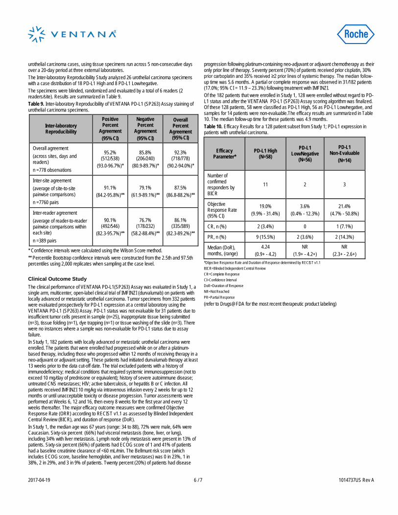
2017-04-19 6 / 7 1014737US Rev A
urothelial carcinoma cases, using tissue specimens run across 5 non-consecutive days over a 20-day period at three external laboratories. The Inter-laboratory Reproducibility Study analyzed 26 urothelial carcinoma specimens with a case distribution of 18 PD-L1 High and 8 PD-L1 Low/negative. The specimens were blinded, randomized and evaluated by a total of 6 readers (2 readers/site). Results are summarized in Table 9. Table 9. Inter-laboratory Reproducibility of VENTANA PD-L1 (SP263) Assay staining of urothelial carcinoma specimens.
Inter-laboratory Reproducibility
Positive Percent
Agreement (95% CI)
Negative Percent
Agreement (95% CI)
Overall Percent
Agreement (95% CI)
Overall agreement (across sites, days and readers) n =778 observations
95.2% (512/538)
(93.0-96.7%)*
85.8% (206/240)
(80.9-89.7%)*
92.3% (718/778)
(90.2-94.0%)*
Inter-site agreement (average of site-to-site pairwise comparisons) n =7760 pairs
91.1% (84.2-95.8%)**
79.1% (61.9-89.1%)**
87.5% (86.8-88.2%)**
Inter-reader agreement (average of reader-to-reader pairwise comparisons within each site) n =389 pairs
90.1% (492/546)
(82.3-95.7%)**
76.7% (178/232)
(58.2-88.4%)**
86.1% (335/389)
(82.3-89.2%)**
* Confidence intervals were calculated using the Wilson Score method. ** Percentile Bootstrap confidence intervals were constructed from the 2.5th and 97.5th percentiles using 2,000 replicates when sampling at the case level. Clinical Outcome Study The clinical performance of VENTANA PD-L1(SP263) Assay was evaluated in Study 1, a single arm, multicenter, open-label clinical trial of IMFINZI (durvalumab) on patients with locally advanced or metastatic urothelial carcinoma. Tumor specimens from 332 patients were evaluated prospectively for PD-L1 expression at a central laboratory using the VENTANA PD-L1 (SP263) Assay. PD-L1 status was not evaluable for 31 patients due to insufficient tumor cells present in sample (n=25), inappropriate tissue being submitted (n=3), tissue folding (n=1), dye trapping (n=1) or tissue washing of the slide (n=3). There were no instances where a sample was non-evaluable for PD-L1 status due to assay failure. In Study 1, 182 patients with locally advanced or metastatic urothelial carcinoma were enrolled. The patients that were enrolled had progressed while on or after a platinum-based therapy, including those who progressed within 12 months of receiving therapy in a neo-adjuvant or adjuvant setting. These patients had initiated durvalumab therapy at least 13 weeks prior to the data cut-off date. The trial excluded patients with a history of immunodeficiency; medical conditions that required systemic immunosuppression (not to exceed 10 mg/day of prednisone or equivalent); history of severe autoimmune disease; untreated CNS metastases; HIV; active tuberculosis, or hepatitis B or C infection. All patients received IMFINZI 10 mg/kg via intravenous infusion every 2 weeks for up to 12 months or until unacceptable toxicity or disease progression. Tumor assessments were performed at Weeks 6, 12 and 16, then every 8 weeks for the first year and every 12 weeks thereafter. The major efficacy outcome measures were confirmed Objective Response Rate (ORR) according to RECIST v1.1 as assessed by Blinded Independent Central Review (BICR), and duration of response (DoR). In Study 1, the median age was 67 years (range: 34 to 88), 72% were male, 64% were Caucasian. Sixty-six percent (66%) had visceral metastasis (bone, liver, or lung), including 34% with liver metastasis. Lymph node only metastasis were present in 13% of patients. Sixty-six percent (66%) of patients had ECOG score of 1 and 41% of patients had a baseline creatinine clearance of <60 mL/min. The Bellmunt risk score (which includes ECOG score, baseline hemoglobin, and liver metastases) was 0 in 23%, 1 in 38%, 2 in 29%, and 3 in 9% of patients. Twenty percent (20%) of patients had disease
progression following platinum-containing neo-adjuvant or adjuvant chemotherapy as their only prior line of therapy. Seventy percent (70%) of patients received prior cisplatin, 30% prior carboplatin and 35% received ≥2 prior lines of systemic therapy. The median follow-up time was 5.6 months. A partial or complete response was observed in 31/182 patients (17.0%; 95% CI = 11.9 – 23.3%) following treatment with IMFINZI. Of the 182 patients that were enrolled in Study 1, 128 were enrolled without regard to PD-L1 status and after the VENTANA PD-L1 (SP263) Assay scoring algorithm was finalized. Of these 128 patients, 58 were classified as PD-L1 High, 56 as PD-L1 Low/negative, and samples for 14 patients were non-evaluable.The efficacy results are summarized in Table 10. The median follow-up time for these patients was 4.9 months. Table 10. Efficacy Results for a 128 patient subset from Study 1; PD-L1 expression in patients with urothelial carcinoma.
Efficacy Parameter*
PD-L1 High (N=58)
PD-L1 Low/Negative
(N=56)
PD-L1 Non-Evaluable
(N=14)
Number of confirmed responders by BICR
11 2 3
Objective Response Rate (95% CI)
19.0% (9.9% - 31.4%)
3.6% (0.4% - 12.3%)
21.4% (4.7% - 50.8%)
CR, n (%) 2 (3.4%) 0 1 (7.1%)
PR, n (%) 9 (15.5%) 2 (3.6%) 2 (14.3%)
Median (DoR), months, (range)
4.24 (0.9+ - 4.2)
NR (1.9+ - 4.2+)
NR (2.3+ - 2.6+)
*Objective Response Rate and Duration of Response determined by RECIST v1.1 BICR=Blinded Independent Central Review CR=Complete Response CI=Confidence Interval DoR=Duration of Response NR=Not Reached PR=Partial Response
(refer to Drugs@FDA for the most recent therapeutic product labeling)

2017-04-19 7 / 7 1014737US Rev A
TROUBLESHOOTING Troubleshooting guidance is provided in Table11. If a problem cannot be attributed to any of these causes, or if the suggested corrective action fails to resolve the problem, consult your local support representative. Table 11. Troubleshooting guidance for VENTANA PD-L1 (SP263) Assay.
Problem Probable Cause Suggested Action
Light or no staining of slides
Incorrect staining protocol selected
Verify that U VENTANA PD-L1 (SP263) Assay procedure was used.
Verify that VENTANA PD-L1 (SP263) was selected for Primary Antibody
Degradation of tissue
Verify tissue was stained within the recommended time frame following sectioning.
Dispenser malfunction
Verify nozzle cap is removed.
Ensure dispenser is primed
Check the priming chamber for foreign materials or particulates, such as fibers or precipitate
Refer to inline dispenser package insert associated with P/N 740-4907 / 07208162001 located at www.ventana.com
Ensure that only recommended fixatives and fixation times are used.
Incorrect or missing bulk reagent
Ensure bulk reagents are correctly filled.
Excessive background staining of slides
Incorrect staining protocol selected
Verify that U VENTANA PD-L1 (SP263) Assay procedure was used.
Incorrect or missing bulk reagent
Ensure bulk reagents are correctly filled.
Inappropriate fixation method used
Ensure that only recommended fixatives and fixation times are used.
Tissue detached from slides
Use of incorrect microscope slides
Ensure positively charged microscope slides are used.
REFERENCES 1. Chalasani V, Chin JL, Izawa JI. Histologic variants of urothelial bladder cancer and
nonurothelial histology in bladder cancer. Can Urol Assoc J. 2009;3(6 Suppl 4):S193-198.
2. Ferlay J, Soerjomataram I, Ervik M, et al. GLOBOCAN 2012 v1.0. Cancer Incidence and Mortality Worldwide: IARC CancerBase No. 11. http://globocan.iarc.fr. Published 2013-12-12. Updated 2014-01-09. Accessed 2014-02-02.
3. American Cancer Society. Cancer Facts & Figures 2015. Atlanta: American Cancer Society; 2015.
4. Howlader N, Noone AM, Krapcho M, et al. (eds). SEER Cancer Statistics Review (CSR),1975-2012. National Cancer Institute. http://seer.cancer.gov/csr/1975_2012/. Published 2015-04-23. Updated 2015-11-18. Accessed 2016-04-19.
5. Keir ME, Butte MJ, Freeman GJ, et al. PD-1 and its ligands in tolerance and immunity. Annu Rev Immunol 2008;26:677-704.
6. Blank C, Mackensen A. Contribution of the PD-L1/PD-1 pathway to T-cell exhaustion: an update on implications for chronic infections and tumor evasion. Cancer Immunol Immunother. 2007;56(5):739-745.
7. Butte MJ, Keir ME, Phamduy TB, et al. Programmed death-1 ligand 1 interacts specifically with the B7-1 costimulatory molecule to inhibit T cell responses. Immunity. 2007;27(1):111-122.
8. Dong H, Zhu G, Tamada K, Chen L. B7-H1, a third member of the B7 family, co-stimulates T-cell proliferation and interleukin-10 secretion. Nat Med. 1999;5(12):1365-1369.
9. Massard C, Gordon MS, Sharma S, et al. Safety and efficacy of durvalumab (MEDI4736), an anti-programmed cell death ligand-1 immune checkpoint inhibitor, in patients with advanced urothelial bladder cancer. J Clin Oncol. 2016;34(26):3119-25.
10. Patel SP, Kurzrock R. PD-L1 Expression as a Predictive Biomarker in Cancer Immunotherapy. Mol Cancer Ther. 2015;14(4) 847-856.
11. Carson F, Hladik C. Histotechnology: A Self Instructional Text, 3rd edition. Hong Kong: American Society for Clinical Pathology Press; 2009.
12. Roche PC, Hsi ED. Immunohistochemistry-Principles and Advances. Manual of Clinical Laboratory Immunology, 6th edition. In: NR Rose, ed. ASM Press; 2002.
13. Anatomic Pathology Checklist. College of American Pathologists. Jul 28, 2015. 14. CLSI. Quality assurance for design control and implementation of
immunohistochemistry assay: approved guidelines. 2nd edition Wayne, PA, USA: Clinical and Laboratory Standards Institute; 2011
15. Herman GE, Elfont EA. The taming of immunohistochemistry: the new era of quality control. Biotech Histochem. 1991;66(4):194-199.
16. Hautzer NW, Wittkuhn JF, McCaughey WT. Trypsin digestion in immunoperoxidase staining. J Histochem Cytochem. 1980;28(1):52-53.
17. Omata M, Liew CT, Ashcavai M, Peters RL. Nonimmunologic binding of horseradish peroxidase to hepatitis B surface antigen. A possible source of error in immunohistochemistry. Am J Clin Pathol. 1980;73(5):626-632.
18. Nadji M, Morales AR. Immunoperoxidase: part 1. The technique and its pitfalls. Lab Med. 1983;14:767.
INTELLECTUAL PROPERTY VENTANA, BENCHMARK, OPTIVIEW, and the VENTANA logo are trademarks of Roche. All other trademarks are the property of their respective owners. © 2017 Ventana Medical Systems, Inc.
CONTACT INFORMATION
Ventana Medical Systems, Inc. 1910 E. Innovation Park Drive Tucson, Arizona 85755 USA +1 520 887 2155 +1 800 227 2155 (USA)
www.ventana.com

VENTANA PD-L1 (SP263) Assay Staining in Urothelial CarcinomaInterpretation Guide

2 VENTANA PD-L1 (SP263) Assay in Urothelial Carcinoma Interpretation Guide

VENTANA PD-L1 (SP263) Assay in Urothelial Carcinoma Interpretation Guide 3
Table of Contents
Introduction 4Intended Use of Product 4Purpose of Interpretation Guide 4
Clinical Evaluation 5Evaluating Staining Patterns and Intensities 5Tissue Requirements 10Morphology and Background Acceptability Criteria 10Positive Tissue Control 11VENTANA PD-L1 (SP263) Assay Scoring Algorithm for Urothelial Carcinoma Tissue 12Overview of PD-L1 (SP263) Scoring Algorithm for Urothelial Carcinoma 14Evaluation of Immune Cell Staining 16
PD-L1 Expression Atlas in Urothelial Carcinoma 25Tumor Cell Cases 25Immune Cell Cases 31
Challenging Cases 39Challenging cases: Weak Tumor Membrane vs Cytoplasmic Staining 40Challenging cases: Strong Immune Cell Staining Overlapping with Tumor Cell Staining 42Challenging cases: Cases with Multiple Tissue Fragments 44Challenging cases: Borderline PD-L1 Status 45Challenging cases: Obscuring Endogenous Material 48
Impact of Pre-Analytical Conditions on VENTANA PD-L1 (SP263) Assay Staining 50Fixative Recommendations to Achieve Optimal Staining Results with the VENTANA PD-L1 (SP263) Assay 50Impact of Tissue Thickness on Assay Staining 51Cut Slide Stability 51
References 51

4 VENTANA PD-L1 (SP263) Assay in Urothelial Carcinoma Interpretation Guide
IntroductionUrothelial carcinoma (also known as urothelial cell carcinoma, transitional cell carcinoma of the urinary tract, or urothelial bladder cancer) is the most common cancer of the urinary system worldwide. The majority of urothelial tumors arise in the bladder with the remainder originating in the renal pelvis, urethra, or ureter. Transitional cell carcinoma (TCC) is the most common histologic subtype associated with bladder cancer and accounts for greater than 90% of all urothelial carcinoma cases in the industrialized world; non-urothelial subtypes (e.g., squamous cell, adenocarcinoma, small cell carcinoma) are more frequent in other areas of the world.1
Globally, there were an estimated 429,793 new cases of bladder cancer and 165,084 deaths in 2012.2 In Europe alone, for 2012, there were an estimated 151,297 new cases of bladder cancer and 52,411 deaths. In 2015, it was estimated that there would be 74,000 new cases of bladder cancer and 16,000 deaths in the United States.3 Urothelial carcinoma presents as non-muscle-invasive, muscle-invasive, or metastatic disease. The overall 5-year survival rate for metastatic urothelial carcinoma (mUC) is approximately 5.4%.4
PD-L1 is a transmembrane protein that downregulates immune responses through binding to its two receptors programmed death-1 (PD-1) and B7-1 (CD80).5 PD-1 is an inhibitory receptor expressed on T cells following T-cell activation, which is sustained in states of chronic stimulation such as in chronic infection or cancer.6 Binding of PD-L1 with PD-1 inhibits T cell proliferation, cytokine production, and cytolytic activity, leading to the functional inactivation or exhaustion of T cells. CD80 is a molecule expressed on antigen presenting cells and activated T cells. PD-L1 binding to CD80 on T cells and antigen presenting cells can mediate downregulation of immune responses, including inhibition of T-cell activation and cytokine production.7 PD-L1 expression has been observed in immune cells and tumor cells.8,9 Aberrant expression of PD-L1 on tumor cells and or tumor-associated immune cells has been reported to impede anti-tumor immunity, resulting in immune evasion.6,9 Therefore, interruption of the PD-L1/PD-1 pathway represents an attractive strategy to reinvigorate tumor-specific T cell immunity suppressed by the expression of PD-L1 in the tumor microenvironment. PD-L1 is expressed in a broad range of cancers including lung, melanoma, urothelial, ovarian, and colorectal cancer. Prevalence of PD-L1 expression has been reported from 12% to 100% depending on the tumor type, anti PD-L1 clone and cutoff for positivity.10
Intended Use of Product
VENTANA PD-L1 (SP263) Assay is a qualitative immunohistochemical assay using rabbit monoclonal anti-PD-L1 clone SP263 intended for use in the assessment of the PD-L1 protein in formalin-fixed, paraffin-embedded (FFPE) urothelial carcinoma tissue stained with OptiView DAB IHC Detection Kit on a VENTANA BenchMark ULTRA instrument.
PD-L1 status is determined by the percentage of tumor cells with any membrane staining above background or by the percentage of tumor-associated immune cells with staining (IC+) at any intensity above background. The percent of tumor area occupied by any tumor-associated immune cells (Immune Cells Present, ICP) is used to determine IC+, which is the percent area of ICP exhibiting PD-L1 positive immune cell staining. PD-L1 status is considered High if any of the following are met:
• ≥ 25% of tumor cells exhibit membrane staining; or,
• ICP > 1% and IC+ ≥ 25%; or,
• ICP = 1% and IC+ = 100%.
PD-L1 High status as determined by VENTANA PD-L1 (SP263) Assay was associated with increased objective response rate (ORR) in a single arm study of IMFINZI™ (durvalumab).
This product is intended for in vitro diagnostic (IVD) use.
Purpose of Interpretation Guide
This guide is intended to aid pathologists in the clinical evaluation of formalin-fixed, paraffin-embedded (FFPE) urothelial carcinoma sections stained with the Assay using the Assay Scoring Algorithms in accordance with the proposed product labeling. Specifically, this guide:
• Provides pathologists with a tool to facilitate the evaluation of formalin-fixed, paraffin-embedded (FFPE) urothelial carcinoma sections stained with the Assay using the Assay Scoring Algorithms in accordance with the proposed product labeling.
• Provides photographic images that illustrate the staining patterns and intensities that may result from staining of urothelial carcinoma tissue samples with the Assay.
• Provides guidance in the use of placenta tissue as a system-level control when stained with the Assay.
• Provides photographic images of internal controls.
• Provides a reference for relating staining patterns to specific PD-L1 scores.
• Provides example images of challenging cases to provide guidance in their evaluation.

VENTANA PD-L1 (SP263) Assay in Urothelial Carcinoma Interpretation Guide 5
Evaluating Staining Patterns and Intensities
Urothelial carcinoma cases stained with the Assay are assessed for both the percentage of tumor cells with membrane staining and the percentage of tumor-associated immune cells with membrane, cytoplasm, or punctate staining.
Tumor Cell Staining:Urothelial carcinoma neoplastic cells labeled with the Assay are evaluated for the percent of the tumor cells with membrane staining at any intensity of the diaminobenzidine (DAB) signal. The immunohistochemical staining in urothelial carcinoma
Clinical Evaluation
Urothelial carcinoma, H&E and Assay: Various cases demonstrating the range of membrane and cytoplasmic staining in tumor cells (10X)
is membranous and/or cytoplasmic, and may be expressed homogeneously or heterogeneously throughout the neoplasm. Membrane staining can have a partial or complete circumferential pattern. Cytoplasmic staining is generally diffuse with some cases displaying a finely granular quality. Tumor cell cytoplasmic staining is disregarded for determining PD-L1 expression. The total percentage of membrane signal intensities is visually estimated and used to generate the PD-L1 expression level. An isotype-matched negative control antibody is used to evaluate the presence of background in test samples and establish a staining intensity baseline.

6 VENTANA PD-L1 (SP263) Assay in Urothelial Carcinoma Interpretation Guide
Urothelial carcinoma, H&E and Assay: Tumor cells with weak (blue arrow) membrane staining pattern (20X)
Urothelial carcinoma, H&E and Assay: Tumor cells show membranous and weak granular cytoplasmic staining (40X)
Urothelial carcinoma, H&E and Assay: Tumor cells with circumferential (black arrow) and partial (blue arrow) membrane staining pattern (20X)

VENTANA PD-L1 (SP263) Assay in Urothelial Carcinoma Interpretation Guide 7
Tumor-Associated Immune Cell Staining:Immune cells exhibit a range of staining intensities and patterns: negative to weak diffuse cytoplasmic and/or weak to strong membranous signal. A punctate pattern of staining may be seen in association with lymphocytes. PD-L1 expression has been observed in lymphocytes, macrophages, histiocytes, plasma cells, and neutrophils.
The percentage of tumor-associated immune cells with staining in urothelial carcinoma cases is evaluated in addition to tumor cell
Urothelial carcinoma, H&E and Assay: Various cases demonstrating the range and patterns of immune cells staining (10X)
staining. Immune cell staining is assessed by initially reviewing the entire tumor area and determining the percentage of the tumor area occupied by immune cells (ICP). Next the percentage of immune cells demonstrating any pattern of PD-L1 expression within the tumor area is visually estimated (IC+).
8 VENTANA PD-L1 (SP263) Assay in Urothelial Carcinoma Interpretation Guide
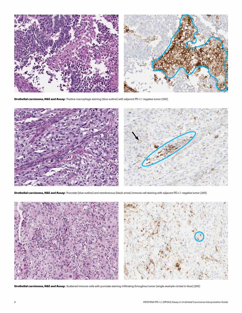
Urothelial carcinoma, H&E and Assay: Punctate (blue outline) and membranous (black arrow) immune cell staining with adjacent PD-L1 negative tumor (20X)
Urothelial carcinoma, H&E and Assay: Scattered immune cells with punctate staining infiltrating throughout tumor (single example circled in blue) (20X)
Urothelial carcinoma, H&E and Assay: Positive macrophage staining (blue outline) with adjacent PD-L1 negative tumor (20X)

VENTANA PD-L1 (SP263) Assay in Urothelial Carcinoma Interpretation Guide 9
NSCLC, H&E and Assay: Case with rare neutrophil-only infiltrate (blue arrow) within the tumor demonstrating PD-L1 staining. Tumor cell membrane staining also present (black arrow) (40X)
10 VENTANA PD-L1 (SP263) Assay in Urothelial Carcinoma Interpretation Guide
Morphology and Background Acceptability Criteria
Table 1: Morphology Acceptability CriteriaInterpretation Microscope Observation
Acceptable Cellular elements of interest are visualized allowing interpretation of the stain.
Not Acceptable Cellular elements of interest are not visualized compromising interpretation of the stain.
Table 2: Background Acceptability CriteriaInterpretation Microscope Observation
Acceptable Non-specific staining that is not obtrusive to interpretation of specific staining.
Not Acceptable Non-specific staining that is obtrusive to interpretation of specific staining.
Tissue morphology and background acceptability are assessed for each patient case using the criteria described in Tables 1 and 2.
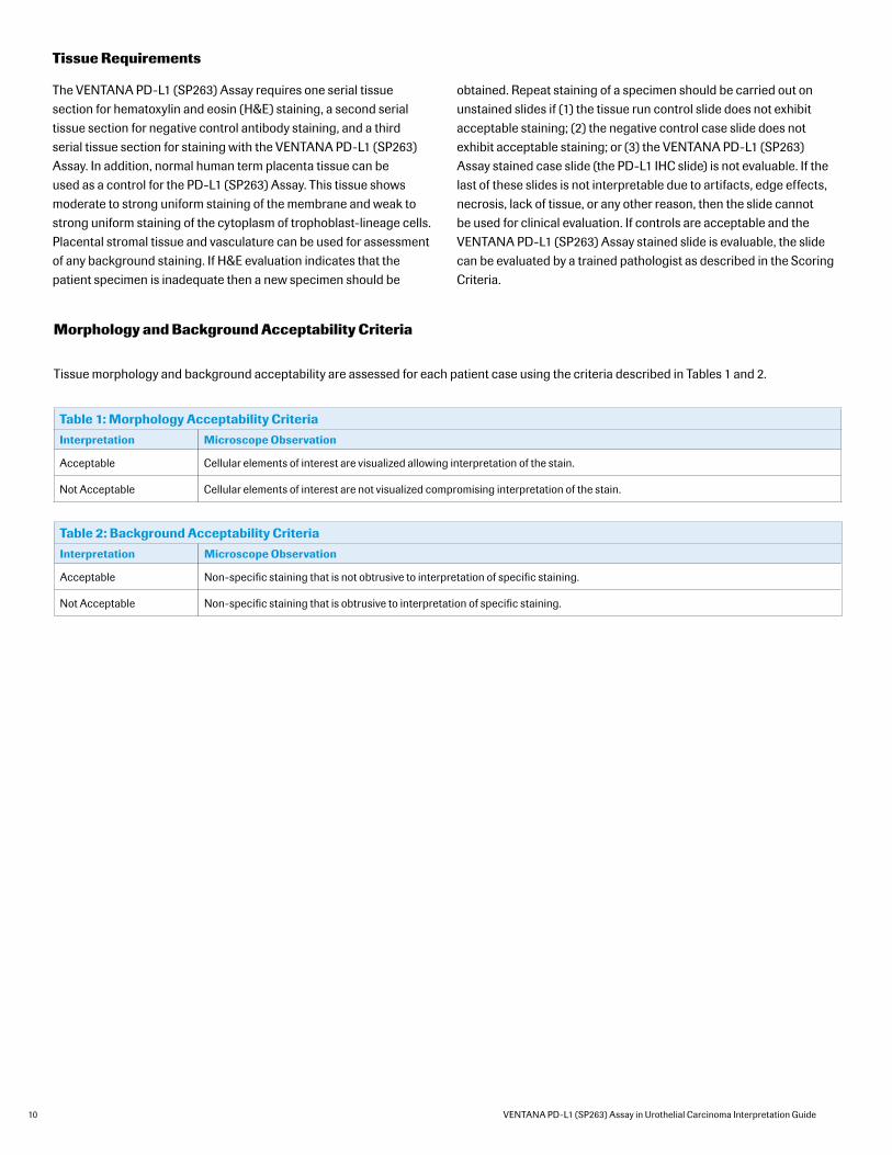
Tissue Requirements
The VENTANA PD-L1 (SP263) Assay requires one serial tissue section for hematoxylin and eosin (H&E) staining, a second serial tissue section for negative control antibody staining, and a third serial tissue section for staining with the VENTANA PD-L1 (SP263) Assay. In addition, normal human term placenta tissue can be used as a control for the PD-L1 (SP263) Assay. This tissue shows moderate to strong uniform staining of the membrane and weak to strong uniform staining of the cytoplasm of trophoblast-lineage cells. Placental stromal tissue and vasculature can be used for assessment of any background staining. If H&E evaluation indicates that the patient specimen is inadequate then a new specimen should be
obtained. Repeat staining of a specimen should be carried out on unstained slides if (1) the tissue run control slide does not exhibit acceptable staining; (2) the negative control case slide does not exhibit acceptable staining; or (3) the VENTANA PD-L1 (SP263) Assay stained case slide (the PD-L1 IHC slide) is not evaluable. If the last of these slides is not interpretable due to artifacts, edge effects, necrosis, lack of tissue, or any other reason, then the slide cannot be used for clinical evaluation. If controls are acceptable and the VENTANA PD-L1 (SP263) Assay stained slide is evaluable, the slide can be evaluated by a trained pathologist as described in the Scoring Criteria.

VENTANA PD-L1 (SP263) Assay in Urothelial Carcinoma Interpretation Guide 11
Placenta, VENTANA PD-L1 (SP263) Assay: Strong uniform membrane staining and moderate cytoplasmic staining of trophoblast-lineage cells (10X)
Placenta, VENTANA PD-L1 (SP263) Assay: Stroma and vasculature within villi show no PD-L1 staining (20X)
A known positive control tissue fixed and processed in the same manner as the patient specimens should be run for each set of test conditions and with every VENTANA PD-L1 (SP263) Assay staining procedure performed. The control tissue (an index case) should be a fresh autopsy, biopsy, surgical specimen prepared and fixed as soon as possible in a manner identical to patient specimens. This tissue may be used to monitor all steps of specimen processing and staining. A tissue section fixed or processed differently from the test specimen can be used as a control for reagents and staining but not for fixation or tissue preparation. A positive urothelial carcinoma case with moderate staining is more suitable for quality control than one that stains strongly; it can be used to detect minor levels of reagent
Positive Tissue Control
degradation or out-of-specification issues that might be instrument-related. Positive membrane staining of neoplastic cells in the control tissue confirms that the VENTANA PD-L1 (SP263) antibody was applied and the instrument functioned properly. The positive tissue control should be used only to monitor performance; it should not be used to aid the clinical diagnosis of patient samples. Additionally, the VENTANA PD-L1 (SP263) Assay can utilize as a positive control human term placental tissue, which shows moderate to strong uniform staining of the membrane and weak to strong uniform staining of the cytoplasm of trophoblast-lineage cells. Placental stromal tissue and vasculature can be used for assessment of any background staining. Please refer to Table 3.
Table 3: Placenta Tissue Control Evaluation Criteria for the Ventana PD-L1 (SP263) Assay
Acceptable Moderate to strong uniform membrane staining of trophoblast-lineage cells, and placental stroma and vasculature with no staining.
Not Acceptable No to weak uniform membrane staining of trophoblast lineage cells and/or specific staining within placental stromal and vascular tissue.

12 VENTANA PD-L1 (SP263) Assay in Urothelial Carcinoma Interpretation Guide
Table 4: VENTANA PD-L1 (SP263) Assay Scoring Algorithm for Urothelial CarcinomaPD-L1 Interpretation Staining Description
PD-L1 status is determined by the percentage of tumor cells with any membrane staining above background or by the percentage of tumor-associated immune cells with staining (IC+) at any intensity above background. Percent of tumor area occupied by any tumor-associated immune cells (ICP) is used to determine IC+, which is the percent area of ICP exhibiting PD-L1 positive immune cell staining.
High
PD-L1 Status is considered high if any of the following are met:• ≥ 25% of tumor cells exhibit membrane staining; or,• ICP > 1% and IC+ ≥ 25%; or, • ICP = 1% and IC+ = 100%.
Low/negativePD-L1 Status is considered low/negative if:• none of the criteria for PD-L1 High Status are met.
VENTANA PD-L1 (SP263) Assay Scoring Algorithm for Urothelial Carcinoma Tissue
PD-L1 status and expression level is assigned by a trained pathologist based on their evaluation of the percentage of specific staining for both tumor and tumor-associated immune cells. PD-L1 status is based on the total percent of tumor cells with membrane staining or the total percent of tumor-associated immune cells with staining (IC+) at any intensity. Specifically, this guide highlights an expression level of greater than or equal to 25% of tumor cells with membrane staining or of tumor-associated immune cells with staining. In cases where the percent of tumor-associated immune cells in the tumor area (ICP) is 1%, IC+ is scored as either 0%, <100% or 100% due to the difficulties in estimating the percent staining in small volumes of immune cells in low measures. The small amount of PD-L1 staining observed in cases with < 100% IC+, should be considered as < 25% PD-L1 expression. Please refer to Table 4.
Interpretation of urothelial carcinoma cases stained with the Assay is based on the criteria noted in the table below. Images of various expression level staining patterns are provided in the subsequent sections. Please refer to Table 4.
PD-L1 LOW/NEGATIVE STATUS: TUMOR CELL EXPRESSION < 25%
0% 5% 10%
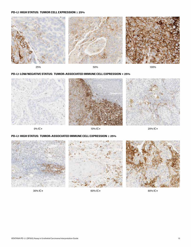
VENTANA PD-L1 (SP263) Assay in Urothelial Carcinoma Interpretation Guide 13
PD-L1 HIGH STATUS: TUMOR CELL EXPRESSION ≥ 25%
25% 50% 100%
PD-L1 LOW/NEGATIVE STATUS: TUMOR-ASSOCIATED IMMUNE CELL EXPRESSION < 25%
PD-L1 HIGH STATUS: TUMOR-ASSOCIATED IMMUNE CELL EXPRESSION ≥ 25%
0% IC+
30% IC+
10% IC+
60% IC+
20% IC+
80% IC+

14 VENTANA PD-L1 (SP263) Assay in Urothelial Carcinoma Interpretation Guide
1. H&E slide is reviewed for viable tumor, tumor-associated immune cells and tumor area. a. Tumor area encompasses the tumor proper, associated desmoplastic stroma and immune cells infiltrating the tumor and contained within the desmoplasia.
2. Visually, tumor-associated immune cells are densely aggregated (with no intervening stroma) within the tumor area when estimating Immune Cells Present (ICP). a. Tumor associated immune cells include those present within the tumor reactive stroma, between the tumor islands and those invading the tumor proper.
3. The ICP percentage is scored as 0%, 1%, 5%, and deciles and quartiles (10, 20, 25, 30, 40, 50, 60, 70, 75, 80, 90, 100). a. If a raw percentage falls between a decile or quartile, standard mathematical rounding is used to score to the nearest decile or quartile. b. When differentiating between 1% and 5% ICP, a raw percentage estimate of 2% is rounded to 1% and an estimate of 3% to 4% is rounded to 5%.
4. PD-L1 stained slide is reviewed for tumor cell and immune cell staining.
5. Estimate the percentage of tumor cells with partial or complete membrane staining at any intensity.
6. The percentage of immune cells (within the ICP) expressing PD-L1 (IC+) is estimated. a. Immune cells with any staining (membranous, cytoplasmic, or punctate) at any intensity are visually aggregated within the ICP to estimate IC+. b. IC+ is scored as deciles and quartiles (0, 10, 20, 25, 30, 40, 50, 60, 70, 75, 80, 90, 100).
7. For cases with 1% ICP, the percentage estimate for IC+ is reported as 0%, <100%, or 100%.
Overview of PD-L1 (SP263) Scoring Algorithm for Urothelial Carcinoma
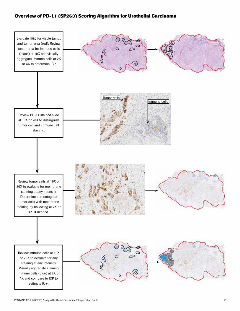
VENTANA PD-L1 (SP263) Assay in Urothelial Carcinoma Interpretation Guide 15
Review immune cells at 10X or 20X to evaluate for any staining at any intensity.
Visually aggregate staining immune cells (blue) at 2X or 4X and compare to ICP to
estimate IC+.
Immune cellsTumor cells
Overview of PD-L1 (SP263) Scoring Algorithm for Urothelial Carcinoma
Review tumor cells at 10X or 20X to evaluate for membrane
staining at any intensity. Determine percentage of
tumor cells with membrane staining by reviewing at 2X or
4X, if needed.
Review PD-L1 stained slide at 10X or 20X to distinguish tumor cell and immune cell
staining.
Evaluate H&E for viable tumor, and tumor area (red). Review tumor area for immune cells (black) at 10X and visually
aggregate immune cells at 2X or 4X to determine ICP.

16 VENTANA PD-L1 (SP263) Assay in Urothelial Carcinoma Interpretation Guide
A variety of immune cells display staining with the Assay, and include lymphocytes, macrophages, histiocytes, reticular dendritic cells, plasma cells and neutrophils. The H&E-stained slide is initially examined to determine the total percentage of the tumor area (tumor cells and any desmoplastic stroma) involved by immune cells with no intervening stroma (ICP). Areas not considered part of the tumor area include non-viable tumor such as those with cautery or crush artifacts, and extensive necrosis. Normal lymphoid tissue uninvolved by the neoplasm, as seen in lymph nodes with metastatic tumor, is not considered as a part of the tumor area or immune cells involving the tumor.
Evaluation of Immune Cell Staining
Tumor-associated immune cell staining percentage estimation:
The PD-L1 IHC slide is then scored for the percentage of tumor-associated immune cells staining for PD-L1 (IC+). Membranous, cytoplasmic, and punctate PD-L1 immune cell staining are all included in the estimation. In cases where positively-staining immune cells are intermixed with positively-staining tumor cells, it can be difficult to quantify the amount of staining for each component. Examples of immune cell interpretation are given in the following photo sets.
The following steps describe the estimation of tumor-associated immune cell staining percentage.
Immune Cell scoring 1: Tumor-associated immune cells (black arrows) within the tumor area (blue outline) are present at the leading edge of the tumor and are included in ICP estimation. A separate aggregate of lymphocytes is not within the tumor area (black outline) (4X)
1. Review case H&E to assess viable tumor with attached desmoplastic stroma containing tumor-associated immune cells. Tumor associated immune cells include those present within the tumor reactive stroma, between the tumor islands and those invading the tumor proper. Tumor within lymphatics is not included within the tumor area.

VENTANA PD-L1 (SP263) Assay in Urothelial Carcinoma Interpretation Guide 17
Immune Cell scoring 2: Tumor area containing tumor cells, desmoplastic stroma and tumor-associated immune cells is outlined in blue, which includes immune cell aggregates identified by black arrows. Immune cells outside of the tumor area are also present (black outlines) (1X)
Immune Cell scoring 3: Tumor area consists of only viable areas of tumor and desmoplasia (areas outlined in blue). Any necrosis, cautery artifact, crush artifact or large areas of non-neoplastic tissue are disregarded (3X)
2. Lower magnification review is recommended to perform the percentage estimate of immune cells within and adjacent to the tumor area (ICP). Immune cells located within blood vessels and lymphatics are disregarded.
3. Exclude non-neoplastic areas not involved by tumor, areas with necrotic tumor, crush and cautery artifacts.

18 VENTANA PD-L1 (SP263) Assay in Urothelial Carcinoma Interpretation Guide
4. If tumor islands are separated by muscle or stroma, they are included as part of the tumor area if the tumor borders on both sides within a 10X field.
Immune Cell scoring 4: The circled areas show large areas of fibromuscular stroma uninvolved by tumor. When viewed at higher power (10X), the stroma with tumor on both sides within a single field of view (blue) is included in the overall tumor area. When the large bundle of fibromuscular stroma is not bordered by tumor within a single 10X field (black), it is not included in the overall tumor area. (1X)
NOT included in tumor area
INCLUDED in tumor area
10X 10X

VENTANA PD-L1 (SP263) Assay in Urothelial Carcinoma Interpretation Guide 19
5. For noninvasive urothelial carcinoma (papillary carcinoma), include the immune cells within the fibrovascular cores as well as within the immediately adjacent base of the frond/stalk.
Immune Cell scoring 5: Double sided arrow (black) shows base of papillary tumor with the tumor area for this noninvasive urothelial carcinoma outlined (blue). Immune cells from the intratumoral fibrovascular core (A) (10X) and adjacent base of the stalk (B) (5X) are included in estimation of ICP (0.8X)
A
B
BA

20 VENTANA PD-L1 (SP263) Assay in Urothelial Carcinoma Interpretation Guide
6. Visually estimate the area occupied by the tumor-associated immune cells relative to the total tumor area (ICP = percent of tumor area occupied by immune cells).
Immune Cell scoring 6: The regions occupied by immune cells within the tumor area in the upper image (blue outlines) are identified in the lower image (gray outlines) and then densely aggregated (illustrated by the smaller black areas) in order to estimate the percentage of the tumor area with immune cells, or ICP – in this example 10% (black areas together relative to the tumor area) (5X)

VENTANA PD-L1 (SP263) Assay in Urothelial Carcinoma Interpretation Guide 21
Immune Cell scoring 7: The stromal regions occupied by widely dispersed immune cells (blue outlines) are identified. When the immune cells are densely aggregated (represented by the black area) in order to estimate the percentage of the tumor area with immune cells, the ICP in this example is 5% (10X)
Immune Cell scoring 8: The immune cell infiltrates with neutrophils and eosinophils, are included in the ICP estimation. The ICP in this example is 25% (10X)
22 VENTANA PD-L1 (SP263) Assay in Urothelial Carcinoma Interpretation Guide
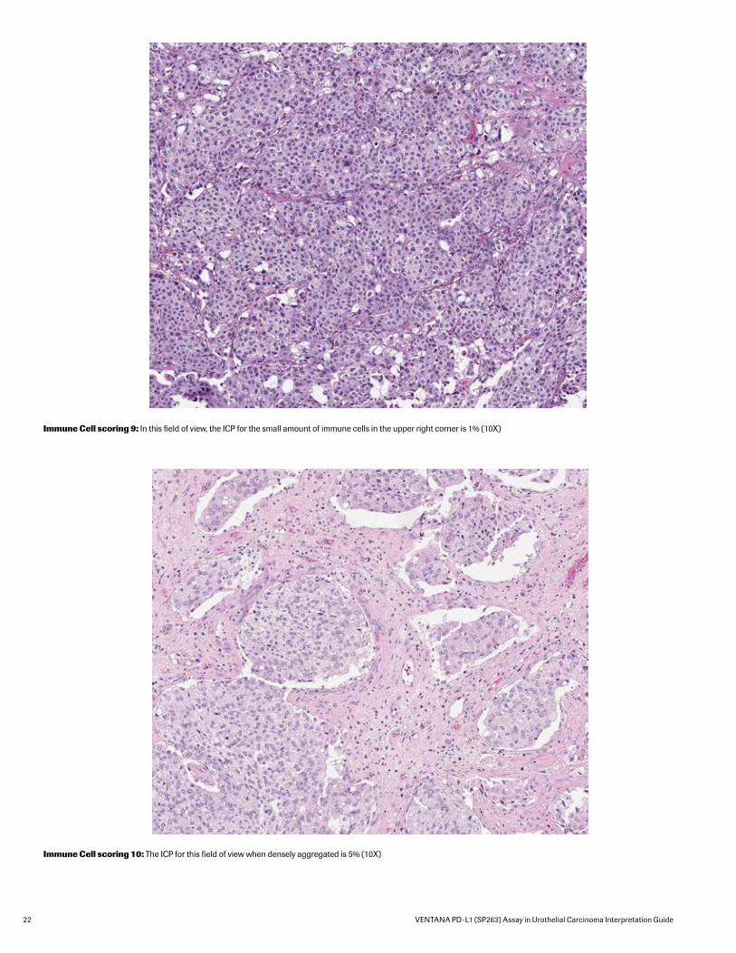
Immune Cell scoring 9: In this field of view, the ICP for the small amount of immune cells in the upper right corner is 1% (10X)
Immune Cell scoring 10: The ICP for this field of view when densely aggregated is 5% (10X)

VENTANA PD-L1 (SP263) Assay in Urothelial Carcinoma Interpretation Guide 23
7. Review PD-L1-stained slide and estimate the percentage of tumor-associated immune cells which demonstrate PD-L1 expression (IC+) including diffuse cytoplasmic, linear membrane and punctate immune cell staining.
Immune Cell scoring 11: The immune cells comprising ICP are outlined in blue (upper image) and can be seen densely aggregated in the lower image. The immune cells with PD-L1 staining (IC+) are highlighted in red (upper image) and when aggregated constitute 25% of the ICP (lower image) (5X). Please refer to the associated H&E image contained within this guide, “Immune Cell scoring 6.”

24 VENTANA PD-L1 (SP263) Assay in Urothelial Carcinoma Interpretation Guide
8. When evaluating lymph node metastases the reactive stroma generated by the tumor is included as part of the tumor area when determining ICP. In cases where the tumor does not generate a stromal response, the tumor area is limited to the tumor nests and adjacent immune cells in direct contact with the tumor only. Any immune cells part of the uninvolved lymphoid tissue are disregarded for ICP.
Immune Cell scoring 12: In this metastatic tumor to a lymph node, the tumor area containing tumor cells, desmoplastic stroma and immune cells is outlined in blue (4X). Higher power shows immune cells that are included as part of ICP within the reactive stroma (inset A (20X)). Along the periphery of the metastatic nodule only immune cells immediately adjacent to the tumor cells outlined in blue are included as part of ICP (inset B (20X)).
9. Only in cases where the ICP equals 1%, the IC+ is only scored as 0%, <100% or 100% due to the difficult nature of quantifying expression is such small amounts of cells.
10. If the PD-L1-stained slide shows more immune cell staining than the initial ICP estimation from the H&E slide, refer back to the H&E slide and revise the ICP percentage, if necessary.

VENTANA PD-L1 (SP263) Assay in Urothelial Carcinoma Interpretation Guide 25
Tumor Cell Cases
PD-L1 Expression Atlas in Urothelial Carcinoma
Tumor Cell Case 1: Tumor cells with 0% staining, Immune cells with 0% staining. (IC+) (Top row H&E 10X, PD-L1 20X. Bottom row PD-L1 10X.)
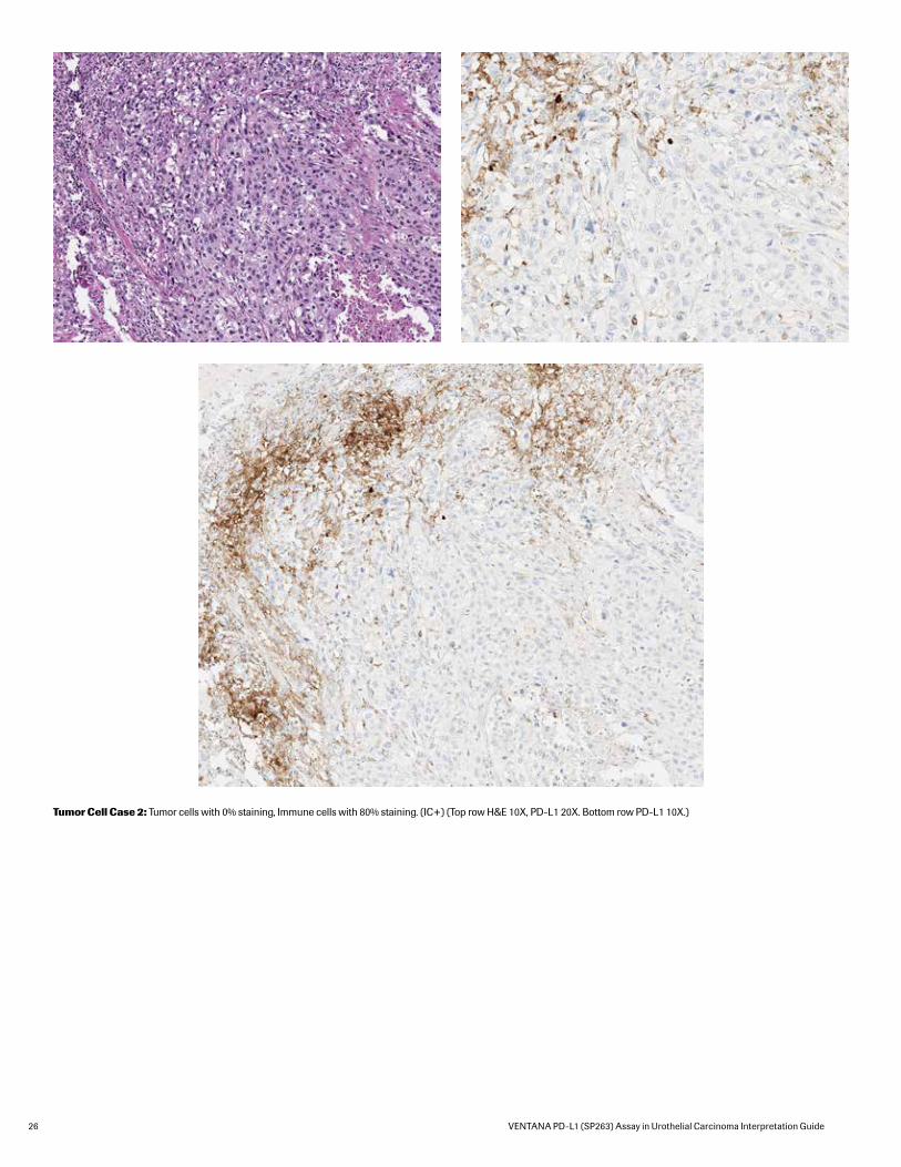
26 VENTANA PD-L1 (SP263) Assay in Urothelial Carcinoma Interpretation Guide
Tumor Cell Case 2: Tumor cells with 0% staining, Immune cells with 80% staining. (IC+) (Top row H&E 10X, PD-L1 20X. Bottom row PD-L1 10X.)

VENTANA PD-L1 (SP263) Assay in Urothelial Carcinoma Interpretation Guide 27
Tumor Cell Case 3: Tumor cells with 10% staining, Immune cells with 40% staining (IC+) (10X)

28 VENTANA PD-L1 (SP263) Assay in Urothelial Carcinoma Interpretation Guide
Tumor Cell Case 4: Tumor cells with 25% staining, Immune cells with 10% staining (IC+) (10X)

VENTANA PD-L1 (SP263) Assay in Urothelial Carcinoma Interpretation Guide 29
Tumor Cell Case 5: Tumor cells with 50% staining, Immune cells with 20% staining (IC+) (10X)

30 VENTANA PD-L1 (SP263) Assay in Urothelial Carcinoma Interpretation Guide
Tumor Cell Case 6: Tumor cells with 100% staining, Immune cells with 20% staining. (IC+) (Top row H&E 10X, PD-L1 20X. Bottom row PD-L1 10X.)
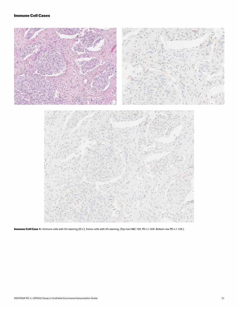
VENTANA PD-L1 (SP263) Assay in Urothelial Carcinoma Interpretation Guide 31
Immune Cell Cases
Immune Cell Case 1: Immune cells with 0% staining (IC+), Tumor cells with 0% staining. (Top row H&E 10X, PD-L1 20X. Bottom row PD-L1 10X.)

32 VENTANA PD-L1 (SP263) Assay in Urothelial Carcinoma Interpretation Guide
Immune Cell Case 2: Immune cells with 10% staining (IC+), Tumor cells with 80% staining. (Top row H&E 10X, PD-L1 20X. Bottom row PD-L1 10X.)

VENTANA PD-L1 (SP263) Assay in Urothelial Carcinoma Interpretation Guide 33
Immune Cell Case 3: Immune cells with 30% staining (IC+), Tumor cells with 10% staining. (Top row H&E 10X, PD-L1 20X. Bottom row PD-L1 10X.)

34 VENTANA PD-L1 (SP263) Assay in Urothelial Carcinoma Interpretation Guide
Immune Cell Case 4: Immune cells with 25% staining (IC+), Tumor cells with 100% staining. (Top row H&E 10X, PD-L1 20X. Bottom row PD-L1 10X.)
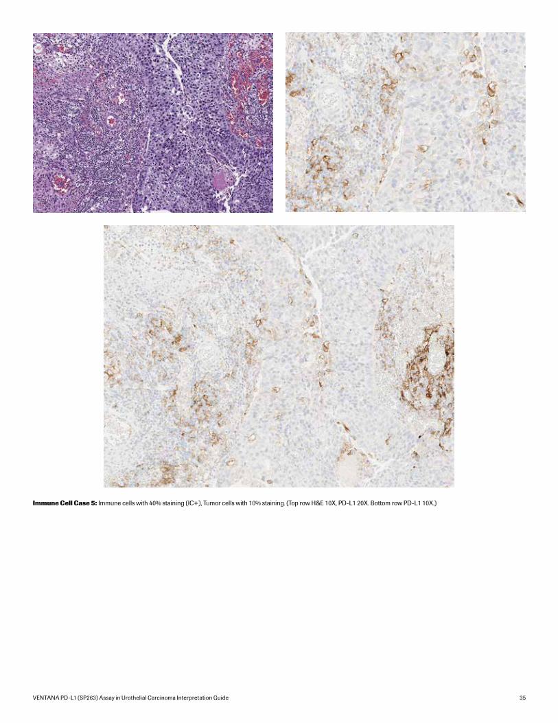
VENTANA PD-L1 (SP263) Assay in Urothelial Carcinoma Interpretation Guide 35
Immune Cell Case 5: Immune cells with 40% staining (IC+), Tumor cells with 10% staining. (Top row H&E 10X, PD-L1 20X. Bottom row PD-L1 10X.)

36 VENTANA PD-L1 (SP263) Assay in Urothelial Carcinoma Interpretation Guide
Immune Cell Case 6: Immune cells with 50% staining (IC+), Tumor cells with 100% staining. (Top row H&E 10X, PD-L1 20X. Bottom row PD-L1 10X.)

VENTANA PD-L1 (SP263) Assay in Urothelial Carcinoma Interpretation Guide 37
Immune Cell Case 7: Immune cells with 60% staining (IC+), Tumor cells with 0% staining. (Top row H&E 10X, PD-L1 20X. Bottom row PD-L1 10X.)
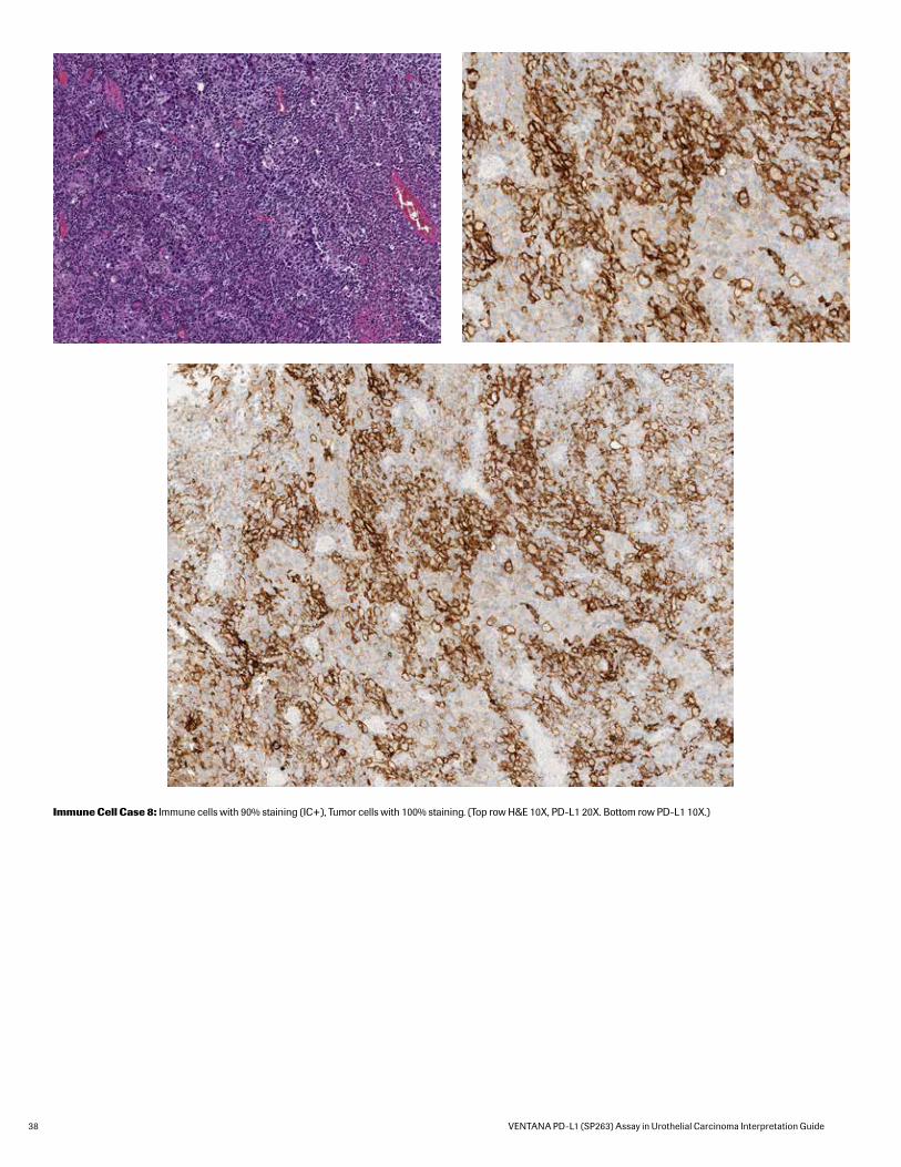
38 VENTANA PD-L1 (SP263) Assay in Urothelial Carcinoma Interpretation Guide
Immune Cell Case 8: Immune cells with 90% staining (IC+), Tumor cells with 100% staining. (Top row H&E 10X, PD-L1 20X. Bottom row PD-L1 10X.)

VENTANA PD-L1 (SP263) Assay in Urothelial Carcinoma Interpretation Guide 39
Challenging CasesCases are given a PD-L1 score according to percentage of tumor cells with membrane staining and tumor-associated immune cells. Various staining patterns and morphologic features may make interpretation and quantification of tumor membrane staining difficult.
Some cases may be particularly challenging due to the following issues:
• Weak Tumor Cell Membrane vs Weak Tumor Cell Cytoplasmic Staining
Some specimens may exhibit weak cytoplasmic staining of the tumor cells that may be confused at low power with weak tumor cell membrane staining. For this reason when evaluating PD-L1 IHC stained slides, weak staining should be confirmed with examination at higher powers to distinguish between tumor cell membranous and cytoplasmic staining.
• Strong Immune Cell Staining Overlapping with Tumor Cell Staining
Some tumors may contain an extensive inflammatory component both surrounding the tumor and infiltrating within the tumor. In instances when significant staining is seen for both tumor and immune cells, it can be challenging to differentiate and quantify the PD-L1 IHC staining between the two cell populations. The presence of immune cells infiltrating the tumor should be confirmed using the H&E slide. The pattern of PD-L1 staining and nuclear morphology are utilized to help attribute expression to immune cells (punctate staining, small uniform nucleus) and tumor cells (linear membrane staining, large irregular nucleus).
• Borderline PD-L1 (SP263) Status
Some cases are near the cut-off between a PD-L1 High and Low/negative status. These cases are particularly challenging to estimate the percent of target cells with staining. For these borderline cases it may be helpful to view the case at a magnification that enables the entire tumor area to be assessed. Consultation with a trained colleague may also be helpful.
• Cases with Multiple Tissue Fragments
Urothelial carcinoma samples resulting from transurethral resections of the bladder (TURB) can result in challenging cases complicated by multiple fragments of tissue scattered across the slide with varying amounts of necrosis, crush/cautery artifact, viable tumor cells and/or tumor-associated immune cells. A fragment-by-fragment examination approach with individual percent staining estimates followed by an overall average can help in the evaluation.
• Obscuring Endogenous Material
Occasionally in urothelial carcinoma samples endogenous material, such as melanin pigment or hemosiderin, may obscure and interfere with interpretation of PD-L1 IHC staining of tumor and immune cells. Comparison of the negative isotype control slide with the PD-L1 stained slide can aid in differentiating between biomarker staining and endogenous material. Non-specific staining can also be seen on the PD-L1 stained slide in areas of necrosis.
Some challenging cases are shown.

40 VENTANA PD-L1 (SP263) Assay in Urothelial Carcinoma Interpretation Guide
Challenging cases: Weak Tumor Membrane vs Cytoplasmic Staining
Challenging Case 1: The area outlined in blue (10X PD-L1 image, upper right) contains weak tumor cell staining that is difficult to distinguish. Examination at higher power (20X) shows there is scattered weak tumor membrane staining. For 10X image: Tumor cells with 40% staining, Immune cells with 80% staining. (IC+) (Top row H&E 10X, PD-L1 10X. Bottom row PD-L1 20X.)

VENTANA PD-L1 (SP263) Assay in Urothelial Carcinoma Interpretation Guide 41
Challenging Case 2: Tumor cells with moderate or stronger membrane staining are readily identified within the right half of this field. Careful examination of the tumor demonstrates cells with weak membrane expression (blue outlines) that should also be counted towards the total percentage of membrane staining.

42 VENTANA PD-L1 (SP263) Assay in Urothelial Carcinoma Interpretation Guide
Challenging cases: Strong Immune Cell Staining Overlapping with Tumor Cell Staining
Challenging Case 3: TC and IC intermixed together with both expressing PD-L1. In areas with strong TC staining, IC can only be evaluated where discernible from tumor cell staining. The different patterns of IC staining (punctate pattern) as well as nuclear morphology can be used to identify PD-L1 expression. The blue outlines contain areas with discernible immune cell staining with the green outlined area having scattered punctate immune cell staining. Tumor cells with 100% staining, Immune cells with 25% staining (IC+) (Top row H&E 10X, PD-L1 10X. Bottom row PD-L1 20X.)

VENTANA PD-L1 (SP263) Assay in Urothelial Carcinoma Interpretation Guide 43
Challenging Case 4: TC and IC intermixed together with TC membrane staining more distinct and IC difficult to evaluate and can only be scored along the periphery of the tumor. Tumor cells with 100% staining. (Top row H&E 10X, PD-L1 10X. Bottom row PD-L1 20X.)

44 VENTANA PD-L1 (SP263) Assay in Urothelial Carcinoma Interpretation Guide
Challenging Case 5: TURB specimen contains multiple pieces with crush artifact, blood clot and necrosis which shows non-specific staining on the IHC slide(1X).
Challenging cases: Cases with Multiple Tissue Fragments

VENTANA PD-L1 (SP263) Assay in Urothelial Carcinoma Interpretation Guide 45
Challenging cases: Borderline PD-L1 Status
Challenging Case 6: Weak tumor cell staining seen at 10X magnification is difficult to distinguish between membrane and cytoplasmic staining. Examination at higher power (20X) reveals tumor cells with 20% weak, delicate membranous staining.

46 VENTANA PD-L1 (SP263) Assay in Urothelial Carcinoma Interpretation Guide
Challenging Case 7: Borderline TC PD-L1 expression with 25% tumor cell membrane staining (10X)

VENTANA PD-L1 (SP263) Assay in Urothelial Carcinoma Interpretation Guide 47
Challenging Case 8: Borderline IC PD-L1 expression with an IC+ of 20% (20X). (Top row H&E 10X, PD-L1 10X. Bottom row PD-L1 20X.)

48 VENTANA PD-L1 (SP263) Assay in Urothelial Carcinoma Interpretation Guide
Challenging Case 9: Case contains anthracotic pigment (red arrow) overlapping with punctate immune cell staining (blue arrow) as well as tumor cell membrane staining (black arrow) (20X). (Top row H&E 10X, Neg Rb 10X. Bottom row PD-L1 10X.)
Challenging cases: Obscuring Endogenous Material

VENTANA PD-L1 (SP263) Assay in Urothelial Carcinoma Interpretation Guide 49
Challenging Case 10: Large necrotic areas can have non-specific DAB staining which may be confused with immune cell staining. (Top row H&E 10X, PD-L1 10X. Bottom row PD-L1 20X.)

50 VENTANA PD-L1 (SP263) Assay in Urothelial Carcinoma Interpretation Guide
VENTANA PD-L1 (SP263) Assay Staining of Placenta Tissue Across Fixatives and Fixation Times
Time Fixative
Point (Hrs)
10% NBF Zinc Formalin PREFER fixative** AFA** Alcoholic Formalin** 95% Ethanol**
1*
6
12
24
48
72
Impact of Pre-Analytical Conditions on VENTANA PD-L1 (SP263) Assay Staining
Fixative Recommendations to Achieve Optimal Staining Results with the VENTANA PD-L1 (SP263) Assay
Ventana recommends fixation in 10% NBF for 6-72 hours. Acceptable fixatives and fixation times are outlined in blue.
NOTES*One hour fixation time is not recommended for all fixative types
**Use of PREFER fixative and 95% Ethanol (weaker staining) or alcoholic fixatives (high background) is not recommended. See additional higher magnification images to the right.

VENTANA PD-L1 (SP263) Assay in Urothelial Carcinoma Interpretation Guide 51
Impact of Tissue Thickness on Assay Staining
Ventana recommends tissue thickness of 4-5 microns for use with the Assay.
Cut Slide Stability
Sections approximately 4-5 microns in thickness should be cut and mounted on positively charged glass slides. Slides should be stained within 6 months of sectioning.
References
1. Chalasani V, Chin JL, Izawa JI. Histologic variants of urothelial bladder cancer and nonurothelial histology in bladder cancer. Can Urol Assoc J. 2009;3(6 Suppl 4):S193-198.
2. Ferlay J, Soerjomataram I, Ervik M, et al. GLOBOCAN 2012 v1.0. Cancer Incidence and Mortality Worldwide: IARC CancerBase No. 11. http://globocan.iarc.fr. Published 2013-12-12. Updated 2014-01-09. Accessed 2014-02-02.
3. American Cancer Society. Cancer Facts & Figures 2015. Atlanta: American Cancer Society; 2015.
4. Howlader N, Noone AM, Krapcho M, et al. (eds). SEER Cancer Statistics Review (CSR),1975-2012. National Cancer Institute. http://seer.cancer.gov/csr/1975_2012/. Published 2015-04-23. Updated 2015-11-18. Accessed 2016-04-19.
5. Keir ME, Butte MJ, Freeman GJ, et al. PD-1 and its ligands in tolerance and immunity. Annu Rev Immunol 2008;26:677-704.
6. Blank C, Mackensen A. Contribution of the PD-L1/PD-1 pathway to T-cell exhaustion: an update on implications for chronic infections and tumor evasion. Cancer Immunol Immunother. 2007;56(5):739-745.
7. Butte MJ, Keir ME, Phamduy TB, et al. Programmed death-1 ligand 1 interacts specifically with the B7-1 costimulatory molecule to inhibit T cell responses. Immunity. 2007;27(1):111-122.
8. Dong H, Zhu G, Tamada K, Chen L. B7-H1, a third member of the B7 family, co-stimulates T-cell proliferation and interleukin-10 secretion. Nat Med. 1999;5(12):1365-1369.
9. Massard C, Gordon MS, Sharma S, et al. Safety and efficacy of durvalumab (MEDI4736), an anti-programmed cell death ligand-1 immune checkpoint inhibitor, in patients with advanced urothelial bladder cancer. J Clin Oncol. 2016; 34(26):3119-25.
10. Patel SP, Kurzrock R. PD-L1 Expression as a Predictive Biomarker in Cancer Immunotherapy. Mol Cancer Ther. 2015;14(4) 847-856.

VENTANA
Empowering | Innovation
Ventana Medical Systems, Inc. 1910 E. Innovation Park Drive Tucson, Arizona 85755 USA +1 520 887 2155 +1 800 227 2155 (USA)
www.ventana.com
©2017 Ventana Medical Systems, Inc. All rights reserved.
VENTANA, BENCHMARK, OPTIVIEW, and the VENTANA logo are trademarks of Roche. All other trademarks are the property of their respective owners.
1014738US Rev A
C Change product name, add “Rx Only” to label See Agile CS C057844
REV DESCRIPTION EFFECTIVE DATE RP CO
TITLE: LBL, 1 DSPNR BRL, 2DBC, 740-4907
This document contains confidential, proprietary information of Ventana Medical Systems, Inc. It may not be copied or reproduced without prior written permission from Ventana Medical Systems, Inc.
REV:
C
Ventana Medical Systems, Inc. Tucson, Arizona
PART NUMBER: DMS MATERIAL NUMBER: SHEET
1 OF 1 07447710001 07447710001
FT0710-300 Rev A
NOTES:
1. Text: Black. Background: White.
2. Make from: Ventana P/N 1442800 blank label stock (DMS Mat. No. 05249368001), approximately 3.00 x 1.75''.
3. The individual label file can be located in the Document Control System (DCS) under 07447710001c.btw.
4. Outside border line is shown here for illustrative purposes only and does not print.
5. Label image is shown here at an approximate size. Label dimensions are contained in the drawing of the label stock found on the bill of materials.
6. Lot Number, Expiry Date, and 2D barcode are shown here for illustrative purposes only. Actual data will vary by job.

C Change product name, add “Rx Only” to label See Agile CS C057844
REV DESCRIPTION EFFECTIVE DATE RP CO
TITLE: LBL, 1 DSPNR BOX, 2DBC, FG 740-4907
This document contains confidential, proprietary information of Ventana Medical Systems, Inc. It may not be copied or reproduced without prior written permission from Ventana Medical Systems, Inc.
REV:
C
Ventana Medical Systems, Inc. Tucson, Arizona
PART NUMBER: DMS MATERIAL NUMBER: SHEET
1 OF 1 07447396001 07447396001
FT0710-300 Rev A
NOTES
1. Text: Black. Background: White.
2. Make from: Ventana P/N 07184603001, approximately 1.75 x 4.0”.
3. The individual label file can be located in the Document Control System (DCS) under 07447396001c.btw.
4. Outside border line is shown here for illustrative purposes only and does not print.
5. Label image is shown here at an approximate size. Label dimensions are contained in the drawing of the label stock found on the bill of materials.
6. Lot Number, Expiry Date, barcodes, and barcode-related human readable text are shown here for illustrative purposes only.
Actual data will vary by job.

Ventana Medical Systems Inc.
REVDWG NUMBERSIZESHEETSCALE
TITLE
ARTWORK, SINGLE DISPENSER,BOX PN 1692500
DO NOT SCALE 1 OF 1 B 000014900
Tucson, AZ
REV DESCRIPTION
A
RELEASED DRAWINGRPEffective Date
C
COGA 10682SEE AGILEA
B UPDATE ARTWORK WITH BLUE BORDER SEE AGILE PM C003389
1692500 C
Ventana Medical Systems, Inc.1910 E. Innovation Park DriveTucson, Arizona 85755USA+1 520 887 2155+1 800 227 2155 (USA)
www.ventanamed.com
Roche Diagnostics GmbHSandhofer Strasse 116D-68305 MannheimGermany
Made in USA
C UPDATE REV FOR SPEC CHANGE ON VARNISH SEE AGILE GV C007663
NOTES:
1. COLOR SWATCHES a.PMS 300