Venous Thromboembolism (VTE) - Healthtrust...
43
Transcript of Venous Thromboembolism (VTE) - Healthtrust...


Venous
Thromboembolism (VTE)

Agenda
What is a DVT?
Virchow’s Triad & Risk Factors
Methods of DVT Prophylaxis
Intermittent Pneumatic Compression Therapy
Diagnosis of DVT
Treatment of DVT
Complications of DVT

A Life-Threatening
Medical Condition
• DVT and PE are collectively known as VTE
• Two million people in the U.S. develop VTE annually
– Nonfatal VTE -- 613,423
– Fatal VTE -- 296,370
• 2/3 of VTE episodes are related to hospitalization
(American Society of Hematology, 2012)

The Economic Effect
“More people die each year from pulmonary
embolism than from motor vehicle accidents,
breast cancer or AIDS.”
Costs of hospitalization (2013) • PE $25,864
• DVT $20,636
• Stroke $14,401
• Acute Myocardial Infarction $11,642

What is a DVT?
• DVT – the formation of a blood clot within a deep vein that may partially or completely block the flow of blood
• Forms in any vein in the body but most commonly in the lower extremities, especially the calf veins (distal DVT)
- 20% will propagate proximally into the thigh (proximal DVT)
• Most clinically significant PEs originate from proximal DVTs of the leg (popliteal, iliac or femoral veins)
- 10% die within the first hour
- PE may be the most common preventable cause of death in the world.

Deep Vein Thrombosis

Anatomy & Physiology
ARTERIAL SYSTEM
• Contains muscles layers
• High pressure system
• Carries oxygenated blood
from heart to tissues
• Low compliance
• Contains 20% of circulating
blood
VENOUS SYSTEM
• Thin, elastic vessels return blood to the heart
• Compress and collapse easily
• Low pressure system
• Valves prevent reflux
• Contains 80% of circulating blood

Venous Return
• Muscle pumping of the legs (esp. calf region) facilitates blood return
to the heart
• Cardiac output - pressure at which blood is pumped out of heart
• Gravity - elevation above heart level assists venous return
• Respiratory pump - diaphragm movements shifts pressure and acts
as a pump in pulling venous blood toward heart
• Pulsations of the arteries - milking effect on veins alongside them
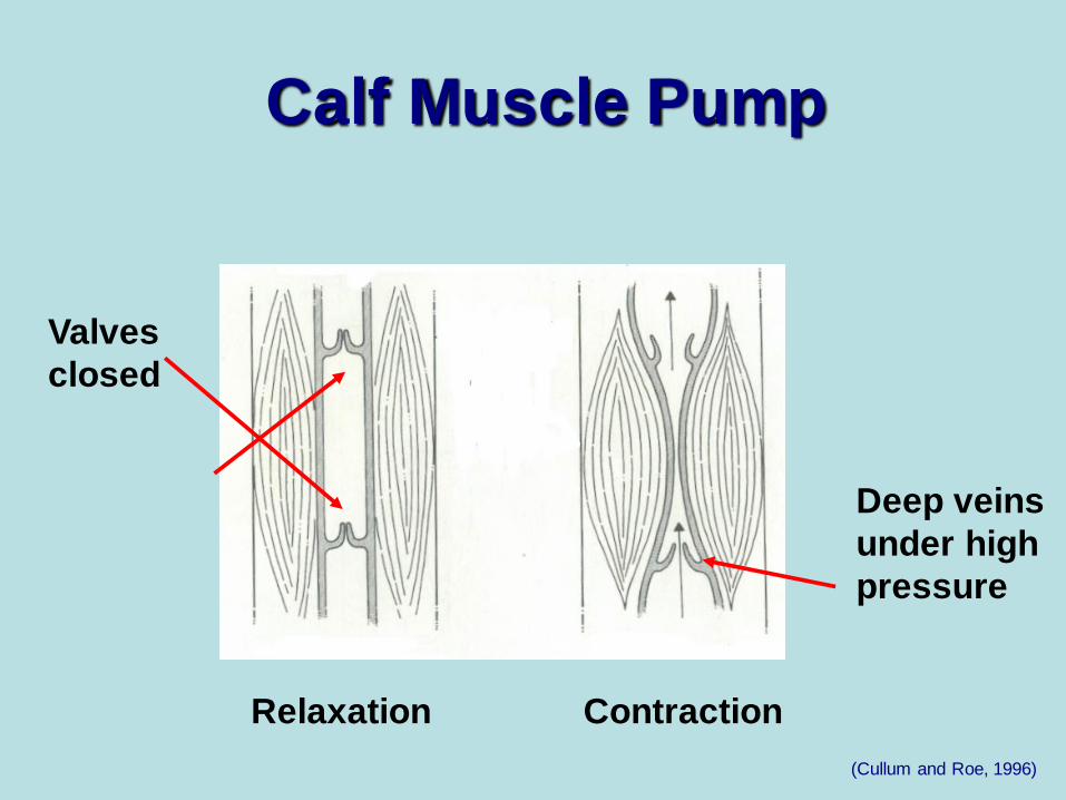
Relaxation Contraction
Valves
closed
Deep veins
under high
pressure
(Cullum and Roe, 1996)
Calf Muscle Pump

Semi-Lunar Valves

Virchow’s Triad
Stasis
Vessel Injury Hypercoagulability
The more risk factors a person has….
the greater the chances of developing a DVT

Blood Clots:
Natural Part of the Healing Process
• Clots enable injured tissue to begin to repair itself without excessive
blood loss
• Platelets clumping and aggregating at site of injury is normal part of
clotting and healing process
• Balance exists between promoting coagulation and retarding
coagulation
• Disturbance in equilibrium may occur and mechanism may backfire
under “ideal” conditions….
Coagulopathies
Coagulation Anti-Coagulation

Virchow’s Triad
Stasis • Surgery longer than 30 min.
– Muscles ineffective due to
anesthesia
– Veins overfill
– Over-distended veins may
develop micro-tears in the
vein wall
• Prolonged Immobility
– Vein distention and blood
pooling in lower extremities
– Long trips
• Paralysis/Stroke
– Muscles can’t help move
blood
• Obesity / Pregnancy
– Excess weight in the
abdomen prevents venous
return
– In pregnancy, risk of VTE
6x greater (fetus increases
pressure on veins in pelvis)
• Edema in lower extremities
– Excess fluid within the tissues
increases pressure against
the veins
• Age over 40
– Loss of muscle tone

Fibrinolytic Shutdown Phenomenon
– Occurs in ALL operative patients to some degree
– Cause uncertain
– Believed to be a stress response to surgery
– Postoperatively, the fibrinolytic activity (body’s natural ability to dissolve blood clots) falls, leaving the patient open to risk of DVT formation
– Begins 30 minutes into surgery and may continue three to four days post-op; has been documented to last up to seven days

Virchow’s Triad
Vessel Injury
• History of DVT or PE
– 4x more likely to develop a
new DVT
• Vessel damage
– Phlebitis, trauma to vein
from bone fractures, IV
punctures and vein-irritating
IV medications, burns
– Central venous catheters
• Immobility
– Over-distention of veins
leads to micro-tears in vein
wall
• Surgery longer than 30 minutes
- Lack of movement -> stasis -> micro-tears
- Surgical trauma
• Age over 40
- Risk increases with age: nearly doubling with each decade of life after age 40

Virchow’s Triad Hypercoagulability
• Surgery lasting longer than 30 minutes
– Anesthesia alters blood chemistry
• Dehydration
– Thickens circulating blood
• Estrogen Therapy
– Birth control pills (3 - 6x greater risk)
– Hormone therapy
• Malignancies - 2x the risk of DVT and 3x the risk of fatal PE as
in non-cancer patients undergoing similar procedures
• Inherited clotting disorders

Public Awareness

American College of Chest Physicians
Patient Group Risk of DVT (%)
Medical patients 10-20
General surgery 15-40
Major gynecologic surgery 15-40
Major urologic surgery 15-40
Neurosurgery 15-40
Stroke 20-50
Hip or knee arthroplasty 40-60
Hip fracture surgery 40-60
Major trauma 40-80
Spinal cord surgery 60-80
Critical care patients 10-80
(ACCP, 2012)

Obesity and DVT
• Obese patients were 2.5x as likely to have DVT and 2.2x as likely to have PE
• Odds of PE and DVT were more than 5x higher for obese patients younger than 40 than their non-obese peers
• Obese men under age 40 had a tripled risk of DVT
• Obese women under age 40 had the highest risk of DVT – 6x as likely as non-obese women under age 40 to have DVT

Risk Stratification for DVT & PE
Low Risk
Uncomplicated minor surgery in patients < 40 years old with no risk factors
Moderate Risk
Major surgery in patients > 40 years old with no risk factors
High Risk
Major surgery in patients > 40 years old who have additional risk factors
Very High Risk
Major surgery in patients > 40 years old plus history of DVT/PE, cancer,
major orthopedic surgery, hip fracture, stroke, spinal cord injury or
hypercoagulability
(ACCP, 2012)

DVT Facts
• Majority of DVTs are clinically silent
• 50 percent of DVTs start on the operating room table, and the majority will be present within 48 hours of surgery
• DVT risk does not end upon discharge
– After joint replacement surgery:
• Highest risk two to five days after surgery
• 2nd peak occurs 10 days after surgery, after most have been discharged from hospital
• Extends for at least three months after surgery

Deep Vein Thrombosis
Prophylaxis Chemical Methods
• The use of anticoagulants includes an inherent risk of increased
bleeding and various other side effects
• All anticoagulants require frequent blood draws in order to monitor
the dosage provided to the patient
• Anticoagulants:
– Warfarin
– Low Molecular Weight Heparin
– Heparin

Deep Vein Thrombosis
Prophylaxis
Physical Methods
• Walking
• Vena Cava Filters
• Gradient Stockings
• Intermittent Pneumatic
Compression (IPC)

MECHANICAL METHODS
Intermittent Pneumatic Compression
• Introduced over 30 years ago
• Considered as effective as anticoagulants but without side
effects
• Addresses all aspects of Virchow’s Triad
(stasis, hypercoagulability, and injury)
• Appropriate modality for all patients at risk
• Low-Moderate Risk -- IPC alone
• High Risk -- IPC + Anticoagulants

How are Pneumatic
Compression Systems
Clinically-Proven?
Blood Flow Studies
DVT Outcome Studies
Compliance Studies
Fibrinolytic Studies

How Does IPC Work?
• Mechanical Effects: – Mimics the action of walking by “squeezing” or compressing
the calf, or calf and thigh muscles, thereby enhancing blood flow, reducing stasis
• Chemical Effects: – Enhances fibrinolytic activity (stimulates fibrinolysis – the
body’s natural method for preventing and breaking down clots) to reduce the risk of clot formation

Intermittent Pneumatic
Compression
Venous Stasis
IPC
Blood Chemistry
Changes
Virchow’s
Triad

When Do You Use Compression?
• Surgical Patient
– Preoperatively, prior to anesthesia induction
– Intraoperatively
– Postoperatively – until fully ambulatory
• Non-Surgical Patient
– Immediately upon identification that the patient
is at risk for DVT

Leg Compression
Sequential • Chambers compress
sequentially
• Distal chamber
pressure 45 mmHg
• Proximal chamber 25
mmHg for 2.5 seconds
Intermittent • Posterior bladder
• 40 mmHg of sustained
pressure
• Distal to proximal
inflation

Foot Compression
• Inflation 0.4 – 3 seconds
• Pressure 80 - 120 mmHg
• Two to three cycles per minute
• Single-Pulse vs Circumferential

• Severe arteriosclerosis or other ischemic vascular
disease
• Phlebitis or any known or suspected DVT
• Pulmonary embolism
• Severe CHF
• Any local condition in which garments would
interfere such as: gangrene, untreated or infected
leg wounds, recent skin grafts or dermatitis
• Extreme deformity of the limb
• Severe congestive heart failure
When is IPC Contraindicated?

Are There Any Reasons to
Discontinue Therapy Early?
• Known or suspected DVT
• Signs of pulmonary embolism
• Tingling of extremity
• Skin breakdown
• Numbness
• Pain

Invasive Diagnostic Tests
Contrast Venography
- Dye injected into vein in the foot or ankle
- X-ray imaging taken to reveal the location of possible clots
- One of the most accurate ways to identify DVT
- Potential complications due to invasiveness
- Expensive
- Requires high degree of expertise to perform and interpret correctly
125I Fibrinogen Leg Scanning
– Radioactive isotope injected into the legs
– Failure to detect approximately 30% of thrombi
– Poor sensitivity for proximal thrombi
– Time consuming

Non-Invasive Diagnostic Tests
Doppler Ultrasound
• Performed using a Doppler
and compression
maneuvers to evaluate the
sound of blood flow in the
veins
• Computerized imaging can
reveal the presence of a
clot

Non-Invasive Diagnostic Tests
Duplex B-Mode Imaging
• Pulsed Doppler (audio interpretation) of the vein with imaging capabilities through visualization on a computer
• Duplex scanning may establish a diagnosis of DVT, but also defines the location and extent of clot
• Color-flow expedites identification of vessels and improves accuracy

Non-Invasive Diagnostic Tests
Helical CT
• Scanners use “slip rings” to power the x-ray tube so
that the ring can spin
throughout the duration of
the scan
• Lower radiation dose to the
patient, decreased artifact,
ability to scan greater body
length and greater
enhancement of body
organs
MRI
• In preliminary stages
• Uses a strong magnet to
visualize internal structures of
the body which generates
high quality images
• Sensitivity and specificity for
DVT have been reported as
97% and 95%, respectively

Treatment of DVT
Short-term
– Bedrest until
confirmed by duplex
– Discontinue IPC
– Anticoagulants
– Monitor for signs of
PE
Long-term – Proper diet and
exercise
– Maintain normal weight
– Avoid standing or sitting for long periods
– Compression garments
– Anticoagulants

• Anticoagulants
– Heparin
– Low Molecular Weight Heparin
– Arixtra (Fondaparinux Sodium)
– Warfarin
• Filters
• Thrombolytic Therapy
• Surgery
Treatment of DVT

• Pulmonary embolism
• Valvular incompetence
• Predisposition for further DVTs
• Post-thrombotic syndrome (occurs in 50-75 percent of
patients diagnosed with DVT)
• Chronic venous insufficiency
• Venous ulceration
• The average cost to treat a venous stasis ulcer is
estimated to be $40,000
Complications of DVT

In the United States,
6-7 million people have evidence of
post-phlebitic syndrome and
500,000-800,000 people have leg ulcers.
Complications of DVT

Venous Thromboembolism
Prevention is the
KEY
to Saving Lives.