Venous thromboembolism and intracranial hemorrhage after ... · Venous thromboembolism and...
11
Vol.:(0123456789) 1 3 J Neurooncol (2018) 136:135–145 DOI 10.1007/s11060-017-2631-5 CLINICAL STUDY Venous thromboembolism and intracranial hemorrhage after craniotomy for primary malignant brain tumors: a National Surgical Quality Improvement Program analysis Joeky T. Senders 1,2 · Nicole H. Goldhaber 1 · David J. Cote 1 · Ivo S. Muskens 1,2 · Hassan Y. Dawood 1 · Filip Y. F. L. De Vos 3 · William B. Gormley 1 · Timothy R. Smith 1 · Marike L. D. Broekman 1,2 Received: 12 July 2017 / Accepted: 29 September 2017 / Published online: 16 October 2017 © The Author(s) 2017. This article is an open access publication operative times were predictive for VTE during hospitaliza- tion, but not for post-discharge events. Admission two or more days before surgery was predictive for DVT, but not for PE. Preoperative steroid usage and male gender were predic- tive for post-discharge DVT and PE, respectively. ICH was associated with various comorbidities and longer operative times. This multicenter study demonstrates distinct critical time periods for the development of thrombotic and hemor- rhagic events after craniotomy. Furthermore, the VTE risk profile depends on the type of VTE (DVT vs. PE) and clini- cal setting (hospitalized vs. post-discharge patients). Keywords Deep venous thrombosis · Intracranial hemorrhage · Primary malignant brain tumor · Pulmonary embolism · Venous thromboembolism Introduction Venous thromboembolism (VTE), including deep venous thrombosis (DVT) and pulmonary embolism (PE), is a major cause of morbidity and mortality in patients undergoing crani- otomy for primary malignant brain tumors [1–6]. In addition to known surgical risk factors, such as venous stasis from periop- erative immobility, endothelial injury, and inflammation from the operation itself, cancer is a recognized risk factor for VTE development [7]. Among all cancer types, high-grade gliomas have been shown to result in second highest lifetime risk for cancer-related VTE, one of the highest risks of perioperative VTE and, when comparing craniotomy for any brain tumor to craniotomy for non-neoplastic disease, rates of postoperative VTE have been reported to be twice as high [8–11]. VTE has been reported as one of the most frequent major complications after craniotomy for brain tumors with incidences up to 21% in the first 3 months after surgery [1–6]. Abstract Venous thromboembolism (VTE), including deep venous thrombosis (DVT) and pulmonary embolism (PE), frequently complicates the postoperative course of primary malignant brain tumor patients. Thromboprophy- lactic anticoagulation is commonly used to prevent VTE at the risk of intracranial hemorrhage (ICH). We extracted all patients who underwent craniotomy for a primary malignant brain tumor from the National Surgical Quality Improve- ment Program (NSQIP) registry (2005–2015) to perform a time-to-event analysis and identify relevant predictors of DVT, PE, and ICH within 30 days after surgery. Among the 7376 identified patients, the complication rates were 2.6, 1.5, and 1.3% for DVT, PE, and ICH, respectively. VTE was the second-most common major complication and third- most common reason for readmission. ICH was the most common reason for reoperation. The increased risk of VTE extends beyond the period of hospitalization, especially for PE, whereas ICH occurred predominantly within the first days after surgery. Older age and higher BMI were overall predictors of VTE. Dependent functional status and longer Timothy R. Smith and Marike L. D. Broekman have shared last author. * Marike L. D. Broekman [email protected] 1 Computational Neurosciences Outcomes Center, Department of Neurosurgery, Brigham and Women’s Hospital, Harvard Medical School, 60 Fenwood Road, Boston, MA 02115, USA 2 Department of Neurosurgery, University Medical Center Utrecht, Heidelberglaan 100, 3584 CX Utrecht, The Netherlands 3 Department of Medical Oncology, University Medical Center Utrecht, Heidelberglaan 100, 3584 CX Utrecht, The Netherlands
Transcript of Venous thromboembolism and intracranial hemorrhage after ... · Venous thromboembolism and...
Vol.:(0123456789)1 3
J Neurooncol (2018) 136:135–145 DOI 10.1007/s11060-017-2631-5
CLINICAL STUDY
Venous thromboembolism and intracranial hemorrhage after craniotomy for primary malignant brain tumors: a National Surgical Quality Improvement Program analysis
Joeky T. Senders1,2 · Nicole H. Goldhaber1 · David J. Cote1 · Ivo S. Muskens1,2 · Hassan Y. Dawood1 · Filip Y. F. L. De Vos3 · William B. Gormley1 · Timothy R. Smith1 · Marike L. D. Broekman1,2
Received: 12 July 2017 / Accepted: 29 September 2017 / Published online: 16 October 2017 © The Author(s) 2017. This article is an open access publication
operative times were predictive for VTE during hospitaliza-tion, but not for post-discharge events. Admission two or more days before surgery was predictive for DVT, but not for PE. Preoperative steroid usage and male gender were predic-tive for post-discharge DVT and PE, respectively. ICH was associated with various comorbidities and longer operative times. This multicenter study demonstrates distinct critical time periods for the development of thrombotic and hemor-rhagic events after craniotomy. Furthermore, the VTE risk profile depends on the type of VTE (DVT vs. PE) and clini-cal setting (hospitalized vs. post-discharge patients).
Keywords Deep venous thrombosis · Intracranial hemorrhage · Primary malignant brain tumor · Pulmonary embolism · Venous thromboembolism
Introduction
Venous thromboembolism (VTE), including deep venous thrombosis (DVT) and pulmonary embolism (PE), is a major cause of morbidity and mortality in patients undergoing crani-otomy for primary malignant brain tumors [1–6]. In addition to known surgical risk factors, such as venous stasis from periop-erative immobility, endothelial injury, and inflammation from the operation itself, cancer is a recognized risk factor for VTE development [7]. Among all cancer types, high-grade gliomas have been shown to result in second highest lifetime risk for cancer-related VTE, one of the highest risks of perioperative VTE and, when comparing craniotomy for any brain tumor to craniotomy for non-neoplastic disease, rates of postoperative VTE have been reported to be twice as high [8–11]. VTE has been reported as one of the most frequent major complications after craniotomy for brain tumors with incidences up to 21% in the first 3 months after surgery [1–6].
Abstract Venous thromboembolism (VTE), including deep venous thrombosis (DVT) and pulmonary embolism (PE), frequently complicates the postoperative course of primary malignant brain tumor patients. Thromboprophy-lactic anticoagulation is commonly used to prevent VTE at the risk of intracranial hemorrhage (ICH). We extracted all patients who underwent craniotomy for a primary malignant brain tumor from the National Surgical Quality Improve-ment Program (NSQIP) registry (2005–2015) to perform a time-to-event analysis and identify relevant predictors of DVT, PE, and ICH within 30 days after surgery. Among the 7376 identified patients, the complication rates were 2.6, 1.5, and 1.3% for DVT, PE, and ICH, respectively. VTE was the second-most common major complication and third-most common reason for readmission. ICH was the most common reason for reoperation. The increased risk of VTE extends beyond the period of hospitalization, especially for PE, whereas ICH occurred predominantly within the first days after surgery. Older age and higher BMI were overall predictors of VTE. Dependent functional status and longer
Timothy R. Smith and Marike L. D. Broekman have shared last author.
* Marike L. D. Broekman [email protected]
1 Computational Neurosciences Outcomes Center, Department of Neurosurgery, Brigham and Women’s Hospital, Harvard Medical School, 60 Fenwood Road, Boston, MA 02115, USA
2 Department of Neurosurgery, University Medical Center Utrecht, Heidelberglaan 100, 3584 CX Utrecht, The Netherlands
3 Department of Medical Oncology, University Medical Center Utrecht, Heidelberglaan 100, 3584 CX Utrecht, The Netherlands
136 J Neurooncol (2018) 136:135–145
1 3
Previous studies have identified older age, male sex, His-panic ethnicity, history of craniotomy, history of VTE, con-gestive heart failure, coagulopathy, seizures, increased stay on the intensive care unit, prolonged hospital stay, medium bed size, residual tumor tissue, and absence of thrombo-prophylactic therapy as predictors of VTE after craniotomy for primary malignant brain tumors [3, 4, 6, 12–15]. Most of these studies have been small, single-center studies, and none of these studies have identified predictors or performed time-to-event analyses separated for VTE type (DVT vs. PE) and the clinical setting (hospitalized vs. post-discharge patients).
Most patients undergoing surgery for brain tumors receive pharmaceutic prophylaxis in combination with mechanical prophylaxis in the perioperative setting [16–19]. However, anticoagulation increases the risk of intracranial hemorrhage (ICH), which is one of the most frequent and feared compli-cations in patients undergoing operations for brain tumors [20]. The increased risk of ICH makes the use of prophy-lactic anticoagulation an issue of great debate and careful balance in this patient population. Although the incidence of ICH is lower compared to VTE events, their outcomes can be at least as detrimental. Only few predictors associated with ICH have been identified including history of crani-otomy, use of bevacizumab, and therapeutic anticoagula-tion for a VTE [13, 20–24]. Adequate assessment of the perioperative risk of both VTE and ICH among this patient population, as well as accurately predicting the typical time to a thrombotic or hemorrhagic event, is meaningful in tai-loring postoperative management to the risk profile of the individual patient.
The American College of Surgeons (ACS) National Surgical Quality Improvement Program (NSQIP) database registers follow-up of neurosurgical patients for 30 days postoperatively, and reports variables relevant to this issue, including the occurrence of DVT, PE, and ICH with related time-to-event data. Because the risk of ICH is intimately tied to the thromboprophylactic treatment used for patients with brain tumors, this study addresses thrombotic as well as hemorrhagic complications. In this study, NSQIP was used to identify predictors and perform time-to-event analyses for VTE and ICH. Assessment of VTE was stratified for VTE type (DVT vs. PE) and clinical setting (hospitalized vs. post-discharge patients).
Methods
Data source
The NSQIP includes surgical patients from over 600 par-ticipating hospitals in the U.S. operated on from 2005 to 2015. This validated dataset is collected by trained surgical
reviewers using a standardized protocol, and includes com-mon postoperative complications, occurrence of reopera-tions and readmissions, together with associated reasons and time-to-event in days. The NSQIP registry has previously been used to study outcomes after neurosurgical procedures [25–36]. Our institutional review board has exempted the NSQIP database from review.
Inclusion criteria
Patients were included who met the following criteria: (1) age 18 years or older; (2) a Current Procedural Terminol-ogy (CPT) code indicating craniotomy for surgical resection of brain tumor (CPT: 61500, 61510, 61512, 61518, 61519, 61520, 61521, 61526, and 61530); (3) a postoperative diag-nosis indicative of primary malignant brain tumor (Interna-tional Classification of Diseases, Ninth Revision [ICD-9]: 191.x).
Covariates
Age, body mass index (BMI), and operative time were assessed continuously in years, kg/m2, and minutes, respec-tively. Other categorized pre- and perioperative covariates included sex, race, American Society of Anesthesiologists (ASA)-classification (I–II, III or IV–V), functional status (dependent or independent), smoking within 1 year prior to surgery, history of hypertension requiring medication, chronic heart failure, COPD, renal failure, dialysis, insulin dependent diabetes, bleeding disorder, weight loss (> 10% loss of body weight in the 6 months prior to surgery), dysp-nea, ventilator dependence, steroid usage, emergency clas-sification, transfer status (admitted from home vs. not from home), creatinine (< 1.4 vs. ≥ 1.4 mg/dL), hematocrit (< 36 vs. ≥36%), platelet count (100–450/µm3, < 100/µm3, vs. > 450/µm3), sodium (135–145 mEq/L, < 135 mEq/L, vs. > 145 mEq/L), white blood cell (WBC) count (≤ 12,000/µL vs. > 12,000/µL), preoperative transfusion, preoperative systemic inflammatory response syndrome, admission two or more days before surgery, and anesthesia type (general vs. no general anesthesia).
Missing data
Covariates with more than 10% missing data or occurring in less than 1% of cases were excluded from multivariable analysis. Cases with missing data in one of the variables of the multivariable analysis were excluded from the analysis. A confirmatory analysis was performed for every multivaria-ble model, in which missing data was coded as an additional group to verify if missing data affected the results.
137J Neurooncol (2018) 136:135–145
1 3
Outcomes
VTE was defined as the occurrence of DVT or PE within 30 days after surgery. The occurrence of other major compli-cations and reasons for readmission and reoperation were extracted by means of ICD-9 and CPT codes, to assess the relative contribution of VTE and ICH to morbidity, read-mission, and reoperation. Based on a previously published definition, major complications were defined as either acute renal failure, cardiac arrest, death, failure to wean from ventilator, myocardial infarction, reintubation, reoperation, stroke, VTE, sepsis, and surgical site infection [37]. ICH was defined as the occurrence of an ICH requiring surgical evacuation and extracted by means of CPT and ICD-9 codes. Reasons for reoperation including ICH were collected since 2012.
Statistical analysis
Statistical analyses were conducted using R 3.3.3 (R Core Team, Auckland, New Zealand). Univariable analysis was performed using logistic regression. Each primary throm-botic outcome (VTE, DVT, PE) was assessed for its occur-rence during the initial hospital stay, after discharge, and within 30 days overall. ICH was assessed within 30 days overall. Potential predictors were selected for inclusion in the multivariable logistic regression analysis based on uni-variable analysis for each outcome. Only pre- and intraop-erative factors were included in the multivariable analysis, because inclusion of postoperative complications other than VTE or ICH would reduce the timeframe in which com-plications can be detected due to the limited 30-day col-lection timeframe of NSQIP, thereby biasing the results of the model. Potential predictors were excluded from the final model if they demonstrated multicollinearity or had a rela-tive low contribution to model fit. A p value below 0.05 was considered statistically significant. Bonferroni correction for multiple testing was deemed to be too rigorous due to the low number of events. The β-coefficients of the continu-ous variables in the final model were multiplied to represent the odds ratios and confidence intervals of meaningful and interpretable units for age (per 10 years increase), BMI (per 5 kg/m2 increase), and operative time (per 60 min increase).
Results
Demographics of study population
The NSQIP registry provided 7376 patients who underwent craniotomy for resection of a primary malignant brain tumor
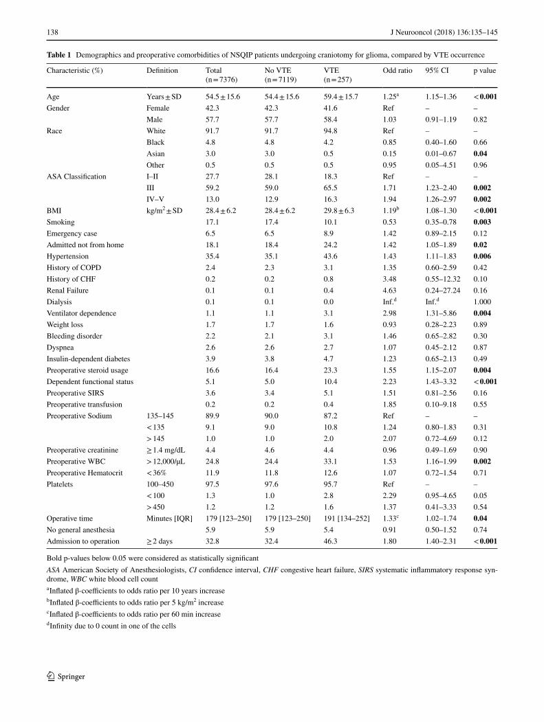
during the study period. Comorbidities, demographics, and preoperative laboratory values are shown separated by the occurrence of VTE (Table 1).
Outcomes
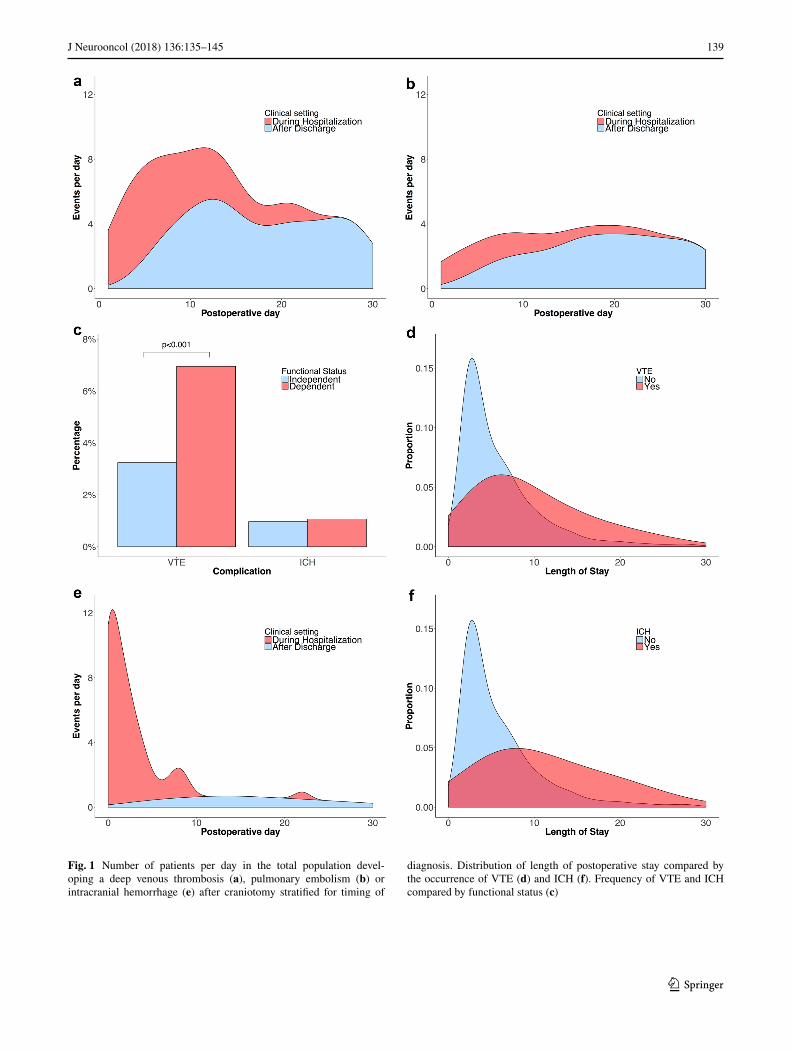
Of the 7376 patients that were identified, 257 (3.5%) devel-oped a VTE within 30 days after surgery, of which 91 (36%) occurred within the initial hospital stay. VTE was the second-most common major complication and included 192 DVTs (2.6%) and 107 PEs (1.5%). Forty-two patients developed both DVT and PE (0.6%). The rate of DVT was highest within the first 2 weeks after surgery, whereas the rate of PE was fairly consistent throughout the first month, occurring predominantly post-discharge (Fig. 1a, b). The rate of VTE was more than twice as high (7.0 vs. 3.2%, p < 0.001) among patients with a preoperative dependent functional status compared to patients with an independent functional status (Fig. 1c).
The median length of stay among VTE patients was 8 days (inter-quartile range [IQR] 5–16 days) compared to 4 days (IQR 3–8 days) in non-VTE patients (Fig. 1d). In-hospital VTE occurred at a median of 6 days (IQR 3–8 days) after surgery, and patients were discharged at a median of 8 days after the occurrence of VTE (IQR 3–16 days). Post-discharge VTE occurred at a median of 13 days after dis-charge (IQR 6–19 days) and resulted in 25% of cases in readmission, making VTE the third-most common reason for readmission (7.4% of total readmissions). Of the patients that developed a PE, 35 (32.7%) were preceded by a DVT. Post-discharge PEs were less frequently preceded by a DVT than hospital acquired PEs (26 vs. 48%, p = 0.048).
Of the 5699 patients that were identified in the NSQIP registry 2012–2015 with data on reoperation and associ-ated reasons, 72 (1.3%) developed an ICH requiring sur-gical evacuation at a median of 2 days after surgery (IQR 0–7.5 days) (Fig. 1e). ICH was the most common reason for reoperation (18.5% of the total number of reoperations). The median length of stay among ICH patients was 12 days (IQR 6–21 days) compared to 4 days (IQR 3–8 days) in non-ICH patients (Fig. 1f). The 55 patients (77.5%) that developed an ICH during the initial hospital stay, were discharged at a median of 10 days (IQR 6–19) after the occurrence of ICH.
Multivariable analysis
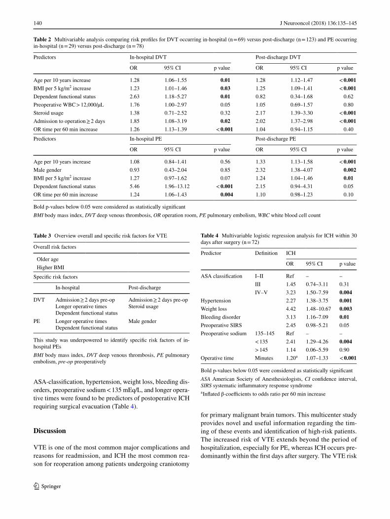
Older age and higher BMI were found to be risk factors of VTE overall (Tables 2, 3). Dependent functional status and longer operative times were predictive for hospital VTE, but not for post-discharge events. Admission two or more days before surgery was a predictor of DVT, but not for PE. Steroid usage was predictive for post-discharge DVT, and male gender was predictive for post-discharge PE. Higher
138 J Neurooncol (2018) 136:135–145
1 3
Table 1 Demographics and preoperative comorbidities of NSQIP patients undergoing craniotomy for glioma, compared by VTE occurrence
Bold p-values below 0.05 were considered as statistically significantASA American Society of Anesthesiologists, CI confidence interval, CHF congestive heart failure, SIRS systematic inflammatory response syn-drome, WBC white blood cell counta Inflated β-coefficients to odds ratio per 10 years increaseb Inflated β-coefficients to odds ratio per 5 kg/m2 increasec Inflated β-coefficients to odds ratio per 60 min increased Infinity due to 0 count in one of the cells
Characteristic (%) Definition Total(n = 7376)
No VTE(n = 7119)
VTE(n = 257)
Odd ratio 95% CI p value
Age Years ± SD 54.5 ± 15.6 54.4 ± 15.6 59.4 ± 15.7 1.25a 1.15–1.36 < 0.001Gender Female 42.3 42.3 41.6 Ref – –
Male 57.7 57.7 58.4 1.03 0.91–1.19 0.82Race White 91.7 91.7 94.8 Ref – –
Black 4.8 4.8 4.2 0.85 0.40–1.60 0.66Asian 3.0 3.0 0.5 0.15 0.01–0.67 0.04Other 0.5 0.5 0.5 0.95 0.05–4.51 0.96
ASA Classification I–II 27.7 28.1 18.3 Ref – –III 59.2 59.0 65.5 1.71 1.23–2.40 0.002IV–V 13.0 12.9 16.3 1.94 1.26–2.97 0.002
BMI kg/m2 ± SD 28.4 ± 6.2 28.4 ± 6.2 29.8 ± 6.3 1.19b 1.08–1.30 < 0.001Smoking 17.1 17.4 10.1 0.53 0.35–0.78 0.003Emergency case 6.5 6.5 8.9 1.42 0.89–2.15 0.12Admitted not from home 18.1 18.4 24.2 1.42 1.05–1.89 0.02Hypertension 35.4 35.1 43.6 1.43 1.11–1.83 0.006History of COPD 2.4 2.3 3.1 1.35 0.60–2.59 0.42History of CHF 0.2 0.2 0.8 3.48 0.55–12.32 0.10Renal Failure 0.1 0.1 0.4 4.63 0.24–27.24 0.16Dialysis 0.1 0.1 0.0 Inf.d Inf.d 1.000Ventilator dependence 1.1 1.1 3.1 2.98 1.31–5.86 0.004Weight loss 1.7 1.7 1.6 0.93 0.28–2.23 0.89Bleeding disorder 2.2 2.1 3.1 1.46 0.65–2.82 0.30Dyspnea 2.6 2.6 2.7 1.07 0.45–2.12 0.87Insulin-dependent diabetes 3.9 3.8 4.7 1.23 0.65–2.13 0.49Preoperative steroid usage 16.6 16.4 23.3 1.55 1.15–2.07 0.004Dependent functional status 5.1 5.0 10.4 2.23 1.43–3.32 < 0.001Preoperative SIRS 3.6 3.4 5.1 1.51 0.81–2.56 0.16Preoperative transfusion 0.2 0.2 0.4 1.85 0.10–9.18 0.55Preoperative Sodium 135–145 89.9 90.0 87.2 Ref – –
< 135 9.1 9.0 10.8 1.24 0.80–1.83 0.31> 145 1.0 1.0 2.0 2.07 0.72–4.69 0.12
Preoperative creatinine ≥ 1.4 mg/dL 4.4 4.6 4.4 0.96 0.49–1.69 0.90Preoperative WBC > 12,000/µL 24.8 24.4 33.1 1.53 1.16–1.99 0.002Preoperative Hematocrit < 36% 11.9 11.8 12.6 1.07 0.72–1.54 0.71Platelets 100–450 97.5 97.6 95.7 Ref – –
< 100 1.3 1.0 2.8 2.29 0.95–4.65 0.05> 450 1.2 1.2 1.6 1.37 0.41–3.33 0.54
Operative time Minutes [IQR] 179 [123–250] 179 [123–250] 191 [134–252] 1.33c 1.02–1.74 0.04No general anesthesia 5.9 5.9 5.4 0.91 0.50–1.52 0.74Admission to operation ≥ 2 days 32.8 32.4 46.3 1.80 1.40–2.31 < 0.001
139J Neurooncol (2018) 136:135–145
1 3
Fig. 1 Number of patients per day in the total population devel-oping a deep venous thrombosis (a), pulmonary embolism (b) or intracranial hemorrhage (e) after craniotomy stratified for timing of
diagnosis. Distribution of length of postoperative stay compared by the occurrence of VTE (d) and ICH (f). Frequency of VTE and ICH compared by functional status (c)
140 J Neurooncol (2018) 136:135–145
1 3
ASA-classification, hypertension, weight loss, bleeding dis-orders, preoperative sodium < 135 mEq/L, and longer opera-tive times were found to be predictors of postoperative ICH requiring surgical evacuation (Table 4).
Discussion
VTE is one of the most common major complications and reasons for readmission, and ICH the most common rea-son for reoperation among patients undergoing craniotomy
for primary malignant brain tumors. This multicenter study provides novel and useful information regarding the tim-ing of these events and identification of high-risk patients. The increased risk of VTE extends beyond the period of hospitalization, especially for PE, whereas ICH occurs pre-dominantly within the first days after surgery. The VTE risk
Table 2 Multivariable analysis comparing risk profiles for DVT occurring in-hospital (n = 69) versus post-discharge (n = 123) and PE occurring in-hospital (n = 29) versus post-discharge (n = 78)
Bold p-values below 0.05 were considered as statistically significantBMI body mass index, DVT deep venous thrombosis, OR operation room, PE pulmonary embolism, WBC white blood cell count
Predictors In-hospital DVT Post-discharge DVT
OR 95% CI p value OR 95% CI p value
Age per 10 years increase 1.28 1.06–1.55 0.01 1.28 1.12–1.47 < 0.001BMI per 5 kg/m2 increase 1.23 1.01–1.46 0.03 1.25 1.09–1.41 < 0.001Dependent functional status 2.63 1.18–5.27 0.01 0.82 0.34–1.68 0.62Preoperative WBC > 12,000/µL 1.76 1.00–2.97 0.05 1.05 0.69–1.57 0.80Steroid usage 1.38 0.71–2.52 0.32 2.17 1.39–3.30 < 0.001Admission to operation ≥ 2 days 1.85 1.08–3.19 0.02 2.02 1.37–2.98 < 0.001OR time per 60 min increase 1.26 1.13–1.39 < 0.001 1.04 0.94–1.15 0.40
Predictors In-hospital PE Post-discharge PE
OR 95% CI p value OR 95% CI p value
Age per 10 years increase 1.08 0.84–1.41 0.56 1.33 1.13–1.58 < 0.001Male gender 0.93 0.43–2.04 0.85 2.32 1.38–4.07 0.002BMI per 5 kg/m2 increase 1.27 0.97–1.62 0.07 1.24 1.04–1.46 0.01Dependent functional status 5.46 1.96–13.12 < 0.001 2.15 0.94–4.31 0.05OR time per 60 min increase 1.24 1.06–1.43 0.004 1.10 0.98–1.23 0.10
Table 3 Overview overall and specific risk factors for VTE
This study was underpowered to identify specific risk factors of in-hospital PEsBMI body mass index, DVT deep venous thrombosis, PE pulmonary embolism, pre-op preoperatively
Overall risk factors
Older age Higher BMI
Specific risk factors
In-hospital Post-discharge
DVT Admission ≥ 2 days pre-opLonger operative timesDependent functional status
Admission ≥ 2 days pre-opSteroid usage
PE Longer operative timesDependent functional status
Male gender
Table 4 Multivariable logistic regression analysis for ICH within 30 days after surgery (n = 72)
Bold p-values below 0.05 were considered as statistically significantASA American Society of Anesthesiologists, CI confidence interval, SIRS systematic inflammatory response syndromea Inflated β-coefficients to odds ratio per 60 min increase
Predictor Definition ICH
OR 95% CI p value
ASA classification I–II Ref – –III 1.45 0.74–3.11 0.31IV–V 3.23 1.50–7.59 0.004
Hypertension 2.27 1.38–3.75 0.001Weight loss 4.42 1.48–10.67 0.003Bleeding disorder 3.13 1.16–7.09 0.01Preoperative SIRS 2.45 0.98–5.21 0.05Preoperative sodium 135–145 Ref – –
< 135 2.41 1.29–4.26 0.004> 145 1.14 0.06–5.59 0.90
Operative time Minutes 1.20a 1.07–1.33 < 0.001
141J Neurooncol (2018) 136:135–145
1 3
profile depends on the type of VTE (DVT vs. PE events) and the clinical setting (hospitalized vs. post-discharge patients).
The patient population in this study was technically clas-sified as all those with primary malignant brain tumors based on ICD-9 codes. Gliomas represent close to 80% of primary malignant brain tumors [38], however, there is no standard ICD-9 code specific for glioma. The Central Brain Tumor Registry of the United States (CBTRUS) argues that multiple combinations of ICD histology codes can be used to define gliomas, and their approach was modeled in this study [38]. Therefore, these results are primarily applicable to glioma patients and should be put in the context of previ-ous outcome research on glioma patients.
Previous work
Several multicenter studies have previously investigated the short-term incidence of and risk factors for VTE after brain tumor surgery [3, 13, 39–45], of which four studies focused on glioma patients [3, 13, 39, 41]. From these studies, the rate of VTE following craniotomy is cited as 3.3–7.5% for glioma patients [3, 13, 39, 41] and 2.3–4.0% for brain tumors patients in general [11, 40–42, 44], with a follow-up ranging from solely the initial hospital stay to 6 weeks after surgery. The 30-day VTE rate was as high as 9.3% when asympto-matic DVTs were included too [3]. These results are com-parable to the VTE rates found in the current study and sug-gest a higher rate of VTE in glioma patients postoperatively compared to other brain tumors.
Simanek et al. assessed the cumulative incidence of VTE over time after craniotomy for gliomas, demonstrating a major increase in the number of events in the first 3 months after surgery; however, no granular insight into the distribu-tion of events within the first few weeks postoperatively was provided due to a low sample size. Neither did this study stratify for VTE type or the clinical setting of the patient [4].
Risk factors identified for VTE after craniotomy for glio-mas are older age, history of craniotomy, history of VTE, coagulopathy, seizures, increased stay on the intensive care unit, prolonged hospital stay, residual tumor tissue, and absence of thromboprophylactic therapy [3, 4, 6, 12–15]. Missios et al. stratified for VTE type demonstrating different risk profiles for postoperative DVT and PE. Male gender, Hispanic ethnicity, and medium bed size were predictive for PE, whereas chronic heart failure was predictive for DVT [3]. Other predictors of postoperative VTE identified in the broader group of brain tumor patients were higher BMI, hypertension, functional dependence, lower Karnofsky Performance Scale (KPS) score, motor deficits, ventilator dependence, steroid usage, preoperative sepsis, longer oper-ative times, and higher World Health Organization (WHO) tumor grade [11, 40, 42, 43, 46].
Prophylactic anticoagulation is a commonly used strategy to prevent VTE but should be carefully balanced against the risk of ICH. In previous studies, the rates of ICH follow-ing craniotomy for brain tumors is cited as 1.0–4.0% with a follow-up ranging between the initial hospital stay and long-term survival after surgery [6, 13–15, 44, 46–48]. However, definitions for major ICH varied between volumetric meas-urement of the hematoma, presence of symptoms, decrease in hemoglobin, or need for surgical evacuation of hematoma [14, 15, 21, 23, 24, 49].
Mantia et al. assessed the cumulative incidence of ICH over time after craniotomy for glioma. However, no time-to-event analysis was provided for the direct postoperative period due to a low sample size [23]. Neither did this study stratify for the clinical setting of the patient. Risk factors associated with ICH were history of craniotomy, use of bevacizumab, and therapeutic anticoagulation for VTE [13, 20–24]. The association between thromboprophylactic anti-coagulation and ICH remains to be elucidated [15].
To our knowledge, the current study is the first large mul-ticenter assessment including a descriptive time-to-event analysis for both VTE and ICH within 30 days after crani-otomy for primary malignant brain tumors. Additionally, it is the first study that uses the NSQIP database to identify predictors of ICH after brain tumor resection. By addressing thrombotic outcomes as well as hemorrhagic outcomes, this study provides a meaningful direction for future research on thromboprophylactic treatment strategies. Lastly, the large sample size allows a stratification of both the descriptive and inferential analysis, demonstrating differences in risk profile and incidence over time based on VTE type (DVT vs. PE) and clinical setting (hospitalized vs. post-discharge patients).
Limitations
Complication rates found in the current study can be con-servative estimates if events were not reported back to the hospitals. VTEs were only coded as events if they were diagnosed and treated, thereby missing asymptomatic and undetected VTEs. The database additionally lacks several demographic variables identified in other studies as predic-tors. Tumor specific information (histology, size, location, residual tumor volume) and complication specific informa-tion (location and classification of DVT, PE, and ICH) was not available. However, both VTE and ICH were defined in the NSQIP database as complications requiring medical and surgical treatment, respectively, resulting in selection of the most clinically relevant events. Perhaps most importantly, no data is available regarding anticoagulation status and non-pharmaceutical prophylactic methods. Therefore, this study offers limited insight in the efficacy of different thrombopro-phylactic treatment strategies and their association with the occurrence of ICH. Selection bias can be introduced since
142 J Neurooncol (2018) 136:135–145
1 3
institutions can selectively contribute patients to the NSQIP registry. There was a lower number of events due to separate analyses based on VTE type and clinical setting; however, our study was not underpowered for most outcome meas-ures according to rule of 10 events per variable in the multi-variable analysis [50]. Lastly, VTE and ICH events after the 30-day time period established in NSQIP are not accounted for in this study, although studies have demonstrated that the risk of VTE events remains non-negligible beyond 30 days postoperatively with incidences up to 26% in the first 12 months postoperatively [4–6]. Despite these limitations, this study provides useful insight into the rates, timing, and predictors of DVT, PE, and ICH after craniotomy for pri-mary malignant brain tumors. Due to the multicenter nature of the NSQIP dataset, the results of this study may be more representative of typical management at all hospitals, includ-ing but not limited to tertiary care academic centers.
Implications
The significant prevalence of VTE and ICH following crani-otomy for primary malignant brain tumors found in the cur-rent study indicates that there is still room for improvement when it comes to monitoring and preventing these events. Rolston et al. demonstrated that the prevalence of VTE fol-lowing a neurosurgical procedure registered in NSQIP has remained consistent over the last years [51]. This suggests that perioperative management still hasn’t improved effec-tively with regards to preventing VTE, despite the attention it receives in neurosurgical literature.
These results particularly encourage the need for contin-ued awareness for VTE post-discharge, especially for PE, which has more lethal consequences. These PEs can also be considered more sudden since they were less often pre-ceded by a known DVT. PEs preceded by a DVT, however, suggest inadequate treatment for the initial VTE event. It is possible that DVTs are less frequent post-discharge. It is our primary suspicion, however, that DVTs are underdetected after leaving the hospital because they are less frequently symptomatic and cannot be effectively screened for. It is also possible that patients develop symptomatic DVTs but are unaware of the signs and symptoms until they progress to PE, implicating a possible role for improved patient edu-cation in preventing morbidity caused by DVT and PE. In prospective randomized control trials investigating different VTE prophylaxis modalities, Goldhaber et al. screened all craniotomy patients prior to discharge and found 9.3% of patients to have VTE, most of which were asymptomatic in both studies [45].
Most guidelines recommend that prophylactic use of low-molecular weight heparin or unfractionated heparin should be considered in all cancer patients undergoing major surgery [16–19]. In patients undergoing operations
for brain tumors, however, the benefits of anticoagulation should be carefully balanced against the risk of ICH [52, 53]. Although most guidelines support the use of pharma-cological prophylaxis in patients with brain tumors, proper timing of prophylaxis remains controversial and the use of anticoagulation often depends on the surgeon’s preference [52–54]. Recommendations vary between administration throughout hospitalization [19], up to 7–10 days after sur-gery [16, 17, 55], until the patient is mobile [52], or timing based on the individual risk profile [56]. A lack of scientific evidence is primarily the cause of this variation in recom-mendations. Recent systematic reviews and meta-analyses of VTE prophylaxis in patients undergoing craniotomy for brain tumors have been performed [42, 57–60]. These analy-ses have compared different VTE prophylaxis modalities, as well as their safety and cost effectiveness, but they do not thoroughly investigate the efficacy of prophylaxis over time to determine a recommended duration. Only one clinical trial studied the effect of continued prophylaxis up to 12 months after surgery [15]. No significant association was found between prolonged prophylaxis and the rate of both VTE and ICH; however, the trial was stopped early because of expiration of study medication, and the control group received placebo instead of short-term prophylaxis. Many patients may not need or benefit from continuing throm-boprophylactic therapy beyond discharge. Algattas et al. reviewed the safety and effectiveness of thromboprophylac-tic strategies and indicated that different regimens may have different efficacies depending on the patient’s VTE risk pro-file [57]. This highlights the importance of using the appro-priate risk profile for optimizing postoperative management.
Since the NSQIP data does not contain information on thromboprophylactic strategies, the current study provides limited insight into the efficacy or safety of prophylactic anticoagulation and insufficient evidence to change the current clinical practice of thromboprophylaxis in patients undergoing operations for primary malignant brain tumors. Therefore, we concur with the current guidelines that rec-ommend pharmaceutic prophylaxis (low-molecular weight heparin or unfractionated heparin) in combination with mechanical prophylaxis (anti-embolism stockings or inter-mittent pneumatic compression devices) postoperatively until the end of hospitalization or until the patient is mobile. Absolute contra-indications for these include recent ICH or another active major bleeding [16–19, 52, 53, 55, 56].
Suggestions for future research
Despite its limitations, this study provides useful insight into the prevalence, timing, and risk factors of postoperative VTE and ICH after craniotomy for primary malignant brain tumors. The results of the current study demonstrate that there is still room for improvement, especially with regard to
143J Neurooncol (2018) 136:135–145
1 3
the prevention of PE after hospitalization. The distinct criti-cal time periods for both thrombotic and hemorrhagic events suggest a potentially safe and effective role for continuing prophylactic anticoagulation post-discharge in high-risk patients. Additionally, the typical patient at risk for develop-ing a VTE during hospitalization is not the same as the typi-cal patient at risk for developing a VTE post-discharge. This is crucial for tailoring post-discharge management to the risk profile of the individual patient and suggests an impor-tant direction for future research. Therefore, future research should study the effects of timing of thromboprophylactic therapy, screening for asymptomatic events, and the effects of patient education on the occurrence of VTE and/or ICH. Additionally, future studies should construct prediction mod-els for DVT, PE, and ICH and examine the effectiveness of tailoring postoperative thromboprophylaxis to the individual risk profile of patients undergoing craniotomy for primary malignant brain tumors.
Conclusion
The increased risk of VTE experienced by patients with primary malignant brain tumors extends beyond the period of hospitalization, especially for PE, whereas ICH occurs predominantly in the first few days after surgery. The risk profile for VTE depends on the type of VTE and the clinical setting of the patient. VTE can have fatal consequences if not recognized early, therefore clinicians should have high suspicion during the postoperative period and a low thresh-old for specific monitoring and prevention.
Funding National Institutes of Health (NIH) Training Grant T32 CA 009001 (DJC).
Compliance with ethical standards
Conflict of interest J.T.S., N.H.G., D.J.C., I.S.M., H.H.D., F.Y.F.L.V., T.R.S., M.L.B., Nothing to Disclose. W.B.G. Codman, Coviden Proctor, Consultant.
Open Access This article is distributed under the terms of the Creative Commons Attribution 4.0 International License (http://crea-tivecommons.org/licenses/by/4.0/), which permits unrestricted use, distribution, and reproduction in any medium, provided you give appro-priate credit to the original author(s) and the source, provide a link to the Creative Commons license, and indicate if changes were made.
References
1. Fadul C, Wood J, Thaler H, Galicich J, Patterson RH Jr, Posner JB (1988) Morbidity and mortality of craniotomy for excision of supratentorial gliomas. Neurology 38:1374–1379
2. Marcus LP, McCutcheon BA, Noorbakhsh A, Parina RP, Gonda DD, Chen C, Chang DC, Carter BS (2014) Incidence and pre-dictors of 30-day readmission for patients discharged home after craniotomy for malignant supratentorial tumors in California (1995–2010). J Neurosurg 120:1201–1211. doi:10.3171/2014.1.JNS131264
3. Missios S, Kalakoti P, Nanda A, Bekelis K (2015) Craniotomy for glioma resection: a predictive model. World Neurosurg 83:957–964. doi:10.1016/j.wneu.2015.04.052
4. Simanek R, Vormittag R, Hassler M, Roessler K, Schwarz M, Zielinski C, Pabinger I, Marosi C (2007) Venous thromboembo-lism and survival in patients with high-grade glioma. Neuro Oncol 9:89–95. doi:10.1215/15228517-2006-035
5. Streiff MB, Ye X, Kickler TS, Desideri S, Jani J, Fisher J, Gross-man SA (2015) A prospective multicenter study of venous thromboembolism in patients with newly-diagnosed high-grade glioma: hazard rate and risk factors. J Neurooncol 124:299–305. doi:10.1007/s11060-015-1840-z
6. Smith TR, Lall RR, Graham RB, McClendon J Jr, Lall RR, Nanney AD, Adel JG, Zakarija A, Chandler JP (2014) Venous thromboembolism in high grade glioma among surgical patients: results from a single center over a 10 year period. J Neurooncol 120:347–352. doi:10.1007/s11060-014-1557-4
7. Khalil J, Bensaid B, Elkacemi H, Afif M, Bensaid Y, Kebdani T, Benjaafar N (2015) Venous thromboembolism in cancer patients: an underestimated major health problem. World J Surg Oncol 13:204. doi:10.1186/s12957-015-0592-8
8. Sartori MT, Della Puppa A, Ballin A, Saggiorato G, Bernardi D, Padoan A, Scienza R, d’Avella D, Cella G (2011) Prothrombotic state in glioblastoma multiforme: an evaluation of the procoagu-lant activity of circulating microparticles. J Neurooncol 104:225–231. doi:10.1007/s11060-010-0462-8
9. Cote DJ, Smith TR (2016) Venous thromboembolism in brain tumor patients. J Clin Neurosci 25:13–18. doi:10.1016/j.jocn.2015.05.053
10. Stein PD, Beemath A, Meyers FA, Skaf E, Sanchez J, Olson RE (2006) Incidence of venous thromboembolism in patients hospitalized with cancer. Am J Med 119:60–68. doi:10.1016/j.amjmed.2005.06.058
11. Kimmell KT, Walter KA (2014) Risk factors for venous thrombo-embolism in patients undergoing craniotomy for neoplastic dis-ease. J Neurooncol 120:567–573. doi:10.1007/s11060-014-1587-y
12. Edwin NC, Khoury MN, Sohal D, McCrae KR, Ahluwalia MS, Khorana AA (2016) Recurrent venous thromboembo-lism in glioblastoma. Thromb Res 137:184–188. doi:10.1016/j.thromres.2015.11.027
13. Chang SM, Parney IF, McDermott M, Barker FG 2nd, Schmidt MH, Huang W, Laws ER Jr, Lillehei KO, Bernstein M, Brem H, Sloan AE, Berger M, Glioma Outcomes I (2003) Perioperative complications and neurological outcomes of first and second cra-niotomies among patients enrolled in the Glioma Outcome Pro-ject. J Neurosurg 98:1175–1181. doi:10.3171/jns.2003.98.6.1175
14. Perry SL, Bohlin C, Reardon DA, Desjardins A, Friedman AH, Friedman HS, Vredenburgh JJ (2009) Tinzaparin prophy-laxis against venous thromboembolic complications in brain tumor patients. J Neurooncol 95:129–134. doi:10.1007/s11060-009-9911-7
15. Perry JR, Julian JA, Laperriere NJ, Geerts W, Agnelli G, Rog-ers LR, Malkin MG, Sawaya R, Baker R, Falanga A, Parpia S, Finch T, Levine MN (2010) PRODIGE: a randomized placebo-controlled trial of dalteparin low-molecular-weight heparin thromboprophylaxis in patients with newly diag-nosed malignant glioma. J Thromb Haemost 8:1959–1965. doi:10.1111/j.1538-7836.2010.03973.x
16. Farge D, Bounameaux H, Brenner B, Cajfinger F, Debourdeau P, Khorana AA, Pabinger I, Solymoss S, Douketis J, Kakkar A
144 J Neurooncol (2018) 136:135–145
1 3
(2016) International clinical practice guidelines including guid-ance for direct oral anticoagulants in the treatment and prophy-laxis of venous thromboembolism in patients with cancer. Lancet Oncol 17:e452–e466. doi:10.1016/S1470-2045(16)30369-2
17. Farge D, Debourdeau P, Beckers M, Baglin C, Bauersachs RM, Brenner B, Brilhante D, Falanga A, Gerotzafias GT, Haim N, Kakkar AK, Khorana AA, Lecumberri R, Mandala M, Marty M, Monreal M, Mousa SA, Noble S, Pabinger I, Prandoni P, Prins MH, Qari MH, Streiff MB, Syrigos K, Bounameaux H, Buller HR (2013) International clinical practice guidelines for the treatment and prophylaxis of venous thromboembolism in patients with can-cer. J Thromb Haemost 11:56–70. doi:10.1111/jth.12070
18. Lyman GH, Bohlke K, Falanga A, American Society of Clini-cal Oncology (2015) Venous thromboembolism prophylaxis and treatment in patients with cancer: American Society of Clini-cal Oncology clinical practice guideline update. J Oncol Pract 11:e442-444. doi:10.1200/JOP.2015.004473
19. Streiff MB (2014) NCCN Clinical Practice Guidelines in Oncol-ogy (NCCN Guidelines®). Cancer-Associated Venous Throm-boembolic Disease. Version 1.2014. National Comprehensive Cancer Network
20. Zwicker JI, Karp Leaf R, Carrier M (2016) A meta-analysis of intracranial hemorrhage in patients with brain tumors receiving therapeutic anticoagulation. J Thromb Haemost 14:1736–1740. doi:10.1111/jth.13387
21. Nghiemphu P, Green RM, Pope WB, Lai A, Cloughesy TF (2008) Safety of anticoagulation use and bevacizumab in patients with glioma. Neuro-Oncology 10:355–360. doi:10.1215/15228517-2008-009
22. Nabi S, Kahlon P, Bozorgnia F, Arshad A, Mikkelsen T, Don-thireddy V (2016) Predictors of venous thromboembolism in patients with glioblastoma. Pathol Oncol Res 22:311–316. doi:10.1007/s12253-015-0008-7
23. Mantia C, Uhlmann EJ, Puligandla M, Weber GM, Neuberg D, Zwicker JI (2017) Predicting the higher rate of intracranial hemor-rhage in glioma patients receiving therapeutic enoxaparin. Blood. doi:10.1182/blood-2017-02-767285
24. Al Megren M, De Wit C, Al Qahtani M, Le Gal G, Carrier M (2017) Management of venous thromboembolism in patients with glioma. Thromb Res 156:105–108. doi:10.1016/j.thromres.2017.06.010
25. Lieber BA, Appelboom G, Taylor BE, Malone H, Agarwal N, Connolly ES Jr (2015) Assessment of the “July Effect”: out-comes after early resident transition in adult neurosurgery. J Neurosurg. doi:10.3171/2015.4.jns142149
26. McGirt MJ, Godil SS, Asher AL, Parker SL, Devin CJ (2015) Quality analysis of anterior cervical discectomy and fusion in the outpatient versus inpatient setting: analysis of 7288 patients from the NSQIP database. Neurosurg Focus 39:E9. doi:10.3171/2015.9.focus15335
27. Lieber BA, Appelboom G, Taylor BE, Lowy FD, Bruce EM, Sonabend AM, Kellner C, Connolly ES Jr, Bruce JN (2015) Preoperative chemotherapy and corticosteroids: independent predictors of cranial surgical-site infections. J Neurosurg. doi:10.3171/2015.4.jns142719
28. Lim S, Parsa AT, Kim BD, Rosenow JM, Kim JY (2015) Impact of resident involvement in neurosurgery: an analysis of 8748 patients from the 2011 American College of Surgeons National Surgical Quality Improvement Program database. J Neurosurg 122:962–970. doi:10.3171/2014.11.jns1494
29. McCutcheon BA, Ciacci JD, Marcus LP, Noorbakhsh A, Gonda DD, McCafferty R, Taylor W, Chen CC, Carter BS, Chang DC (2015) Thirty-day perioperative outcomes in spinal fusion by specialty within the NSQIP Database. Spine 40:1122–1131. doi:10.1097/brs.0000000000000599
30. Dasenbrock HH, Devine CA, Liu KX, Gormley WB, Claus EB, Smith TR, Dunn IF (2016) Thrombocytopenia and craniotomy for tumor: a National Surgical Quality Improvement Program analysis. Cancer. doi:10.1002/cncr.29984
31. Lieber BA, Han J, Appelboom G, Taylor BE, Han B, Agarwal N, Connolly ES Jr (2016) Association of steroid use with deep venous thrombosis and pulmonary embolism in neurosurgi-cal patients: A National Database analysis. World Neurosurg. doi:10.1016/j.wneu.2016.01.033
32. Kim BD, Smith TR, Lim S, Cybulski GR, Kim JY (2014) Pre-dictors of unplanned readmission in patients undergoing lumbar decompression: multi-institutional analysis of 7016 patients. J Neurosurg Spine 20:606–616. doi:10.3171/2014.3.spine13699
33. Dasenbrock HH, Liu KX, Devine CA, Chavakula V, Smith TR, Gormley WB, Dunn IF (2015) Length of hospital stay after craniotomy for tumor: a National Surgical Quality Improvement Program analysis. Neurosurg Focus 39:E12. doi:10.3171/2015.10.focus15386
34. Sellers MM, Merkow RP, Halverson A, Hinami K, Kelz RR, Bentrem DJ, Bilimoria KY (2013) Validation of new readmis-sion data in the American College of Surgeons National Surgi-cal Quality Improvement Program. J Am Coll Surg 216:420–427. doi:10.1016/j.jamcollsurg.2012.11.013
35. Hackett NJ, De Oliveira GS, Jain UK, Kim JY (2015) ASA class is a reliable independent predictor of medical complica-tions and mortality following surgery. Int J Surg 18:184–190. doi:10.1016/j.ijsu.2015.04.079
36. Abt NB, De la Garza-Ramos R, Olorundare IO, McCutcheon BA, Bydon A, Fogelson J, Nassr A, Bydon M (2016) Thirty day postoperative outcomes following anterior lumbar inter-body fusion using the National Surgical Quality Improve-ment Program database. Clin Neurol Neurosurg 143:126–131. doi:10.1016/j.clineuro.2016.02.024
37. Lukasiewicz AM, Grant RA, Basques BA, Webb ML, Samuel AM, Grauer JN (2016) Patient factors associated with 30-day morbidity, mortality, and length of stay after surgery for sub-dural hematoma: a study of the American College of Surgeons National Surgical Quality Improvement Program. J Neurosurg 124:760–766. doi:10.3171/2015.2.jns142721
38. Ostrom QT, Gittleman H, Xu J, Kromer C, Wolinski Y, Kruchko C, Barnholtz-Sloan JS (2016) CBTRUS Statistical Report: Pri-mary brain and other central nervous system tumors diganosed int he United States in 2009–2013. Neuro-Oncology 18:v1–v75. doi:10.1093/neuonc/now207
39. Auguste KI, Quinones-Hinojosa A, Gadkary C, Zada G, Lamborn KR, Berger MS (2003) Incidence of venous thromboembolism in patients undergoing craniotomy and motor mapping for glioma without intraoperative mechanical prophylaxis to the contralateral leg. J Neurosurg 99:680–684. doi:10.3171/jns.2003.99.4.0680
40. Chaichana KL, Pendleton C, Jackson C, Martinez-Gutierrez JC, Diaz-Stransky A, Aguayo J, Olivi A, Weingart J, Gallia G, Lim M, Brem H (2013) Deep venous thrombosis and pulmonary embo-lisms in adult patients undergoing craniotomy for brain tumors. Neurol Res 35:206–211. doi:10.1179/1743132812y.0000000126
41. Chan AT, Atiemo A, Diran LK, Licholai GP, McLaren M, Black PM, Creager MA, Goldhaber SZ (1999) Venous thromboembo-lism occurs frequently in patients undergoing brain tumor surgery despite prophylaxis. J Thromb Thrombolysis 8:139–142
42. Cote DJ, Dubois HM, Karhade AV, Smith TR (2016) Venous thromboembolism in patients undergoing craniotomy for brain tumors: A U.S. Nationwide Analysis. Semin Thromb Hemost 42:870–876. doi:10.1055/s-0036-1592306
43. Dasenbrock HH, Liu KX, Chavakula V, Devine CA, Gormley WB, Claus EB, Smith TR, Dunn IF (2017) Body habitus, serum albumin, and the outcomes after craniotomy for tumor: a National
145J Neurooncol (2018) 136:135–145
1 3
Surgical Quality Improvement Program analysis. J Neurosurg 126:677–689. doi:10.3171/2016.2.jns152345
44. Nuno M, Carico C, Mukherjee D, Ly D, Ortega A, Black KL, Patil CG (2015) Association between in-hospital adverse events and mortality for patients with brain tumors. J Neurosurg 123:1247–1255. doi:10.3171/2014.10.jns141516
45. Goldhaber SZ, Dunn K, Gerhard-Herman M, Park JK, Black PM (2002) Low rate of venous thromboembolism after crani-otomy for brain tumor using multimodality prophylaxis. Chest 122:1933–1937
46. Smith TR, Nanney AD 3rd, Lall RR, Graham RB, McClendon J Jr, Lall RR, Adel JG, Zakarija A, Cote DJ, Chandler JP (2015) Devel-opment of venous thromboembolism (VTE) in patients undergo-ing surgery for brain tumors: results from a single center over a 10 year period. J Clin Neurosci 22(3):519–525. doi:10.1016/j.jocn.2014.10.003
47. Portillo J, de la Rocha IV, Font L, Braester A, Madridano O, Peromingo JA, Apollonio A, Pagan B, Bascunana J, Monreal M (2015) Venous thromboembolism in patients with glioblas-toma multiforme: findings of the RIETE registry. Thromb Res 136:1199–1203. doi:10.1016/j.thromres.2015.10.043
48. Pan E, Tsai JS, Mitchell SB (2009) Retrospective study of venous thromboembolic and intracerebral hemorrhagic events in glioblas-toma patients. Anticancer Res 29:4309–4313
49. Auer TA, Renovanz M, Marini F, Brockmann MA, Tanyildizi Y (2017) Ischemic stroke and intracranial hemorrhage in patients with recurrent glioblastoma multiforme, treated with bevaci-zumab. J Neurooncol. doi:10.1007/s11060-017-2467-z
50. Peduzzi P, Concato J, Kemper E, Holford TR, Feinstein AR (1996) A simulation study of the number of events per variable in logistic regression analysis. J Clin Epidemiol 49:1373–1379
51. Rolston JD, Han SJ, Bloch O, Parsa AT (2014) What clinical fac-tors predict the incidence of deep venous thrombosis and pulmo-nary embolism in neurosurgical patients? J Neurosurg 121:908–918. doi:10.3171/2014.6.JNS131419
52. Strangman D (2009) Clinical Practice Guidelines for the man-agement of adult gliomas: astrocytomas and oligodendrogliomas. Cancer Council Australia/Australian Cancer Network/Clinical Oncological Society of Australia
53. Guyatt GH, Akl EA, Crowther M, Gutterman DD, Schuune-mann HJ, American College of Chest Physicians Antithrombotic
T, Prevention of Thrombosis P (2012) Executive summary: Antithrombotic Therapy and Prevention of Thrombosis. 9th edn: American College of Chest Physicians Evidence-Based Clinical Practice Guidelines. Chest 141:7S-47S. doi:10.1378/chest.1412S3
54. Carman TL, Kanner AA, Barnett GH, Deitcher SR (2003) Prevention of thromboembolism after neurosurgery for brain and spinal tumors. South Med J 96:17–22. doi:10.1097/01.SMJ.0000047628.44490.B2
55. Lyman GH, Bohlke K, Khorana AA, Kuderer NM, Lee AY, Arce-lus JI, Balaban EP, Clarke JM, Flowers CR, Francis CW, Gates LE, Kakkar AK, Key NS, Levine MN, Liebman HA, Tempero MA, Wong SL, Somerfield MR, Falanga A, American Society of Clinical O (2015) Venous thromboembolism prophylaxis and treatment in patients with cancer: American Society Of Clinical Oncology clinical practice guideline update 2014. J Clin Oncol 33:654–656. doi:10.1200/JCO.2014.59.7351
56. Prevention and treatment of venous thromboembolism (2013) International Consensus Statement 2013 Guidelines According to Scientific Evidence/Cardiovascular Disease Educational and Research Trust (UK)/European Venous Forum/North American Thrombosis Forum International Union of Angiology and Union Internationale du Phlebologie
57. Algattas H, Damania D, DeAndrea-Lazarus I, Kimmell KT, Marko NF, Walter KA, Vates GE, Jahromi BS (2017) Systematic review of safety and cost-effectiveness of venous thromboembo-lism prophylaxis strategies in patients undergoing craniotomy for brain tumor. Neurosurgery. doi:10.1093/neuros/nyx156
58. Alshehri N, Cote DJ, Hulou MM, Alghamdi A, Alshahrani A, Mekary RA, Smith TR (2016) Venous thromboembolism prophy-laxis in brain tumor patients undergoing craniotomy: a meta-anal-ysis. J Neurooncol 130:561–570. doi:10.1007/s11060-016-2259-x
59. Cote DJ, Dawood HY, Smith TR (2016) Venous thromboembo-lism in patients with high-grade glioma. Semin Thromb Hemost 42:877–883. doi:10.1055/s-0036-1592334
60. Salmaggi A, Simonetti G, Trevisan E, Beecher D, Carapella CM, DiMeco F, Conti L, Pace A, Filippini G (2013) Periopera-tive thromboprophylaxis in patients with craniotomy for brain tumours: a systematic review. J Neurooncol 113:293–303. doi:10.1007/s11060-013-1115-5