Venom as a source of useful biologically active molecules
9
Emergency Medicine (2001) 13 , 28–36 Blackwell Science Asia Wilderness Medicine Series Venom as a source of useful biologically active molecules Paul Bailey 1 and Jacqueline Wilce 2 1 Departments of Biochemistry and Emergency Medicine, University of Western Australia and 2 Departments of Chemistry and Biochemistry, University of Western Australia and The Western Australian Institute for Medical Research, Department of Emergency Medicine, Sir Charles Gairdner Hospital, Perth, Western Australia, Australia Abstract In the specialty area of venomology, emergency physicians traditionally have been most interested in the description of a variety of envenomation syndromes and, subsequent to this, the most appropriate investigative and therapeutic strategies to employ when envenomation is present. Taking an alternative viewpoint, in this paper we have reviewed a selection of interesting areas of biomedical research in which venom components are being investigated for their potential as novel therapeutic agents, pesticides and ion-channel probes. In addition, we describe the molecular imaging tools of X-ray crystallography and nuclear magnetic res- onance spectroscopy, key techniques in the development of rationally designed therapeutic agents. See Commentary, page 5. Key words: drug design , ion channel , toxin structure , structural studies , venom . Introduction The world’s venomous animals remain responsible for an astonishing number of deaths. In West Africa, it is estim- ated that snakebite alone is responsible for approxim- ately 23 000 deaths per year, with fewer than 10% of patients being treated in modern medical facilities. 1 It is from this vantage point that the majority of medical inter- est in the area of venom research has been in the areas of recognition and treatment of envenomation syndromes. Within Australia, venomous animals loom large in the population’s psyche, 2 and with good reason. The line-up of venomous animals includes the following: • Snakes: Twenty-one of the world’s 25 most-deadly snakes by mouse lethality assay reside in Australia, including all of the top 10. 3 • Jellyfish: The deadly box jellyfish Chironex fleckeri , widely recognized as the planet’s most deadly venomous animal and its elusive cousin the Irukandji, Carukia barnesi . • Spiders: The potentially lethal spiders Hadronyche versuta (the Blue Mountain funnel web) and the red- back spider Latrodectus hasseltii . • Cone shells: Carnivorous marine snails that immobil- ize their prey with a potent cocktail of toxins known as conotoxins. Correspondence: Dr Paul Bailey, PhD Scholar, University Department of Emergency Medicine, Sir Charles Gairdner Hospital, Hospital Avenue, Nedlands, Western Australia 6009, Australia. Email: [email protected] Paul Bailey, MB BS, FACEM, Emergency Physician and PhD Scholar; Jacqueline Wilce, BSc, PhD, Research Fellow.
-
Upload
paul-bailey -
Category
Documents
-
view
214 -
download
1
Transcript of Venom as a source of useful biologically active molecules
Emergency Medicine
(2001)
13
, 28–36
Blackwell Science Asia
Wilderness Medicine Series
Venom as a source of useful biologically active molecules
Paul Bailey
1
and Jacqueline Wilce
2
1
Departments of Biochemistry and Emergency Medicine, University of Western Australia and
2
Departments of Chemistry and Biochemistry, University of Western Australia and The Western Australian Institute for Medical Research, Department of Emergency Medicine, Sir Charles
Gairdner Hospital, Perth, Western Australia, Australia
Abstract
In the specialty area of venomology, emergency physicians traditionally have been mostinterested in the description of a variety of envenomation syndromes and, subsequent to this,the most appropriate investigative and therapeutic strategies to employ when envenomationis present. Taking an alternative viewpoint, in this paper we have reviewed a selection ofinteresting areas of biomedical research in which venom components are being investigatedfor their potential as novel therapeutic agents, pesticides and ion-channel probes. In addition,we describe the molecular imaging tools of X-ray crystallography and nuclear magnetic res-onance spectroscopy, key techniques in the development of rationally designed therapeuticagents.
See Commentary, page 5.
Key words:
drug design
,
ion channel
,
toxin structure
,
structural studies
,
venom
.
Introduction
The world’s venomous animals remain responsible for anastonishing number of deaths. In West Africa, it is estim-ated that snakebite alone is responsible for approxim-ately 23 000 deaths per year, with fewer than 10% ofpatients being treated in modern medical facilities.
1
It isfrom this vantage point that the majority of medical inter-est in the area of venom research has been in the areas ofrecognition and treatment of envenomation syndromes.
Within Australia, venomous animals loom large in thepopulation’s psyche,
2
and with good reason. The line-upof venomous animals includes the following:
• Snakes: Twenty-one of the world’s 25 most-deadlysnakes by mouse lethality assay reside in Australia,including all of the top 10.
3
• Jellyfish: The deadly box jellyfish
Chironex fleckeri
,widely recognized as the planet’s most deadly venomousanimal and its elusive cousin the Irukandji,
Carukiabarnesi
.• Spiders: The potentially lethal spiders
Hadronycheversuta
(the Blue Mountain funnel web) and the red-back spider
Latrodectus hasseltii
.• Cone shells: Carnivorous marine snails that immobil-
ize their prey with a potent cocktail of toxins known asconotoxins.
Correspondence: Dr Paul Bailey, PhD Scholar, University Department of Emergency Medicine, Sir Charles Gairdner Hospital, Hospital Avenue, Nedlands, Western Australia 6009, Australia. Email: [email protected]
Paul Bailey, MB BS, FACEM, Emergency Physician and PhD Scholar; Jacqueline Wilce, BSc, PhD, Research Fellow.
EMM174.fm Page 28 Wednesday, February 21, 2001 10:01 AM

Wilderness medicine
29
Rather than being the subject of generalized fear andloathing and perhaps because, at least in most developedcountries, death from envenomation is uncommon, it has beenrealized that these venomous animals represent a vast andlargely untapped source of potent biologically active molecules.
Venoms consist of a complex mixture of toxic com-ponents that include protein and peptide toxins, enzymesand other active agents. These molecules serve the dualpurposes of prey capture and digestion and/or defenceagainst predators. While the precise mechanisms of actionof many venom constituents remain unknown, what isknown is that many exert their effects via interactionswith specific ion channels, enzymes and membranecomponents. These highly potent and specific inter-actions make venom constituents attractive candidatesfor the development of novel therapeutics, pesticidesand as molecular probes of target molecules.
Scope of this review
In this review, we will examine the following areas of interest:• Sea anemone toxins: several of these have been found
to modulate sodium-channel function and are beinginvestigated as drug leads for the development ofnovel cardioactive agents.
• Conotoxins: potent ion channel modulators found inthe venom of a range of marine molluscs known ascone shells. The conotoxins have yielded a novel anal-gesic agent, known as ziconotide, that is used in thetreatment of a variety of pain syndromes.
• Insect-specific toxins from venoms: these peptides,which block insect but not mammalian voltage-gatedcalcium channels, have tremendous potential in thearea of bioinsecticidal control of agricultural pests.
• Antibacterial components of snake and spider venoms:the increasing prevalence of multi-drug-resistantbacteria has led to the screening of a wide number ofnatural molecules as potential new antibiotic agents.
• The angiotensin converting enzyme inhibitors (ACEinhibitors): for many years, captopril and the ACEinhibitors have been the shining light of practicalapplication of both venom research and rational drugdesign. As we shall see, captopril was rationally syn-thesized after isolation and study of an ACE-inhibitingprotein from the venom of the snake
Bothrops jararaca.
The study of these toxin components has encompassedboth the mode of action of the molecules and the rigorousanalyses of their 3-D structure. Such structure–functionstudies are at the cutting edge of modern medical science.It is believed that through an understanding of these
potent molecules at a structural level, they can be adaptedfor use as optimized ‘rationally designed’ agents. Thus thefinal section in this review outlines the techniques cur-rently employed for such studies of molecular structure.
Sea anemone toxins as modulators of sodium channel function
Sea anemones contain potent venom for the purpose ofprey capture. The venom is fired into the victim via abarbed tubule that emerges from venom-containing cysts(nematocysts) on their tentacles. This both paralyses theprey and allows it to be drawn towards the digestiveapparatus of the anemone.
The paralytic action of the venom is due to a mixtureof protein and peptide toxins, several of which are potentinhibitors of sodium channel inactivation.
4–6
One of the bestcharacterized of these is anthopleurin-A (AP-A) (Fig. 1)
Figure 1. The sodium channel modulator anthopleurin-A from the venom of the giant green sea anemone Xanthopleura xanthogrammica: a. the backbone fold of the 20 nuclear magnetic resonance (NMR)-derived structures (black) and the three disulphide bonds which stabilize the structure (grey); b. the space-filling model of one of the NMR-derived structures (pdb code: 1AHL).
EMM174.fm Page 29 Wednesday, February 21, 2001 10:01 AM
P Bailey and J Wilce
30
which was found to be active as a cardiac stimulant atnanomolar concentrations
in vivo
and not associatedwith any significant effects on heart rate or blood pres-sure in mammals.
7,8
This was of great interest, especiallysince it was found to be approximately 200-fold morepotent than digoxin and to possess fewer undesirableside-effects. It was thus a potentially valuable lead in thedevelopment of a novel drug for the treatment of chronicheart failure.
Since AP-A was discovered, a whole family of seaanemone sodium channel modulators have been isolatedfrom various species of anemone. Many are polypeptidesof about 49 amino-acid residues in length and containthree disulfide bonds which help to stabilize their 3-Dstructure. Despite their potency, however, the use of these‘natural products’ as drugs is not ideal because of theirlow stability following oral administration and their anti-genicity.
9
The research effort has thus been to use the3-D structural information and knowledge of the modeof action of these peptides in order to design smaller,non-peptide molecules with the same activity.
The toxins are known to bind to specific sites on themyocardial voltage-gated sodium channel,
10
known as‘site 3’. Other polypeptides that also bind to site 3 includeseveral ‘short’ anemone polypeptides
6,11
that are neuro-toxic to crustacea, a class of anemone peptides with anti-hypertensive and antiviral activity
12,13
and
α
-scorpiontoxins.
14
By the comparison of the structures of thesedifferent classes of peptides, the important structuralfeatures of the molecules can be identified. This, alongwith knowledge of their relative binding affinities, under-pins the effort to define the cardioactive pharmacaphore.
The sodium channel site 3 binding face of the toxinshas now been identified
9
and both charged and hydro-phobic residues are thought to be important for bindingaffinity. It now remains for the synthesis and testing ofmimics of this pharmacaphore to be carried out. Thismay be achieved by designing a linear peptide mimetic,or it may be necessary to develop a molecule of a com-pletely different architecture which maintains the spatialarrangement and chemical properties of the toxin bind-ing face. Such ‘rational drug design’ is at the cutting edgeof medical research today.
Cone shell venom: The conotoxins
Conotoxins have been the subject of intensive researchefforts in recent times and much of this work has beenbased in Australia. Animals of the genus
Conus
are predat-ory molluscs, found in tropical waters such as the GreatBarrier Reef area of Queensland, that also immobilize
their prey by injecting them with venom via a harpoon-like apparatus. The conotoxins of each species of coneshells are targeted to specific receptors in the cone shells’prey.
15
There are over 500 species currently identified,with both inter- and intraspecies variation of venom com-ponents. In fact, within the same animal there can be sig-nificant variation in the composition of venom over time.Seemingly minor variations to the basic protein structureof these conotoxins can result in widely varying affinitiesfor molecular targets.
Conotoxins consist of a variety of peptidic compon-ents, notable for their relatively small size (9–35 aminoacids
15
), complex post-translational modifications, dis-tinctive disulphide bonds and potent inhibitory effecton a variety of neuronal ion channels. Thus far, around60 unique conotoxins have been identified. This is, how-ever, only the tip of the iceberg. Analysis of whole coneshell venoms suggests that this represents less than 0.1%of the total peptide load contained within these venoms.
16
The majority of conotoxins are potent neurotoxins,being highly selective for neuronal ion channel subtypes.They are divided into six major classes, based upon theirstructure and channel-blocking activity. Conotoxins havebeen shown to have activity, usually antagonistic on nico-tinic acetylcholine receptors, sodium channels, calciumchannels and
N
-methyl-
D
-aspartate (NMDA) glutamatereceptors.
16
They were initially used as ion-channel probesand have assisted in both the characterization of specificion-channel subtypes and the elucidation of the 3-D struc-ture of the ion-channel binding surface.
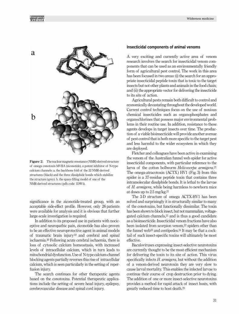
Conotoxins have been recognized for their potential astherapeutic agents owing to their relatively small size andthe excellent stability conferred by the disulphide bonds.The most spectacular current therapeutic application ofthe conotoxins is, however, that of the omega conotoxinMVIIA, a 25-amino-acid disulfide-bridged polypeptide derivedfrom the venom of
Conus magnus
. This conotoxin, whichhas been chemically synthesized and will be marketedunder the generic name of ziconotide, is an antagonist ofN-type neuronal calcium channels
16
(Fig. 2), which playan important role in the spinal transmission of pain.
Ziconotide has been shown to be effective in the treat-ment of both nociceptive and neuropathic pain syndromesin both animal
17
and human models of pain. Atanassoff
et al.
18
recently reported a randomized, double-blindedclinical trial of postoperative intrathecal ziconotideinfusions in the treatment of patients undergoing elect-ive total abdominal hysterectomy, radical prostatectomyor total hip replacement. Both outcome measures of meandaily morphine patient-controlled analgesia consumptionand visual-analogue pain-intensity scores reached statistical
EMM174.fm Page 30 Wednesday, February 21, 2001 10:01 AM
Wilderness medicine
31
significance in the ziconotide-treated group, with anacceptable side-effect profile. However, only 26 patientswere available for analysis and it is obvious that furtherlarge-scale investigation is required.
In addition to its proposed use in patients with nocic-eptive and neuropathic pain, ziconotide has also provento be an effective neuroprotective agent in animal modelsof traumatic brain injury
19
and cerebral and spinalischaemia.
20
Following acute cerebral ischaemia, there isloss of cytosolic calcium homeostasis, with increasedlevels of intracellular calcium, which in turn leads tomitochondrial dysfunction. Use of N-type calcium-channelblocking agents partially reverses this rise of intracellularcalcium, which is seen particularly in the setting of reper-fusion injury.
The search continues for other therapeutic agentsbased on the conotoxins. Potential therapeutic applica-tions include the setting of severe head injury, epilepsy,cerebrovascular disease and spinal cord injury.
Insecticidal components of animal venoms
A very exciting and currently active area of venomresearch involves the search for insecticidal venom com-ponents that can be used as an environmentally friendlyform of agricultural pest control. The work in this areahas been focused in two areas: (i) the search for an appro-priate insecticidal peptide toxin that is toxic to the targetinsects but not other plants and animals in the food chain;and (ii) the appropriate vector for delivering the insecticideto its site of action.
Agricultural pests remain both difficult to control andeconomically devastating throughout the developed world.Current control techniques focus on the use of noxiouschemical insecticides such as organophosphates andorganochlorines that possess major environmental prob-lems in their routine use. In addition, resistance to theseagents develops in target insects over time. The produc-tion of a viable bioinsecticide will provide another avenueof pest control that is both more specific to the target pestand less harmful to the wider ecosystem in which theyare deployed.
Fletcher and colleagues have been active in examiningthe venom of the Australian funnel web spider for activeinsecticidal components, with particular reference to thelarva of the cotton bollworm
Helicoverpa armigera.
21
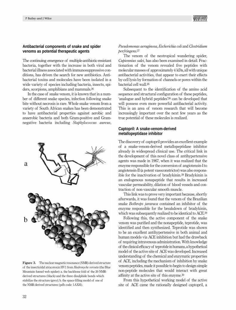
The omega-atracotoxin (ACTX) HV1 (Fig. 3) from thisspider is a 37-residue peptide toxin that contains threeintramolecular disulphide bonds. It is lethal to the larvaeof
H. armigera
, while being harmless to newborn miceat doses up to 2.5 mg/kg.
21
The 3-D structure of omega ACTX-HV1 has beensolved and surprisingly it is structurally similar to manyof the conotoxins, but functionally dissimilar. The toxinhas been shown to block insect, but not mammalian, voltage-gated calcium channels,
21
and is thus a good candidateas a bioinsecticide. Insecticidal venom fractions have alsobeen isolated from scorpion venom,
22
spiders other thanthe funnel web
23
and centipedes.
24
It may be that a cock-tail of such insect-specific toxins will ultimately be mosteffective.
Baculoviruses expressing insect-selective neurotoxinsare currently thought to be the most efficient mechanismfor delivering the toxin to its site of action. This virusspecifically infects
H. armigera
, but without the additionof a venom-derived neurotoxin they are very slow tocause larval mortality. This enables the infected larvae tocontinue their course of crop destruction prior to dying.The addition of one or more insect-selective neurotoxinsprovides a method for rapid attack of insect hosts, withgreatly reduced time to host death.
25
Figure 2. The nuclear magnetic resonance (NMR)-derived structure of omega conotoxin MVIIA (ziconotide), a potent inhibitor of N-type calcium channels: a. the backbone fold of the 32 NMR-derived structures (black) and the three disulphide bonds which stabilize the structure (grey). b. the space-filling model of one of the NMR-derived structures (pdb code: 1DW4).
EMM174.fm Page 31 Wednesday, February 21, 2001 10:01 AM
P Bailey and J Wilce
32
Antibacterial components of snake and spider venoms as potential therapeutic agents
The continuing emergence of multiple-antibiotic-resistantbacteria, together with the increase in both viral andbacterial illness associated with immunosuppressive con-ditions, has driven the search for new antibiotics. Anti-bacterial toxins and molecules have been isolated in awide variety of species including bacteria, insects, spi-ders, scorpions, amphibians and mammals.
26
In the case of snake venom, it is known that in a num-ber of different snake species, infection following snakebite without necrosis is rare. Whole snake venom from avariety of South African snakes has been demonstratedto have antibacterial properties against aerobic andanaerobic bacteria and both Gram-positive and Gram-negative bacteria including
Staphylococcus aureus
,
Pseudomonas aeruginosa
,
Escherichia coli
and
Clostridiumperfringens.
27
The venom of the neotropical wandering spider,
Cupiennius salei
,
has also been examined in detail. Frac-tionation of the venom revealed five peptides withmolecular masses of approximately 4 kDa, all with uniqueantibacterial activities, that appear to exert their effectsby cell lysis by formation of channels or pores within thebacterial cell wall.
26
Subsequent to the identification of the amino acidsequence and structural configuration of these peptides,‘analogue and hybrid peptides’
26
can be developed thatwill possess even more powerful antibacterial activity.This is an area of venom research that will becomeincreasingly important over the next few years as thetrue potential of these molecules is realized.
Captopril: A snake-venom-derived metallopeptidase inhibitor
The discovery of captopril provides an excellent exampleof a snake-venom-derived metallopeptidase inhibitoralready in widespread clinical use. The critical link inthe development of this novel class of antihypertensiveagents was made in 1967, when it was realized that theenzyme responsible for the conversion of angiotensin-I toangiotensin-II (a potent vasoconstrictor) was also respons-ible for the inactivation of bradykinin.
28
Bradykinin isan endogenous nonapeptide that results in increasedvascular permeability, dilation of blood vessels and con-traction of non-vascular smooth muscle.
This link was to prove very important because, shortlyafterwards, it was found that the venom of the Braziliansnake
Bothrops jararaca
contained an inhibitor of theenzyme responsible for the breakdown of bradykinin,which was subsequently realized to be identical to ACE.
29
Following this, the active component of the snakevenom was purified and the nonapeptide, teprotide, wasidentified and then synthesized. Teprotide was shownto be an excellent antihypertensive in both animal andhuman models via ACE inhibition but had the drawbackof requiring intravenous administration. With knowledgeof the clinical efficacy of teprotide in humans, a hypotheticalmodel of the active site of ACE was developed. Increasedunderstanding of the chemical and enzymatic propertiesof ACE, including the mechanism of inhibition by snakevenom peptides, made it possible to begin to design simplenon-peptide molecules that would interact with greataffinity at the active site of this enzyme.
30
From this hypothetical working model of the activesite of ACE came the rationally designed captopril, a
Figure 3. The nuclear magnetic resonance (NMR)-derived structure of the insecticidal atracotoxin HV1 from Hadronyche versuta (the Blue Mountain funnel web spider): a. the backbone fold of the 20 NMR-derived structures (black) and the three disulphide bonds which stabilize the structure (grey); b. the space-filling model of one of the NMR-derived structures (pdb code: 1AXH).
EMM174.fm Page 32 Wednesday, February 21, 2001 10:01 AM

Wilderness medicine
33
novel and highly effective antihypertensive drug,
30
andthe prototype for a new class of therapeutic agents, theACE inhibitors (Fig. 4).
Studies of molecular structure for ‘rational drug design’
Rational drug design, as opposed to serendipitous drugdiscovery, depends upon knowledge of either the molecu-lar structure of the target molecule or the ligands whichbind it. Ideally the full 3-D structure of the ligand : targetcomplex to atomic resolution should be known, but thisis often not the case. Some proteins, particularly largemultidomain proteins or those that are membrane bound,are difficult to structurally determine and thus not avail-able to assist in the process. The chemical features of thebinding site may alternatively be inferred from the struc-ture of the natural ligand, often a small, more solublecompound which is more easily structurally analysed.When many ligands to the target molecule are structur-ally characterized, their common chemical features canprovide a very good picture of the active pharmacophore.This then, can be used as a template for the design of adrug molecule with potent binding characteristics that issuitable for commercial production and use.
Determining the 3-D structure of any biological macro-molecule requires the use of X-ray crystallographic,nuclear magnetic resonance (NMR) spectroscopic or elec-tron diffraction techniques. None of these methods areroutine in their application and some are better suited toparticular structural problems than others. This final
section aims to provide a brief overview of the capabil-ities and limitations of these technologies.
X-ray crystallography is a technique which directlyimages the molecule of interest. X-rays (with a wavelengthof approximately 1 Å) rather than light (with a minimumwavelength of approximately 4000 Å) must be used becausetheir shorter wavelength is sensitive to the distances ofinterest (a typical covalent bond is 1.5 Å). The molecule isexposed to a beam of monochromatic X-rays, which arediffracted by the electrons surrounding and connectingeach atom in the molecule. The diffraction pattern isrecorded onto an X-ray sensitive recording plate and isthen mathematically re-focused in order to visualize the‘electron density’ of the molecule. This is analogous to theneed for reflected light rays to be re-focused by the lensesin our eyes before we can interpret visual information.
In order to be able to measure the X-ray diffractionsignal, the molecules must be prepared in crystallineform. When the individual molecules are in such anordered arrangement, their diffraction pattern is effect-ively amplified and can be recorded (Fig. 5). The require-ment for a good diffracting crystal, however, is notalways readily achieved. There is no guaranteed methodfor growing good quality diffracting crystals and macro-molecules, particularly those which occur naturally in alipid environment of the cell, are notoriously difficult to
Figure 4. Development of captopril. The amino acid sequence of the snake-venom-derived peptide, teprotide, was used to develop a model of the active site of the enzyme angiotensin converting enzyme (ACE). The synthetic drug captopril was designed to mimic the C-terminus of teprotide.
Figure 5. Typical protein crystal X-ray diffraction pattern from which the position and intensity of each diffraction spot can be measured. A set of such images contains the experimental information used to create an electron density map which defines the structure of the macromolecule.
EMM174.fm Page 33 Wednesday, February 21, 2001 10:01 AM

P Bailey and J Wilce
34
crystallize. In these cases it is sometimes possible to useone of the other methods of structure determination.
The electron density image that the X-ray experimentgives rise to can then be used to build in the atomic coor-dinates of the molecule (Fig. 6). A macromolecular struc-ture determined by this method may be highly accurate(to almost 1 resolution), or it might be less well defined (to4 resolution or greater) depending on the quality of thecrystals and the diffraction images collected.
Nuclear magnetic resonance spectroscopy is an altern-ative method for macromolecular structure determina-tion. It is an indirect method which uses radiofrequencysignals emitted from the nuclei of the molecule beinginvestigated to ascertain their spatial arrangement. Themolecule is studied while in solution, a fundamentaldifference from the X-ray crystallographic method. Thesample is place in a strong magnetic field in which themagnetically susceptible nuclei (which include
1
H nucleiand the less naturally abundant
13
C and
15
N nuclei)respond like tiny bar magnets and tend to align with themagnetic field. In the NMR experiment, radiofrequencypulses are applied to the sample, which perturb themagnetization of the nuclei from their aligned positions.After the radiofrequency ‘excitation’ pulse is turned off,the magnetization of the nuclei returns to equilibriumand in doing so each nucleus emits its own characteristic
radiofrequency signal. Depending on the radiofrequencypulses that are applied, the emitted radiofrequenciesmeasured can contain information about which signalis coming from which nucleus and, importantly, whichnucleus is close in space to which other nucleus (Fig. 7).This provides the information required to determine the3-D structure of the molecule.
For small macromolecules (< 5 kDa), only two-dimensional (2-D)
1
H experiments are required in orderto interpret the NMR data. In order to determine largerstructures, more sophisticated 3-D and 4-D experiments inwhich
15
N and/or
13
C nuclei are detected must be employed.This requires that the macromolecule sample is enrichedwith
15
N and/or
13
C isotopes and thus limits the techniqueto molecules that can be prepared with isotopic enrich-ment. Nuclear magnetic resonance is also limited tomolecules that remain soluble at high concentration(preferably mM concentrations) and that are not greaterthan approximately 40 kDa (although advances in the fieldmay bring this limit closer to 100 kDa). The final structurestend not to be as well defined as those determined by
Figure 6. An example of 2.5 Å resolution electron density, shown as a 3-D contour map (grey), into which the atoms and bonds of the macromolecule can be built. The accuracy of the molecular model depends upon the resolution of the map.
Figure 7. A 2-D 1H nuclear magnetic resonance spectrum of a peptide molecule (recorded at 500 MHz) in which 1H nuclei are distinguished by their resonance frequencies (in p.p.m.). The cross peaks arise from 1H nuclei (e.g. labelled A–D) which exist close to each other in space within the macromolecule. These can be used to define the fold of the molecule in solution.
EMM174.fm Page 34 Wednesday, February 21, 2001 10:01 AM

Wilderness medicine
35
X-ray crystallography but may more accurately depictmotion which occurs in the molecule in solution.
Finally, electron diffraction is becoming an increas-ingly useful technique for visualizing macromolecules.Electron diffraction is analagous to X-ray diffractionexcept that lower wavelength electron frequency wavesinteract with nuclei as well as the electrons and are usedto obtain somewhat lower resolution structural informa-tion. Electron diffraction can be conducted in transmis-sion mode through thin films of aligned molecules (or2-D crystals), which is sometimes the only way thatmembrane-bound molecules may be structurally analysed.The information that is gained tends to be low resolution(4 Å or greater), but it is anticipated that this techniquewill also assist in drug design in the future.
The resulting picture of the molecule by these tech-niques provides information about the arrangement of allof the functional groups of the molecule. Both the chem-ical nature of the groups and their surface shape form thebasis of their binding interaction with their target mole-cule and therefore provide the information required for
rational drug design.
References
1. Spawls S, Branch B.
The Dangerous Snakes of Africa
. SanibelIsland, FL: Ralph Curtis, 1995.
2. Currie B. Toxinology research and clinical controversies: AnAustralian perspective.
Toxicon
1999;
37
: 261–2.
3. Broad AJ, Sutherland SK, Coulter AR. The lethality in mice ofdangerous Australian and other snake venom.
Toxicon
1979;
17
:661–4.
4. Narahashi T, Moore JW, Shapiro BI. Condylactis toxin: Interactionwith nerve membrane ionic conductances.
Science
1969;
163
(3868):680–1.
5. Romey G, Abita JP, Schweitz H, Wunderer G, Lazdunski M. Seaanemone toxin: A tool to study molecular mechanisms of nerveconduction and excitation-secretion coupling.
Proc. Natl. Acad. Sci.USA
1976;
73
: 4055–9.
6. Beress L. Biologically active compounds from coelenterates.
PureAppl. Chem
. 1982;
54
: 1981–94.
7. Shibata S, Norton TR. Potent cardiotonic action of polypeptidesisolated from sea anemone.
Recent Dev. Card. Muscle Pharmacol
.Tokyo, Japan: Igaku-Shoin, 1982; 13–33.
8. Blair RW, Peterson DF, Bishop VS. The effects of anthopleurine-Aon cardiac dynamics in conscious dogs.
J. Pharmacol. Exp. Ther
.1978;
207
: 271–6.
9. Norton RS. Polypeptide modulators of sodium channel functionas a basis for the development of novel cardiac stimulants.In: Veerapandian P (ed).
Structure-based Drug Design
. New York:Marcel Dekker, 1997; 295–319.
10. Caterall WA. Structure and function of voltage-sensitive ionchannels.
Science
1988;
242
(4875): 50–61.
11. Norton RS. Structure and structure-function relationships of seaanemone proteins that interact with the sodium channel.
Toxicon
1991;
29
: 1051–84.
12. Driscoll PC, Gronenborn AM, Beress L, Clore GM. Determina-tion of the 3-D solution structure of the antihypertensive and anti-viral protein BDS-1 from the sea anemone
Anemnia sulcata
: Astudy using nuclear magnetic resonance and hybrid distancegeometry-dynamical simulated annealing.
Biochemistry
1989;
28
:2188–98.
13. Llewellyn LE, Norton RS. Binding of the sea anemone polypeptideBDS II to the voltage gated sodium channel.
Biochem. Intl
. 1991;
24
: 937–46.
14. Catterall WA, Beress L. Sea anemone toxin and scorpion toxinshare a common receptor site associated with the action potentialsodium ionophore.
J. Biol. Chem
. 1978;
253
: 7393–6.
15. Atkinson RA, Kieffer B, Dejaegere A
et al.
Structural and dynamiccharacterization of omega-Conotoxin MVIIA. The binding loopexhibits slow conformational exchange.
Biochemistry
2000;
39
:3908–19.
16. Adams DJ, Alewood PF, Craik DJ
et al.
Conotoxins and theirpotential pharmaceutical applications.
Drug Dev. Res
. 1999;
46
:219–34.
17. Wang YX, Gao D, Pettus M
et al.
Interactions of intrathecallyadministered ziconotide, a selective blocker of neuronal N-typevoltage-sensitive calcium channels, with morphine on nociceptionin rats.
Pain
2000;
84
: 271–81.
18. Atanassoff PG, Hartmannsgruber MWB, Thrasher J
et al.
Ziconotide,a new N-type calcium channel blocker, administered intrathecallyfor acute postoperative pain.
Reg. Anesth. Pain Med
. 2000;
25
:274–8.
19. Samii A, Badie H, Fu K
et al.
Effects of an N-type calcium channelantagonist (SNX 111; ziconotide) on calcium-45 accumula-tion following fluid-percussion injury.
J. Neurotrauma
1999;
16
:879–92.
20. Ridgeway B, Wallace M, Gerayli A. Ziconotide for the treatment ofsevere spasticity after spinal cord injury.
Pain
2000;
85
: 287–9.
21. Fletcher KI, Smith R, O’Donaghue SI
et al.
The structure of a novelinsecticidal neurotoxin, omega-atracotoxin-HV1, from the venomof an Australian funnel web spider.
Nat. Struct. Biol
. 1997;
4
: 559–566.
22. Xiong YM, Ling MH, Lan ZD
et al.
The cDNA sequence of anexcitatory insect selective neurotoxin from the scorpion
Buthusmartensi
Karsch.
Toxicon
1999;
37
: 335–41.
23. Penaforte CL, Prado VF, Prado MAM
et al.
Molecular cloning of acDNA encoding insecticidal neurotoxic peptides from the spider
Phoneutria nigriventer
.
Toxicon
2000;
38
: 562 (Abstract).
24. Stankiewicz M, Hamon A, Benkhalifa R
et al.
Effects of a centipedevenom fraction on insect nervous system, a native Xenopus oocytereceptor and on an expressed Drosophilia muscarinic receptor.Toxicon 1999; 37: 1431–45.
25. McCuthen B. Efficacy of recombinant baculoviruses expressinginsect-selective toxins. Toxicon 1999; 37: 1474 (Abstract).
26. Haeberli S, Kuhn-Nentwig L, Schaller J, Nentwig W. Char-acterisation of antibacterial activity of peptides isolated fromthe venom of the spider Cupiennius salei. Toxicon 2000; 38: 373–80.
EMM174.fm Page 35 Wednesday, February 21, 2001 10:01 AM

P Bailey and J Wilce
36
27. Blaylock RSM. Antibacterial properties of KwaZulu Natal snakevenoms. Toxicon 2000; 38: 1529–34.
28. Yang HYT, Erdos EG, Levin Y. A dipeptidyl carboxypeptidasethat converts angiotensin-I and inactivates bradykinin. Biochim.Biophys. Acta 1970; 214: 374–6.
29. Opie LH, Kowolik H. The discovery of captopril: From animals tosmall molecules. Cardiovascular Res. 1995; 30 (1005): 18–25.
30. Cushman DW, Cheung HS, Sabo EF, Ondetti MA. Developmentand design of specific inhibitors of angiotensin-converting enzyme.Am. J. Cardiol. 1982; 49: 1390–4.
EMM174.fm Page 36 Wednesday, February 21, 2001 10:01 AM