P. Christakoglou, A. Petridis, M. Vassiliou University of Athens, for the NA49 collaboration.

Vassiliou, VS; Flynn, PD; Raphael, CE; Newsome, S; Khan, T; Ali,A; Halliday, B; Studer Bruengger, A; Malley, T; Sharma, P; Sel-vendran, S; Aggarwal, N; Sri, A; Berry, H; Donovan, J; Lam, W;Auger, D; Cook, SA; Pennell, DJ; Prasad, SK (2017) Lipoprotein(a)in patients with aortic stenosis: Insights from cardiovascular mag-netic resonance. PLoS One, 12 (7). e0181077. ISSN 1932-6203 DOI:10.1371/journal.pone.0181077
Downloaded from: http://researchonline.lshtm.ac.uk/4086870/
DOI: 10.1371/journal.pone.0181077
Usage Guidelines
Please refer to usage guidelines at http://researchonline.lshtm.ac.uk/policies.html or alterna-tively contact [email protected].
Available under license: http://creativecommons.org/licenses/by/2.5/

RESEARCH ARTICLE
Lipoprotein(a) in patients with aortic stenosis:
Insights from cardiovascular magnetic
resonance
Vassilios S. Vassiliou1,2*, Paul D. Flynn3, Claire E. Raphael1, Simon Newsome1,4,
Tina Khan1, Aamir Ali1, Brian Halliday1*, Annina Studer Bruengger1,5, Tamir Malley1,
Pranev Sharma1, Subothini Selvendran1, Nikhil Aggarwal1, Anita Sri1, Helen Berry6,
Jackie Donovan6, Willis Lam1, Dominique Auger1, Stuart A. Cook1,7,8, Dudley J. Pennell1,
Sanjay K. Prasad1
1 CMR Unit, Royal Brompton Hospital and NIHR Biomedical Research Unit, Royal Brompton and Harefield
Hospitals and Imperial College London, London, United Kingdom, 2 Norwich Medical School, University of
East Anglia, Bob Champion Research & Education Building, Norwich, United Kingdom, 3 The Lipid Clinic,
Addenbrooke’s Hospital, Cambridge University Foundation NHS Trust, UK and University of Cambridge,
Cambridge, United Kingdom, 4 Department of Statistics, London School of Hygiene and Tropical Medicine,
London, United Kingdom, 5 Clinic of Cardiology, Stadtspital Triemli, Zurich, Switzerland, 6 Department of
Biochemistry, Royal Brompton and Harefield NHS Trust, London, United Kingdom, 7 Duke National
University Hospital, Singapore, Singapore, 8 Cardiovascular Magnetic Resonance Imaging and Genetics,
MRC London Institute of Medical Sciences, Imperial College London, Hammersmith Hospital Campus,
London, United Kingdom
* [email protected] (BH); [email protected] (VSV)
Abstract
Background
Aortic stenosis is the most common age-related valvular pathology. Patients with aortic ste-
nosis and myocardial fibrosis have worse outcome but the underlying mechanism is unclear.
Lipoprotein(a) is associated with adverse cardiovascular risk and is elevated in patients with
aortic stenosis. Although mechanistic pathways could link Lipoprotein(a) with myocardial
fibrosis, whether the two are related has not been previously explored. In this study, we
investigated whether elevated Lipoprotein(a) was associated with the presence of myocar-
dial replacement fibrosis.
Methods
A total of 110 patients with mild, moderate and severe aortic stenosis were assessed by late
gadolinium enhancement (LGE) cardiovascular magnetic resonance to identify fibrosis.
Mann Whitney U tests were used to assess for evidence of an association between Lp(a)
and the presence or absence of myocardial fibrosis and aortic stenosis severity and com-
pared to controls. Univariable and multivariable linear regression analysis were undertaken
to identify possible predictors of Lp(a).
Results
Thirty-six patients (32.7%) had no LGE enhancement, 38 (34.6%) had midwall enhance-
ment suggestive of midwall fibrosis and 36 (32.7%) patients had subendocardial myocardial
PLOS ONE | https://doi.org/10.1371/journal.pone.0181077 July 13, 2017 1 / 19
a1111111111
a1111111111
a1111111111
a1111111111
a1111111111
OPENACCESS
Citation: Vassiliou VS, Flynn PD, Raphael CE,
Newsome S, Khan T, Ali A, et al. (2017) Lipoprotein
(a) in patients with aortic stenosis: Insights from
cardiovascular magnetic resonance. PLoS ONE 12
(7): e0181077. https://doi.org/10.1371/journal.
pone.0181077
Editor: Xianwu Cheng, Nagoya University, JAPAN
Received: November 21, 2016
Accepted: June 26, 2017
Published: July 13, 2017
Copyright: © 2017 Vassiliou et al. This is an open
access article distributed under the terms of the
Creative Commons Attribution License, which
permits unrestricted use, distribution, and
reproduction in any medium, provided the original
author and source are credited.
Data Availability Statement: Data are available
from public repository: https://figshare.com/s/
7fdaf66233596705c187.
Funding: The authors would like to acknowledge
financial support from Rosetrees Charity Trust
(VSV, SKP), London, UK, and the NIHR Biomedical
Research Unit, Royal Brompton and Harefield NHS
Foundation Trust and Imperial College London, UK
(TK, DJP, SKP). CR and BH were supported by a
British Heart Foundation Clinical Research Training
fellowship (FS/14/13/30619 and FS/15/29/31492).

fibrosis, typical of infarction. The aortic stenosis patients had higher Lp(a) values than con-
trols, however, there was no significant difference between the Lp(a) level in mild, moderate
or severe aortic stenosis. No association was observed between midwall or infarction pat-
tern fibrosis and Lipoprotein(a), in the mild/moderate stenosis (p = 0.91) or severe stenosis
patients (p = 0.42).
Conclusion
There is no evidence to suggest that higher Lipoprotein(a) leads to increased myocardial
midwall or infarction pattern fibrosis in patients with aortic stenosis.
Introduction
Aortic stenosis is the most common age-related valvular pathology. Symptomatic patients with
severe aortic stenosis have a poor prognosis and there is a need for identification of markers
that are mechanistically associated with disease progression. Recently, there has been much
interest in the role of Lipoprotein(a) [Lp(a)], a lipoprotein subclass first detected by Berg in
1963,[1] whose physiological function still remains elusive.[2] Lp(a) consists of a cholesterol-
rich LDL particle with one molecule of apolipoprotein B100 and an additional protein, apoli-
poprotein(a), attached via a disulphide bond.[3–5] Increased levels of Lp(a) have been associ-
ated with increased risk of calcification of the aortic valve, leading to aortic stenosis.[6,7] Lp(a)
has further been associated with an increase in the rate of progression of aortic stenosis, and
need for intervention to relieve the pressure overload.[8]
Various mechanisms have been proposed as an explanation for the association between Lp
(a) and aortic valve calcification and stenosis. One possible mechanism suggests that after
transfer from the bloodstream into the wall of the aortic valve cusps, Lp(a) leads to cholesterol
deposition in a manner similar to LDL cholesterol. This is supported by the similarity of the
structure of Lp(a) to LDL, particularly as Lp(a) consists of a low-density LDL cholesterol-rich
particle bound covalently to apolipoprotein(a), leading to thickening of the aortic valve cusps.
[3] Another possible mechanism relates to Lp(a) promoting thrombosis by competing with
plasminogen and preventing plasmin from dissolving fibrous clots. This could lead to fibrin
deposition and aortic valve calcification.[9] A further mechanism suggests that Lp(a) may bind
to fibrin and deliver cholesterol to sites of tissue injury, thus promoting calcification in patients
with mild aortic stenosis.[10,11] In addition, it has recently been proposed that autotaxin
derived from Lp(a) could promote inflammation and mineralisation promoting valve stenosis.
[12]
Aortic stenosis is not merely a pathology of the valve, but affects the left ventricular myocar-
dium as well.[13–16] In a recent study only 35% of patients with moderate or severe aortic ste-
nosis had normal myocardium when assessed by cardiovascular magnetic resonance (CMR),
whilst 38% had evidence of midwall myocardial fibrosis and 28% had evidence of subendocar-
dial infarction pattern fibrosis. Myocardial fibrosis, both midwall and infarction pattern, is a
strong predictor of adverse outcome in AS. [17] Although it is uncertain by which mechanism
Lp(a) promotes aortic calcification and stenosis,[10] if an association of Lp(a) with myocardial
fibrosis were to be shown this could have clinical implications as patients with fibrosis have
worse outcome.[17,18] Furthermore this could provide an explanation why some patients
develop fibrosis whilst others with the same degree of valve stenosis do not, and allow us to
better risk-stratify patients from the outpatient setting.
Lipoprotein(a) in aortic stenosis
PLOS ONE | https://doi.org/10.1371/journal.pone.0181077 July 13, 2017 2 / 19
SAC was supported by the Medical Research
Council, UK.
Competing interests: Professor Pennell is a
consultant to ApoPharma, director of
Cardiovascular Imaging Solutions., London, United
Kingdom and reports personal fees from Siemens
outside the submitted work. Dr Prasad reports
personal fees from Bayer outside the submitted
work. This does not alter our adherence to PLOS
ONE policies on sharing data and materials. The
other authors have declared that no competing
interests exist.
As Lp(a) can affect multiple pathways at a cellular level it is uncertain what contribution, if
any, it might have in the development of myocardial fibrosis. On one hand Lp(a) can compete
with plasminogen for binding to lysine residues on the surface of fibrin, leading to a reduction
of plasmin generation[19] and associated fibrinolysis. This impairment of clot lysis can then
lead to increased accumulation of cholesterol[20] and (micro) thrombosis thus increasing the
risk of myocardial fibrosis. On the other hand, Lp(a) has been shown to decrease the level of
transforming growth factor beta (TGF- β)[21]; a factor promoting myocardial fibrosis in aortic
stenosis[15] and other conditions[22] therefore leading to a reduced risk of fibrosis.
The potential association of Lp(a) with myocardial fibrosis in patients with aortic stenosis
has not been previously studied. In this study we investigated whether myocardial fibrosis was
associated with higher levels of Lp(a) and compared the Lp(a) values in the mild/moderate and
severe aortic stenosis groups.
Methods
Between 2011–2013, consecutive patients with aortic stenosis who underwent CMR with late
gadolinium enhancement (LGE) were prospectively included in this sub-study of CMR use in
cardiomyopathy (ClinicalTrials.gov Identifier: NCT00930735). The degree of severity of aortic
stenosis was defined according to American College of Cardiology/American Heart Associa-
tion criteria.[23] Patients with clinical suspicion or evidence of current infection or acute coro-
nary syndrome were excluded. Volunteer controls were recruited following local advertising
and also underwent CMR. The study was approved by the Royal Brompton Hospital Institu-
tional Review Board and NHS England Research Committee, and undertaken in accordance
with the ethical standards of the Declaration of Helsinki. All patients and volunteers provided
a signed consent form. Blood tests were collected on the same day as the CMR and analysed as
one batch in a biochemistry approved laboratory.
In our institution, CMR is recommended routinely for all patients with severe aortic steno-
sis and where the clinical team requires further information regarding the severity of aortic ste-
nosis or left ventricular function or aortic dimensions. We excluded patients with
disseminated malignancy, severe aortic regurgitation, moderate or severe mitral regurgitation/
stenosis, patients with previous valve replacement operations, patients with contraindications
to CMR (including pacemaker and defibrillator implantation) and estimated glomerular filtra-
tion rate (Cockcroft-Gault equation) of<30 ml/min.
Data collection
Demographic characteristics and medical history were collected from the patient as well as
their hospital records or community records on the day of the CMR. All medical conditions
and prescribed medication were recorded. The presence of coronary artery disease was defined
as prior coronary revascularization or the presence of significant coronary artery stenosis as
assessed by invasive or computed tomography coronary angiography by>50% lumen diame-
ter narrowing of a vessel of 2mm diameter or greater.
Cardiovascular magnetic resonance
CMR was performed using a 1.5T scanner (Magnetom Sonata or Avanto, Siemens, Erlangen,
Germany) and a standardized protocol. The patients were scanned in a supine position with
an anterior coil placed over the heart and advanced into the magnet. Initial localiser images
were acquired in the transaxial plane with half-Fourier acquisition single short turbo spin echo
(HASTE) and free breathing. These images were then utilised to guide acquisition of a vertical
long axis (VLA) cine with balanced steady state free precession (SSFP) with breathholding
Lipoprotein(a) in aortic stenosis
PLOS ONE | https://doi.org/10.1371/journal.pone.0181077 July 13, 2017 3 / 19

preferably at end expiration- as this is more reproducible. Breathhold SSFP cines in the 2,3
and 4 chamber views were then taken using the short axis scout and VLA images. Four- cham-
ber and 2-chamber cine images at end diastole were then used to plan a stack of short-axis
SSFP cine images, from the level of the AV groove and perpendicular to the left ventricular
long axis. Subsequently, 10mm contiguous short axis slices were acquired (7mm thickness,
3mm gap) from base to apex. Retrospective ECG gating was predominantly utilised for the
cine acquisition. However, prospective triggering was used in patients with arrhythmia, e.g.
atrial fibrillation. The sequence parameters for the SSFP cines were TE 1.6ms, TR 3.2 ms, in
plane pixel size 2.1 x 1.3mm and flip angle 60˚. Aortic valve planimetry and LV volume and
mass were calculated from SSFP sequences as previously described by our group.[17] In the
aortic stenosis patients ten minutes after injection of 0.1 mmol/kg of gadolinium contrast
agent (Gadovist, Schering AG, Berlin, Germany) followed by 10ml saline flush to ensure com-
plete delivery, inversion recovery–prepared spoiled gradient echo images were acquired in
standard long- and short-axis views to detect areas of LGE as described for aortic stenosis
patients previously [17][24]. Inversion times were optimized to null normal myocardium with
images repeated in two separate phase-encoding directions to exclude artifact.
Image analysis
For quantification of LV function, volumes, mass and aortic valve severity assessment a dedi-
cated software was used (CMR Tools, www.cmrtools.com, Cardiovascular Imaging Solutions.,
London, United Kingdom) and for quantification of myocardial fibrosis a separate dedicated
software was used (CVI 42, www.circlecvi.com, Circle Cardiovascular Imaging, Calgary,
Canada).
In CMR Tools the endocardial and epicardial contours were semi-automatically applied in
end-diastole and end-systole and the diastolic LV mass was calculated from the total end-dia-
stolic myocardial volume multiplied by the specific density of the myocardium, as previously
described [17]. The severity of aortic stenosis was assessed using CMR-derived planimetry of
the aortic valve area. This technique has been validated against echocardiographic measures of
aortic stenosis severity.[24] The aortic stenosis was graded using the CMR aortic valve area
(AVA) as follows: mild, >1.5 to 2.5 cm2; moderate, 1.5 to 1.0 cm2; and severe, <1.0 cm2 in
accordance with the American College of Cardiology/American Heart Association guidelines.
[23] For the final classification of stenosis severity for our cohort this method was used.
The presence and pattern of LGE were assessed by two independent expert observers
(SCMR/ EuroCMR Level III) to categorise each patient according to the visual presence or
absence of myocardial fibrosis, and if present whether this was midwall fibrosis or infarction
pattern fibrosis with examples shown in Fig 1. Both observers were blinded to clinical data. A
third blinded observer adjudicated when there was a disparity between the initial two observers.
Patients with a mixed pattern of LGE were categorized according to the predominant pattern
of fibrosis. The anonymised images of the patients who had fibrosis were then quantified using
CVI 42 with the established “full with half maximum” [17] technique and presented as the per-
centage of enhanced mass in the late phase following gadolinium administration (LGE mass)
divided by the total LV mass giving % LGE mass (LGE mass/ total mass) as shown in (Fig 2).
Lipoprotein(a)
Lp(a) was measured using Sentinel Diagnostics Lp(a) Ultra, an isoform independent latex
immunoassay developed for Lp(a) levels. When an antigen-antibody reaction occurred
between Lp(a) in a sample and anti-Lp(a) antibody, this resulted in agglutination detected as
an absorbance change, with the magnitude of the change being proportional to the quantity of
Lipoprotein(a) in aortic stenosis
PLOS ONE | https://doi.org/10.1371/journal.pone.0181077 July 13, 2017 4 / 19

Lp(a) contained in the sample. This analysis was undertaken on serum from our patients taken
on the day of CMR and stored in a dedicated space in a biobank freezer at -80˚C until the day
of analysis.
Statistical analysis
Baseline patient characteristics are presented as mean and standard deviation for continuous
variables and number (percentage) for categorical variables. The mild and moderate groups
were merged into one group to increase group numbers and directly compared with the severe
group. Mann Whitney U tests were used to assess whether there was evidence of an association
between Lp(a) and aortic stenosis severity (mild/moderate or severe), and also between Lp(a)
and presence or absence of myocardial fibrosis. Finally, univariable and multivariable linear
regression analysis were undertaken to identify possible predictors of Lp(a). A p value of
<0.05 was taken as significant. All analyses were undertaken using Stata 14.0 (College Station,
Texas, USA).
Results
In total, 110 patients with mild/moderate or severe aortic stenosis were recruited and com-
pleted CMR examination and 55 control volunteers. Patient baseline characteristics are shown
in Table 1. The baseline pharmacotherapy is shown in Table 2.
Fig 1. The top panels (A, B, C) represent graphical sketches of a mid-ventricular short axis slice through the
myocardium using an inversion recovery sequence. The bottom panels (D, E,F) show the corresponding
images obtained with CMR. Panels A and D show normal myocardium with no evidence of fibrosis
(homogeneously black following gadolinium administration), panels B and E show infarction pattern fibrosis
(subendocardial white enhancement following gadolinium administration) and panels C and F show midwall
fibrosis (midwall enhancement following gadolinium administration with normal (black) myocardium both
towards the epicardium and endocardium).
https://doi.org/10.1371/journal.pone.0181077.g001
Lipoprotein(a) in aortic stenosis
PLOS ONE | https://doi.org/10.1371/journal.pone.0181077 July 13, 2017 5 / 19

CMR assessment of myocardial fibrosis
Of the cohort, 36 patients (32.7%) did not show any LGE indicating that there was no macro-
scopic myocardial replacement fibrosis. A total of 38 (34.6%) patients had midwall enhance-
ment suggestive of midwall fibrosis and 36 (32.7%) patients showed evidence of
subendocardial myocardial fibrosis, a pattern typical for myocardial infarction. CMR and
important biochemical data are shown in Table 3.
Lipoprotein(a) level
The controls had a lower median Lp(a) valued compared to the whole cohort of aortic stenosis
patients (100 mg/L (41–266) vs 309 mg/L (75–688), p<0.001 as shown in Fig 3).
Even when compared to the mild/moderate and severe aortic stenosis group separately
there was still a significant difference as shown in Fig 4. Linear regression adjusted for age and
sex also confirmed that controls had lower Lp(a) values compared to the patients with mild/
moderate (p = 0.013) or severe aortic stenosis (p = 0.019).
Furthermore, there was no significant difference between the Lp(a) level seen in the mild,
moderate and severe aortic stenosis group, values 541 (91–1043), 368 (94–619) and 242 (72–
700) respectively (Fig 5).
The concentration of Lp(a) seen in mild/ moderate aortic stenosis (AVA = 1.0–2.5cm2) and
severe aortic stenosis (AVA<1.0cm2) was compared using the Mann-Whitney U test. The
median value for the mild/moderate aortic stenosis group was 384mg/L (91–656) and for the
Fig 2. Example demonstrating the quantification of the left ventricular myocardium. Panel A shows the visual late gadolinium
enhancement whilst panel B shows the quantified enhanced mass. Once completed for all the myocardial slices then the overall absolute
enhanced mass or % mass can be calculated.
https://doi.org/10.1371/journal.pone.0181077.g002
Lipoprotein(a) in aortic stenosis
PLOS ONE | https://doi.org/10.1371/journal.pone.0181077 July 13, 2017 6 / 19

Table 1. Patient and control demographic characteristics.
Demographics Mild / Moderate (N = 35) Severe (N = 75) P-Value
Age, years 71 ± 10 78 ± 9 <0.001
Male, n (%) 26 (74.3) 51 (68.0) 0.66
Hypertension, n (%) 13 (40.6) 44 (58.7) 0.096
Diabetes mellitus, n (%) 1 (3.8) 4 (6.1) 1.000
Any coronary artery disease, n (%) 13 (37.1) 27 (36.0) 1.00
Previous stroke, n (%) 1 (2.9) 2 (2.7) 1.00
Atrial Fibrillation, n (%) 5 (14.3) 6 (8.0) 0.32
Hypercholesterolaemia, n (%) 18 (58.1) 50 (67.6) 0.38
NYHA� II 19 (59.4) 60 (81.1) 0.028
Demographics All Aortic Stenosis (N = 110) Controls (N = 55) P-Value
Age, years 76 ± 10 74 ± 7 0.052
Male, n (%) 77 (70.0) 39 (70.9) 1.00
Hypertension, n (%) 57 (53.3) 20 (36.4) 0.047
Diabetes mellitus, n (%) 5 (5.4) 10 (18.2) 0.022
Any coronary artery disease, n (%) 40 (36.4) 26 (47.3) 0.18
Previous stroke, n (%) 3 (2.7) 1 (1.8) 1.00
Atrial Fibrillation, n (%) 11 (10.9) 2 (3.6) 0.14
Hypercholesterolaemia, n (%) 68 (64.8) 27 (49.1) 0.064
NYHA� II 79 (71.8) 6 (11.5) <0.0001
Top panel comparison between mild/moderate and severe patients with aortic stenosis. Bottom panel comparison between all patients with aortic stenosis
and controls. NYHA = New York Heart Association classification.
https://doi.org/10.1371/journal.pone.0181077.t001
Table 2. Baseline pharmacotherapy of patients and controls at the time of inclusion in the study.
Medical therapy Mild / Moderate Severe P-Value
Aspirin, n (%) 19 (61.3) 44 (59.5) 1.00
Clopidogrel, n (%) 4 (13.8) 12 (16.7) 1.00
ACE I/ ARB, n (%) 13 (43.3) 38 (51.4) 0.52
Beta Blocker, n (%) 14 (46.7) 32 (44.4) 1.00
Calcium channel blocker, n (%) 8 (26.7) 6 (8.7) 0.028
Diuretic, n (%) 14 (43.8) 43 (58.1) 0.21
Warfarin, n (%) 4 (14.3) 6 (8.3) 0.46
Amiodarone, n (%) 0 (0.0) 4 (5.6) 0.32
Statin, n (%) 20 (66.7) 54 (72.0) 0.64
Medical therapy All Aortic Stenosis Controls P-Value
Aspirin, n (%) 63 (60.0) 26 (47.3) 0.14
Clopidogrel, n (%) 16 (15.8) 25 (45.5) <0.001
ACE I/ ARB, n (%) 51 (49.0) 24 (43.6) 0.62
Beta Blocker, n (%) 46 (45.1) 20 (36.4) 0.31
Calcium channel blocker, n (%) 14 (14.1) 7 (12.7) 1.00
Diuretic, n (%) 57 (53.8) 3 (5.5) <0.0001
Warfarin, n (%) 10 (10.0) 0 (0.0) 0.015
Amiodarone, n (%) 4 (4.0) 0 (0.0) 0.30
Statin, n (%) 74 (70.5) 27 (49.1) 0.010
Top panel comparison between patients with mild/ moderate and severe aortic stenosis. Bottom panel comparison between all patients with aortic stenosis
and controls. ACE I = Angiotensin converting enzyme inhibitor, ARB = Angiotensin II blocker
https://doi.org/10.1371/journal.pone.0181077.t002
Lipoprotein(a) in aortic stenosis
PLOS ONE | https://doi.org/10.1371/journal.pone.0181077 July 13, 2017 7 / 19

severe group was 242 mg/L (72–700). There was no significant difference between aortic steno-
sis severity (mild/moderate vs severe) and level of Lp(a), p = 0.64 (Fig 6). Even when the mean
Lp(a) values were compared this did not show any statistical difference between the mild/
moderate group and severe aortic stenosis groups (420±344 vs. 404±390, p = 0.84).
Although we did not expect statin use to be a confounder, as statins do not appear to affect
Lp(a) especially in non-Familial Hypercholesterolemia populations [25] this was further inves-
tigated. Median Lp(a) for the patients not taking statin vs. patients on statins was not different
(321mg/L (63–582) vs 324mg/L (97–732), p = 0.25. Moreover, the effect of severity of aortic
stenosis on Lp(a) was assessed using multivariable regression including statin use, age, sex,
coronary artery disease and presence of fibrosis which failed to show any association
(p = 0.78).
We further evaluated whether Lp(a) was associated with midwall or infarction pattern
fibrosis. As there was no difference between the mild/moderate and severe aortic stenosis
groups and Lp(a) level these were merged for subsequent analysis. No association between the
presence and absence of fibrosis and Lp(a) was identified (Fig 7). Similarly, there was no asso-
ciation between an increase in the enhanced absolute mass or % enhanced mass (defined by
enhanced mass/overall mass) as shown in Fig 8.
We also investigated the prognostic role of Lp(a) in patients developing post-operative
LBBB or requiring a pacemaker following surgical aortic valve replacement (AVR) or percuta-
neous transcatheter aortic valve replacement (TAVI). As shown in Table 4 we found no such
association.
Furthermore, we evaluated associations between other potential adverse predictors in AS
with Lp(a) value. Univariable linear analysis per Lp(a) 100mg/L, was undertaken between
patients in the midwall fibrosis vs. no fibrosis, infarction pattern fibrosis vs. no fibrosis and
any fibrosis (midwall or infarction) vs. no fibrosis. No association was found between Lp(a)
and either fibrosis pattern (midwall p = 0.77; infraction pattern p = 0.62, any fibrosis p = 0.91).
There was no correlation between Lp(a) and any other parameters including left ventricular
ejection fraction (Spearman correlation 0.14, p = 0.14), left ventricular hypertrophy (Mann-
Whitney U Test p = 0.22), left ventricular mass (correlation 0.04, p = 0.68), gender (female
median = 577, (IQR 111–741), men 172, (72–558), Mann-Whitney U test p = 0.10); age (Spear-
man correlation 0.03, p = 0.72); aortic valve area (Spearman correlation 0.09, p = 0.35),
Table 3. Patient biochemical and CMR characteristics per aortic stenosis severity group.
Biochemical and CMR data Mild / Moderate Severe P-Value
Lp(a), mg/L 420 ± 344 404 ± 390 0.64
Creatinine, μmol/L 93 ± 30 102 ± 36 0.20
CMR aortic valve area, cm2 1.2 ± 0.3 0.7 ± 0.1 <0.00001
LVEF, % 62 ± 14 57 ± 17 0.11
LV Mass, g 166 ± 47 168 ± 57 0.87
CMR Myocardial Tissue Characterisation
No Myocardial Fibrosis, n (%) 13 (37.1) 23 (30.7) 0.82
Midwall Fibrosis, n (%) 11 (31.4) 27 (36.0)
Infarction Pattern Fibrosis, n(%) 11 (31.4) 25 (33.3)
Lp(a) by CMR Fibrosis Group
No Myocardial Fibrosis, mg/L 377 ± 416 418 ± 406 0.93
Midwall Fibrosis, mg/L 421 ± 333 360 ± 389 0.46
Infarction Pattern fibrosis, mg/L 469 ± 278 438 ± 389 0.74
https://doi.org/10.1371/journal.pone.0181077.t003
Lipoprotein(a) in aortic stenosis
PLOS ONE | https://doi.org/10.1371/journal.pone.0181077 July 13, 2017 8 / 19

Fig 3. Box plots comparing controls vs. the whole cohort of aortic stenosis patients indicating that
the controls had significantly lower Lp(a), median 100mg/L vs 309mg/L, p<0.001.
https://doi.org/10.1371/journal.pone.0181077.g003
Lipoprotein(a) in aortic stenosis
PLOS ONE | https://doi.org/10.1371/journal.pone.0181077 July 13, 2017 9 / 19

Fig 4. Box plots comparing the controls vs the mild/moderate aortic and severe aortic stenosis
patients confirming a significant difference between the controls and either of the groups.
https://doi.org/10.1371/journal.pone.0181077.g004
Lipoprotein(a) in aortic stenosis
PLOS ONE | https://doi.org/10.1371/journal.pone.0181077 July 13, 2017 10 / 19

Fig 5. Box-plots of groups of patients with mild, moderate and severe aortic stenosis (AS) and
lipoprotein(a) (Lp[a]) level, showing no significant difference between the groups by severity of AS
and Lp(a) levels.
https://doi.org/10.1371/journal.pone.0181077.g005
Lipoprotein(a) in aortic stenosis
PLOS ONE | https://doi.org/10.1371/journal.pone.0181077 July 13, 2017 11 / 19

Fig 6. Box-plots of lipoprotein(a) (Lp[a]) concentration in groups of patients with mild/moderate and
severe aortic stenosis (AS). There was no difference in the level of Lp(a) in the patients whether they had
mild/moderate or severe AS.
https://doi.org/10.1371/journal.pone.0181077.g006
Lipoprotein(a) in aortic stenosis
PLOS ONE | https://doi.org/10.1371/journal.pone.0181077 July 13, 2017 12 / 19
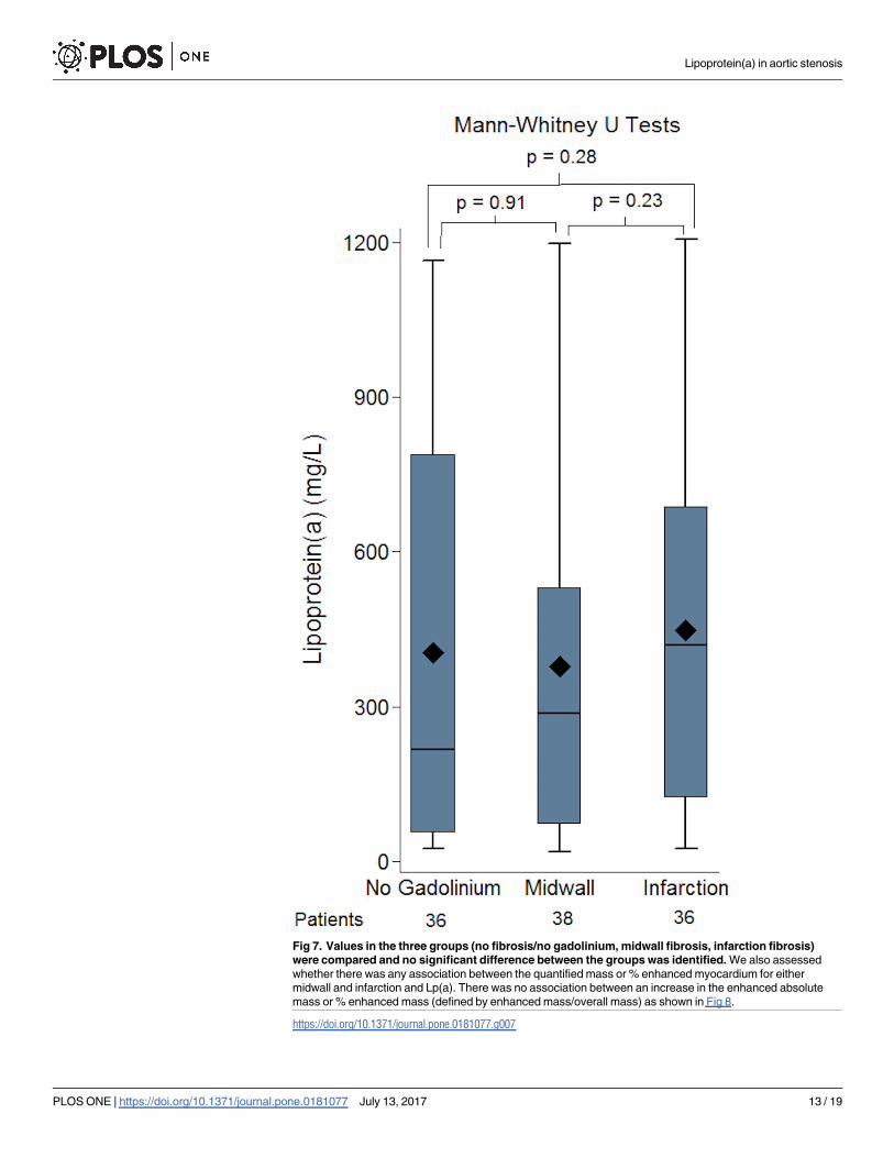
Fig 7. Values in the three groups (no fibrosis/no gadolinium, midwall fibrosis, infarction fibrosis)
were compared and no significant difference between the groups was identified. We also assessed
whether there was any association between the quantified mass or % enhanced myocardium for either
midwall and infarction and Lp(a). There was no association between an increase in the enhanced absolute
mass or % enhanced mass (defined by enhanced mass/overall mass) as shown in Fig 8.
https://doi.org/10.1371/journal.pone.0181077.g007
Lipoprotein(a) in aortic stenosis
PLOS ONE | https://doi.org/10.1371/journal.pone.0181077 July 13, 2017 13 / 19

evidence of pre-existing coronary artery disease (Mann-Whitney U test p = 0.61) or C-Reactive
Protein (Spearman correlation 0.16, p = 0.13).
Our intention in this manuscript was to investigate a potential mechanistic association
between Lp(a) and fibrosis. As such as our cohort included patients with moderate and severe
aortic stenosis who had medical or interventional therapy. Nonetheless, despite the heteroge-
neity of patients, it is of interest to review the impact of Lp(a) in outcomes. In our cohort 79
people (71.8%) died or had an aortic valve intervention over a median of 1.9 years (1.2–2.7
years). As Lp(a) has been shown to associate with worse outcome only if very high, we have
investigated whether the patients in the highest decile of Lp(a) had worse outcome, defined as
overall death or aortic valve intervention, compared to the rest of the cohort. Using Cox
Fig 8. investigating a potential association between quantified enhanced myocardial mass or % mass and Lp(a).
As shown in panel A, there was no association between enhanced mass and Lp(a) (p = 0.28). Reviewed separately there
was no association between Lp(a) and midwall fibrosis (p = 0.20) or infarction (p = 0.31). Similarly there was no
significance for enhanced % mass as shown in panel B.
https://doi.org/10.1371/journal.pone.0181077.g008
Table 4. Investigating the potential association between Lp(a) and post-operative new LBBB or need for pacemaker implantation in patients with
TAVI or AVR.
TAVI/AVR TAVI AVR
In those with intervention: n Lp(a) [Median (IQR)] p n Lp(a) [Median (IQR)] p n Lp(a) [Median (IQR)] p
No post-intervention LBBB 49 297 (72–721) 0.92 22 153 (63–558) 0.61 28 412 (86–866) 0.53
Post-Intervention LBBB 10 287 (86–577) 6 362 (35–1077) 4 258 (88–432)
TAVI/AVR TAVI AVR
In those with intervention: n Lp(a) [Median (IQR)] p n Lp(a) [Median (IQR)] p n Lp(a) [Median (IQR)] p
No post-intervention PPM 41 297 (86–838) 0.45 17 242 (70–628) 0.65 25 423 (86–858) 0.85
Post-Intervention PPM 15 156 (63–577) 10 151 (35–577) 5 401 (89–427)
TAVI/AVR TAVI AVR
In those with intervention: n Lp(a) [Median (IQR)] p n Lp(a) [Median (IQR)] p n Lp(a) [Median (IQR)] p
No post-intervention LBBB/PPM 38 275 (72–838) 0.72 16 196 (58–593) 0.88 23 423 (72–873) 0.71
Post-Intervention LBBB/PPM 18 245 (75–577) 11 156 (35–1063) 7 401 (86–436)
There was no association between Lp(a) and either post-operative LBBB or need for PPM in TAVI, AVR or the combination of the two.
https://doi.org/10.1371/journal.pone.0181077.t004
Lipoprotein(a) in aortic stenosis
PLOS ONE | https://doi.org/10.1371/journal.pone.0181077 July 13, 2017 14 / 19

proportional modeling we observed a trend towards worse survival or need for intervention in
those in the higher decile HR = 1.47 CI 0.76–2.85, p = 0.26, but this did not reach statistical
significance.
Discussion
There has been recent growing interest in the role of Lp(a) in aortic stenosis as Lp(a) has been
shown to be causally associated with increased calcification and the need for aortic valve
replacement [8,26]. Plasma Lp(a) level is mostly genetically determined by a variation in krin-
gle IV type 2 (KIV-2) repeat numbers at the LPA gene, which encodes for apolipoprotein(a)
[3]. Recently, genome wide association studies have identified more frequent genetic varia-
tions such as the SNP rs104555872 at the LPA gene in patients with aortic valve calcification
and stenosis and importantly, presence of such genetic variations in the LPA gene led to
increased levels of both Lp(a) [27] and clinical aortic stenosis, confirming the causal role of Lp
(a) [6]. Despite this breakthrough however, the mechanism by which Lp(a) might promote
this remains unclear. Seminal to this, it also remains to be seen whether Lp(a) associates with
increased likelihood of myocardial changes as reflected by increased myocardial fibrosis. In
addition, clinically what needs to be determined is whether lowering Lp(a) in patients with
mild/ moderate aortic stenosis might alter the rate of stenosis progression and subsequent
need for intervention. An initial pilot study is currently recruiting to investigate this, the Early
Aortic Valve Lipoprotein(a) Lowering Trial (EAVaLL) (ClinicalTrials.gov Identifier:
NCT02109614) [28] where patients with aortic sclerosis or mild stenosis are randomized to
niacin or placebo. The primary end-point is calcium score progression by cardiac CT in the
patients randomized to niacin vs. placebo at two years. Therefore, Lp(a) could provide a novel
therapeutic target in addressing this clinically unmet need. At the same time, presence of myo-
cardial fibrosis is an adverse predictor of survival and higher levels of Lp(a) could potentially
lead to increased or decreased myocardial fibrosis, depending on the dominant signalling
pathway.
This is the first report to explore the potential mechanistic role of Lp(a) in contributing to
left ventricular myocardial fibrosis and we observed no evidence to support an association
between Lp(a) and ventricular fibrosis. Moreover, in the patients with CMR evidence of myo-
cardial infarction, the Lp(a) level was not significantly different to the other groups, supporting
that Lp(a) mediated thrombosis is less likely to be implicated, perhaps as the Lp(a) is not very
high. Furthermore, it is also likely that the two opposing mechanisms influenced by Lp(a), one
promoting and one reducing fibrosis are running in parallel leading to an overall neutral effect.
This finding is reassuring as it suggests that although Lp(a) increases calcification and need for
intervention, it is not per se associated with the increased arrythmogenicity and mortality seen
in patients with myocardial fibrosis.
Moreover, we observed no association between the level of Lp(a) concentration and the
severity of aortic stenosis. Although this was an unexpected finding this lack of association
could suggest that Lp(a) might potentially have an initial effect in promoting early calcifica-
tion; however, once beyond initial stenosis the calcification pathway is independent of Lp(a)
hence explaining why in our study we observed no significant difference between mild/moder-
ate and severe.
Finally, although our study was not intended to investigate a potential association of Lp(a)
and outcomes in view of the heterogeneity of this cohort, nonetheless, we observed that the
patients in the highest decile of Lp(a) levels had an almost 50% higher risk of mortality or need
for intervention compared to the patients in the lowest decile, although this did not reach sta-
tistical significance.
Lipoprotein(a) in aortic stenosis
PLOS ONE | https://doi.org/10.1371/journal.pone.0181077 July 13, 2017 15 / 19

Study limitations
A limitation of our study is that it is from a single centre with a high proportion of Caucasian
patients. It remains to be shown whether these results could be extended to other races. Sec-
ondly, in aortic stenosis the myocardial subendocardial infarction pattern could relate to either
atherosclerotic coronary disease or embolic disease, therefore skewing the potential effect of
Lp(a). However, even when a history of documented CAD was adjusted for, there was no asso-
ciation of fibrosis with Lp(a) level. It is important to note however, that Lp(a) is only signifi-
cantly associated with increased risk of myocardial infarction when >500mg/L [3]. As the
majority of our patients had lower values this could explain the lack of association. Thirdly,
our cohort included 14 patients with bicuspid aortic valve and given that Lp(a) has a genetic
component this could have influenced the results. However, even when the 14 patients with
bicuspid aortic valve were excluded from the analysis, this did not influence the result out-
come. Fourthly, our study of 110 patients is the largest study to date comparing Lp(a) and
myocardial fibrosis assessed with CMR. Despite this, there were only 38 patients in the midwall
fibrosis group and 36 in the infarction pattern group. It is possible therefore that with higher
numbers a small association might have been observed between Lp(a) and fibrosis pattern. We
have estimated however, that using the currently available sample size and the standard devia-
tion of Lp(a), at a significance level of 0.05 there would be 80% power to detect a mean differ-
ence of 200 mg/L or greater between mild/moderate and severe groups. Combined with the
fact that we did observe significant difference between controls and patients with any degree of
aortic stenosis this would further support that our study was powered to detect clinically rele-
vant differences in Lp(a).
Likewise, our mild and moderate groups had low numbers which could have stopped us
from observing a small difference in Lp(a) level. A final limitation of our study is that we did
not have a comparison between Lp(a) and interstitial diffuse fibrosis as quantified by T1 map-
ping sequences as this might have provided further useful information. When we undertook
this study we did not have a validated T1 mapping sequence with appropriate quality assurance
to use. It was only subsequently that validated T1 mapping sequences with appropriate quality
assurance have been used in aortic stenosis [29][30][31]. Nonetheless, although the strong
prognostic role between midwall fibrosis and infarction pattern fibrosis in aortic stenosis has
been shown [32], it remains unclear whether T1 mapping could offer incremental benefit, and
the association of T1 mapping with Lp(a) remains to be studied.
Conclusion
In conclusion, our study has shown no association between Lp(a) and left ventricular midwall
or infarction pattern fibrosis when compared to patients with no fibrosis, therefore suggesting
that Lp(a) is unlikely to mediate fibrosis in patients with aortic stenosis. Additionally, patients
with mild/moderate and severe aortic stenosis have similar levels of Lp(a). The mechanistic
influence of Lp(a) in patients with aortic stenosis remains uncertain and future studies should
aim not only to identify this, but also establish whether a reduction in Lp(a) in the early stages
of mild aortic stenosis in the patients with very high Lp(a) levels, either using high dose niacin
[5], apheresis[33][34] or novel PKCS9 inhibitors[35] might improve stenosis progression and
outcomes.
Acknowledgments
The authors would like to acknowledge the assistance of the NIHR Biomedical research nurses
at the Royal Brompton Hospital for their support in recruiting patients for this study.
Lipoprotein(a) in aortic stenosis
PLOS ONE | https://doi.org/10.1371/journal.pone.0181077 July 13, 2017 16 / 19

Author Contributions
Conceptualization: Vassilios S. Vassiliou, Paul D. Flynn, Tina Khan, Anita Sri, Helen Berry,
Jackie Donovan, Dominique Auger, Stuart A. Cook, Dudley J. Pennell, Sanjay K. Prasad.
Data curation: Vassilios S. Vassiliou, Claire E. Raphael, Simon Newsome, Aamir Ali, Annina
Studer Bruengger, Tamir Malley, Pranev Sharma, Subothini Selvendran, Nikhil Aggarwal,
Anita Sri, Willis Lam, Dominique Auger.
Formal analysis: Simon Newsome, Aamir Ali, Brian Halliday, Annina Studer Bruengger,
Tamir Malley, Pranev Sharma, Subothini Selvendran, Nikhil Aggarwal, Anita Sri, Willis
Lam, Dominique Auger.
Funding acquisition: Vassilios S. Vassiliou, Dudley J. Pennell, Sanjay K. Prasad.
Investigation: Vassilios S. Vassiliou, Paul D. Flynn, Claire E. Raphael, Aamir Ali, Brian Halli-
day, Annina Studer Bruengger, Tamir Malley, Pranev Sharma, Subothini Selvendran,
Nikhil Aggarwal, Helen Berry, Jackie Donovan, Willis Lam, Sanjay K. Prasad.
Methodology: Vassilios S. Vassiliou, Paul D. Flynn, Tina Khan, Stuart A. Cook, Dudley J.
Pennell.
Project administration: Dudley J. Pennell.
Resources: Tina Khan, Dudley J. Pennell.
Software: Simon Newsome.
Supervision: Paul D. Flynn, Sanjay K. Prasad.
Validation: Brian Halliday.
Visualization: Paul D. Flynn, Claire E. Raphael, Sanjay K. Prasad.
Writing – original draft: Vassilios S. Vassiliou.
Writing – review & editing: Paul D. Flynn, Claire E. Raphael, Simon Newsome, Tina Khan,
Aamir Ali, Brian Halliday, Annina Studer Bruengger, Tamir Malley, Pranev Sharma, Sub-
othini Selvendran, Nikhil Aggarwal, Anita Sri, Helen Berry, Jackie Donovan, Willis Lam,
Dominique Auger, Stuart A. Cook, Dudley J. Pennell, Sanjay K. Prasad.
References1. Berg K. A new serum type system in man- the LP system. Acta Pathol Microbiol Scand. 1963; 59: 369–
82. PMID: 14064818
2. Kostner KM, Marz W, Kostner GM. When should we measure lipoprotein (a)? Eur Heart J. 2013; 34:
3268–76. https://doi.org/10.1093/eurheartj/eht053 PMID: 23735860
3. Nordestgaard BG, Chapman MJ, Ray K, Boren J, Andreotti F, Watts GF, et al. Lipoprotein(a) as a car-
diovascular risk factor: current status. Eur Heart J. 2010; 31: 2844–53. https://doi.org/10.1093/
eurheartj/ehq386 PMID: 20965889
4. Dube JB, Boffa MB, Hegele RA, Koschinsky ML. Lipoprotein(a): more interesting than ever after 50
years. Curr Opin Lipidol. 2012; 23: 133–40. https://doi.org/10.1097/MOL.0b013e32835111d8 PMID:
22327610
5. Reiner Z, Catapano AL, De Backer G, Graham I, Taskinen M-R, Wiklund O, et al. ESC/EAS Guidelines
for the management of dyslipidaemias: the Task Force for the management of dyslipidaemias of the
European Society of Cardiology (ESC) and the European Atherosclerosis Society (EAS). Eur Heart J.
2011; 32: 1769–818. https://doi.org/10.1093/eurheartj/ehr158 PMID: 21712404
6. Thanassoulis G, Campbell CY, Owens DS, Smith JG, Smith A V, Peloso GM, et al. Genetic associa-
tions with valvular calcification and aortic stenosis. N Engl J Med. 2013; 368: 503–12. https://doi.org/10.
1056/NEJMoa1109034 PMID: 23388002
Lipoprotein(a) in aortic stenosis
PLOS ONE | https://doi.org/10.1371/journal.pone.0181077 July 13, 2017 17 / 19

7. Kamstrup PR, Tybjærg-Hansen A, Nordestgaard BG. Elevated lipoprotein(a) and risk of aortic valve
stenosis in the general population. J Am Coll Cardiol. 2014; 63: 470–7. https://doi.org/10.1016/j.jacc.
2013.09.038 PMID: 24161338
8. Capoulade R, Chan KL, Yeang C, Mathieu P, Bosse Y, Dumesnil JG, et al. Oxidized Phospholipids,
Lipoprotein(a), and Progression of Calcific Aortic Valve Stenosis. J Am Coll Cardiol. 2015; 66: 1236–
1246. https://doi.org/10.1016/j.jacc.2015.07.020 PMID: 26361154
9. Simon DI, Fless GM, Scanu AM, Loscalzo J. Tissue-type plasminogen activator binds to and is inhibited
by surface-bound lipoprotein(a) and low-density lipoprotein. Biochemistry. 1991; 30: 6671–7. PMID:
1829635
10. Nordestgaard BG, Langsted A. How Does Elevated Lipoprotein(a) Cause Aortic Valve Stenosis? J Am
Coll Cardiol. 2015; 66: 1247–1249. https://doi.org/10.1016/j.jacc.2015.07.045 PMID: 26361155
11. Brown MS, Goldstein JL. Plasma lipoproteins: teaching old dogmas new tricks. Nature. 330: 113–4.
https://doi.org/10.1038/330113a0 PMID: 3670399
12. Bouchareb R, Mahmut A, Nsaibia MJ, Boulanger M-C, Dahou A, Lepine J-L, et al. Autotaxin Derived
From Lipoprotein(a) and Valve Interstitial Cells Promotes Inflammation and Mineralization of the Aortic
ValveCLINICAL PERSPECTIVE. Circulation. 2015; 132: 677–690. https://doi.org/10.1161/
CIRCULATIONAHA.115.016757 PMID: 26224810
13. Weidemann F, Herrmann S, Stork S, Niemann M, Frantz S, Lange V, et al. Impact of myocardial fibrosis
in patients with symptomatic severe aortic stenosis. Circulation. 2009; 120: 577–84. https://doi.org/10.
1161/CIRCULATIONAHA.108.847772 PMID: 19652094
14. Azevedo CF, Nigri M, Higuchi ML, Pomerantzeff PM, Spina GS, Sampaio RO, et al. Prognostic signifi-
cance of myocardial fibrosis quantification by histopathology and magnetic resonance imaging in
patients with severe aortic valve disease. J Am Coll Cardiol. Elsevier Inc.; 2010; 56: 278–87. https://doi.
org/10.1016/j.jacc.2009.12.074 PMID: 20633819
15. Dweck MR, Boon N a, Newby DE. Calcific aortic stenosis: a disease of the valve and the myocardium. J
Am Coll Cardiol. Elsevier Inc.; 2012; 60: 1854–63. https://doi.org/10.1016/j.jacc.2012.02.093 PMID:
23062541
16. Chin CW, Vassiliou V, Jenkins WS, Prasad SK, Newby DE, Dweck MR. Markers of left ventricular
decompensation in aortic stenosis. Expert Rev Cardiovasc Ther. 2014; 12: 901–12. https://doi.org/10.
1586/14779072.2014.923307 PMID: 24866867
17. Dweck MR, Joshi S, Murigu T, Alpendurada F, Jabbour A, Melina G, et al. Midwall fibrosis is an inde-
pendent predictor of mortality in patients with aortic stenosis. J Am Coll Cardiol. Elsevier Inc.; 2011; 58:
1271–9. https://doi.org/10.1016/j.jacc.2011.03.064 PMID: 21903062
18. Barone-Rochette G, Pierard S, De Meester de Ravenstein C, Seldrum S, Melchior J, Maes F, et al.
Prognostic Significance of LGE by CMR in Aortic Stenosis Patients Undergoing Valve Replacement. J
Am Coll Cardiol. 2014; 64: 144–154. https://doi.org/10.1016/j.jacc.2014.02.612 PMID: 25011718
19. Angles-Cano E, de la Peña Dıaz A, Loyau S. Inhibition of fibrinolysis by lipoprotein(a). Ann N Y Acad
Sci. 2001; 936: 261–75. PMID: 11460483
20. Angles-Cano E, Hervio L, Rouy D, Fournier C, Chapman JM, Laplaud M, et al. Effects of lipoprotein(a)
on the binding of plasminogen to fibrin and its activation by fibrin-bound tissue-type plasminogen activa-
tor. Chem Phys Lipids. 1994; 67-68: 369–80.
21. Grainger DJ, Kemp PR, Liu AC, Lawn RM, Metcalfe JC. Activation of transforming growth factor-beta is
inhibited in transgenic apolipoprotein(a) mice. Nature. 1994; 370: 460–2. https://doi.org/10.1038/
370460a0 PMID: 8047165
22. Teekakirikul P, Eminaga S, Toka O, Alcalai R, Wang L, Wakimoto H, et al. Cardiac fibrosis in mice with
hypertrophic cardiomyopathy is mediated by non-myocyte proliferation and requires Tgf-β. J Clin Invest.
2010; 120: 3520–9. https://doi.org/10.1172/JCI42028 PMID: 20811150
23. Nishimura R a, Otto CM, Bonow RO, Carabello B a, Erwin JP, Guyton R a, et al. 2014 AHA/ACC Guide-
line for the Management of Patients With Valvular Heart Disease: Executive Summary: A Report of the
American College of Cardiology/American Heart Association Task Force on Practice Guidelines. J Am
Coll Cardiol. 2014; 148: e1–e132. https://doi.org/10.1016/j.jacc.2014.02.537 PMID: 24603192
24. Gulati A, Jabbour A, Ismail TF, Guha K, Khwaja J, Raza S, et al. Association of fibrosis with mortality
and sudden cardiac death in patients with nonischemic dilated cardiomyopathy. JAMA. 2013; 309: 896–
908. https://doi.org/10.1001/jama.2013.1363 PMID: 23462786
25. Lippi G, Targher G. Optimal therapy for reduction of lipoprotein(a). J Clin Pharm Ther. 2012; 37: 1–3.
https://doi.org/10.1111/j.1365-2710.2011.01244.x PMID: 22276844
26. Hung M-Y, Tsimikas S. What is the ultimate test that lowering lipoprotein(a) is beneficial for cardiovas-
cular disease and aortic stenosis? Curr Opin Lipidol. 2014; 25: 423–30. https://doi.org/10.1097/MOL.
0000000000000131 PMID: 25340480
Lipoprotein(a) in aortic stenosis
PLOS ONE | https://doi.org/10.1371/journal.pone.0181077 July 13, 2017 18 / 19

27. Clarke R, Peden JF, Hopewell JC, Kyriakou T, Goel A, Heath SC, et al. Genetic variants associated
with Lp(a) lipoprotein level and coronary disease. N Engl J Med. 2009; 361: 2518–28. https://doi.org/10.
1056/NEJMoa0902604 PMID: 20032323
28. Kotar K, Thanassoulis G. Early Aortic Valve Lipoprotein(a) Lowering Trial (EAVaLL). In: ClinicalTrials.
gov. 2015 p. NCT02109614.
29. Lee S-P, Lee W, Lee JM, Park E-A, Kim H-K, Kim Y-J, et al. Assessment of diffuse myocardial fibrosis
by using MR imaging in asymptomatic patients with aortic stenosis. Radiology. 2015; 274: 359–69.
https://doi.org/10.1148/radiol.14141120 PMID: 25251584
30. Vassiliou VS, Heng EL, Gatehouse PD, Donovan J, Raphael CE, Giri S, et al. Magnetic resonance
imaging phantoms for quality-control of myocardial T1 and ECV mapping: specific formulation, long-
term stability and variation with heart rate and temperature. J Cardiovasc Magn Reson. 2016; 18: 62.
https://doi.org/10.1186/s12968-016-0275-9 PMID: 27659737
31. Vassiliou V, Wassilew K, Cameron D, Heng EL, Nyktari E, Asimakopoulos G, et al. Identification of myo-
cardial diffuse fibrosis by 11 heartbeat MOLLI T1 mapping: Averaging to improve precision and correla-
tion with collagen volume fraction. MAGMA. 2017; https://doi.org/10.1007/s10334-017-0630-3 (in
press). PMID: 28608326
32. Vassiliou VS, Perperoglou A, Raphael CE, Joshi S, Malley T, Everett R, et al. Midwall Fibrosis and 5-
Year Outcome In Patients With Moderate and Severe Aortic Stenosis. J Am Coll Cardio. 2017; 69:
1755–1756.
33. Khan TZ, Pottle A, Pennell DJ, Barbir MS. The impact of lipoprotein apheresis in patients with refractory
angina and raised lipoprotein(a): Objectives and methods of a randomised controlled trial. Atheroscler
Suppl. 2015; 18: 103–8. https://doi.org/10.1016/j.atherosclerosissup.2015.02.019 PMID: 25936312
34. Khan TZ, Hsu L-Y, Arai AE, Rhodes S, Pottle A, Wage R, et al. Apheresis as novel treatment for refrac-
tory angina with raised lipoprotein(a): a randomized controlled cross-over trial. Eur Heart J. 2017; 38:
1561–1569. https://doi.org/10.1093/eurheartj/ehx178 PMID: 28453721
35. Sabatine MS, Giugliano RP, Wiviott SD, Raal FJ, Blom DJ, Robinson J, et al. Efficacy and Safety of
Evolocumab in Reducing Lipids and Cardiovascular Events. N Engl J Med. 2015; 372:
150315080057008. https://doi.org/10.1056/NEJMoa1500858 PMID: 25773607
Lipoprotein(a) in aortic stenosis
PLOS ONE | https://doi.org/10.1371/journal.pone.0181077 July 13, 2017 19 / 19