VASCULARIZATION IN PAX 6+/+ AND PAX 6+/- MICE PETER … · EVALUATION OF CORNEAL HEALING, LIMBAL...
68
EVALUATION OF CORNEAL HEALING, LIMBAL PROGENITOR CELLS, AND VASCULARIZATION IN PAX 6 +/+ AND PAX 6 +/- MICE by PETER ACCOLA (Under the Direction of James D. Lauderdale and K. Paige Carmichael) ABSTRACT Heterozygous deficiency in the Pax6 gene results in the analogous condition of aniridia in humans and small eye (Sey) in mice. The underlying pathogenesis is incompletely understood. An in vivo, murine, corneal wounding model was developed to study healing capacity, evaluate the limbal progenitor cell population (p63), and explore the role of soluble vascular endothelial growth factor receptor-1 (sVEGFR-1) in aniridia related keratopathy. Results demonstrated a statistically significant delay in corneal wound healing in Sey mice at days 2 and 3 when compared to WT mice (p<0.05). There was no significant difference in corneal p63 staining (p>0.05). All corneas exhibited comparable sVEGFR-1 staining. In conclusion, our in vivo wounding model revealed delayed corneal healing in Sey mice that does not appear to be due to deficiency in p63 cellular expression. The comparable expression of sVEGFR-1 suggests that it alone is likely not responsible for corneal vascularization present in Sey mice. INDEX WORDS: Aniridia related keratopathy, Pax6, Cornea, Aniridia, in vivo, p63, sVEGFR-1, Wound
Transcript of VASCULARIZATION IN PAX 6+/+ AND PAX 6+/- MICE PETER … · EVALUATION OF CORNEAL HEALING, LIMBAL...

EVALUATION OF CORNEAL HEALING, LIMBAL PROGENITOR CELLS, AND
VASCULARIZATION IN PAX 6+/+ AND PAX 6+/- MICE
by
PETER ACCOLA
(Under the Direction of James D. Lauderdale and K. Paige Carmichael)
ABSTRACT
Heterozygous deficiency in the Pax6 gene results in the analogous condition of
aniridia in humans and small eye (Sey) in mice. The underlying pathogenesis is
incompletely understood. An in vivo, murine, corneal wounding model was developed to
study healing capacity, evaluate the limbal progenitor cell population (p63), and explore
the role of soluble vascular endothelial growth factor receptor-1 (sVEGFR-1) in aniridia
related keratopathy. Results demonstrated a statistically significant delay in corneal
wound healing in Sey mice at days 2 and 3 when compared to WT mice (p<0.05). There
was no significant difference in corneal p63 staining (p>0.05). All corneas exhibited
comparable sVEGFR-1 staining. In conclusion, our in vivo wounding model revealed
delayed corneal healing in Sey mice that does not appear to be due to deficiency in p63
cellular expression. The comparable expression of sVEGFR-1 suggests that it alone is
likely not responsible for corneal vascularization present in Sey mice.
INDEX WORDS: Aniridia related keratopathy, Pax6, Cornea, Aniridia, in vivo, p63,
sVEGFR-1, Wound

EVALUATION OF CORNEAL HEALING, LIMBAL PROGENITOR CELLS, AND
VASCULARIZATION IN PAX 6+/+ AND PAX 6+/- MICE
by
PETER ACCOLA
B.S., University of Wisconsin – River Falls, 1997
D.V.M., University of Wisconsin – Madison, 2001
A Thesis Submitted to the Graduate Faculty of The University of Georgia in Partial
Fulfillment of the Requirements for the Degree
MASTER OF SCIENCE
ATHENS, GEORGIA
2009

© 2009
Peter Accola
All Rights Reserved

EVALUATION OF CORNEAL HEALING, LIMBAL PROGENITOR CELLS, AND
VASCULARIZATION IN PAX 6+/+ AND PAX 6+/- MICE
by
PETER ACCOLA
Major Professors: James D. Lauderdale K. Paige Carmichael Committee: Phillip A. Moore Angela E. Ellis Electronic Version Approved: Maureen Grasso Dean of the Graduate School The University of Georgia December 2009

iv
DEDICATION
This work is dedicated to my beautiful wife and our family to come.

v
ACKNOWLEDGEMENTS
I would like to thank my wonderful committee members for their encouragement
and dedication. Your direct influence is inextricably linked to this project. I also am
grateful to Abbie Butler, Lynn Reece, Nadia Gadsden, and Rob Miller for their technical
contributions. I thank my wife, Laurie, for her unwavering support and valuable
discussion. Finally I wish to recognize my parents, James and Ida Accola. The
foundation you provided guides me daily.

vi
TABLE OF CONTENTS
Page
ACKNOWLEDGEMENTS .................................................................................................v
CHAPTER
1 INTRODUCTION AND LITERATURE REVIEW ........................................1
2 ANTERIOR SEGMENT PATHOLOGY AND CORNEAL WOUND
HEALING IN PAX6 HETEROZYGOTE MICE ............................................5
INTRODUCTION AND OBJECTIVE ......................................................5
MATERIALS AND METHODS ................................................................6
RESULTS .................................................................................................11
DISCUSSION ...........................................................................................15
REFERENCES .........................................................................................21
FIGURES ..................................................................................................34
3 CONCLUSION ...............................................................................................61

1
CHAPTER 1
INTRODUCTION and LITERATURE REVIEW
Ocular development depends on an organized interaction of tissues from different
embryologic origins. This process proceeds in a predictable and stepwise fashion under
the control of various transcription factors and induction signals which direct coordinated
differentiation of all ocular structures [1]. Alterations in the delicate balance of these
controlling factors results in abnormal ocular development.
The Pax multigene family is a group of transcription factors that are responsible
for the development of numerous tissues [2]. Pax6, in addition to its contributions to
brain, olfactory, and pancreatic formation, plays a principal role in ocular development
[3-16]. This gene has been highly conserved in species throughout evolution. A
heterozygous loss-of-function mutation in the Pax6 gene results in a congenital ocular
condition in humans termed aniridia and a Small Eye (Sey) phenotype in rodents. These
two conditions are widely considered homologous given their genotypic and phenotypic
similarities [6, 11, 17-35]. In addition to its importance in the developing embryo, Pax6
also plays a regulatory role within the eye postnatally [29, 36, 37].
Aniridia is a panocular developmental disorder that affects the cornea, anterior
chamber, iris, lens, retina, and optic nerve [38-41]. Aniridia related keratopathy (ARK)
denotes the complex pathology observed in the cornea of both humans with aniridia and
in Sey mice. In humans, the cornea develops a peripheral keratopathy which advances
towards the central cornea with age [42]. The progressive nature of ARK results in

2
devastating consequences for vision in these patients. Despite important advances in the
understanding of its pathophysiology, the cause of ARK remains elusive [35].
Deficiency in the limbal stem cell population has been proposed as a cause based on,
among other things, corneal conjunctivalization [38, 43-47]; however evaluation of cell
proliferation and epithelial progenitor cell markers suggest an adequate stem cell
population [48]. The prominent corneal neovascularization is thought to be due to
deficiency in a soluble receptor for vascular endothelial growth factor A (VEGF-A) in the
cornea called soluble VEGF receptor-1 (sVEGFR-1, also known as sflt-1) [49-51].
Additional proposed mechanisms of ARK include abnormal corneal wound healing
characterized by an increased epithelial cell fragility secondary to abnormal expression of
cytokeratin 12 [48, 52-54], glycosaminoglycan down regulation with resultant deleterious
effects on epithelial cell migration [55], defective calcium signaling in Pax6+/- cells [56],
and abnormal remodeling of the corneal extracellular matrix [57]. Furthermore, a
positive correlation has been made between the severity of ARK and abnormal tear film
stability and meibomian gland dysfunction [58]. More recently, an increased
susceptibility of the Sey cornea to oxidative stress was shown to result in metaplastic
epithelial changes and opacity [59]. Finally, a study of corneal innervation in Sey mice
did not support neurogenic keratitis as a cause for ARK [60]. It is clear, therefore, that
the underlying mechanism of ARK is multifactorial.
Current treatment options for ARK include penetrating keratoplasty and
keratolimbal grafting techniques; however success rates have generally been poor with
recurrent keratopathy in grafted tissue [12, 13, 61-64]. Protection of the cornea with long
term bandage contact lens [65] or surface lubricants [66] may improve outcome.
3
Identification of the mechanisms underlying ARK will be important in determining
effective future treatments. Ultimately, techniques employing transplantation of
genetically repaired Pax6 stem cells may be necessary for definitive treatment [67, 68].
The corneal abnormalities seen in Sey mice provide a useful model for study of ARK in
humans [6, 53].
The vast majority of studies evaluating corneal biology in ARK have employed in
vitro murine models in which wounding is performed in epithelial cell culture, or in
which wounding is followed by enucleation and placement of the eye in organ culture.
However, these studies have resulted in discordant results of corneal wound healing
capacity in Sey mice [55, 69, 70]. This is likely because in vitro studies cannot provide
the complex cues that in vivo cells must integrate. Corneal wounding initiates a series of
coordinated events that serve to maintain its refractive and protective functions. Optimal
healing involves interaction of not only the resident cells of the cornea and immune
system, but contributions from lacrimal glands, conjunctiva, meibomian glands, eyelids,
and the afferent and efferent nervous system. This integrated concept of the ocular
surface as a functional unit highlights the complexities inherent to corneal wound healing
[71, 72]. An in vivo study would incorporate the necessary components important in
corneal wound healing and thus identify a basal healing capacity in Sey mice from which
further work can be compared.
Thus, the objective of this study is to establish and evaluate a prospective, in vivo
corneal wounding model in Sey mice to aid in future study of the pathophysiology and
potential treatment of ARK. This model will then be used to compare corneal wound
healing characteristics in Wild Type (WT) and Sey mice, evaluate changes over time in

4
the limbal progenitor cell population (p63) after wounding, and explore the role of
sVEGFR-1 in the vascularization of ARK.

5
CHAPTER 2
ANTERIOR SEGMENT PATHOLOGY AND CORNEAL WOUND HEALING
IN PAX6 HETEROZYGOTE MICE
1. Introduction and objective
Corneal wound healing involves a complex interaction of all required components
in order to achieve the exquisite clarity which is necessary for normal vision.
Heterozygous deficiency in Pax6, a gene important to the development and postnatal
function of the eye, results in the analogous condition of aniridia in humans and small eye
(Sey) in mice. Humans with aniridia suffer from progressive corneal pathology termed
aniridia related keratopathy (ARK) for which the Sey mouse is a model. The underlying
mechanisms of ARK are unclear, but may include deficiency in the limbal progenitor cell
population and disturbance of factors involved in maintaining corneal avascularity. The
hypothesis of this study is that the Pax6 heterozygote genotype will result in defective
corneal wound healing, a deficiency in limbal progenitor cells, and impairment of corneal
anti-angiogenesis. The objective of this study is twofold. First, we will establish a
prospective, in vivo corneal wounding model in Sey mice for future study of the
pathophysiology, and potential treatment, of ARK. Second, this model will be used to
determine corneal wound healing capacity in Wild Type (WT) and Sey mice, compare
their limbal progenitor cell population (p63), and explore the role of a VEGF-A receptor
(sVEGFR-1) in the vascularization of ARK.
6
2. Materials and Methods
2.1. Experimental Animals
A total of 79 Pax6+/+ (CD1; Charles River Laboratories) and 40 SeyNeu (Pax6+/-)
mice on CD1 background (J.D. Lauderdale murine breeding colony, Department of
Cellular Biology, University of Georgia, Athens, GA) between the ages of 8 and 12
weeks were included in this study. Heterozygous Pax6+/- mice were produced from
crosses between WT (CD1 x CD1) F1 females and Pax6+/Sey-Neu males on a CD1
background. Housing consisted of mice separated by sex in cages within a controlled
room temperature (20˚C - 22˚C), humidity (33-48%), and a constant 12 hour light – dark
cycle. Mice had access to free choice water and were fed Laboratory Rat Diet 5001
(LabDiet®, Richmond, Indiana). Animals in this study were handled in accordance with
the regulations in the ARVO Statement for the Use of Animals in Ophthalmic and Vision
Research and the National Institutes of Health Guide for the Care and Use of Laboratory
Animals. This study was approved by The University of Georgia Institutional Animal
Care and Use Committee.
2.2. Genotype confirmation
Tail biopsy samples from all WT and Sey mice were placed in individual tubes
with tail lysis buffer (100mM Tris HCl pH 8.5, 5mM EDTA, 0.2% SDS, and 200mM
NaCl) and 100µg Proteinase K/mL. Following complete lysis, the tubes were vortexed
and centrifuged @ 13 600 rcf for 10 minutes. The supernatant was mixed with 500µL
isopropanol to induce genomic DNA precipitation. After centrifugation (5 min. @ 11
333 rcf), the supernatant was removed and the DNA was allowed to dry briefly at RT.

7
The DNA was then dissolved in 100µL Tris EDTA (10mM Tris HCl, 1mM EDTA; pH
7.5) and incubated at 37˚C overnight.
Genotyping was performed similar to a previously described method [23, 37].
Briefly, 3µL of genomic DNA was added to a 22µL mixture of dNTP (200 µM each
dNTP, Fisher Scientific), PCR buffer with 1.5 mM Mg2+ (Roche Diagnostics), Pax6
forward (5' GAG GAA CCA GAG AAG ACA GGC 3') and reverse (5' GCA TAG GCA
GGT TGT TTG CC 3') primer (0.2µM each), Taq DNA polymerase (1.25 U per reaction,
Sigma), and molecular grade, distilled, and deionized water. For PCR, samples were
placed in a PTC-100 Programmable Thermal Controller (MJ Research Inc.) for the
following: 5min @ 95˚C; 40 cycles of [1min. @ 95˚C, 1min. @ 60˚C, 2min. @ 72˚C]; 7
min. @ 72˚C. A 5µL mixture of 10x Buffer (Buffer 3; New England Biolabs) and HincII
restriction enzyme (New England Biolabs) was added to the PCR sample and tubes were
incubated at 37˚C overnight. Ethidium bromide stained PCR products were fractionated
on a 2% agarose gel.
2.3. In vivo corneal epithelial wounding
A pilot study using WT mice (n=21) was performed to develop the corneal
wounding protocol. Ophthalmic examination of the WT mice using slit lamp
biomicroscopy and fluorescein stain was normal. The mice were anesthetized with
intraperitoneal ketamine hydrochloride (100mg/kg) and xylazine hydrochloride
(10mg/kg). Wounding of the central cornea was performed in the left eye under 2.5x
magnification by applying for 1 minute a sterile, 1.75mm disk of single-ply tissue
(Kimwipe®) saturated in 2µL of n-heptanol [68, 73]. An identical procedure was
repeated on the right eye using physiologic saline instead of n-heptanol. Buprenorphine

8
hydrochloride (0.05-0.01mg/kg SQ) was administered pre- and post-operatively every 6-
12 hours as needed for analgesia. Mice were evaluated on days 1, 2, 4, 7, 14, and 28 post
wounding. Ophthalmic examination included slit lamp biomicroscopy (SL-15; Kowa
Optimed, Inc.; Torrance, CA), application of fluorescein stain (Fluor-I-Strip-A.T.;
Bausch & Lomb Pharmaceuticals, Inc.; Tampa, FL) to identify the area of epithelial loss
(ulcer), and digital photography (Nikon D-100; Melville, NY) of both eyes in all mice. A
total of 3-5 randomly selected mice were euthanized (CO2 and cervical dislocation) on
each examination day.
In the wound healing comparison phase, WT (n=58) and Sey (n=40) mice were
wounded using the protocol established in the pilot study, except no disk was applied to
the right eye. To account for difference in corneal size between groups, a 1.75mm disk
was used for WT and a 1.2mm disk for Sey. The 1.2mm diameter disk size was
determined by comparing numerous disk sizes from 0.75 – 1.5mm and estimating the
amount of cornea wounded for the average Sey cornea. The 1.2mm diameter disk was
chosen as it most closely approximated the amount of cornea wounded for WT mice.
Mice were evaluated on days 1, 2, 3 (if fluorescein positive on day 2), 4, 7, 14, and 28
post wounding. A total of 3-5 mice were euthanized (CO2 and cervical dislocation) on
each examination day.
2.4. Comparison of corneal wound healing
Digital photographs of the cornea were taken immediately post wounding and
fluorescein staining (WT=43; Sey=29). The total corneal area and the area of the corneal
wound on day 0 was determined for each mouse by evaluation of digital photographs
using image analysis software (ImageJ [74]) on a Power Macintosh G4 computer running

9
OSX (Fig. 1). The number of days to negative fluorescein staining (wound healing) was
recorded for each mouse. Mice were examined as indicated above until complete corneal
wound healing.
2.5. Histology
Immediately after euthanasia, both eyes from each mouse (WT=79; Sey=40) were
immersed in 4% paraformaldehyde and stored at 4˚C. Globes were measured
(Castroviejo calipers) at the horizontal equator using 2.5x magnification and then
processed and embedded in paraffin wax. For routine microscopy, vertical sections one
quarter of the way into the globe were made based on prior measurements. Four micron
sections were then deparaffinized in two changes of xylene, rehydrated in graded ethanol
(100%, 100%, 95%, 95%, and 70%), and rinsed in water. Application of Gill II
Hematoxylin (Surgipath™, Richmond, Il.), 95% ethanol, and Eosin (Surgipath™,
Richmond, Il.) was then followed by 4 changes of 100% ethanol, 1 change of
acetone/xylene, and 3 changes of xylene. Samples were mounted on a slide and a
coverslip applied using PermaMount (Vector Laboratories). Corneal morphology of non-
wounded and wounded WT and Sey mice was described and compared.
2.6. Molecular Marker Analysis
Sections of each eye were deparaffinized, rehydrated, and incubated with 3%
hydrogen peroxide for 5 minutes to quench endogenous peroxidase activity. The number
of mice evaluated for p63 staining is as follows: day 1 WT/Sey = 3/4; day 2 WT/Sey =
4/4; day 4 WT/Sey = 8/5; day 28 WT/Sey = 7/4, respectively. For p63 immunostaining,
monoclonal anti-p63 primary antibody derived from the 4A4 hybridoma in BALB/c mice
raised against mouse ΔNp63 (Sigma, St. Louis, MO; stock diluted 1:500) was used. As

10
the primary antibody was made in a mouse and being used on mouse tissue, a MOM kit
(Vector Laboratories, Burlingame, CA) which is specifically designed to decrease
background was used following manufacturer’s directions. The primary antibody was
applied for 30 minutes. The non-wounded corneas of WT (n=46) and Sey (n=24) mice
were evaluated for expression of sVEGFR-1. For sVEGFR-1 immunostaining, Power
Block™ Universal Blocking Reagent (BioGenex, San Ramon, CA) was applied for 5
minutes to block non-specific binding of antibody. Membrane bound VEGFR-1 antibody
was used (R&D Systems, Minneapolis, MN; rat monoclonal; stock diluted 1:100).
Samples were incubated with the primary antibody for 60 minutes, followed by 10
minutes with biotinylated anti-rat IgG (Vector Laboratories, Burlingame, CA) and 10
minutes with LSAB2 Streptavidin-HRP (Dako, Carpinteria, CA). For both
immunostains, incubation with 3,3'-diaminobenzidine (DAB; Dako, Carpinteria, CA) was
performed to localize peroxidase activity. A Dako Autostainer Plus Universal Staining
System (Dako, Carpinteria, CA) was used for all immunostaining. Counterstaining with
Gills II hematoxylin (Surgipath™, Richmond, Il.) was performed followed by graded
ethanol dehydration (70%, 95%, 95%, 100%, 100%) and 2 changes of xylene. Sections
were mounted on a slide and a coverslip applied. Positive controls (mouse kidney) for
both antibodies were performed at the same time under the same conditions as the sample
tissue. Two negative controls (PBS and normal mouse serum for p63; PBS and normal
rat serum for sVEGFR-1) were performed by incubating the tissue (mouse kidney) with
the negative control instead of the primary antibody under the same conditions.
For analysis of p63 expression, the corneal epithelium was divided into three
sections. First, the limbal epithelium was identified based on its location adjacent to the

11
base of the iris, as well as cellular and underlying stromal morphology characterized by a
thinner epithelial layer (2-4 cell layers thick) with a loose, well vascularized underlying
stroma and lack of goblet cells [75, 76]. The peripheral and central regions were defined
by counting the remaining basal epithelial cells and dividing in half. All basal epithelial
cells from the limbal and peripheral regions were scored as either positive or negative.
The central region was not counted due to wounding. The expression of sVEGFR-1 was
scored as either positive or negative and anatomic localization was recorded.
2.7. Statistical Analysis
All analyses were performed using SAS V 9.2 (Cary, NC). The proportion of the
cornea wounded was compared between groups using a Student’s t-test. The number of
days to negative fluorescein staining was compared using the Chi-Square test. The
number of p63 staining basal epithelial cells was recorded and compared between time
points and groups for each section of cornea (limbal and peripheral) by ANOVA.
3. Results
3.1. Ophthalmic examination and histopathologic findings
On evaluation prior to wounding, slit lamp biomicroscopy of the anterior segment
of all WT mice was normal (Fig’s. 2, 3a). Examination of Sey mice revealed variation in
phenotype, however most expressed corneal opacity, neovascularization, iris hypoplasia,
and anterior cortical cataract (Fig’s. 2, 4a).
Histology of the non-wounded WT cornea (Fig. 3b) revealed a basal layer in
which the epithelial cells varied in shape from cuboidal to short columnar to polygonal
and contained dark to variably basophilic round to oval nuclei and lightly eosinophilic
cytoplasm. As epithelial cells migrated superficially, nuclei became more oval and then

12
horizontally elongated, while the cytoplasm became more darkly eosinophilic. The
epithelial layer was 5-7 cells thick centrally and 3-4 cells thick peripherally and did not
rest upon a clearly defined basal lamina. The corneal stroma consisted of organized,
tightly arranged collagen fibers. Evenly distributed within the collagen were keratocytes
with fusiform to polygonal cell nuclei and indistinct cell borders. The endothelial layer
consisted of a single layer of cuboidal shaped cells with basophilic nuclei and a clear
vacuolated to eosinophilic cytoplasm. Separating the endothelial layer and the deep
stroma was a faint, deeply eosinophilic, hyaline membrane (Descemet’s membrane).
Comparatively, histopathology of the non-wounded Sey cornea was markedly
different (Fig. 4). The central epithelium was typically only 3-4 cells (rarely 1-2) thick
while the periphery was usually 1-3 cells thick. The epithelial layer of most Sey mice
(31/39) contained a variable number of epithelial cells with a large, lightly basophilic
cytoplasmic vacuole. These vacuoles were consistent with mucin on H&E and this was
confirmed with PAS staining (Fig. 5). The cuboidal basal epithelial cells contained
basophilic nuclei in which the chromatin was intermittently dispersed and a lightly
eosinophilic cytoplasm with tiny clear vacuoles. There was no discernible underlying
basal lamina even with PAS staining. The more superficial epithelial cells had nuclei that
were more basophilic and cigar shaped with a more hypereosinophilic cytoplasm. In
general, the corneal (stromal) thickness in the Sey was greater than that of WT. The
anterior stroma typically contained densely packed and disorganized collagenous lamella.
The amount of collagenous stroma appears to be increased over that seen in the WT. In
all samples with sufficient cornea for evaluation, the peripheral to mid anterior stroma
contained small caliber blood vessels (39/39). Associated with the vascularization was

13
mild to moderate corneal edema. Additional histopathologic findings included iris
hypoplasia (Fig’s. 6, 7), anterior cortical cataract (Fig. 8), and anterior lens dislocation
(Fig. 9). All Sey had iris hypoplasia of varying degrees. While anterior cortical cataract
was present in some mice, accurate quantification of the actual number with this lesion
was difficult due to the relatively small size of the lesion. A single mouse had lens
dislocation.
Corneal wounding with n-heptanol removed the epithelial layer in all mice (Fig.
10). Corneal histopathology post wounding in both groups was similar with a few minor
differences. In addition to those findings previously described in non wounded corneas,
there was mild to moderate, anterior stromal, purulent keratitis characterized by
infiltrations of neutrophils with rare lymphocytes and plasma cells. Anterior stromal
neovascularization and focal superficial ulceration was present in eyes that were
fluorescein positive at enucleation. In some cases, there appeared to be a more prominent
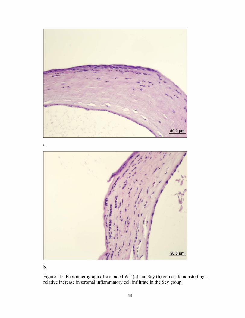
inflammatory cell response in the corneal stroma of Sey mice (Fig. 11), however overall
the variation in inflammatory cell infiltrate did not appear to differ dramatically enough
to adequately quantify.
Typical findings in ulcerated areas of both wounded WT and Sey eyes included a
focal area of stroma that was devoid of an overlying epithelial layer. The epithelium
adjacent to the ulcer was usually one cell layer thick and progressively increased in cell
thickness further from the ulcerated site. Many of the basal epithelial cells adjacent to the
ulcer and at the limbus contained clear cytoplasmic vacuoles, consistent with intracellular
edema. The mid to anterior corneal stroma contained an infiltration of blood vessels and

14
mature neutrophils. There was no difference in the number of goblet cells in wounded
vs. non-wounded Sey corneas.
3.2. In vivo corneal wound healing
The genotype for all mice was confirmed (Fig. 12). The pilot study allowed time
to perfect the wounding technique. All left eyes revealed axial corneal ulceration (n=21).
A single right eye sustained a pinpoint paraxial ulcer that healed uneventfully by the next
day’s examination. In the comparison phase, the proportion of cornea wounded between
WT and Sey mice was not significantly different (WT=56.3% +/- 13.5% vs. Sey=52.0%
+/- 12.2%; p=0.1585). Time to corneal wound healing is summarized in Fig. 13. The
wounded cornea of all mice was fluorescein positive on day 0. On days 1 and 4, there
was no significant difference in number of mice that were fluorescein positive
(p=0.2201). However, the proportion of eyes that were fluorescein positive was
significantly higher in the Sey group than the WT group on days 2 (p=0.0187) and 3
(p=0.0312). All corneal ulcers were completely healed (fluorescein negative) by day 7.
3.3. Expression of p63 and sVEGFR-1
All samples were strongly positive for p63 in the nuclei of limbal and peripheral
basal corneal epithelium. Statistical evaluation revealed no significant difference in basal
epithelial p63 expression between WT and Sey mice or time points for any section of
cornea for either eye (p>0.05). There was no staining within the more superficial
epithelium or corneal stroma (Fig’s. 14-20) in all WT and most Sey. Occasionally, Sey
mice (29%) had epithelial nuclei superficial to the basal layer which stained positive for
p63.

15
Expression of sVEGFR-1 in the cornea was comparable in all WT and Sey mice
(Fig’s. 21-25). In both non-wounded groups there was prominent basal staining within
the perilimbal cornea localizing most prominently within the cytoplasm of stromal
keratocytes and endothelium (posterior epithelium) and within the extracellular space of
the associated corneal stroma. Rarely, there was light staining within the cytoplasm of
adjacent epithelial (anterior) cells. In WT mice, the central cornea was devoid of staining
(Fig 26). However, in the Sey group, a total of 8 (33%) mice had central to peripheral
corneal staining (Fig 27).
4. Discussion
4.1. Anterior segment evaluation
Ophthalmic examination using a slit lamp biomicroscope allows for thorough
analysis of the anterior segment. These findings are helpful in correlating observed gross
changes with microscopic pathology. This is the first detailed slit lamp examination of
SeyNeu mice performed to indicate the prevalence of specific anterior segment pathology.
Corneal opacity was common and associated with vascularization. Interestingly, while
corneal vascularization was clinically detected in only 50% (20/40) of Sey mice at initial
exam, histopathologically 100% (39/39) of the examined corneas were vascularized.
This is likely secondary to the very fine, small caliber vessels in some mice and indicates
an underlying disturbance in the mechanism(s) responsible for maintenance of corneal
avascularity even in grossly avascular eyes. The relatively thin corneal epithelial layer,
dense and disorganized stromal collagen, and basophilic cytoplasmic vacuole within the
epithelial cells (PAS positive goblet cell) are similar to previous findings [53]. The thin
epithelial layer has been suggested to be secondary to increased shearing of squamous

16
cells from the corneal surface due to impaired cytokeratin expression. This morphology
may also be secondary to decreased levels of Pax6 resulting in abnormal epithelial
development [48]. The increased corneal thickness in Sey is likely secondary to stromal
edema, neovascularization and increased collagen production, however the underlying
mechanism of this pathology is not understood. Iris hypoplasia was identified with
biomicroscopy and confirmed with histopathology.
While there is variation in the severity of cataract and portion of lens affected, the
anterior cortical cataract observed clinically appears microscopically as an accumulation
of disorganized and proliferating anterior lens epithelial cells with an intact overlying
lens capsule. The disorganized anterior epithelial cells appear to be secreting a PAS
positive material (Fig 8b). This may represent a continued attempt at lens capsule
production. Given the gross and microscopic findings, this pathology may represent a
defective detachment of the lens vesicle from the overlying surface ectoderm during
development. To the authors’ knowledge, this cataract has not been described in the Sey
mouse.
A single Sey mouse expressed unique lenticular pathology (Fig. 9). Clinically,
the lens was within the anterior chamber and there was corneal opacity which was most
prominent centrally. Microscopically, there was a relative thickening and fibrovascular
pannus with edema associated with the central corneal stroma. The anterior lens capsule
was complete and in contact with the posterior cornea. There was disorganization and
proliferation of the anterior lens epithelium similar to cataractous changes in other Sey
mice. One potential mechanism for this pathology is incomplete separation during
embryology, as suggested above. Alternatively, this may represent an anterior lens

17
luxation secondary to hypoplasia of the anterior uvea (ciliary body and lens) and
malformation of the suspensory apparatus (zonules) necessary for maintaining the lens in
normal anatomic position. Further study to characterize the lens and anterior uvea /
zonules will be important in understanding the lenticular pathology inherent to Pax6
deficiency.
Finally, there was an increase in inflammatory cell infiltrate in the wounded Sey
eyes compared to the WT eyes at the same time post wounding. This has been previously
reported [70]. This phenomenon may be secondary to an increased stimulus for
infiltration or a perturbation of mechanisms inherent to its regulation.
4.2. In vivo corneal wound healing
In vivo corneal wounding with n-heptanol as described was a valid technique to
provide evaluation of wound healing ability in the mice of this study. The corneal
epithelium was consistently removed without histopathologic evidence of stromal loss.
Options for corneal wounding are numerous and include n-heptanol, sodium hydroxide,
and mechanical removal. Sodium hydroxide can induce dramatic anterior uveitis
(including hyphema and hypopyon) in the murine species [77]. Mechanical removal,
even under an operating microscope, has been shown to be inaccurate and imprecise in
the rabbit model [73]. Thus, in the significantly smaller eye of the mouse, it is not
reasonable to assume that the amount of cornea wounded by mechanical removal will be
consistent enough for valid comparisons of wound healing rates. Application of n-
heptanol has been shown to be a fast, accurate, and reproducible technique for consistent
removal of corneal epithelium [73]. The vast majority of mice in this study did not
exhibit outward signs of pain (blepharospasm, epiphora, rubbing, or withdrawal from the

18
group) the day after wounding and there was no clinical or histologic evidence of
infectious keratitis. Topical ophthalmic anti-inflammatory or antibiotic medication was
not used in this study due to their potential to inhibit wound healing [78, 79]. This would
have complicated interpretation of wound healing data.
In the in vivo corneal wounding model reported here, Sey mice exhibited a
statistically significant delay in corneal healing compared to WT mice at days 2 and 3
post wounding. This delay may in part be secondary to depletion in the cellular
machinery required for wound healing or a deficiency in limbal stem cells. Overall, the
difference between wounding times were not as dramatic as expected. The majority of
WT and Sey mice healed within the first 48 hours, which suggests that there is not a
dramatic difference in wound healing ability between these two groups of mice after a
single wounding. This would be consistent with the progressive corneal pathology in
humans with ARK and Sey mice from the cumulative effects of a Pax6 deficiency over
time. Chronic corneal microtrauma or ulceration with concomitant abnormal healing in
Pax6 deficient humans and mice may lead to epithelial and stromal opacity. This
wounding model provides a baseline to further study the pathophysiology of ARK.
4.3. Evaluation of limbal progenitor cells
In this study we evaluated p63, a marker of the proliferative cell pool of the
corneal epithelium, to determine if these cells, in an in vivo model of corneal wounding,
are depleted or become depleted over time. The corneal epithelial stem cells are widely
accepted to reside within the basal layer of the corneoscleral limbus. Support for this
location is based on its unique presence of slow-cycling cells (label-retaining cells) [75,
76, 80-82], primitive differentiation compared to the rest of the cornea [83-89], capacity

19
for unlimited self renewal [90, 91], high proliferative potential after activation by
wounding [75, 92, 93], morphologic criteria [94], and abnormal corneal wound repair
after removal of the limbal epithelium [95-97]. To date, a cell marker has not been
discovered that definitively labels corneal stem cells. This study revealed no significant
difference in p63 expression in the basal layer of the corneal epithelium of either eye
between WT and Sey mice at any time point before or after wounding. These findings
suggest that the delay in corneal healing does not appear to be due to depletion in p63
cellular expression over time, which is consistent with previous studies [48]. Expression
of p63 within the nuclei of epithelial cells superficial to the basal layer in Sey mice may
indicate defective cell fate decision making. Identification of specific markers for
corneal epithelial stem cells in addition to assessing the function of these cells will allow
for a better assessment of this cell population in ARK. It is possible that a model which
induces multiple woundings (chronic wound model) would identify a deficiency in the
proliferative cell pool over time in Sey mice. The results reported in this study support a
combination of underlying wound healing deficiency and inherent abnormal epithelial
differentiation and stromal changes as the underlying etiology of ARK, rather than
overall depletion in limbal stem cells.
4.4 Localization of sVEGFR-1 in WT and Sey mice cornea
One of the most prominent manifestations of ARK is widespread corneal
vascularization [38, 39], a change which significantly compromises vision. The unique
quality of corneal avascularity is crucial in maintaining a clear visual axis. Vascular
endothelial growth factor (VEGF)-A is a potent stimulator of angiogenesis in numerous
tissues and is expressed in the cornea [98]. A soluble factor which binds VEGF-A with

20
high affinity (sVEGFR-1) and inhibits its activity has been identified and suggested to
play a role in maintaining corneal avascularity [99]. Inhibition of VEGF-A with
sVEGFR-1 reduces corneal vascularization [51, 100]. A more recent study in mice
shows inhibition of corneal sVEGFR-1 with an antagonist resulted in corneal
vascularization [101]. sVEGFR-1 is reported to be deficient in Pax6+/- mice [50].
A commercially available sVEGFR-1 antibody is not available; therefore,
membrane bound VEGFR-1 antibody was used. This antibody detects both the soluble
and membrane bound protein (extracellular domain). Previous work has shown the
membrane bound form to be absent in murine cornea, and thus positive staining using
this antibody should identify sVEGFR-1 [50]. In this study, we have shown for the first
time comparable immunolocalization of sVEGFR-1 in the cornea of WT and Sey mice.
Given that corneal vascularization initiates from the limbus, the prominence of perilimbal
staining in this study is suggestive of a role for this protein in the maintenance of an
avascular cornea. In a recent study of sVEGFR-1 in the cornea, there was no detectable
basal level of sVEGFR-1 within the corneal epithelial cells, while it was constitutively
secreted within stromal fibroblasts [102]. These findings are consistent with the
localization of sVEGFR-1 in this study.
Given the previous study of sVEGFR-1 in Sey mice [50], these were unexpected
findings. One explanation may be the use of different Sey strains, however the strain
used in the previous report was not indicated. There are numerous mice with various
mutations in the Pax6 gene which result in varying phenotypic expression and genetic
modifier effects. Immunohistochemical analysis is, inherently, qualitative and thus
further quantitative work (western blot) is required to determine specific levels of this

21
protein in the cornea of these mice. This investigation is underway. Regardless, the
comparable expression of sVEGFR-1 in mice with avascular (WT) and vascular (Sey)
corneal phenotypes reported here suggests that it alone is likely not responsible for the
corneal vascularization present in Sey mice.
5. References
1. Graw, J., Genetic aspects of embryonic eye development in vertebrates. Dev
Genet, 1996. 18(3): p. 181-97.
2. Dahl, E., H. Koseki, and R. Balling, Pax genes and organogenesis. Bioessays,
1997. 19(9): p. 755-65.
3. Ashery-Padan, R., et al., Pax6 activity in the lens primordium is required for lens
formation and for correct placement of a single retina in the eye. Genes Dev,
2000. 14(21): p. 2701-11.
4. Azuma, N., et al., Mutations of the PAX6 gene detected in patients with a variety
of optic-nerve malformations. Am J Hum Genet, 2003. 72(6): p. 1565-70.
5. Collinson, J.M., R.E. Hill, and J.D. West, Analysis of mouse eye development with
chimeras and mosaics. Int J Dev Biol, 2004b. 48(8-9): p. 793-804.
6. Davis, J., et al., Requirement for Pax6 in corneal morphogenesis: a role in
adhesion. J Cell Sci, 2003. 116(Pt 11): p. 2157-67.
7. Davis-Silberman, N., et al., Genetic dissection of Pax6 dosage requirements in the
developing mouse eye. Hum Mol Genet, 2005. 14(15): p. 2265-76.
22
8. Kanakubo, S., et al., Abnormal migration and distribution of neural crest cells in
Pax6 heterozygous mutant eye, a model for human eye diseases. Genes Cells,
2006. 11(8): p. 919-33.
9. Mathers, P.H. and M. Jamrich, Regulation of eye formation by the Rx and pax6
homeobox genes. Cell Mol Life Sci, 2000. 57(2): p. 186-94.
10. Nishina, S., et al., PAX6 expression in the developing human eye. Br J
Ophthalmol, 1999. 83(6): p. 723-7.
11. Tzoulaki, I., I.M. White, and I.M. Hanson, PAX6 mutations: genotype-phenotype
correlations. BMC Genet, 2005. 6(1): p. 27.
12. Halder, G., P. Callaerts, and W.J. Gehring, New perspectives on eye evolution.
Curr Opin Genet Dev, 1995b. 5(5): p. 602-9.
13. Halder, G., P. Callaerts, and W.J. Gehring, Induction of ectopic eyes by targeted
expression of the eyeless gene in Drosophila. Science, 1995a. 267(5205): p. 1788-
92.
14. Kozmik, Z., The role of Pax genes in eye evolution. Brain Res Bull, 2008. 75(2-
4): p. 335-9.
15. Reza, H.M., Y. Takahashi, and K. Yasuda, Stage-dependent expression of Pax6 in
optic vesicle/cup regulates patterning genes through signaling molecules.
Differentiation, 2007. 75(8): p. 726-36.
16. Favor, J., et al., Relationship of Pax6 activity levels to the extent of eye
development in the mouse, Mus musculus. Genetics, 2008. 179(3): p. 1345-55.

23
17. Gehring, W.J., The master control gene for morphogenesis and evolution of the
eye. Genes Cells, 1996. 1(1): p. 11-5.
18. Gehring, W.J., New perspectives on eye development and the evolution of eyes
and photoreceptors. J Hered, 2005. 96(3): p. 171-84.
19. Hogan, B.L., et al., Small eyes (Sey): a homozygous lethal mutation on
chromosome 2 which affects the differentiation of both lens and nasal placodes in
the mouse. J Embryol Exp Morphol, 1986. 97: p. 95-110.
20. Glaser, T., J. Lane, and D. Housman, A mouse model of the aniridia-Wilms tumor
deletion syndrome. Science, 1990. 250(4982): p. 823-7.
21. van der Meer-de Jong, R., et al., Location of the gene involving the small eye
mutation on mouse chromosome 2 suggests homology with human aniridia 2
(AN2). Genomics, 1990. 7(2): p. 270-5.
22. Ton, C.C., et al., Positional cloning and characterization of a paired box- and
homeobox-containing gene from the aniridia region. Cell, 1991. 67(6): p. 1059-
74.
23. Hill, R.E., et al., Mouse small eye results from mutations in a paired-like
homeobox-containing gene. Nature, 1991. 354(6354): p. 522-5.
24. Ton, C.C., H. Miwa, and G.F. Saunders, Small eye (Sey): cloning and
characterization of the murine homolog of the human aniridia gene. Genomics,
1992. 13(2): p. 251-6.
24
25. Hill, R.E., et al., Mouse Small eye results from mutations in a paired-like
homeobox-containing gene. Nature, 1992. 355(6362): p. 750.
26. Quinn, J.C., J.D. West, and R.E. Hill, Multiple functions for Pax6 in mouse eye
and nasal development. Genes Dev, 1996. 10(4): p. 435-46.
27. Collinson, J.M., et al., Primary defects in the lens underlie complex anterior
segment abnormalities of the Pax6 heterozygous eye. Proc Natl Acad Sci U S A,
2001. 98(17): p. 9688-93.
28. Ziman, M.R., et al., Pax genes in development and maturation of the vertebrate
visual system: implications for optic nerve regeneration. Histol Histopathol, 2001.
16(1): p. 239-49.
29. Zhang, W., et al., Quantitation of PAX6 and PAX6(5a) transcript levels in adult
human lens, cornea, and monkey retina. Mol Vis, 2001. 7: p. 1-5.
30. Ashery-Padan, R. and P. Gruss, Pax6 lights-up the way for eye development. Curr
Opin Cell Biol, 2001. 13(6): p. 706-14.
31. Simpson, T.I. and D.J. Price, Pax6; a pleiotropic player in development.
Bioessays, 2002. 24(11): p. 1041-51.
32. Pichaud, F. and C. Desplan, Pax genes and eye organogenesis. Curr Opin Genet
Dev, 2002. 12(4): p. 430-4.
33. Collinson, J.M., et al., The roles of Pax6 in the cornea, retina, and olfactory
epithelium of the developing mouse embryo. Dev Biol, 2003. 255(2): p. 303-12.

25
34. Hanson, I.M., PAX6 and congenital eye malformations. Pediatr Res, 2003. 54(6):
p. 791-6.
35. Ramaesh, K., et al., Evolving concepts on the pathogenic mechanisms of aniridia
related keratopathy. Int J Biochem Cell Biol, 2005a. 37(3): p. 547-57.
36. Koroma, B.M., J.M. Yang, and O.H. Sundin, The Pax-6 homeobox gene is
expressed throughout the corneal and conjunctival epithelia. Invest Ophthalmol
Vis Sci, 1997. 38(1): p. 108-20.
37. Kim, J. and J.D. Lauderdale, Analysis of Pax6 expression using a BAC transgene
reveals the presence of a paired-less isoform of Pax6 in the eye and olfactory
bulb. Dev Biol, 2006. 292(2): p. 486-505.
38. Margo, C.E., Congenital aniridia: a histopathologic study of the anterior segment
in children. J Pediatr Ophthalmol Strabismus, 1983. 20(5): p. 192-8.
39. Nelson, L.B., et al., Aniridia. A review. Surv Ophthalmol, 1984. 28(6): p. 621-42.
40. Khaw, P.T., Aniridia. J Glaucoma, 2002. 11(2): p. 164-8.
41. Walton, D.S., Aniridia (PAX6(+/-)). J Pediatr Ophthalmol Strabismus, 2005.
42(2): p. 128.
42. Mackman, G., F.S. Brightbill, and J.M. Optiz, Corneal changes in aniridia. Am J
Ophthalmol, 1979. 87(4): p. 497-502.
43. Puangsricharern, V. and S.C. Tseng, Cytologic evidence of corneal diseases with
limbal stem cell deficiency. Ophthalmology, 1995. 102(10): p. 1476-85.

26
44. Nishida, K., et al., Ocular surface abnormalities in aniridia. Am J Ophthalmol,
1995. 120(3): p. 368-75.
45. Dua, H.S. and A. Azuara-Blanco, Limbal stem cells of the corneal epithelium.
Surv Ophthalmol, 2000. 44(5): p. 415-25.
46. Holland, E.J., A.R. Djalilian, and G.S. Schwartz, Management of aniridic
keratopathy with keratolimbal allograft: a limbal stem cell transplantation
technique. Ophthalmology, 2003. 110(1): p. 125-30.
47. Collinson, J.M., et al., Corneal development, limbal stem cell function, and
corneal epithelial cell migration in the Pax6(+/-) mouse. Invest Ophthalmol Vis
Sci, 2004a. 45(4): p. 1101-8.
48. Ramaesh, T., et al., Developmental and cellular factors underlying corneal
epithelial dysgenesis in the Pax6+/- mouse model of aniridia. Exp Eye Res,
2005b. 81(2): p. 224-35.
49. Ambati, B.K., et al., Soluble vascular endothelial growth factor receptor-1
contributes to the corneal anti-angiogenic barrier. Br J Ophthalmol, 2006b.
50. Ambati, B.K., et al., Corneal avascularity is due to soluble VEGF receptor-1.
Nature, 2006a. 443(7114): p. 993-7.
51. Lai, C.M., et al., Inhibition of angiogenesis by adenovirus-mediated sFlt-1
expression in a rat model of corneal neovascularization. Hum Gene Ther, 2001.
12(10): p. 1299-310.

27
52. Liu, J.J., W.W. Kao, and S.E. Wilson, Corneal epithelium-specific mouse keratin
K12 promoter. Exp Eye Res, 1999. 68(3): p. 295-301.
53. Ramaesh, T., et al., Corneal abnormalities in Pax6+/- small eye mice mimic
human aniridia-related keratopathy. Invest Ophthalmol Vis Sci, 2003. 44(5): p.
1871-8.
54. Shiraishi, A., et al., Identification of the cornea-specific keratin 12 promoter by in
vivo particle-mediated gene transfer. Invest Ophthalmol Vis Sci, 1998. 39(13): p.
2554-61.
55. Kucerova, R., et al., Cell surface glycoconjugate abnormalities and corneal
epithelial wound healing in the pax6+/- mouse model of aniridia-related
keratopathy. Invest Ophthalmol Vis Sci, 2006. 47(12): p. 5276-82.
56. Leiper, L.J., et al., The roles of calcium signaling and ERK1/2 phosphorylation in
a Pax6+/- mouse model of epithelial wound-healing delay. BMC Biol, 2006. 4: p.
27.
57. Sivak, J.M., et al., Pax-6 expression and activity are induced in the
reepithelializing cornea and control activity of the transcriptional promoter for
matrix metalloproteinase gelatinase B. Dev Biol, 2000. 222(1): p. 41-54.
58. Jastaneiah, S. and A.A. Al-Rajhi, Association of aniridia and dry eyes.
Ophthalmology, 2005. 112(9): p. 1535-40.
59. Ou, J., et al., Chronic wound state exacerbated by oxidative stress in Pax6+/-
aniridia-related keratopathy. J Pathol, 2008. 215(4): p. 421-30.

28
60. Leiper, L.J., et al., Control of patterns of corneal innervation by Pax6. Invest
Ophthalmol Vis Sci, 2009. 50(3): p. 1122-8.
61. Tiller, A.M., et al., The influence of keratoplasty on visual prognosis in aniridia:
a historical review of one large family. Cornea, 2003. 22(2): p. 105-10.
62. Gomes, J.A., et al., Recurrent keratopathy after penetrating keratoplasty for
aniridia. Cornea, 1996. 15(5): p. 457-62.
63. Kremer, I., et al., Results of penetrating keratoplasty in aniridia. Am J
Ophthalmol, 1993. 115(3): p. 317-20.
64. Lopez-Garcia, J.S., et al., Congenital aniridia keratopathy treatment. Arch Soc
Esp Oftalmol, 2006. 81(8): p. 435-44.
65. Ozbek, Z. and I.M. Raber, Successful management of aniridic ocular surface
disease with long-term bandage contact lens wear. Cornea, 2006. 25(2): p. 245-7.
66. Rivas, L., et al., [Impression cytology study of dry eyes in patients with congenital
aniridia]. Arch Soc Esp Oftalmol, 2003. 78(11): p. 615-22.
67. Ueno, H., et al., Experimental transplantation of corneal epithelium-like cells
induced by Pax6 gene transfection of mouse embryonic stem cells. Cornea, 2007.
26(10): p. 1220-7.
68. Homma, R., et al., Induction of epithelial progenitors in vitro from mouse
embryonic stem cells and application for reconstruction of damaged cornea in
mice. Invest Ophthalmol Vis Sci, 2004. 45(12): p. 4320-6.

29
69. Ramaesh, T., et al., Increased apoptosis and abnormal wound-healing responses
in the heterozygous Pax6+/- mouse cornea. Invest Ophthalmol Vis Sci, 2006.
47(5): p. 1911-7.
70. Sivak, J.M., et al., Transcription Factors Pax6 and AP-2alpha Interact To
Coordinate Corneal Epithelial Repair by Controlling Expression of Matrix
Metalloproteinase Gelatinase B. Mol Cell Biol, 2004. 24(1): p. 245-57.
71. Lemp, M.A., Baudouin, C., Baum, J., Dogru, M., Foulks, G.N., Kinoshita, S.,
Laibson, P., and J. McCulley, Murube, J., Pflugfelder, S.C., Roloando, M., Toda,
I., The definition and classification of dry eye disease: report of the Definition
and Classification Subcommittee of the International Dry Eye WorkShop (2007).
Ocul Surf, 2007. 5(2): p. 75-92.
72. Rolando, M. and M. Zierhut, The ocular surface and tear film and their
dysfunction in dry eye disease. Surv Ophthalmol, 2001. 45 Suppl 2: p. S203-10.
73. Cintron C, et al., A simple method for the removal of rabbit corneal epithelium
utilizing n-heptanol. Ophthalmic Res., 1979(11): p. 90-96.
74. Rasband, W.S., ImageJ, U. S. National Institutes of Health, Bethesda, Maryland,
USA, http://rsb.info.nih.gov/ij/, 1997-2008. 1997-2008.
75. Cotsarelis, G., et al., Existence of slow-cycling limbal epithelial basal cells that
can be preferentially stimulated to proliferate: implications on epithelial stem
cells. Cell, 1989. 57(2): p. 201-9.

30
76. Lavker, R.M., et al., Relative proliferative rates of limbal and corneal epithelia.
Implications of corneal epithelial migration, circadian rhythm, and suprabasally
located DNA-synthesizing keratinocytes. Invest Ophthalmol Vis Sci, 1991. 32(6):
p. 1864-75.
77. Sosne, G., et al., Thymosin-beta4 modulates corneal matrix metalloproteinase
levels and polymorphonuclear cell infiltration after alkali injury. Invest
Ophthalmol Vis Sci, 2005. 46(7): p. 2388-95.
78. Hendrix, D.V., D.A. Ward, and M.A. Barnhill, Effects of antibiotics on
morphologic characteristics and migration of canine corneal epithelial cells in
tissue culture. Am J Vet Res, 2001. 62(10): p. 1664-9.
79. Hendrix, D.V., D.A. Ward, and M.A. Barnhill, Effects of anti-inflammatory drugs
and preservatives on morphologic characteristics and migration of canine
corneal epithelial cells in tissue culture. Vet Ophthalmol, 2002. 5(2): p. 127-35.
80. Bickenbach, J.R., Identification and behavior of label-retaining cells in oral
mucosa and skin. J Dent Res, 1981. 60 Spec No C: p. 1611-20.
81. Lehrer, M.S., T.T. Sun, and R.M. Lavker, Strategies of epithelial repair:
modulation of stem cell and transit amplifying cell proliferation. J Cell Sci, 1998.
111 (Pt 19): p. 2867-75.
82. Tseng, S.C. and S.H. Zhang, Limbal epithelium is more resistant to 5-fluorouracil
toxicity than corneal epithelium. Cornea, 1995. 14(4): p. 394-401.
31
83. Abe, K., et al., Establishment of an efficient BAC transgenesis protocol and its
application to functional characterization of the mouse Brachyury locus. Exp
Anim, 2004. 53(4): p. 311-20.
84. Kiritoshi, A., N. SundarRaj, and R.A. Thoft, Differentiation in cultured limbal
epithelium as defined by keratin expression. Invest Ophthalmol Vis Sci, 1991.
32(12): p. 3073-7.
85. Kurpakus, M.A., M.T. Maniaci, and M. Esco, Expression of keratins K12, K4 and
K14 during development of ocular surface epithelium. Curr Eye Res, 1994.
13(11): p. 805-14.
86. Kurpakus, M.A., E.L. Stock, and J.C. Jones, Expression of the 55-kD/64-kD
corneal keratins in ocular surface epithelium. Invest Ophthalmol Vis Sci, 1990.
31(3): p. 448-56.
87. Liu, C.Y., et al., Cornea-specific expression of K12 keratin during mouse
development. Curr Eye Res, 1993. 12(11): p. 963-74.
88. Matic, M., et al., Stem cells of the corneal epithelium lack connexins and
metabolite transfer capacity. Differentiation, 1997. 61(4): p. 251-60.
89. Schermer, A., S. Galvin, and T.T. Sun, Differentiation-related expression of a
major 64K corneal keratin in vivo and in culture suggests limbal location of
corneal epithelial stem cells. J Cell Biol, 1986. 103(1): p. 49-62.
90. Lindberg, K., et al., In vitro propagation of human ocular surface epithelial cells
for transplantation. Invest Ophthalmol Vis Sci, 1993. 34(9): p. 2672-9.

32
91. Pellegrini, G., et al., Location and clonal analysis of stem cells and their
differentiated progeny in the human ocular surface. J Cell Biol, 1999. 145(4): p.
769-82.
92. Ebato, B., J. Friend, and R.A. Thoft, Comparison of limbal and peripheral human
corneal epithelium in tissue culture. Invest Ophthalmol Vis Sci, 1988. 29(10): p.
1533-7.
93. Hernandez Galindo, E.E., et al., Expression of Delta Np63 in response to phorbol
ester in human limbal epithelial cells expanded on intact human amniotic
membrane. Invest Ophthalmol Vis Sci, 2003. 44(7): p. 2959-65.
94. Romano, A.C., et al., Different cell sizes in human limbal and central corneal
basal epithelia measured by confocal microscopy and flow cytometry. Invest
Ophthalmol Vis Sci, 2003. 44(12): p. 5125-9.
95. Chen, J.J. and S.C. Tseng, Corneal epithelial wound healing in partial limbal
deficiency. Invest Ophthalmol Vis Sci, 1990. 31(7): p. 1301-14.
96. Chen, J.J. and S.C. Tseng, Abnormal corneal epithelial wound healing in partial-
thickness removal of limbal epithelium. Invest Ophthalmol Vis Sci, 1991. 32(8):
p. 2219-33.
97. Huang, A.J. and S.C. Tseng, Corneal epithelial wound healing in the absence of
limbal epithelium. Invest Ophthalmol Vis Sci, 1991. 32(1): p. 96-105.
98. Chang, J.H., et al., Corneal neovascularization. Curr Opin Ophthalmol, 2001.
12(4): p. 242-9.

33
99. Kendall, R.L. and K.A. Thomas, Inhibition of vascular endothelial cell growth
factor activity by an endogenously encoded soluble receptor. Proc Natl Acad Sci
U S A, 1993. 90(22): p. 10705-9.
100. Lai, C.M., et al., Inhibition of corneal neovascularization by recombinant
adenovirus mediated antisense VEGF RNA. Exp Eye Res, 2002. 75(6): p. 625-34.
101. Ponticelli, S., et al., Modulation of angiogenesis by a tetrameric tripeptide that
antagonizes vascular endothelial growth factor receptor 1. J Biol Chem, 2008.
283(49): p. 34250-9.
102. Kommineni, V.K., et al., IFN-gamma acts as anti-angiogenic cytokine in the
human cornea by regulating the expression of VEGF-A and sVEGF-R1. Biochem
Biophys Res Commun, 2008. 374(3): p. 479-84.

34
Figure 1: Schematic drawing showing method to determine total corneal size and wound (ulcer) size in WT and Sey mice. Using Image J software, corneal (continuous line) and wound (dotted line) perimeter were outlined. The proportion of wound area to total corneal area was determined for each mouse and compared between groups.
Corneal perimeter
Wound perimeter

35
Figure 2: Sey mice anterior segment slit lamp biomicroscopic examination findings. All WT mice ophthalmic examinations were normal.
Pathology Total
Microophthalmia 39/40
Iris hypoplasia 36/40
Corneal opacity 35/40
Cataract 27/40
Corneal vascularization 20/40
Keratolenticular adhesions 8/40
Lens luxation 1/40
Normal 0/40

36
a.
b.
Figure 3: Representative clinical photograph (a) and corneal photomicrograph (b) of WT mouse prior to wounding. In the clinical photograph, note the normal ocular shape and clear cornea. Microscopically, the cornea was characterized by a stratified, squamous epithelial layer 5-7 cells thick and an organized underlying collagenous stroma.

37
a.
b.
Figure 4: Representative clinical photograph (a) and corneal photomicrograph (b) of Sey mouse prior to wounding. In the clinical photograph, note the relative microophthalmia, corneal opacification and vascularization. Microscopically, the cornea was thicker than WT and was characterized by a thin (3-4 cell layers) squamous epithelium with a dense, disorganized, edematous, fibrovascular stroma.

38
a.
b.
Figure 5: Photomicrograph of Sey cornea, showing a large, lightly basophilic cytoplasmic vacuole (arrows) within epithelial cells on staining with H&E (a) and same vacuole (arrows) staining positive with PAS (b).

39
a.
b.
Figure 6: WT iris for subsequent Sey comparison. In the clinical photograph (a), note the normal appearing WT iris and circular, central pupil. In the photomicrograph (b), the WT iris is of normal size with an anterior and posterior epithelium, fibrovascular stroma, and smooth muscle.
Normal iris

40
a.
b.
Figure 7: Representative clinical photograph and photomicrograph of Sey anterior segment pathology. In the clinical photograph (a), note the iris hypoplasia with dyscoric pupil in this Sey mouse. In the photomicrograph (b), the Sey iris is hypoplastic with a lack of fibrovascular stroma and smooth muscle when compared to WT.
Iris hypoplasia

41
a.
b.
Figure 8: Representative clinical photograph and photomicrograph of Sey anterior segment pathology. In the clinical photograph (a), note the anterior cortical cataract. In the photomicrograph (b), the cataract is characterized by disorganized and proliferating anterior lens epithelial cells with associated PAS positive material. There is an intact overlying PAS positive lens capsule.
Anterior cortical cataract
Disorganized anterior lens epithelial cells with associated PAS positive material (magenta)

42
a.
b.
Figure 9: Clinical photograph and photomicrograph of Sey anterior segment pathology. In the clinical photograph (a), note the lens within the anterior chamber. In the photomicrograph (b), the anterior lens capsule is in contact with the posterior cornea. There is disorganization and proliferation of the anterior lens epithelial cells.
Cornea
Disorganized anterior lens epithelial cells

43
a.
c.
b.
c.
Figure 10: Representative wounding sequence showing application of n-heptanol saturated disk to central cornea (a), resultant fluorescein positive corneal ulcer (b), and corresponding histopathology (c).
Ulcer
Epithelium
Epithelium
44
a.
b.
Figure 11: Photomicrograph of wounded WT (a) and Sey (b) cornea demonstrating a relative increase in stromal inflammatory cell infiltrate in the Sey group.

45
Figure 12: Murine genotype confirmation showing fractionated PCR products. Bands for WT show normal complement of Pax6, while bands for Sey demonstrate single normal Pax6 allele and characteristic two products of the HincII digestion (mutant allele).
WT Sey
Mutant allele
Normal allele

46
Figure 13: Days to corneal healing after wounding with n-heptanol. All mice were fluorescein positive on day 0. There was a statistically significant delay in wound healing between groups on days 2 and 3. All mice were healed by day 7.
% F
luor
esce
in P
osit
ive
Days Post Wounding
Sey (n=29)
WT (n=43)
* *
* Statistically significant difference (p<0.05)

47
a.
b.
Figure 14: Expression of p63 in the epithelium of WT mice. Positive staining (brown) in each slide is nuclei of basal epithelial cells (arrow). Representative image (a,b) with different magnification show staining in non-wounded WT mice on day 1.

48
a.
b.
Figure 15: Expression of p63 in the epithelium of Sey mice. Positive staining (brown) in each slide is nuclei of basal epithelial cells (arrow). Representative image (a,b) with different magnification show staining in non-wounded Sey mice on day 1.

49
a.
b.
Figure 16: Expression of p63 in the epithelium of WT mice. Positive staining (brown) in each slide is nuclei of basal epithelial cells (arrow). Representative image (a,b) with different magnification show staining in WT mice on post wounding day 28.

50
a.
b.
Figure 17: Expression of p63 in the epithelium of Sey mice. Positive staining (brown) in each slide is nuclei of basal epithelial cells (arrow). Representative image (a,b) with different magnification show staining in Sey mice on post wounding day 28.

51
Figure 18: Photomicrograph of mouse renal pelvis showing p63 positive control. As expected, the transitional epithelium is positive for p63 (brown nuclear staining; arrow).

52
Figure 19: Photomicrograph of mouse renal pelvis showing p63 negative control (normal horse serum). As expected, the transitional epithelium is devoid of staining.

53
Figure 20: Photomicrograph of mouse renal pelvis showing p63 negative control (PBS). As expected, the transitional epithelium is devoid of staining.

54
a.
b.
Figure 21: Expression of sVEGFR-1 in non-wounded WT mice. Positive staining in each slide is brown. Representative image (a,b) with different magnification show staining in non-wounded WT mice on day 1 (red arrow = endothelial staining; block arrow = keratocyte staining; thin arrow = stromal staining).

55
a.
b.
Figure 22: Expression of sVEGFR-1 in non-wounded Sey mice. Positive staining in each slide is brown. Representative image (a,b) with different magnification show staining in non-wounded Sey mice on day 1 (red arrow = endothelial staining; block arrow = keratocyte staining; thin arrow = stromal staining).

56
Figure 23: Photomicrograph of mouse kidney showing sVEGFR-1 positive control. As expected, the proximal tubular cells are positive for sVEGFR-1 (brown staining; arrow).
57
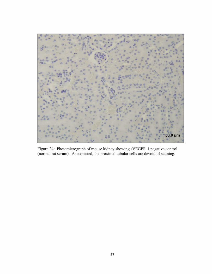
Figure 24: Photomicrograph of mouse kidney showing sVEGFR-1 negative control (normal rat serum). As expected, the proximal tubular cells are devoid of staining.
58
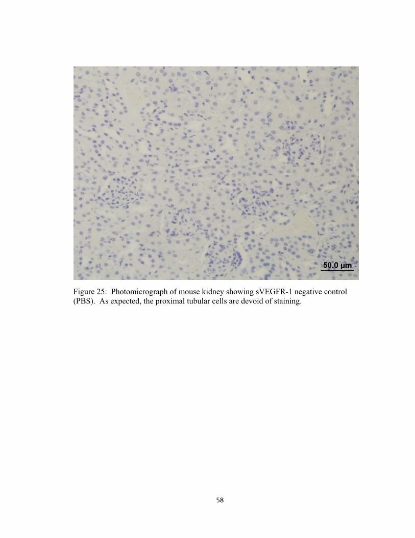
Figure 25: Photomicrograph of mouse kidney showing sVEGFR-1 negative control (PBS). As expected, the proximal tubular cells are devoid of staining.

59
a.
b.
Figure 26: Immunohistochemistry for sVEGFR-1 in central cornea. There was no sVEGFR-1 staining within the central corneal epithelium or stroma in non-wounded WT eyes (a) and the majority of Sey eyes (b).

60
a.
b.
Figure 27: Immunohistochemistry for sVEGFR-1 in central cornea. In the Sey group, a total of 8 mice (33%) had staining within the central corneal stroma (a,b). Keratocyte staining is indicated (arrow).
61
CHAPTER 3
CONCLUSION
The results of this study have shown precise and accurate corneal epithelial
removal with n-heptanol in an in vivo model using WT and Sey mice. This model has
confirmed an inherent deficiency in corneal wound healing in the Sey mouse that is likely
partly responsible for the ARK phenotype. While additional study is needed, the wound
healing deficiency observed in Sey mice did not appear to be caused by depletion in the
limbal progenitor cell population (p63 expressing cells). Localization of p63 within the
wing cells and superficial cells of the cornea suggests an impairment of epithelial cell
differentiation and further study is needed to evaluate the significance of this finding.
Comparable anatomic localization of sVEGFR-1 suggests a role for this factor in the
maintenance of corneal avascularity. However, given the results reported here, it is
unlikely that this factor alone is responsible for the prominent corneal vascularization in
Sey mice. This wounding paradigm provides an option for future in vivo evaluation of
corneal pathophysiology in Pax6 deficiency and a model for testing and comparing
potential therapeutic intervention. Further characterization of the epithelial and stromal
pathology, including limbal stem cell function, in Sey mice may lead to therapeutic
strategies (corrective gene therapy) in the future.