Vascular Medicine - Seoul National University
11
ISSN: 1524-4539 Copyright © 2005 American Heart Association. All rights reserved. Print ISSN: 0009-7322. Online 72514 Circulation is published by the American Heart Association. 7272 Greenville Avenue, Dallas, TX DOI: 10.1161/CIRCULATIONAHA.104.503433 2005;112;1618-1627; originally published online Sep 6, 2005; Circulation Han-Kwang Yang, Byung-Hee Oh, Young-Bae Park and Hyo-Soo Kim Il-Young Oh, Tae-Youn Kim, Hyun-Jai Cho, Hyun-Jae Kang, In-Ho Chae, Chang-Hwan Yoon, Jin Hur, Kyung-Woo Park, Ji-Hyun Kim, Choon-Soo Lee, Cytokines and Matrix Metalloproteinases Progenitor Cells and Late Outgrowth Endothelial Cells: The Role of Angiogenic Synergistic Neovascularization by Mixed Transplantation of Early Endothelial http://circ.ahajournals.org/cgi/content/full/CIRCULATIONAHA.104.503433/DC1 Data Supplement (unedited) at: http://circ.ahajournals.org/cgi/content/full/112/11/1618 located on the World Wide Web at: The online version of this article, along with updated information and services, is http://www.lww.com/reprints Reprints: Information about reprints can be found online at [email protected] 410-528-8550. E-mail: Fax: Kluwer Health, 351 West Camden Street, Baltimore, MD 21202-2436. Phone: 410-528-4050. Permissions: Permissions & Rights Desk, Lippincott Williams & Wilkins, a division of Wolters http://circ.ahajournals.org/subscriptions/ Subscriptions: Information about subscribing to Circulation is online at at SAROK SEOUL NATIONAL UNIV on August 27, 2009 circ.ahajournals.org Downloaded from
Transcript of Vascular Medicine - Seoul National University

ISSN: 1524-4539 Copyright © 2005 American Heart Association. All rights reserved. Print ISSN: 0009-7322. Online
72514Circulation is published by the American Heart Association. 7272 Greenville Avenue, Dallas, TX
DOI: 10.1161/CIRCULATIONAHA.104.503433 2005;112;1618-1627; originally published online Sep 6, 2005; Circulation
Han-Kwang Yang, Byung-Hee Oh, Young-Bae Park and Hyo-Soo Kim Il-Young Oh, Tae-Youn Kim, Hyun-Jai Cho, Hyun-Jae Kang, In-Ho Chae,
Chang-Hwan Yoon, Jin Hur, Kyung-Woo Park, Ji-Hyun Kim, Choon-Soo Lee, Cytokines and Matrix Metalloproteinases
Progenitor Cells and Late Outgrowth Endothelial Cells: The Role of Angiogenic Synergistic Neovascularization by Mixed Transplantation of Early Endothelial
http://circ.ahajournals.org/cgi/content/full/CIRCULATIONAHA.104.503433/DC1Data Supplement (unedited) at:
http://circ.ahajournals.org/cgi/content/full/112/11/1618
located on the World Wide Web at: The online version of this article, along with updated information and services, is
http://www.lww.com/reprintsReprints: Information about reprints can be found online at
[email protected]. E-mail:
Fax:Kluwer Health, 351 West Camden Street, Baltimore, MD 21202-2436. Phone: 410-528-4050. Permissions: Permissions & Rights Desk, Lippincott Williams & Wilkins, a division of Wolters
http://circ.ahajournals.org/subscriptions/Subscriptions: Information about subscribing to Circulation is online at
at SAROK SEOUL NATIONAL UNIV on August 27, 2009 circ.ahajournals.orgDownloaded from

Synergistic Neovascularization by Mixed Transplantationof Early Endothelial Progenitor Cells and Late
Outgrowth Endothelial CellsThe Role of Angiogenic Cytokines and Matrix Metalloproteinases
Chang-Hwan Yoon, MD*; Jin Hur, MS*; Kyung-Woo Park, MD; Ji-Hyun Kim, MS;Choon-Soo Lee, MS; Il-Young Oh, MD; Tae-Youn Kim, MS; Hyun-Jai Cho, MD;
Hyun-Jae Kang, MD; In-Ho Chae, MD; Han-Kwang Yang, MD; Byung-Hee Oh, MD;Young-Bae Park, MD; Hyo-Soo Kim, MD
Background—Two types of cells are cultured from the human peripheral blood, early endothelial progenitor cells (EPCs)and outgrowth endothelial cells (OECs), as previously reported. Here, we further characterize these cells, especially withrespect to their different origins and functions both in vitro and in vivo. We also investigated whether the combinationof these different cell types shows synergism during neovascularization.
Methods and Results—Early EPCs were heterogeneously made up of both CD14� monocyte-derived cells, which secretecytokines, and CD14�-derived cells, which contain high levels of CD34�KDR� cells. OECs were cultured almostexclusively from CD14� cells, not CD14� cells, and were distinct from mature endothelial cells in terms of proliferationpotential, KDR� expression level, and telomerase activity. A portion of cells from CD14� cells and early EPCs producedrapidly proliferating, capillary-forming cells in both the Matrigel plug and the ischemic hind limb similar to OECs. EarlyEPCs and OECs expressed receptors for vascular endothelial growth factor and interleukin-8, cytokines secreted byearly EPCs. There was a differential increase in matrix metalloproteinases (MMPs): MMP-9 in early EPCs and MMP-2in OECs. In vitro, the angiogenic capability of the 2 cell types was augmented by mutual interaction through cytokinesand MMPs. Injection of a mixture of the 2 cells resulted in superior neovascularization in vivo to any single-cell-typetransplantation.
Conclusions—Distinct origins of the different types of EPCs exist that have different functions in neovascularization.Mixed transplantation of these cells results in synergistic neovascularization through cytokines and MMPs.(Circulation. 2005;112:1618-1627.)
Key Words: angiogenesis � cells � endothelium � ischemia � revascularization
Although therapeutic angiogenesis with endothelial pro-genitor cells (EPCs) is under intense investigation as a
treatment modality for ischemic disease, many questionsabout what EPCs really are and how they interact with oneanother and with cytokines to contribute to new vesselformation remain unanswered.1
See p 1522Recently, we reported that 2 types of cells cultured from
human peripheral blood had comparable angiogenic capabil-ities, which we called early EPCs and late EPCs,respectively.2
Early EPCs are similar to the progenitor cells first reportedby Asahara and colleagues3 that have been used in most of thetherapeutic angiogenesis trials.4,5 Recently, they have beenreferred to as monocyte-derived circulating angiogenic cellsby March and colleagues6,7 or as early EPCs by Gulati andcolleagues.8 Gulati and colleagues also showed that CD14�
cells produced these spindle-shaped cells but did not give riseto endothelial outgrowth. Instead, the CD14� cells gave riseto endothelial outgrowth.
Late EPCs, named after their late outgrowth potential, aresimilar to the circulating bone marrow–derived endothelialcells reported by Shi et al9 or outgrowth endothelial cells
Received August 30, 2004; revision received May 20, 2005; accepted May 25, 2005.From the Cardiovascular Laboratory, Clinical Research Institute, Seoul National University Hospital (C.-H.Y., J.H., K.-W.P., J.-H.K., C.-S.L., I.-Y.O.,
T.-Y.K., H.-J.C., H.-J.K., I.-H.C., B.-H.O., Y.-B.P., H.-S.K.), and Departments of Internal Medicine (C.-H.Y., K.-W.P., I.-Y.O., H.-J.C., H.-J.K., I.-H.C.,B.-H.O., Y.-B.P., H.-S.K.) and Surgery (H.-K.Y.), Seoul National University College of Medicine, Seoul, Korea.
*Drs Yoon and Hur contributed equally to this work.The online-only Data Supplement can be found at http://circ.ahajournals.org/cgi/content/full/CIRCULATIONAHA.104.503433/DC1.Correspondence to Hyo-Soo Kim, MD, or Young-Bae Park, MD, Department of Internal Medicine, Seoul National University College of Medicine,
28 Yongon-Dong Chongno-Gu, Seoul 110-744, Korea. E-mail [email protected]© 2005 American Heart Association, Inc.
Circulation is available at http://www.circulationaha.org DOI: 10.1161/CIRCULATIONAHA.104.503433
1618
Vascular Medicine
at SAROK SEOUL NATIONAL UNIV on August 27, 2009 circ.ahajournals.orgDownloaded from

(OECs) reported by Lin et al10 and Gulati et al,8 who noticedlate outgrowth and cobblestone appearances. We use the term“OEC” instead of “late EPC” in the present study to avoidconfusion of terminology. The various characteristics of the 2types of cells, which we and other investigators reported, aresummarized in the Table.
In the present study, the main purpose was to examine thesynergism between different types of EPCs during neovascu-larization in terms of cytokines and matrix metalloproteinases(MMPs), 2 factors that recently have been spotlighted as keyfactors in angiogenesis.11–14 In addition, we further investi-gated the heterogeneous origin of these different types ofEPCs by culturing different groups of peripheral mononu-clear cells separated by CD14 positivity, and we examinedwhich group or groups of cells give rise to early EPCs orOECs in vitro. Furthermore, we confirmed in vivo thedifferentiation of certain cell lineages into OECs wheninjected into the ischemic limb of nude mice.
MethodsExpanded methods are available in the Data Supplement, foundonline at http://circ.ahajournals.org/cgi/content/full/CIRCULATIONAHA.104.503433/DC1.
CellsAll human projects in this study were approved by the institutionalreview board of Seoul National University Hospital. See the DataSupplement for information on cell culture, fluorescence-activatedcell sorter (FACS) analysis, telomeric repeat amplification protocol,evaluation of the effects of cytokines and MMPs (including acqui-sition of conditioned medium, assay of tube formation on Matrigel,proliferation assay, vertical chamber migration assay, zymography
for MMP activity, quantitative RT-PCR, and vertical collagen gelinvasion assay), and in vivo vasculogenesis of mixed cells inMatrigel or ischemic limb of nude mouse.
All procedures were approved by the Experimental AnimalCommittee of Clinical Research Institute, Seoul National UniversityHospital (Seoul, Korea). Female athymic nude mice (Jackson Lab-oratory) that were 8 to 9 weeks old and weighed 17 to 20 g wereanesthetized with 50 mg/kg IP pentobarbital. In vivo experimentsconsisted of a 3D Matrigel plug assay and hind-limb ischemic model.See the Data Supplement for detailed procedures.
Statistical AnalysisAll data are presented as mean�SEM. Intergroup comparisons wereperformed by Student t test, ANOVA with Duncan post hoc analysis,or the Kruskall-Wallis test. The comparative incidence of limbsalvage was evaluated by �2 tests. SPSS version 11.0 was used foranalysis, and P�0.05 was considered statistically significant.
ResultsDistinct Precursor Cells Exist in the PeripheralBlood for Early EPCs and OECsFreshly isolated human peripheral blood mononuclear cells(MNCs) were divided into 3 groups: 50.4�14.1%CD14�AC133�, 0.01�0.01% CD14�AC133�, and49.4�14.2% CD14�AC133� (Figure 1a). CD14�AC133�
cells were not detected. After CD14 selection with MACS, noAC133� cells were detected in the CD14� fraction, andvirtually all of the AC133� cells existed in the CD14�
fraction (0.02�0.01%) (Figure 1a). CD34�KDR� double-positive cells, which made up �0.16�0.10% of the entireMNCs, existed mostly in the CD14� fraction (0.81�0.4%)(Figure 1b).
Various Characteristics of Early EPCs and OECs
Early EPCs OECs
Morphology
Growth pattern in vitro Early growth2 Late outgrowth2,8,10
Surface antigen expression CD34, CD31, Flk-1, Tie-23 Flk-1, vWF, CD36, Ve-cadherin10
CD34, CD31, Ve-cadherin, KDR, CD144 CD31, VEGFR-2, Tie-2, Ve-cadherin8
KDR, CD105, vWF, CD315
CD31, CD45, CD14, CD11c, CD1636
CD14, CD31, VEGFR-2, Tie-28
AcLDL uptake and lectin binding Positive2 Positive2
Incorporation into HUVECs Good2 Better2
Tube formation Unable2,8 Able2,8
Cytokine secretion High level2,6 Low level2
VEGF, IL-82
VEGF, HGF, G-CSF6
NO production Low level2 High level2,8
Population Heterogeneous2 Homogeneous2
Angiogenic potential Good2 Good2
vWF indicates von Willebrand factor; VEGFR-2, VEGF receptor 2; HUVECs, human umbilical vain endothelial cells;and NO, nitric oxide.
Yoon et al Synergism of 2 Types of EPC 1619
at SAROK SEOUL NATIONAL UNIV on August 27, 2009 circ.ahajournals.orgDownloaded from

Next, to find out whether different cell groups give rise todifferent EPCs, entire MNCs, CD14� cells, and CD14� cellswere cultured separately. On day 7, all MNC-derived earlyEPCs showed an increase in CD34�KDR� cells(1.32�0.43%) (Figure 1c). CD14� cells rarely containedCD34�KDR� double positive cells on day 7 (0.08�0.06%;Figure 1d). CD14� cells, however, showed a 4-fold increasein CD34�KDR� cells (3.2�1.1%; Figure 1e). OECs showedthe largest CD34�KDR� fraction (58.4�12.5%; Figure 1f).CD14� cells gave rise to spindle-shaped cells on day 7
(Figure 1g). OECs, however, did not appear from this group.In contrast, CD14� cells gave rise to fewer spindle-like cellson day 7 (data not shown), whereas 2 or 3 weeks of culture ofthis group gave rise to OECs (Figure 1h), which showedproliferation up to 40 passages in vitro.
In terms of cytokines, the CD14� group of cells secreted asignificantly higher level of interleukin (IL)-8 than the CD14�
group of cells (Figure 1i). In addition, vascular endothelial growthfactor (VEGF), albeit small, was also higher in the supernatant ofCD14� cells than in that of CD14� cells (Figure 1j).
Figure 1. The different lineages of early EPCs and OECs. a, Freshly isolated MNCs stained with dual antibodies against AC133and CD14. AC133� cells were discovered only in the CD14� fraction. a’, a”, CD14� and CD14� selection by magnetic activatedcell sorting (MACS). b, Freshly isolated MNCs stained with dual antibodies against CD34 and KDR. b’, b”, Double FACS of CD14�
and CD14� cells against CD34 and KDR. c, d, e, Double FACS against CD34 and KDR after 7 days of culture of entire MNCs (c),CD14� cells (d), and CD14� cells (e) in the same condition. CD14� cells showed the greatest increase in CD34�KDR� cells. f,OECs had a large CD34�KDR� fraction. g, CD14� cells gave rise to spindle-shaped cells on day 7, but OECs did not appear fromthis group. Scale bar�100 �m. h, Representative figure showing a colony of OECs appearing from CD14� cells after 2 or 3 weeksof culture. i, j, Level of IL-8 (i) and VEGF (j) from CD14�- and CD14�-derived cells after 7 days of culture. k, Surface antigens ofearly EPCs, late EPCs, and GEAECs by FACS.
1620 Circulation September 13, 2005
at SAROK SEOUL NATIONAL UNIV on August 27, 2009 circ.ahajournals.orgDownloaded from

Taken together, early EPCs were made up of heteroge-neous cells of both CD14� and CD14� origin, and these earlyEPCs collectively showed expressions of both endothelialand monocytic antigens as analyzed by FACS (Figure 1k).Late-appearing OECs did not express CD14 or CD45 butwere positive for endothelial surface antigens similar togastroepiploic artery endothelial cells (GEAECs) (Figure 1k).
Heterogeneous Fates of Blood-Derived Cells InVivo: Supporting Evidence for the Existence ofDifferent Types of EPCs In VivoTo examine whether the heterogeneous origins of the EPCsactually give rise to OECs in vivo, we transplanted and thentraced fluorescent dye–labeled CD14� cells, CD14� cells, andthe early EPCs in the ischemic muscle of nude mice. On day
3, we found that in all 3 groups, most of the transplanted cellswere located in the interstitium of muscle tissue (Figure 2a).On day 7, we could observe some cells that migrated toadjacent muscles and proliferated. On day 21, the cells in theCD14� cell transplanted group and most of the cells in theearly EPC transplanted group looked similar to those ob-served on day 7, suggesting low-grade proliferation. How-ever, in the CD14� cell transplanted group and in a portion ofthe cells from the early EPC transplanted group, we were ableto observe a wider distribution of the transplanted cells in themuscle tissue, and the number of cells was remarkablyincreased, resulting in fluorescence dilution. Moreover, it isinteresting to note that, even in the samples injected withCD14� cells, not all of the injected cells resulted in migrationand proliferation; we often observed nonproliferating nonmi-
Figure 2. Heterogeneous fates of various blood-derived cells in vivo. a, Freshly isolated DiI-labeled CD14� cells were located in theinterstitium of the muscle on day 3 and did not show any significant proliferation up to day 21. CD14� cells were also located in theinterstitium on day 3. However, from day 7, a small portion migrated into the capillary spaces and proliferated; at day 21, we observeda large number of cells in both the muscular capillaries (day 21; left column) and the interstitial capillaries (day 21; right column). Usu-ally, most of the transplanted DiO (green)-labeled early EPCs behaved similarly to CD14� cells with only low-grade proliferation. How-ever, some cells from these early EPCs proliferated in the ischemic muscle and constructed capillary structures similar to CD14� cellson day 21. b, CD14� cells (red) in a Matrigel plug on day 21 were stained by anti-CD14 antibody (green) and thus look yellow in themerged figure. These cells were rarely positive for KDR and VE-cadherin and thus look red. CD14� cells (red) in the Matrigel plug werepositive for KDR and VE-cadherin (both green) but not CD14. Early EPCs (green) were usually scarcely positive (red) for KDR andVE-cadherin, but again we observed some cells from this early EPC population that were CD14�, KDR�, and VE-cadherin�.
Yoon et al Synergism of 2 Types of EPC 1621
at SAROK SEOUL NATIONAL UNIV on August 27, 2009 circ.ahajournals.orgDownloaded from

grating cells remaining near the interstitium that retained ahigh level of fluorescence. This suggests that there is asubpopulation of cells in both the CD14� cells and earlyEPCs that eventually migrate and proliferate when injectedinto the ischemic muscle.
In addition, the widely distributed cells we observed in theCD14� cell transplanted group and the early EPC trans-planted group stained positive for KDR and VE-cadherin butnot CD14. In contrast, cells in the CD14� cells transplantedgroup retained CD14 positivity and showed only focal posi-tivity for KDR and VE-cadherin (Figure 2b).
OECs Show Distinct Features From MatureEndothelial Cells Both In Vitro and In VivoBecause OECs did not express CD14 or CD45 and were positivefor endothelial surface antigens similar to mature endothelialcells, we examined the differences between these cells in termsof other surface markers, telomerase activity, and capillaryforming capacity. See the Data Supplement for results.
Cytokines Secreted From Early EPCs EnhanceAngiogenic Capacity of OECs In Vitro: ParacrineEffects of IL-8 and VEGFAs we previously reported, early EPCs secrete high levels ofVEGF and Il-8. Early EPCs and OECs expressed receptorsfor both VEGF (KDR and Flt-1) and IL-8 (CXCR-1 andCXCR-2). In particular, OECs showed significantly higher
expressions of KDR and CXCR-1 (Figure 3a). Therefore, weexamined the effect of early EPC-secreted cytokines on thebehavior of OECs using the culture supernatant of early EPCs(early EPC–conditioned medium).
When the early EPC–conditioned medium was added toOECs, OECs showed significantly higher proliferation (Fig-ure 3b) and significantly more complete tubes as measured bythe area and length of the tubes (Figure 3c through 3e) thanOECs exposed to unconditioned medium or the OEC-conditioned medium. The enhanced proliferation and capil-lary tube formation, induced by early EPC-secreted cyto-kines, was attenuated by neutralizing antibodies againstVEGF and IL-8, but not by isotype antibody (Figure 3b, 3d,and 3e). Next, OECs showed better migratory capacity thanearly EPCs in the vertical chamber (Figure 3f and 3g). Whenwe mixed both cells and incubated them together, the migrat-ing distance of both types of cells was significantly increasedbeyond that of any single cell alone (Figure 3f and 3g),suggesting synergism between the 2 types of cells. Theneutralizing antibodies also significantly inhibited migrationin contrast to isotype antibody control (Figure 3g).
Two Types of EPCs Show Different Patterns ofMMP Secretion That Are Affected by IL-8 andVEGF: Autocrine and Paracrine ActionsFreshly isolated CD14� cells, the main fraction containing theorigin of OECs, secreted mainly MMP-2, whereas CD14�
Figure 3. Paracrine effects of early EPCsaugment tube formation, proliferation,and migration of OECs. a, RT-PCR of the2 types of EPCs showing expression ofreceptors for cytokines secreted fromearly EPCs: KDR and Flt-1 for VEGF,CXCR-1 and 2 for IL-8. b, Cell viability ofOECs in various media. OECs prolifer-ated most rapidly in the presence of theearly EPC-conditioned medium (ECM),which was significantly blocked by neu-tralizing antibodies (Abs) against IL-8 andVEGF. Values are given as mean�SEM(n�4). c, Representative figure of OECs,incubated in the ECM, forming signifi-cantly more capillary tubes than OECsincubated in OCM. Scale bar�100 �m.d, e, Quantitative data of total completetube area (d) and total tube length (e)formed by OECs under various condi-tions. The enhanced tube-forming capa-bility of OECs in ECM was again attenu-ated by blocking the action of IL-8 andVEGF. f, g, Mixed incubation of earlyEPCs and OECs (mix) significantlyincreased the migration distance of the 2cells compared with early EPCs (E-EPC)or OECs alone, which was significantlyattenuated in the presence of neutralizingantibody against IL-8 and VEGF(mix�Abs) in contrast to isotype anti-body (mix�iso). Scale bar�1 mm. Greenbar denotes E-EPCs; red bar, OECs. Val-ues are mean�SEM (n�3). †P�0.01.
1622 Circulation September 13, 2005
at SAROK SEOUL NATIONAL UNIV on August 27, 2009 circ.ahajournals.orgDownloaded from
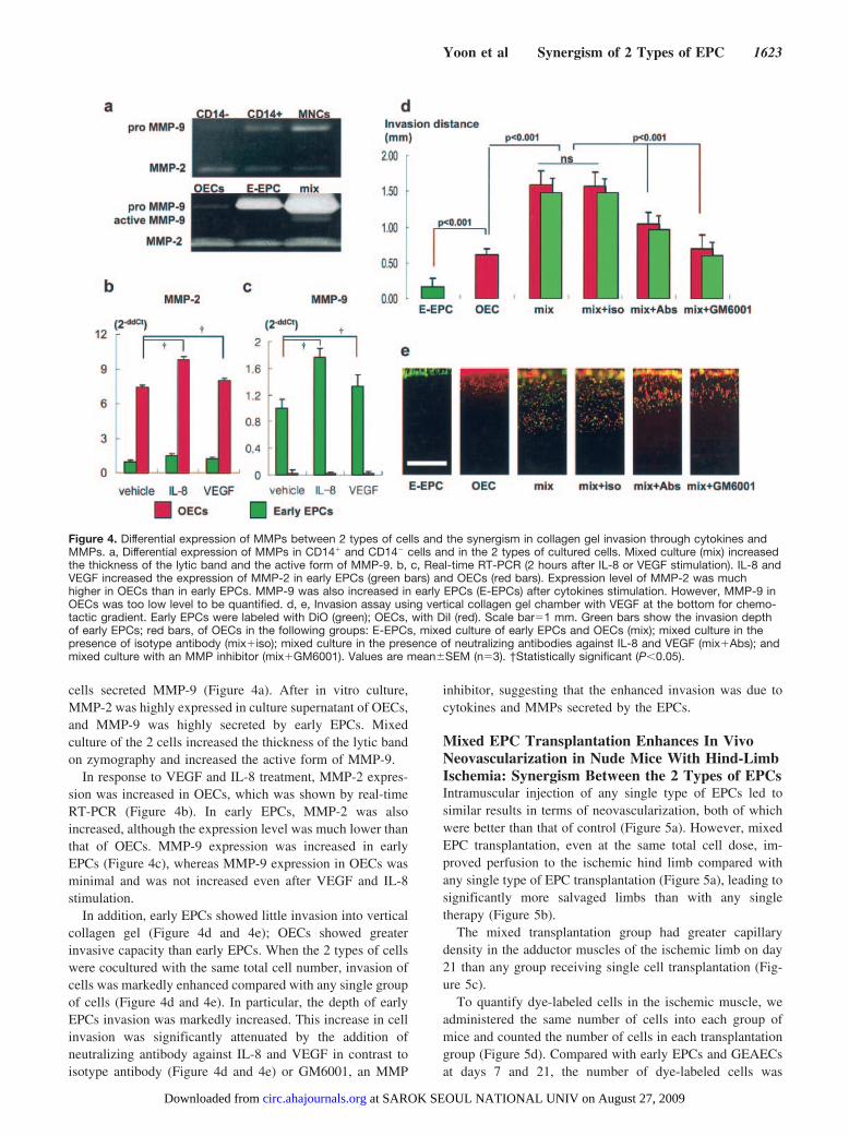
cells secreted MMP-9 (Figure 4a). After in vitro culture,MMP-2 was highly expressed in culture supernatant of OECs,and MMP-9 was highly secreted by early EPCs. Mixedculture of the 2 cells increased the thickness of the lytic bandon zymography and increased the active form of MMP-9.
In response to VEGF and IL-8 treatment, MMP-2 expres-sion was increased in OECs, which was shown by real-timeRT-PCR (Figure 4b). In early EPCs, MMP-2 was alsoincreased, although the expression level was much lower thanthat of OECs. MMP-9 expression was increased in earlyEPCs (Figure 4c), whereas MMP-9 expression in OECs wasminimal and was not increased even after VEGF and IL-8stimulation.
In addition, early EPCs showed little invasion into verticalcollagen gel (Figure 4d and 4e); OECs showed greaterinvasive capacity than early EPCs. When the 2 types of cellswere cocultured with the same total cell number, invasion ofcells was markedly enhanced compared with any single groupof cells (Figure 4d and 4e). In particular, the depth of earlyEPCs invasion was markedly increased. This increase in cellinvasion was significantly attenuated by the addition ofneutralizing antibody against IL-8 and VEGF in contrast toisotype antibody (Figure 4d and 4e) or GM6001, an MMP
inhibitor, suggesting that the enhanced invasion was due tocytokines and MMPs secreted by the EPCs.
Mixed EPC Transplantation Enhances In VivoNeovascularization in Nude Mice With Hind-LimbIschemia: Synergism Between the 2 Types of EPCsIntramuscular injection of any single type of EPCs led tosimilar results in terms of neovascularization, both of whichwere better than that of control (Figure 5a). However, mixedEPC transplantation, even at the same total cell dose, im-proved perfusion to the ischemic hind limb compared withany single type of EPC transplantation (Figure 5a), leading tosignificantly more salvaged limbs than with any singletherapy (Figure 5b).
The mixed transplantation group had greater capillarydensity in the adductor muscles of the ischemic limb on day21 than any group receiving single cell transplantation (Fig-ure 5c).
To quantify dye-labeled cells in the ischemic muscle, weadministered the same number of cells into each group ofmice and counted the number of cells in each transplantationgroup (Figure 5d). Compared with early EPCs and GEAECsat days 7 and 21, the number of dye-labeled cells was
Figure 4. Differential expression of MMPs between 2 types of cells and the synergism in collagen gel invasion through cytokines andMMPs. a, Differential expression of MMPs in CD14� and CD14� cells and in the 2 types of cultured cells. Mixed culture (mix) increasedthe thickness of the lytic band and the active form of MMP-9. b, c, Real-time RT-PCR (2 hours after IL-8 or VEGF stimulation). IL-8 andVEGF increased the expression of MMP-2 in early EPCs (green bars) and OECs (red bars). Expression level of MMP-2 was muchhigher in OECs than in early EPCs. MMP-9 was also increased in early EPCs (E-EPCs) after cytokines stimulation. However, MMP-9 inOECs was too low level to be quantified. d, e, Invasion assay using vertical collagen gel chamber with VEGF at the bottom for chemo-tactic gradient. Early EPCs were labeled with DiO (green); OECs, with DiI (red). Scale bar�1 mm. Green bars show the invasion depthof early EPCs; red bars, of OECs in the following groups: E-EPCs, mixed culture of early EPCs and OECs (mix); mixed culture in thepresence of isotype antibody (mix�iso); mixed culture in the presence of neutralizing antibodies against IL-8 and VEGF (mix�Abs); andmixed culture with an MMP inhibitor (mix�GM6001). Values are mean�SEM (n�3). †Statistically significant (P�0.05).
Yoon et al Synergism of 2 Types of EPC 1623
at SAROK SEOUL NATIONAL UNIV on August 27, 2009 circ.ahajournals.orgDownloaded from

significantly greater in the OEC-injected mice, suggestinggreater proliferation. Early EPCs did not increase much untilday 7 yet surpassed GEAECs on day 21, which suggests thata certain portion of early EPCs rapidly proliferated after day7 up to day 21. The transplantation of mixed EPCs resulted ina greater number of cells at all time points than any other typeof cell, again suggesting synergism between the 2 types ofEPCs.
When we stained the murine capillaries blue, both types ofcells were colocalized in the capillary spaces (Figure 5e). Weobserved that 33�4.4% of the capillaries in the neovasculo-genic foci of the ischemic muscle were lined by deliveredcells in the mixed EPC group (n�4). This was significantlygreater than that of GEAECs (7.5�1.4%), early EPCs(13.3�3.5%), or OECs (22�3.9%) (n�4 for all; P�0.003).
We confirmed that the transplanted cells retained KDRexpression by immunofluorescent staining (blue) againsthuman KDR (Figure 5f).
Transplanted cells took part in not only forming muscularcapillaries but also making subcutaneous (Figure 5g) orinterstitial (Figure 5h) capillaries with murine cells, whichmay also have contributed to improved perfusion and thusenhanced limb salvage.
To analyze the mechanism of the mixed effect in vivo, weinjected cells into subcutaneous Matrigel plugs. Mixed EPCssecreted high levels of MMP-2, MMP-9, and VEGF (Figure6a). Mixed EPCs showed more rapid proliferation and moreinvasive migration than any other kinds of cells (Figure 6b)and thus formed compact dense capillaries in the Matrigelplugs (Figure 6c) compared with other cells. When we
Figure 5. Two types of EPCs synergistically improve neovasculogenesis in hind-limb ischemia. a, Laser Doppler perfusion analysisshowing superior recovery of perfusion as early as day 14 in the mixed EPC group (perfusion ratio at day 21, 0.47�0.07 vs 0.50�0.09vs 0.65�0.09 for early EPC only vs OEC only vs mixed group; n�17, n�17, n�15, respectively) (mixed vs OECs or early EPCs,P�0.05; OECs vs early EPCs, P�NS). Each type of cell alone still results in better perfusion recovery than GEAEC or media (OECs orearly EPCs vs GEAEC or media, P�0.05). Values are mean�SEM. b, Graph showing the superior effect of mixed cell transplantation onlimb salvage. Limb salvage/tip necrosis/autoamputation: 6/5/6 in early EPC group, 6/4/7 in OECs group, and 9/5/1 in mixed group(mixed versus early EPCs or OECs, P�0.001). c, Capillary density among the 5 groups of mice, with the mixed transplantation groupshowing the best results (599�26, n�4; P�0.043 vs OECs; P�0.021 vs early EPCs). d, Number of fluorescent-labeled cells (counted in10 sections at 100-�m intervals) on day 3, 7, and 21 (n�4 in each group). In the mixed group, the early EPC and OEC subtypes wereseparately denoted. e, On day 21, the 2 types of cells (green, early EPC derived; red, OEC derived) were colocalized with the capillaryspace (blue, stained murine endothelial cell with anti-mouse CD31 antibody) among muscle fibers (unstained light microscopic figure ofthe same field). Scale bar�100 �m. f, Red OEC-derived cells stained with anti-human KDR antibody (blue) look purple (arrow), andgreen early EPC-derived cells positive for KDR look bright sky-blue (arrowhead). g, We were also able to observe development of sub-cutaneous capillary networks in the ischemic muscle. FITC (green)-BS-1 lectin was used to demonstrate murine vessels, and DAPIstaining was performed to visualize the nucleus. We could see that EPC-derived capillaries (red) were connected to murine capillariesin the subcutaneous tissue. h, We also found that these networks developed in the interstitial spaces in the ischemic muscle. †Statisti-cally significant.
1624 Circulation September 13, 2005
at SAROK SEOUL NATIONAL UNIV on August 27, 2009 circ.ahajournals.orgDownloaded from

quantified the neovascularization within the Matrigel bymeasuring the percentages of capillary lumen area againsttotal Matrigel area, we found significant augmentation ofneovascularization in the mixed EPC group compared withthe other groups (Figure 6d).
DiscussionWe and others have suggested that there might be differenttypes of EPCs derived from the human peripheral blood.2,7,8
Here, we show that early EPCs are a heterogeneous mixtureof cells of both CD14� and CD14� origin and that OECs arisefrom cells of CD14� origin. We also showed that the origin ofthese different cells exists in vivo by injecting CD14� orCD14� cells into ischemic limbs and confirming the differ-entiation to OECs by certain cells from the CD14� fraction.In addition, injection of early EPCs in vivo resulted in thedifferentiation of a portion of these cells into OECs, suggest-ing that OECs may arise from the differentiation of both earlyEPCs and certain CD14� cells. The early EPCs, specificallythe CD14�-derived early EPCs, secreted high levels of IL-8and VEGF, and both early EPCs and OECs expressed thereceptors for these cytokines. In a paracrine manner, earlyEPCs augmented proliferation, migration, and the capillarytube forming capabilities of OECs.
More important, we also demonstrate for the first time thatearly EPCs and OECs synergistically cooperate to enhanceneovasculogenesis in the ischemic hind limb. We found thatearly EPCs and OECs show different patterns of MMPsecretion, which were also seen in CD14� and CD14� cells inthe blood. The expression of these MMPs was affected by theparacrine and autocrine effects of VEGF and IL-8, whichwere secreted by early EPCs, and mixed culture of these 2types of cells resulted in enhanced invasion into collagen gel,suggesting synergism between them. The synergism wasconfirmed in vivo by mixed transplantation of the 2 types ofEPCs to both the murine ischemic limb and subcutaneousMatrigel plugs, which significantly improved neovascularization.
Endothelial Progenitor CellsEarly EPCs that arise from the culture of entire MNCs are aheterogeneous group of cells containing both progenitors ofOECs, which are probably CD14� and secrete low levels ofcytokines, and monocyte-derived cells, which are probablyCD14� and secrete high levels of cytokines. This wasconfirmed in vivo when we injected CD14� cells, CD14�
cells, and early EPCs and found that CD14� but not CD14�
cells gave rise to OECs. In the group that received early EPCinjection, we observed endothelial proliferation in the ische-mic limb similar to OECs, although less frequently than thegroup that received only CD14� cells.
Figure 6. The mechanism of theenhanced neovascularization by themixed transplantation in vivo Matrigelplug experiment. a, Primary antibodiesagainst MMP-2, MMP-9, and VEGF andFITC-conjugated secondary antibodieswere used for immunofluorescence. Bluedots were DAPI-stained nucleus. Wefound that MMP-2, MMP-9, and VEGFwere secreted around the cell clumps ofthe mixed group on day 3. b, MixedEPCs showed more rapid proliferationand invasive migration than any othertype of cell. Early EPCs were stainedgreen (DiO labeled); OECs and GEAECswere stained red (DiI labeled). c, Phase-contrast microscopy also shows thatmixed cell transplantation results in moredense capillaries in the Matrigel plugs. d,After 2 weeks, capillary lumens (L)appeared lined with EPCs (red with bluenucleus) or murine endothelial cells (bluenucleus only) in the Matrigel (M, green).When we quantified the neovasculariza-tion within the Matrigel by measuring thepercentages of capillary lumen areaagainst that of Matrigel, we found a sig-nificant augmentation of neovasculariza-tion in the group of mixed EPCs com-pared with the other groups (n�4). Connindicates connective tissue.
Yoon et al Synergism of 2 Types of EPC 1625
at SAROK SEOUL NATIONAL UNIV on August 27, 2009 circ.ahajournals.orgDownloaded from

In early EPCs, the difference between CD14� cells andCD14� cells was the number of AC133� cells or CD34�KDR�
cells. Therefore, AC133, KDR, and CD34 without CD14 orCD45 may be critical markers of OEC-producing EPCs, whichare different from hematopoietic progenitors or leukocytes.Because AC133 rapidly disappeared and CD34�KDR� cellsincreased, CD34/KDR double positivity may be considered animportant marker of EPCs in vitro. Many reports support theimportance of these markers in defining and quantifyingEPCs.7,15–17 Because different subpopulations of cells seem todifferentiate into different types of EPCs, it would be interestingto investigate which cell types are closely related to the riskfactors for or severity of atherosclerosis. From our data, theclinical relevance of the variation in the number of CD34/KDRdouble-positive cells in blood or in culture with risk factors foror severity of atherosclerosis in individuals should be studied ina future study.
OECs are closer to mature endothelial cells in phenotypebut show surprising proliferative, migrating, and tube-forming capabilities. In the present study, we further eluci-dated that OECs showed relatively high KDR expression andtelomerase activity in vitro and formed more capillaries invivo. Others have also reported that OECs are different frommature ECs in terms of caveolae,8 expression of integrins,18
resistance to oxidative stress,19 and angiogenic potency invivo.2,20 Therefore, OECs are supposed to be in lineages ofEPCs.
Autocrine and Paracrine Network in EPCsPrevious studies have shown that only a small fraction of theprogenitor cells infused or mobilized actually incorporate intothe ischemic tissue.1,21,22 Considering the marked improve-ment in neovascularization induced by such cells, it may bepossible that progenitor cells not only incorporate into newvessels and mechanically improve neovascularization butalso secrete potent angiogenic cytokines that enhance thesurvival, proliferation, and function of other surroundingprogenitor cells and mature endothelial cells through auto-crine and paracrine networks. In contrast to OECs, earlyEPCs secrete large amounts of VEGF and IL-8. Both of thesecytokines are proangiogenic molecules that increase endothe-lial proliferation, tube formation, and migration.11,23 IL-8 andVEGF are also known to increase MMP secretion in endo-thelial cells.23,24 In the present study, we confirmed that bothtypes of cells express receptors for VEGF and IL-8. Inaddition, the early EPCs had a paracrine effect on OECperformance from the early EPC-secreted cytokines. Further-more, these early EPC-secreted cytokines stimulated bothearly EPCs and OECs in an autocrine and paracrine mannerto increase differential expressions of MMPs, contributing tothe enhanced invasiveness of mixed EPCs.
MMPs and EPCsThe importance of MMP-9 in angiogenesis was reported inrelation to angiogenic switch during carcinogenesis.25 Thissupports the notion that MMP secretion by EPCs may beimportant in neovascularization of ischemic organ.
MMP-2 and MMP-9 are produced by CD14� or CD14�
cells in blood and by OECs or early EPCs in vitro, respec-
tively. The existence of cowork between the 2 cell typesthrough MMPs and cytokines is suggested by the fact thatcoculture increased the active form of MMP-9 and theinvasion depth of early EPCs, which were not observed inearly EPCs alone. Fridman et al26 reported that activation ofMMP-9 is mediated by MMP-2 species that may be localizedin the cell surface and enhance matrix degradation. Accord-ingly, the interaction through MMPs should be investigatedfurther in future studies.
Synergism of Mixed Cell Transplantation onNeovascularizationThe 2 types of EPCs showed comparable angiogenic poten-tials when each type was transplanted alone. Early EPCs maycontribute to neovascularization by secretion of cytokines andMMP-9, whereas OECs participate by providing buildingblocks and secreting MMP-2. As discussed, cytokines,MMPs, and other functions of EPCs are closely connectedthrough autocrine and paracrine networks. Therefore, it wasnot surprising to observe a markedly improved perfusion tothe ischemic limb and subsequent improved limb salvage inmice receiving mixed EPC transplantation through cross-talkbetween 2 types of EPCs. Rafii and Lyden27 suggested thepossibility of synergistic combined stem or progenitor celltransplantation to improve the efficacy of cell therapy fororgan regeneration.
In conclusion, 2 different types of cells are produced fromculture of peripheral MNCs. Paracrine and autocrine net-works of cytokines and MMPs between the 2 types of cellsmay be an important mechanism of neovascularization. Con-sequently, the transplantation of mixed EPCs results insynergistic augmentation of angiogenesis in athymic nudemice with hind-limb ischemia. Such synergistic interactionsmay also be present among other types of stem or progenitorcells that may shed light on the future direction of stem celltherapy.
AcknowledgmentsThis study was supported by a grant from the Korea Health 21 R&Dproject, Ministry of Health & Welfare (02-PJ10-PG8-EC01-0026)and from Stem Cell Research Center, Republic of Korea (SC13122).
References1. Urbich C, Dimmeler S. Endothelial progenitor cells: characterization and
role in vascular biology. Circ Res. 2004;95:343–353.2. Hur J, Yoon CH, Kim HS, Choi JH, Kang HJ, Hwang KK, Oh BH, Lee
MM, Park YB. Characterization of two types of endothelial progenitorcells and their different contributions to neovasculogenesis. ArteriosclerThromb Vasc Biol. 2004;24:288–293.
3. Asahara T, Murohara T, Sullivan A, Silver M, van der Zee R, Li T,Witzenbichler B, Schatteman G, Isner JM. Isolation of putative progenitorendothelial cells for angiogenesis. Science. 1997;275:964–967.
4. Kalka C, Masuda H, Takahashi T, Kalka-Moll WM, Silver M, KearneyM, Li T, Isner JM, Asahara T. Transplantation of ex vivo expandedendothelial progenitor cells for therapeutic neovascularization. Proc NatlAcad Sci U S A. 2000;97:3422–3427.
5. Assmus B, Schachinger V, Teupe C, Britten M, Lehmann R, Dobert N,Grunwald F, Aicher A, Urbich C, Martin H, Hoelzer D, Dimmeler S,Zeiher AM. Transplantation of Progenitor Cells and RegenerationEnhancement in Acute Myocardial Infarction (TOPCARE-AMI). Circu-lation. 2002;106:3009–3017.
6. Rehman J, Li J, Orschell CM, March KL. Peripheral blood “endothelialprogenitor cells” are derived from monocytes/macrophages and secreteangiogenic growth factors. Circulation. 2003;107:1164–1169.
1626 Circulation September 13, 2005
at SAROK SEOUL NATIONAL UNIV on August 27, 2009 circ.ahajournals.orgDownloaded from

7. Rehman J, Li J, Parvathaneni L, Karlsson G, Panchal VR, Temm CJ,Mahenthiran J, March KL. Exercise acutely increases circulating endo-thelial progenitor cells and monocytes-/macrophage-derived angiogeniccells. J Am Coll Cardiol. 2004;43:2314–2318.
8. Gulati R, Jevremovic D, Peterson TE, Chatterjee S, Shah V, Vile RG,Simari RD. Diverse origin and function of cells with endothelial pheno-type obtained from adult human blood. Circ Res. 2003;93:1023–1025.
9. Shi Q, Rafii S, Wu MH, Wijelath ES, Yu C, Ishida A, Fujita Y, KothariS, Mohle R, Sauvage LR, Moore MA, Storb RF, Hammond WP.Evidence for circulating bone marrow-derived endothelial cells. Blood.1998;92:362–367.
10. Lin Y, Weisdorf DJ, Solovey A, Hebbel RP. Origins of circulatingendothelial cells and endothelial outgrowth from blood. J Clin Invest.2000;105:71–77.
11. Carmeliet P. Angiogenesis in health and disease. Nat Med. 2003;9:653–660.
12. McCawley LJ, Matrisian LM. Matrix metalloproteinases: they’re not justfor matrix anymore! Curr Opin Cell Biol. 2001;13:534–540.
13. Hiraoka N, Allen E, Apel IJ, Gyetko MR, Weiss SJ. Matrix metallopro-teinases regulate neovascularization by acting as pericellular fibrino-lysins. Cell. 1998;95:365–377.
14. Stetler-Stevenson WG. Matrix metalloproteinases in angiogenesis: amoving target for therapeutic intervention. J Clin Invest. 1999;103:1237–1241.
15. Valgimigli M, Rigolin GM, Fucili A, Porta MD, Soukhomovskaia O,Malagutti P, Bugli AM, Bragotti LZ, Francolini G, Mauro E, Castoldi G,Ferrari R. CD34� and endothelial progenitor cells in patient with variousdegrees of congestive heart failure. Circulation. 2004;110:1209–1212.
16. Ingram DA, Mead LE, Tanaka H, Meade V, Fenoglio A, Mortell K,Pollok K, Ferkowicz MJ, Gilley D, Yoder MC. Identification of a novelhierarchy of endothelial progenitor cells utilizing human peripheral andumbilical cord blood. Blood. 2004;104:2752–2760.
17. Powell TM, Paul JD, Hill JM, Thompson M, Benjamin M, Rodrigo M,McCoy JP, Read EJ, Khuu HM, Leitman SF, Finkel T, Cannon RO 3rd.Granulocyte colony-stimulating factor mobilizes functional endothelial
progenitor cells in patients with coronary artery disease. ArteriosclerThromb Vasc Biol. 2005;25:1–6.
18. Deb A, Skelding KA, Wang S, Reeder M, Simper D, Caplice NM.Integrin profile and in vivo homing of human smooth muscle progenitorcells. Circulation. 2004;110:1–5.
19. He T, Peterson TE, Holmuhamedov EL, Terzic A, Caplice NM, OberleyLW, Katusic ZS. Human endothelial progenitor cells tolerate oxidativestress due to intrinsically high expression of manganese superoxide dis-mutase. Arterioscler Thromb Vasc Biol. 2004;24:2021–2027.
20. Reyes M, Dudek A, Jahagirdar B, Koodie L, Marker PH, Verfaillie CM.Origin of endothelial progenitors in human postnatal bone marrow. J ClinInvest. 2002;109:337–346.
21. Urbich C, Heeschen C, Aicher A, Dernbach E, Zeiher AM, Dimmeler S.Relevance of monocytic features for neovascularization capacity of cir-culating endothelial progenitor cells. Circulation. 2003;108:2511–2516.
22. Aicher A, Heeschen C, Mildner-Rihm C, Urbich C, Ihling C,Technau-Ihling K, Zeiher AM, Dimmeler S. Essential role of endothelialnitric oxide synthase for mobilization of stem and progenitor cells. NatMed. 2003;9:1370–1376.
23. Li A, Dubey S, Varney ML, Dave BJ, Singh RK. IL-8 directly enhancedendothelial cell survival, proliferation, and matrix metalloproteinasesproduction and regulated angiogenesis. J Immunol. 2003;170:3369–3376.
24. Zucker S, Mirza H, Conner CE, Lorenz AF, Drews MH, Bahou WF, JestyJ. Vascular endothelial growth factor induces tissue factor and matrixmetalloproteinase production in endothelial cells: conversion of pro-thrombin to thrombin results in progelatinase A activation and cell pro-liferation. Int J Cancer. 1998;75:780–786.
25. Bergers G, Brekken R, McMahon G, Vu TH, Itoh T, Tamaki K, TanzawaK, Thorpe P, Itohara S, Werb Z, Hanahan D. Matrix metalloproteinase-9triggers the angiogenic switch during carcinogenesis. Nat Cell Biol.2000;2:737–744.
26. Fridman R, Toth M, Pena D, Mobashery S. Activation of progelatinase B(MMP-9) by gelatinase A (MMP-2). Cancer Res. 1995;55:2548–2555.
27. Rafii S, Lyden D. Therapeutic stem and progenitor cell transplantation fororgan vascularization and regeneration. Nat Med. 2003;9:702–712.
CLINICAL PERSPECTIVEFor the clinical cardiologist confronted with the daunting task of treating patients with ischemic heart disease, recentclinical studies showing that transplantation of endothelial progenitor cells (EPCs) through the coronary catheter ormobilization of EPCs using cytokines improves myocardial function of the ischemic heart, gives new hope, and pushes abit further the boundaries of treatment that we can provide. However, multiple unsolved questions remain to be answeredbefore this technique can be accepted for routine clinical practice. From which origin should we obtain EPCs? If there aredifferent types of EPCs, what are the different roles that these cell types play in neovascularization? Which EPCs wouldbe best for therapy? If combined, do these cells have additive or synergistic effects? These are some questions that wesought to answer in the present study. Here, we further characterized the different type of EPCs (early EPCs and late EPCs)that we reported previously, especially with respect to the different origins and the different functions of these cells in vivo.Early EPCs secreted angiogenic cytokines and MMPs and thus provided the “software,” whereas late EPCs differentiatedinto endothelial cells and thus provided the “hardware” for neovascularization. Furthermore, we found that the combinationof 2 types of EPCs might provide greater benefit than any single type of EPC. This study provides further insight into thefunctional differences between early and late EPCs and their distinct therapeutic roles in clinical trials. In addition, fromour data, we propose with caution that transplantation of heterogeneous EPCs or mixed types of EPCs would be better thantransplantation of a homogeneous population of EPCs for future clinical trials because of the synergism between the cells.
Yoon et al Synergism of 2 Types of EPC 1627
at SAROK SEOUL NATIONAL UNIV on August 27, 2009 circ.ahajournals.orgDownloaded from