Vascular and Solid Organ Trauma - Interventional Radiology John Kuo, MD Chief, Interventional...
94
Vascular and Solid Organ Trauma Vascular and Solid Organ Trauma - Interventional Radiology - Interventional Radiology John Kuo, MD John Kuo, MD Chief, Interventional Chief, Interventional Radiology Radiology
-
Upload
allan-glenn -
Category
Documents
-
view
242 -
download
0
Transcript of Vascular and Solid Organ Trauma - Interventional Radiology John Kuo, MD Chief, Interventional...
Vascular and Solid Organ Trauma Vascular and Solid Organ Trauma - Interventional Radiology - Interventional Radiology
John Kuo, MDJohn Kuo, MD
Chief, Interventional Chief, Interventional RadiologyRadiology
Background: Background: Interventional Radiology Interventional Radiology
(IR) & Trauma(IR) & Trauma Interventional radiologists are subspecialized physicians Interventional radiologists are subspecialized physicians
with 6 years formal residency and fellowship training who with 6 years formal residency and fellowship training who specialize in minimally invasive, targeted treatments specialize in minimally invasive, targeted treatments performed using imaging for guidance. Their procedures performed using imaging for guidance. Their procedures are less invasive and have less risk, less pain and less are less invasive and have less risk, less pain and less recovery time compared to open surgery.recovery time compared to open surgery.
First description of transcatheter embolization of the First description of transcatheter embolization of the internal iliac artery to control hemorrhage associated internal iliac artery to control hemorrhage associated with pelvic fractures published in 1972. with pelvic fractures published in 1972.
Since that time, the role of IR in trauma has evolved from Since that time, the role of IR in trauma has evolved from the initial diagnosis of vascular and solid organ injuries to the initial diagnosis of vascular and solid organ injuries to temporizing or definitive treatment. temporizing or definitive treatment.
Diagnostic imaging, angiography & Diagnostic imaging, angiography & transcatheter therapytranscatheter therapy
Technical innovations in imaging and angiography & Technical innovations in imaging and angiography & new developments in transcatheter therapy, have new developments in transcatheter therapy, have paved this trend in minimally invasive paved this trend in minimally invasive managementmanagement. .
State-of-the-art digital subtraction angiography State-of-the-art digital subtraction angiography Helical, spiral, multislice (multidetector) CT Helical, spiral, multislice (multidetector) CT Microcatheters, steerable and hydrophilic Microcatheters, steerable and hydrophilic
guidewires, and coaxial guiding catheters and guidewires, and coaxial guiding catheters and sheaths sheaths
New embolization materials and delivery systems New embolization materials and delivery systems Stents and covered stents (stent-grafts) Stents and covered stents (stent-grafts)
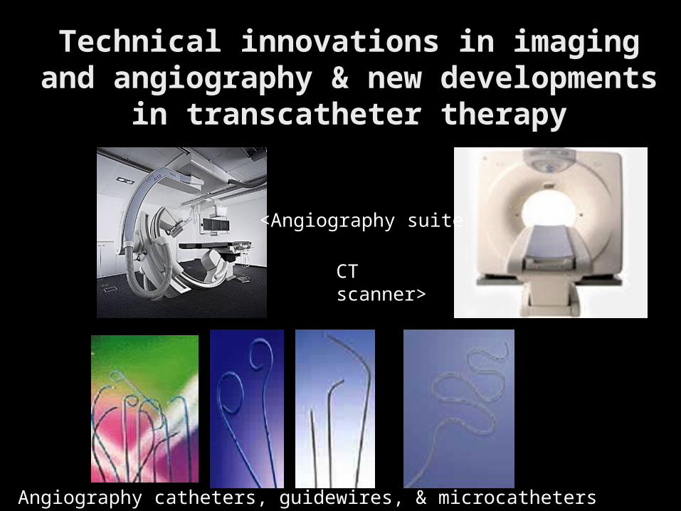
Technical innovations in imaging and Technical innovations in imaging and angiography & new developments in angiography & new developments in
transcatheter therapytranscatheter therapy
<Angiography suite
CT scanner>
Angiography catheters, guidewires, & microcatheters
CT: Imaging study of CT: Imaging study of
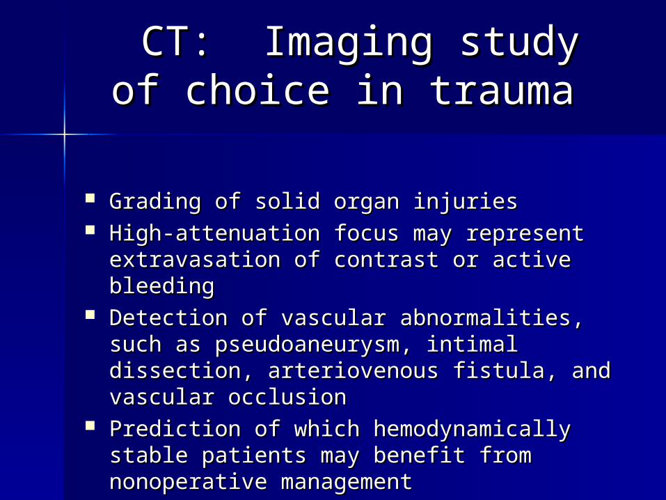
choice in traumachoice in trauma
Grading of solid organ injuries Grading of solid organ injuries High-attenuation focus may represent High-attenuation focus may represent
extravasation of contrast or active bleeding extravasation of contrast or active bleeding Detection of vascular abnormalities, such as Detection of vascular abnormalities, such as
pseudoaneurysm, intimal dissection, pseudoaneurysm, intimal dissection, arteriovenous fistula, and vascular occlusion arteriovenous fistula, and vascular occlusion
Prediction of which hemodynamically stable Prediction of which hemodynamically stable patients may benefit from nonoperative patients may benefit from nonoperative management management
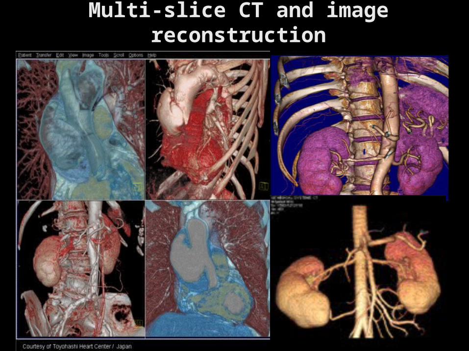
Multi-slice CT and image Multi-slice CT and image reconstructionreconstruction
Transcatheter embolization Transcatheter embolization TCE: EMBOLOTHERAPYTCE: EMBOLOTHERAPY
Transcatheter embolization TCE Transcatheter embolization TCE (embolotherapy) is the intentional (embolotherapy) is the intentional occlusion of a vessel by deposition occlusion of a vessel by deposition of thrombogenic materials directly of thrombogenic materials directly into the vessel via an angiographic into the vessel via an angiographic catheter under remote control catheter under remote control (femoral or brachial access) (femoral or brachial access)
EMBOLOTHERAPY: Mainstay of EMBOLOTHERAPY: Mainstay of modern interventional trauma modern interventional trauma
radiology. radiology.
Treated arteries must be expendable or supply a relative Treated arteries must be expendable or supply a relative infarction resistant vascular bed or must be associated with infarction resistant vascular bed or must be associated with distal collateral vessels. If end arteries, they must have distal collateral vessels. If end arteries, they must have adequate parenchymal reserve. Typically these are arteries adequate parenchymal reserve. Typically these are arteries which can be ligated surgically. which can be ligated surgically.
Transcatheter embolization of active hemorrhage or Transcatheter embolization of active hemorrhage or vascular injury often may be preferable to surgical vascular injury often may be preferable to surgical alternatives in the following instances: alternatives in the following instances: – When rapid occlusion is desired When rapid occlusion is desired – When surgical access is difficult When surgical access is difficult – When the patient is a poor operative risk When the patient is a poor operative risk – When selective transcatheter embolization may limit the When selective transcatheter embolization may limit the
amount of normal tissue or parenchyma necrotizedamount of normal tissue or parenchyma necrotized
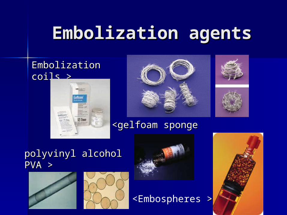
Embolization agentsEmbolization agents
Embolization coilsEmbolization coils >>
<gelfoam sponge<gelfoam sponge
polyvinyl alcohol polyvinyl alcohol PVA >PVA >
<Embospheres >>
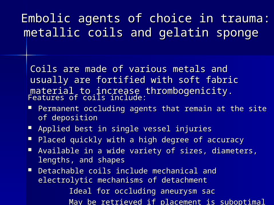
Embolic agents of choice in trauma: Embolic agents of choice in trauma: metallic coils and gelatin spongemetallic coils and gelatin sponge
Features of coils include: Features of coils include: Permanent occluding agents that remain at the site of Permanent occluding agents that remain at the site of
deposition deposition Applied best in single vessel injuries Applied best in single vessel injuries Placed quickly with a high degree of accuracy Placed quickly with a high degree of accuracy Available in a wide variety of sizes, diameters, lengths, and Available in a wide variety of sizes, diameters, lengths, and
shapes shapes Detachable coils include mechanical and electrolytic Detachable coils include mechanical and electrolytic
mechanisms of detachment mechanisms of detachment
Ideal for occluding aneurysm sac Ideal for occluding aneurysm sac
May be retrieved if placement is suboptimalMay be retrieved if placement is suboptimal
Coils are made of various metals and usually are Coils are made of various metals and usually are fortified with soft fabric material to increase fortified with soft fabric material to increase thrombogenicity.thrombogenicity.
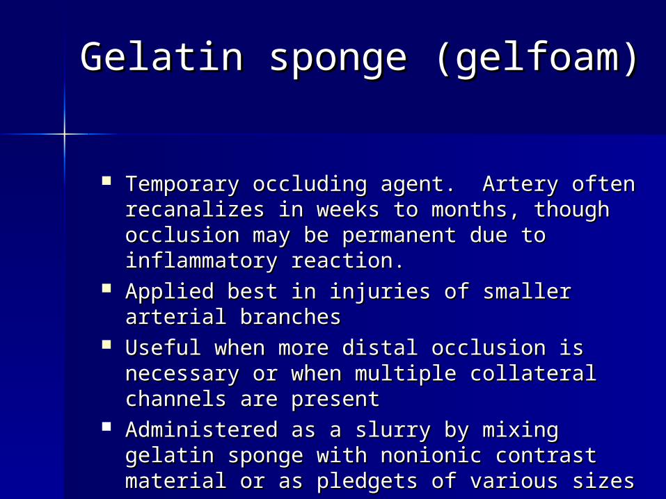
Gelatin sponge (gelfoam)Gelatin sponge (gelfoam)
Temporary occluding agent. Artery often Temporary occluding agent. Artery often recanalizes in weeks to months, though recanalizes in weeks to months, though occlusion may be permanent due to occlusion may be permanent due to inflammatory reaction.inflammatory reaction.
Applied best in injuries of smaller arterial Applied best in injuries of smaller arterial branchesbranches
Useful when more distal occlusion is Useful when more distal occlusion is necessary or when multiple collateral necessary or when multiple collateral channels are present channels are present
Administered as a slurry by mixing gelatin Administered as a slurry by mixing gelatin sponge with nonionic contrast material or as sponge with nonionic contrast material or as pledgets of various sizespledgets of various sizes
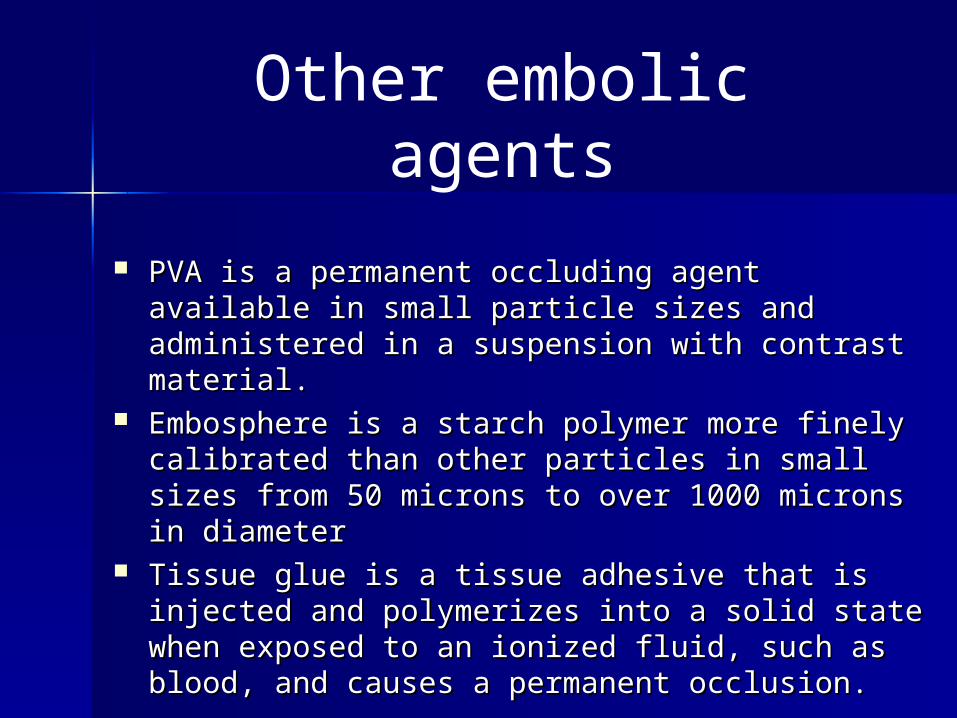
PVA is a permanent occluding agent available in PVA is a permanent occluding agent available in small particle sizes and administered in a small particle sizes and administered in a suspension with contrast material. suspension with contrast material.
Embosphere is a starch polymer more finely Embosphere is a starch polymer more finely calibrated than other particles in small sizes from calibrated than other particles in small sizes from 50 microns to over 1000 microns in diameter50 microns to over 1000 microns in diameter
Tissue glue is a tissue adhesive that is injected Tissue glue is a tissue adhesive that is injected and polymerizes into a solid state when exposed and polymerizes into a solid state when exposed to an ionized fluid, such as blood, and causes a to an ionized fluid, such as blood, and causes a permanent occlusion. permanent occlusion.
Other embolic agents
Stent graftsStent grafts
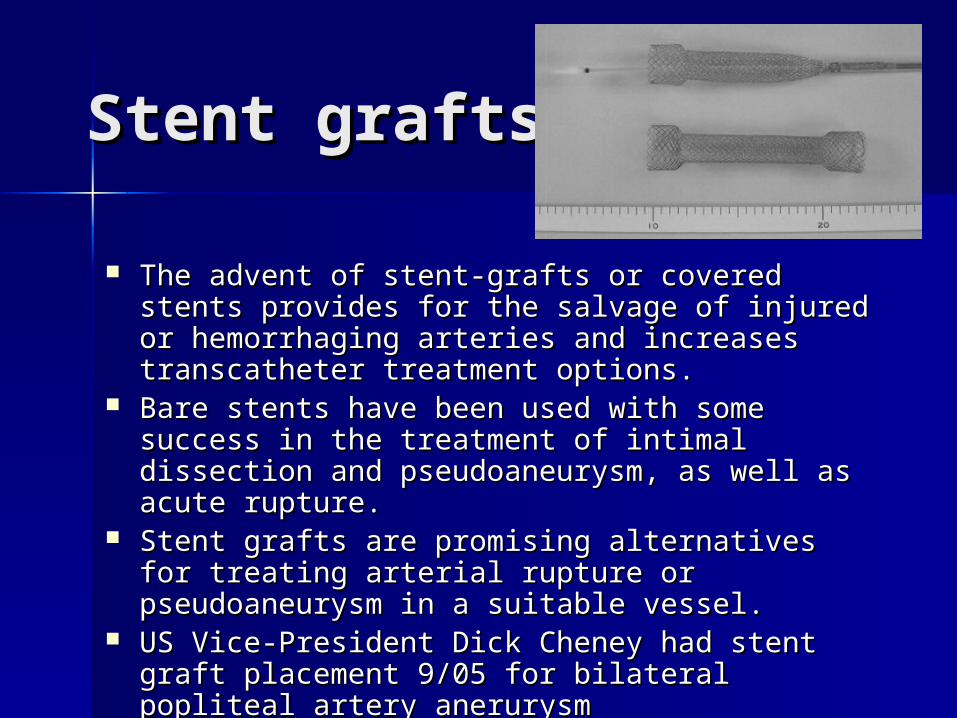
The advent of stent-grafts or covered stents The advent of stent-grafts or covered stents provides for the salvage of injured or provides for the salvage of injured or hemorrhaging arteries and increases hemorrhaging arteries and increases transcatheter treatment options. transcatheter treatment options.
Bare stents have been used with some success Bare stents have been used with some success in the treatment of intimal dissection and in the treatment of intimal dissection and pseudoaneurysm, as well as acute rupture. pseudoaneurysm, as well as acute rupture.
Stent grafts are promising alternatives for Stent grafts are promising alternatives for treating arterial rupture or pseudoaneurysm in treating arterial rupture or pseudoaneurysm in a suitable vessel. a suitable vessel.
US Vice-President Dick Cheney had stent graft US Vice-President Dick Cheney had stent graft placement 9/05 for bilateral popliteal artery placement 9/05 for bilateral popliteal artery anerurysmanerurysm
Stent-graft featuresStent-graft features
Stents are covered with vein or synthetic Stents are covered with vein or synthetic materials, such as materials, such as polytetrafluoroethylene, polyethylene polytetrafluoroethylene, polyethylene terephthalate (eg, Dacron), polycarbonate terephthalate (eg, Dacron), polycarbonate urethane compounds, or other proprietary urethane compounds, or other proprietary materials. materials.
The balloons are expandable or self-The balloons are expandable or self-expandable. expandable.
Stents exclude and effectively repair the Stents exclude and effectively repair the injured arterial segment injured arterial segment
ACUTE THORACIC AORTIC ACUTE THORACIC AORTIC INJURY (ATAI)INJURY (ATAI)
Incidence and natural historyIncidence and natural history
Only 10-23% survive long enough to present to the hospital. Only 10-23% survive long enough to present to the hospital. Of those patients, approximately 30% are fatal within 6 hours Of those patients, approximately 30% are fatal within 6 hours and 40% are fatal within 24 hours if undiagnosed and left and 40% are fatal within 24 hours if undiagnosed and left untreated. Only 2-10% of untreated patients survive longer untreated. Only 2-10% of untreated patients survive longer than 6 months. than 6 months.
Acute thoracic aortic injury (ATAI) aka aortic transection, aortic traumatic pseudoaneurysm, aortic rupture, aortic laceration, and aortic tear.
ATAIs are responsible for 10-20% of high-speed traffic accident fatalities. More than 8000 cases per year in the US. Most ATAIs represent full-thickness tears, and most cases ATAIs are fatal at the scene of the accident
Pathophysiology of ATAIPathophysiology of ATAI
ATAIs caused by rapid deceleration forces produced by high-ATAIs caused by rapid deceleration forces produced by high-speed MVA or falls from great heights. speed MVA or falls from great heights. – During acute deceleration, the thoracic aorta is relatively During acute deceleration, the thoracic aorta is relatively
fixed in position at the aortic root, isthmus, and fixed in position at the aortic root, isthmus, and diaphragm. diaphragm.
– Movement of the aorta about these fixation points causes Movement of the aorta about these fixation points causes stress and tethering, resulting in a tear at these locations.stress and tethering, resulting in a tear at these locations.
Among patients who survive with ATAI, the locations of tears Among patients who survive with ATAI, the locations of tears are: are: – Aortic isthmus, 80-90% Aortic isthmus, 80-90% – Ascending aorta, 5-9% Ascending aorta, 5-9% – Diaphragmatic aorta, 1-3%Diaphragmatic aorta, 1-3%
Of ATAI cases fatal at the scene, a higher percentage of Of ATAI cases fatal at the scene, a higher percentage of lacerations involve the aortic root. lacerations involve the aortic root.
Pathologically, a transverse tear of the aortic intima and Pathologically, a transverse tear of the aortic intima and media is found, and the adventitia is intact in approximately media is found, and the adventitia is intact in approximately 60% of patients. 60% of patients.
Multiple sites of ATAI occur in 6-20% of patients, and aortic Multiple sites of ATAI occur in 6-20% of patients, and aortic arch branch artery injuries occur in 4-10% of patients.arch branch artery injuries occur in 4-10% of patients.
Imaging studies of ATAIImaging studies of ATAI
Imaging of ATAIs consists of plain Imaging of ATAIs consists of plain radiography, CT, conventional thoracic radiography, CT, conventional thoracic aortography, transesophageal aortography, transesophageal echocardiography, intravascular echocardiography, intravascular ultrasound, MRI, and magnetic ultrasound, MRI, and magnetic resonance angiography. Of these, the 3 resonance angiography. Of these, the 3 most commonly used modalities in the most commonly used modalities in the assessment of ATAI are plain assessment of ATAI are plain radiography, CT, and conventional radiography, CT, and conventional thoracic aortography. thoracic aortography.
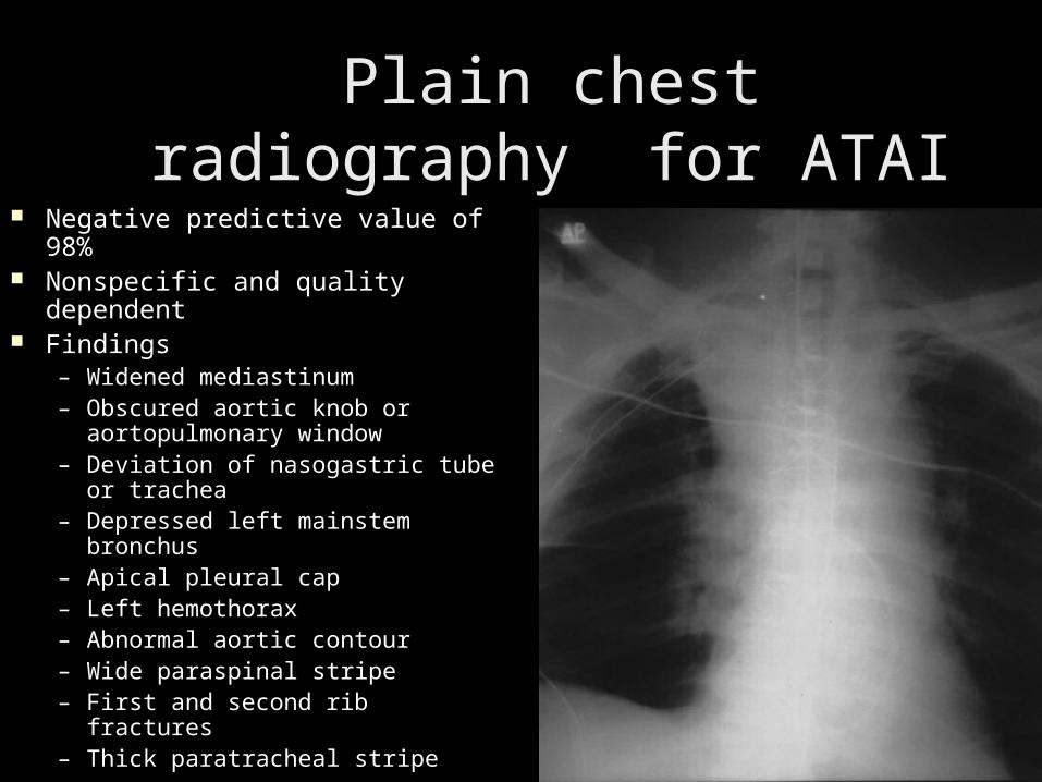
Plain chest radiography Plain chest radiography for ATAIfor ATAI
Negative predictive value of Negative predictive value of 98% 98%
Nonspecific and quality Nonspecific and quality dependentdependent
Findings Findings – Widened mediastinum Widened mediastinum – Obscured aortic knob or Obscured aortic knob or
aortopulmonary window aortopulmonary window – Deviation of nasogastric tube or Deviation of nasogastric tube or
trachea trachea – Depressed left mainstem Depressed left mainstem
bronchus bronchus – Apical pleural cap Apical pleural cap – Left hemothorax Left hemothorax – Abnormal aortic contour Abnormal aortic contour – Wide paraspinal stripe Wide paraspinal stripe – First and second rib fractures First and second rib fractures – Thick paratracheal stripeThick paratracheal stripe
CT - Detection of thoracic CT - Detection of thoracic aortic injury and mediastinal aortic injury and mediastinal hematomahematoma Findings for thoracic aortic injuryFindings for thoracic aortic injury Intraluminal low-attenuation focus Intraluminal low-attenuation focus Contour abnormality Contour abnormality Pseudoaneurysm Pseudoaneurysm Intramural hematoma Intramural hematoma Localized dissectionLocalized dissectionIsolated aortic injury without a mediastinal Isolated aortic injury without a mediastinal
hematoma is rare. hematoma is rare. Screening for mediastinal hematoma by CT can Screening for mediastinal hematoma by CT can
increase the positive conventional thoracic increase the positive conventional thoracic aortogram rate. aortogram rate.
CT demonstrates a sensitivity of nearly 100% in CT demonstrates a sensitivity of nearly 100% in aortic injury but is slightly nonspecific. aortic injury but is slightly nonspecific.
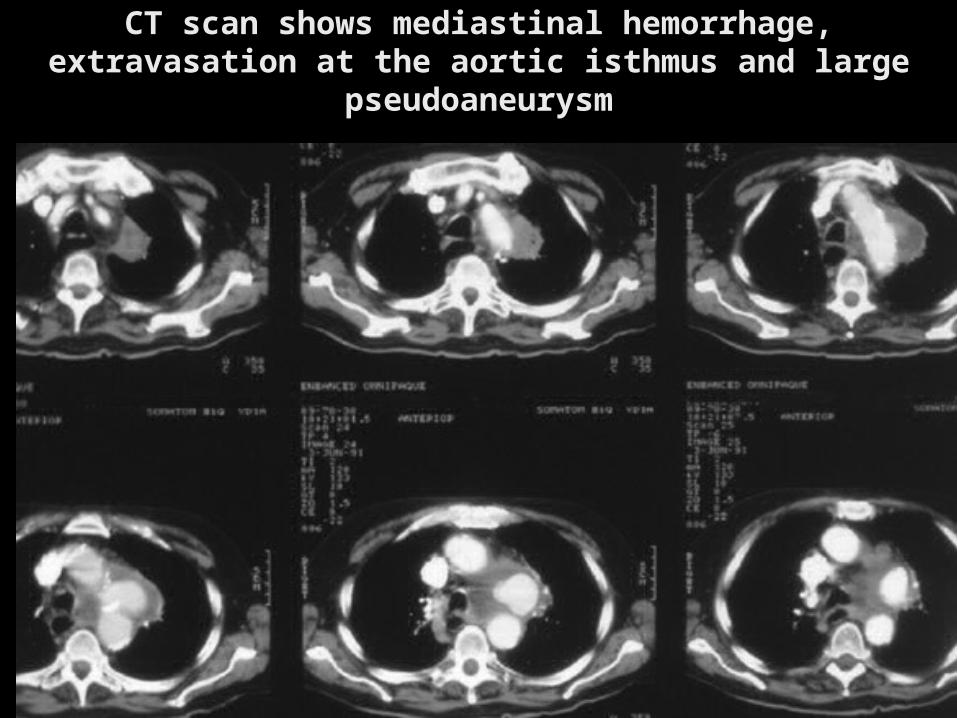
CT scan shows mediastinal hemorrhage, CT scan shows mediastinal hemorrhage, extravasation at the aortic isthmus and large extravasation at the aortic isthmus and large
pseudoaneurysmpseudoaneurysm
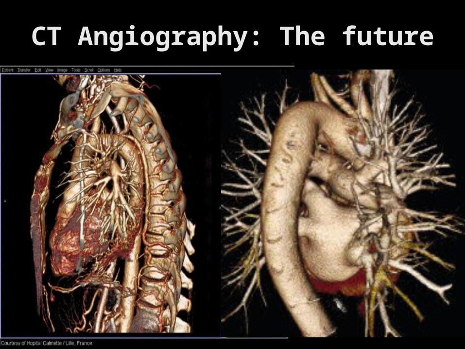
CT Angiography: The futureCT Angiography: The future
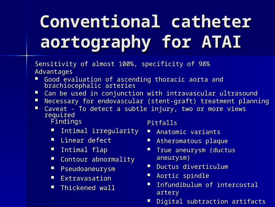
Conventional catheter Conventional catheter aortography for ATAIaortography for ATAI
Findings Findings Intimal irregularity Intimal irregularity Linear defect Linear defect Intimal flap Intimal flap Contour abnormality Contour abnormality Pseudoaneurysm Pseudoaneurysm Extravasation Extravasation Thickened wallThickened wall
Pitfalls Pitfalls Anatomic variants Anatomic variants Atheromatous plaque Atheromatous plaque True aneurysm (ductus aneurysm) True aneurysm (ductus aneurysm) Ductus diverticulum Ductus diverticulum Aortic spindle Aortic spindle Infundibulum of intercostal artery Infundibulum of intercostal artery Digital subtraction artifactsDigital subtraction artifacts
Sensitivity of almost 100%, specificity of 98% Sensitivity of almost 100%, specificity of 98% Advantages Advantages Good evaluation of ascending thoracic aorta and brachiocephalic Good evaluation of ascending thoracic aorta and brachiocephalic
arteries arteries Can be used in conjunction with intravascular ultrasound Can be used in conjunction with intravascular ultrasound Necessary for endovascular (stent-graft) treatment planningNecessary for endovascular (stent-graft) treatment planning Caveat - To detect a subtle injury, two or more views required Caveat - To detect a subtle injury, two or more views required
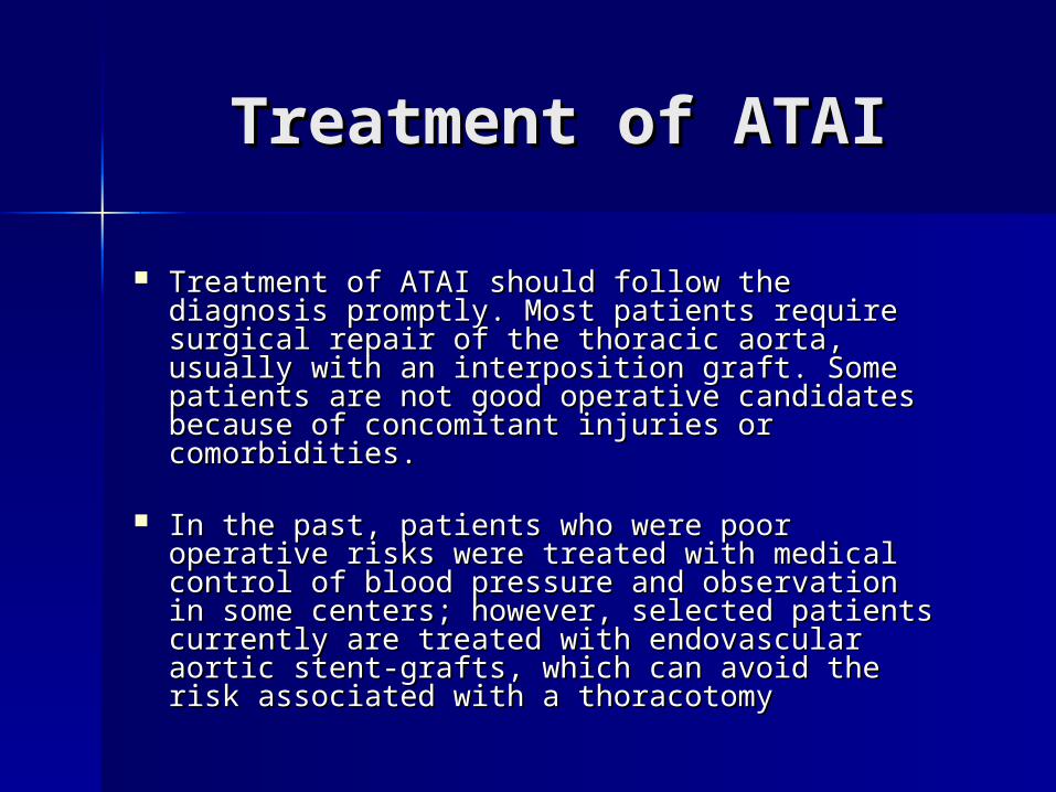
Treatment of ATAITreatment of ATAI
Treatment of ATAI should follow the Treatment of ATAI should follow the diagnosis promptly. Most patients require diagnosis promptly. Most patients require surgical repair of the thoracic aorta, usually surgical repair of the thoracic aorta, usually with an interposition graft. Some patients with an interposition graft. Some patients are not good operative candidates because are not good operative candidates because of concomitant injuries or comorbidities. of concomitant injuries or comorbidities.
In the past, patients who were poor In the past, patients who were poor operative risks were treated with medical operative risks were treated with medical control of blood pressure and observation in control of blood pressure and observation in some centers; however, selected patients some centers; however, selected patients currently are treated with endovascular currently are treated with endovascular aortic stent-grafts, which can avoid the risk aortic stent-grafts, which can avoid the risk associated with a thoracotomy associated with a thoracotomy
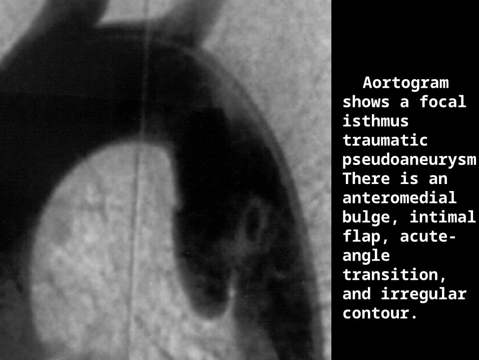
Aortogram shows a focal isthmus traumatic pseudoaneurysmThere is an anteromedial bulge, intimal flap, acute-angle transition, and irregular contour.
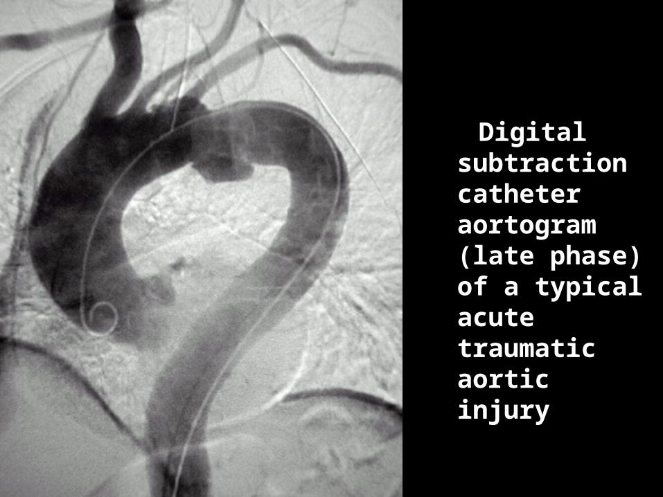
Digital Digital subtraction catheter catheter aortogram aortogram (late phase) (late phase) of a of a typical acute acute traumatic traumatic aortic injury aortic injury
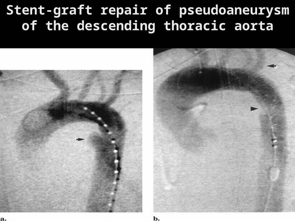
Stent-graft repair of pseudoaneurysm Stent-graft repair of pseudoaneurysm of the descending thoracic aortaof the descending thoracic aorta
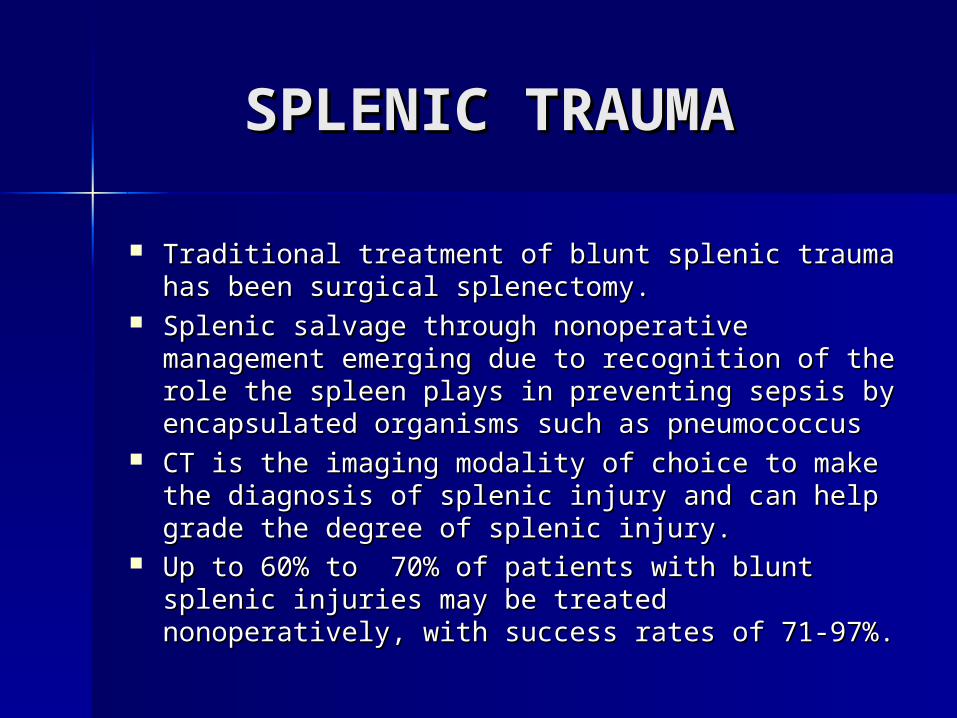
SPLENIC TRAUMA SPLENIC TRAUMA
Traditional treatment of blunt splenic trauma has Traditional treatment of blunt splenic trauma has been surgical splenectomy. been surgical splenectomy.
Splenic salvage through nonoperative Splenic salvage through nonoperative management emerging due to recognition of the management emerging due to recognition of the role the spleen plays in preventing sepsis by role the spleen plays in preventing sepsis by encapsulated organisms such as pneumococcus encapsulated organisms such as pneumococcus
CT is the imaging modality of choice to make the CT is the imaging modality of choice to make the diagnosis of splenic injury and can help grade the diagnosis of splenic injury and can help grade the degree of splenic injury.degree of splenic injury.
Up to 60% to 70% of patients with blunt splenic Up to 60% to 70% of patients with blunt splenic injuries may be treated nonoperatively, with injuries may be treated nonoperatively, with success rates of 71-97%.success rates of 71-97%.
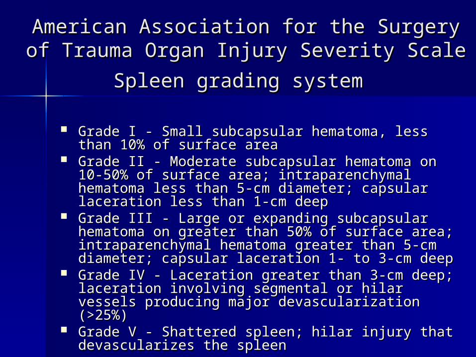
American Association for the Surgery of American Association for the Surgery of Trauma Organ Injury Severity Scale Spleen Trauma Organ Injury Severity Scale Spleen
grading systemgrading system
Grade I - Small subcapsular hematoma, less than 10% Grade I - Small subcapsular hematoma, less than 10% of surface area of surface area
Grade II - Moderate subcapsular hematoma on 10-Grade II - Moderate subcapsular hematoma on 10-50% of surface area; intraparenchymal hematoma 50% of surface area; intraparenchymal hematoma less than 5-cm diameter; capsular laceration less than less than 5-cm diameter; capsular laceration less than 1-cm deep 1-cm deep
Grade III - Large or expanding subcapsular hematoma Grade III - Large or expanding subcapsular hematoma on greater than 50% of surface area; on greater than 50% of surface area; intraparenchymal hematoma greater than 5-cm intraparenchymal hematoma greater than 5-cm diameter; capsular laceration 1- to 3-cm deep diameter; capsular laceration 1- to 3-cm deep
Grade IV - Laceration greater than 3-cm deep; Grade IV - Laceration greater than 3-cm deep; laceration involving segmental or hilar vessels laceration involving segmental or hilar vessels producing major devascularization (>25%) producing major devascularization (>25%)
Grade V - Shattered spleen; hilar injury that Grade V - Shattered spleen; hilar injury that devascularizes the spleen devascularizes the spleen
Splenic trauma: Splenic trauma: Imaging studies Imaging studies
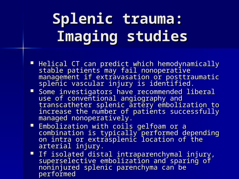
Helical CT can predict which hemodynamically Helical CT can predict which hemodynamically stable patients may fail nonoperative stable patients may fail nonoperative management if extravasation or posttraumatic management if extravasation or posttraumatic splenic vascular injury is identified. splenic vascular injury is identified.
Some investigators have recommended liberal use Some investigators have recommended liberal use of conventional angiography and transcatheter of conventional angiography and transcatheter splenic artery embolization to increase the number splenic artery embolization to increase the number of patients successfully managed nonoperatively. of patients successfully managed nonoperatively.
Embolization with coils gelfoam or a combination Embolization with coils gelfoam or a combination is typically performed depending on intra or is typically performed depending on intra or extrasplenic location of the arterial injury. extrasplenic location of the arterial injury.
If isolated distal intraparenchymal injury, If isolated distal intraparenchymal injury, superselective embolization and sparing of superselective embolization and sparing of noninjured splenic parenchyma can be performednoninjured splenic parenchyma can be performed
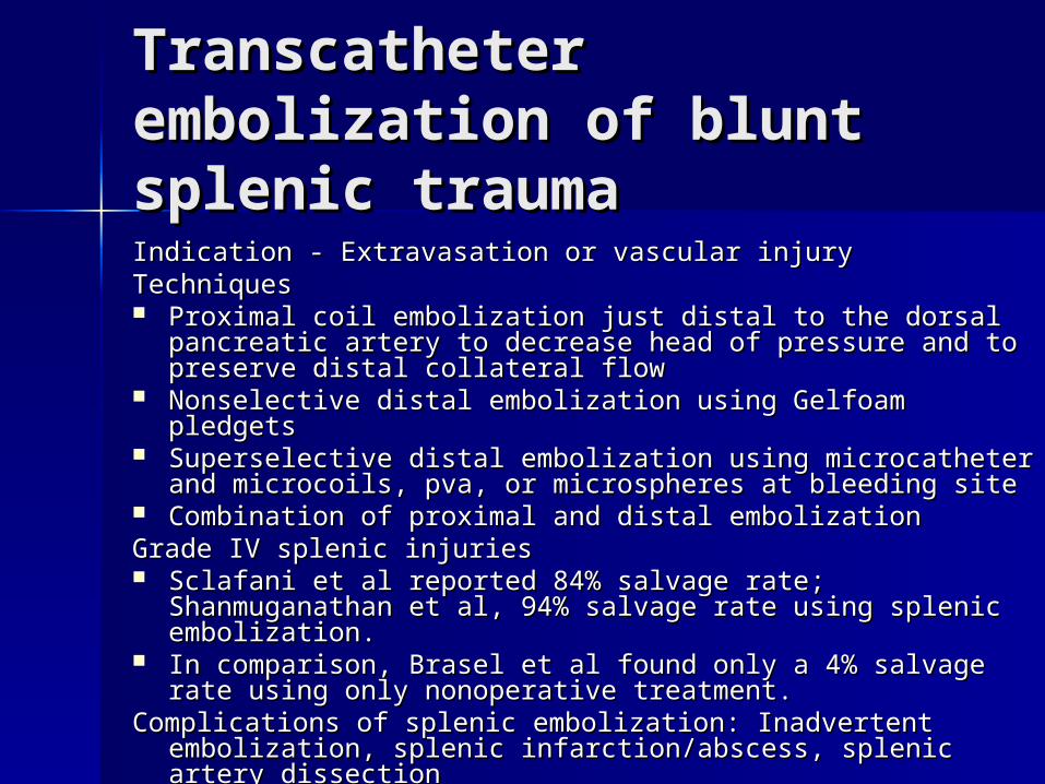
Transcatheter Transcatheter embolization of blunt embolization of blunt splenic trauma splenic trauma Indication - Extravasation or vascular injury Indication - Extravasation or vascular injury Techniques Techniques Proximal coil embolization just distal to the dorsal Proximal coil embolization just distal to the dorsal
pancreatic artery to decrease head of pressure and to pancreatic artery to decrease head of pressure and to preserve distal collateral flow preserve distal collateral flow
Nonselective distal embolization using Gelfoam pledgets Nonselective distal embolization using Gelfoam pledgets Superselective distal embolization using microcatheter Superselective distal embolization using microcatheter
and microcoils, pva, or microspheres at bleeding site and microcoils, pva, or microspheres at bleeding site Combination of proximal and distal embolizationCombination of proximal and distal embolizationGrade IV splenic injuries Grade IV splenic injuries Sclafani et al reported 84% salvage rate; Sclafani et al reported 84% salvage rate;
Shanmuganathan et al, 94% salvage rate using splenic Shanmuganathan et al, 94% salvage rate using splenic embolization. embolization.
In comparison, Brasel et al found only a 4% salvage rate In comparison, Brasel et al found only a 4% salvage rate using only nonoperative treatment.using only nonoperative treatment.
Complications of splenic embolization: Inadvertent Complications of splenic embolization: Inadvertent embolization, splenic infarction/abscess, splenic artery embolization, splenic infarction/abscess, splenic artery dissectiondissection
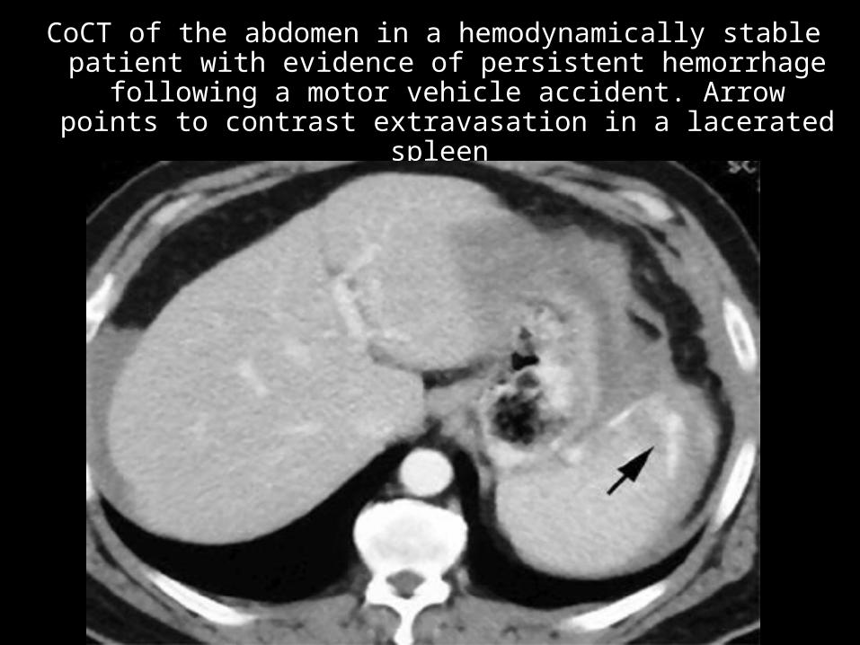
CoCT of the abdomen in a hemodynamically stable CoCT of the abdomen in a hemodynamically stable patient with evidence of persistent hemorrhage patient with evidence of persistent hemorrhage
following a motor vehicle accident. Arrow points to following a motor vehicle accident. Arrow points to contrast extravasation in a lacerated spleen contrast extravasation in a lacerated spleen
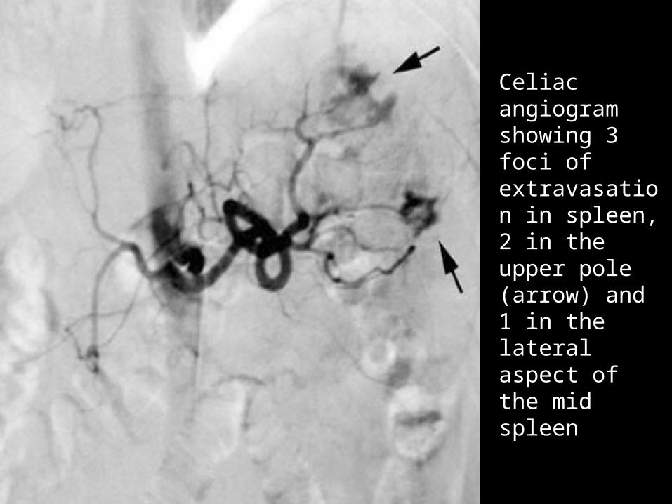
Celiac Celiac angiogram angiogram showing 3 showing 3 foci of foci of extravasatioextravasation in spleen, n in spleen, 2 in the 2 in the upper pole upper pole (arrow) and (arrow) and 1 in the 1 in the lateral lateral aspect of the aspect of the mid spleen mid spleen
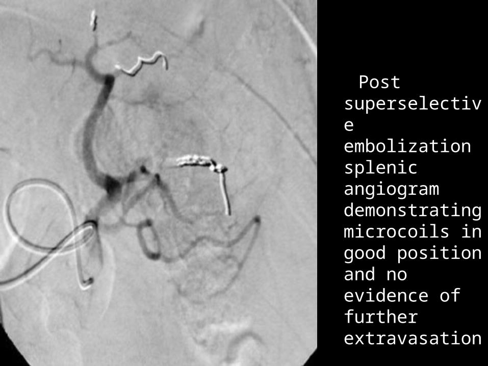
Post superselective embolization splenic angiogram demonstrating microcoils in good position and no evidence of further extravasation
HEPATIC TRAUMA HEPATIC TRAUMA
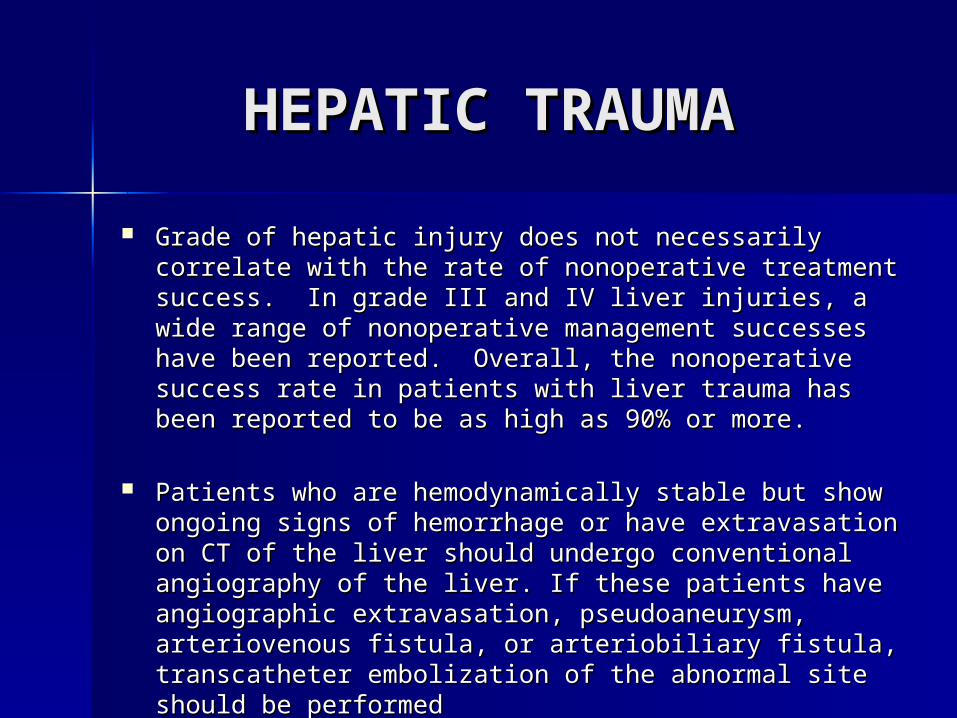
Grade of hepatic injury does not necessarily correlate Grade of hepatic injury does not necessarily correlate with the rate of nonoperative treatment success. In with the rate of nonoperative treatment success. In grade III and IV liver injuries, a wide range of grade III and IV liver injuries, a wide range of nonoperative management successes have been nonoperative management successes have been reported. Overall, the nonoperative success rate in reported. Overall, the nonoperative success rate in patients with liver trauma has been reported to be as patients with liver trauma has been reported to be as high as 90% or more. high as 90% or more.
Patients who are hemodynamically stable but show Patients who are hemodynamically stable but show ongoing signs of hemorrhage or have extravasation on ongoing signs of hemorrhage or have extravasation on CT of the liver should undergo conventional CT of the liver should undergo conventional angiography of the liver. If these patients have angiography of the liver. If these patients have angiographic extravasation, pseudoaneurysm, angiographic extravasation, pseudoaneurysm, arteriovenous fistula, or arteriobiliary fistula, arteriovenous fistula, or arteriobiliary fistula, transcatheter embolization of the abnormal site should transcatheter embolization of the abnormal site should be performed be performed
American Association for the Surgery of American Association for the Surgery of Trauma Organ Injury Severity Scale Liver Trauma Organ Injury Severity Scale Liver
grading systemgrading system
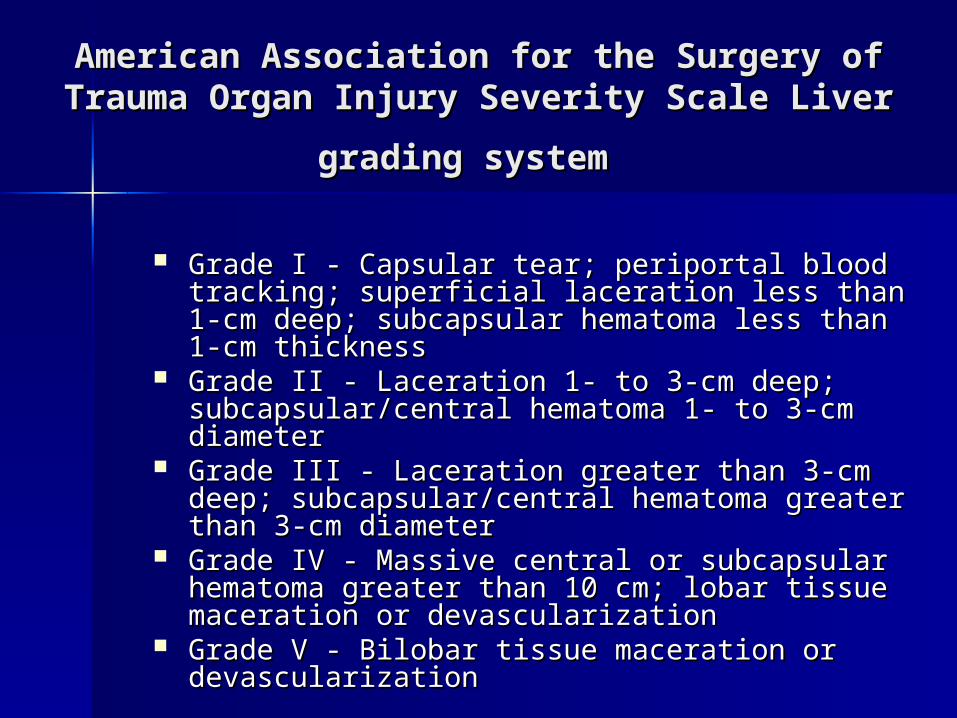
Grade I - Capsular tear; periportal blood Grade I - Capsular tear; periportal blood tracking; superficial laceration less than 1-cm tracking; superficial laceration less than 1-cm deep; subcapsular hematoma less than 1-cm deep; subcapsular hematoma less than 1-cm thickness thickness
Grade II - Laceration 1- to 3-cm deep; Grade II - Laceration 1- to 3-cm deep; subcapsular/central hematoma 1- to 3-cm subcapsular/central hematoma 1- to 3-cm diameter diameter
Grade III - Laceration greater than 3-cm deep; Grade III - Laceration greater than 3-cm deep; subcapsular/central hematoma greater than 3-subcapsular/central hematoma greater than 3-cm diameter cm diameter
Grade IV - Massive central or subcapsular Grade IV - Massive central or subcapsular hematoma greater than 10 cm; lobar tissue hematoma greater than 10 cm; lobar tissue maceration or devascularization maceration or devascularization
Grade V - Bilobar tissue maceration or Grade V - Bilobar tissue maceration or devascularization devascularization
Transcatheter Transcatheter embolization embolization of the liver of the liver
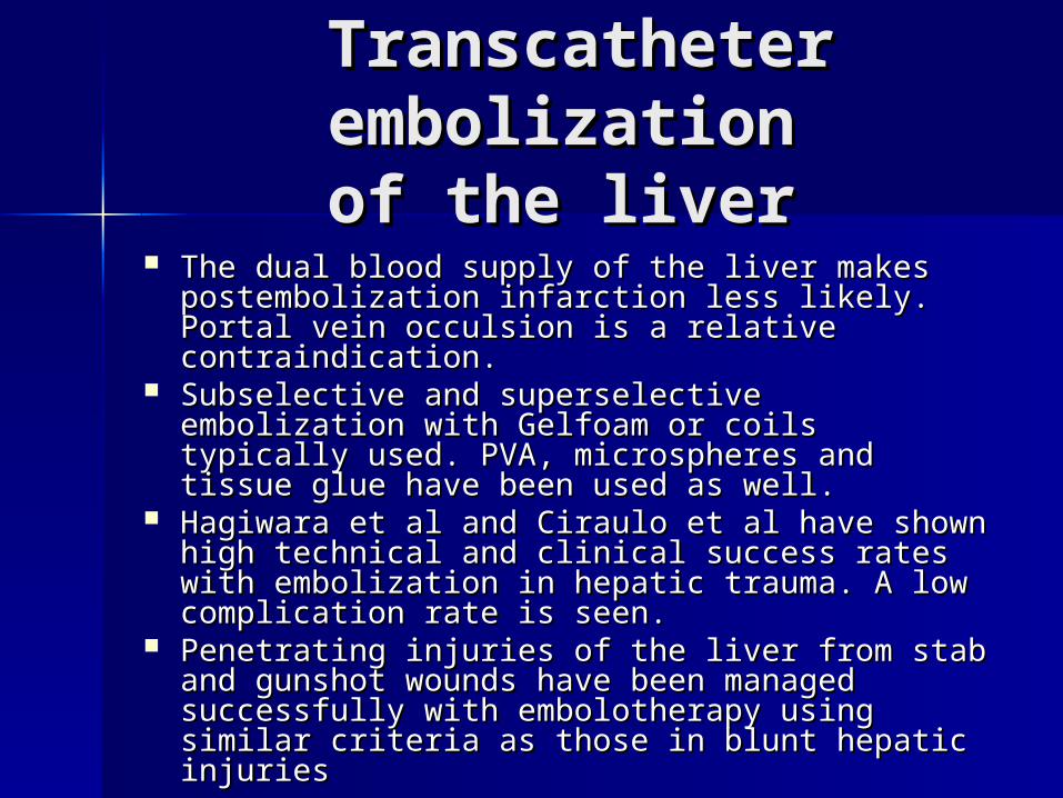
The dual blood supply of the liver makes The dual blood supply of the liver makes postembolization infarction less likely. Portal postembolization infarction less likely. Portal vein occulsion is a relative contraindication. vein occulsion is a relative contraindication.
Subselective and superselective embolization Subselective and superselective embolization with Gelfoam or coils typically used. PVA, with Gelfoam or coils typically used. PVA, microspheres and tissue glue have been used microspheres and tissue glue have been used as well. as well.
Hagiwara et al and Ciraulo et al have shown Hagiwara et al and Ciraulo et al have shown high technical and clinical success rates with high technical and clinical success rates with embolization in hepatic trauma. A low embolization in hepatic trauma. A low complication rate is seen.complication rate is seen.
Penetrating injuries of the liver from stab and Penetrating injuries of the liver from stab and gunshot wounds have been managed gunshot wounds have been managed successfully with embolotherapy using similar successfully with embolotherapy using similar criteria as those in blunt hepatic injuries criteria as those in blunt hepatic injuries
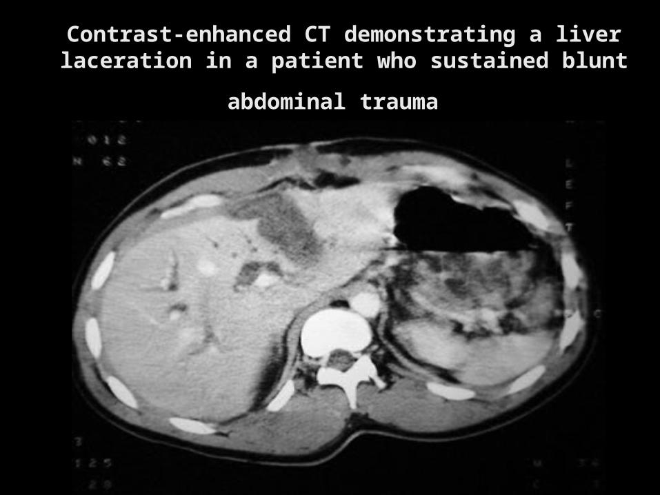
Contrast-enhanced CT demonstrating a liver Contrast-enhanced CT demonstrating a liver laceration in a patient who sustained blunt laceration in a patient who sustained blunt
abdominal traumaabdominal trauma
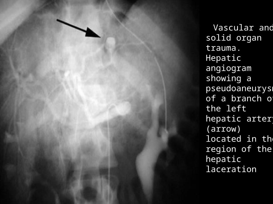
Vascular and Vascular and solid organ solid organ trauma. trauma. Hepatic Hepatic angiogram angiogram showing a showing a pseudoaneuryspseudoaneurysm of a branch m of a branch of the left of the left hepatic artery hepatic artery (arrow) located (arrow) located in the region of in the region of the hepatic the hepatic laceration laceration
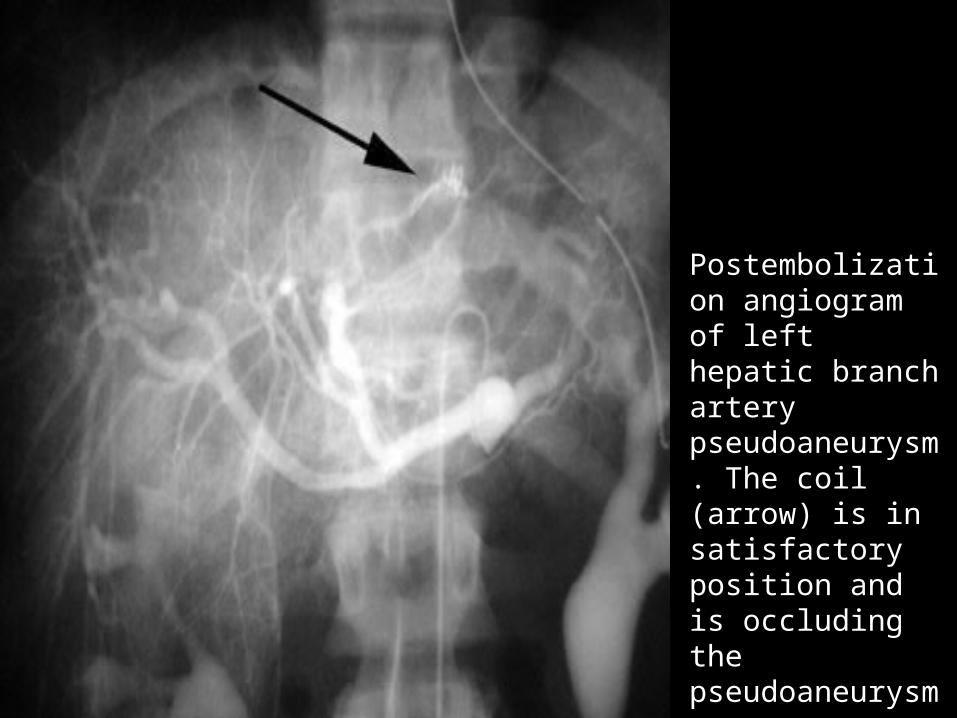
PostembolizatioPostembolization angiogram of n angiogram of left hepatic left hepatic branch artery branch artery pseudoaneuryspseudoaneurysm. The coil m. The coil (arrow) is in (arrow) is in satisfactory satisfactory position and is position and is occluding the occluding the pseudoaneuryspseudoaneurysm m
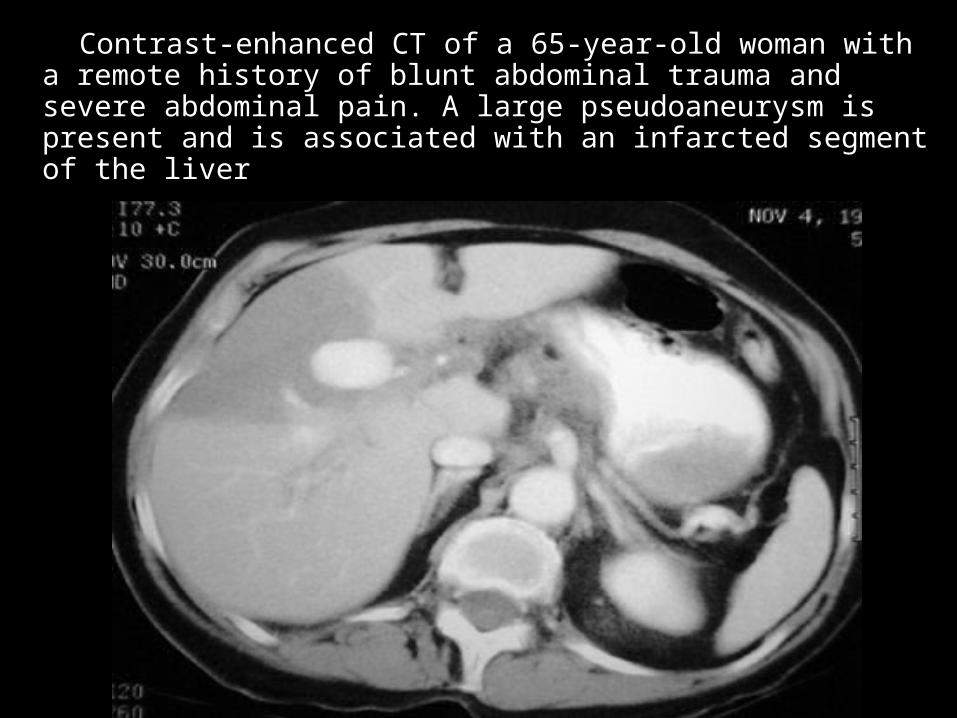
Contrast-enhanced CT of a 65-year-old woman with a Contrast-enhanced CT of a 65-year-old woman with a remote history of blunt abdominal trauma and severe remote history of blunt abdominal trauma and severe abdominal pain. A large pseudoaneurysm is present abdominal pain. A large pseudoaneurysm is present and is associated with an infarcted segment of the liver and is associated with an infarcted segment of the liver
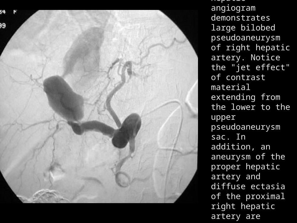
Hepatic angiogram Hepatic angiogram demonstrates large demonstrates large bilobed bilobed pseudoaneurysm of pseudoaneurysm of right hepatic artery. right hepatic artery. Notice the "jet Notice the "jet effect" of contrast effect" of contrast material extending material extending from the lower to from the lower to the upper the upper pseudoaneurysm pseudoaneurysm sac. In addition, an sac. In addition, an aneurysm of the aneurysm of the proper hepatic proper hepatic artery and diffuse artery and diffuse ectasia of the ectasia of the proximal right proximal right hepatic artery are hepatic artery are apparentapparent
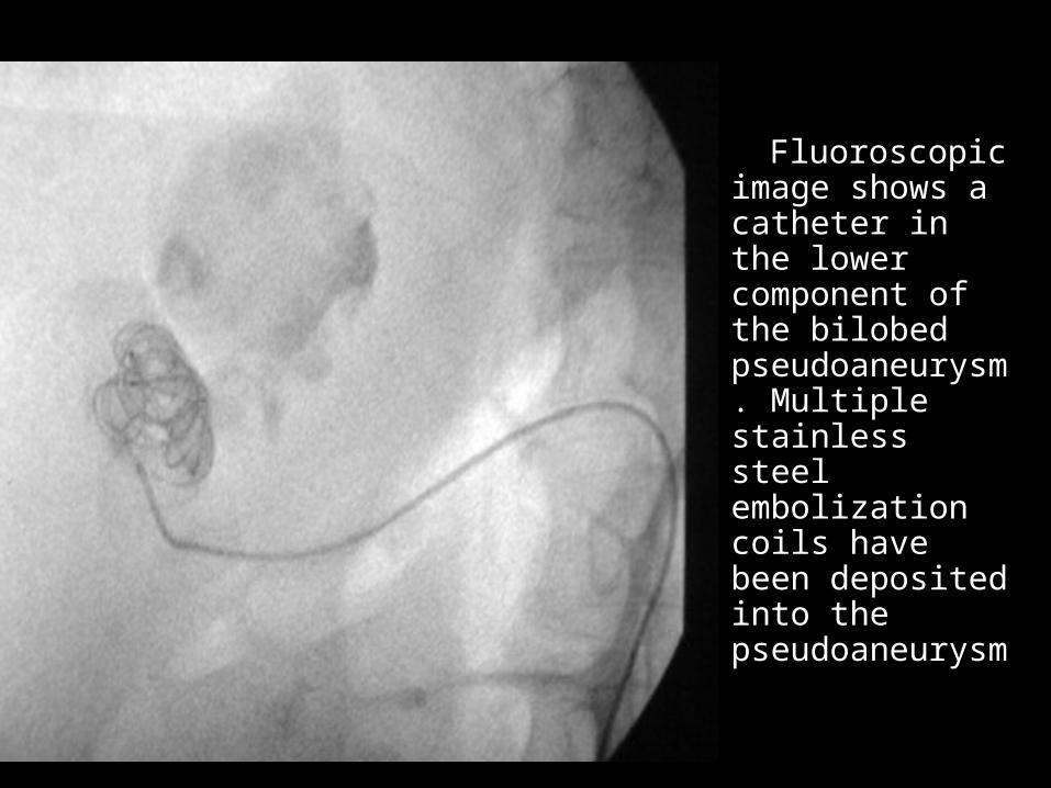
Fluoroscopic Fluoroscopic
image shows a image shows a catheter in the catheter in the lower lower component of component of the bilobed the bilobed pseudoaneuryspseudoaneurysm. Multiple m. Multiple stainless steel stainless steel embolization embolization coils have been coils have been deposited into deposited into the the pseudoaneuryspseudoaneurysm m
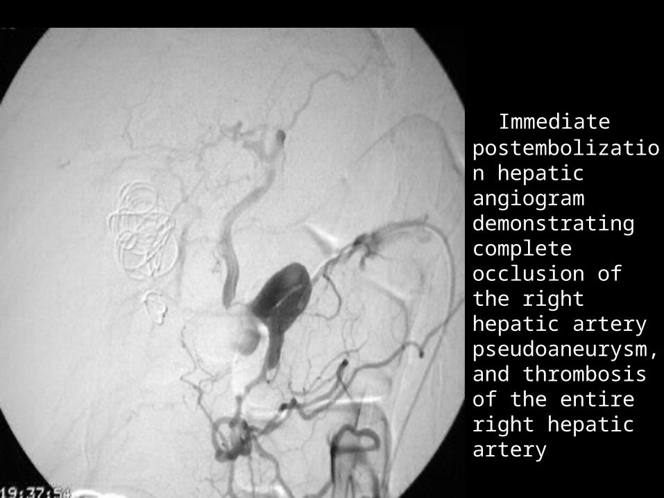
Immediate Immediate postembolization postembolization hepatic hepatic angiogram angiogram demonstrating demonstrating complete complete occlusion of the occlusion of the right hepatic right hepatic artery artery pseudoaneurysmpseudoaneurysm, and thrombosis , and thrombosis of the entire right of the entire right hepatic artery hepatic artery
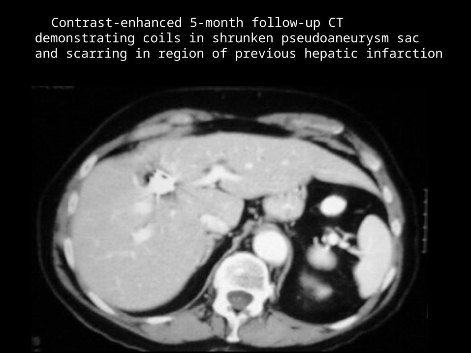
Contrast-enhanced 5-month follow-up CT demonstrating Contrast-enhanced 5-month follow-up CT demonstrating coils in shrunken pseudoaneurysm sac and scarring in coils in shrunken pseudoaneurysm sac and scarring in region of previous hepatic infarction region of previous hepatic infarction
RENAL TRAUMA RENAL TRAUMA
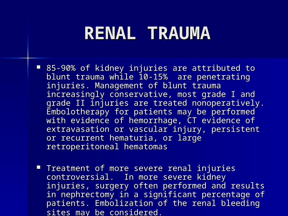
85-90% of kidney injuries are attributed to blunt 85-90% of kidney injuries are attributed to blunt trauma while 10-15% are penetrating injuries. trauma while 10-15% are penetrating injuries. Management of blunt trauma increasingly Management of blunt trauma increasingly conservative, most grade I and grade II injuries conservative, most grade I and grade II injuries are treated nonoperatively. Embolotherapy for are treated nonoperatively. Embolotherapy for patients may be performed with evidence of patients may be performed with evidence of hemorrhage, CT evidence of extravasation or hemorrhage, CT evidence of extravasation or vascular injury, persistent or recurrent hematuria, vascular injury, persistent or recurrent hematuria, or large retroperitoneal hematomasor large retroperitoneal hematomas
Treatment of more severe renal injuries Treatment of more severe renal injuries
controversial. In more severe kidney injuries, controversial. In more severe kidney injuries, surgery often performed and results in surgery often performed and results in nephrectomy in a significant percentage of nephrectomy in a significant percentage of patients. Embolization of the renal bleeding sites patients. Embolization of the renal bleeding sites may be considered.may be considered.
Renal TraumaRenal Trauma
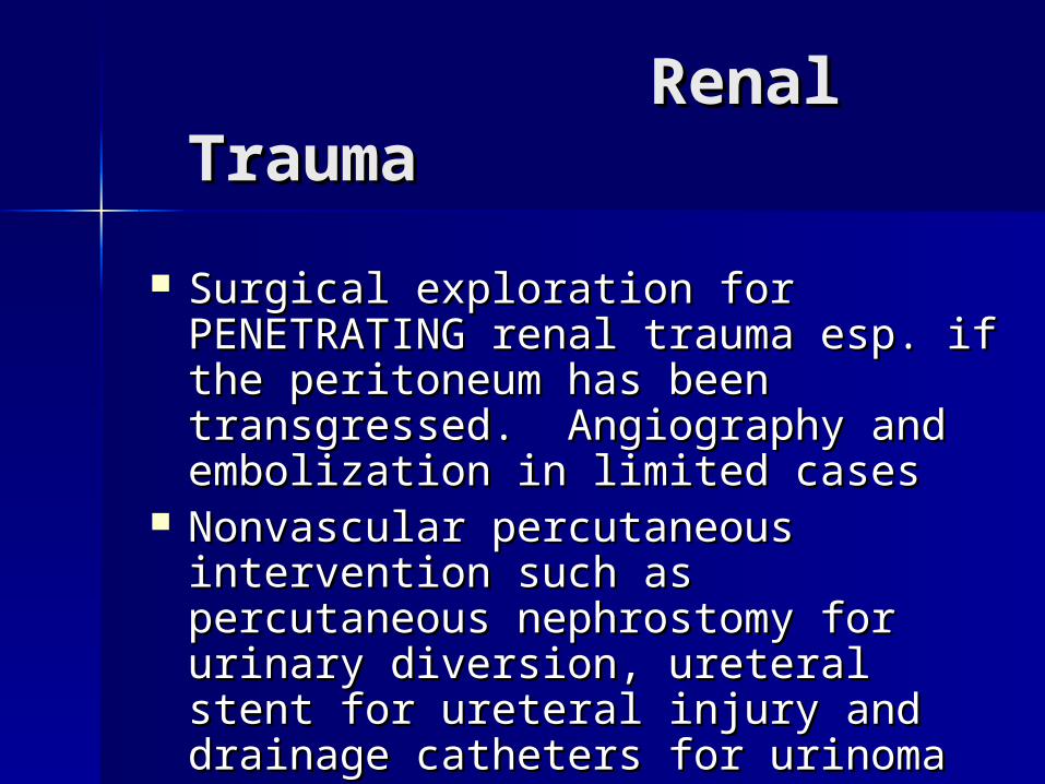
Surgical exploration for Surgical exploration for PENETRATING renal trauma esp. if PENETRATING renal trauma esp. if the peritoneum has been the peritoneum has been transgressed. Angiography and transgressed. Angiography and embolization in limited casesembolization in limited cases
Nonvascular percutaneous Nonvascular percutaneous intervention such as percutaneous intervention such as percutaneous nephrostomy for urinary diversion, nephrostomy for urinary diversion, ureteral stent for ureteral injury and ureteral stent for ureteral injury and drainage catheters for urinomadrainage catheters for urinoma
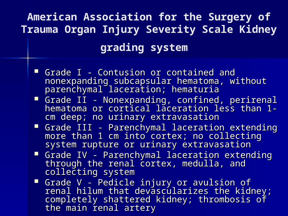
American Association for the Surgery of Trauma Organ Injury Severity Scale Kidney
grading system Grade I - Contusion or contained and nonexpanding Grade I - Contusion or contained and nonexpanding
subcapsular hematoma, without parenchymal subcapsular hematoma, without parenchymal laceration; hematuria laceration; hematuria
Grade II - Nonexpanding, confined, perirenal Grade II - Nonexpanding, confined, perirenal hematoma or cortical laceration less than 1-cm hematoma or cortical laceration less than 1-cm deep; no urinary extravasation deep; no urinary extravasation
Grade III - Parenchymal laceration extending more Grade III - Parenchymal laceration extending more than 1 cm into cortex; no collecting system rupture than 1 cm into cortex; no collecting system rupture or urinary extravasation or urinary extravasation
Grade IV - Parenchymal laceration extending Grade IV - Parenchymal laceration extending through the renal cortex, medulla, and collecting through the renal cortex, medulla, and collecting system system
Grade V - Pedicle injury or avulsion of renal hilum Grade V - Pedicle injury or avulsion of renal hilum that devascularizes the kidney; completely that devascularizes the kidney; completely shattered kidney; thrombosis of the main renal shattered kidney; thrombosis of the main renal arteryartery
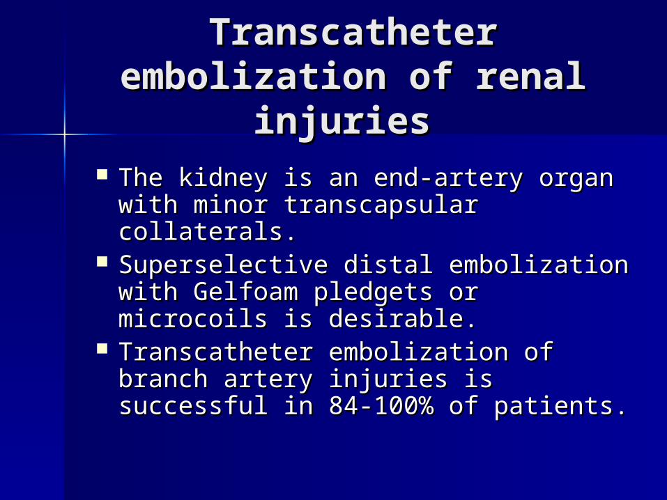
Transcatheter Transcatheter embolization of renal embolization of renal
injuries injuries The kidney is an end-artery organ The kidney is an end-artery organ
with minor transcapsular collaterals. with minor transcapsular collaterals. Superselective distal embolization Superselective distal embolization
with Gelfoam pledgets or microcoils with Gelfoam pledgets or microcoils is desirable. is desirable.
Transcatheter embolization of Transcatheter embolization of branch artery injuries is successful branch artery injuries is successful in 84-100% of patients.in 84-100% of patients.
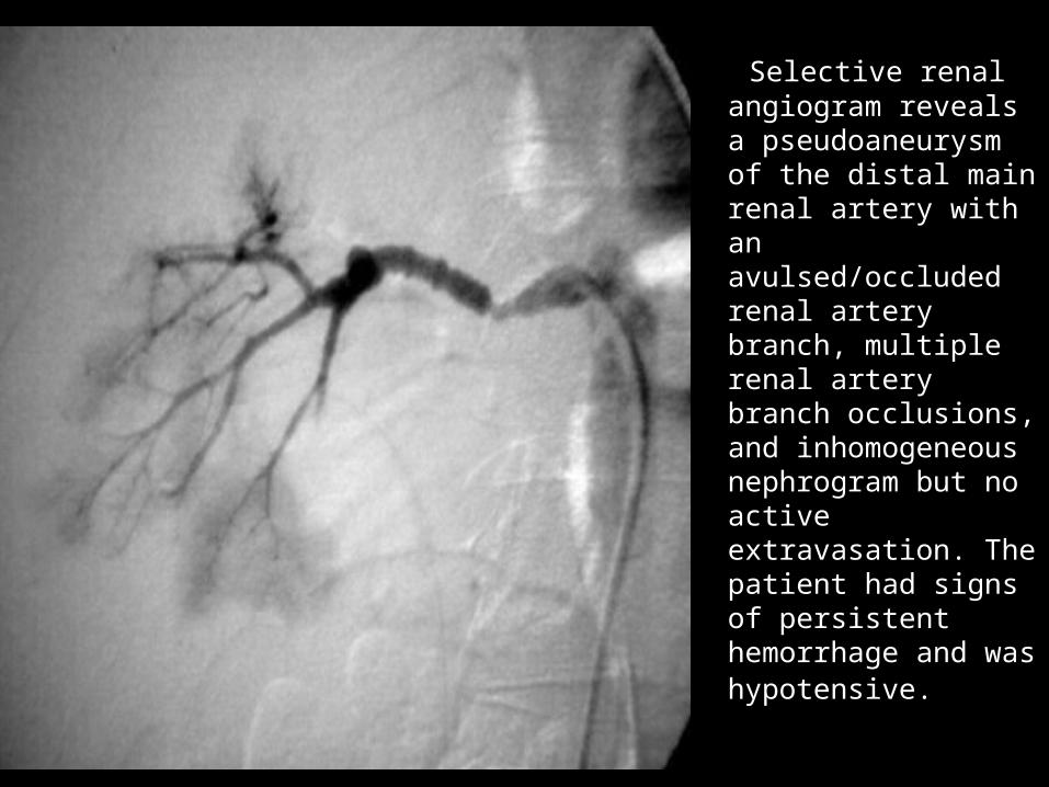
Selective renal Selective renal angiogram reveals angiogram reveals a pseudoaneurysm a pseudoaneurysm of the distal main of the distal main renal artery with an renal artery with an avulsed/occluded avulsed/occluded renal artery renal artery branch, multiple branch, multiple renal artery branch renal artery branch occlusions, and occlusions, and inhomogeneous inhomogeneous nephrogram but no nephrogram but no active active extravasation. The extravasation. The patient had signs of patient had signs of persistent persistent hemorrhage and hemorrhage and was hypotensive.was hypotensive.
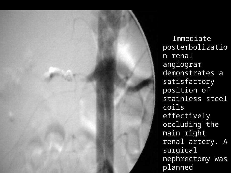
Immediate Immediate postembolization postembolization renal angiogram renal angiogram demonstrates a demonstrates a satisfactory satisfactory position of position of stainless steel stainless steel coils effectively coils effectively occluding the occluding the main right renal main right renal artery. A surgical artery. A surgical nephrectomy nephrectomy was planned was planned
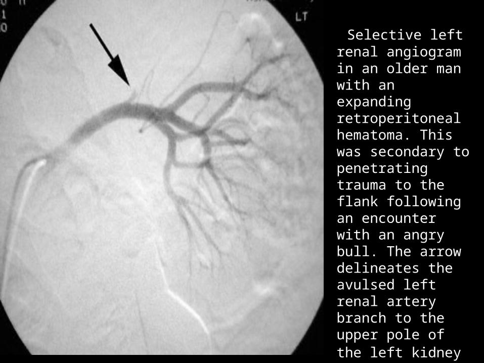
Selective left Selective left renal angiogram renal angiogram in an older man in an older man with an with an expanding expanding retroperitoneal retroperitoneal hematoma. This hematoma. This was secondary to was secondary to penetrating penetrating trauma to the trauma to the flank following an flank following an encounter with encounter with an angry bull. an angry bull. The arrow The arrow delineates the delineates the avulsed left renal avulsed left renal artery branch to artery branch to the upper pole of the upper pole of the left kidneythe left kidney
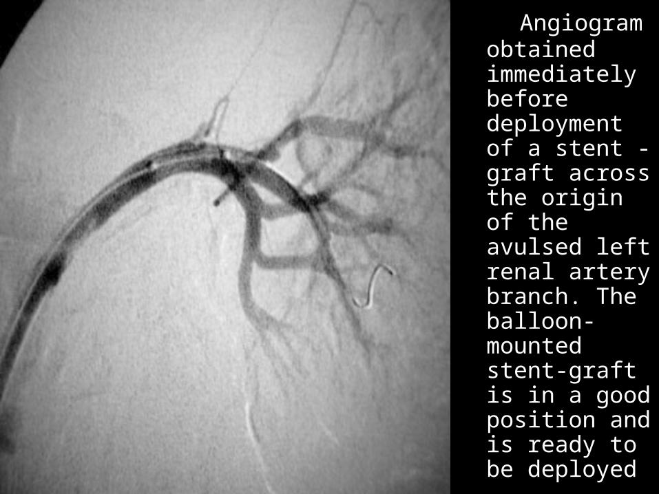
Angiogram Angiogram obtained obtained immediately immediately before before deployment deployment of a stent -of a stent -graft across graft across the origin of the origin of the avulsed the avulsed left renal left renal artery artery branch. The branch. The balloon-balloon-mounted mounted stent-graft is stent-graft is in a good in a good position and position and is ready to be is ready to be deployed deployed
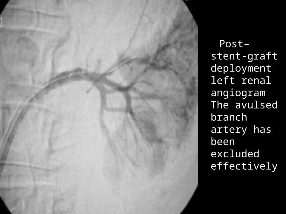
Post–stent-Post–stent-graft graft deployment deployment left renal left renal angiogram angiogram The The avulsed avulsed branch branch artery has artery has been been excluded excluded effectively effectively
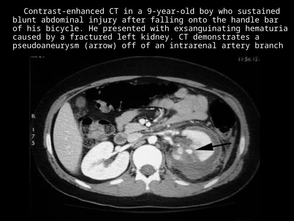
Contrast-enhanced CT in a 9-year-old boy who sustained Contrast-enhanced CT in a 9-year-old boy who sustained blunt abdominal injury after falling onto the handle bar of his blunt abdominal injury after falling onto the handle bar of his bicycle. He presented with exsanguinating hematuria caused bicycle. He presented with exsanguinating hematuria caused by a fractured left kidney. CT demonstrates a by a fractured left kidney. CT demonstrates a pseudoaneurysm (arrow) off of an intrarenal artery branch pseudoaneurysm (arrow) off of an intrarenal artery branch
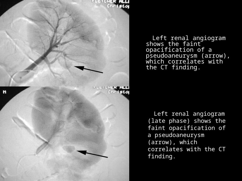
Left renal angiogram Left renal angiogram (late phase) shows the (late phase) shows the faint opacification of a faint opacification of a pseudoaneurysm pseudoaneurysm (arrow), which correlates (arrow), which correlates with the CT finding. with the CT finding.
Left renal angiogram shows Left renal angiogram shows the faint opacification of a the faint opacification of a pseudoaneurysm (arrow), pseudoaneurysm (arrow), which correlates with the which correlates with the CT finding. CT finding.
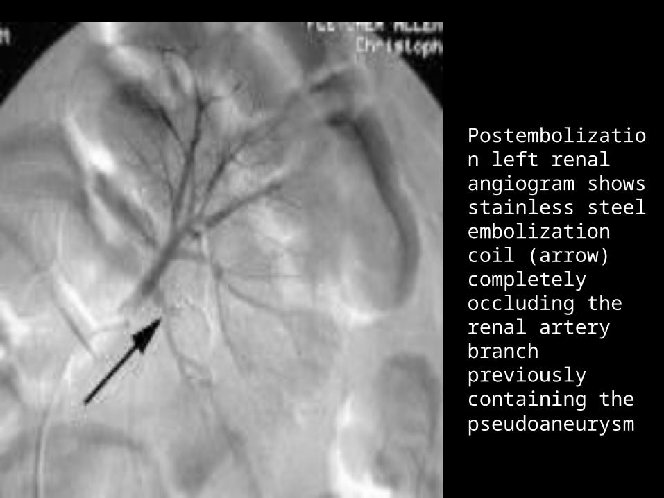
Postembolization Postembolization left renal left renal angiogram shows angiogram shows stainless steel stainless steel embolization coil embolization coil (arrow) (arrow) completely completely occluding the occluding the renal artery renal artery branch previously branch previously containing the containing the pseudoaneurysmpseudoaneurysm
PELVIC TRAUMA PELVIC TRAUMA
Hemorrhage associated with pelvic trauma, Hemorrhage associated with pelvic trauma, +/- pelvic fracture, is common and can arise +/- pelvic fracture, is common and can arise from venous, osseous, or arterial sources or from venous, osseous, or arterial sources or any combination any combination
Pelvic hemorrhage is treated first using Pelvic hemorrhage is treated first using external fixation, which usually is successful external fixation, which usually is successful in treating venous and osseous bleeding in treating venous and osseous bleeding through a tamponade effect through a tamponade effect
Continued bleeding may indicate an arterial Continued bleeding may indicate an arterial source and is associated with a high source and is associated with a high morbidity and mortality rate. Intractable morbidity and mortality rate. Intractable hemorrhage associated with pelvic fracture hemorrhage associated with pelvic fracture is a large contributor to the overall mortality is a large contributor to the overall mortality rate. rate.
PELVIC TRAUMAPELVIC TRAUMA
Surgical exploration and intervention Surgical exploration and intervention of a pelvic hematoma often is of a pelvic hematoma often is complex because of difficulties in complex because of difficulties in visualizing the hemorrhaging artery visualizing the hemorrhaging artery or arteries within the extraperitoneal or arteries within the extraperitoneal hematoma and in gaining arterial hematoma and in gaining arterial control. control.
Risk of increased blood loss through Risk of increased blood loss through the surgical disruption of the pelvic the surgical disruption of the pelvic fascia, which may be important in fascia, which may be important in tamponade of the hematoma tamponade of the hematoma
Transcatheter Transcatheter embolization of pelvic embolization of pelvic
trauma trauma
Early transcatheter embolization of Early transcatheter embolization of pelvic trauma, within 3 hours of pelvic trauma, within 3 hours of presentation, shown to lower the presentation, shown to lower the mortality rate. Angiography required mortality rate. Angiography required in fewer than 10% of patients with in fewer than 10% of patients with pelvic trauma pelvic trauma
When angiography performed, When angiography performed, extravasation is documented in extravasation is documented in about 50% of patients and warrants about 50% of patients and warrants transcatheter embolization. transcatheter embolization.
Imaging and Pelvic Imaging and Pelvic traumatrauma
The sensitivity and specificity demonstrated The sensitivity and specificity demonstrated by CT of active extravasation pelvic trauma by CT of active extravasation pelvic trauma is 80-84% and 85-98%, respectively is 80-84% and 85-98%, respectively
All branches of the internal iliac artery are at All branches of the internal iliac artery are at risk of bleeding. Arterial bleeding most risk of bleeding. Arterial bleeding most frequently occurs from superior gluteal, frequently occurs from superior gluteal, internal pudendal, and obturator arteries. internal pudendal, and obturator arteries. The fascia of the piriformis muscle can The fascia of the piriformis muscle can lacerate the superior gluteal artery, even lacerate the superior gluteal artery, even without fracture. without fracture.
Pelvic and retroperitoneal hemorrhage also Pelvic and retroperitoneal hemorrhage also may arise from the lumbar, inferior may arise from the lumbar, inferior epigastric, deep circumflex iliac, and middle epigastric, deep circumflex iliac, and middle sacral arteries sacral arteries
Pelvic angiography Pelvic angiography
Initially nonselective pelvic angiogram from a Initially nonselective pelvic angiogram from a femoral artery catheter contralateral to the femoral artery catheter contralateral to the trauma with tip in the lower abdominal aorta. trauma with tip in the lower abdominal aorta.
Select the contralateral and subsequently the Select the contralateral and subsequently the ipsilateral internal iliac artery of interest and ipsilateral internal iliac artery of interest and perform selective internal iliac angiography. perform selective internal iliac angiography.
Microcatheters occasionally are needed for Microcatheters occasionally are needed for superselective angiography and embolization. superselective angiography and embolization.
Brisk hemorrhage may be evident on Brisk hemorrhage may be evident on nonselective pelvic angiogram but subtle nonselective pelvic angiogram but subtle extravasation may require selective or extravasation may require selective or subselective angiography for detection subselective angiography for detection
Pelvic transcatheter Pelvic transcatheter embolization embolization technique technique
– If the source of extravasation is defined, If the source of extravasation is defined, superselective embolization with gelatin superselective embolization with gelatin sponge pledgets of 1-2 mm in diameter or sponge pledgets of 1-2 mm in diameter or slurry is optimal slurry is optimal
– Proximial coil embolization for proximal Proximial coil embolization for proximal internal iliac artery injury or following distal internal iliac artery injury or following distal gelatin sponge pledget embolization. gelatin sponge pledget embolization.
– With clinical and CT evidence of massive With clinical and CT evidence of massive hemorrhage, emperic nonselective gelfoam hemorrhage, emperic nonselective gelfoam embolization of both internal iliac arteries is embolization of both internal iliac arteries is acceptable and can arrest bleeding quicklyacceptable and can arrest bleeding quickly
Pelvic transcatheter Pelvic transcatheter embolization efficacy embolization efficacy
Postembolization nonselective Postembolization nonselective angiogram to exclude additional angiogram to exclude additional extravasation sites or collateral vessels extravasation sites or collateral vessels causing retrograde (backfill) causing retrograde (backfill) hemorrhage requiring further hemorrhage requiring further embolization embolization
The success rate of stopping The success rate of stopping hemorrhage is 85-100%. hemorrhage is 85-100%.
Despite high technical success rates, Despite high technical success rates, the mortality rate is significant because the mortality rate is significant because of concomitant injuries of concomitant injuries
Pelvic transcatheter Pelvic transcatheter embolization embolization
complications complications – Inadvertent embolization - Should be rare Inadvertent embolization - Should be rare
if catheter position is satisfactory and if catheter position is satisfactory and embolization procedure is terminated embolization procedure is terminated once occlusion is established once occlusion is established
– Ischemic tissue necrosis or infarction - Ischemic tissue necrosis or infarction - Rare if particle sizes remain larger than Rare if particle sizes remain larger than 500 mm because of extensive distal 500 mm because of extensive distal collateralization of pelvic vasculature collateralization of pelvic vasculature
– Impotence in males - Difficult to Impotence in males - Difficult to differentiate from neurogenic causes of differentiate from neurogenic causes of impotence related to lumbosacral plexus impotence related to lumbosacral plexus injuriesinjuries
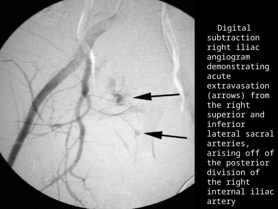
Digital Digital subtraction subtraction right iliac right iliac angiogram angiogram demonstrating demonstrating acute acute extravasation extravasation (arrows) from (arrows) from the right the right superior and superior and inferior lateral inferior lateral sacral arteries, sacral arteries, arising off of arising off of the posterior the posterior division of the division of the right internal right internal iliac artery iliac artery
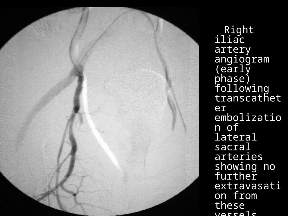
Right iliac Right iliac artery artery angiogram angiogram (early (early phase) phase) following following transcathettranscatheter er embolizatioembolization of lateral n of lateral sacral sacral arteries arteries showing no showing no further further extravasatioextravasation from n from these these vessels. vessels.
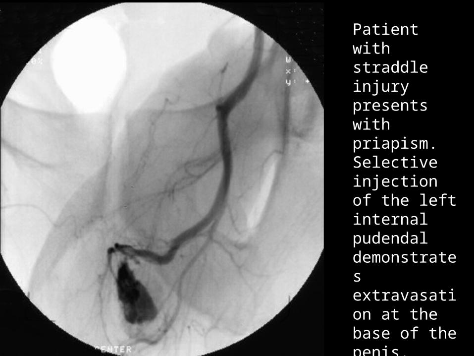
Patient with straddle injury presents with priapism. Selective injection of the left internal pudendal demonstrates extravasation at the base of the penis.
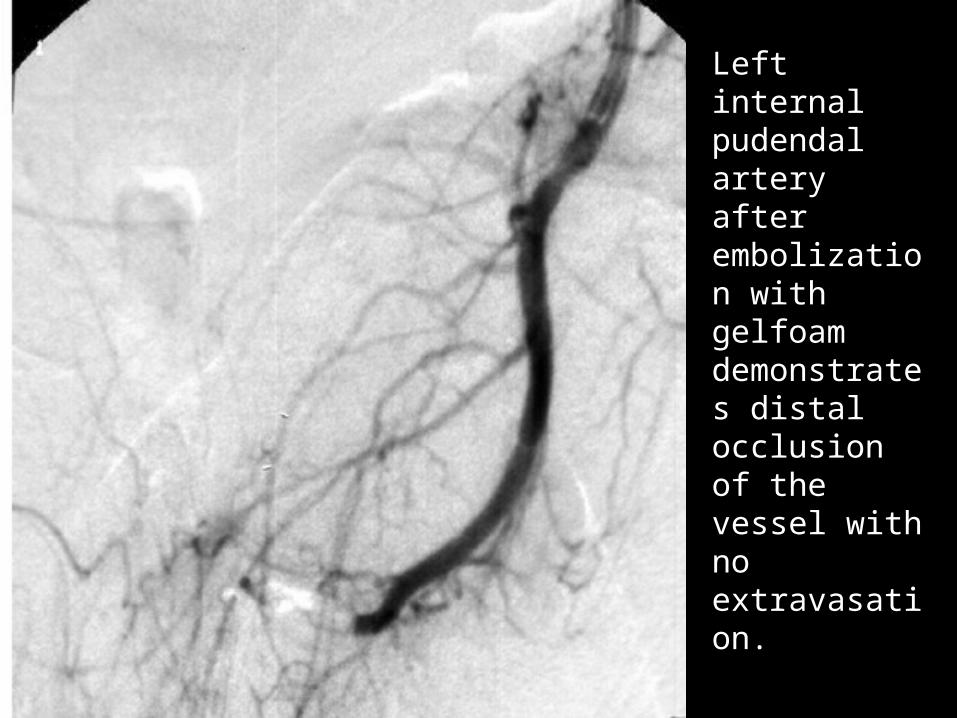
Left internal pudendal artery after embolization with gelfoam demonstrates distal occlusion of the vessel with no extravasation.
PERIPHERAL PERIPHERAL VASCULAR TRAUMA VASCULAR TRAUMA
Peripheral vascular trauma is Peripheral vascular trauma is relatively common in urban settings relatively common in urban settings where penetrating injuries often occurwhere penetrating injuries often occur
Elsewhere, nonpenetrating peripheral Elsewhere, nonpenetrating peripheral vascular injuries, such as occur with vascular injuries, such as occur with blunt trauma, crush injuries, injuries blunt trauma, crush injuries, injuries associated with displaced skeletal associated with displaced skeletal fractures and joint dislocations, and fractures and joint dislocations, and degloving injuries, are seen more degloving injuries, are seen more often often
Penetrating peripheral Penetrating peripheral vascular trauma vascular trauma
Direct penetration of the object through the Direct penetration of the object through the vessel, with resulting disruption, or by dissipation vessel, with resulting disruption, or by dissipation of kinetic energy within the tissues adjacent to of kinetic energy within the tissues adjacent to the vessel the vessel
Low-velocity objects such as knives, the object Low-velocity objects such as knives, the object must traverse the vessel, and the object must must traverse the vessel, and the object must penetrate it to cause injury penetrate it to cause injury
High-velocity weapons e.g. rifles, kinetic energy High-velocity weapons e.g. rifles, kinetic energy expelled and dissipated within the surrounding expelled and dissipated within the surrounding tissues as object decelerates. This causes shock tissues as object decelerates. This causes shock waves and cavitation, which produce injury to waves and cavitation, which produce injury to vessels distant from the trajectory vessels distant from the trajectory
Any of these mechanisms may cause laceration, Any of these mechanisms may cause laceration, pseudoaneurysm formation, transection, pseudoaneurysm formation, transection, arteriovenous fistula, or thrombosis of the vessel. arteriovenous fistula, or thrombosis of the vessel.
Blunt peripheral Blunt peripheral vascular trauma vascular trauma
Direct compression or crushing force may produce Direct compression or crushing force may produce vascular injury such as a vascular mural contusion vascular injury such as a vascular mural contusion
Shearing mechanism, which occurs with stretching Shearing mechanism, which occurs with stretching or traction forces, produces complete transection or or traction forces, produces complete transection or intimal or medial dissection, which may result in a intimal or medial dissection, which may result in a pseudoaneurysm formation pseudoaneurysm formation
Severe extrinsic compression, such as from Severe extrinsic compression, such as from adjacent hematoma, fracture fragment, dislocation, adjacent hematoma, fracture fragment, dislocation, or edema, may cause severe narrowing of the or edema, may cause severe narrowing of the vessel, which in turn may result in thrombosis vessel, which in turn may result in thrombosis
Vasospasm may occur as an isolated injury or as an Vasospasm may occur as an isolated injury or as an associated response to the above-mentioned associated response to the above-mentioned vascular insults vascular insults
Catheter angiography Catheter angiography peripheral vascular injuryperipheral vascular injury
Indicated in peripheral vascular injury Indicated in peripheral vascular injury when the location of injury not certain, when the location of injury not certain, when multiple injury sites may be when multiple injury sites may be present, when the diagnosis requires present, when the diagnosis requires confirmation, or when transcatheter confirmation, or when transcatheter treatment may be the therapy of choice treatment may be the therapy of choice
Some peripheral vascular injuries may Some peripheral vascular injuries may be treated by transcatheter be treated by transcatheter embolization or with stent or stent-graft embolization or with stent or stent-graft placement. placement.
Catheter angiography Catheter angiography indications indications
peripheral vascular injury peripheral vascular injury – Major indications Major indications
Active arterial bleeding or expanding hematoma Active arterial bleeding or expanding hematoma Peripheral pulse deficit Peripheral pulse deficit Bruit over the site of injury Bruit over the site of injury Isolated neurologic deficit Isolated neurologic deficit Hypotension or other sign of ongoing Hypotension or other sign of ongoing
hemorrhagehemorrhage– Minor indications Minor indications
Proximity of a wound or trajectory to a major Proximity of a wound or trajectory to a major blood vessel blood vessel
Nonexpanding hematoma Nonexpanding hematoma Posterior dislocation of the knee joint and Posterior dislocation of the knee joint and
anterior dislocation of the elbow jointanterior dislocation of the elbow joint
Catheter angiography Catheter angiography technique peripheral vascular technique peripheral vascular
injuriesinjuries – Examination of inflow and outflow. Examination of inflow and outflow. – A minimum of two angiographic views A minimum of two angiographic views
centered on the region of injury usually centered on the region of injury usually is required. is required.
– Examine outflow to exclude a distal Examine outflow to exclude a distal embolization from a proximal injury site. embolization from a proximal injury site.
– In gunshot injuries, perform angiography In gunshot injuries, perform angiography or fluoroscopy of the entire extremity to or fluoroscopy of the entire extremity to exclude embolization of metallic exclude embolization of metallic gunshot, shrapnel, or fragments.gunshot, shrapnel, or fragments.
Transcatheter treatment Transcatheter treatment peripheral vascular peripheral vascular
trauma trauma – The artery to be embolized must be nonessential, ie artery The artery to be embolized must be nonessential, ie artery
may be surgically ligated. may be surgically ligated. – This provides optimal treatment when surgical access This provides optimal treatment when surgical access
difficult. difficult. – Embolization can be performed for pseudoaneurysm and Embolization can be performed for pseudoaneurysm and
arteriovenous fistula. arteriovenous fistula. Embolize proximally and distally to the lesion to prevent Embolize proximally and distally to the lesion to prevent
backfilling through collateral vessels. backfilling through collateral vessels. Consider embolizing the neck of a pseudoaneurysm or Consider embolizing the neck of a pseudoaneurysm or
arteriovenous fistula to preserve the parent vessel.arteriovenous fistula to preserve the parent vessel. Direct percutaneous thrombin injection of Direct percutaneous thrombin injection of
pseudoaneurysm pseudoaneurysm
-- A success rate of 85-100% is reported -- A success rate of 85-100% is reported
-- Stents and Stent graft have the potential for preservation -- Stents and Stent graft have the potential for preservation
of the injured vesselof the injured vessel
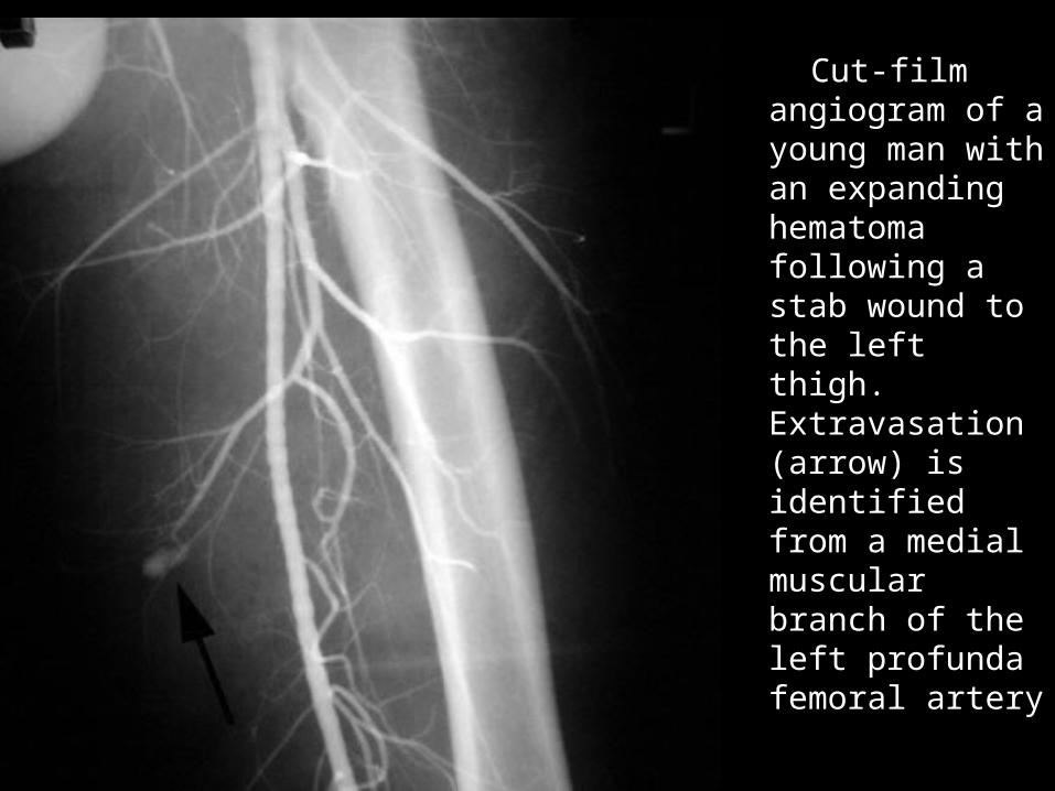
Cut-film Cut-film angiogram of a angiogram of a young man young man with an with an expanding expanding hematoma hematoma following a following a stab wound to stab wound to the left thigh. the left thigh. Extravasation Extravasation (arrow) is (arrow) is identified from identified from a medial a medial muscular muscular branch of the branch of the left profunda left profunda femoral artery femoral artery
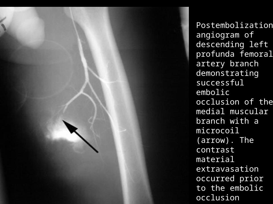
Postembolization Postembolization angiogram of angiogram of descending left descending left profunda femoral profunda femoral artery branch artery branch demonstrating demonstrating successful successful embolic occlusion embolic occlusion of the medial of the medial muscular branch muscular branch with a microcoil with a microcoil (arrow). The (arrow). The contrast material contrast material extravasation extravasation occurred prior to occurred prior to the embolic the embolic occlusion occlusion
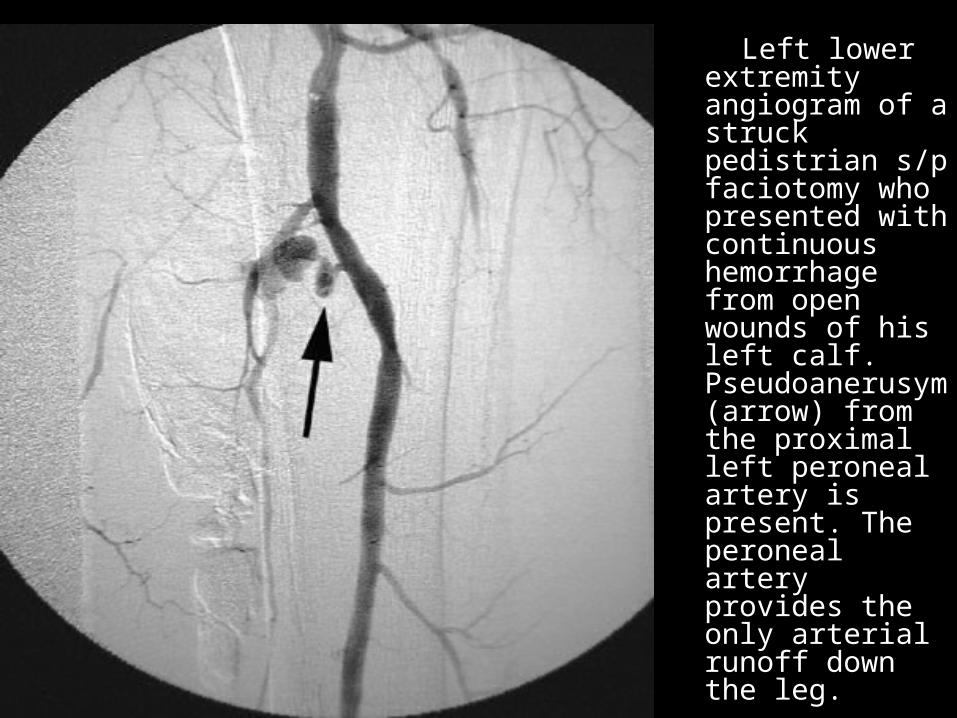
Left lower Left lower extremity extremity angiogram of a angiogram of a struck struck pedistrian s/p pedistrian s/p faciotomy who faciotomy who presented with presented with continuous continuous hemorrhage hemorrhage from open from open wounds of his wounds of his left calf. left calf. PseudoanerusyPseudoanerusym (arrow) from m (arrow) from the proximal the proximal left peroneal left peroneal artery is artery is present. The present. The peroneal artery peroneal artery provides the provides the only arterial only arterial runoff down runoff down the leg. the leg.
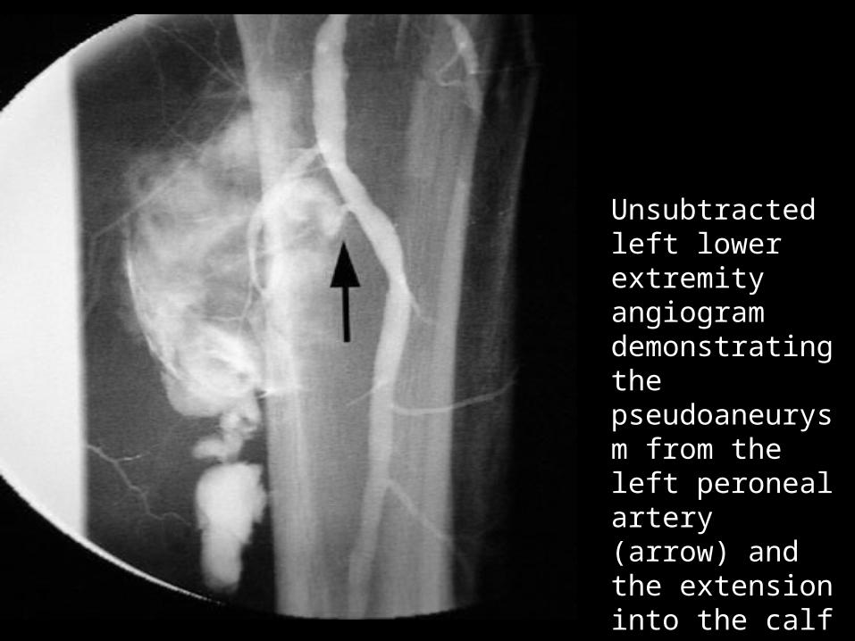
Unsubtracted Unsubtracted left lower left lower extremity extremity angiogram angiogram demonstrating demonstrating the the pseudoaneuryspseudoaneurysm from the left m from the left peroneal artery peroneal artery (arrow) and the (arrow) and the extension into extension into the calf the calf
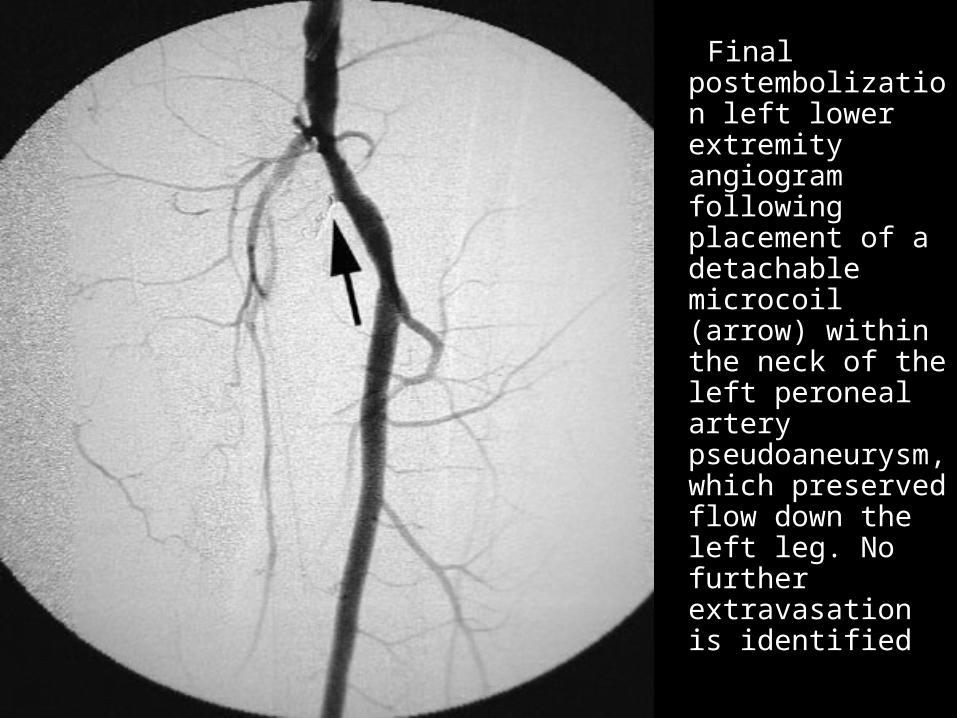
Final Final postembolizatiopostembolization left lower n left lower extremity extremity angiogram angiogram following following placement of a placement of a detachable detachable microcoil microcoil (arrow) within (arrow) within the neck of the the neck of the left peroneal left peroneal artery artery pseudoaneuryspseudoaneurysm, which m, which preserved flow preserved flow down the left down the left leg. No further leg. No further extravasation is extravasation is identified identified
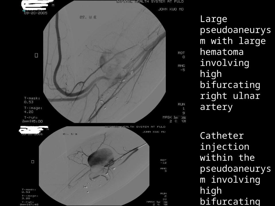
Large pseudoaneurysm with large hematoma involving high bifurcating right ulnar artery
Catheter injection within the pseudoaneurysm involving high bifurcating right ulnar artery
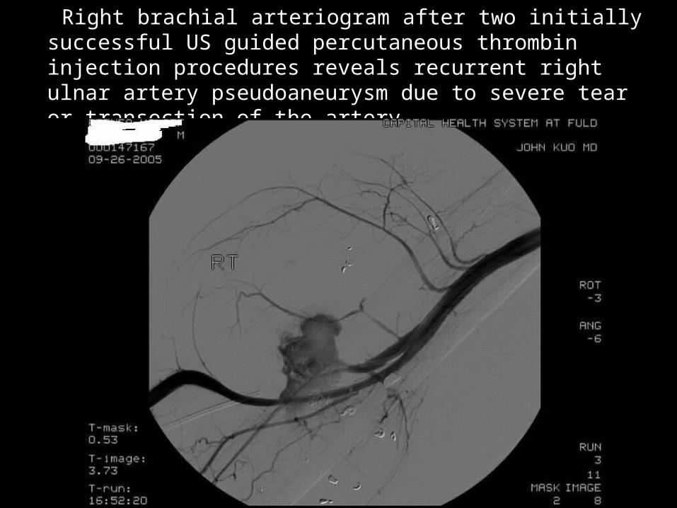
Right brachial arteriogram after two initially successful Right brachial arteriogram after two initially successful US guided percutaneous thrombin injection US guided percutaneous thrombin injection procedures reveals recurrent right ulnar artery procedures reveals recurrent right ulnar artery pseudoaneurysm due to severe tear or transection of pseudoaneurysm due to severe tear or transection of the arterythe artery
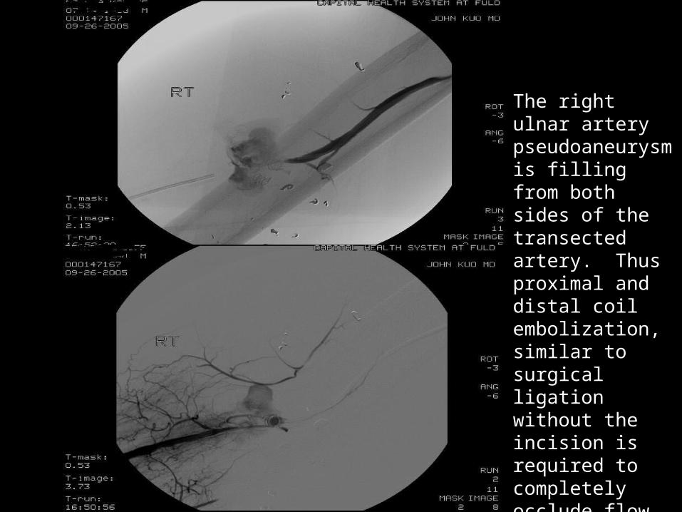
The right ulnar artery pseudoaneurysm is filling from both sides of the transected artery. Thus proximal and distal coil embolization, similar to surgical ligation without the incision is required to completely occlude flow to the pseudoaneurysm
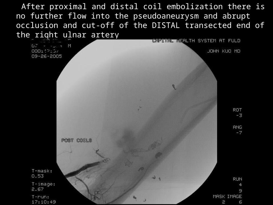
After proximal and distal coil embolization there is no further flow into the pseudoaneurysm and abrupt occlusion and cut-off of the DISTAL transected end of the right ulnar artery
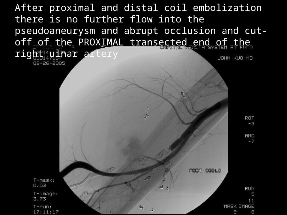
After proximal and distal coil embolization there is no further flow into the pseudoaneurysm and abrupt occlusion and cut-off of the PROXIMAL transected end of the right ulnar artery
VASCULAR TRAUMA OF VASCULAR TRAUMA OF THE NECK THE NECK
Significant penetrating injuries usually require Significant penetrating injuries usually require surgical exploration surgical exploration
Less accessible zone 1 and zone 3 penetrating Less accessible zone 1 and zone 3 penetrating neck injuries may benefit from angiographic neck injuries may benefit from angiographic screening and transcatheter embolization if the screening and transcatheter embolization if the injury involves a branch of the external carotid injury involves a branch of the external carotid artery artery
Pathophysiology of blunt carotid and cervical Pathophysiology of blunt carotid and cervical injuries usually is dissection, which may result in a injuries usually is dissection, which may result in a stenosis, occlusion, or pseudoaneurysm formationstenosis, occlusion, or pseudoaneurysm formation
Extracranial internal carotid injuries are much more Extracranial internal carotid injuries are much more common than intracranial internal carotid injuries common than intracranial internal carotid injuries and usually originate at the C2- to C3-vertebral and usually originate at the C2- to C3-vertebral level and terminate at the base of the carotid canal level and terminate at the base of the carotid canal
Grading scale for blunt Grading scale for blunt carotid arterial injury carotid arterial injury
Grade I - Luminal irregularity or dissection Grade I - Luminal irregularity or dissection with less than 25% luminal narrowing with less than 25% luminal narrowing
Grade II - Dissection or intramural Grade II - Dissection or intramural hematoma with greater than or equal to hematoma with greater than or equal to 25% luminal narrowing, intramural 25% luminal narrowing, intramural thrombus, or raised intimal flap thrombus, or raised intimal flap
Grade III - Pseudoaneurysm Grade III - Pseudoaneurysm Grade IV - Occlusion Grade IV - Occlusion Grade V - Transection with free Grade V - Transection with free
extravasationextravasation
Findings associated with Findings associated with blunt carotid or vertebral blunt carotid or vertebral
injuryinjury Early diagnosis and treatment of these injuries Early diagnosis and treatment of these injuries
improves neurologic outcome improves neurologic outcome Expanding cervical hematoma Expanding cervical hematoma Hemorrhage from mouth, nose, ears, or wounds Hemorrhage from mouth, nose, ears, or wounds Massive facial fractures Massive facial fractures Cervical bruit in patients younger than 50 years Cervical bruit in patients younger than 50 years Evidence of stroke on CT Evidence of stroke on CT Unexplained or incongruous central or lateralizing Unexplained or incongruous central or lateralizing
neurologic deficit, Horner syndrome, transient neurologic deficit, Horner syndrome, transient ischemic attack, or amaurosis fugax ischemic attack, or amaurosis fugax
Basilar skull fracture through or near the carotid Basilar skull fracture through or near the carotid canal canal
Fracture through the foramen transversarium Fracture through the foramen transversarium Severe flexion or extension cervical spine fracture Severe flexion or extension cervical spine fracture
or subluxation or subluxation
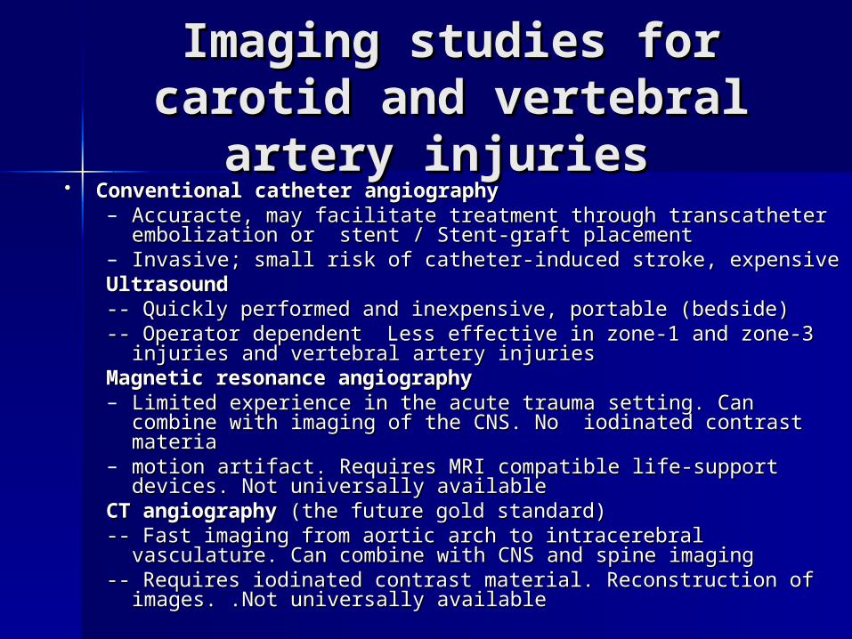
Imaging studies for Imaging studies for carotid and vertebral carotid and vertebral
artery injuries artery injuries Conventional catheter angiographyConventional catheter angiography
– Accuracte, may facilitate treatment through transcatheter Accuracte, may facilitate treatment through transcatheter embolization or stent / Stent-graft placementembolization or stent / Stent-graft placement
– Invasive; small risk of catheter-induced stroke, expensive Invasive; small risk of catheter-induced stroke, expensive UltrasoundUltrasound-- Quickly performed and inexpensive, portable (bedside)-- Quickly performed and inexpensive, portable (bedside)-- Operator dependent Less effective in zone-1 and zone-3 injuries -- Operator dependent Less effective in zone-1 and zone-3 injuries
and vertebral artery injuriesand vertebral artery injuriesMagnetic resonance angiographyMagnetic resonance angiography– Limited experience in the acute trauma setting. Can combine Limited experience in the acute trauma setting. Can combine
with imaging of the CNS. No iodinated contrast materiawith imaging of the CNS. No iodinated contrast materia– motion artifact. Requires MRI compatible life-support devices. Not motion artifact. Requires MRI compatible life-support devices. Not
universally available universally available CT angiographyCT angiography (the future gold standard) (the future gold standard)-- Fast imaging from aortic arch to intracerebral vasculature. Can -- Fast imaging from aortic arch to intracerebral vasculature. Can
combine with CNS and spine imagingcombine with CNS and spine imaging-- Requires iodinated contrast material. Reconstruction of -- Requires iodinated contrast material. Reconstruction of
images. .Not universally available images. .Not universally available
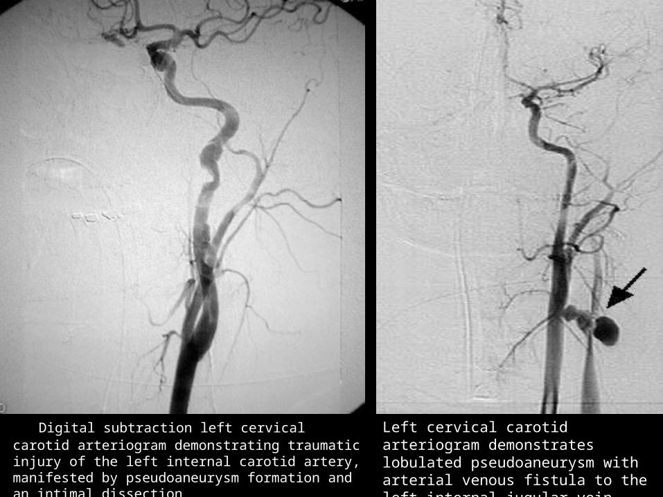
Digital subtraction left cervical carotid Digital subtraction left cervical carotid arteriogram demonstrating traumatic injury of arteriogram demonstrating traumatic injury of the left internal carotid artery, manifested by the left internal carotid artery, manifested by pseudoaneurysm formation and an intimal pseudoaneurysm formation and an intimal dissection dissection
Left cervical carotid arteriogram demonstrates lobulated pseudoaneurysm with arterial venous fistula to the left internal jugular vein
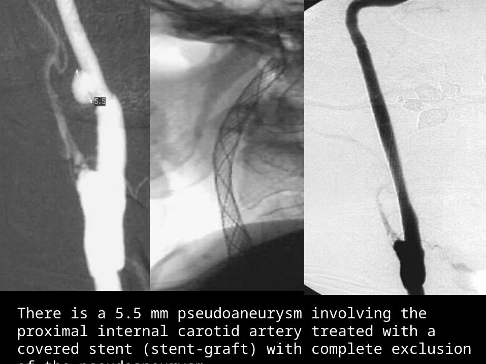
There is a 5.5 mm pseudoaneurysm involving the proximal internal carotid artery treated with a covered stent (stent-graft) with complete exclusion of the pseudoaneurysm.
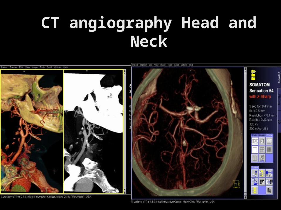
CT angiography Head CT angiography Head and Neckand Neck
Vascular and Solid Organ Trauma Vascular and Solid Organ Trauma - Interventional Radiology - Interventional Radiology
John Kuo, MDJohn Kuo, MD
Chief, Interventional Chief, Interventional RadiologyRadiology

My son My son JustinJustin