Variations and Bony Landmarks of the Maxillary Artery: a ...
50
VARIATIONS AND BONY LANDMARKS OF THE MAXILLARY ARTERY: A CADAVERIC STUDY INVESTIGATOR DR.TIMOTHY M. WARUI V60/83836/2012 BDS (UoN) A dissertation submitted in partial fulfillment for the award of the degree of Master of Dental Surgery in Oral and Maxillofacial Surgery of the University of Nairobi 2017
Transcript of Variations and Bony Landmarks of the Maxillary Artery: a ...

VARIATIONS AND BONY LANDMARKS OF THE MAXILLARY
ARTERY: A CADAVERIC STUDY
INVESTIGATOR
DR.TIMOTHY M. WARUI
V60/83836/2012
BDS (UoN)
A dissertation submitted in partial fulfillment for the award of the degree of Master of
Dental Surgery in Oral and Maxillofacial Surgery of the University of Nairobi
2017
i

DECLARATIONS
This dissertation is my original work and has not been presented for the award of a degree in
any other university.
Dr. Timothy M. Warui BDS (UoN)
University of Nairobi.
Signed……………………………… Date……………….
This dissertation has been presented with my approval as supervisor.
Dr. Fawzia M.A. Butt BDS (UoN), FDSRCS (Eng), MDS-OMFS (UoN), FICD
Lecturer, Department of Human Anatomy, University of Nairobi.
Signed ………………………………………… Date ………………….
This dissertation has been presented with my approval as supervisor.
Dr. Matthew Akama BDS (UoN), MDS-OMFS (UoN), FAOCMF.
Senior lecturer, Department of Oral and Maxillofacial Surgery, Oral Medicine and Oral
Pathology, University of Nairobi.
Signed ………………………………………… Date ………………….
This dissertation has been presented with my approval as supervisor.
Prof. Mark L. Chindia BDS (UoN), MSc (London), FFDRCSI (Ireland), FIAOMS.
Professor of oral and maxillofacial surgery, Department of Oral and Maxillofacial Surgery,
Oral Medicine and Oral Pathology, University of Nairobi.
Signed ………………………………………… Date ………………….
ii

DECLARATION FORM FOR STUDENT
UNIVERSITY OF NAIROBI
DECLARATION OF ORIGINALITY
This form must be completed and signed for all works submitted to the University for
Examination.
Name of student…………………………………………………………………………………
Registration number…………………………………………………………………………….
College…………………………………………………………………………………………
Falculty/school/institute………………………………………………………………………...
Department……………………………………………………………………………………...
Course name…………………………………………………………………………………….
Title of the work………………………………………………………………………………
Declaration
1. I understand what plagiarism is and I am aware of the universitys policy in this
regard.
2. I declare that this …………………. (thesis, paper, essay dissertation, project,
assignment, report etc) is my original work and has not been submitted elsewhere for
examination, award of degree or publication. Where other peoples work or my own
work has been used, this has been properly acknowledged and referenced in
accordance with the University of Nairobi’s Requirement.
3. I have not sought or used the services of any professional agencies to produce this
work.
4. I have not allowed, and shall not allow anyone to copy my work with the intention of
passing it off as his/her own work.
5. I understand that any false claim in respect of this work shall result in disciplinary
action, in accordance with university of Nairobi plagiarism policy.
Signature………………………………….
Date………………………………………..
iii

ACKNOWLEDGEMENTS
I would like to thank the Almighty God for His strength and providence. I acknowledge my
supervisors Dr. F.M. Butt, Prof. M. L. Chindia and Dr. M. K. Akama for the guidance and
limitless counsel during the study. I would like to thank Dr. B. Olabu of the Department of
Human Anatomy, University of Nairobi for his encouragement and guidance during the
study. Many thanks to Dr. S.K. Kabinga, physician/biostatician at Maralal county hospital for
his tireless guidance and encouragement throughout the study. I am grateful to the members
of staff in the department of Human Anatomy, University of Nairobi.
May God bless you all.
iv

DEDICATION
I would like to dedicate this study to my parents for their support and prayers and for
continuously teaching me that: Knowledge is the most precious possession one can ever
acquire.
v

TABLE OF CONTENTS
DECLARATIONS .................................................................................................................................. ii
DECLARATION FORM FOR STUDENT ........................................................................................... iii
ACKNOWLEDGEMENTS ................................................................................................................... iv
DEDICATION ........................................................................................................................................ v
LIST OF TABLES ............................................................................................................................... viii
LIST OF FIGURES ............................................................................................................................... ix
LIST OF ABBREVIATIONS AND ACRONYMS ................................................................................ x
ABSTRACT ........................................................................................................................................... xi
CHAPTER 1 ....................................................................................................................................... 1
1. INTRODUCTION ...................................................................................................................... 1
1.1 BACKGROUND ...................................................................................................................... 1
1.1.2 REPORTED INJURIES TO THE MAXILLARY ARTERY IN THE CLINICAL PRACTICE ............................................................................................................................................................ 2
CHAPTER 2 .............................................................................................................................................. 4
2. LITERATURE REVIEW ............................................................................................................... 4
2.1 ANATOMICAL VARIATIONS OF THE MAXILLARY ARTERY ...................................... 4
2.2 DISTANCE OF THE MA FROM SELECTED BONY ANATOMICAL LANDMARKS ............. 6
2.3 PROBLEM STATEMENT ........................................................................................................... 7
2.4 JUSTIFICATION OF THE STUDY ................................................................................................ 8
2.5 RESEARCH QUESTION ............................................................................................................. 8
2.6 OBJECTIVES ............................................................................................................................... 8
2.6.1 BROAD OBJECTIVE ........................................................................................................... 8
2.6.2 SPECIFIC OBJECTIVES ...................................................................................................... 8
CHAPTER 3 ........................................................................................................................................... 9
3. MATERIALS AND METHODS .................................................................................................... 9
3.1 STUDY DESIGN ...................................................................................................................... 9
3.2 STUDY AREA ......................................................................................................................... 9
3.3 STUDY POPULATION ......................................................................................................... 10
3.5 SAMPLE SIZE CALCULATION .......................................................................................... 10
3.4 SAMPLING METHOD .......................................................................................................... 10
3.6 SELECTION CRITERIA ....................................................................................................... 10
3.7 STUDY VARIABLES ............................................................................................................ 11
3.8 DISSECTION METHOD ....................................................................................................... 12
3.9 QUALITY CONTROL IN THE STUDY ............................................................................. 13
vi

3.10 DATA MANAGEMENT ...................................................................................................... 13
3.11 ETHICAL CONSIDERATION ............................................................................................ 15
CHAPTER 4 ......................................................................................................................................... 16
4.0. RESULTS .......................................................................................................................................................... 16
4.1 DISTANCES OF THE MAXILLARY ARTERY (MA) FROM SELECTED BONY LANDMARKS .......................................................................................................................................................... 16
4.2 COURSE OF THE MAXILLARY ARTERY .......................................................................................... 19
4.3 RELATIONSHIP OF THE MAXILLARY ARTERY TO THE LATERAL PTERYGOID MUSCLE .......................................................................................................................................................... 19
4.4 BRANCHING PATTERN OF THE 1ST PART OF THE MAXILLARY ARTERY ......................... 21
CHAPTER 5 ......................................................................................................................................... 24
5.1 DISCUSSION ............................................................................................................................. 24
CONCLUSION ..................................................................................................................................... 27
RECOMMENDATIONS ...................................................................................................................... 28
REFERENCES ..................................................................................................................................... 29
APPENDIX I ........................................................................................................................................ 33
DATA COLLECTION SHEET ........................................................................................................ 33
APPENDIX II ....................................................................................................................................... 35
ETHICAL APPROVAL ................................................................................................................... 35
APPENDIX III ...................................................................................................................................... 37
PLAGIARISM REPORT .................................................................................................................. 37
vii

LIST OF TABLES
Table 1.4: Comparison of the measured distances between the right and the left side in
relation to selected bony landmarks. ........................................................................................ 18
Table 2.4: Relationship of the maxillary artery with the parotid gland ................................... 19
Table 3.4: Relationship of the maxillary artery (MA) to the lateral pterygoid muscle (LPM) 20
Table 4.4: Pattern of origin of the middle meningeal artery (MMA) and the inferior alveolar
artery (IAA) from the maxillary artery (MA). ......................................................................... 22
Table 5.4: Variations in origin of the middle meningeal artery (MMA) and accessory
meningeal artery (AMA).......................................................................................................... 23
viii

LIST OF FIGURES
Figure 1.1: Anatomy of the maxillary artery (MA). .................................................................. 1
Figure 2.2: Selected bony landmarks. ........................................................................................ 7
Figure 3.3: Flow chart of specimens available for dissection .................................................. 11
Figure 4.3: Photomacrograph of the right side of the face showing perpendicular distances
measured between the Maxillary artery and the selected bony landmarks. ............................. 13
Figure 5.3:Vernier callipers ..................................................................................................... 14
Figure 6.3: Protractor used during measurement ..................................................................... 14
Figure 7.4: Relationship between the maxillary artery and the lateral pterygoid muscle ........ 20
Figure 8.4: Illustration of the branching pattern of the 1st part of the right maxillary artery
(MA). ....................................................................................................................................... 22
Figure 9.4: Illustration of the inferior alveolar artery (IAA) originating before the middle
meningeal artery (MMA). ........................................................................................................ 23
ix
LIST OF ABBREVIATIONS AND ACRONYMS
AMA - Accessory Meningeal artery
ECA - External Carotid artery
IAA - Inferior Alveolar artery
IAN - Inferior Alveolar nerve
KNH - Kenyatta National Hospital
LPM - Lateral Pterygoid muscle
MA - Maxillary artery
MMA - Middle Meningeal artery
TMJ - Temporomandibular joint
UoN - University of Nairobi
ATA - Anterior Tympanic Artery
x

ABSTRACT
BACKGROUND: The maxillary artery (MA) is known to vary in the branching pattern,
relation with the lateral pterygoid muscle (LPM) and its distances from conventional bony
landmarks. These variations pose surgical challenges in procedures around the
temporomandibular joint (TMJ) and infratemporal fossa and, therefore, their knowledge is
vital. Data on African populations are scanty. Therefore, this cadaveric study sought to
provide information pertaining the MA in a Kenyan population.
STUDY OBJECTIVE: To describe the variations and anatomical landmarks of the MA.
STUDY DESIGN AND POPULATION: This was a descriptive cross-sectional cadaveric
study, carried out in the department of human anatomy at the University of Nairobi. Cadavers
of adults available in the University of Nairobi Human Anatomy dissection laboratory with
intact structures in the parotid and infratemporal region were included in the study.
STUDY DURATION: The study was conducted between November 2015 and May 2016.
MATERIAL AND METHODS: Ninety three (93) hemi-sections from 48 cadavers were
used for the study. The side of the face dissected was noted. Dissection of the infratemporal
fossa was done to expose the MA and its branches on both the right and left sides. The
distance of the MA from the articular eminence, mandibular neck, mandibular notch and
pterygoid fovea were measured in millimetres (mm) using digital Vernier calipers and
protractor. The relationship of the MA with the LPM was noted and the branching pattern of
the first part of the MA was described.
DATA MANAGEMENT: Photo-macrographs were used to demonstrate the variations in
branching and relationship of the MA with the LPM. Data were coded and analyzed using the
Statistical Package for Social Science (SPSS) version 17.0 and presented in tables.
xi

Independent paired student’s t test was used to test the level of significance for the parametric
data.
RESULTS: The average perpendicular distances between the MA and the posterior cortex at
the centre of the condylar neck of the mandible, inferior most part of the mandibular notch,
inferior part of the articular eminence and inferior part of the pterygoid fovea were
determined as 8.58±2.69 mm, 5.76±2.8 mm, 14.20±3.89 mm and 14.28±6.62 mm
respectively. Out of the 93 hemi-sections dissected 67% of the MA was within the parotid
gland while 33% of the MA was medial to the gland. Thirty five of the MA had a medial
relationship to the LPM while 58 of the MA had a lateral relationship to the LPM. Four
(8.9%) of the cadavers showed asymmetry whereby the MA passed on the medial side of the
LPM on one side while on the other side it passed on the lateral aspect of the LPM. Six
(6.5%) hemi-sections had the middle meningeal artery (MMA) and the inferior alveolar
artery (IAA) originating from a common trunk on the MA. The accessory meningeal artery
(AMA) was absent in 3 (3.2%) hemi-sections on the right side and in 2 (2.2%) on the left
side. On the right, the MMA branched off from the MA before the IAA in 18 (37.5%) of the
hemi-sections whereas in 27 (56.3%) of the hemi-sections it was given out after the IAA. On
the left side the MMA was given off before the IAA in 10 (22.8%) of the hemi-sections and
branched off after the IAA in 32 (71.1%) of the hemi-sections.
CONCLUSION: The variations of the MA in this population displayed patterns comparable
to other populations. The distance of the MA to selected bony landmarks between the right
and left sides was comparable. Remarkably, in this Kenyan sample population, the MA
passed significantly further from the articular eminence compared to other populations.
Majority of the MAs were within the parotid gland in this population. There was high
prevalence of the MA passing lateral to the LPM which was consistent with studies done
xii

xiii
elsewhere. A branching pattern similar to other studies was observed on the first part of the
MA.

CHAPTER 1
1. INTRODUCTION
1.1 BACKGROUND
1.1.1 ANATOMICAL DESCRIPTION OF THE MAXILLARY ARTERY
The maxillary artery (MA) is one of the terminal branches of the external carotid artery (ECA). It
supplies deep structures of the face and the dura in the anterior and the middle cranial fossae.
The artery branches off from the ECA at the neck of the mandible and courses into the
infratemporal fossa from where it enters the pterygopalatine fossa through the pterygopalatine
fissure (1).
Figure 1.1: Anatomy of the maxillary artery (MA). Standring et al 2005
It is divided into three parts anatomically. The 1st part (the mandibular part) is the region
between where the artery branches off to where it reaches the lateral pterygoid muscle ( LPM).
1
This segment gives rise to branches that mainly enter various foramina, namely the deep
auricular artery, the anterior tympanic artery (ATA), the middle meningeal artery (MMA), the
accessory meningeal artery (AMA) and the inferior alveolar artery (IAA). The MA through its
MMA supplies the dura in the anterior cranial fossa while the AMA supplies the dura in the
middle cranial fossa. The 2nd part (the pterygoid part) is the region where the artery traverses the
LPM. This segment gives branches to soft tissues primarily muscles of mastication. They include
the deep temporal, pterygoid, masseteric and buccal arteries. The 3rd part (the pterygopalatine
part) is the segment which lies within the pterygopalatine fossa. This part gives rise to: the
posterior superior alveolar, infraorbital, descending palatine, sphenopalatine, pharyngeal and
pterygoid canal arteries.
1.1.2 REPORTED INJURIES TO THE MAXILLARY ARTERY IN THE CLINICAL
PRACTICE
The MA is a large vessel and is known to have a high blood flow. Accordingly, its iatrogenic
injury during surgeries around the temporomandibular joint (TMJ), the mandible, the maxilla and
the infratemporal fossa can lead to massive haemorrhage or formation of pseudoaneurysms (2-4).
This kind of injury is made possible due to its variability in landmark and branching, and its
proximity with the parotid gland, the temporomandibular joint (TMJ), the mandibular neck and
ramus. Although there are few studies focusing on the MA injuries, there are several isolated
reports regarding the same (5-9). Hemorrhage following such injuries have been reported to be
so severe in some cases that require blood transfusion (5, 6), temporal pressure application on
the external carotid artery (ECA) through a neck incision and direct ligation of the artery (10-12).
Similarly, cases of pseudoaneurysms that sometimes complicate with severe epistaxis have been
reported (10-12), and some of these may require embolization (13). Therefore, given the fact that
2

the MA and its branches are vulnerable to iatrogenic injury, precautions have to be taken in order
to minimize or prevent hemorrhage and improve intraoperative safety (14).
1.1.3 RATIONALE OF THE STUDY
Despite the existence of population differences in the variations of the MA, there is paucity of
data on such variations in the African population. Knowledge of the reliable landmarks of the
MA will help reduce inadvertent injuries to the vessel in procedures around the parotid region
and infratemporal fossa. The study aimed at providing information about operative landmarks
relations of the MA.
3

CHAPTER2
2. LITERATURE REVIEW
2.1 ANATOMICAL VARIATIONS OF THE MAXILLARY ARTERY
Variations of the MA have been reported with regard to its relation to the parotid gland, the LPM
and the branching pattern of its first part. These variations display population differences (7, 15,
16).
2.1.1 RELATIONSHIP OF THE MA AND THE LPM
The relationship of the MA to the LPM can either be medial or lateral which vary depending on
the gender and the population studied. Hussain et al. studied the relationship of the MA and the
LPM in a Caucasian population in Canada and found that in the majority (68%) the MAs were
lateral to the lower head of the LPM (71% in men and 65% in women). There were no variations
in the course of the MA noted between the right and the left side and between genders (17).
However, a study by Pretterklieber et al. showed asymmetry in the same cadavers (18). Among
the Turkish population the MA has been reported to be lateral to the LPM in 57.1% and medial
to the LPM in 42.9% of cases (19). These findings are similar to those reported in New Zealand
(20). Among the Asian population, the MA travelled on the lateral aspect of the LPM in 82.0%
of the cases and on the medial side in 18.0% of the cases which was consistent with a study done
by Sashi et al. in the Japanese population (15, 21). In another study among the Japanese, in 3.6%
of the cases the MA passed medial to the LPM and in 96.4% of the cases it passed lateral to the
LPM (22). Verma et al. described a case where the MA was seen passing medial to the LPM and
crossed through the nerve loop formed between the two roots of the auriculotemporal nerve and
the posterior division of the mandibular nerve. Further, the course of the MA was medial to the
4

posterior division of the mandibular nerve. The MA gave its MMA branch as it traversed through
the nerve loop. A tortuous course taken by the MA can lead to its entrapment which is associated
with headaches or nerve irritation presenting with neuralgia (23). In a study by Fujimura et al.,
cases were described where the MA passed laterally and superficially to the LPM in 9 to 55% in
whites, 69% in blacks, and in about 90% in the Japanese (7). Racial variation is also reported by
Lasker et al. where the MA passed on the medial aspect of the LPM in 46% of white individuals
and in 31% of an African-American population (16).
2.1.2 BRANCHING PATTERNS OF THE FIRST PART OF THE MA
The branching patterns are the MA originating from the ECA in a single trunk or forming a loop,
the MMA being given off the MA before or after the IAA, the MMA and the AMA being given
off from the MA separately or in a common trunk, missing AMA, two IAAs, the IAA originating
directly from the ECA and the anterior tympanic artery and the AMA being given off from the
MMA. This branching pattern of the first part of the MA has been described in various studies.
Uysal et al. found out that while the MMA was present in all cases, the AMA was observed only
in 57.1 % of the cases. In 35.7% of the cases, the IAA arose from the MA before the MMA while
in 35.7 % it arose after the MMA. The IAA and the MMA branched from the same area of the
MA in 14.3 % of the cases. In others, the IAA branched from the beginning of the MA in 14.3%
of the cases (19).
Looping of the MA in the infratemporal region has been described. Maeda et al. reported a case
where the MA bifurcated into a superficial trunk and a deep trunk in the proximal part, with the
MMA and the AMA originating from the deep trunk while the IAA originated from the
5

superficial trunk. The trunks reunited to form a complete loop (24). Claire et al. described a case
where the MA bifurcated into deep and superficial branches at the distal part of the divergence
of the anterior tympanic artery which reunited to form a complete loop at the infratemporal
region (25). Bhat et al. reported a case where the MA passed through the loop of the
auriculotemporal nerve (26). Lydia et al. reported a case whereby the MMA and the AMA
originated from the second part of the MA and the deep temporal arteries arose from
the first part in a common trunk with the IAA (27).
Rao et al. described different variations in the branching of the MA. They described a case
whereby the MMA originated directly from the ECA. They also described a case in whom there
were two IAAs 1.5 cm apart arising from the first part of the MA. The first artery went to the
mandibular canal along with the inferior alveolar nerve. The second artery accompanied the
lingual nerve to the last molar tooth. Another variation described showed the anterior tympanic
artery and the AMA arising from the MMA (28). Khaki et al. also reported a case in which the
IAA originated directly from the ECA (29).
2.2 DISTANCE OF THE MA FROM SELECTED BONY ANATOMICAL LANDMARKS
The distances of the MA from different bony landmarks have been reported in the literature (4,
30, 31). These bony landmarks include the articular eminence, posterior cortex at the centre of
condylar neck of the mandible, mandibular notch and the pterygoid fovea (Fig.2.2). These
landmarks are used as intraoperative reference points during bone osteotomies and are dissected
to some extent intraoperatively (30). Although there are few studies focusing on the landmarks
6

of the MA, these distances seem to vary in various populations.
Figure 2.2: Selected bony landmarks. Maxillary artery (MA), Articular eminence (AE), Mandibular notch (MN), Midcervical (M), supracervical (S), infracervical (I).(30) Balcioglua et al 2010
For example a study done by Orbay et al in the Turkish population, the distance between the MA
to the mandibular notch was 5.1 mm .The same distance was, however, 3.3mm as reported by
Aziz et al. in their study of the Columbians. Similarly, Balcioglu et al. reported the average
distance of 5.38 mm between the MA and the medial cortex of the mandible in a study of a
Turkish population, while Orbay et al. reported an average distance of 6.8 mm in a different
Turkish population.
2.3 PROBLEM STATEMENT
Distances from different bony landmarks and anatomical variations of the MA has been reported
in different populations, however there is no data that is available in this population. Cases of
iatrogenic injury to the MA with hemorrhage and formation of pseudoaneurysms have been
documented.
7

2.4 JUSTIFICATION OF THE STUDY
Knowledge on the findings about the variations in the anatomy and distances from different bony
landmarks of the MA may help in reduction of the iatrogenic injuries to the MA in surgical
procedures involving the parotid region, infratemporal fossa and the TMJ. Knowledge of the
relationship of the MA with different anatomical landmarks may be utilized to avoid iatrogenic
injuries during surgical procedures involving the TMJ, condyle, mandibular ramus osteotomy
and total maxillectomy.
2.5 RESEARCH QUESTION
What are the variation patterns and distances from selected bony landmarks of the MA in this
population?
2.6 OBJECTIVES
2.6.1 BROAD OBJECTIVE
To describe the variations and anatomical landmarks of the MA.
2.6.2 SPECIFIC OBJECTIVES
1. To determine the distances between the MA and the following bony landmarks: the
most inferior part of the mandibular notch, posterior cortex at the centre of the
condylar neck of the mandible, inferior part of the articular eminence and the inferior
border of pterygoid fovea.
2. To describe the relationship of the MA to the parotid gland.
3. To describe the relationship between the MA and the LPM.
4. To describe the branching pattern of the first part of the MA.
8

CHAPTER 3
3. MATERIALS AND METHODS
3.1 STUDY DESIGN
Data about this study was collected at the Department of Human Anatomy dissection laboratory,
University of Nairobi in duration of 2 months. This was a cadaveric descriptive cross-sectional
study.
3.2 STUDY AREA
The study was carried out in the Department of Human Anatomy dissection laboratory,
University of Nairobi at the Chiromo campus which is approximately 2 km from the Nairobi
Central Business District, Riverside Drive off Waiyaki Way. The University of Nairobi is the
largest public universities in Kenya with a Department of Human Anatomy in the College of
Health Sciences where training of both undergraduate and postgraduate students in health related
disciplines is undertaken. The Human Anatomy Department acquires human cadavers for
teaching purposes mainly from pastoralist communities in the country as well as unclaimed
human bodies at the Kenyatta national hospital mortuary. Annually there are between 50-60
cadavers available for teaching human anatomy. The Kenyan law, under the Human Tissue Act
Chapter 252, allows dissection of cadavers for purposes of medical education and research. CAP
252…̏An Act of Parliament to make provision with respect to the use of parts of bodies of
deceased persons for therapeutic purposes and purposes of medical education and research; and
for matters connected therewith and incidental thereto.”
9

3.3 STUDY POPULATION
The study included all the available cadavers for dissection for the academic year 2015/2016 in
the Human anatomy laboratory, University of Nairobi.
3.5 SAMPLE SIZE CALCULATION
With finite population correction (32).
P = expected proportion (0.5)
Z = normal standard deviate corresponding to α=0.05(1.96)
d = Precision (0.05)
N = the size of the study population (50)
n' = Sample size with finite population correction (45) (minimum number of cadavers to be
dissected).
3.4 SAMPLING METHOD
All cadavers meeting the inclusion criteria were utilized as this was a population study.
3.6 SELECTION CRITERIA
3.6.1 INCLUSION CRITERIA
Cadavers from adult Kenyans with the following intact structures: the preauricular regions,
parotid areas, the rami of the mandible and infratemporal region. The cadavers which were in
occlusion.
10
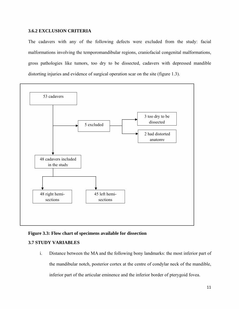
3.6.2 EXCLUSION CRITERIA
The cadavers with any of the following defects were excluded from the study: facial
malformations involving the temporomandibular regions, craniofacial congenital malformations,
gross pathologies like tumors, too dry to be dissected, cadavers with depressed mandible
distorting injuries and evidence of surgical operation scar on the site (figure 1.3).
53 cadavers
Figure 3.3: Flow chart of specimens available for dissection
3.7 STUDY VARIABLES
i. Distance between the MA and the following bony landmarks: the most inferior part of
the mandibular notch, posterior cortex at the centre of condylar neck of the mandible,
inferior part of the articular eminence and the inferior border of pterygoid fovea.
48 cadavers included in the study
5 excluded
3 too dry to be dissected
2 had distorted anatomy
48 right hemi-sections
45 left hemi-sections
11

ii. The relationship of the MA to the parotid gland.
iii. Relationship between the MA and the LPM.
iv. Branching pattern of the first part of the MA.
3.8 DISSECTION METHOD
A pre-auricular incision was made and skin reflected anteriorly to expose the parotid gland. The
superficial lobe of the parotid gland and masseter muscle were removed to expose the
mandibular condyle and ramus. The ECA was identified in the neck and was dissected rostrally.
The origin of the MA was identified and its course noted if it was within or medial to the
substance of the parotid gland. The zygomatic arch was resected using an oscillating saw and the
coronoid process was transected with reflection of the temporalis muscle superiorly to expose the
MA within the infratemporal fossa. The perpendicular distances between the MA and the
following bony landmarks were measured using Vernier calipers (Fig. 5.3) and protractor
(Fig.6.3): the most inferior part of the mandibular notch, posterior cortex at the centre of the
condylar neck of the mandible, inferior part of the articular eminence and the inferior border of
the pterygoid fovea were taken at least three times. The measurements were taken by drawing a
perpendicular line from the selected bony landmarks to the MA. The relationship between the
LPM and the MA was recorded. Photographs of the exposed region were taken using a digital
camera. The condyle was transected together with the LPM to further expose the MA. The MA
was dissected out to display the branches of the first part.
12

Figure 4.3: Photomacrograph of the right side of the face showing perpendicular distances measured between the Maxillary artery and the selected bony landmarks. Maxillary artery (MA), articular eminence (AE), mandibular notch (MN), external carotid artery (ECA), pterygoid fovea (PF). NOTE: the MA passing lateral to the lateral pterygoid muscle (LPM).
3.9 QUALITY CONTROL IN THE STUDY
Calibration of the digital Vernier calipers was done. The landmarks were clearly defined. To
reduce the intra-observer bias at least three measurements were taken and averaged. The mean
distance measured by different research assistants were averaged and recorded as the final
measurement in the data collection sheets.
3.10 DATA MANAGEMENT
3.10.1 DATA COLLECTION TOOLS
i. Dissection kit- The instruments in the dissection kit included blade holders, blade size 23,
tissue forceps, dissecting scissors and an oscillating saw.
13
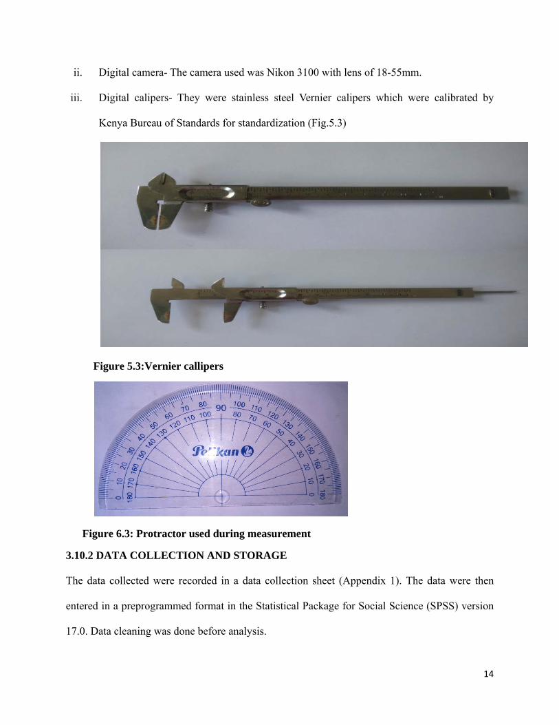
ii. Digital camera- The camera used was Nikon 3100 with lens of 18-55mm.
iii. Digital calipers- They were stainless steel Vernier calipers which were calibrated by
Kenya Bureau of Standards for standardization (Fig.5.3)
Figure 5.3:Vernier callipers
Figure 6.3: Protractor used during measurement
3.10.2 DATA COLLECTION AND STORAGE
The data collected were recorded in a data collection sheet (Appendix 1). The data were then
entered in a preprogrammed format in the Statistical Package for Social Science (SPSS) version
17.0. Data cleaning was done before analysis.
14

Back-up for the data was saved in an external hard drive and written on a digital versatile
compact disk (DVD). Access to these back-ups was limited to the investigator only.
3.10.3 DATA ANALYSIS
Data analysis was done using the Statistical Package for Social Science (SPSS) version 17.0. The
distance of the MA from the selected bony landmarks on the right and the left side were tested
for significance using paired students t-test.
3.10.4 DATA PRESENTATION
The results of the study were summarized and presented as tables.
3.11 ETHICAL CONSIDERATION
The study proposal was approved by Kenyatta national hospital-university of Nairobi
(KNH/UoN) Ethics and Research Committee (approval No.p660/10/2015).
15

CHAPTER 4
4.0. RESULTS
There were 53 cadavers available in the dissection laboratory. Forty eight cadavers (46 male and
2 female) were included in the study. Five cadavers were excluded from the study, three were too
dry to be dissected and the other two had distorted anatomy where one had deep cuts bilaterally
in the preauricular region extending into the submandibular regions and the other had bilateral
lazy – S surgical scars. Out of the 48 cadavers, three had the left sides mutilated by the medical
students, therefore, the data were collected from the right sides only (Fig. 3.3). Therefore, there
were 48 right hemi-sections and 45 left hemi-sections. Because of inadequate number of female
cadavers, gender differences were not determined in this study.
4.1 DISTANCES OF THE MAXILLARY ARTERY (MA) FROM SELECTED BONY
LANDMARKS
The distances of the MA were measured from the following selected bony landmarks; the
posterior cortex at the centre of condylar neck of the mandible, from the inferior-most part of the
mandibular notch, the inferior part of the articular eminence and the inferior part of pterygoid.
(Fig.4.3). The mean distance of the MA from the inferior-most part of the mandibular notch on
the right side was 5.72±2.82 mm (mode = 5.25 mm) while on the left side it was 5.80±2.85 mm
with a mode of 5.75 mm. On the right side the average distance of the MA from the posterior
cortex at the centre of the condylar neck of the mandible was 8.39±2.67 mm (mode =6 mm)
whereas on the left side the mean distance was 8.78±2.70 mm (mode =3.60 mm). Whereas the
mean distance of the MA from the inferior part of the pterygoid on the right was 13.93±6.17
mm; (mode =7.25 mm), on the left side the mean distance was 14.61±7.07 mm (mode =5.25
mm). The mean distance of the MA from the inferior part of articular eminence on the right was
16

17
14.05±3.77 mm mode of 6.62 mm while on the left side the mean distance was 14.47±4.01 mm;
mode of 14.25 mm. The difference between the measurements from the MA and the selected
bony landmarks on the right and left sides were not statistically significant (Table 1.4).

Table 1.4: Comparison of the measured distances between the right and the left side in relation to selected bony landmarks.
Bony landmarks Mean Distance (mm)±SD(mm)
Median Distance (mm)
Minimum Distance (mm)
Maximum Distance (mm)
95 % confidence interval of the difference
Independent student’s t Test
Lower Upper P value <0.05
Distance of MA from inferior most part of mandibular notch on the right side (n=48)
5.72±2.82 5.44 1.03
14.76
-0.64402 0.12947 0.187
Distance of MA from inferior most part of mandibular notch on the left side(n=45)
5.80±2.85 6.00 0.00
14.00
Distance of MA from posterior cortex at the centre of condylar neck of the mandible on the right side(n=48)
8.39±2.67 8.30 3.87
18.27
-0.93597 0.16143 0.162
Distance of MA from posterior cortex at the centre of condylar neck of the mandible on the left side (n=45)
8.78±2.70 9.15 3.60
17.81
Distance of MA from inferior of articular eminence on the right side (n=48)
14.05±3.77 14.19 6.62
21.48
-1.53776 0.25185 0.155
Distance of MA from inferior part of articular eminence on the left side (n=45)
14.47±4.01 14.38 6.50
24.08
Distance of MA from inferior part of pterygoid fovea on the right side (n=48)
13.93±6.17 11.70 4.94
24.50
-1.50507 0.23689 0.149
Distance of MA from inferior part of ptrerygoid fovea on the left side (n=45)
14.61±7.07 13.38 3.98
26.76
18

4.2COURSEOFTHEMAXILLARYARTERY
The MA originated directly from the ECA in a single trunk bilaterally in all the cadavers. Out of
the 93 hemi-sections dissected, 62 (67%) of the MAs were within the parotid gland while 31
(33%) of the MA were outside the gland. The MA on the right side passed within the parotid
gland in 32 hemi-sections (68.1%) while in 16 hemi-sections (31.9%), the MA passed medial to
the parotid gland. A similar pattern was observed on the left side, whereby 30 hemi-sections
(67%) had the MA within the parotid gland while in 15 hemi-sections (33%) the MA passed
medial to the parotid gland (Table 2.4). One cadaver had asymmetry whereby on the left side the
artery was within the gland while on the right side the artery passed medial to the gland.
Table 2.4: Relationship of the maxillary artery with the parotid gland
Side MA within the parotid gland n (%)
MA medial to the parotid gland n (%)
Total n (%)
Right side 32 (68.1) 16 (31.9) 48 (100)
Left side 30 (67.0) 15 (33.0) 45 (100)
Total 62 (67) 31 (33) 93 (100)
4.3 RELATIONSHIP OF THE MAXILLARY ARTERY TO THE LATERAL PTERYGOID
MUSCLE
The relationship of the MA to the LPM was described as either lateral or medial to the LPM
(Fig.7.4). Out of the 93 hemi-sections dissected, 35 (38%) of the MA had a medial relationship
to the LPM while 58 (62%) of the MA had a lateral relationship to the LPM (Fig. 7.4).
19
A B
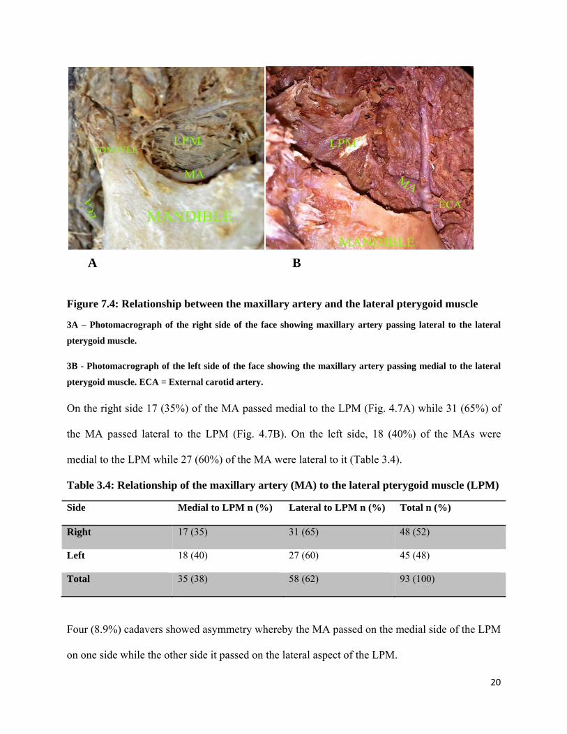
Figure 7.4: Relationship between the maxillary artery and the lateral pterygoid muscle
3A – Photomacrograph of the right side of the face showing maxillary artery passing lateral to the lateral
pterygoid muscle.
3B - Photomacrograph of the left side of the face showing the maxillary artery passing medial to the lateral
pterygoid muscle. ECA = External carotid artery.
On the right side 17 (35%) of the MA passed medial to the LPM (Fig. 4.7A) while 31 (65%) of
the MA passed lateral to the LPM (Fig. 4.7B). On the left side, 18 (40%) of the MAs were
medial to the LPM while 27 (60%) of the MA were lateral to it (Table 3.4).
Table 3.4: Relationship of the maxillary artery (MA) to the lateral pterygoid muscle (LPM)
Side Medial to LPM n (%) Lateral to LPM n (%) Total n (%)
Right 17 (35) 31 (65) 48 (52)
Left 18 (40) 27 (60) 45 (48)
Total 35 (38) 58 (62) 93 (100)
Four (8.9%) cadavers showed asymmetry whereby the MA passed on the medial side of the LPM
on one side while the other side it passed on the lateral aspect of the LPM.
20
4.4 BRANCHING PATTERN OF THE 1ST PART OF THE MAXILLARY ARTERY
The branching pattern of the first part of the MA was successfully bilaterally dissected in forty
five bilaterally whereas 3 were dissected only the right side. Branching patterns observed were,
in 6 (6.5%) different cadavers the MMA and the IAA originated at the same point off the MA.
The AMA was absent in 3 (3.2%) cadavers on the right side and in 2 (2.2%) cadavers on the left
side. A different branching pattern observed was on the right side, the MMA branched off from
the MA before the IAA in 18 (37.5%) of the cadavers (Fig. 8.4) whereas in 27 (56.3%) of the
hemi-sections it was given out after the IAA (Table 4.4). On the left side the MMA was given
out before (Fig. 9.4) the IAA in 10 (22.8%) of the cadavers and branched out after the IAA in 32
(71.1%) of the cases. In all the cadavers the IAA originated directly from the MA. There was one
case whereby two IAAs were given out. One branch entered the mandibular canal while the other
one accompanied the nerve to the mylohyoid muscle. Another pattern observed was where the
deep auricular artery and the anterior tympanic artery originated from the same trunk of the MA.
Also the deep auricular artery was abnormally larger than would have been expected (Fig. 8.4).
21

Figure 8.4: Illustration of the branching pattern of the 1st part of the right maxillary artery (MA). The inferior alveolar artery (IAA) branching after the middle meningeal artery (MMA). Separate origin of the MMA and the accessory meningeal artery (AMA). Note also an abnormally large deep auricular A.
Table 4.4: Pattern of origin of the middle meningeal artery (MMA) and the inferior alveolar artery (IAA) from the maxillary artery (MA).
Description Right n (%) Left n (%) Total n (%)
The
MMA
Branching before the IAA 18 (37.5) 10 (22.8) 28 (32)
Branching after the IAA 27 (57.4) 32 (71.1) 59 (68)
Total n (%) 45 (53) 42 (47) 87 (100)
The MMA and AMA originated from a common trunk off the MA on the right side in 28
(58.3%) of the cases (Table 5.4). On the left side the MMA and the AMA originated from the
same trunk on the MA in 25 (55.6%) of the cadavers (Fig. 9.4). There was asymmetry in the
22
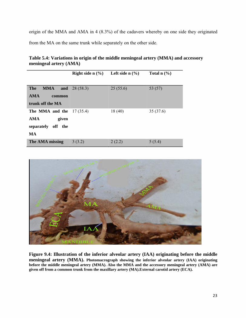
origin of the MMA and AMA in 4 (8.3%) of the cadavers whereby on one side they originated
from the MA on the same trunk while separately on the other side.
Table 5.4: Variations in origin of the middle meningeal artery (MMA) and accessory meningeal artery (AMA)
Right side n (%) Left side n (%) Total n (%)
The MMA and
AMA common
trunk off the MA
28 (58.3) 25 (55.6) 53 (57)
The MMA and the
AMA given
separately off the
MA
17 (35.4) 18 (40) 35 (37.6)
The AMA missing 3 (3.2) 2 (2.2) 5 (5.4)
Figure 9.4: Illustration of the inferior alveolar artery (IAA) originating before the middle meningeal artery (MMA). Photomacrograph showing the inferior alveolar artery (IAA) originating before the middle meningeal artery (MMA). Also the MMA and the accessory meningeal artery (AMA) are given off from a common trunk from the maxillary artery (MA).External carotid artery (ECA).
23

CHAPTER 5
5.1 DISCUSSION
Different specialities have interest in understanding the MA and studies have been done on
different aspects of the artery. Being one of the most vulnerable structures in the craniofacial
region, knowledge of the MA may help in reduction of its injury and intraoperative haemorrhage.
In the present study, 67% of the MA was given within the parotid gland as compared to 33% of
the arteries which passed medial to the gland. This relationship is important when doing total
parotidectomy as it may help in the reduction of iatrogenic injury to the MA. Remarkably, there
is hardly any study describing the relationship between the MA and the parotid gland.
Different bony landmarks have been studied to try locating the MA which acts as guide to
prevent its injury. The studied parameters are key bony land marks that are used intraoperatively
during osteotomies. The distance from the mandibular notch to the MA at 5.76±2.82 mm was
consistent with other studies done elsewhere (4, 30, 31). The average distance of the MA from
the articular eminence has been reported as 1.67±0.48 mm (n=34) (30). There was great variation
from the current study whereby the average distance from the articular eminence to the MA was
14.25±3.87 mm. However, the distance from the pterygoid fovea to the MA was 14.21±6.61
mm, which was consistent with a study done by Balcioglu et al. (30). There are few cadaveric
studies focusing on the risk of the MA injury during mandibular osteotomies (7, 8). Care is
needed in intraoral vertical ramus osteotomies to prevent injury to the MA when performing
bicortical osteotomy inferiorly since the MA passes upward across the lower head of the LPM
inside the mandibular notch (7, 33). This necessitates exposure of the medial aspect of the
mandibular notch such that special retractors (Bauers retractor) should be inserted to prevent
24

injury to the MA (7). There is a high probability of injuring the MA when the medial cut of
sagittal split ramus osteotomy is placed very close to the mandibular notch which may lead to
severe bleeding(2).
In the current study, the majority of the MAs were lateral to the LPM, with 58 (62%) of the MA
having been lateral and 35 (38%) of the MA having been medial to the muscle. The MA was
lateral in 62% of the hemi-sections in the current study which was comparable to a study by
Fujimura et al. (69% in blacks) and Lasker et al. (69 % in an African American population)(7).
This was also consistent with other studies which showed that the MA passed lateral to the LPM
in most of the hemi-sections(17, 19, 21, 22).The MA passed medial to the LPM (38%) in the
present study which was comparable to a study by Lasker et al (31% of an African American
population) (16) and a Caucasian population whereby the MA passed medial in 32 % of hemi-
sections(17). However, the lateral relationship of the MA to the LPM has been reported as 9 to
55% and 90% in the white and Japanese respectively(7). In most of the cadavers, there was
symmetry in the courses of the MA on both the left and the right sides. However, four (8.9%)
cadavers showed asymmetry whereby the MA passed on the medial side of the LPM on one side
while the other side it passed on the lateral aspect of the LPM. The prevalence of the asymmetry
in the course of the right and the left MAs has been reported to be 6.8% by Takarada (34) and
6.3% by Ikakura (35).
Though the relationship between the MA and the LPM is not consistent, given most of the MA
passes lateral to the LPM, the pterygoid part can be identified after blunt dissection of the buccal
fat pad. During total maxillectomy, some studies have shown that tying the MA in advance
reduces the risks of intraoperative bleeding (36, 37).The MA passes in close proximity with the
subcondylar region of the mandible. This is of great importance when managing subcondylar
25

fractures to avoid iatrogenic injury to the MA. In the infratemporal fossa the MA is closely
related to the LPM with the artery passing either medial or lateral to the muscle. The relationship
has been widely studied and has been shown to differ depending on the race and population. The
most common source of bleeding during fixation of subcondylar fractures of the mandible is
due to accidental puncture of the MA(2, 4). Therefore, it should be prudent for surgeons to know
the location of the MA in order to mitigate the risks of intraoperative bleeding. The Gow-Gates
technique is commonly applied in the administration of local anaesthesia in clinical dentistry
(38) and is associated with a higher response rate (>95%) than other techniques with a low risk
of puncturing vessels. However, the response of the Gow-Gates technique has been shown to
vary depending on the population, which is attributed to differences in the course of the MA.
Toki reported that the technique had a response rate of 75% in Japanese patients whereby most
of the MAs are lateral to the LPM consistent also with the present study, which was lower than
that in Australian patients and has increased risk of intravenous puncture (38).
The IAA is the main source of blood supply to the mandible. It mainly branches from the
mandibular part of the MA, though rarely can originate from the ECA (29, 39). In the current
study, the IAA originated directly from the MA in all cadavers. In the present study, the IAA
branched off before the MMA in 59 (63.4%) of the hemi-sections, branched after the MMA in 28
(30.1%) of the hemi-sections and at the same localization with the MMA in 6 (6.5%) hemi
sections. Otake et al found that the IAA arose distal to the MMA in 25 of 28 hemi sections
(89.3%) (22). In a study by Uysal et al, in 35.7% of the cases the IAA arose from the MA before
the MMA while in 35.7 % it was given off after the MMA. The IAA and the MMA branched off
from the same area of the MA in 14.3 % of the cases while in others, the IAA branched off from
the beginning of the MA in 14.3% of the cases (19). To prevent negative influence on blood
26
supply to the mandible, knowledge of the branching pattern of the IAA is important when
performing surgical procedures around this area.
In a study by Uysal et al., while the MMA was present in all cases, the AMA was observed in
57.1% of the cases (19). In this study, the AMA was missing in 3 (6.3%) cadavers on the right
side and in 2 (4.4%) cadavers on the left side. Rao et al. described a variation whereby the
anterior tympanic and the AMA arose from the MMA (28). In the present study the AMA and
the MMA were given off from the MA in the common trunk in 53 (57%) of the hemi-sections.
The prevalence of the AMA and MMA being given off from a common trunk from the MA has
been reported to have been 18 (64.3%) by Otake et al. (22) and as high as 73 of 76 subjects
(96%) by Baumel et al. (40). The limitation of the study was tissue shrinkage especially the soft
tissue.
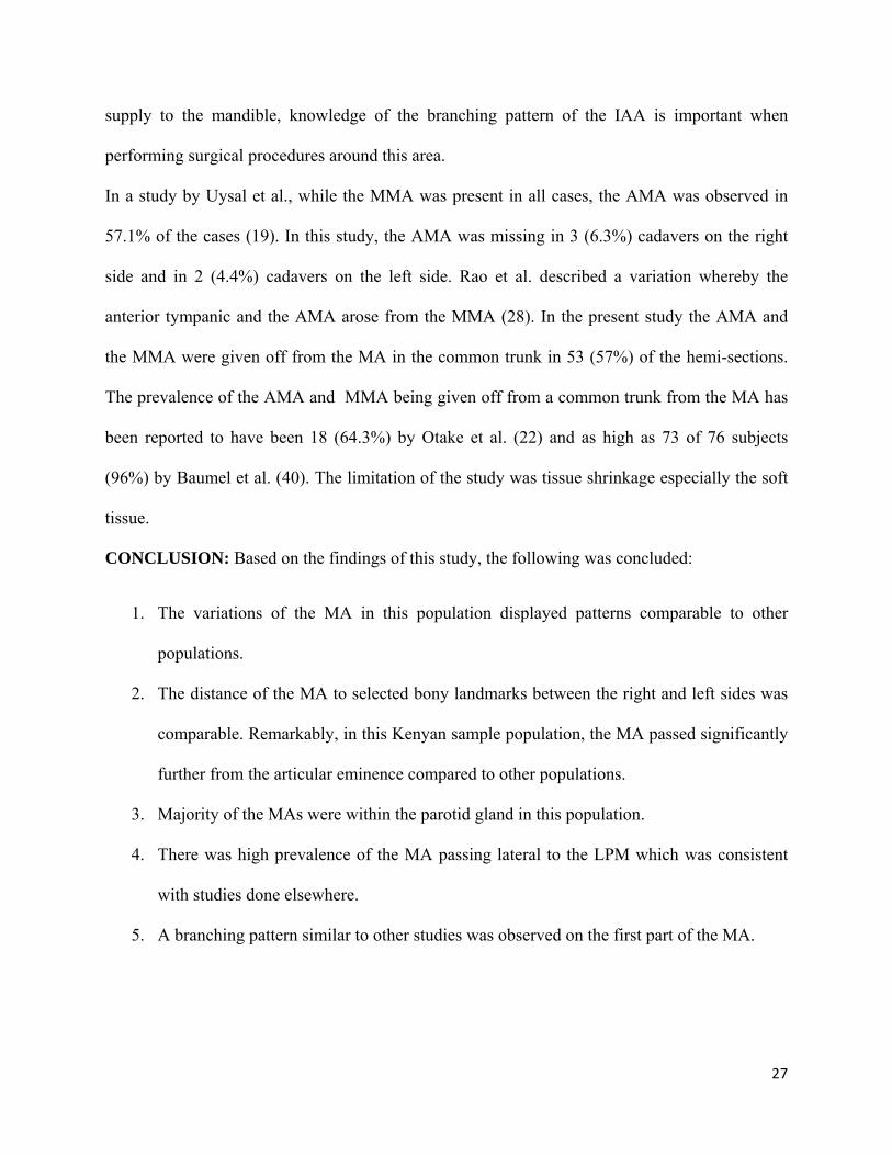
CONCLUSION: Based on the findings of this study, the following was concluded:
1. The variations of the MA in this population displayed patterns comparable to other
populations.
2. The distance of the MA to selected bony landmarks between the right and left sides was
comparable. Remarkably, in this Kenyan sample population, the MA passed significantly
further from the articular eminence compared to other populations.
3. Majority of the MAs were within the parotid gland in this population.
4. There was high prevalence of the MA passing lateral to the LPM which was consistent
with studies done elsewhere.
5. A branching pattern similar to other studies was observed on the first part of the MA.
27

RECOMMENDATIONS
Based on the findings of this study, the following is recommended:
1. The closeness of the MA to the subcondylar region of the mandible and its relationship
to the TMJ, caution needs to be exercised in procedures involving these regions.
2. Given the distance of the MA from the articular eminence was significantly longer
compared with studies done elsewhere, a multicenter cadaveric studies in Kenyan
population should be done to ascertain this finding.
3. Another study with a bigger sample size to determine if there are gender variations on the
MA since there were few female cadavers in this study.
28

REFERENCES
1. Ngassapa D, Hassanali J, Amwayi P, Guthua S. Essentials of Orofacial Anatomy.
Tanzania: Dar es Saalam University Press; 1996. 196-200 p.
2. Lanigan D, Hey J, West R. Hemorrhage following mandibular osteotomies: a report of 21
cases. J Oral Maxillofac Surg. 1991;47(7):713-24.
3. Elton V, Turnbull I, Foster M. An overview of the management of pseudoaneurysm of
the maxillary artery: A report of a case following mandibular subcondylar osteotomy.
Craniomaxillofac Surg. 2007;35:52-6.
4. Orbay H, Kerem M, Unlü R, Cömert A, Tüccar E, Sensöz O. Maxillary artery:
anatomical landmarks and relationship with the mandibular subcondyle. Plast Reconstr Surg
2007;120:1865-70.
5. Panula K, Finne K, Oikarinen K. Incidence of complications and problems related to
orthognathic surgery: a review of 655 patients. J Oral Maxillofac Surg 2001;59:1128-37.
6. Iannetti G, Fadda TM, Riccardi E, Mitro V, Filiaci F. Our experience in complications of
orthognathic surgery: a retrospective study on 3236 patients. European Review for Medical and
Pharmacological Sciences. 2013;17:379-84.
7. Fujimura K, Segami N, Kobayashi S. Anatomical study of the complications of intraoral
vertico-sagittal ramus osteotomy. J Oral Maxillofac Surg 2006;64:384-9.
8. Fontoura Rd, Vasconcellos H, Campos A. Morphologic basis for the intraoral vertical
ramus osteotomy: anatomic and radiographic localization of the mandibular foramen. J Oral
Maxillofac Surg. 2002;60:660-5.
9. Yeo MS, Goh TLH, Nallathamby V, Cheong EC, Lim TC. Maxillary Artery Injury
Associated with Subcondylar Mandible Fractures:A Novel Treatment Algorithm.
Craniomaxillofac Trauma Reconstruction 2012;5:83-8.
10. Chakrabarty S, Majumdar SK, Bansal A. Management of Pseudoaneurysm of Internal
Maxillary Artery Resulting from Trauma. J Maxillofac Oral Surg. 2015;14:203-8.
11. Pinjala RK, Joshi S, Rammurti S. Traumatic Pseudoaneurysm of the Internal Maxillary
Artery. European journal of Vascular and Endovascular Surgery 2007;14:54-5.
12. Rogers S, Patel M, Beirne J, Nixon T. Traumatic aneurysm of the maxillary artery: the
role of interventional radiology. A report of two cases. Int J Oral Maxillofac Surg 1995;24:336-
9.
29

13. Singam P, Thanabalan J, Mohammed Z. Superselective embolisation for control of
intractable epistaxis from maxillary artery injury. Biomedical Imaging and Intervention Journal.
2011;7:e3.
14. Lanigan D, Hey J, West R. Hemorrhage following mandibular osteotomies. J Oral
Maxillofac Surg. 1991;49:13-7.
15. Sashi R, Tomura N, Hashimoto M, Kobayashi M, Watarai J. Angiographic anatomy of
the first and second segments of the maxillary artery. Radiat Med. 1996;14:133-8.
16. Lasker G, Opdyke D, Miller H. The position of the internal maxillary artery and its
questionable relation to the cephalic index. Anat Rec. 1951;109(1):119-26.
17. Hussain A, Binahmed A, Karim A, Sándor G. Relationship of the maxillary artery and
lateral pterygoid muscle in a caucasian sample. Oral Surg Oral Med Oral Pathol Oral Radiol
Endod 2008;105:32-6.
18. Pretterklieber M, Skopakoff C, Mayr R. The human maxillaryartery reinvestigated:
topographical relations in the infratemporalfossa. Acta Anat 1991;142:281-7.
19. Uysal I. I, Buyukmumcu M, Dogan NU, Seker M, Ziylan T. Clinical Significance of
Maxillary Artery and its Branches:A Cadaver Study and Review of the Literature. International
journal of morphology. 2011;29:1274-81.
20. Dennison J, Batra A, Herbison P. The maxillary artery and the lateral pterygoid muscle:
the New Zealand story. Oral and maxillofacial surgery. 2009;108:e26–e9.
21. Kim JK, Cho JH, Lee Y-J, Kim C-H, Lee JHBJ-G, Yoon J-H. Anatomical Variability of
the Maxillary Artery Findings From 100 Asian Cadaveric Dissections. Arch Otolaryngol Head
Neck Surg 2010;136:813-8.
22. Otake I, Kageyama I, Mataga I. Clinical Anatomy of the Maxillary Artery. Okajimas
Folia Anatomy Japan. 2011;87:155-64.
23. Verma S, Fasil M, Murugan M, Sakkarai J. Unique variation in the course of maxillary
artery in infratemporal fossa: a case report. Surgical and Radiologic Anatomy. 2014;36:507-9.
24. Maeda S, Aizawa Y, Kumaki K, Kageyama I. Variations in the course of the maxillary
artery in Japanese adults. Anatomy Science International. 2012;87:187-94.
25. Claire PG, Gibbs K, Hwang SH, Hill RV. Divided and reunited maxillary artery:
developmental and clinical considerations. Anatomical Science International. 2011;86:232-6.
30

26. Bhat K, Madhyastha S, R B. course of the maxillary artery through the loop of the
auriculotemporal nerve. Rev Arg de Anat Clin. 2013;5:235-9.
27. lydia Q, Babu A, Ankolekar VH, D'souza AS. Anatomical variations in the branches
of maxillary artery: A case report. Journal of pharmaceutical and biomedical sciences.
2013;32:1271-3.
28. Rao NS, Manivannan K, Gangadhara, Rao HRK. Trifurcation of external carotid artery
and variant branches of first part of maxillary artery. International Journal of Anatomy and
Research. 2014;2:561-65.
29. Khaki AA, Tubbs RS, Shoja MM, Shokouhi G, Farahani RM. A rare variation of the
inferior alveolar artery with potential clinical consequences. Folia Morphology. 2005;64:345-6.
30. Balcioglua HA, Kilicb C, Varolc A, Ozanb H, Kocabiyikb N, Yildirimd M. A
Morphometric Study of the Maxillary Artery and Lingula in Relation to Mandibular Ramus
Osteotomies and TMJ Surgery. European Journal of Dentistry. 2010;4:166-9.
31. Aziz sR, Dorfman BJ, Ziccardi VB, Janal MN. Accuracy of using the antilingula as a sole
determinant of vertical ramus osteotomy position. J Oral Maxillofac Surg 2007 65:859-62.
32. Daniel ww. Biostatistics: A Foundation for Analysis in the Health Sciences. 7th edition.
New York: John Wiley & Sons. 1999.
33. Tuinzing D, Grebe R. Complications related to intraoral vertical ramus osteotomy. Int J
Oral Surg. 1985;14:319-24.
34. Takarada T. Anatomical studies on the maxillary artery. Shikwa gakuho. 1958;58:1-20.
35. Ikakura K. On the origin, course and distribution of the maxillary artery in Japanese. .
Arch Dept of Anat Tokyo Dent Coll (Kouku Kaibou Kenkyu). 1961;18:91-122.
36. Choi E, Choi Y, Kim C. Surgical outcome of radical maxillectomy in advanced maxillary
sinus cancers. Yonsei Med J 2004;45:621-8.
37. Wang C, Yang T, Ko J, Lou P. Ligation of the internal maxillary artery to reduce
intraoperative bleeding during total maxillectomy. Laryngoscope. 2007;117(11):1978-81.
38. Toki S. Gow-Gates mandibular block Technique. Nippon Dental Review 1982;478:177-
85.
39. Jergenson MA, Norton NS, Opack JM, Barritt LC. Unique origin of the inferior alveolar
artery. . Clin Anat,. 2005;18:597-601.
31

40. Baumel J, Beard D. The accessory meningeal artery of man. Journal of Anatomy.
1961;95:386-402.
32

APPENDIX I
DATA COLLECTION SHEET Case No………………sex…………….
1. Distance of maxillary artery from the following bony landmarks
Right side Distance
(mm)
Left side Distance
(mm)
i Inferior most part of
mandibular notch
Inferior most part of
mandibular notch
ii posterior cortex at the
centre of condyler neck of
the mandible
posterior cortex at the
centre of condyler
neck of the mandible
iii Inferior part of articular
eminence
Inferior part of
articular eminence
iv Inferior part of pterygoid
fovea
Inferior part of
pterygoid fovea
2. Course of the maxillary artery
Forming a
loop
Single trunk Within the
parotid
gland
Outside the
parotid
gland
Others
(specify)
Right side
Left side
3. Relationship of maxillary artery to lateral pterygoid muscle
Right side Yes No Left side Yes No
i Medial Medial
ii Lateral Lateral
iii Others (specify) Others
(specify)
33

4. Branching pattern of the maxillary artery
A).The MMA
Branching
before the
IAA
Branching
after the IAA
Branching
from one
trunk with
AMA
Missing Others(specify)
Right
Left
B).The IAA
Originating
directly from the
ECA
Branching
before the MMA
Originating after
the MMA
Others (specify)
Right side
Left side
34

APPENDIX II
ETHICAL APPROVAL
35

36

APPENDIX III
PLAGIARISM REPORT
37