Root canal morphology and its relationship to endodontic ...
Upload
nguyenliemCategory
view
222download
0
1 d-J- ~ ~ f VARIANT ROOT MORPHOLOGY OF THE THIRD MANDIBULAR
MOLAR IN NORMAL AND IMPACTED TEETH
Investigator Bokindo Isaac Kipyator
V2819582010
BDS III
-1- bull - C) CJ
sect ~
-I- C-J
A research proposal submitted in partial fulfillment of the requirements for the
award of the degree of Bachelor of Dental Surgery University of Nairobi
copy 2014
DECLARA TION
I Bokindo Isaac Kipyator hereby declare that this research proposal is my original work and has
not been presented to any other institution for examination or any other purpose
Sign
Date
APPROVAL I Bokindo Isaac Kipyator IS submitting this research proposal to the Kenyatta National
Hospital University of Nairobi Research Ethics and Standards committee for approval
ln~tF Date Sign
This research proposal has been submitted with our approval as the University of Nairobi
supervisors
Supervisors
IDr Fawzia Butt BDS (Nbi) FDSRCS MDS-OMFS
Department of Oral and Maxillofacial Surgery
School of Dental Sciences
University of Nairobi
Date Sign
2Prof Francis G Macigo BDS (Nbi) MPH (Nbi) PGD-STI (Nbi)
Department of Periodontology I Community and Preventive Dentistry
School of Dental Sciences
University of Nairobi
110~J-DILfSign~ Date
ii
TABLE OF CONTENTS DECLARATION i
APPROVAL ii
TABLE OF CONTENTS iii
LIST OF ABBREVIATIONS iv
SUMMARy 1
CHAPTER 1 INTRODUCTION AND LITERATURE REVIEW 3
11 INTRODUCTION 3
12 LITERATURE REVIEW 5
121 Types of third molar im paction 5
122 Factors involved in third molar impaction 6
123 Root morphology 6
Chapter 2 PROBLEM STATEMENT AND JUSTIFICATION 8
21 Problem statement 8
22 Justification 8
23 Objectives 8
231 Broad objective 8
232 Specific objectives 8
24 Hypothesis 9
24 Study variables 9
Chapter 3 MATERIALS AND METHODS 10
31 Study area 10
32 Study Population 10
33 Study Design 10
34 Sample size 10
35 Sampling methods 10
36 Inclusion and exclusion criteria 11
37 Data collection instruments and techniques 11
38 Data analysis and presentation 11
39 Ethical Consideration 12
310 Study benefits 12
BUDGET 13
CHEDULE AND TIME FRAME 14
60 REFERENCES 15
DATA SHEET 19
iii
LIST OF ABBREVIATIONS
DA - disto-angular
H - Horizontal
IAN - Inferior alveolar nerve
MA - mesio-angular
OMFS - Oral and Maxillofacial Surgery
SDS- School of Dental Sciences
SPSS - Statistical Package for Social Sciences
UoN - University of Nairobi
V - vertical
iv
SUMMARY Background The prevalence of third molar impaction is high across various population
Extraction of the impacted tooth is usually indicated where there is associated pathology
Surgical difficulty in extraction is described in various classification the most commonly used
ones are the Winters and Pell and Gregory The downside to these classification methods is that
morphology of the roots is not put into consideration Root morphology is known to influence
the eruption of teeth therefore it may have a role in impaction of these teeth The study aims to
describe various root morphologies in different types of impactions and establish any association
between the morphology of the teeth and type of impaction
Objective To describe the various root morphologies in different types of third molar impaction
among residents of Nairobi County visiting the School of Dental Sciences University of Nairobi
Study design Descriptive cross-sectional study
Study Population and Study Area This study will be carried out among patients visiting the
School of Dental Sciences University of Nairobi
Materials and methods 359 panoramic radiographs (179 male and 179 female) will be
obtained from the Radiology Division of the Oral and Maxillofacial department School of
Dental Sciences University of Nairobi The morphology of the roots of the third molar will be
described as straight or dilacerated The number of roots will also be recorded The crown and
root lengths will be measured using a Vernier caliper and the root to crown length ratio will be
calculated
Data management Measurements will be coded tabulated and analyzed using SPSS 17
(version 200 Chicago Illinois) Means standard deviations and variances will be calculated A
P-value of ~005 will be considered significant at a confidence interval of 95 Data will be
1
presented in form of graphs pie-charts and tables Photographs will be used for pictorial
representation
Study benefits The root morphology is useful for a surgeon operatingon and around the third
molar region to plan well and in extension enrich the current methods utilized in grading the
surgical difficulty in extraction of the third molar in which the root component has not been
regarded
2
CHAPTER 1 INTRODUCTION AND LITERATURE REVIEW
11 INTRODUCTION The mandibular third molars are the most frequently impacted teeth in the human dentition 1
accounting for 98 of all impacted teeth2 The incidence of impaction of the third molar has
been reported to vary between 8-84 in various studies There is higher prevalence in females
as compared to males5 Various theories have been put forward to explain the cause of
impaction The main factor has been lack of space in the jaw Others include late eruption of the
tooth 7 and the size of the third molar8
The level of difficulty in extracting impacted third molar has been described in the Pell and
Gregory and the Winters classification9 Various aspects such as level of eruption position of
the tooth in relation to the ramus of the mandible and the angulation of the tooth have been
considered Despite the useful parameters used root morphology of the tooth is not put into
consideration in assessing difficulty in these classification methods The third molar shows the
greatest variation in the root morphology The variation in morphology accounts for the
complications that occur during disimpaction most common being laceration of the inferior
alveolar nerve 10
Majority of the third molars (60-70) studied have two roots 1112 The variations documented on
the mandibular third molar include presence of three roots 13 fused roots one rootll Most
studies on the morphologic variants of the third molar have focused on the number of roots
Literature describing the shape of the root of third molars is scarce despite its importance in third
molar disimpaction The shape of the root may be influenced by the nature of impaction since
developmentally growth of tissue has been shown to be determined by thesurrounding
structures as described in the functional matrix theory proposed by Moss (1962)14 Following
3
this theory it is expected that the nature of the third molar impaction will have a considerable
effect on the shape of the morphology of the third molar Knowledge on the root morphology
will help the surgeon to evaluate the difficulty of the operation and anticipate the complications
that may occur The study therefore aims to describe the various root morphologies occurring in
different types of impaction
4
12 LITERATURE REVIEW The third mandibular molar is the most frequently impacted tooth in the human dentition
Various types of impaction have been observed and documented and classified The role of the
root morphology of the third mandibular molar in impaction is yet to be established
121 Types of third molar impaction The most commonly used classification to assess the difficulty of extracting impacted
mandibular molars are the Wintersl5 and the Pel and Gregory Winters classification is based
on the long axis of the impacted tooth in relation to the long axis of the second molar and as
such the types of impaction in this category include mesial angular horizontal bony vertical and
distal angular impactions The Pell and Gregory classification is based on the vertical relation of
the third molar to the second molar as wel as the relationship of the tooth to the anterior border
of the ramus of the mandible
The Pel Gregory classification
i) Based on vertical relation of the third molar to the second molar
A- The occlusal plane of the impacted tooth is at the same level as the occlusal plane of the
second molar
B- The occlusal plane of the impacted tooth is between the occlusal plane and the cervical line of
the second molar
c- The impacted tooth is below the cervical line of the second molar
ii) Based on the relationship with the anterior border of ramus of the mandible
1- There is sufficient space between the ramus and the distal part of the second molar for the
accommodation of the mesiodistal diameter of the third molar
2- The space between the second molar and the ramus of the
mandible is less than the mesiodistal diameter of the third molar
3- Allor most of the third molar is in the ramus of the mandible
5
The above classifications have been shown not to be a reliable indicator of surgical difficulty in
extraction of impacted lower molars with variable intraexaminer and interexaminer agreementl7
Garcia et al(2000) obtained low sensitivity test on the Pell and Gregory classification and this
can be attributed to the variant root morphology of these teeth in terms the length and the shape
or the roots 18
122 Factors involved in third molar impaction Several theories have been put forward to describe causes of third molar impaction The most
popular has been insufficient development of retromolar space 1920 and this may also be related to
imbalance in the pattern of bone remodeling at the mandibular ramus22 Growth of the condyle
occurs in a vertical direction and has been shown to limit resorption at the anterior aspect of the
ramus of the mandible Unfavourable path of eruption has also been implicated in impactiorr
for instance if the tooth bud is in an abnormal position during development and eruption the
tooth ends up impacted Emes at al(20 11) described the evolutionary decrease in the size of the
jaw disproportionately with teeth and this may been due to the change in diet from the hard
unprocessed foods to the soft processed ones2526 The relatively small jaw is more susceptible to
impactions General factors such as genetics race gender and environmental factors including
dietary habits also playa role in impaction2728 Third molar impaction have been associated with
anterior teeth crowding therefore features of the latter seen in a young patient has been an
indicator of third molar impaction
123 Root morphology Literature has focused on the pattern of impaction of the third molar with little mention of the
role the roots of the third molar play in the management of the condition Carvalho and
Vasconcelos(2011) put forward that the number of root (Plt 0004) and the morphology
(Plt0031) were significant predictors of surgical difficulty The main parameters in root
morphology are dilaceration and length Dilaceration is a developmental disturbance in the shape
6
of teeth whereby there is a sharp bend or curvature in the root of a formed tooth A curvature of
greater than 10deg posses a greater risk than lower values Yamaoka et al(2009) found the relation
between the root angulation and impaction whereby impacted tooth had a higher incidence of
angulated roots The reported prevalence of dilaceration of the roots are very high at 81 30
There is little literature on the length of the roots of the third molar which may influence its
closeness to the mandibular canal and thus the risk of injuring the inferior alveolar nerve (IAN)
during extraction Crown to root length ratios have been shown to show the development of
roots Unfavorable ratios have been found in females31 and this may explain the higher
prevalence of impacted teeth among women Some authors have recommended coronectomy of
impacted wisdom teeth in case the roots are surrounding the mandibular canal32J3 The
morphology of the roots has been shown to influence autotransplantation of the third molar34 in
that the morphology of the root may not favor successful transfer of the third molar into the
socket of another missing molar
Park et al(2013) assessed the number of roots of third mandibular molars in a Korean
population whereby there was high prevalence of two rooted teeth (569) and one rooted
(379i5bull Three rooted teeth were seen in only 19 Higher prevalence of two rooted teeth was
also observed in the Iran population at 73 II Panoramic radiography is the standard imaging
technique for evaluating third molars The sensitivity of these radiographs have been reported to
be fair but the specificity of the radiographs is quite high36
The study therefore aims to describe the various root morphologies occurring in different types
of impaction which will help in surgical approach to this region
7
Chapter 2 PI~OBLEM STATEMENT AND JUSTIFICATION
21 Problem statement
The morphology of the roots of the third molar has been shown to infl uence the clinical decision
pertaining the management of impacted third molars There is high variability in the size and
shape and number of the roots of the third molar more than any other teeth in human dentition
Iatrogenic damage to the IAN is the main complication arising from third molar disimpaction
and it is highly related to the closeness of the tooth and its form Current classifications have not
put into consideration the impact of root morphology in grading the difficulty in surgical
extraction
22 Justification
Although there is overwhelming data on the prevalence of third molar impaction in various populations
including the Kenyan there is little literature that focusses in the role played by root morphology in
assessing surgical difficulty in third molar disirnpaction The present study will aim to investigate the
root forms in various types of impactions Data obtained will enable surgeons have more
informed decision during third molar disimpaction
23 Objectives
231 Broad objective
To describe root morphologies in various types of impaction of the third molar in a Kenyan
population
232 Specific objectives
2 To classify various impacted third molar teeth radiographically
3 To describe the number and the shape of the roots in each of the class of impaction
4 To obtain the ratio of crown to root length of the impacted third molar
8
4 To establish the relationship between the morphology of root and the type of impaction
24 Hypothesis
Alternative hypothesis
Angulated roots increases the risk of occurrence of impacted teeth
24 Study variables
Variables Measures
Social- Demographic Variables Age of the patient in years
Independent Variables Root morphology
Dependent Variables Type of third molar impaction
9
Chapter 3 MATERIALS AND METHODS
31 Study area The study is to be carried out at the Radiology division of the Oral and Maxillofacial(OMFS) Department
School of Dental Sciences (SDS) University of Nairobi (UoN) The SDS is located 5 kilometers from the
Central Business District of Nairobi City along valley road
32 Study Population The population will comprise of patients who have come to seek dental treatment in the SDS
33 Study Design A descriptive cross - sectional study on the root morphology of the impacted third molar
34 Sample size Sample size will be computed using the following formula
Where
z = z value according to the confidence level chosen
P = prevalence of impacted teeth recorded at SDS (62837)
c = 1- confidence interval
Using a confidence level of95 and a Z value of l96
(196)20628(1 - 0628) n=
(1 - 095)2 n= 35898 ~ 359 radiographs
35 Sampling methods Probability sampling will be employed whereby simple random sampling will be utilized
Panoramic radiographs of patients taken from year 2010 until current date of study at the
radiology divisionwill be assed Those that will havemet the selection criteria will be listedfrom
10
the oldest to the newest in terms of the date taken starting from number one A fare coin wi II
tossed with the heads meaning the even numbers selected and the tail odd numbers
36 Inclusion and exclusion criteria
361 Inclusion criteria
1) Radiographs from patients 30 years or older
2) Presence of the 3 molars in ei ther quadrant
362 Exclusion criteria
1) Radiographs lacking good contrast
2) Presence of pathologies such as tumors and cysts
37 Data collection instruments and techniques Radiographs will be retrieved from the computers archives by the radiology assistant The main
researcher will examine the radiographs selecting those that have met the requirements The type
of impaction will be classified using the Pell and Gregory classification as
AlA2A3BIB2B3ClC2C3 and also using the Winters method as mesio-angular (MA)
horizontal (H) vertical (V) and disto-angular (OA) The morphology of the roots will also be
studied under each classification and categorized as either straight or dilacerated with the
number of roots recorded in each The lengths of the longest root and crown will be measured
using a Vernier caliper in rnillimetres
38 Data analysis and presentation The ratio of the crown to the root height will also calculated The results will be coded and
tabulated and analyzed using SPSS v17 (SPSS Inc Chicago IL USA) Students t- test will be
performed to test the relationship between root morphology and type of impaction A p value of
11
lt005 will be considered significant at a confidence interval of95 Photographs tables and
charts will be used for data presentation
39 Ethical Consideration Ethical approval will be sought from the Kenyatta National Hospital-University of Nairobi-
Ethics and Standards Committee before the commencement of the study Permission and
requisite authority will be obtained from the administration of the Radiology Division of OMFS
department SDS VoN Information regarding the subjects will be held with maximum
confidentiality and will not be disclosed to any unauthorized persons Case numbers and not
names will be used throughout the study At the end of the study data sheets will be shredded
310 Study benefits The study will be useful to surgeons operating on the third molar Morphology of the roots of the
third molars will enable proper planning and to anticipate difficulties The research proposal and
report will be presented in partial fulfillment of the requirements for the award of the degree of
Bachelor of Dental Surgery in the University of Nairobi
12
BUDGET ITEM NUMBER OF COST OF TOT AL (Kshs)
UNITS UNIT (Kshs) Proposal Typing and 30 pages of 4 10 1200 development Printing booklets
Internet 15 GB 1500 1500 Stationary(printing 1 ream 1000 1000 papers) Flash disk 1000 1000 Binding proposal 3 copies 100 300
Subtotals 5000 Data Collection Typing and 5 pages each 4 10 200
printing copies
Subtotals 200 Data analysis Data analysis 1000 1000 and report computer package writing Typing and 30 pages of 4 10 1200
printing report booklets Internet 14 GB 1400 1400 Binding report 3 copies 100 300
Subtotals 3900 Miscellaneous 15 of total 1365 expenses Grand Total 10465
13
SCHEDULE AND TIME FRAME
Time of the year Activity
May-July Proposal writing
August Approval
August - September Data Collection
September - October Data analysis and writing of dissertation
November Presentation
14
REFERENCES
I Kaya GS Asian M Omezli MM Dayi E Some morphological features related to
mandibular third molar impaction I ClinExp Dent 20102 12-19
2
Fayad BJ Levy JC Yazbeck C Cavezian R Cabanis EA Eruption of third molars
relationship to inclination of adjacent molars Am J OrthodDentofacialOrthop 2004
F eb 125(2) 200-202
3 Ahlquist M Grondahl H Prevalence of impacted teeth and associated pathology in
middle-aged and older Swedish women Community Dent Oral Epidemiol 1991 19 116-
119
4 Venta I Ylipaavalnierni P Turtola L Clinical outcome of third molars in adults followed
during 18 years I Oral Maxillofac Surg 200462 182-5
5 Hashemipour MA Tahmasbi-Arashlow M Fahimi-Hnzaei F Incidence of impacted
mandibular and maxillary third molars A radiographic study in a Southern Iran
Population Med Oral Patol Oral Cir Bucal 20131340-45
6 Sadetaxecic Samir Prohic Sanjalxomsic AmraVukovic Incidence of impacted
mandibular third molars in population of Bosnia and Herzegovina a retrospective
radiographic study Journal of Health Sciences 20133(2) 151-158
7 Hassan Ali Mandibular cephalometric characteristics of a Saudi sample of patients
having impacted third molars Saudi Dent J 2011 23(2) 73-80
8 Forsberg CM Tooth size spacing and crowding in relation to eruption or impaction of
third molars Am I OrthodDentofacialOrthop 198894( 1 )57-62
9 Susarla SM Dodson TB Estimating Third Molar Extraction Difficulty Subjective and
Objective Factors J Oral and Maxillofac Surg 200563427-34
15
10 HoseiniZarch SH Bagherpour A Javadianlangaroodi A Ahmadian Yazdi A Safaei A
Evaluation of the accuracy of panoramic radiography in linear measurements of the jaws
Iran J Radiol 2011897-102
11 Kuzekanani M Haghani 1 Nosrati H Root and canal morphology of mandibular third
molars in an Iranian population Dent Res Dent Clin Dent Prospects 20126(3)85-8
12 Danilo M Zanello G Rusiel AS Manoel D de Sousa Nero Ricardo GS Jesus DPBraz
Dent J 19989(2) 91-94
13 Plotino G A mandibular third molar with three mesial roots A case report J Endod
200834224-6
14 Moss ML The functional matrix In Kraus BS Reidel RA editors Vistas in
orthodontics Philadelphia Lea and Febiger 1962
15 Winter GB Impacted Mandibular Third Molar St Lous American Medical Book Co
1926
16 Pell GJ Gregory B1 Impacted mandibular third molars Classification and modified
techniques for removal Dent Digest 193339330-338
17 Alrnendros-Marques N Berini-Aytes L Gay-Escoda C Influence of lower third molar
position on the incidence of preoperative complications Oral Surg Oral Med Oral Pathol
Oral RadiolEndod 2006 1 02725-32
18 Garcia A G Sampedro F G Rey 1 G Vila P G Martin M S Pell-Gregory classification
is unreliable as a predictor of difficulty in extracting impacted lower third molars Br I
Oral MaxillofacSurg 200038585-587
19 Bishara SA Andreasen G Third molars a review American Journal of Orthodontics
198383131-137
16
20 Grover PS Lorton L The incidence of unerupted permanent teeth and related clinical
cases Oral Surgery Oral Medicine Oral Pathology 198559420-425
21 Ricardo WF Carvalho Belmiro CEo Assessment of Factors Associated With Surgical
Difficulty During Removal of Impacted Lower Third Molars J Oral Maxillofac Surg
2011 692714-2721
22 Bjork A Prediction of mandibular growth rotation Am J Orthod 196955585-599
23 Lakhani ~J Kadri W Mehdi H Sukhia H Bano A Yaqoob S Anterior crowding - a
possible predictor formandibular third molar impaction J AyubColl Abbottabad
20112363-5
24 Richardson M Changes in lower third molar position in the young adult American
Journal of Orthodontics and Dentofacial Orthopedics 1992 1 02320-327
25 Emes Y Aybar B Serhat Y On the evolution of human jaws and teeth A review Bull
IntAssocPaleodont 2011 537-47
26 Garn SM Leonard WR What did our ancestors eat Nutr Rev 1989 Nov47(11 )337-45
27 Hattab FN Alhaija ES Radiographic evaluation of mandibular third molar eruption
space Oral Surg Oral Med Oral Pathol Oral RadiolEndod 1999 Sep88(3)285-91
28 Polat HB Ozan F Kara I Ozdemir H Ay S Prevalence of commonly found pathoses
associated with mandibular impacted third molars based on panoramic radiographs in
Turkish population Oral Surg Oral Med Oral Pathol Oral RadiolEndod 2008 10541-47
29 Yamaoka M Furusawa K Hayama H Kura T Relationship of third molar development
and root angulation J Oral Rehabil 2001 28 198-205
30 Saraswati FK Balajirao B Mamatha GPClinical and orthopantomographic evaluation of
mandibular third molar 2010 1 (1) 27-30
17
31 Holtta P Nystrom M Evalahti M Alaluusua S Root-crown ratios of permanent teeth in a
healthy Finnish population assessed from panoramic radiographs European Journal of
Orthodontics 200426491-497
32 Matzen LH Christensen J Hintze H Schou S Wenzel A Influence of cone beam CT on
treatment plan before surgical intervention of mandibular third molars and impact of
radiogr~phic factors on deciding on coronectomyvs surgical removal
DentomaxillofacRadiol 201342(1)
33 Pogrel MA Lee JS Muff DF Coronectomy A technique to protect the inferior alveolar
nerve J Oral Maxillofac Surg 2004 621447-52
34 Mendes RA Rocha G Mandibular third molar autotransplantation - literature review
with clinical cases J Can Dent Assoc 200470761-6
35 Jun-Beom Park NamRyang Kim Seojin Park YoungkyungKo Evaluation of number of
roots and root anatomy of permanent mandibular third molars in a Korean population
using cone-beam computed tomography 20137(3) 296-301
36 Atieh MA Diagnostic accuracy of panoramic radiography in determining relationship
between inferior alveolar nerve and mandibular third molar J Oral Maxillofac Surg 2010
Jan68(1)74-82
37 Guthua SW Mwaniki DL A retrospective study of characteristics of impacted
mandibular wisdom teeth in 110 patients treated in Nairobi KenyaAfr Dent J
1992630-3
18
DATA SHEET
Radiograph no
CrICK WHERE APPROPRIATE)
Pell and Gregory (A I) (A2) (A3) (8 I) (82) (83) (C I) (C2) (C3)
Winters (MA) (H) (V) (DA)
Straight ( ) d i lacerated ( )
No of roots (I) (2) (3)
Root length (R)
Crown length (C)
CR ratio
19
UNIVERSITY OF NAIROBI COLLEGE OF HEALTH SCIENCES POBOX 19676 Code 00202 Telegrams varsity (254-020) 2726300 Ext 44355
Ref KNH-ERCUAl153
Bokindo Isaac Kipyator V2819582010 School of Dental Sciences University of Nairobi
Dear Isaac
_-t G (-I
cO ~
KNHuON-ERC Email uonknh_ercuonbiacke Website wwwuonbiacke LinkwwwuonbiackeactivitiesIKNHUoN
KENY ATT A NATIONAL HOSPITAL POBOX 20723 Code 00202 Tel 726300-9
Fax 725272 Telegrams MEDSUP Nairobi
24th August 2014
Research Proposal - clearance -Variant Root morphology of the third mandibular molar in normal and impacted teeth (UP523072014)
Your above proposal refers
This is to inform you that permission has been granted by the KNHUON-Ethics amp Research Committee to carry out research on study titled - Variant Root morphology of the third mandibular molar in normal and impacted teeth
By a copy of this letter I am requesting the relevant persons to accord you the professional support and other materials that may be useful to your research
P ML CHINDIA SECRETARY KNHUONmiddotERC
cc The Principal College of Health Sciences UON The Deputy Director CS KNH The Chairperson KNHUoN-ERC AD Health Information KNH The Dean School of Dental Sciences UoN Supervisors Dr Fawzia Butt Prof Francis G Macigo
Protect to discover
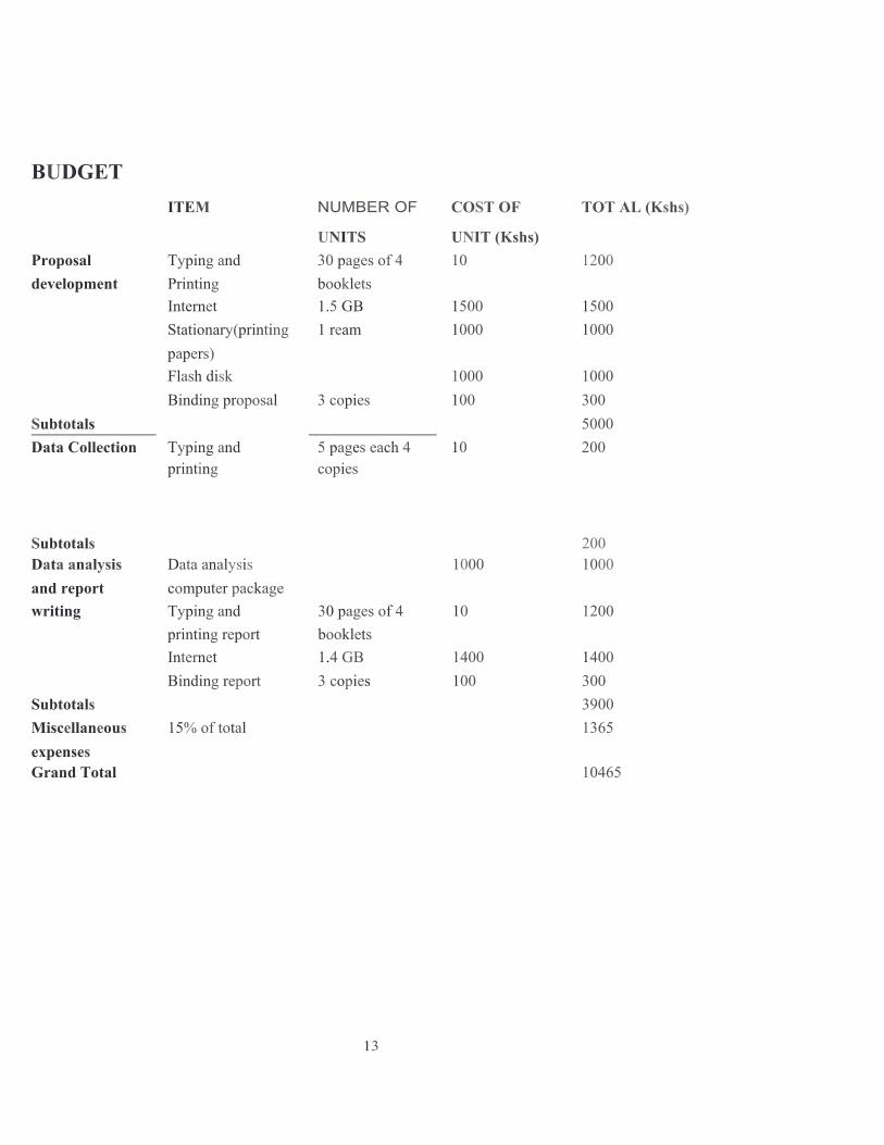