Vander’s Human Physiology The Mechanisms of Body Function Tenth Edition by Widmaier Raff Strang ©...
26
Vander’s Human Physiology The Mechanisms of Body Function Tenth Edition by Widmaier • Raff • Strang © The McGraw-Hill Companies, Inc. Figures and tables from the book, with additional comments by: John J. Lepri, Ph. D., The University of North Carolina at Greensboro Chapter 5
-
Upload
rosa-morton -
Category
Documents
-
view
224 -
download
2
Transcript of Vander’s Human Physiology The Mechanisms of Body Function Tenth Edition by Widmaier Raff Strang ©...

Vander’s
Human PhysiologyThe Mechanisms of Body Function
Tenth Editionby
Widmaier • Raff • Strang© The McGraw-Hill Companies, Inc.
Figures and tables from the book, with additional comments by: John J. Lepri, Ph. D.,
The University of North Carolina at Greensboro
Chapter 5
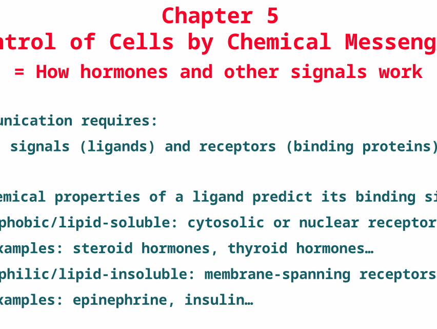
The chemical properties of a ligand predict its binding site:
• Hydrophobic/lipid-soluble: cytosolic or nuclear receptors
examples: steroid hormones, thyroid hormones…
• Hydrophilic/lipid-insoluble: membrane-spanning receptors
examples: epinephrine, insulin…
Chapter 5Control of Cells by Chemical Messengers
= How hormones and other signals work
Communication requires:
signals (ligands) and receptors (binding proteins).
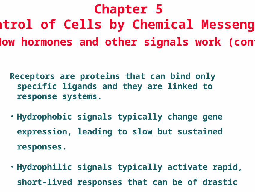
Receptors are proteins that can bind only specific ligands and they are linked to response systems.
• Hydrophobic signals typically change gene expression,
leading to slow but sustained responses.
• Hydrophilic signals typically activate rapid, short-lived
responses that can be of drastic impact.
Chapter 5Control of Cells by Chemical Messengers= How hormones and other signals work (cont.)

Figure 5-1
Receptors on the surface of a cell are typically proteins that span the membrane.


Figure 5-2
Cells B & C lack the matching receptorsTherefore are not directly affected by the signal.
Only Cell A has thematching receptorsfor this chemical messenger, so it is the only one that responds.
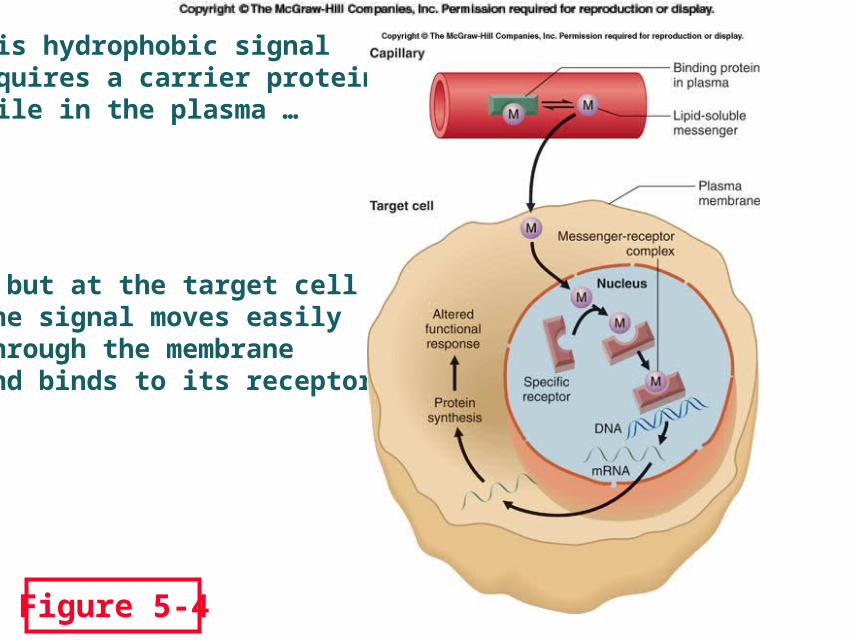
Figure 5-4
… but at the target cellthe signal moves easily through the membraneand binds to its receptor.
This hydrophobic signal requires a carrier protein while in the plasma …

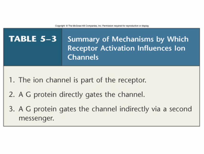
Binding of ligands to membrane-spanning receptorsactivates diverse response mechanisms.
Figure 5-5
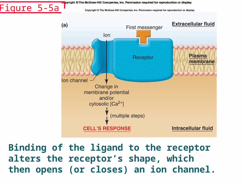
Binding of the ligand to the receptoralters the receptor’s shape, which then opens (or closes) an ion channel.
Figure 5-5a
Binding of the ligand to the receptor alters the receptor’s shape, which activates its enzyme function, phosphorylating an intracellular protein.
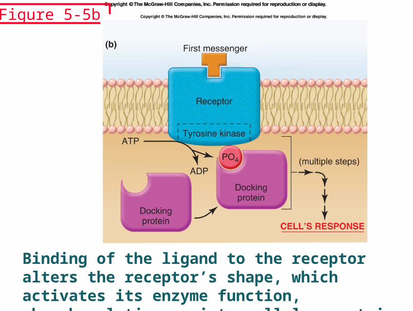
Figure 5-5b
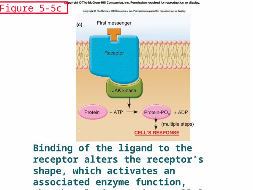
Binding of the ligand to the receptor alters the receptor’s shape, which activates an associated enzyme function, phosphorylating an intracellular protein.
Figure 5-5c

Binding of the ligand to the receptor alters the receptor’s shape, which activates an associated G-protein, which then activates effector proteins,i.e., enzyme functions or ion channels.
Figure 5-5d
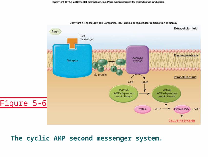
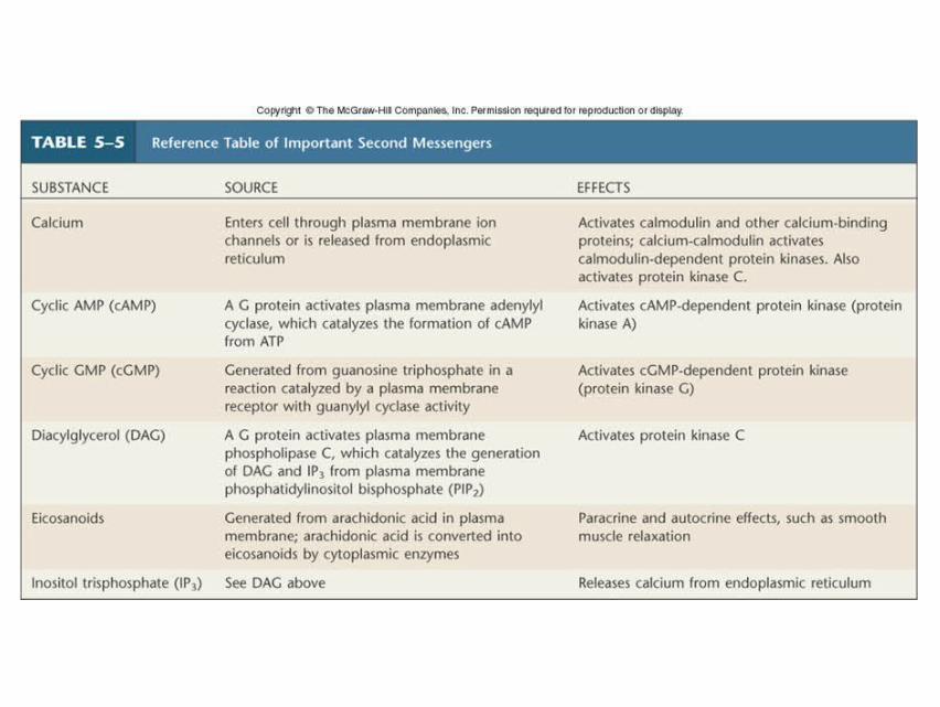
The cyclic AMP second messenger system.
Figure 5-6
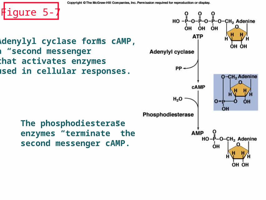
Adenylyl cyclase forms cAMP,a “second messenger” that activates enzymes used in cellular responses.
The phosphodiesterase enzymes “terminate” thesecond messenger cAMP.
Figure 5-7

Figure 5-8
The cAMP system rapidly amplifies the responsecapacity of cells: here, one “first messenger” ledto the formation of one million product molecules.
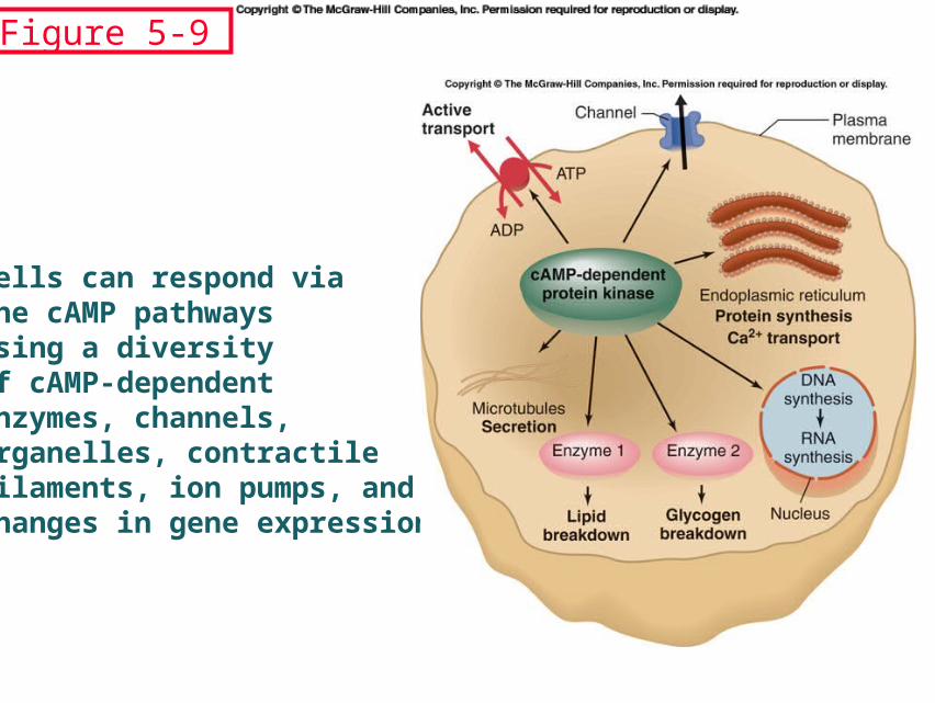
Cells can respond via the cAMP pathwaysusing a diversity of cAMP-dependentenzymes, channels,organelles, contractile filaments, ion pumps, and changes in gene expression.
Figure 5-9

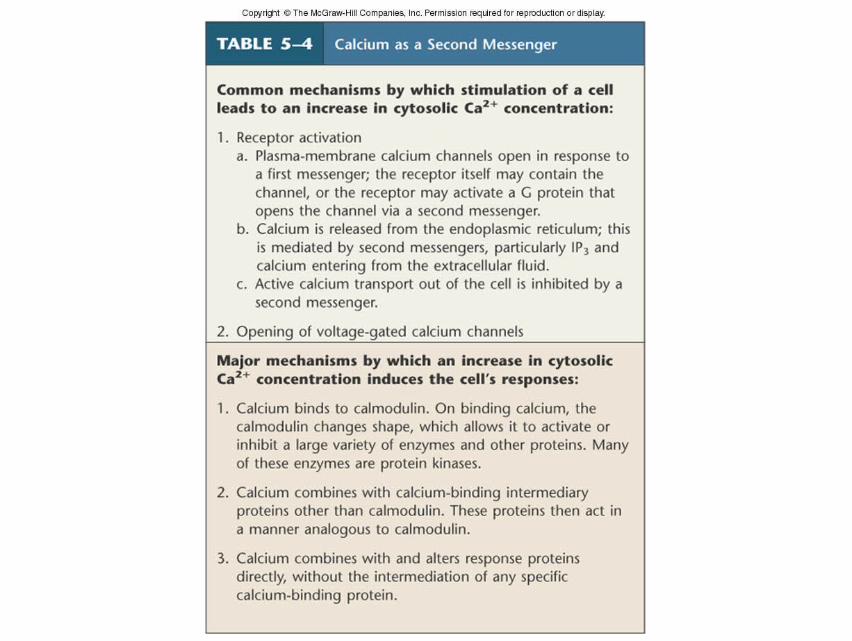
This receptor-G-protein complex is linked to and activates phospholipase C, leading to an increase in IP3 and DAG, which work together to activate enzymes and to increase intracellular calcium levels.
Figure 5-10
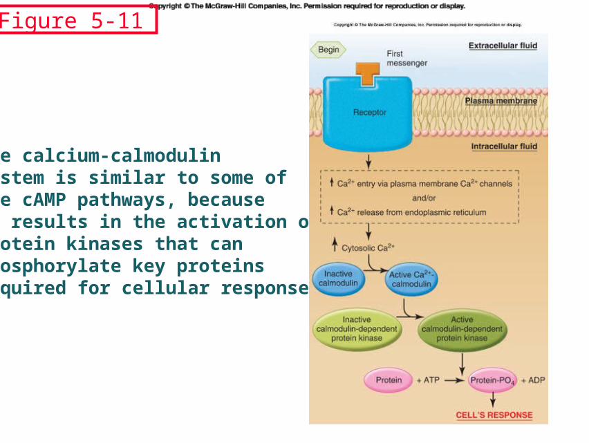
The calcium-calmodulinsystem is similar to some of the cAMP pathways, because it results in the activation of protein kinases that can phosphorylate key proteins required for cellular responses.
Figure 5-11
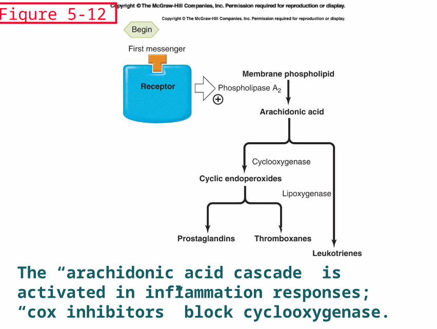
The “arachidonic acid cascade” is activated in inflammation responses; “cox inhibitors” block cyclooxygenase.
Figure 5-12
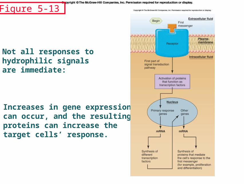
Not all responses to hydrophilic signals are immediate:
Increases in gene expression can occur, and the resulting proteins can increase the target cells’ response.
Figure 5-13

Guanylate cyclase
1.Receptor forms: intracellular carboxyl terminal has a tyrosine kinase-like and a guanylate cyclase domain.
Two such guanylate cyclases are ANP receptors.
2. Soluble forms: are intracellular enzymes, and activated by NO.
HA20HA20
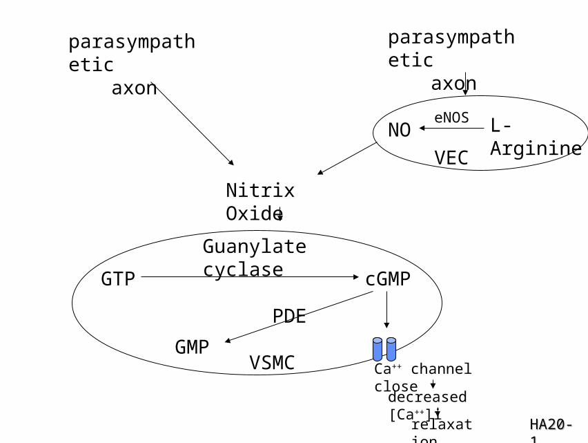
parasympatheticaxon
parasympatheticaxon
eNOS L-Arginine
VEC
Nitrix Oxide
Guanylate cyclase
GTP cGMP
PDE
GMPCa++ channel close
decreased [Ca++]i
relaxation
VSMC
HA20-1HA20-1
NO

The End.