Valiente, E; Bouch, L; Hitchen, P; Faulds-Pain, A; Songane ... · restored upon complementation in...
19
Valiente, E; Bouch, L; Hitchen, P; Faulds-Pain, A; Songane, M; Daw- son, LF; Donahue, E; Stabler, RA; Panico, M; Morris, HR; Bajaj- Elliott, M; Logan, SM; Dell, A; Wren, BW (2016) Role of glyco- syltransferases modifying type B flagellin of emerging hypervirulent Clostridium difficile lineages and their impact on motility and biofilm formation. The Journal of biological chemistry. ISSN 0021-9258 DOI: 10.1074/jbc.M116.749523 Downloaded from: http://researchonline.lshtm.ac.uk/2965146/ DOI: 10.1074/jbc.M116.749523 Usage Guidelines Please refer to usage guidelines at http://researchonline.lshtm.ac.uk/policies.html or alterna- tively contact [email protected]. Available under license: http://creativecommons.org/licenses/by/2.5/
Transcript of Valiente, E; Bouch, L; Hitchen, P; Faulds-Pain, A; Songane ... · restored upon complementation in...

Valiente, E; Bouch, L; Hitchen, P; Faulds-Pain, A; Songane, M; Daw-son, LF; Donahue, E; Stabler, RA; Panico, M; Morris, HR; Bajaj-Elliott, M; Logan, SM; Dell, A; Wren, BW (2016) Role of glyco-syltransferases modifying type B flagellin of emerging hypervirulentClostridium difficile lineages and their impact on motility and biofilmformation. The Journal of biological chemistry. ISSN 0021-9258 DOI:10.1074/jbc.M116.749523
Downloaded from: http://researchonline.lshtm.ac.uk/2965146/
DOI: 10.1074/jbc.M116.749523
Usage Guidelines
Please refer to usage guidelines at http://researchonline.lshtm.ac.uk/policies.html or alterna-tively contact [email protected].
Available under license: http://creativecommons.org/licenses/by/2.5/

Role of Glycosyltransferases Modifying Type B Flagellin ofEmerging Hypervirulent Clostridium difficile Lineages andTheir Impact on Motility and Biofilm Formation*□S
Received for publication, July 20, 2016, and in revised form, September 27, 2016 Published, JBC Papers in Press, October 4, 2016, DOI 10.1074/jbc.M116.749523
Esmeralda Valiente‡1, Laura Bouche§, Paul Hitchen§, Alexandra Faulds-Pain‡, Mario Songane¶2, Lisa F. Dawson‡,Elizabeth Donahue‡, Richard A. Stabler‡, Maria Panico§, Howard R. Morris§�, Mona Bajaj-Elliott¶, Susan M. Logan**,Anne Dell§, and Brendan W. Wren‡3
From the ‡Department of Pathogen Molecular Biology, London School of Hygiene and Tropical Medicine, Keppel Street, LondonWC1E 7HT, United Kingdom, the §Department of Life Sciences, Imperial College London, South Kensington Campus, London SW72AZ, United Kingdom, the ¶Institute of Child Health, University College London, 30 Guilford Street, London WC1N 1EH, UnitedKingdom, the **Vaccine Program, Human Health Therapeutics Portfolio, National Research Council, Ottawa, Ontario K1A 0R6,Canada, and �BioPharmaSpec Ltd., Suite 3.1, Lido Medical Centre, St. Saviours Road, Jersey JE2 7LA, United Kingdom
Edited by Charles Samuel
Clostridium difficile is the principal cause of nosocomialinfectious diarrhea worldwide. The pathogen modifies its flagel-lin with either a type A or type B O-linked glycosylation system,which has a contributory role in pathogenesis. We study thefunctional role of glycosyltransferases modifying type B fla-gellin in the 023 and 027 hypervirulent C. difficile lineages bymutagenesis of five putative glycosyltransferases and biosyn-thetic genes. We reveal their roles in the biosynthesis of theflagellin glycan chain and demonstrate that flagellar post-trans-lational modification affects motility and adhesion-related bac-terial properties of these strains. We show that the glycosyl-transferases 1 and 2 (GT1 and GT2) are responsible for thesequential addition of a GlcNAc and two rhamnoses, respec-tively, and that GT3 is associated with the incorporation of anovel sulfonated peptidyl-amido sugar moiety whose structureis reported in our accompanying paper (Bouche, L., Panico, M.,Hitchen, P., Binet, D., Sastre, F., Faulds-Pain, A., Valiente, E.,Vinogradov, E., Aubry, A., Fulton, K., Twine, S., Logan, S. M.,Wren, B. W., Dell, A., and Morris, H. R. (2016) J. Biol. Chem. 291,25439 –25449). GT2 is also responsible for methylation of therhamnoses. Whereas type B modification is not required for fla-gellar assembly, some mutations that result in truncation orabolition of the glycan reduce bacterial motility and promoteautoaggregation and biofilm formation. The complete lack offlagellin modification also significantly reduces adhesion ofC. difficile to Caco-2 intestinal epithelial cells but does not affectactivation of human TLR5. Our study advances our understand-ing of the genes involved in flagellar glycosylation and their bio-logical roles in emerging hypervirulent C. difficile strains.
Clostridium difficile, recently reclassified as Peptoclos-tridium difficile (1), has emerged since the mid-2000s as themain cause of antibiotic-associated diarrhea within health-care environments (2). This Gram-positive spore-forminganaerobe is an opportunistic pathogen that colonizes thegastro-intestinal tracts of susceptible individuals, leading toC. difficile infection (3). Those at risk include patientsundergoing antibiotic therapy, the immunocompromised,and the elderly (4), although increasingly younger people areaffected by C. difficile infection (5).
Genetic typing and sequence analysis have confirmed thatthere are at least five distinct lineages of C. difficile (6, 7), whichcan be divided according to the most important PCR ribotypes(RTs)4: RT017, RT023, RT027, RT078, and a large groupincluding the 630 strain and the other RTs (8, 9). Our work isfocused on two lineages (RT023 and RT027). The lineage com-posed primarily of the RT027 strains has spread globally. It hasalso been associated with increased severity of disease and isdescribed as “hypervirulent” (10 –12). RT023 is a recentlyemerging lineage, prevalent in Europe, and is also often associ-ated with more severe disease (13). Most strains of C. difficileproduce two cytotoxins, toxin A (TcdA) and toxin B (TcdB),which cause extensive damage to the epithelial cells of the gas-tro-intestinal tract and are considered to be essential for C. dif-ficile disease (14). The production of either TcdA or TcdB hasbeen found to be essential for C. difficile infection (15, 16).However, the existence of naturally toxin A-negative strainssuggests that other factors are involved in C. difficile transmis-sibility, survival, and colonization. C. difficile expresses multi-ple surface proteins and colonization factors, including cellsurface-associated proteins (surface layer proteins), fibronec-tin-binding protein A, proteases, heat-shock proteins, and fla-gella (17–19). Flagella biosynthesis is known to be linked to anumber of other cellular processes, including sporulation,
* This work was supported by Wellcome Trust Grants 102979/Z/13/Z and105609/Z/14/Z, Medical Research Council Grant MR/K000551/1, andBiotechnology and Biological Sciences Research Council GrantsBB/K016164/1 and BB/F008309/1. The authors declare that they have noconflicts of interest with the contents of this article.Author’s Choice—Final version free via Creative Commons CC-BY license.
□S This article contains supplemental Table 1 and Figs. 1 and 2.1 Supported by Marie Curie Intra-European Fellowship for Career Develop-
ment PIEF-GA-2009-252207-CDI.2 Recipient of Commonwealth Scholarship MZCS-2011-280.3 To whom correspondence should be addressed. Tel.: 44-2079272288; Fax:
44-207-6374314; E-mail: [email protected].
4 The abbreviations used are: RT, ribotype; GT, glycosyltransferase; HexNAc,hexose N-acetylhexosamine; GlcNAc, N-acetylglucosamine; PTM, post-translational modification; TEM, transmission electron microscopy; IEC,intestinal epithelial cell; BisTris, 2-[bis(2-hydroxyethyl)amino]-2-(hydro-xymethyl)propane-1,3-diol.
THE JOURNAL OF BIOLOGICAL CHEMISTRY VOL. 291, NO. 49, pp. 25450 –25461, December 2, 2016Author’s Choice © 2016 by The American Society for Biochemistry and Molecular Biology, Inc. Published in the U.S.A.
crossmark
25450 JOURNAL OF BIOLOGICAL CHEMISTRY VOLUME 291 • NUMBER 49 • DECEMBER 2, 2016
at LO
ND
ON
SCH
OF H
YG
IEN
E &
TR
OPIC
AL
ME
DIC
INE
on Decem
ber 2, 2016http://w
ww
.jbc.org/D
ownloaded from
at L
ON
DO
N SC
H O
F HY
GIE
NE
& T
RO
PICA
L M
ED
ICIN
E on D
ecember 2, 2016
http://ww
w.jbc.org/
Dow
nloaded from
at LO
ND
ON
SCH
OF H
YG
IEN
E &
TR
OPIC
AL
ME
DIC
INE
on Decem
ber 2, 2016http://w
ww
.jbc.org/D
ownloaded from
at L
ON
DO
N SC
H O
F HY
GIE
NE
& T
RO
PICA
L M
ED
ICIN
E on D
ecember 2, 2016
http://ww
w.jbc.org/
Dow
nloaded from
at LO
ND
ON
SCH
OF H
YG
IEN
E &
TR
OPIC
AL
ME
DIC
INE
on Decem
ber 2, 2016http://w
ww
.jbc.org/D
ownloaded from
at L
ON
DO
N SC
H O
F HY
GIE
NE
& T
RO
PICA
L M
ED
ICIN
E on D
ecember 2, 2016
http://ww
w.jbc.org/
Dow
nloaded from
at LO
ND
ON
SCH
OF H
YG
IEN
E &
TR
OPIC
AL
ME
DIC
INE
on Decem
ber 2, 2016http://w
ww
.jbc.org/D
ownloaded from

adhesion, metabolism, and toxin production (20). The C. diffi-cile flagella are composed of a single flagellin protein encodedby a single flagellin gene (fliC), which is called CD0239 in 630strain and CDR20291_0240 in R20291 strain. C. difficile flagel-lin is post-translationally modified by an O-linked glycan, andthis post-translational modification (PTM) is essential formotility (21). Two types of flagellin PTM have been identified inC. difficile, which we have previously defined as type A and B,where type A has a simpler structural modification of the flagel-lin than type B (21–23). The flagellin of the first C. difficilesequenced strain, 630, is modified at up to seven sites with atype A PTM (21–23), which is composed of an O-linkedN-acetyl glucosamine (GlcNAc) linked to a methylated threo-nine via a phosphodiester bond (21–23). The genes responsiblefor this modification (CD0240, CD0241, CD0242, CD0243, andCD0244) are encoded immediately downstream of the flagellingene, fliC (21–24). Previous genetic analysis of the hyperviru-lent RT027 strain revealed that the gene lying downstream offliC in the type B modification, glycosyltransferase 1 (GT1(CDR20291_0241)), was highly similar to CD0240 of 630 (21).Although the function of the GT1 protein is yet to be con-firmed, the first sugar of the type B PTM is also an O-linkedN-acetylhexosamine (HexNAc) residue, indicating that theGT1 protein may have a function similar to the CD0240 protein(22, 25). Beyond this initial sugar, however, the RT027 flagellindoes not contain a phosphodiester-linked amino acid as in typeA flagellin. Instead, it has recently been found to be extended by
two rhamnoses and a novel sulfonated peptidyl amidoglycan(25).
The differences between type A and type B flagellin struc-tures are reflected at the genetic level. In this study, bioin-formatic analyses reveal the presence of two putative gly-cosyltransferase genes (GT2 (CDR20291_0242) and GT3(CDR20291_0243)) in type B that are absent in the type A PTMgene cluster. Additionally, immediately downstream of GT2and GT3, there are four further predicted coding sequences(CDR20291_0244-0247) that do not appear to code for glyco-syltransferases. We also identified putative glycosylation genesupstream of fliC (CDR20291_0223-0226), which are similar torhamnose biosynthesis genes. In addition, we explored whichgenes affect flagellin type B modification and investigated thebiological role of flagellin glycosyltransferases from two hyper-virulent C. difficile lineages, namely RT027 (strain R20291) andRT023 (strain CD1426). We show that flagellin PTM type Baffects motility and adhesion properties, including adhesion tointestinal cells, but does not activate human TLR5.
Results
Flagella Glycosylation Genes in RT027 and RT023 Are HighlySimilar—We reported the presence of orthologues of theRT027 flagellin type B modification genes in RT023 strains.Thus, a BLAST analysis upstream and downstream of theRT027 flagellin biosynthetic locus identified glycosyltrans-ferases and glycan biosynthesis genes (Fig. 1A). Downstream of
FIGURE 1. A, a schematic diagram illustrates flagellin type A (present in RT012 and RT017 strains) and type B (present in RT027 and RT023 strains) modificationgenes from C. difficile. Flagellar structural genes (fliC and flgB) are colored in white, and predicted GTs and remaining putative modification genes are colored ingray. The dotted lines indicate that there are homologous genes in PCR ribotypes 012/017 (CD0241–CD0244) and homologous genes in PCR ribotypes 023/027(CDR20291_0242– 0247). The asterisk indicates predicted GTs that are different in RT027 and RT023 compared with strain 630. CDR20291_0223– 0226 aresimilar to rmlD, rmlA, rmlC, and rmlB, respectively. The number of coding sequences from CDR20291_R0226 and the fliC gene is also indicated. B, flagellaglycosylation gene RT-PCR. Top, rhamnose biosynthetic genes. Bars below the genes illustrate the amplified regions (1–3) and internal region (Int) amplified asa control of gene expression by RT-PCR. Bottom, flagella glycosylation locus located downstream of the fliC gene. Bars below the genes illustrate the amplifiedregions (4 –9). luxS gene amplification was used as a control for constitutive expression, and an internal primer was used to amplify fliC as a control of geneexpression. Control reactions were carried out without reverse transcriptase (No RT) and with a DNA template from genomic DNA (gDNA). RT-PCR was carriedout on RNA extracted during early exponential growth (A) and during late exponential growth (B).
C. difficile Type B Flagellin Affects Motility and Biofilm
DECEMBER 2, 2016 • VOLUME 291 • NUMBER 49 JOURNAL OF BIOLOGICAL CHEMISTRY 25451
at LO
ND
ON
SCH
OF H
YG
IEN
E &
TR
OPIC
AL
ME
DIC
INE
on Decem
ber 2, 2016http://w
ww
.jbc.org/D
ownloaded from

the RT027 flagellin gene there were three putative glycosyl-transferase genes (CDR20291_0241, CDR20291_0242, andCDR20291_0243, which we designate GT1, GT2, GT3, respec-tively) encoding predicted proteins with similarity to the groupII superfamily of glycosyltransferases, each having a Pfamdomain 00535 (26). In addition, GT2 also appears to have amethyltransferase domain (Pfam 08241). We identified fourother predicted coding sequences (CDR20291_0244 – 0247)downstream of GT3 in the chromosome from strains repre-senting RT027 and RT023, which may also be involved in thisflagellin PTM (Fig. 1A). The CDR20291_0244 predicted proteinshares some similarity with FemAB superfamily proteins,which contain an acyltransferase domain (Pfam 02388).CDR20291_0245 is a putative carbamoyl-phosphate synthetasewith an ATP-grasp domain (Pfam 13535). This family includesa diverse set of enzymes that possess ATP-dependent carboxy-late-amine ligase activity. CDR20291_0246 is a putative orni-thine cyclodeaminase with a conserved domain PRK06046 alsopresent in alanine dehydrogenase enzymes. CDR20291_0247shows significant similarity to FdtB (dTDP-6-deoxy-D-xylohex-3-uloseaminase) from Aneurinibacillus thermoaerophilus andXanthamonas campestris. In these organisms, this enzyme hasbeen shown to be part of the biosynthetic pathway of TDP-D-Fucp3NAc producing dTDP-D-Fucp3N from dTDP-6-deoxy-D-xylohex-3-ulose (27, 28). Upstream of the flagellin biosynthe-sis genes, in both RT027 and RT023 strains, there were fourgenes (CDR20291_0223– 0226) that are similar to rhamnosebiosynthetic genes (rmlD, rmlA, rmlC, and rmlB, respectively)(Fig. 1A).
Additionally, none of the studied genes in Fig. 1A corre-sponds to a glycosulfotransferase that could potentially beinvolved in the biosynthesis of the sulfonated peptidylamido-sugar moiety.
To determine whether the putative flagellin type B modifica-tion genes upstream and downstream of fliC are co-transcribedwith fliC, RT-PCR was carried out in R20291 (RT027) over thejunctions between CDR20291_0223– 0226 and GT1, GT2,GT3, and CDR20291_0244 – 0246. We found that the predictedtype B modification genes were co-transcribed in both expo-nential and stationary growth phase (Fig. 1B).
Mutations in Flagellin Modification Type B Genes Resultin Truncation or Abolition of Flagellin Post-translationalModification—Disruption of the glycosyltransferase geneCD0240, as well as CD0241, CD0242, CD0243, CD0244 in 630strain was previously reported to result in flagellin of differentsizes (23). To further characterize the role of type B modifica-tion genes in flagellin glycosylation, R20291 mutants in GT1,GT2, GT3, CDR20291_0245, and CDR20291_0246 were gener-ated by ClosTron mutagenesis. Flagellin samples were studiedby SDS-PAGE and mass spectrometry. On SDS-PAGE, flagellinof R20291 had a molecular mass around 60 kDa (Fig. 2A). Wealso observed a shift in the mass of the flagellin of all flagellaglycosylation mutants, especially in GT1 and GT2, towardlower molecular weight. Wild type molecular weight wasrestored upon complementation in trans of GT1 mutant(GT1comp) (Fig. 2A).
Gel bands corresponding to flagellin of mutants and corre-sponding complements were analyzed by nano-LC-MS/MS.
Spectra were manually compared, and the identified glycopep-tides in the mutants are listed in Table 1. Tandem mass spec-trometric analyses of flagellin tryptic digests from the GT1mutant showed peptides to be non-glycosylated. Flagellin iso-lated from the GT2 mutant harbored glycopeptides with a sin-gle HexNAc moiety but lacking any further extension of theglycan. Flagellin isolated from the GT3 mutant was modified bya deoxyHex-deoxyHex-HexNAc (with and without methylgroups) but lacking the terminal sulfonated peptidylamido-sugar moiety observed in the wild type (25). Similarly, theCDR20291_0245 and CDR20291_0246 mutants carried thenon-extended core trisaccharide (Table 1). The analysis ofthe complemented mutants by mass spectrometry demon-strated that the flagellar glycan chain is fully restored (data notshown).
Based on the mass spectrometric data of the mutants andtaking into account the flagellin type B sulfonated peptidyl-amido-glycan structure reported by Bouche et al. (25), weconclude that the glycosyltransferases GT1 and GT2 areresponsible for the sequential addition of the GlcNAcand two rhamnoses sugars, respectively, and that GT3,CDR20291_0245, and CDR20291_0246 are all involved in thebiosynthesis of the terminal sulfonated peptidylamido-sugarmoiety. Given that our bioinformatics analysis suggests thatGT2 could also function as a methyltransferase, we hypothesizethat GT2 is additionally responsible for methylation of therhamnoses (Table 1 and Fig. 2B).
Motility, Cell Aggregation, and Biofilm Formation AreAffected by Flagellin Glycosylation Mutants in RT027 andRT023 Strains—Flagella type A modification is essential formotility in C. difficile 630 and RT017 strains (23). To determinewhether flagellin modification type B is also required for motil-ity, GT1, GT2, GT3, CDR20291_0245, and CDR20291_0246mutants were assessed. The R20291fliC mutant was used as anegative control. Measuring bacterial motility in agar, we foundthat GT1 and GT2 knock-out strains were significantly lessmotile than wild type R20291 (diameter of 0.8 � 0.1 cm versus2.0 � 0.2 cm) (Fig. 3A). Complementation of these knockoutsrestored the motility phenotype (Fig. 3A). Furthermore, GT3,
FIGURE 2. A, Western blotting of flagellin extractions of RT027 wild type andflagellin modification mutants. Lane M, marker (prestained protein marker;Fermentas); lane 1, R20291; lane 2, R20291fliC; lane 3, GT1; lane 4, GT2; lane 5,GT3; lane 6, CDR20291_0245; lane 7, CDR20291_0246; lane 8, GT1comp. B,schematic diagram illustrates the structure of flagellin type B modificationand the contribution of each GT. Flagellin type B is modified with oneN-acetylglucosamine (white square) linked to two rhamnoses (white circles)(with or without a methyl group (Me)). White hexagon, terminal peptidyl-sugar moiety.
C. difficile Type B Flagellin Affects Motility and Biofilm
25452 JOURNAL OF BIOLOGICAL CHEMISTRY VOLUME 291 • NUMBER 49 • DECEMBER 2, 2016
at LO
ND
ON
SCH
OF H
YG
IEN
E &
TR
OPIC
AL
ME
DIC
INE
on Decem
ber 2, 2016http://w
ww
.jbc.org/D
ownloaded from

CDR20291_0245, and CDR20291_0246 mutants were as motileas the parent R20291 strain, suggesting that the first three sug-ars of the flagella glycan chain are both sufficient and necessaryfor full motility of C. difficile.
To study the effect of PTM type B on flagellar assembly, indi-vidual bacteria were imaged by TEM. The images revealed thepresence of flagella filaments associated with the surface ofR20291 and mutants. The R20291fliC mutant was used as anegative control (Fig. 3B). In addition, we quantified flagellafilament length of R20291 and its corresponding mutants, andno significant differences were observed (Table 2). Our resultssuggest that type B modification is not required for flagellaassembly.
We next investigated whether flagellin PTM affects cellaggregation of hypervirulent C. difficile RT027 and observedincreased aggregation in all glycosylation mutants (83 � 3 �m)versus wild type (R20291) (52 � 5 �m) (Fig. 4A). These resultsindicate that flagella post-translational modification type Baffects C. difficile cell aggregation. However, a FliC mutantstrain that completely lacks flagellin aggregates similarly toR20291, suggesting that flagella per se are not required for cellaggregation. Complementation of the mutants restored theoriginal aggregation phenotype (Fig. 4A). Cell surface hydro-phobicity of wild type and flagellin glycosylation mutants wasmeasured, and our results show that flagellin PTM mutants aremore hydrophobic than the wild-type and/or R20291fliC (Fig.4C). Cell viability in PBS1x was affected after 16-h incubation(Table 3) but in the same way in the wild types (R20291 andCD1426) and corresponding mutants. Cell aggregation wasvisualized by optical microscopy, where mutants showed a cleartendency to cell-to-cell aggregate (supplemental Fig. 1).
To determine whether these observations can be extended toanother strain from a different lineage, we repeated theseexperiments using the GT2 mutant in CD1426 (RT023). Anal-ogous to R20291 (RT027), this mutant (supplemental Fig. 2A)was less motile compared with the wild type strain (diameterof 0.8 � 0.1 cm versus 2.1 � 0.1 cm) and aggregated morethan the CD1426 mutant (85 � 1 versus 60 � 3, respectively)(supplemental Fig. 2B).
TABLE 1Glycan structures identified by mass spectrometry present on C. difficile flagellin purified from mutant strains belonging to ribotypes 027 and 023A check mark indicates presence, and an X indicates absence. The glycan structures of wild-type of both RT027 and RT023 have been described in the accompanying article(25) and are shown as a reference.
FIGURE 3. Motility and flagella production in flagellin modificationmutants. A, quantification of the motility halo of RT027 WT (R20291) andcorresponding mutants (fliC, GT1, GT2, GT3, R20291_0245, and R20291_0246)and complements (GT1comp and GT2comp). Measurements are in cm. Aster-isks, significant differences compared with WTs (p � 0.05). Data are presentedas the mean � S.E. (error bars), n � 3. B, electron transmission images ofR20291 and its corresponding flagellin modification mutants. Arrows, flagella;bar, 0.5 nm.
TABLE 2Measurement of flagella filament length
Strains Length of flagella filament � S.E.a
�mR20291 3.62 � 0.42GT1 3.55 � 0.46GT2 3.60 � 0.40GT3 3.63 � 0.33CDR20291_0245 3.66 � 0.45CDR20291_0246 3.64 � 0.42
a The flagella of 100 bacteria were measured in each strain, and TEM images wereanalyzed using the line measuring tool of the Image J 1.50i software (NationalInstitutes of Health).
C. difficile Type B Flagellin Affects Motility and Biofilm
DECEMBER 2, 2016 • VOLUME 291 • NUMBER 49 JOURNAL OF BIOLOGICAL CHEMISTRY 25453
at LO
ND
ON
SCH
OF H
YG
IEN
E &
TR
OPIC
AL
ME
DIC
INE
on Decem
ber 2, 2016http://w
ww
.jbc.org/D
ownloaded from

Finally, we determined the role of flagellin modification inRT027 and RT023 on biofilm on abiotic surface (polystyreneplates). As shown in Fig. 4B, flagellin type B modification facil-itates adherence of cells to abiotic surfaces when comparedwith the wild type. Complementation of the mutants restoredthe original phenotype.
C. difficile Flagellin Glycosylation Modulates BacterialAdherence to Intestinal Epithelial Cells—Having observed dif-ferences in the glycosylation mutants in aggregation and bio-film on an abiotic surface, we investigated whether adhesion toa physiologically relevant biological surface, such as humanintestinal epithelial cells (IECs), may also be affected. Previous
work has shown that the C. difficile R20291fliC mutant adheresless well to human IECs than the wild type strain (29), butwhether adhesion is influenced by changes in flagellin glycosyl-ation is unknown. Therefore, we measured adherence of FITC-labeled R20291 C. difficile, as well as the respective flagellin typeB modification mutants, to cultured Caco-2 cells by flowcytometry (Fig. 5A). Compared with R20291, only the GT1mutant showed reduced adherence, and complementationrestored the phenotype (Fig. 5B). All other mutants adheredsimilarly to R20291 on the tested cell line. These data suggestthat flagellin glycans facilitate adhesion of C. difficile to IECsand that the presence of just the first sugar in the glycan chain issufficient to restore adhesion to wild type levels.
Similarly, the CD1426 GT2 mutant showed no difference inadherence compared with the WT strain, confirming that GT2does not affect bacterial adherence to IECs (data not shown). Ofnote, no difference in FITC labeling was observed between WTand glycosylation mutants (data not shown).
C. difficile Flagellar Glycosylation Is Not Involved in TLR5Recognition—TLR5 is part of the human innate immune systemand specifically binds to bacterial flagellin. TLR5 activationresults in the secretion of the cytokine IL-8. The potentialimpact of flagellin glycosylation (e.g. masking) on host TLR5engagement was investigated. Untransfected and TLR5-trans-fected human HEK293 cells were co-cultured with wild typeC. difficile RT027 and RT023 strains and their respective fla-gella modification-deficient mutants. 8 h after infection, IL-8
FIGURE 4. Cell aggregation in PBS (A), biofilm formation (B), and cell hydrophobicity (C). All of the type B modification mutants showed a significantincrease in cell aggregation, biofilm formation, and hydrophobicity compared with R20291. Asterisks, significant differences compared with WT (p � 0.05). Dataare presented as the mean � S.E. (error bars) from three independent experiments with three technical replicates each.
TABLE 3C. difficile viability in PBS1xcfu/ml were counted when the experiment was set up (initial cfu/ml) and after 16 hof incubation (final cfu/ml).
Strains Initial cfu/ml � S.E. Final cfu/ml � S.E.
R20291 2.3 � 0.2E�09 1.3 � 0.3E�07GT1 2.1 � 0.4E�09 1.0 � 0.3E�07GT2 2.5 � 0.3E�09 1.5 � 0.1E�07GT3 2.4 � 0.3E�09 1.3 � 0.3E�07CDR20291_0245 2.2 � 0.1E�09 1.4 � 0.2E�07CDR20291_0246 2.2 � 0.3E�09 1.9 � 0.5E�07GT1comp 2.6 � 0.2E�09 1.3 � 0.2E�07GT2comp 2.7 � 0.2E�09 1.5 � 0.3E�07GT3comp 2.2 � 0.4E�09 1.1 � 0.3E�07CDR20291_0245 comp 2.3 � 0.2E�09 1.3 � 0.1E�07CDR20291_0246 comp 2.2 � 0.4E�09 1.5 � 0.3E�07CD1426 2.1 � 0.2E�09 1.6 � 0.1E�07CD1426_GT2 2.2 � 0.3E�09 1.2 � 0.2E�07CD1426_GT2comp 2.4 � 0.5E�09 1.5 � 0.3E�07
C. difficile Type B Flagellin Affects Motility and Biofilm
25454 JOURNAL OF BIOLOGICAL CHEMISTRY VOLUME 291 • NUMBER 49 • DECEMBER 2, 2016
at LO
ND
ON
SCH
OF H
YG
IEN
E &
TR
OPIC
AL
ME
DIC
INE
on Decem
ber 2, 2016http://w
ww
.jbc.org/D
ownloaded from

secretion into the medium was quantified by ELISA. MinimalIL-8 was recorded in untransfected cells; in contrast, significantincrease in IL-8 was observed in response to the wild typeR20291 (Fig. 6A). The increase was completely abrogated inresponse to the R20291fliC mutant, whereas the flagella glyco-sylation mutants activated TLR5 to a similar extent as the WTstrain (Fig. 6A). The CD1426 glycosylation mutant also dis-played similar TLR5 engagement as its WT counterpart (Fig.6B). These observations suggest that the interaction betweenTLR5 and C. difficile flagellin is maintained even in the absenceof glycans and therefore that glycosylation of flagellin in C. dif-ficile does not influence TLR5 binding or activation.
Discussion
C. difficile is a major global healthcare problem. In recentyears, hypervirulent strains, including RT027 and RT023, haveemerged, causing more severe infections (29, 30). Although thetoxins are the main virulence factors, other features, such as
flagella (31), S-layer protein (32), and sporulation (33), also con-tribute to bacteria colonization and transmission of the disease.
C. difficile flagellin (FliC) has a role in motility, colonization,biofilm formation, and toxin production. Moreover, flagellin isimmunogenic and protective in a murine model of C. difficileinfection (34). However, little is known about the role of proteinPTM in C. difficile disease, which has a type A or type B modi-fication (23, 25). It has also been reported that type A post-translational modification of flagellin in C. difficile 630 strainhas a contributory effect in pathogenesis (23). However, therole of type B post-translational modification of flagellinremains unclear. In this work, we investigated the function ofputative type B flagellin modification genes and demonstratedthe importance of flagellar modification in motility, cell aggre-gation, biofilm formation, epithelial cell adhesion, and TLR5recognition.
To study the function of the flagellin type B modificationgenes, we generated mutants and studied the consequences for
FIGURE 5. Cell adhesion in RT027 flagellin modification mutants. C. difficile flagella glycosylation moieties contribute to bacterial adherence to human IECs.1 � 106 Caco-2 cells were infected with 2.5 � 107 FITC-labeled WT R20291 and flagellin glycosylation isogenic mutants and co-cultured for 1.5 h underanaerobic conditions. Bacterial adherence was assessed by flow cytometry (A) and quantified as change in median fluorescence intensity (B). Data arepresented as the mean � S.E. (error bars) from three independent experiments with three technical replicates each. **, p � 0.01; ns, non-significant.
C. difficile Type B Flagellin Affects Motility and Biofilm
DECEMBER 2, 2016 • VOLUME 291 • NUMBER 49 JOURNAL OF BIOLOGICAL CHEMISTRY 25455
at LO
ND
ON
SCH
OF H
YG
IEN
E &
TR
OPIC
AL
ME
DIC
INE
on Decem
ber 2, 2016http://w
ww
.jbc.org/D
ownloaded from

flagellin post-translational modification using nano-LC-MS/MS. Based on our results and knowledge of type B glycosylation(25), we demonstrated that the flagellin type B PTM locus con-tains two sequential glycosyltransferase genes (GT1 and GT2)lying downstream of the fliC gene. Our data suggest that GT1 isresponsible for GlcNAc addition to the flagellin polypeptide,whereas GT2 appears to be responsible for the extension of thisO-linked monosaccharide with an additional two rhamnosemoieties. Bioinformatic analysis showed that GT2 has a meth-yltransferase domain (Pfam 08241). We propose that GT2 isresponsible for both the addition and the monomethylation ofthese rhamnose residues. Bifunctional glycosyltransferaseshave also been reported in other human pathogens, such asListeria monocytogenes (35). Interestingly, we found that meth-ylation of the second rhamnose only occurred in mutants thatwere truncated at the trisaccharide, and not in the WT, suggest-ing that methylation of this sugar may prevent further exten-sion of the glycan. Three mutants were found to carry glycansthat were not extended from the trisaccharide and are thereforepredicted to be involved in the biosynthesis of the unusual sul-fonated peptidylamido-sugar moiety that is found in the WT(25). These were the putative glycosyltransferase GT3, theputative carbamoyl-phosphate synthetase CDR20291_0245,and the putative ornithine cyclodeaminase CDR20291_R0246.Although it is clear that these enzymes are involved in theflagellin post-translational modification, their precise rolesremain to be determined. Despite several attempts, we wereunsuccessful in creating mutants for the two remaining genes inthe PTM locus, namely CDR20291_0244 and CDR20291_0247.These genes are located on either side of CDR20291_0245 andCDR20291_0246, downstream from GT1, GT2, and GT3. Ourfailure to produce mutants strongly suggests that both genesare important for the viability of C. difficile and may be involvedin essential metabolic pathways.
In many bacteria, such as C. jejuni, the genes coding for thebiosynthesis of the glycan and the corresponding glycosyltrans-ferases are located adjacent to the biosynthetic flagellar genes(36). For type B flagellin, it appears that the rhamnose biosyn-thetic group of genes (CDR20291_0223– 0226), similar tormlD, rmlA, rmlC, and rmlB, lie upstream of fliC and are co-
transcribed with both the fliC gene and the downstream GTloci. Our repeated efforts to create mutants in the rhamnosegenes were unsuccessful, which could suggest that these geneswould be involved in metabolic pathways that are essential forC. difficile viability.
In some bacterial pathogens, flagellin PTM is essential forbacterial flagellar assembly (37), although in Pseudomonas spp.and Burkholderia spp., glycosylation is not required for flagellarassembly (38). Type B flagellin PTM in C. difficile is notrequired for flagellar assembly because flagellar GT mutantsstill produce flagella. This is similar to type A flagellar modifi-cation, where glycosylation was also not required for flagellarfilament assembly (21, 23). Several bacterial pathogens, such asPseudomonas aeruginosa, decorate their flagellin with sugarsthat are not required for motility (39). However, in most of thebacterial pathogens, flagellar glycosylation does affect motility(23, 40). The latter was previously shown to be the case forC. difficile 630, which produces a type A modification (21, 23).Our current study demonstrates that the flagellin PTM type Bsimilarly facilitates the motility of C. difficile RT027 and RT023strains, possibly by altering the physical properties of the cell.
Flagellin glycosylation can also alter adhesion properties ofthe bacterial cells (23). Our results suggest that flagellin PTMtype B has an impact on biofilm formation on abiotic surfaces.Additionally, flagellin type B glycosylation affects adhesion toCaco-2 cells, which is a cell model widely used for studyingC. difficile cell adhesion, but it has also its limitations due to anabsence of mucus layer. Further studies on a mucus-producingcell line could give us a better understanding of the interactionbetween C. difficile flagellin glycan and the intestinal tissue.
Cell autoaggregation is a phenomenon observed as theclumping of cells. The importance of aggregation in virulencehas been strongly implicated for other pathogenic bacteria,including Yersinia enterocolitica, Vibrio cholerae, and Campy-lobacter jejuni. In those cases, a defect in cell aggregationshowed a defective colonization phenotype in the correspond-ing animal model (41– 43). However, in C. difficile 630 strain,an increase in cell aggregation attenuates mouse colonization(23). Our results indicate that flagellin modification type Baffects C. difficile cell aggregation and biofilm formation. We
FIGURE 6. TLR5 activation in RT027 and corresponding mutants. Glycosylation moieties on C. difficile flagellin do not contribute to TLR5 activation.Untransfected (HEK-WT) and TLR5-transfected (HEK-TLR5) HEK cells were co-cultured with WT R20291 (A) or CD1426 (B) and their respective flagellin mutantsat a multiplicity of infection of 10. IL-8 protein was quantified 8 h post-infection. Data are presented as the mean � S.E. from three independent experimentswith three technical replicates each. *, p � 0.05; **, p � 0.01; ***, p � 0.001.
C. difficile Type B Flagellin Affects Motility and Biofilm
25456 JOURNAL OF BIOLOGICAL CHEMISTRY VOLUME 291 • NUMBER 49 • DECEMBER 2, 2016
at LO
ND
ON
SCH
OF H
YG
IEN
E &
TR
OPIC
AL
ME
DIC
INE
on Decem
ber 2, 2016http://w
ww
.jbc.org/D
ownloaded from

also showed that differences in flagellin modification also alterbacterial cell hydrophobicity, causing increased aggregation ofthe cells, which could prevent them from swimming efficiently.This phenotype might affect the fitness of the bacteria in thehost and also interfere with the immune system. Although theflagellum is important for bacterial motility, cell aggregation,and colonization, little is known about the functional role offlagellar glycosylation in host-bacteria interactions. Activationof TLR5 by flagellin initiates a powerful host response that pro-vides essential signals for maintaining intestinal immunehomeostasis (44). In C. difficile, it has been described that theflagellin protein activates TLR5 in epithelial cells (45), but littleis known about the interaction between flagellin glycosylationand the immune response in C. difficile. In P. aeruginosa andBurkholderia cenocepacia flagellin, post-translational modifi-cation modulates innate immune responses in human epithelialcells (46). In the current study, we confirmed that C. difficileflagellin is required for activation of TLR5, but glycosylationdeficiency did not affect TLR5 signaling. Our data do not, how-ever, preclude the possibility that the C. difficile flagellin sulfo-nated peptidylamido-glycans alter the host immune responsein other ways. For example, it has recently been reported thatpseudaminic acid on C. jejuni flagellin interacts with the host ina TLR5-independent manner by engaging with a sialic acidbinding lectin, consequently promoting anti-inflammatoryimmunity (47).
Our study provides clear evidence that the emerging hyper-virulent C. difficile RT027 and RT023 strains possess at leasttwo sequential glycosyltransferases (GT1 and GT2), which addGlcNAc, methyl-rhamnoses, and/or rhamnoses to flagellin,and highlights the importance of flagellin type B modificationin motility, cell adhesion, and TLR5 engagement.
Experimental Procedures
Bacterial Growth and Culture—C. difficile was routinelygrown on Brazier’s CCEY agar (BioConnection, Leeds, UK)containing 4% (w/v) egg yolk, 250 mg/ml cycloserine, 8 mg/mlcefoxitin (Bioconnections), and 1% defibrinated horse blood(TCS Biosciences, Buckingham, UK), in blood agar (Oxoid,Hampshire, UK) and in BHI (brain heart infusion medium(Oxoid)) and BHIS (BHI supplemented with 0.5% (w/v) yeast(Sigma, Gillingham, UK), 0.1% L-cysteine (Sigma)) broth oragar. All cultures were grown from glycerol stocks in an anaer-obic atmosphere (10% CO2, 10% H2, 80% N2) in a Don WhitneyMG500 anaerobic workstation (Don Whitney Scientific Ltd.) at37 °C. All Escherichia coli strains were grown using Luria-Ber-tani broth or agar supplemented with 12.5 mg/ml chloram-phenicol (Sigma) at 37 °C. Plasmid DNA was transferred intoC. difficile R20291 and CD1426 by conjugation from CA434E. coli, as described previously (48, 49). Strains and plasmids,used in this study, are shown in Table 4.
TABLE 4Strains and plasmids used in this study
Strains and plasmids Characteristics Source
StrainsC. difficile
R20291 Hypervirulent PCR ribotype 027, isolated from an outbreak in 2004–2005 Ref. 24R20291fliC::CT fliC mutant derived from R20291 by ClosTron insertion This studyGT1::CT GT1 mutant derived from R20291 by ClosTron insertion This studyGT2::CT GT2 mutant derived from R20291 by ClosTron insertion This studyGT3::CT GT3 mutant derived from R20291 by ClosTron insertion This studyCDR20291_0245::CT CDR20291_0245 mutant derived from R20291 by ClosTron insertion This studyCDR20291_0246::CT CDR20291_0246 mutant derived from R20291 by ClosTron insertion This studyR20291fliC:: CT::fliC R20291fliC mutant derived from R20291 containing the pMTL-fliC plasmid This studyGT1::CT::GT1 (GT1comp) GT1 mutant derived from R20291 containing the pMTL-GT1 plasmid This studyGT2::CT::GT2 (GT2comp) GT2 mutant derived from R20291 containing the pMTL-GT2 plasmid This studyGT3::CT::GT3 (GT3comp) GT3 mutant derived from R20291 containing the pMTL-GT3 plasmid This studyCDR20291_0245::CT::R0245
(CDR20291_0245comp)ORF5 mutant derived from R20291 containing the pMTL-CDR20291_0245 plasmid This study
CDR20291_0246::CT::R0246(CDR202911_0246comp)
ORF6 mutant derived from R20291 containing the pMTL-CDR20291_0246 plasmid This study
CD1426 Hypervirulent PCR ribotype 023, isolated from an outbreak in 2010 Queens Hospital RemfordCD1426_GT2::CT GT2 mutant derived from CD1426 by ClosTron insertion This studyCD1426_GT2::CT::GT2
(CD1426_GT2comp)GT2 mutant derived from CD1426 containing the pMTL-GT2 plasmid This study
E. coliTop10 F� mcrA �(mrr-hsdRMS-mcrBC) �80lacZ�M15 �lacX74 nupG recA1 araD139
�(ara-leu)7697 galE15 galK16 rpsL(StrR) endA1 ��Invitrogen
CA434 E. coli HB101 F� mcrB mrr hsdS20(rB� mB
�) recA13 leuB6 ara-14 proA2 lacY1galK2 xyl-5 mtl-1 rpsL20(Smr) glnV44 �� containing plasmid R702
Ref. 50
PlasmidspMLT007C-E2-R20291fliC ClosTron plasmid retargeted to fliC at 344/345a DNA 2.0pMLT007C-E2-GT1 ClosTron plasmid retargeted to GT1 at 478/479s DNA 2.0pMLT007C-E2-GT2 ClosTron plasmid retargeted to GT2 at 1293/1294s DNA 2.0pMLT007C-E2-GT3 ClosTron plasmid retargeted to GT3 at 1800/1801s DNA 2.0pMTL007C-E2-CDR20291_0245 ClosTron plasmid retargeted to CDR20291_0245 at 147/148s DNA 2.0pMTL007C-E2-CDR20291_0246 ClosTron plasmid retargeted to CDR20291_0246 at 392/393a DNA 2.0pMTL84153 E. coli–C. difficile shuttle plasmid (pCD6;catP; ColE1�tra; fdx promoter) Ref. 51pMTL-fliC pMTL84151 with fliC cloned with its native promoter This studypMTL-GT1 pMTL84153 with GT1 cloned behind the fdx promoter This studypMTL-GT2 pMTL84153 with GT2 and GT3 cloned behind the fdx promoter This studypMTL-GT3 pMTL84153 with GT3 cloned behind the fdx promoter This studypMTL-CDR20291_0245 pMTL84153 with CDR20291_0245 cloned behind the fdx promoter This studypMTL-CDR20291_0246 pMTL84153 with CDR20291_0246 cloned behind the fdx promoter This study
C. difficile Type B Flagellin Affects Motility and Biofilm
DECEMBER 2, 2016 • VOLUME 291 • NUMBER 49 JOURNAL OF BIOLOGICAL CHEMISTRY 25457
at LO
ND
ON
SCH
OF H
YG
IEN
E &
TR
OPIC
AL
ME
DIC
INE
on Decem
ber 2, 2016http://w
ww
.jbc.org/D
ownloaded from

Growth curves were performed using BHIS medium, andsamples were incubated at 37 °C in an anaerobic atmosphere.Samples were taken at different time points during the first24 h. Experiments were performed in three separate biologicalreplicates, measured as three technical replicates each.
Mutagenesis and Complementation Method—Knockouts ofpost-translational flagellin type B genes were constructed byinsertional inactivation of the target genes with the ClosTronsystem as described previously (49). Primers are shown in sup-plemental Table 1. The retargeted plasmids were designedusing the Perutka algorithm at the ClosTron website and syn-thesized by DNA 2.0. They were transferred from E. coli CA434to wild-type R20291 and CD1426 by conjugation, andtransconjugants were selected for on BHIS agar containing 15�g/ml thiamphenicol (Sigma). Chromosomal insertion of theintron was selected for on BHIS agar containing 20 – 40 �g/mllincomycin (Sigma) in R20291 and with 5 �g/ml erythromycin(Sigma) in CD1426. Mutants were checked by PCR and genomesequencing.
For complementation studies, genes were amplified by PCRusing primer pairs (supplemental Table 1) and cloned into plas-mid pMTL84153 to generate the plasmids listed in Table 4.Each of the constructs was transferred from E. coli CA434 intoC. difficile mutants by conjugation, and transformants wereselected on BHIS plates containing 15 �g/ml thiamphenicolto select for C. difficile containing the retargeted plasmidintegrants.
RNA Isolation and RT-PCR—Bacteria were grown in 10 ml ofBHIS overnight at 37 °C. The cell pellet was lysed using theFASTRNA Pro Blue kit (MP Biomedicals) according to themanufacturer’s instructions for lysis of Gram-positives. TotalRNA was extracted using the Qiagen RNeasy kit, as described inthe manufacturer’s instructions. The integrity and concentra-tion of extracted RNA were determined using a Nanodropspectrophotometer. RT-PCRs were carried out with randomprimers and Superscript II reverse transcriptase (Invitrogen)according to the manufacturer’s instructions, using 1 �g of totalRNA. The cDNA was used as amplicon to perform furtherPCRs. The PCR program was the following: 94 °C for 2 min; 35cycles of denaturation (94 °C for 30 s); annealing (50 – 60 °C for30 s); 72 °C for 3 min; and one extension cycle (72 °C for 5 min).The negative control without reverse transcriptase was carriedout in 5 �l of diethylpyrocarbonate-treated water. Primers usedare listed in supplemental Table 1. luxS gene amplification wasused as a control for constitutive expression, and an internalprimer was used to amplify fliC as a control of gene expression.We performed experiments in three separate biological repli-cates, measured as two technical replicates each.
Flagellin Extraction and Western Blotting—Liquid cultureswere grown on BHIS broth overnight, at 37 °C. C. difficilestrains were harvested, washed in phosphate-buffered saline,and resuspended in a 1:100 volume of low pH glycine (0.2 M
glycine-HCl, pH 2.2) and incubated at room temperature for 30min with gentle shaking. The cell pellets were removed by cen-trifugation at 4 °C, and the supernatant was neutralized withthe addition of 2 M Tris to a neutral pH. Flagellin samples werenormalized according to the A600 of the cultures, and a totalvolume of 10 �l was run on a Nu-Page 4 –12% BisTris SDS-
polyacrylamide gel in MOPS running buffer (both from LifeTechnologies, Inc.) for 1.5 h. Proteins were transferred to anitrocellulose membrane using the iBLOT system (Life Tech-nologies). The membrane was blocked in PBS plus 2% milk andprobed with an anti-FliC antibody (a generous gift from theArmstrong laboratory, University of Calgary, Alberta, Canada)in PBS plus 0.1% skimmed milk and 0.1% Tween 20 at a 1:10,000dilution for 1 h at room temperature. Following standard wash-ing with PBS plus 0.1% Tween 20, a donkey anti-chicken IR dye680RD secondary antibody (LI-COR) was added at 1:5000 inPBS plus 0.1% skimmed milk, 0.01% Tween 20, and 0.01% SDSand incubated in the dark for 1 h. The membrane was washed asbefore and visualized using a LI-COR Odyssey system.
Nano-LC-MS/MS Analysis—In preparation for mass spec-trometry, flagellin samples were digested with trypsin (EC3.4.21.4; Promega) overnight. Tryptic digests were analyzed bynano-LC-MS/MS using a reverse-phase nano-HPLC system(Dionex, Sunnyvale, CA) connected to a quadrupole TOF massspectrometer (Q-STAR Pulsar I, MDS Sciex). The digests wereseparated by a binary nano-HPLC gradient generated by anUltimate pump fitted with a Famos autosampler and a Switchosmicrocolumn switching module (LC Packings, Amsterdam,The Netherlands). An analytical C18 nanocapillary (75-meterinside diameter � 15 cm; PepMap) and a micro-precolumn C18cartridge were employed for on-line peptide separation. Thedigest was first loaded onto the precolumn and eluted with 0.1%formic acid (Sigma) in water (HPLC grade; Purite) for 4 min.The eluant was then transferred onto an analytical C18 nano-capillary HPLC column and eluted at a flow rate of 150 nl/minusing the following gradient of solvent A (0.05% (v/v) formicacid in a 95:5 (v/v) water/acetonitrile mixture) and solvent B(0.04% formic acid in a 95:5 (v/v) acetonitrile/water mixture):99% A from 0 to 5 min, 99 to 90% A from 5 to 10 min, 90 to 60%A from 10 to 70 min, 60 to 50% A from 70 to 71 min, 50 to 5% Afrom 71 to 75 min, 5% A from 75 to 85 min, 5 to 95% A from 85to 86 min, and 95% A from 86 to 90 min. Data acquisitionwas performed using Analyst QS software with an automaticinformation-dependent acquisition function.
Motility and EM—Bacteria were grown on BHIS plates for24 h at 37 °C. One colony was inoculated in BHIS with 0.3% agar(BD Bacto-agar) and incubated in the anaerobic hood at 37 °Cfor 48 h. Photographs were captured after the incubation timewith a Canon 600D SLR camera. The diameter of the halo wasalso measured. We performed experiments in three separatebiological replicates, measured as three technical replicateseach.
For C. difficile visualization by EM, strains were grown inBHIS tubes at 37 °C overnight under anaerobic conditions.Each strain was grown in a total volume of 1 ml and was incu-bated for 24 h. 500 �l of 2.5% paraformaldehyde, 2.5% glutaral-dehyde, 0.1 M sodium cacodylate, pH 7.4, was added to 500 �l ofthe bacterial culture. Five microliters of each sample wereadded to 200 �l of deionized water (Sigma). Five microliterswere placed onto a platform-coated 300 mesh copper grid for 1min. The sample was then stained with 10 �l of 0.3% phospho-tungstic acid, pH 7, for 1 min. The phosphotungstic acid wasdrained, and the grid was air-dried before examining on the Jeol1200EX transmission electron microscope. Digital images were
C. difficile Type B Flagellin Affects Motility and Biofilm
25458 JOURNAL OF BIOLOGICAL CHEMISTRY VOLUME 291 • NUMBER 49 • DECEMBER 2, 2016
at LO
ND
ON
SCH
OF H
YG
IEN
E &
TR
OPIC
AL
ME
DIC
INE
on Decem
ber 2, 2016http://w
ww
.jbc.org/D
ownloaded from

recorded using a side-mounted AMT 2K CCD Digital camerasupplied by Deben UK Ltd., IP30 9QS.
Cell Aggregation and Hydrophobicity Assays—The cell aggre-gation assay was performed according to Faulds-Pain et al. (23).Briefly, C. difficile strains were grown on BHIS agar for 24 h andsuspended in PBS to obtain an A600 nm of 10.0 � 0.1 in a volumeof 5 ml. Following incubation for 16 h at 37 °C in the anaerobiccabinet, the top 1 ml was removed, and the A600 nm was mea-sured. The A600 nm of the whole solution was also measured.Cell aggregation was calculated as the difference between theA600 nm of the whole solution and the A600 nm of the top 1 ml asa percentage of the whole solution OD. Bacterial cell suspen-sions in 1� PBS were fixed with cold methanol 100% andstained with 1% crystal violet for 1 min. After washing withdistilled water, bacterial cells were mounted on glass slidesusing PBS/glycerol (1:1). Imaging was performed with a Leicamicroscope (Leith DMRB), Qimaging Retina 2000R FAST1394camera, and Volocity software (PerkinElmer Life Sciences).
The mutants and their corresponding complements werecompared with the wild type. Cell viability of bacterial suspen-sions in PBS1X was checked by counting the cfu/ml at thebeginning of the aggregation experiment and after a 16-h incu-bation. We performed experiments in three separate biologicalreplicates, measured as three technical replicates each.
Hydrophobicity was determined by a salting out method asdescribed by Misawa and Blaser (52). Sodium phosphate at 2mM was used to make serial 2-fold dilutions of 4 M ammoniumsulfate (25 �l each) to a final concentration of 0.00195 M in aU-bottomed 96-well plate. C. difficile strains were grown inBHIS agar, and a bacterial suspension in 2 mM sodium phos-phate was used, and the A600 nm was adjusted to 1.0. Twenty-five microliters of the bacterial suspensions were dispensed intoeach well, and the plate was incubated for 24 h statically inanaerobic conditions. The minimum concentration of ammo-nium sulfate allowing cells to aggregate defined the point ofhydrophobicity. We performed experiments in three separatebiological replicates, measured as three technical replicateseach.
Biofilm on Abiotic Surface—This assay was performed asdescribed (23, 53). Briefly C. difficile strains were grown in 5 mlovernight. Then 2 ml of BHIS were inoculated in low evapora-tion 24-well plates at a 1:100 dilution. The cultures were grownfor 6 days, after which the supernatant was carefully removed,and the wells were washed with PBS. 1% crystal violet was addedto the wells and incubated at room temperature for 20 min. Thewells were then washed with PBS, and the remaining crystalviolet was detached from the cells attached to the surface of thewells with methanol for 15 min at room temperature. TheA595 nm was measured in a spectrophotometer (BioTek, Swin-don, UK). We performed experiments in three separate biolog-ical replicates, measured as three technical replicates each.
Adherence of C. difficile to Caco-2 IECs—Caco-2 cells grownin DMEM supplemented with 10% FCS and 1% penicillin-streptomycin (Gibco, Paisley, UK) were seeded at a density of5 � 105/well in a 24-well plate and grown for 9 days to obtaindifferentiated confluent monolayers (54). Cells were then co-cultured with 2.5 � 107 cfu/ml FITC-labeled WT R20291, WTCD1426, or their respective flagellin glycosylation isogenic
mutants for 1.5 h under anaerobic conditions. Bacterial adher-ence was assessed by flow cytometry and expressed as change inmedian fluorescent intensity of FITC. C. difficile flagellin-hu-man TLR5 interaction HEK293 cells were kindly provided byDr. D. Guilliano (University College London). UntransfectedHEK293 cells were grown in DMEM supplemented with 10%FCS, 1% penicillin-streptomycin (Gibco), and 100 �g/mlNormocin (InvivoGen, Toulouse, France). TLR5-transfectedHEK293 cells were grown in the same medium with the addi-tion of 10 �g/ml Blasticidin (InvivoGen). Cells were seededovernight at a density of 5 � 105 cells/well in a 24-well plate andthen co-cultured with WT and mutants at a multiplicity ofinfection of 10 for 8 h. Supernatants were collected, and IL-8was measured by ELISA (Peprotech, London, UK) according tothe manufacturer’s instructions. We performed experiments inthree separate biological replicates, measured as three technicalreplicates each.
Statistical Analysis—Data were analyzed by Tukey’s multi-ple-comparison test using Prism software 9 version 4.0(GraphPad Software, Inc., San Diego, CA). p � 0.05 was con-sidered statistically significant.
Author Contributions—E. V., M. B. E., A. D., and B. W. W. designedresearch; E. V., A. F.-P., L. F. D., and E. D. constructed mutants; E. V.and M. S. performed phenotypic assays; R. A. S. sequenced themutants; P. H. and L. B. produced mass spectrometry data; E. V.,S. L., P. H., and A. D. analyzed data; M. P., H. R. M., S. M. L., E. V.,and B. W. W. wrote the manuscript; all authors edited the paper.
Acknowledgments—We thank Maria McCrossan for assistance withthe TEM images and Dinah Rahman for mass spectrometry technicalsupport.
References1. Yutin, N., and Galperin, M. Y. (2013) A genomic update on clostridial
phylogeny: Gram negative spore-formers and other misplaced clostridia.Environ. Microbiol. 15, 2631–2641
2. McFarland, L. V. (2008) Update on the changing epidemiology of Clos-tridium difficile-associated disease. Nat. Clin. Pract. Gastroenterol. Hepa-tol. 5, 40 – 48
3. Wilson, K. H., and Perini, F. (1988) Role of competition for nutrients insuppression of Clostridium difficile by the colonic microflora. Infect. Im-mun. 56, 2610 –2614
4. Loo, V. G., Bourgault, A. M., Poirier, L., Lamothe, F., Michaud, S., Tur-geon, N., Toye, B., Beaudoin, A., Frost, E. H., Gilca, R., Brassard, P., Den-dukuri, N., Beliveau, C., Oughton, M., Brukner, I., and Dascal, A. (2011)Host and pathogen factors for Clostridium difficile infection and coloni-zation. N. Engl. J. Med. 365, 1693–1703
5. Schutze, G. E., Willoughby, R. E., Committee on Infectious Diseases, andAmerican Academy of Pediatrics (2013) Clostridium difficile infection ininfants and children. Pediatrics 131, 196 –200
6. He, M., Sebaihia, M., Lawley, T. D., Stabler, E. A., Dawson, L. F., Martin,M. J., Holt, K. E., Seth-Smith, H. M., Quail, M. A., Rance, R., Brooks, K.,Churcher, C., Harris, D., Bentley, S. D., Burrows, C., et al. (2010) Evolu-tionary dynamics of Clostridium difficile over short and long time scales.Proc. Natl. Acad. Sci. U.S.A. 107, 7527–7532
7. Stabler, R. A., Dawson, L. F., Valiente, E., Cairns, M. D., Martin, M. J.,Donahue, E. H., Riley, T. V., Songer, J. G., Kuijper, E. J., Dingle, K. E., andWren, B. W. (2012) Macro and micro diversity of Clostridium difficileisolates from diverse sources and geographical locations. PLoS One 7,e31559
C. difficile Type B Flagellin Affects Motility and Biofilm
DECEMBER 2, 2016 • VOLUME 291 • NUMBER 49 JOURNAL OF BIOLOGICAL CHEMISTRY 25459
at LO
ND
ON
SCH
OF H
YG
IEN
E &
TR
OPIC
AL
ME
DIC
INE
on Decem
ber 2, 2016http://w
ww
.jbc.org/D
ownloaded from

8. Griffiths, D., Fawley, W., Kachrimanidou, M., Bowden, R., Crook, D. W.,Fung, R., Golubchik, T., Harding, R. M., Jeffery, K. J., Jolley, K. A., Kirton,R., Peto, T. E., Rees, G., Stoesser, N., Vaughan, A., et al. (2010) Multilocussequence typing of Clostridium difficile. J. Clin. Microbiol. 48, 770 –778
9. Dingle, K. E., Griffiths, D., Didelot, X., Evans, J., Vaughan, A., Kachrimani-dou, M., Stoesser, N., Jolley, K. A., Golubchik, T., Harding, R. M., Peto,T. E., Fawley, W., Walker, A. S., Wilcox, M., and Crook, D. W. (2011)Clinical Clostridium difficile: clonality and pathogenicity locus diversity.PLoS One 6, e19993
10. Goorhuis, A., Van der Kooi, T., Vaessen, N., Dekker, F. W., Van den Berg,R., Harmanus, C., van den Hof, S., Notermans, D. W., and Kuijper, E. J.(2007) Spread and epidemiology of Clostridium difficile polymerase chainreaction ribotype 027/toxinotype III in The Netherlands. Clin. Infect Dis.45, 695–703
11. Hubert, B., Loo, V. G., Bourgault, A. M., Poirier, L., Dascal, A., Fortin, E.,Dionne, M., and Lorange, M. (2007) A portrait of the geographic dissem-ination of the Clostridium difficile North American pulsed-field type 1strain and the epidemiology of C. difficile-associated disease in Quebec.Clin. Infect Dis. 44, 238 –244
12. Valiente, E., Cairns, M. D., and Wren, B. W. (2014) The Clostridium dif-ficile PCR ribotype 027 lineage: a pathogen on the move. Clin. Microbiol.Infect. 20, 396 – 404
13. Cairns, M. D., Stabler, R. A., Shetty, N., and Wren, B. W. (2012) Thecontinually evolving Clostridium difficile species. Future Microbiol. 7,945–957
14. Voth, D. E., and Ballard, J. D. (2005) Clostridium difficile toxins: mecha-nism of action and role in disease. Clin. Microbiol. Rev. 18, 247–263
15. Lyras, D., O’Connor, J. R., Howarth, P. M., Sambol, S. P., Carter, G. P.,Phumoonna, T., Poon, R., Adams, V., Vedantam, G., Johnson, S., Gerding,D. N., and Rood, J. I. (2009) Toxin B is essential for virulence of Clostrid-ium difficile. Nature 458, 1176 –1179
16. Kuehne, S. A., Cartman, S. T., Heap, J. T., Kelly, M. L., Cockayne, A., andMinton, N. P. (2010) The role of toxin A and toxin B in Clostridiumdifficile infection. Nature 467, 711–713
17. Calabi, E., and Fairweather, N. (2002) Patterns of sequence conservation inthe S-layer proteins and related sequences in Clostridium difficile. J. Bac-teriol. 184, 3886 –3897
18. Hennequin, C., Janoir, C., Barc, M. C., Collignon, A., and Karjalainen, T.(2003) Identification and characterization of a fibronectin-binding pro-tein from Clostridium difficile. Microbiology 149, 2779 –2787
19. Waligora, A. J., Hennequin, C., Mullany, P., Bourlioux, P., Collignon, A.,and Karjalainen, T. (2001) Characterization of a cell surface protein ofClostridium difficile with adhesive properties. Infect. Immun. 69,2144 –2153
20. Barketi-Klai, A., Monot, M., Hoys, S., Lambert-Bordes, S., Kuehne, S.A.,Minton, N., Collignon, A., Dupuy, B., and Kansau, I. (2014) The flagellinFliC of Clostridium difficile is responsible for Pleiotropic gene regulationduring in vivo infection. PLoS One 9, e96876
21. Twine, S. M., Reid, C. W., Aubry, A., McMullin, D. R., Fulton, K. M.,Austin, J., and Logan, S. M. (2009) Motility and flagellar glycosylation inClostridium difficile. J. Bacteriol. 191, 7050 –7062
22. Hitchen, P. G., Twigger, K., Valiente, E., Langdon, R. H., Wren, B. W., andDell, A. (2010) Glycoproteomics: a powerful tool for characterizing thediverse glycoforms of bacterial pilins and flagellin. Biochem. Soc. Trans.38, 1307–1313
23. Faulds-Pain, A., Twine, S. M., Vinogradov, E., Strong, P. C. R., Dell, A.,Buckley, A. M., Douce, G. R., Valiente, E., Logan, S. M., and Wren, B. W.(2014) The post-translational modification of the Clostridium difficileflagellin affects motility, cell surface proterties and virulence. Mol. Micro-biol. 94, 272–289
24. Stabler, R. A., He, M., Dawson, L., Martin, M., Valiente, E., Corton, C.,Lawley, T. D., Sebaihia, M., Quail, M. A., Rose, G., Gerding, D. N., Gibert,M., Popoff, M. R., Parkhill, J., Dougan, G., and Wren, B. W. (2009) Com-parative genome and phenotypic analysis of Clostridium difficile 027strains provides insight into the evolution of a hypervirulent bacterium.Genome Biol. 10, R102
25. Bouche, L., Panico, M., Hitchen, P., Binet, D., Sastre, F., Faulds-Pain, A.,Valiente, E., Vinogradov, E., Aubry, A., Fulton, K., Twine, S., Logan, S. M.,
Wren, B. W., Dell, A., and Morris, H. R. (2016) The type B flagellin ofhypervirulent Clostridium difficile is modified with novel sulfonated pep-tidylamido-glycans. J. Biol. Chem. 291, 25439 –25449
26. Finn, R. D., Coggill, P., Eberhardt, R. Y., Eddy, S. R., Mistry, J., Mitchell,A. L., Potter, S. C., Punta, M., Qureshi, M., Sangrador-Vegas, A., Salazar,G. A., Tate, J., and Bateman, A. (2016) The Pfam protein families database:towards a more sutainable future. Nucleic Acids Res. 44, D279 –D285
27. Pfoestl, A., Hofinger, A., Kosma, P., and Messner, P. (2003) Biosynthesis ofdTDP-3-acetamido-3,6-dideoxy-�-D-galactose in Aneurinibacillus ther-moaerophilus L420-91T. J. Biol. Chem. 278, 26410 –26417
28. Vorholter, F. J., Niehaus, K., and Puhler, A. (2001) Lipopolysaccharidebiosynthesis in Xanthomonas campestris pv. campestris: a cluster of 15genes is involved in the biosynthesis of the LPS O-antigen and the LPScore. Mol. Genet. Genomics 266, 79 –95
29. Wren, M. W., Kinson, R., Sivapalan, M., Shemko, M., and Shetty, N. R.(2009) Detection of Clostridium difficile infection: a suggested laboratorydiagnostic algorithm. Br. J. Biomed. Sci. 66, 175–179
30. Lessa, F. C., Gould, C. V., and McDonald, L. C. (2012) Current status ofClostridium difficile infection epidemiology. Clin. Infect. Dis. 55, 65–70
31. Tasteyre, A., Barc, M. C., Collignon, A., Boureau, H., and Karjalainen, T.(2001) Role of FLiC and FliD flagellar proteins of Clostridium difficile inadherence and gut colonization. Infect. Immun. 69, 7937–7940
32. Reynolds, C. B., Emerson, J. E., de la Riva, L., Fagan, R. P., and Fairweather,N. F. (2011) The Clostridium difficile cell wall protein CwpV is antigeni-cally variable between strains, but exhibits conserved aggregation-pro-moting function. PLoS Pathog. 7, e1002024
33. Deakin, L. J., Clare, S., Fagan, R. P., Dawson, L. F., Pickard, D. J., West,M. R., Wren, B. W., Fairweather, N. F., Dougan, G., and Lawley, T. D.(2012) The Clostridium difficile spoA gene isa persistence and transmis-sion factor. Infect Immun. 80, 2704 –2711
34. Ghose, C., Eugenis, I., Sun, X., Edwards, A. N., McBride, S. M., Pride, D. T.,Kelly, C. P., and Ho, D. D. (2016) Immunogenicity and protective efficacyof recombinant Clostridium difficile flagellar protein FliC. Emerg. Mi-crobes Infect. 5, e8
35. Zawadzka-Skomial, J., Markiewicz, Z., Nguyen-Disteche, M., Devreese, B.,Frere, J. M., and Terrak, M. (2006) Characterization of the bifunctionalglycosyltransferase/acyltransferase penicillin-binding protein 4 of Listeriamonocytogenes. J. Bacteriol. 188, 1875–1881
36. Dorrell, N., Mangan, J. A., Laing, K. G., Hinds, J., Linton, D., Al-Ghusein,H., Barrell, B. G., Parkhill, J., Stoker, N. G., Karlyshev, A. V., Butcher, P. D.,and Wren, B. W. (2001) Whole genome comparison of Campylobacterjejuni human isolates using a low-cost microarray reveals extensive ge-netic diversity. Genome Res. 11, 1706 –1715
37. Ewing, C. P., Andreishcheva, E., and Guerry, P. (2009) Functional charac-terization of flagellin glycosylationin Campylobacter jejuni 81-176. J. Bac-teriol. 191, 7086 –7093
38. Scott, N. E., Parker, B. L., Connolly, A. M., Paulech, J., Edwards, A. V.,Crossett, B., Falconer, L., Kolarich, D., Djordjevic, S. P., Højrup, P., Packer,N. H., Larsen, M. R., and Cordwell, S. J. (2011) Simultaneous gycan-pep-tide characterization using hydrophilic interaction chromatography andparallel fragmentation by CID, higher energy collisional dissociation, andelectron transfer dissociation MS applied to the N-linked glycoproteomeof Campylobacter jejuni. Mol. Cell Proteomics 10, M000031–MCP201
39. Schirm, M., Arora, S. K., Verma, A., Vinogradov, E., Thibault, P., Ramphal,R., and Logan, S. M. (2004) Structural and genetic characterization ofglycosylation of type a flagellin in Pseudomonas aeruginosa. J. Bacteriol.186, 2523–2531
40. Arora, S. K., Wolfgang, M. C., Lory, S., and Ramphal, R. (2004) Sequencepolymorphism in the glycosylation island and flagellins of Pseudomonasaeruginosa. J. Bacteriol. 186, 2115–2122
41. Roggenkamp, A., Neuberger, H. R., Flugel, A., Schmoll, T., and Heese-mann, J. (1995) Substitution of two histidine residues in YadA protein ofYersinia enterocolitica abrogates collagen binding, cell adherence andmouse virulence. Mol. Microbiol. 16, 1207–1219
42. Chiang, S. L., Taylor, R. K., Koomey, M., and Mekalanos, J. J. (1995) Singleamino acid substitutions in the N-terminus of Vibrio cholerae TcpA affectcolonization, autoagglutination, and serum resistance. Mol. Microbiol. 17,1133–1142
C. difficile Type B Flagellin Affects Motility and Biofilm
25460 JOURNAL OF BIOLOGICAL CHEMISTRY VOLUME 291 • NUMBER 49 • DECEMBER 2, 2016
at LO
ND
ON
SCH
OF H
YG
IEN
E &
TR
OPIC
AL
ME
DIC
INE
on Decem
ber 2, 2016http://w
ww
.jbc.org/D
ownloaded from

43. Howard, S. L., Jagannathan, A., Soo, E. C., Hui, J. P., Aubry, A. J., Ahmed,I., Karlyshev, A., Kelly, J. F., Jones, M. A., Stevens, M. P., Logan, S. M., andWren, B. W. (2009) Campylobacter jejuni glycosylation island importantin cell charge, legionaminic acid biosynthesis, and colonization of chick-ens. Infect. Immun. 77, 2544 –2556
44. Vijay-Kumar, M., Aitken, J. D., and Gewirtz, A. T. (2008) Toll like recep-tor-5: protecting the gut from enteric microbes. Semin. Immunopathol.30, 11–21
45. Batah, J., Deneve-Larrazet, C., Jolivot, P. A., Kuehne, S., Collignon, A.,Marvaud, J. C., and Kansau, I. (2016) Clostridium difficile flagella predom-inantly activate TLR5-linked NF kB pathway in epithelial cells. Anaerobe38, 116 –124
46. Hanuszkiewicz, A., Pittock, P., Humphries, F., Moll, H., Rosales, A. R.,Molinaro, A., Moynagh, P. N., Lajoie, G. A., and Valvano, M. A. (2014)Identification of the flagellin glycosylation system in Burkholderia cenoce-pacia and the contribution of glycosylated flagellin to evasion of humaninnate immune responses. J. Biol. Chem. 289, 19231–19244
47. Stephenson, H. N., Mills, D. C., Jones, H., Milioris, E., Copland, A., Dorrell,N., Wren, B. W, Crocker, P. R., Escors, D., and Bajaj-Elliott, M. (2014)Pseudaminic acid on Campylobacter jejuni flagella modulates dendriticcell IL-10 expression via Siglec-10 receptor: a novel flagellin-host interac-tion. J. Infect. Dis. 210, 1487–1498
48. Purdy, D., O’Keeffe, T. A., Elmore, M., Herbert, M., McLeod, A., Bokori-Brown, M., Ostrowski, A., and Minton, N. P. (2002) Conjugative transferof clostridial shuttle vectors from Escherichia coli to Clostridium throughcircumvention of the restriction barrier. Mol. Microbiol. 46, 439 – 452
49. Heap, J. T., Pennington, O. J., Cartman, S. T., Carter, G. P., and Minton,N. P. (2007) The ClosTron: a universal gene knock-out system for thegenus Clostridium. J. Microbiol. Methods 70, 452– 464
50. Williams, D. R, Young, D. I., and Young, M. (1990) Conjugative plasmidtransfer from Escherichia coli to Clostridium acetobutylicum. J. Gen. Mi-crobiol. 136, 819 – 826
51. Heap, J. T., Pennington, O. J., Cartman, S. T., and Minton, N. P. (2009) Amodular system for Clostridium shuttle plasmids. J. Microbiol. Methods78, 79 – 85
52. Misawa, N., and Blaser, M. J. (2000) Detection and characterization ofautoagglutination activity by Campylobacter jejuni. Infect. Immun. 68,6168 – 6175
53. Dawson, L. F., Valiente, E., Faulds-Pain, A., Donahue, E. H., and Wren,B. W. (2012) Characterisation of Clostridium difficile biofilm formation, arole for SpoOA. PLoS One 7, e50527
54. Baban, S. T., Kuehne, S. A., Barketi-Klai, A., Cartman, S. T., Kelly,M. L., Hardie, K. R., Kansau, I., Collignon, A., and Minton, N. P. (2013)The role of flagella in Clostridium difficile pathogenesis: comparisonbetween a non-epidemic and an epidemic strain. PLoS One 8, e73026
C. difficile Type B Flagellin Affects Motility and Biofilm
DECEMBER 2, 2016 • VOLUME 291 • NUMBER 49 JOURNAL OF BIOLOGICAL CHEMISTRY 25461
at LO
ND
ON
SCH
OF H
YG
IEN
E &
TR
OPIC
AL
ME
DIC
INE
on Decem
ber 2, 2016http://w
ww
.jbc.org/D
ownloaded from

S1
Role of glycosyltransferases modifying type B flagellin of emerging hypervirulent Clostridium difficile
lineages and their impact on motility and adhesion
Esmeralda Valiente1, Laura Bouché2, Paul Hitchen2, Alexandra Faulds-Pain1, Mario Songane3,
Lisa F. Dawson1, Elizabeth Donahue1, Richard A. Stabler1, Maria Panico2, Howard R. Morris2,5,
Mona Bajaj-Elliott3, Susan M. Logan4, Anne Dell2 and Brendan W. Wren1*
Supplementary figure 1
R20291 GT1 GT2
CDR20291_0245
GT3
CDR20291_0246 R20291fliC

S2
Supplementary figure 2
A B
CD
1426
CD
1426_G
T2
CD
1426_G
T2co
mp
5 0
6 0
7 0
8 0
9 0
1 0 0
% a
uto
ag
glu
tin
ati
on
*
CD
1426
CD
1426_G
T2
CD
1426_G
T2co
mp
0 .0
0 .5
1 .0
1 .5
2 .0
2 .5
Ha
lo d
iam
ete
r (c
m)
*

S3
Supplementary table 1. List of oligonucleotides used in this study
Primers used for screening mutants and make complements Nucleotide sequence (5’—3’)
fliCF GGGAAGAAACGTAAATGCACA
fliCR TTGTGCCCCTAATTTTGCTC
GT1F TCCTATGCTAAAGGTGAATGGTT
GT1R TCCCTAAATCTACTTGTGCTTG
T
GT2F ACACCAATAGTGGTTAATGA GT2R TATGTTTTCACCAGTAGCCA
GT3F ACCTGTGTAAATGATGAAAG
GT3R GCATCTTGATGTAAGTATACCT CDR20291_0245F TGCGAAAAAGATTTGTGATGA
CDR20291_0245R CCATCTTGTGAGGCTTCCTT
CDR20291_0246F CAGTAGAAATAACTACATC
CDR20291_0246R CAGTAGAAATAACTACATC GT2-EBS1δ CAGATTGTACAAATGTGGTGAT
AACAGATAAGTCGATCCAACT
AACTTACCTTTCTTTGT GT2-EBS2 TGAACGCAAGTTTCTAATTTCG
GTTGAAGATCGATAGAGGAAA
GTGTCT GT2-IBS AAAAGCTTATAATTATCCTTAT
CTTCCGATGGAGGTGCGCCCAG
ATAGGGTG
GT3-EBS1δ CAGATTGTACAAATGTGGTGATAACAGATAAGTCGAGGAATGT
AACTTACCTTTCTTTGT
GT3-EBS2 TGAACGCAAGTTTCTAATTTCGGTTTAAAGTCGATAGAGGAAA
GTGTCT
GT3-IBS AAAAAGCTTATAATTATCCTTA
CTTTACGAGGAAGTGCGCCCAGATAGGGTG
EBS universal CGAAATTAGAAACTTGCGTTCA
GTAAAC RAM-F ACGCGTTATATTGATAAAAATA
ATAATAGTGGG
RAM-R ACGCGTGCGACTCATAGAATTATTTCCTCCCG
GT1EcoRI_F ATAGAATTCATTGGATATAGAA
GAAAGGG
GT1XbaI_R ATATCTAGATCTATTTGATTATATGTC
GT2NdeI_F TATGAATACACCAATAGTGGT *
GT2 EcoRI_R ATCGAATTCACAAGACATTCCTCATAAAGT
GT3_EcoRI_F ATAGAATTCAATGAATCACCAA
TAGTG
GT3_XbaI_R ATATCTAGAGTCCTCAGTGAAT

S3
TTTCC
CDR20291_0245EcoRI_F ATAGAATTCATGAAAAGATTA
ATGATTTTAGG
CDR20291_0245XbaI_R ATATCTAGATTAAAAATTTTCTATCAT
CDR20291_0246EcoRI_F ATAGAATTCTATGATAGATATG
AAGCTA CDR20291_0246XbaI_R ATATCTAGATTGCTTTATTTGA
TTCTA
Primers used for RT-PCR
1F GTCTCAAAGACCACTATATTCTG
1R GGTAATATATCATTGGTTTATG
G
2F CCAAGCCACTGATAAAAACTG
2R CAAGACCCTGCTCTAT
3F GGTTTATATGGAATGACAAGC
3R ACCTTTCTTCTTCAAATAGCT
4F GACAGTATTAGAGATGGATAGAG
4R CAATAGAATCGGCTCCT
5F GTGAATTAGAAGAATTTGATACTG
5R GCAATAGAATCGGCTCCTCT
6F GATGAAATCGAAGACTATCAG
C
6R CATAAGTTTCACATGCAGG
7F GGCAAATACTTAGACGAGG
7R GAGTATCAATTCTATTATCAC
8F GATGAGAGACAGGATTGTGG
8R CTCCCTGGCAATCTTGTAACC
9F GATAGAATTAAAAGAACC
9R GCCTTACTTCATTTCCAC
luxS-F GTACTTGATGGAGTAAAGG
luxS-R TTGCATATTCCTTTGCC Int-F GTGTCTTGTATTGGGGG
Int-R CCAGTATCAAGCCAAGCC fliCF GGGAAGAAACGTAAATGCACA
fliCR TTGTGCCCCTAATTTTGCTC
*5’end is phosphorilated
S4
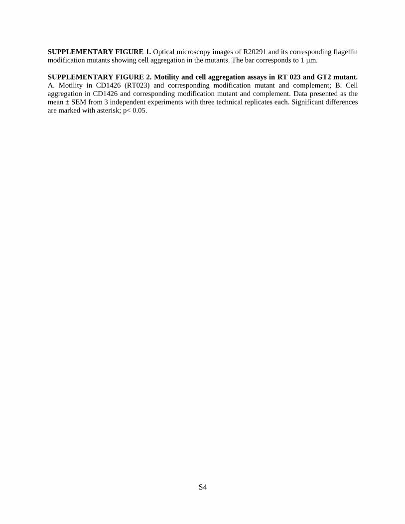
SUPPLEMENTARY FIGURE 1. Optical microscopy images of R20291 and its corresponding flagellin
modification mutants showing cell aggregation in the mutants. The bar corresponds to 1 µm.
SUPPLEMENTARY FIGURE 2. Motility and cell aggregation assays in RT 023 and GT2 mutant. A. Motility in CD1426 (RT023) and corresponding modification mutant and complement; B. Cell
aggregation in CD1426 and corresponding modification mutant and complement. Data presented as the
mean ± SEM from 3 independent experiments with three technical replicates each. Significant differences
are marked with asterisk; p< 0.05.

WrenHoward R. Morris, Mona Bajaj-Elliott, Susan M. Logan, Anne Dell and Brendan W.
Songane, Lisa F. Dawson, Elizabeth Donahue, Richard A. Stabler, Maria Panico, Esmeralda Valiente, Laura Bouché, Paul Hitchen, Alexandra Faulds-Pain, Mario
Biofilm Formation Lineages and Their Impact on Motility andClostridium difficileHypervirulent
Role of Glycosyltransferases Modifying Type B Flagellin of Emerging
doi: 10.1074/jbc.M116.749523 originally published online October 4, 20162016, 291:25450-25461.J. Biol. Chem.
10.1074/jbc.M116.749523Access the most updated version of this article at doi:
Alerts:
When a correction for this article is posted•
When this article is cited•
to choose from all of JBC's e-mail alertsClick here
Supplemental material:
http://www.jbc.org/content/suppl/2016/10/04/M116.749523.DC1.html
http://www.jbc.org/content/291/49/25450.full.html#ref-list-1
This article cites 54 references, 25 of which can be accessed free at
at LO
ND
ON
SCH
OF H
YG
IEN
E &
TR
OPIC
AL
ME
DIC
INE
on Decem
ber 2, 2016http://w
ww
.jbc.org/D
ownloaded from