Vaginal Bleeding in Late Pregnancy
33
Vaginal Bleeding in Late Pregnancy
description
dvdsavdas
Transcript of Vaginal Bleeding in Late Pregnancy

Vaginal Bleeding in Late Pregnancy
Objectives
Identify major causes of vaginal bleeding in Identify major causes of vaginal bleeding in the second half of pregnancythe second half of pregnancy
Describe a systematic approach to Describe a systematic approach to identifying the cause of bleedingidentifying the cause of bleeding
Describe specific treatment options based Describe specific treatment options based on diagnosison diagnosis

Causes of Late Pregnancy Bleeding
Placenta PreviaPlacenta Previa AbruptionAbruption Ruptured vasa previaRuptured vasa previa Uterine scar disruptionUterine scar disruption Cervical polypCervical polyp Bloody showBloody show Cervicitis or cervical ectropionCervicitis or cervical ectropion Vaginal traumaVaginal trauma Cervical cancerCervical cancer
Life-Threatening

Prevalence of Placenta Previa
Occurs in 1/200 pregnancies that reach 3Occurs in 1/200 pregnancies that reach 3 rdrd trimestertrimester
Low-lying placenta seen in 50% of Low-lying placenta seen in 50% of ultrasound scans at 16-20 weeksultrasound scans at 16-20 weeks 90% will have normal implantation when 90% will have normal implantation when
scan repeated at >30 weeksscan repeated at >30 weeks No proven benefit to routine screening No proven benefit to routine screening
ultrasound for this diagnosisultrasound for this diagnosis

Risk Factors for Placenta Previa
Previous cesarean deliveryPrevious cesarean delivery Previous uterine instrumentationPrevious uterine instrumentation High parityHigh parity Advanced maternal ageAdvanced maternal age SmokingSmoking Multiple gestationMultiple gestation

Morbidity with Placenta Previa
Maternal hemorrhageMaternal hemorrhage Operative delivery complicationsOperative delivery complications TransfusionTransfusion Placenta accreta, increta, or percretaPlacenta accreta, increta, or percreta PrematurityPrematurity

Patient History – Placenta Previa
Painless bleedingPainless bleeding 22ndnd or 3 or 3rdrd trimester, or at term trimester, or at term Often following intercourseOften following intercourse May have preterm contractionsMay have preterm contractions
““Sentinel bleed”Sentinel bleed”

Physical Exam – Placenta Previa
Vital signsVital signs Assess fundal heightAssess fundal height Fetal lieFetal lie Estimated fetal weight (Leopold)Estimated fetal weight (Leopold) Presence of fetal heart tonesPresence of fetal heart tones Gentle speculum examGentle speculum exam NO NO digital vaginal exam digital vaginal exam unlessunless placental location placental location
knownknown

Laboratory – Placenta Previa
Hematocrit or complete blood countHematocrit or complete blood count Blood type and RhBlood type and Rh Coagulation testsCoagulation tests
While waitingWhile waiting – serum clot tube taped to – serum clot tube taped to wallwall

Ultrasound – Placenta Previa
Can confirm diagnosisCan confirm diagnosis Full bladder can create false appearance of Full bladder can create false appearance of
anterioranterior previa previa Presenting part may overshadow Presenting part may overshadow posterior posterior
previaprevia Transvaginal scan can locate placental edge Transvaginal scan can locate placental edge
and internal osand internal os

Treatment – Placenta Previa
With no active bleedingWith no active bleeding Expectant managementExpectant management No intercourse, digital examsNo intercourse, digital exams
With late pregnancy bleedingWith late pregnancy bleeding Assess overall status, circulatory stabilityAssess overall status, circulatory stability Full dose Rhogam if Rh-Full dose Rhogam if Rh- Consider maternal transfer if prematureConsider maternal transfer if premature May need corticosteroids, tocolysis, May need corticosteroids, tocolysis,
amniocentesisamniocentesis

Double Set-Up Exam
Appropriate Appropriate onlyonly in marginal previa with vertex in marginal previa with vertex presentationpresentation
Palpation of placental edge and fetal head with set Palpation of placental edge and fetal head with set up for immediate surgeryup for immediate surgery
Cesarean delivery Cesarean delivery under regional anesthesiaunder regional anesthesia if: if: Complete previaComplete previa Fetal head not engagedFetal head not engaged Non-reassuring tracingNon-reassuring tracing Brisk or persistent bleedingBrisk or persistent bleeding Mature fetusMature fetus

Placental Abruption
Premature separation of placenta from Premature separation of placenta from uterine walluterine wall Partial or completePartial or complete
““Marginal sinus separation” or “marginal Marginal sinus separation” or “marginal sinus rupture”sinus rupture” Bleeding, but abnormal implantation or Bleeding, but abnormal implantation or
abruption never establishedabruption never established

Epidemiology of Abruption
Occurs in 1-2% of pregnanciesOccurs in 1-2% of pregnancies Risk factorsRisk factors
Hypertensive diseases of pregnancyHypertensive diseases of pregnancy Smoking or substance abuse (e.g. cocaine)Smoking or substance abuse (e.g. cocaine) TraumaTrauma Overdistention of the uterusOverdistention of the uterus History of previous abruptionHistory of previous abruption Unexplained elevation of MSAFPUnexplained elevation of MSAFP Placental insufficiencyPlacental insufficiency Maternal thrombophilia/metabolic Maternal thrombophilia/metabolic
abnormalitiesabnormalities
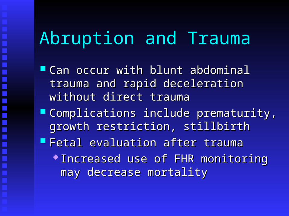
Abruption and Trauma
Can occur with blunt abdominal trauma and Can occur with blunt abdominal trauma and rapid deceleration without direct traumarapid deceleration without direct trauma
Complications include prematurity, growth Complications include prematurity, growth restriction, stillbirthrestriction, stillbirth
Fetal evaluation after traumaFetal evaluation after trauma Increased use of FHR monitoring may Increased use of FHR monitoring may
decrease mortalitydecrease mortality
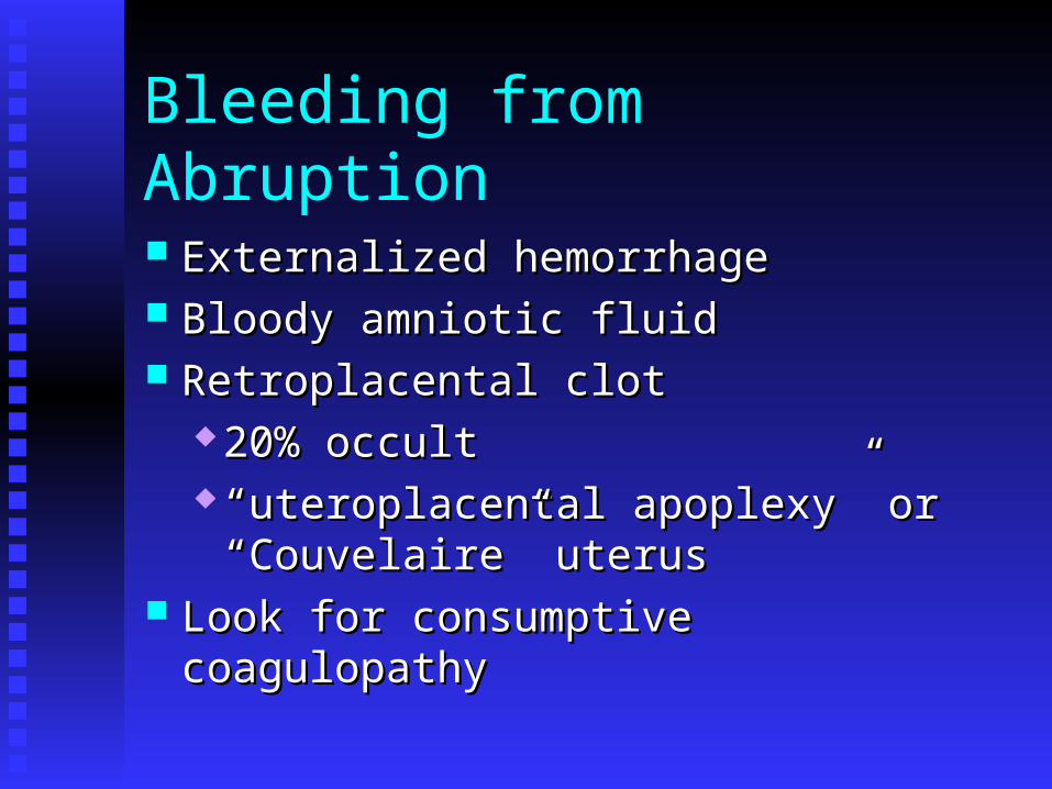
Bleeding from Abruption
Externalized hemorrhageExternalized hemorrhage Bloody amniotic fluidBloody amniotic fluid Retroplacental clotRetroplacental clot
20% occult20% occult ““uteroplacental apoplexy” or uteroplacental apoplexy” or
“Couvelaire” uterus“Couvelaire” uterus Look for consumptive coagulopathyLook for consumptive coagulopathy

Patient History - Abruption
Pain = hallmark symptomPain = hallmark symptom Varies from mild cramping to severe painVaries from mild cramping to severe pain Back pain – think posterior abruptionBack pain – think posterior abruption
BleedingBleeding May not reflect amount of blood lossMay not reflect amount of blood loss Differentiate from exuberant bloody showDifferentiate from exuberant bloody show
TraumaTrauma Other risk factors (e.g. hypertension)Other risk factors (e.g. hypertension) Membrane ruptureMembrane rupture

Physical Exam - Abruption
Signs of circulatory instabilitySigns of circulatory instability Mild tachycardia normalMild tachycardia normal Signs and symptoms of shock represent >30% Signs and symptoms of shock represent >30%
blood lossblood loss Maternal abdomenMaternal abdomen
Fundal heightFundal height Leopold’s: estimated fetal weight, fetal lieLeopold’s: estimated fetal weight, fetal lie Location of tendernessLocation of tenderness Tetanic contractionsTetanic contractions
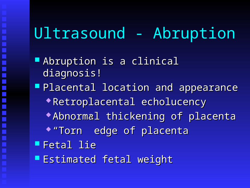
Ultrasound - Abruption
Abruption is a clinical diagnosis!Abruption is a clinical diagnosis! Placental location and appearancePlacental location and appearance
Retroplacental echolucencyRetroplacental echolucency Abnormal thickening of placentaAbnormal thickening of placenta ““Torn” edge of placentaTorn” edge of placenta
Fetal lieFetal lie Estimated fetal weightEstimated fetal weight

Laboratory - Abruption
Complete blood countComplete blood count Type and RhType and Rh Coagulation tests + “Clot test”Coagulation tests + “Clot test” Kleihauer-Betke not diagnostic, but useful Kleihauer-Betke not diagnostic, but useful
to determine Rhogam doseto determine Rhogam dose Preeclampsia labs, if indicatedPreeclampsia labs, if indicated Consider urine drug screenConsider urine drug screen

Sher’s Classification - Abruption
Grade IGrade I
Grade IIGrade II
Grade IIIGrade III with fetal demisewith fetal demise III AIII A - without coagulopathy (2/3) - without coagulopathy (2/3) III BIII B - with coagulopathy (1/3) - with coagulopathy (1/3)
mild, often retroplacental clot identified at delivery
tense, tender abdomen and live fetus

Treatment – Grade II Abruption
Assess fetal and maternal stabilityAssess fetal and maternal stability
AmniotomyAmniotomy
IUPC to detect elevated uterine toneIUPC to detect elevated uterine tone
Expeditious operative or vaginal deliveryExpeditious operative or vaginal delivery
Maintain urine output > 30 cc/hr and Maintain urine output > 30 cc/hr and hematocrit > 30%hematocrit > 30%
Prepare for neonatal resuscitationPrepare for neonatal resuscitation

Treatment – Grade III Abruption
Assess mother for hemodynamic and Assess mother for hemodynamic and coagulation statuscoagulation status
Vigorous replacement of fluid and blood Vigorous replacement of fluid and blood productsproducts
Vaginal delivery preferred, unless severe Vaginal delivery preferred, unless severe hemorrhagehemorrhage

Coagulopathy with Abruption
Occurs in 1/3 of Grade III abruptionOccurs in 1/3 of Grade III abruption Usually not seen if live fetusUsually not seen if live fetus Etiologies: consumption, DICEtiologies: consumption, DIC Administer platelets, FFPAdminister platelets, FFP Give Factor VIII if severeGive Factor VIII if severe

Epidemiology of Uterine Rupture
Occult dehiscence vs. symptomatic ruptureOccult dehiscence vs. symptomatic rupture 0.03 – 0.08% of all women0.03 – 0.08% of all women 0.3 – 1.7% of women with uterine scar0.3 – 1.7% of women with uterine scar Previous cesarean incision most common Previous cesarean incision most common
reason for scar disruptionreason for scar disruption Other causes: previous uterine curettage or Other causes: previous uterine curettage or
perforation, inappropriate oxytocin usage, perforation, inappropriate oxytocin usage, traumatrauma

Risk Factors – Uterine Rupture
Previous uterine surgeryPrevious uterine surgery AdenomyosisAdenomyosisCongenital uterine Congenital uterine anomalyanomaly
Fetal anomalyFetal anomaly
Uterine overdistensionUterine overdistension Vigorous uterine Vigorous uterine pressurepressure
Gestational trophoblastic Gestational trophoblastic neoplasianeoplasia
Difficult placental Difficult placental removalremoval
Placenta increta or Placenta increta or percretapercreta

Morbidity with Uterine Rupture
MaternalMaternal Hemorrhage with anemiaHemorrhage with anemia Bladder ruptureBladder rupture HysterectomyHysterectomy Maternal deathMaternal death
FetalFetal Respiratory distressRespiratory distress HypoxiaHypoxia AcidemiaAcidemia Neonatal deathNeonatal death
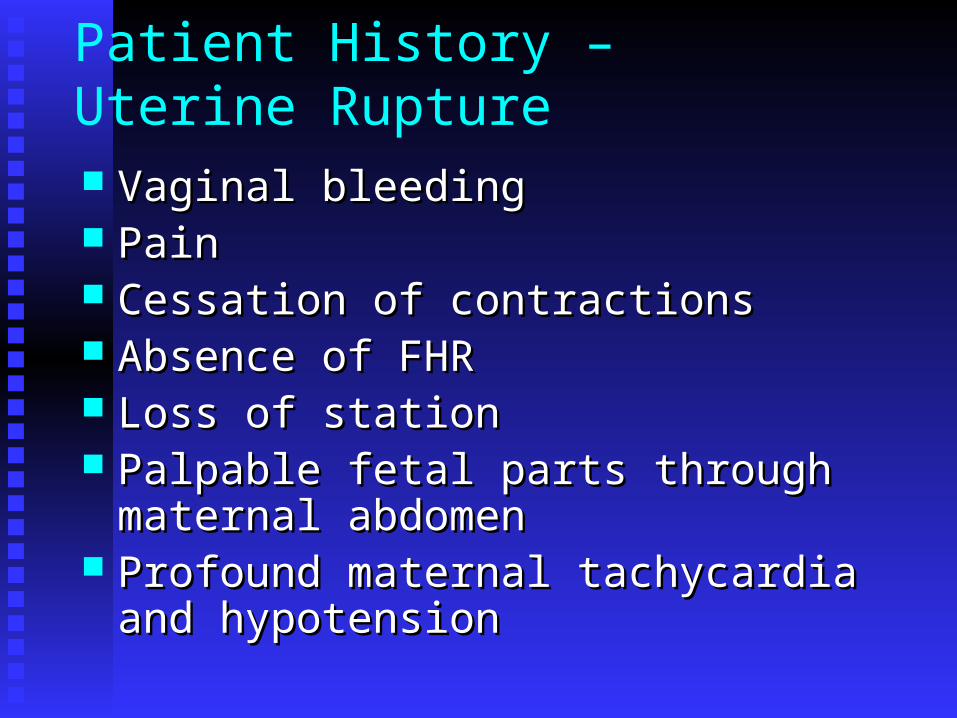
Patient History – Uterine Rupture
Vaginal bleedingVaginal bleeding PainPain Cessation of contractionsCessation of contractions Absence of FHRAbsence of FHR Loss of stationLoss of station Palpable fetal parts through maternal Palpable fetal parts through maternal
abdomenabdomen Profound maternal tachycardia and Profound maternal tachycardia and
hypotensionhypotension

Uterine Rupture
Sudden deterioration of FHR pattern is most Sudden deterioration of FHR pattern is most frequent findingfrequent finding
Placenta may play a role in uterine rupturePlacenta may play a role in uterine rupture Transvaginal ultrasound to evaluate uterine Transvaginal ultrasound to evaluate uterine
wallwall MRI to confirm possible placenta accretaMRI to confirm possible placenta accreta
TreatmentTreatment Asymptomatic scar disruption – expectant Asymptomatic scar disruption – expectant
managementmanagement Symptomatic rupture – emergent cesarean Symptomatic rupture – emergent cesarean
deliverydelivery

Vasa Previa
Rarest cause of hemorrhageRarest cause of hemorrhage Onset with membrane ruptureOnset with membrane rupture Blood loss is fetal, with 50% mortalityBlood loss is fetal, with 50% mortality Seen with low-lying placenta, velamentous Seen with low-lying placenta, velamentous
insertion of the cord or succenturiate lobeinsertion of the cord or succenturiate lobe Antepartum diagnosisAntepartum diagnosis
AmnioscopyAmnioscopy Color doppler ultrasoundColor doppler ultrasound Palpate vessels during vaginal examinationPalpate vessels during vaginal examination

Diagnostic Tests – Vasa Previa
Apt test – based on colorimetric response of Apt test – based on colorimetric response of fetal hemoglobinfetal hemoglobin
Wright stain of vaginal blood – for Wright stain of vaginal blood – for nucleated RBCsnucleated RBCs
Kleihauer-Betke test – 2 hours delay Kleihauer-Betke test – 2 hours delay prohibits its useprohibits its use
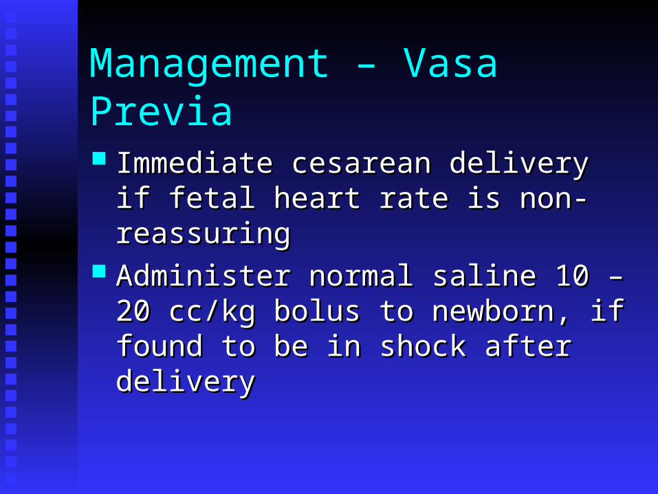
Management – Vasa Previa
Immediate cesarean delivery if fetal heart Immediate cesarean delivery if fetal heart rate is non-reassuringrate is non-reassuring
Administer normal saline 10 – 20 cc/kg Administer normal saline 10 – 20 cc/kg bolus to newborn, if found to be in shock bolus to newborn, if found to be in shock after deliveryafter delivery

Summary
Late pregnancy bleeding may herald Late pregnancy bleeding may herald diagnoses with significant diagnoses with significant morbidity/mortalitymorbidity/mortality
Determining diagnosis important, as Determining diagnosis important, as treatment dependent on causetreatment dependent on cause
Avoid vaginal exam when placental Avoid vaginal exam when placental location not knownlocation not known