Uveitis Review for the OKAPS… - School of Medicine · – A chronic posterior uveitis...
111
Uveitis Review for the OKAPS… What you should and should not know! By: Armando L. Oliver, MD Ocular Immunology and Uveitis Specialist Vitreoretinal Surgeon Assistant Professor, UPR
Transcript of Uveitis Review for the OKAPS… - School of Medicine · – A chronic posterior uveitis...
Uveitis Review for the OKAPS…
What you should and should not know!
By: Armando L. Oliver, MD
Ocular Immunology and Uveitis Specialist
Vitreoretinal Surgeon
Assistant Professor, UPR
Disclaimer...
• The information you are about to whiteness has
been oversimplified for the purpose of passing the
OKAPs, boards, etc…
• Some of this information sounds OK, and you
may have been brought up to believe is true, but it
isn’t!
• Using this information for real life patient
management may have deleterious consequences.
• Contrary to popular believe, I’m not insane!
Anterior Uveitis
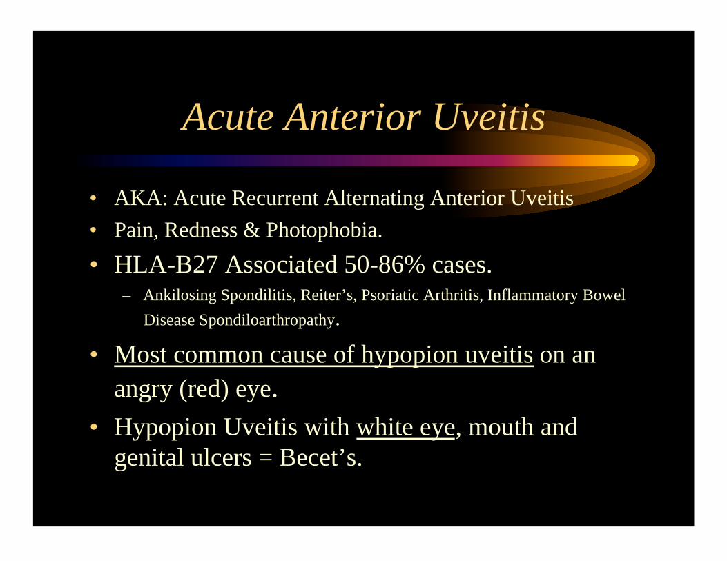
Acute Anterior Uveitis
• AKA: Acute Recurrent Alternating Anterior Uveitis
• Pain, Redness & Photophobia.
• HLA-B27 Associated 50-86% cases. – Ankilosing Spondilitis, Reiter’s, Psoriatic Arthritis, Inflammatory Bowel
Disease Spondiloarthropathy.
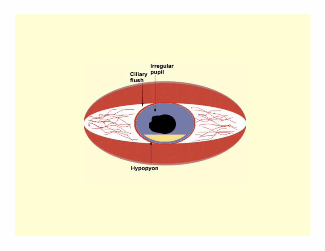
• Most common cause of hypopion uveitis on an
angry (red) eye.
• Hypopion Uveitis with white eye, mouth and
genital ulcers = Becet’s.
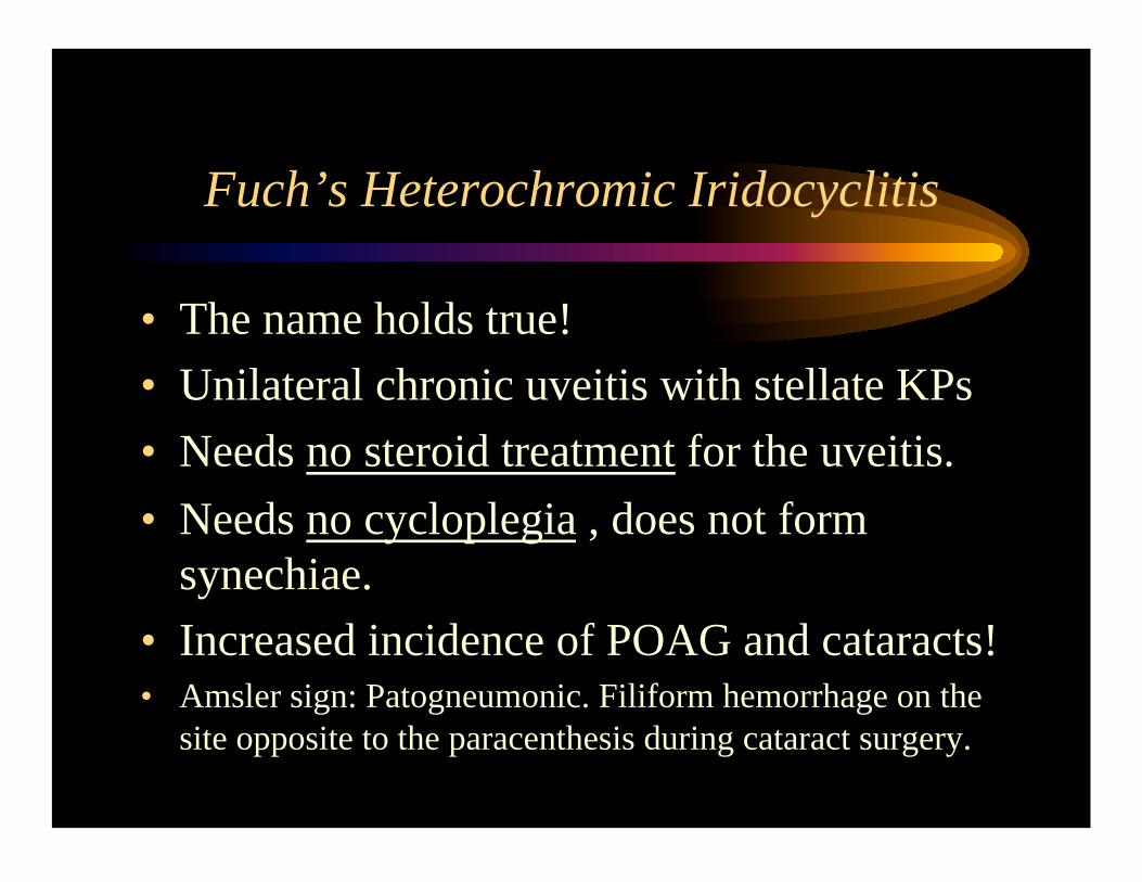
Fuch’s Heterochromic Iridocyclitis
• The name holds true!
• Unilateral chronic uveitis with stellate KPs
• Needs no steroid treatment for the uveitis.
• Needs no cycloplegia , does not form
synechiae.
• Increased incidence of POAG and cataracts! • Amsler sign: Patogneumonic. Filiform hemorrhage on the
site opposite to the paracenthesis during cataract surgery.
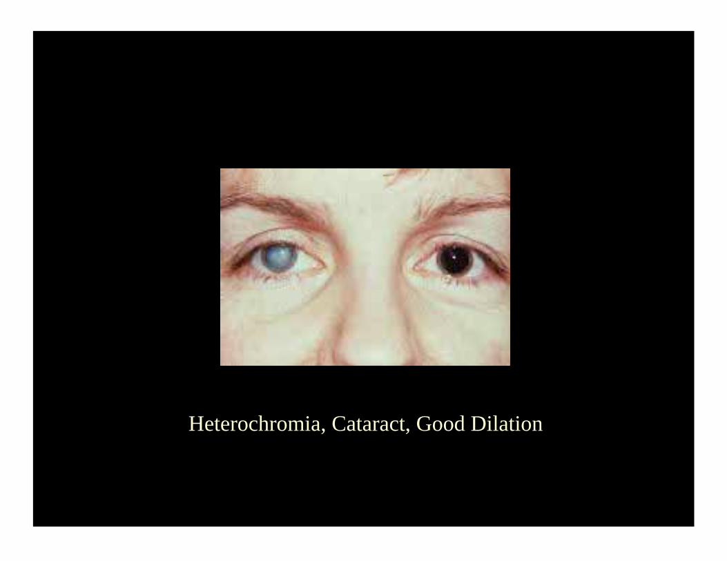
Heterochromia, Cataract, Good Dilation
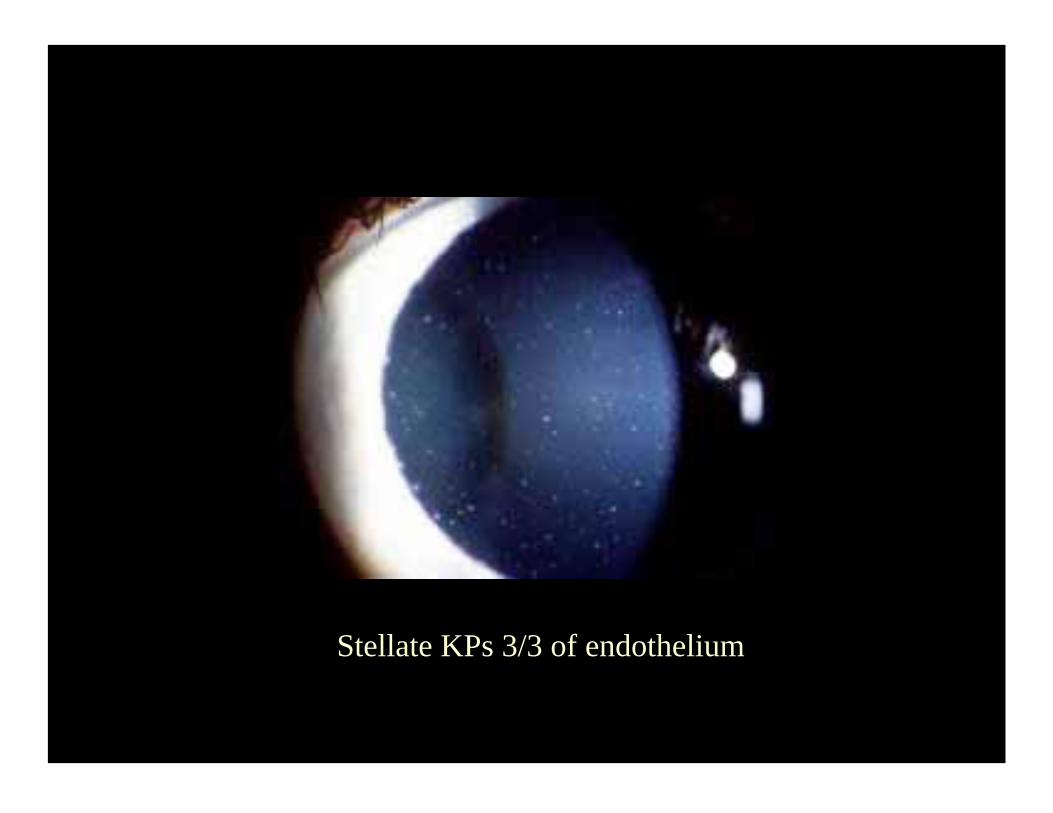
Stellate KPs 3/3 of endothelium
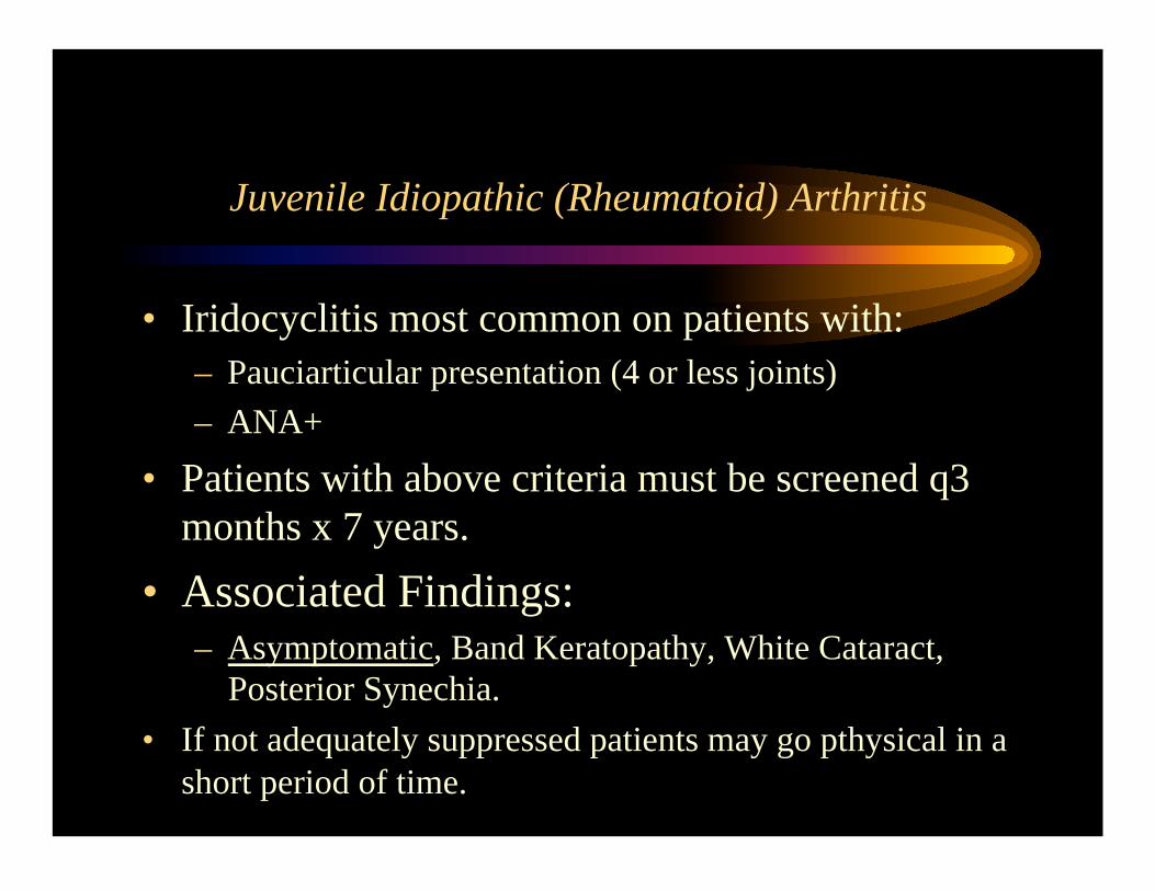
Juvenile Idiopathic (Rheumatoid) Arthritis
• Iridocyclitis most common on patients with:
– Pauciarticular presentation (4 or less joints)
– ANA+
• Patients with above criteria must be screened q3
months x 7 years.
• Associated Findings: – Asymptomatic, Band Keratopathy, White Cataract,
Posterior Synechia.
• If not adequately suppressed patients may go pthysical in a
short period of time.
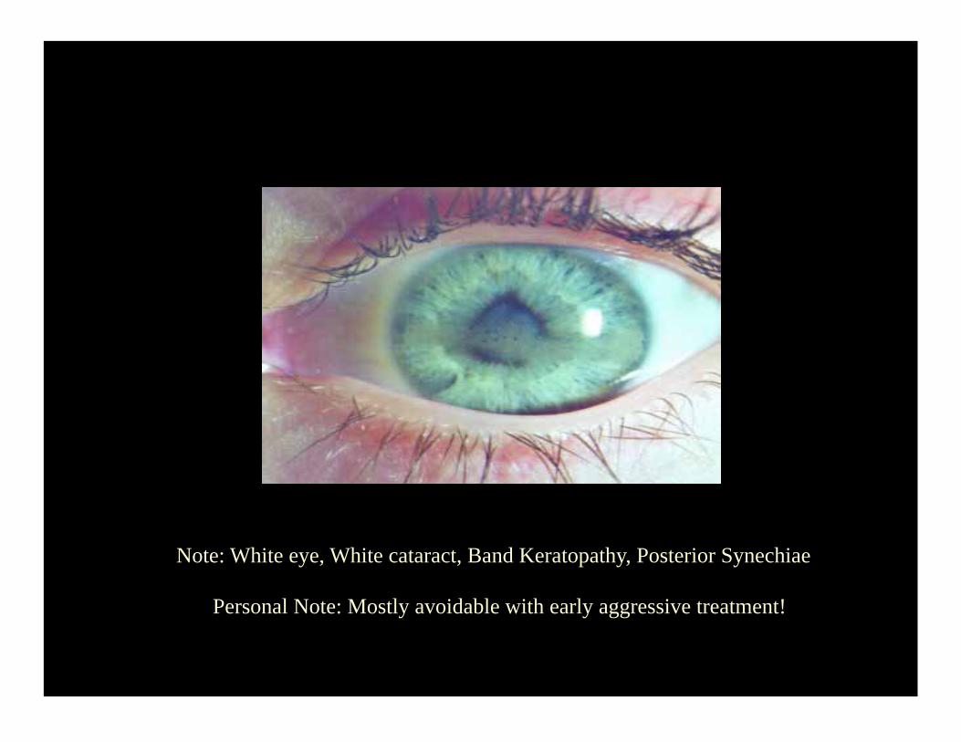
Note: White eye, White cataract, Band Keratopathy, Posterior Synechiae
Personal Note: Mostly avoidable with early aggressive treatment!
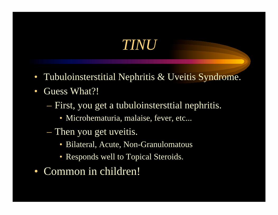
TINU
• Tubuloinsterstitial Nephritis & Uveitis Syndrome.
• Guess What?!
– First, you get a tubuloinstersttial nephritis.
• Microhematuria, malaise, fever, etc...
– Then you get uveitis.
• Bilateral, Acute, Non-Granulomatous
• Responds well to Topical Steroids.
• Common in children!
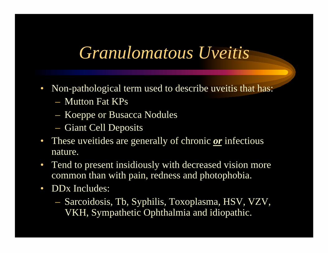
Granulomatous Uveitis
• Non-pathological term used to describe uveitis that has:
– Mutton Fat KPs
– Koeppe or Busacca Nodules
– Giant Cell Deposits
• These uveitides are generally of chronic or infectious nature.
• Tend to present insidiously with decreased vision more common than with pain, redness and photophobia.
• DDx Includes:
– Sarcoidosis, Tb, Syphilis, Toxoplasma, HSV, VZV, VKH, Sympathetic Ophthalmia and idiopathic.
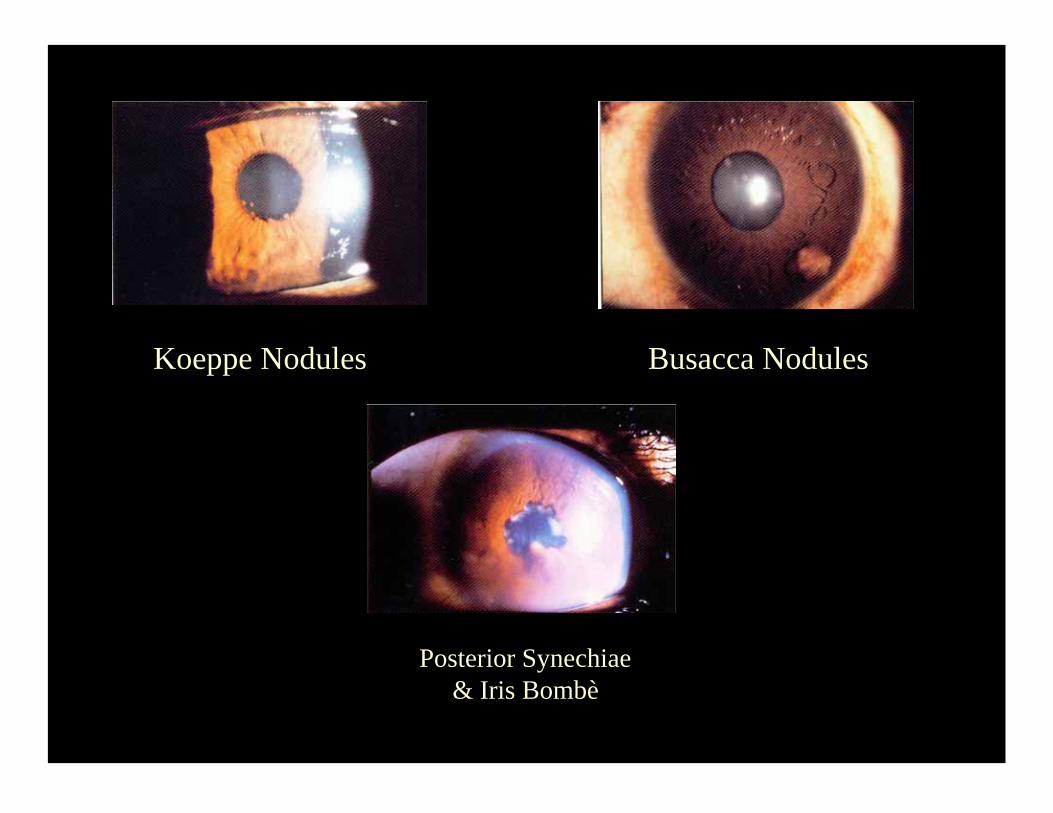
Koeppe Nodules Busacca Nodules
Posterior Synechiae
& Iris Bombè
Herpes Simplex Virus Uveitis
• Unilateral Granulomatous Uveitis with Iris
transillumination defects in a patient that
suffers recurrent cold sores who has corneal
aesthesia and possibly, high IOP and a corneal
stromal haze.
• Treatment: – Acyclovir 400mg 5times/d, Pred Forte and Atropine.
• Note: Acute if treated very early otherwise may become
chronic.
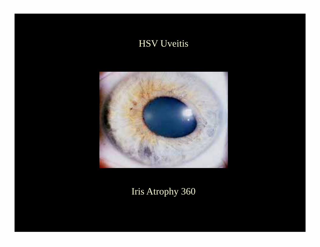
Iris Atrophy 360
HSV Uveitis
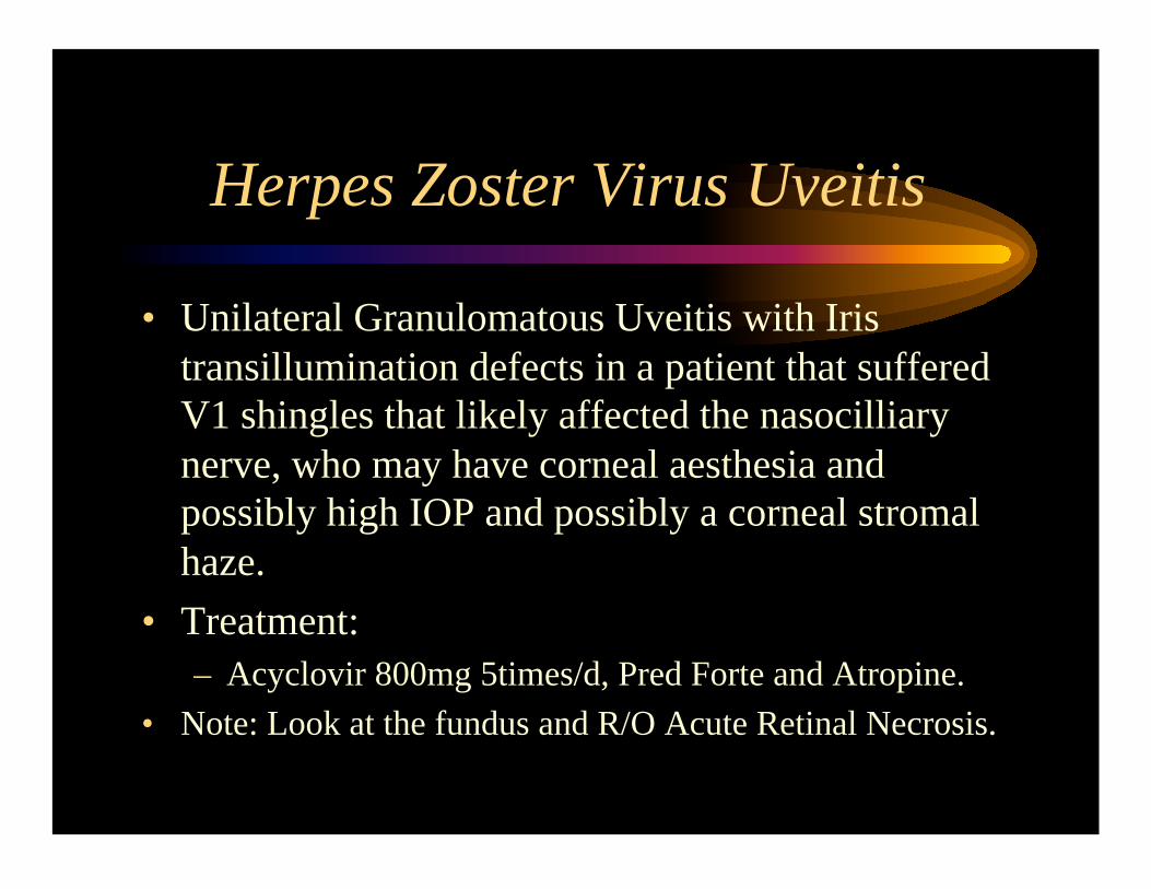
Herpes Zoster Virus Uveitis
• Unilateral Granulomatous Uveitis with Iris
transillumination defects in a patient that suffered
V1 shingles that likely affected the nasocilliary
nerve, who may have corneal aesthesia and
possibly high IOP and possibly a corneal stromal
haze.
• Treatment: – Acyclovir 800mg 5times/d, Pred Forte and Atropine.
• Note: Look at the fundus and R/O Acute Retinal Necrosis.
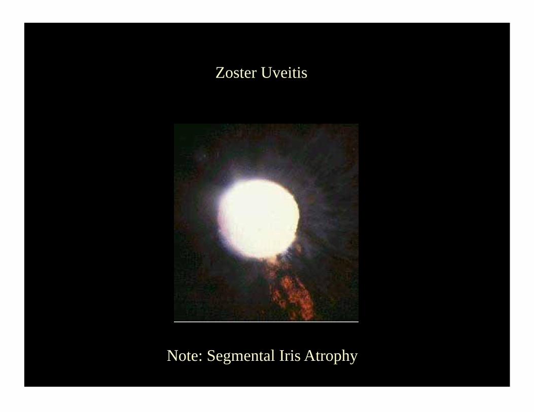
Zoster Uveitis
Note: Segmental Iris Atrophy
Uveitis that may have high IOP upon presentation.
• DDX for the OKAPS: – HSV & VZV
– CMV
– Toxoplasma
– Posner-Schlossman Syndrome (Dx of Exclusion)
• Rare in real life, common in the OKAP!
After the initial presentation, the differential of glaucoma in uveitis patients includes: anterior synechiae, bombè,
steroid response, and pigment deposition (HSV/VZV).
Intermediate Uveitis
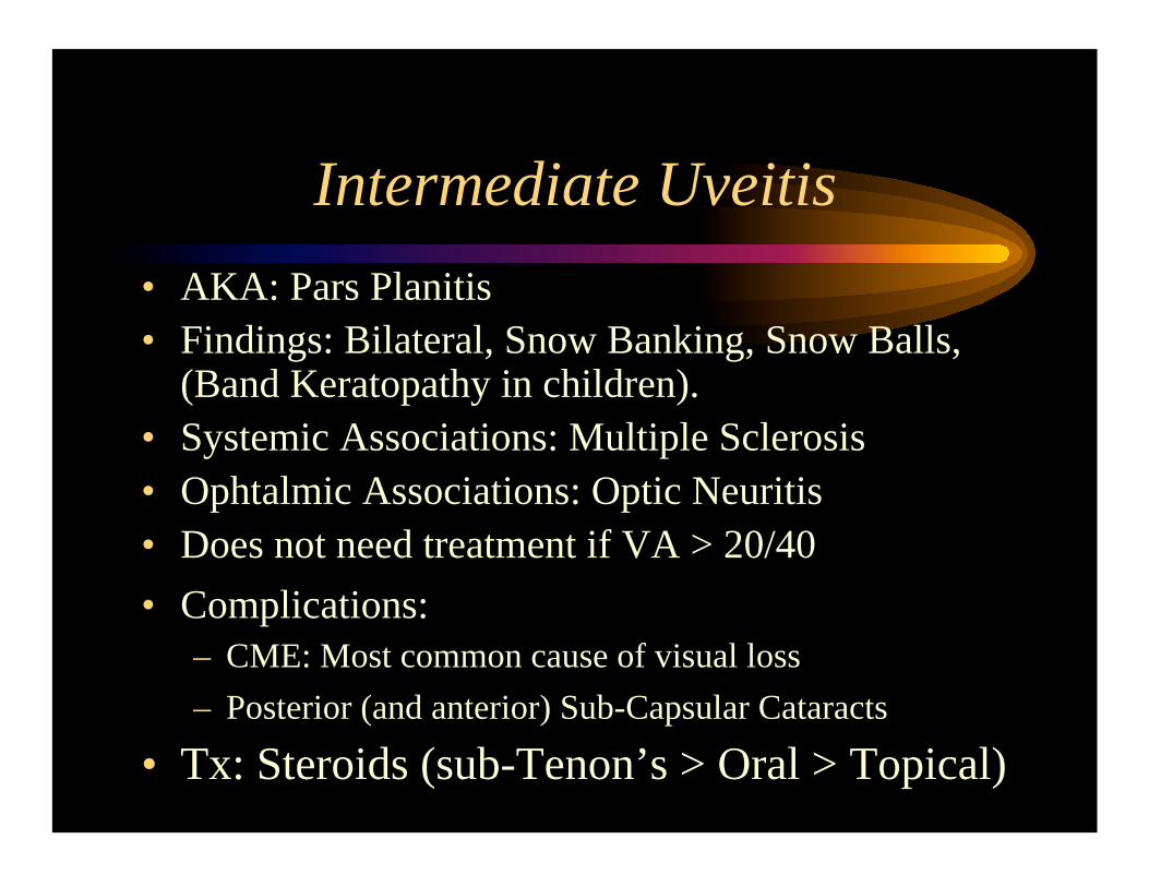
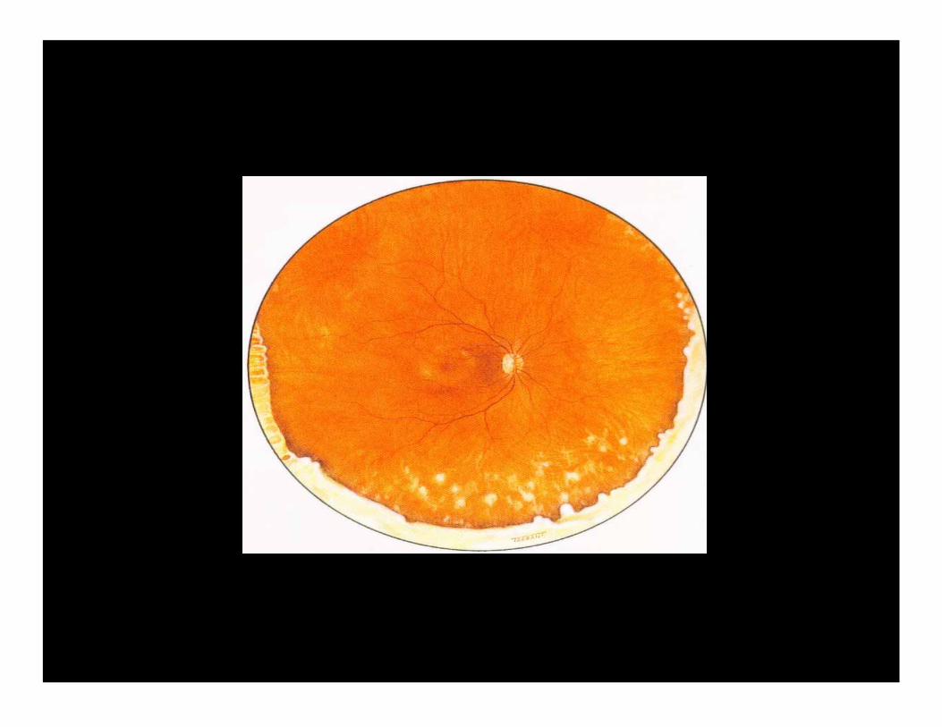
Intermediate Uveitis
• AKA: Pars Planitis
• Findings: Bilateral, Snow Banking, Snow Balls, (Band Keratopathy in children).
• Systemic Associations: Multiple Sclerosis
• Ophtalmic Associations: Optic Neuritis
• Does not need treatment if VA > 20/40
• Complications: – CME: Most common cause of visual loss
– Posterior (and anterior) Sub-Capsular Cataracts
• Tx: Steroids (sub-Tenon’s > Oral > Topical)
Posterior Uveitis
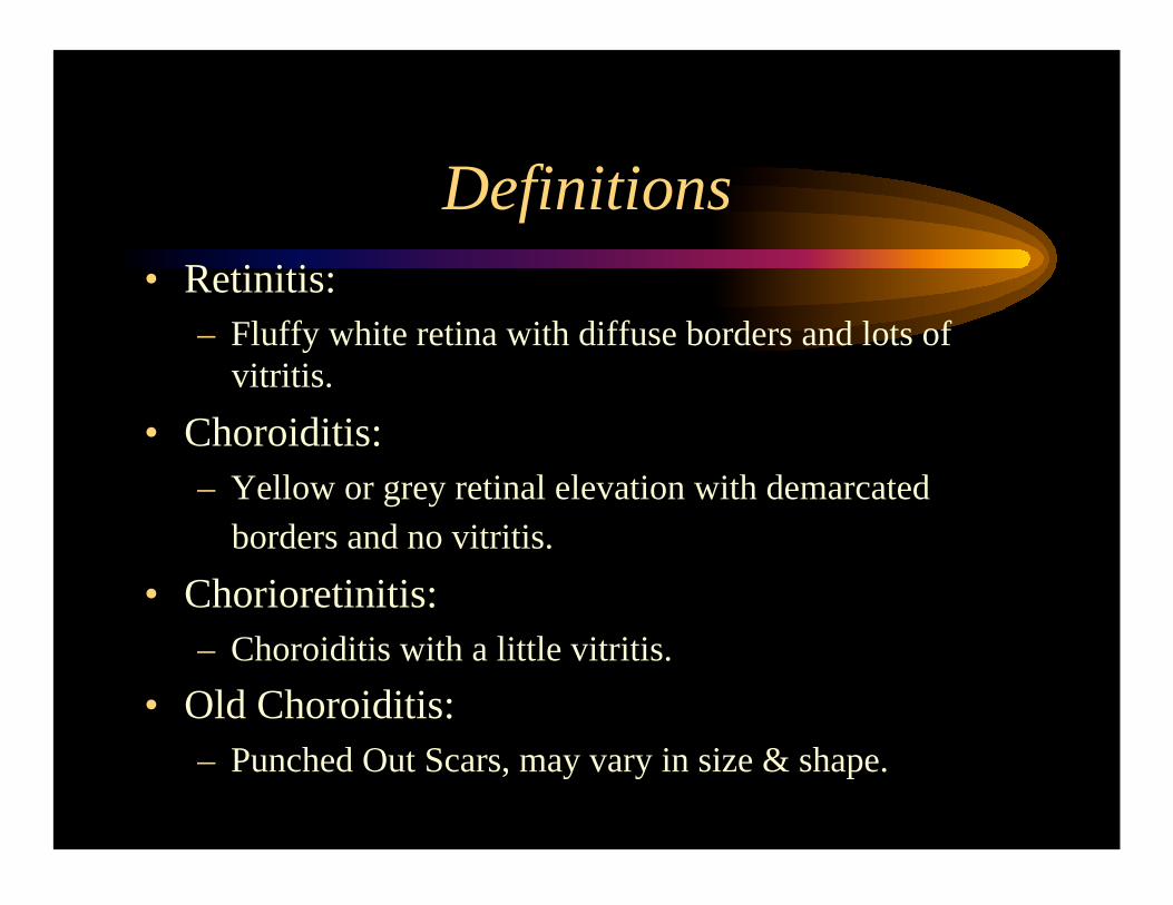
Definitions
• Retinitis:
– Fluffy white retina with diffuse borders and lots of
vitritis.
• Choroiditis: – Yellow or grey retinal elevation with demarcated
borders and no vitritis.
• Chorioretinitis:
– Choroiditis with a little vitritis.
• Old Choroiditis:
– Punched Out Scars, may vary in size & shape.
Definitions
• Plaque:
– A lesion approximately 1DA.
• Spots & Dots:
– Lesions approximately 100-200 microns.

Definitions
• Focal:
– Single lesion, may be localized like in
Toxoplasma or spread like CMV retinitis.
• Multifocal:
– Multiple lesions, I.e: MEWDS.
• Diffuse:
– Sympathetic Ophthalmia.
Choroiditis and Chorioretinitis
Which is the unilateral one?
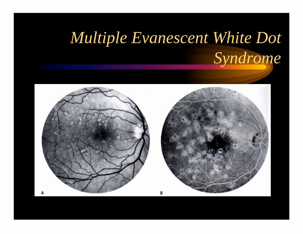
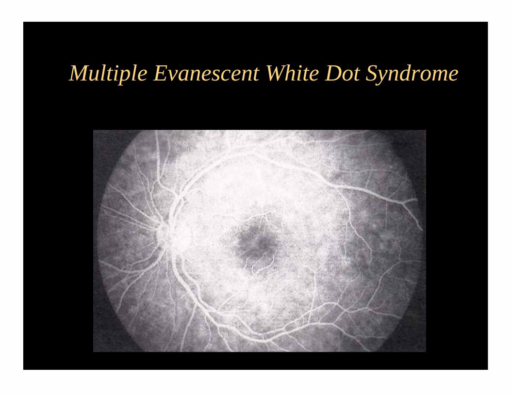
MEWDS:
Multiple Evanescent White Dot Syndrome.
• Multiple dots that subsequently disappear are
required for the diagnosis!
• Has a good prognosis, without treatment!
• Late wreath pattern fluorescein.
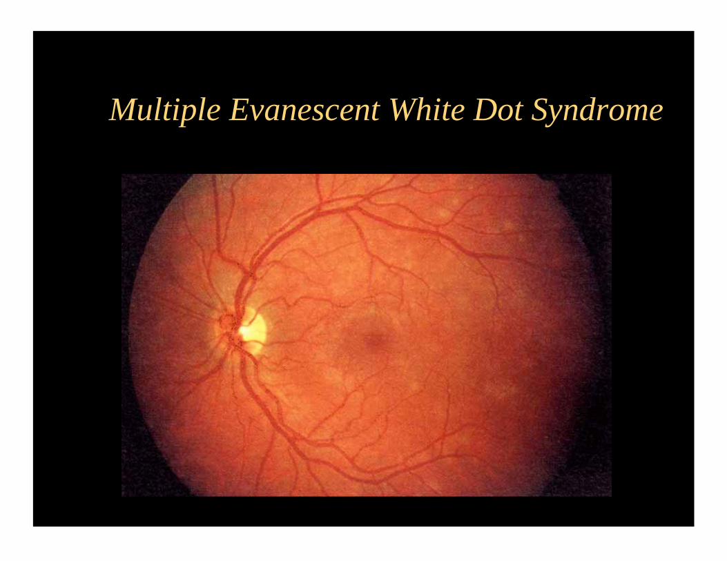
Multiple Evanescent White Dot Syndrome
Multiple Evanescent White Dot
Syndrome
Multiple Evanescent White Dot Syndrome
OK lets talk about choroiditis!
Which is the purest form of choroiditis?
• OHS: Ocular Histoplasmosis Syndrome
• Classic triad of:
– Peripapilary atrophy
– Punched Out Chorioretinal Lesions
– Macular CNVM
• By definition: No vitritis.
• For more info ask the expert: Dr. Feinberg
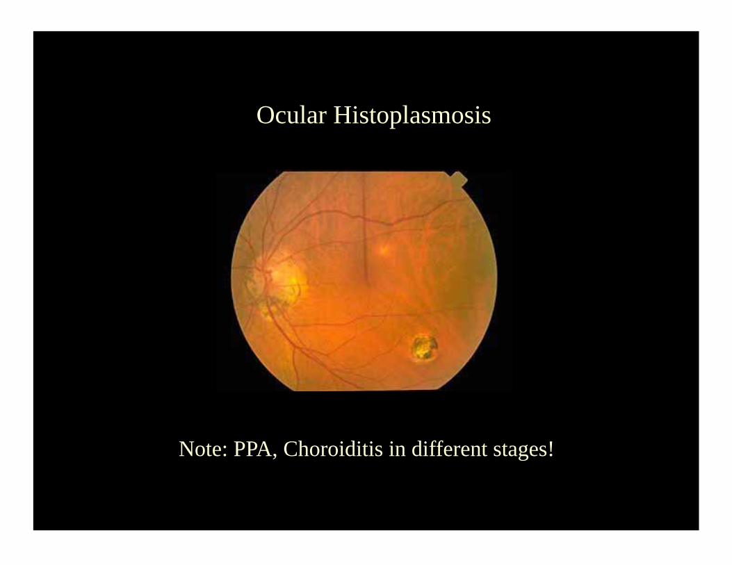
Ocular Histoplasmosis
Note: PPA, Choroiditis in different stages!
MFC PU
Multifocal choroiditis with panuveitis syndrome.
• OHS + Vitritis = MFC PU
• The lesions are somewhat smaller and more
peripheral than OHS, if untreated may look like
large dark plaques.
• Needs chronic systemic treatment or may loose
peripheral vision over the years.
• Sub-retinal fibrosis variant 2ry to CNVM.
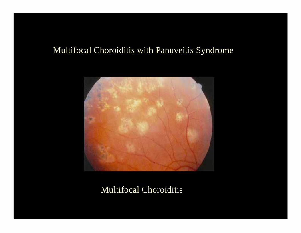
Multifocal Choroiditis
Multifocal Choroiditis with Panuveitis Syndrome
PIC
Punctate Inner Choroiditis
• As the name (choroiditis) implies, has no
vitritis.
• Bilateral, macular dots, (puncta = dot).
• High risk for CNVM.
• May follow a uniphasic course which is
better off if treated with systemic steroids.
Note: Whether PIC exists or not is controversial among uveitis specialists, I doubt they would ask this one.
I have seen it, I understand it; therefore, I am a believer!
Now…
Two syndromes with
early blockage and late
leakage...
APMPPE
Acute Posterior Multifocal Placoid Pigment Epitheliopathy
• Bilateral, confluent plaques deep to the retina.
• Early blockage, late staining of the entire lesion.
• In the OKAPs patients may get a cold or flu!
• Good prognosis, no treatment needed!
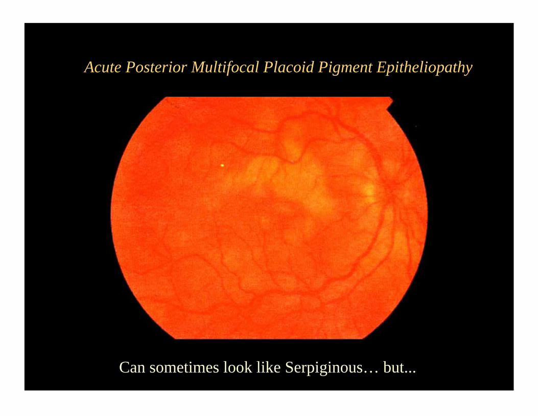
Acute Posterior Multifocal Placoid Pigment Epitheliopathy
Can sometimes look like Serpiginous… but...
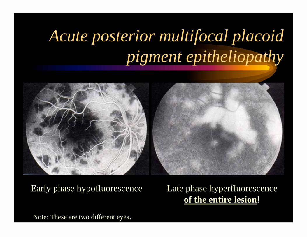
Early phase hypofluorescence Late phase hyperfluorescence
of the entire lesion!
Acute posterior multifocal placoid
pigment epitheliopathy
Note: These are two different eyes.
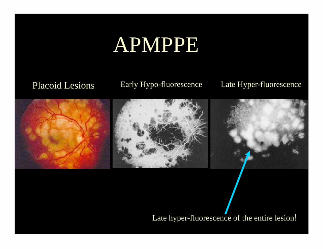
APMPPE
Placoid Lesions Early Hypo-fluorescence Late Hyper-fluorescence
Late hyper-fluorescence of the entire lesion!
Serpiginous Choroiditis
• Bilateral Serpentine/Ameboid choroidal lesions that most
of the time emerge from the optic nerve.
• Follows a relapsing and remitting course.
• If the fovea becomes involve the prognosis is poor. No
kidding!
• Early blockage, late staining at the border of the lesion!
• Tx: Cyclophosphamide x 1yr with oral prednisone for
acute suppression of the lesions.
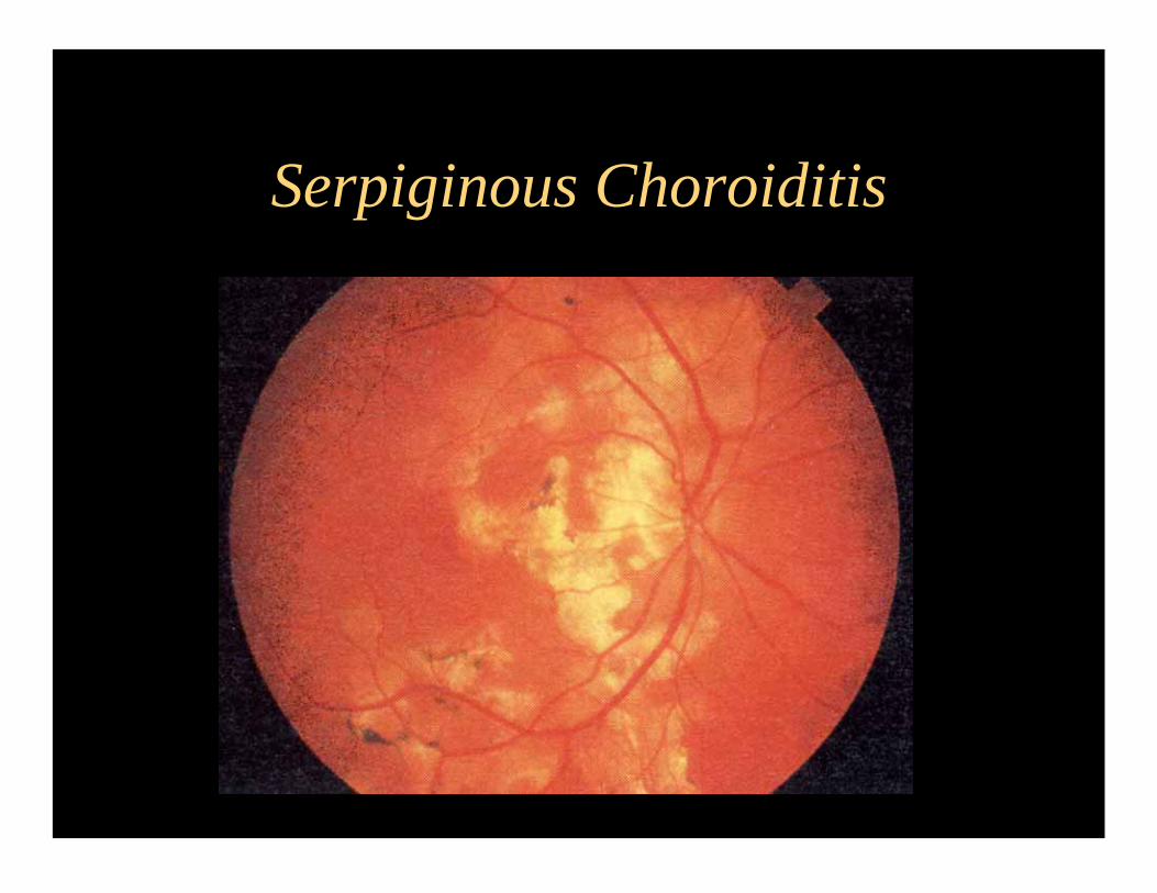
Serpiginous Choroiditis
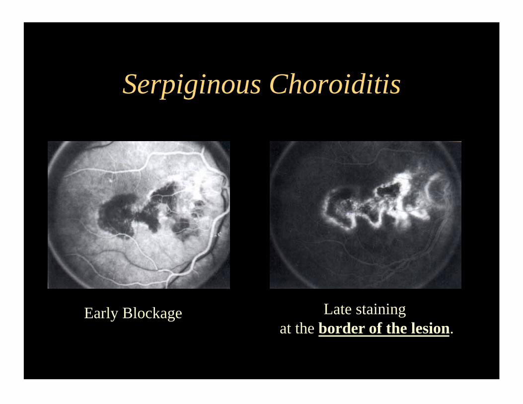
Serpiginous Choroiditis
Early Blockage Late staining
at the border of the lesion.
Now 2 very similar
panuveitis syndromes
which differ on the
systemic manifestations!
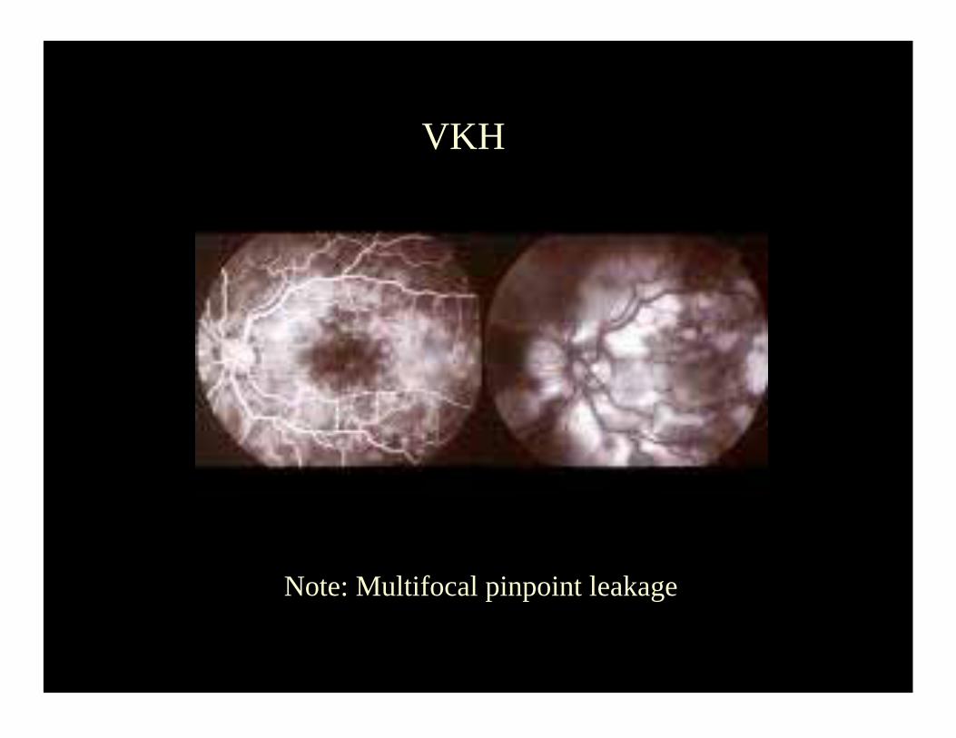
Vogt Koyanagi Harada
Bilateral Granulomatous Panuveitis with serous RD
• Latinos, asians, arabs and american indians.
• IVFA: Stars in the night appaerance
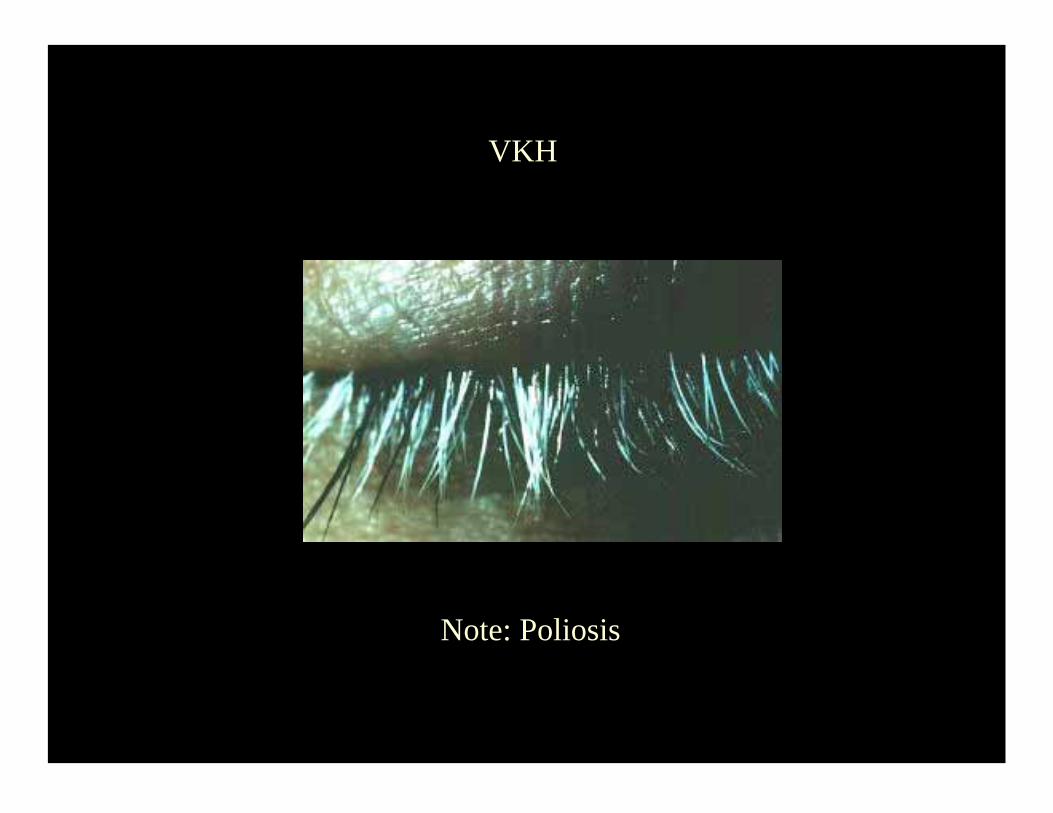
• Alopecia, Poliosis, Vitiligo,
• Meningismus, Tinitus, Sensorineural Hearing loss
• Tx: Systemic prednisone.
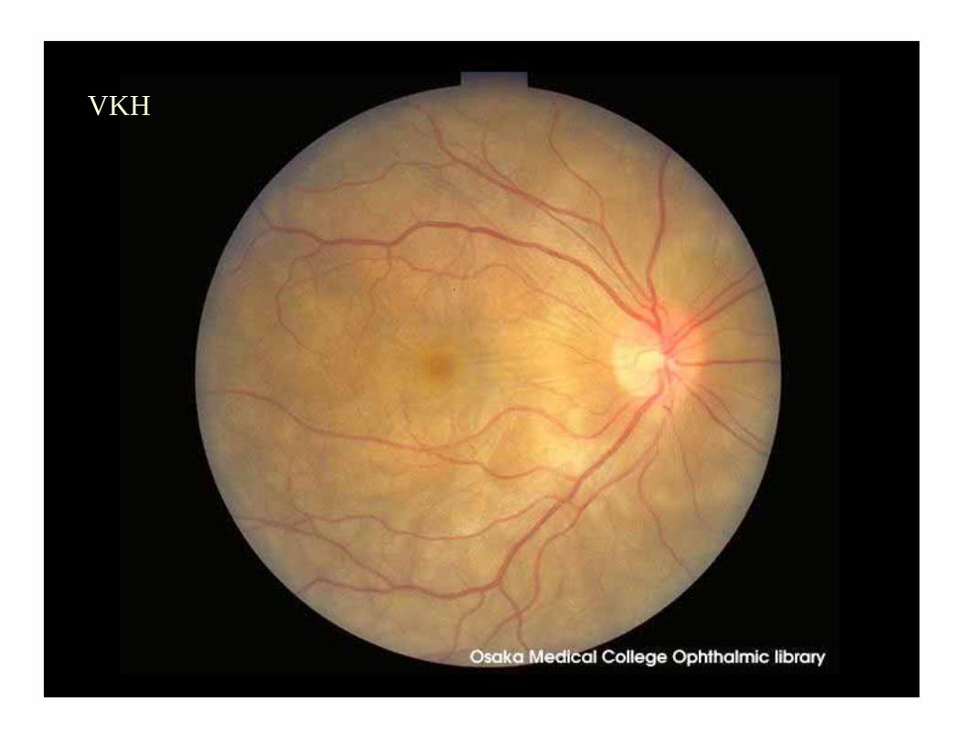
VKH
VKH
Note: Multifocal pinpoint leakage
VKH
Note: Poliosis
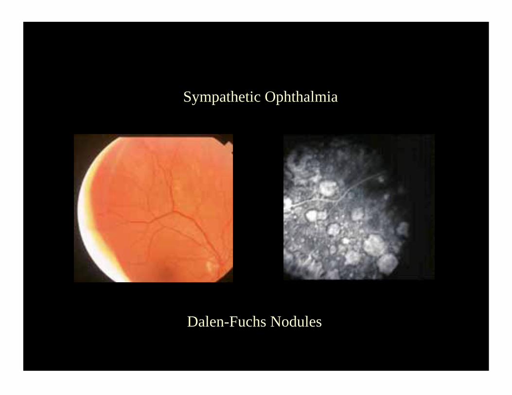
Sympathetic Ophthalmia
Bilateral Panuveitis with Dalen-Fuchs Nodules, in a
patient with history of eye surgery or trauma to one eye.
• Pathology: Choriocapillaris is spared
• Ophthalmic findings virtually indistinguishable from VKH
• May have serous RDs just like VKH
• Fundus findings indistinguishable from VKH in the late phases
• Follows a chronic course, needs prednisone and immunosuppression.
Sympathetic Ophthalmia
Dalen-Fuchs Nodules
Finally…
a retinochoroiditis...
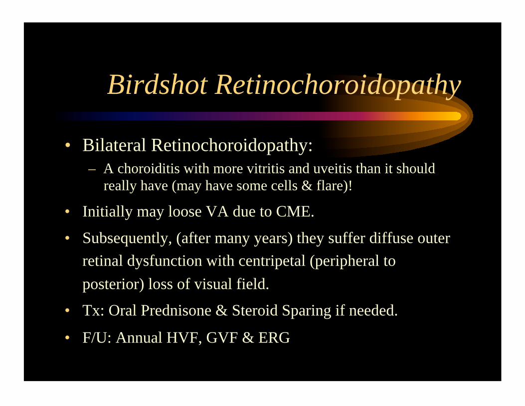
Birdshot Retinochoroidopathy
• Bilateral Retinochoroidopathy: – A choroiditis with more vitritis and uveitis than it should
really have (may have some cells & flare)!
• Initially may loose VA due to CME.
• Subsequently, (after many years) they suffer diffuse outer
retinal dysfunction with centripetal (peripheral to
posterior) loss of visual field.
• Tx: Oral Prednisone & Steroid Sparing if needed.
• F/U: Annual HVF, GVF & ERG
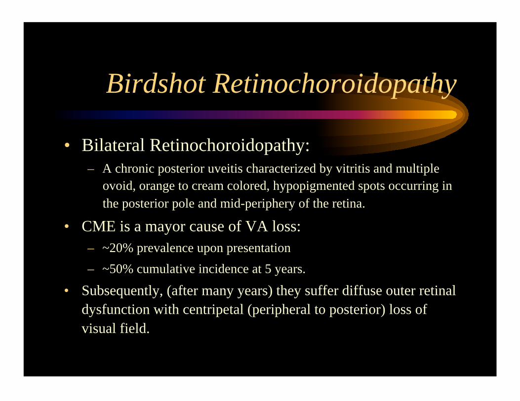
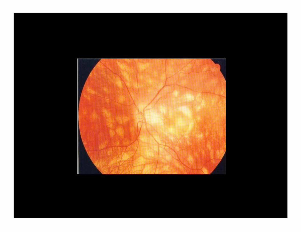
Birdshot Retinochoroidopathy
• Bilateral Retinochoroidopathy: – A chronic posterior uveitis characterized by vitritis and multiple
ovoid, orange to cream colored, hypopigmented spots occurring in
the posterior pole and mid-periphery of the retina.
• CME is a mayor cause of VA loss:
– ~20% prevalence upon presentation
– ~50% cumulative incidence at 5 years.
• Subsequently, (after many years) they suffer diffuse outer retinal
dysfunction with centripetal (peripheral to posterior) loss of
visual field.
Birdshot Retinochoroidopathy
• HLA-A29 gene has been reported to have a
relative risk of 50 - 200 for this disease!
Birdshot Retinochoroidopathy
• Treatment:
– Immunosupressive therapy reduces the risk of CME,
which is an important cause of visual acuity loss.
– Low dose oral corticosteroids (10mg or less), does not
seem to prevent the occurrence of CME.
– Immunosupressive therapy may reduce the risk
progressive diffuse retinal dysfuntion.
JE Thorne, DA Jabs, et al. AJO 2005;140:45-51.
Now,
two focal retinitis
that differ on the
clinical scenario...
Toxoplasma Retinitis
• Unilateral Focal localized retinitis with lots of
vitritis, “light at the end of the fog”, in an
ambulatory patient.
• Granulomatous A/C findings.
• Tx: – Pyrimethamine/Sulfadiazine
– Clindamycin
– Zithromax
– Bactrim (1-2) DS qid.
– Please do not forget Pred Forte and Atropine (nothing personal)!
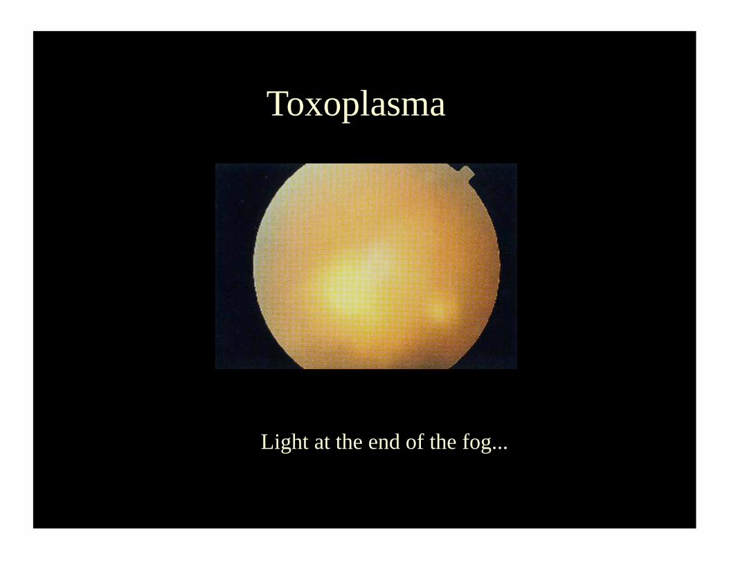
Toxoplasma
Light at the end of the fog...
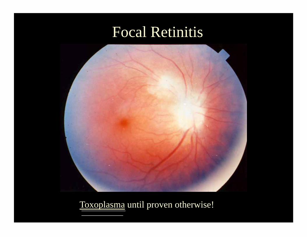
Focal Retinitis
Toxoplasma until proven otherwise!
Candida Retinochoroiditis
• In the OKAPS: Focal Retinochoroiditis.
– Unilateral Focal localized retinitis with some vitritis, in
a bedridden patient on TPN.
• In real life: Bilateral Large Multifocal
Choroiditis & Chorioretinitis.
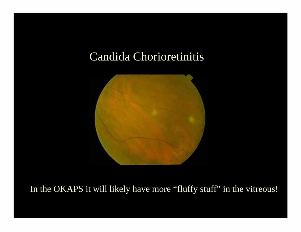
Candida Chorioretinitis
In the OKAPS it will likely have more “fluffy stuff” in the vitreous!
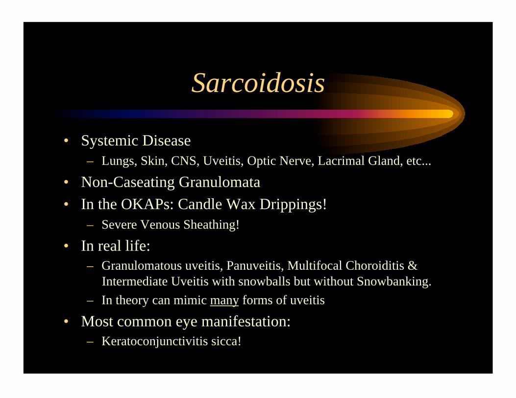
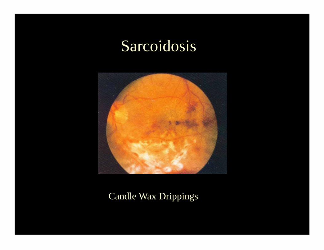
Sarcoidosis
• Systemic Disease
– Lungs, Skin, CNS, Uveitis, Optic Nerve, Lacrimal Gland, etc...
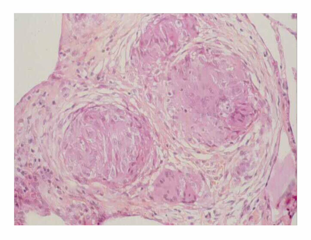
• Non-Caseating Granulomata
• In the OKAPs: Candle Wax Drippings!
– Severe Venous Sheathing!
• In real life:
– Granulomatous uveitis, Panuveitis, Multifocal Choroiditis &
Intermediate Uveitis with snowballs but without Snowbanking.
– In theory can mimic many forms of uveitis
• Most common eye manifestation:
– Keratoconjunctivitis sicca!
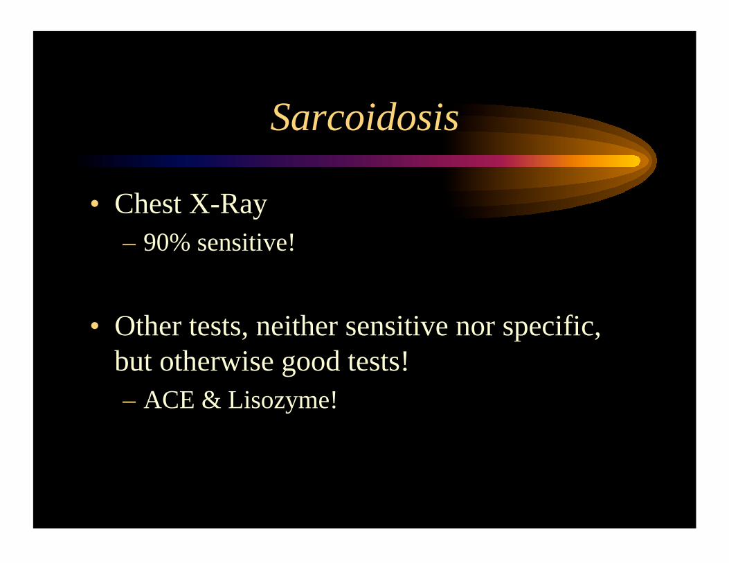
Sarcoidosis
• Chest X-Ray
– 90% sensitive!
• Other tests, neither sensitive nor specific,
but otherwise good tests!
– ACE & Lisozyme!
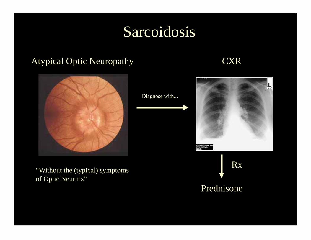
Sarcoidosis
Atypical Optic Neuropathy
“Without the (typical) symptoms
of Optic Neuritis”
Diagnose with...
CXR
Rx
Prednisone
Sarcoidosis
Candle Wax Drippings
Syphilis
• A treponema that can mimic any form of:
– Scleritis
– Uveitis:
– Retinitis
– Choroiditis: Think, all of the white dot Syndromes!
• Therefore all of the above must always get an
FTA-Abs (positive for life)!
• Nothing Personal!
Syphilis
• Most Uveitis:
– Secondary Stage
• Most Retinitis:
– Latent Secondary Stage
• Treatment needed:
– IV Penicillin: As in tertiary stage!
Syphilis
• Can also Cause:
– Interstitial Keratitis (congenital syphilis)
– Argyll-Robertson Pupil (tertiary syphilis)
• Accommodates but does not react!
– Optic Neuropathy
• Mostly Atypical
– CN III & CN VI palsies
– Visual Field Defects
• Gummae in the brain.
Syphilis
Your neighbor can have it!
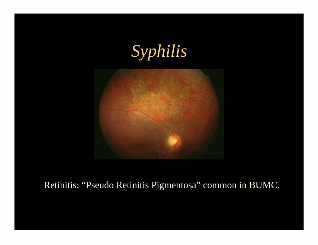
Syphilis
Retinitis: “Pseudo Retinitis Pigmentosa” common in BUMC.
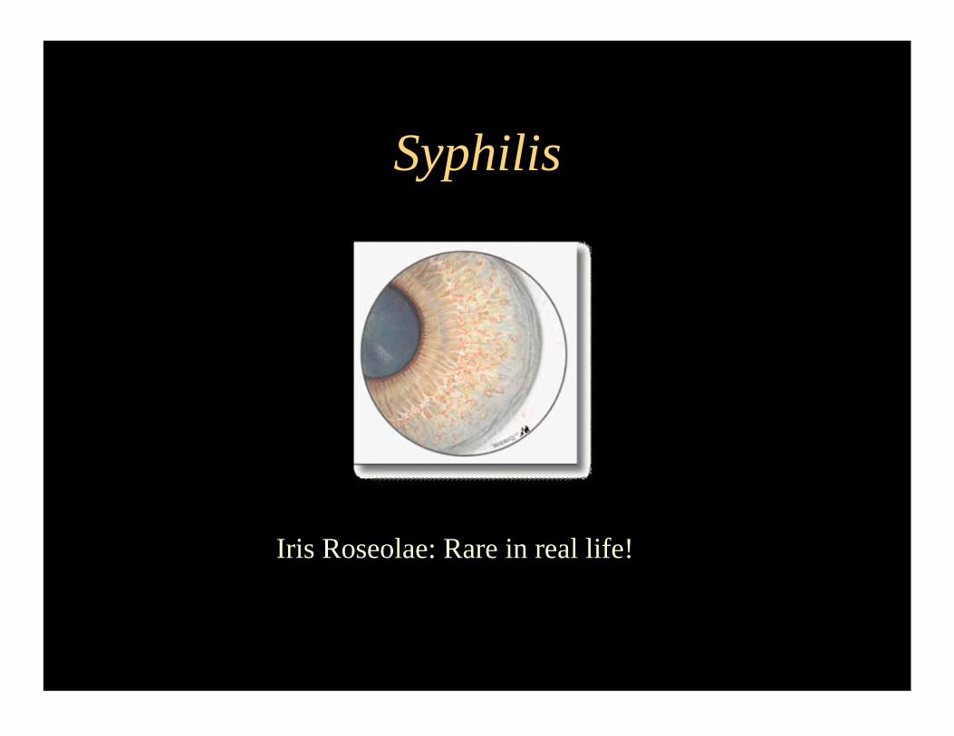
Syphilis
Iris Roseolae: Rare in real life!
Lyme Disease
• Close relative of Syphilis, a spirochette!
– Borrelia Burdoferi
• Your neighbor can also have it!
• Can cause: – Interstitial Keratitis, just like Syphilis!
• Non-Congenital, this is different!
– Granulomatous Uveitis:
• Just like Syphilis!
– Intermediate Uveitis:
• Guess what?, just like syphilis!
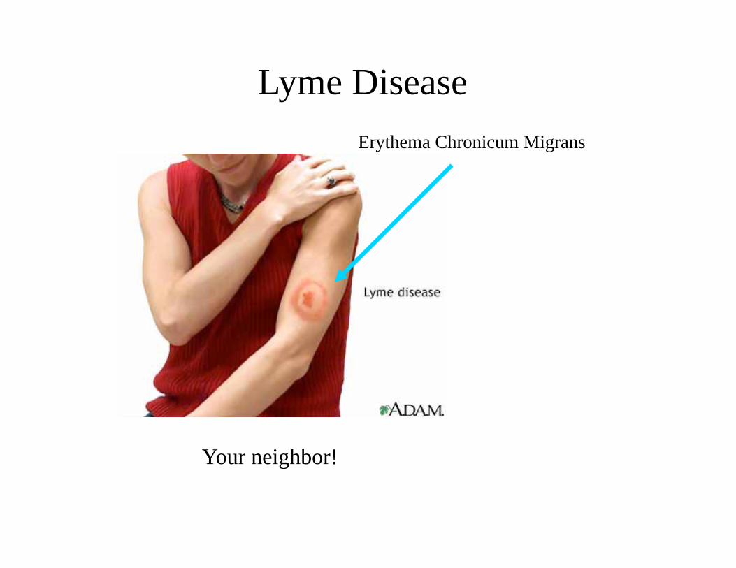
Lyme Disease
Your neighbor!
Erythema Chronicum Migrans
Tuberculosis
• Rare cause of Uveitis in the USA
• Overstated historically as a cause of uveitis! • Likely ocular manifestations:
– A Multifocal Choroiditis or Granulomatous Uveitis on someone
with Active Pulmonary Involvement.
– Focal Retinal Tuberculoma (possible & rare).
• Scleritis:
– Rare cause of scleritis, does not need lung involvement because it
occurs due to direct contact trough the eye surface, can be caused
by atypical mycobacteria.
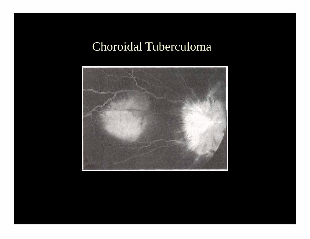
Choroidal Tuberculoma

A few more things...
Acute Retinal Necrosis
• HSV/VZV associated focal, rapidly
spreading retinitis that usually starts in the
retinal periphery and may turn bilateral.
• Tx: Acyclovir, Valacyclovir, Foscarnet.
• May become complicated with RRD
– OKAP Tx: PPV & Silicone Oil.
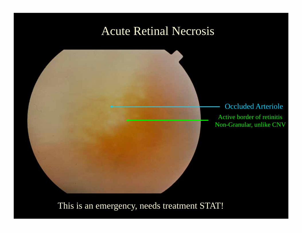
Acute Retinal Necrosis
Occluded Arteriole Active border of retinitis
Non-Granular, unlike CNV
This is an emergency, needs treatment STAT!
Toxocara Canis
• Answer to the OKAP question where you see a granuloma
possibly in the periphery connected to the optic disk by a
vitreous traction band.
• Etiology:
– Dog roundworm ingested by children who eat dirt (pica).
– Average age 7.5 years.
– Typically no eosinophilia, unlike Visceral Larva Migrans, which
is the systemic version of this.
– Tx: Ocular disease: Prednisone if active.
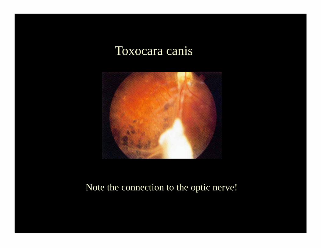
Toxocara canis
Note the connection to the optic nerve!
Briefly…
HIV eye disease...
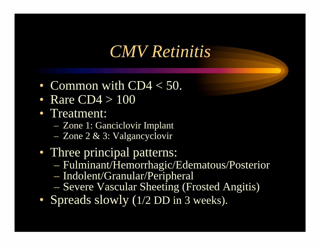
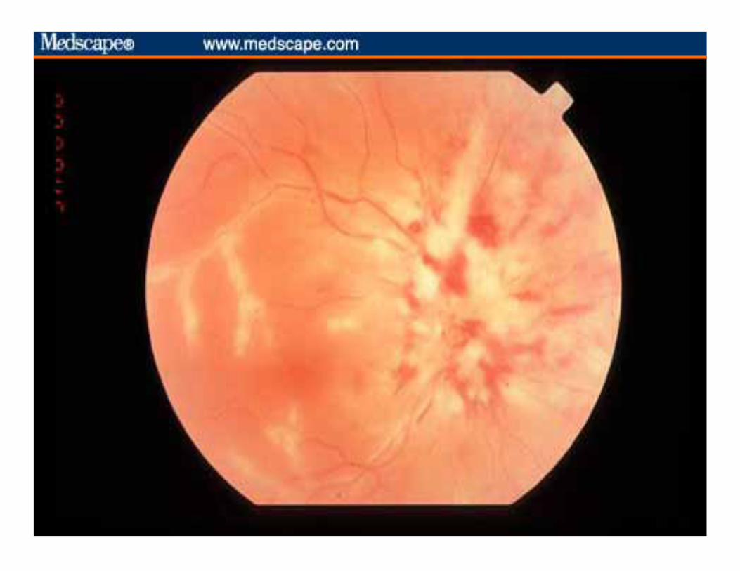
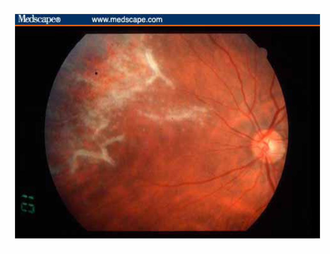
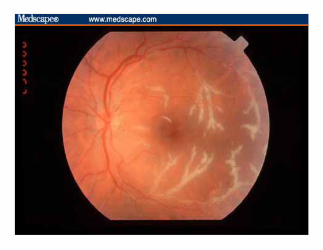
CMV Retinitis
• Common with CD4 < 50. • Rare CD4 > 100 • Treatment:
– Zone 1: Ganciclovir Implant – Zone 2 & 3: Valgancyclovir
• Three principal patterns: – Fulminant/Hemorrhagic/Edematous/Posterior – Indolent/Granular/Peripheral – Severe Vascular Sheeting (Frosted Angitis)
• Spreads slowly (1/2 DD in 3 weeks).
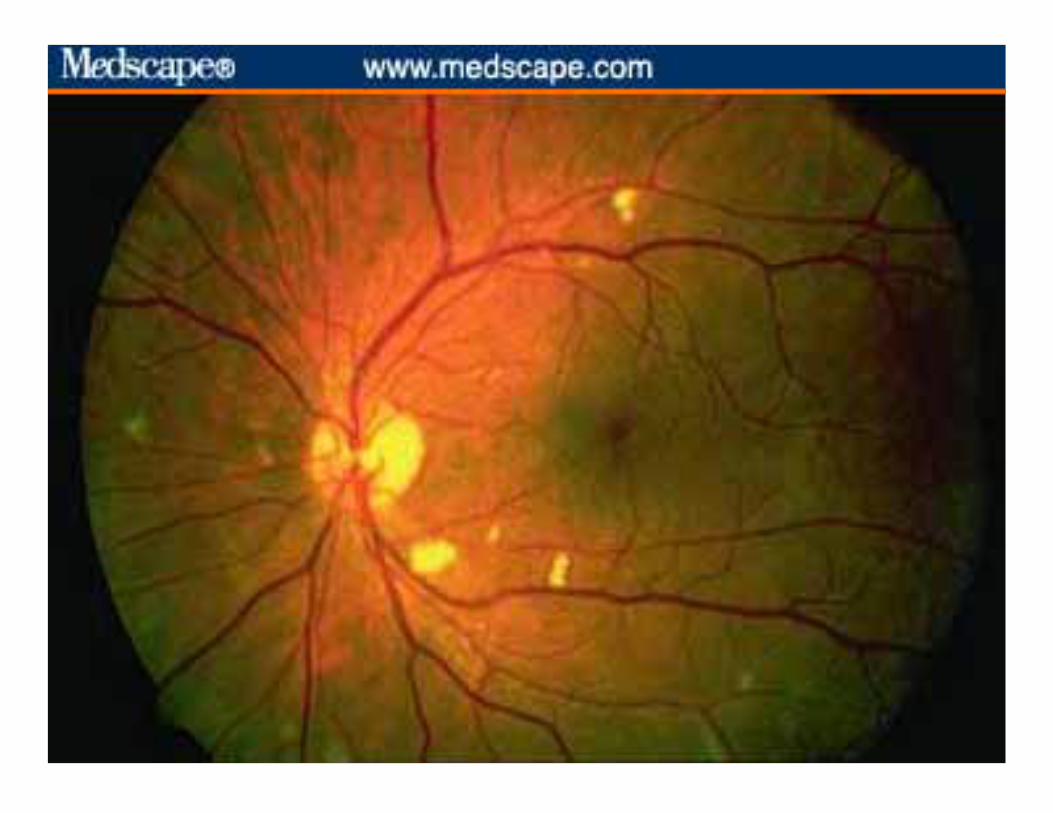
Background HIV Retinopathy
• Common on patients with low CD4 counts
(<50cells/mcL).
• If the diagnosis of CMVR is being considered, the
patient should be observed in 2-3 weeks as CMVR
may be expected to slowly progress in the absence
of treatment.
Progressive Outer Retinal Necrosis (PORN)
• AKA: Herpetic Necrotizing Retinitis
• Etiology: HSV & HZV
• The “ARN” of the immunosuppressed.
• It represents a clinical emergency!
Progressive Outer Retinal Necrosis (PORN)
• Treatment Alternatives:
– Foscarnet I.V.
– Valtrex 1gm po tid.
– Intravitreal Foscarnet.
• This is a very rapidly progressing retinitis!
• 67% of eyes are completely blind in 1 month!
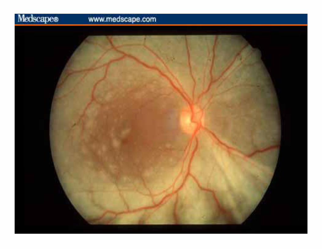
Infectious Choroiditis of Systemic Origin
• They can all look the same!
– Multifocal Choroiditis!
• As a general rule whatever the patient has at the
moment of the active choroiditis phase is the
diagnosis!
– I.e. Candida, Criptococcus, Tb, PCP, etc…
– No need to order CH50, Rheumatoid Factor, etc…
– You get the idea!
Anecdote
• It was my first weekend of call as the
Uveitis Fellow!
• The second year resident calls me!
• Hey doc, there is a patient with CMVR!
• Guess what?
Copyright restrictions may apply.
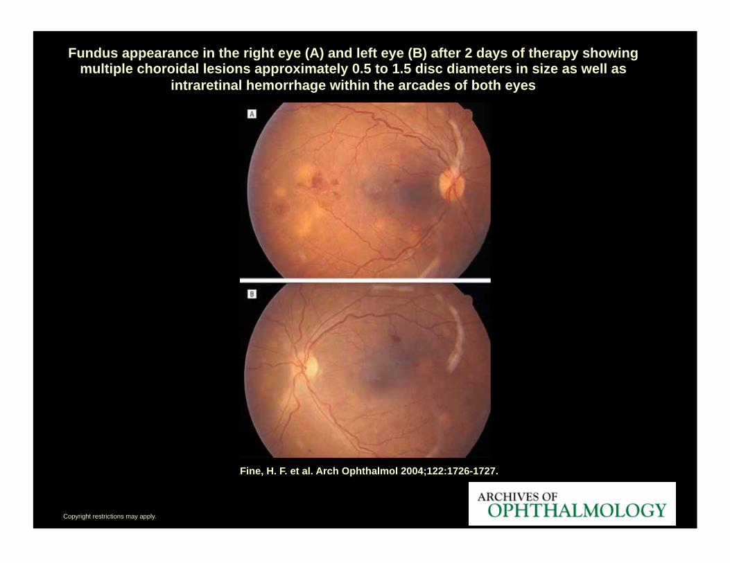
Fine, H. F. et al. Arch Ophthalmol 2004;122:1726-1727.
Fundus appearance in the right eye (A) and left eye (B) after 2 days of therapy showing multiple choroidal lesions approximately 0.5 to 1.5 disc diameters in size as well as
intraretinal hemorrhage within the arcades of both eyes
Copyright restrictions may apply.
Fine, H. F. et al. Arch Ophthalmol 2004;122:1726-1727.
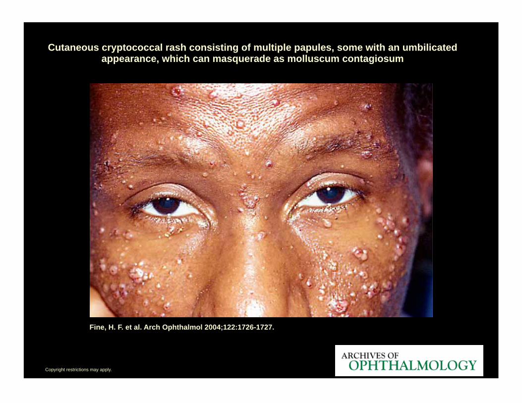
Cutaneous cryptococcal rash consisting of multiple papules, some with an umbilicated appearance, which can masquerade as molluscum contagiosum
Copyright restrictions may apply.
Fine, H. F. et al. Arch Ophthalmol 2004;122:1726-1727.
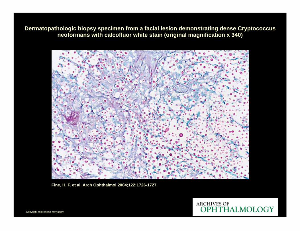
Dermatopathologic biopsy specimen from a facial lesion demonstrating dense Cryptococcus neoformans with calcofluor white stain (original magnification x 340)
Copyright restrictions may apply.
Fine, H. F. et al. Arch Ophthalmol 2004;122:1726-1727.
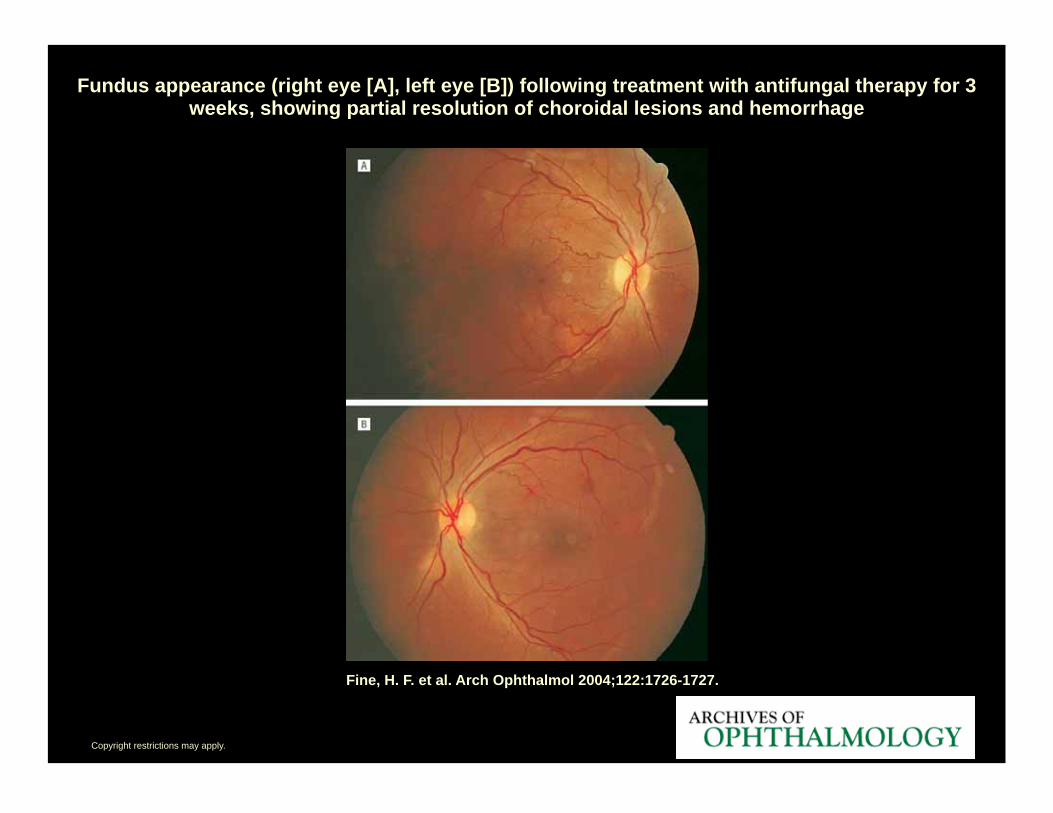
Fundus appearance (right eye [A], left eye [B]) following treatment with antifungal therapy for 3 weeks, showing partial resolution of choroidal lesions and hemorrhage
Scleritis
Scleritis
• 50 % Identified Etiology
– 10 % Infectious
• Zoster (most common)
• HSV, Lyme, Syphilis
– 40% Systemic Disease
• Rheumatoid Arthritis (most common)
• Vasculitis: Wegener’s & PAN
• IBD, Lupus & Relapsing Polychondritis
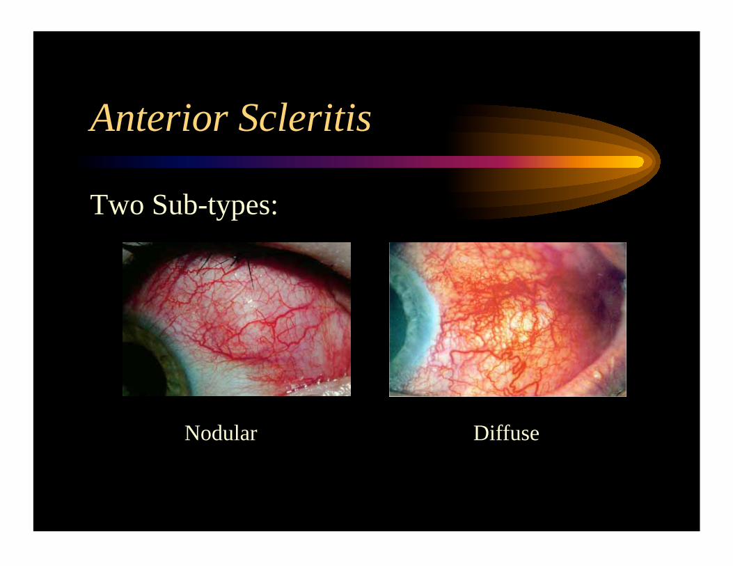
Anterior Scleritis
Two Sub-types:
Nodular Diffuse
Necrotizing Scleritis
• In a red eye,
– AKA (necrotizing with inflammation):
– Poor life prognosis: • A Systemic Vasculitis is likely.
• Sleromalacia Perforans:
– White eye necrotizing scleritis
– Rheumatoid Arthritis Associated
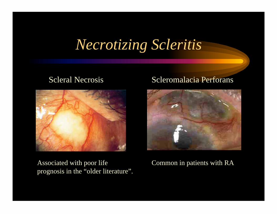
Necrotizing Scleritis
Scleral Necrosis Scleromalacia Perforans
Associated with poor life
prognosis in the “older literature”. Common in patients with RA
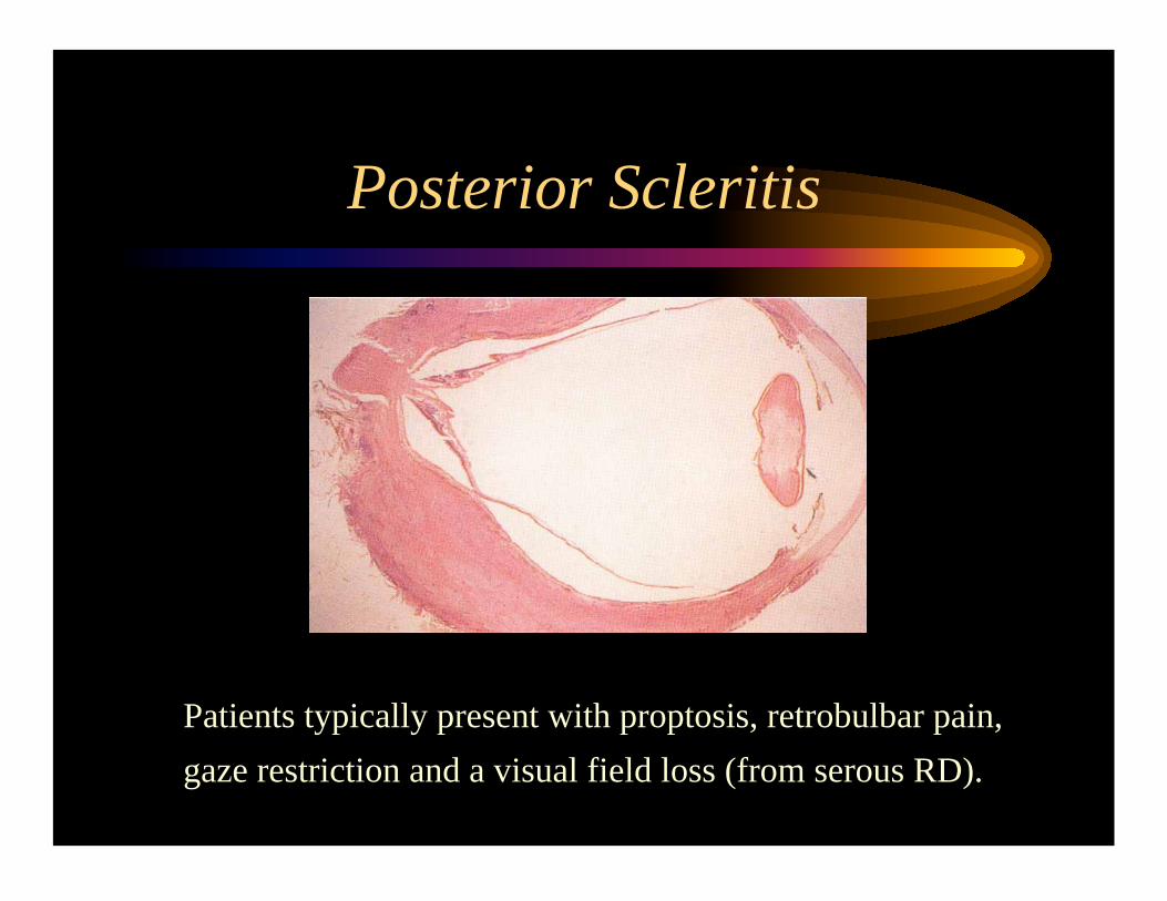
Posterior Scleritis
Patients typically present with proptosis, retrobulbar pain,
gaze restriction and a visual field loss (from serous RD).
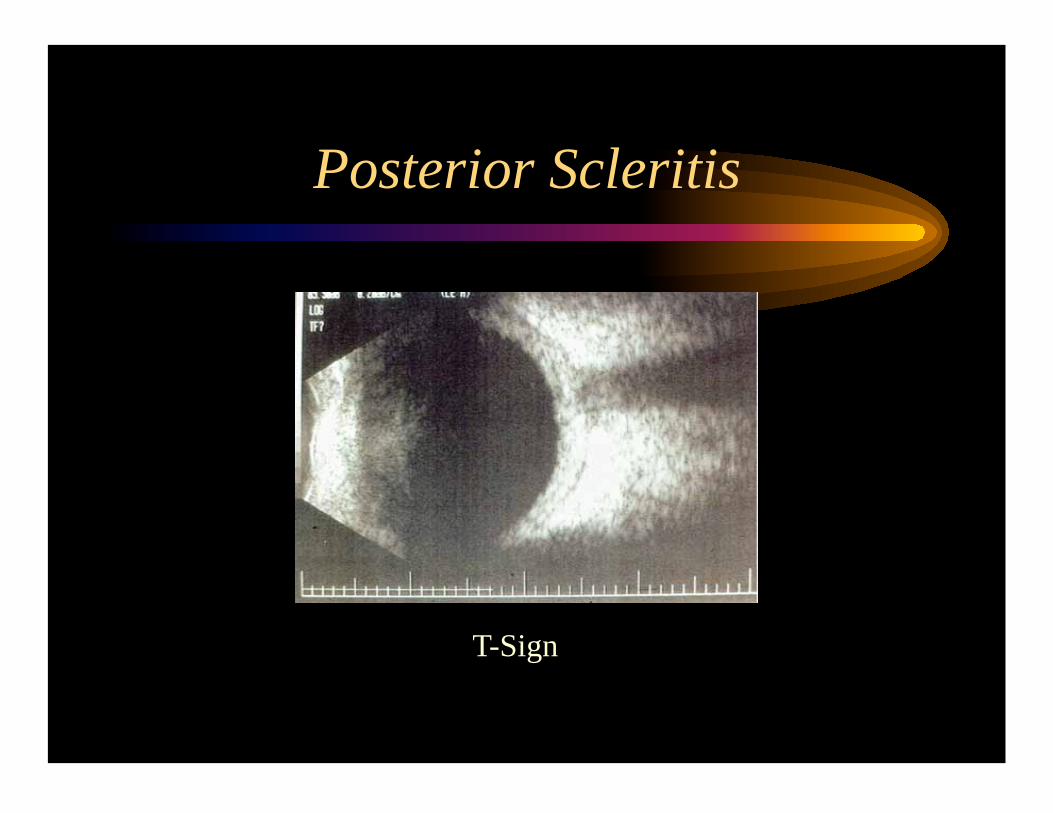
Posterior Scleritis
T-Sign
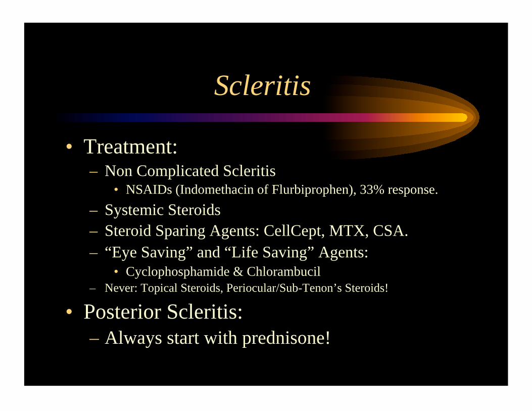
Scleritis
• Treatment: – Non Complicated Scleritis
• NSAIDs (Indomethacin of Flurbiprophen), 33% response.
– Systemic Steroids
– Steroid Sparing Agents: CellCept, MTX, CSA.
– “Eye Saving” and “Life Saving” Agents:
• Cyclophosphamide & Chlorambucil
– Never: Topical Steroids, Periocular/Sub-Tenon’s Steroids!
• Posterior Scleritis:
– Always start with prednisone!

The last one….
I promise!...
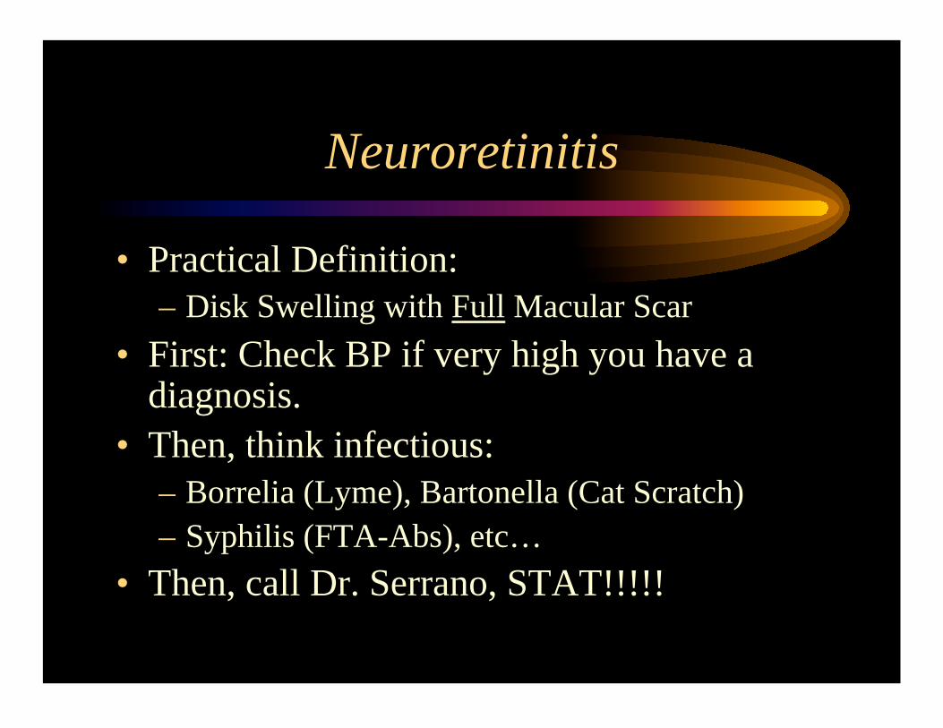
Neuroretinitis
• Practical Definition:
– Disk Swelling with Full Macular Scar
• First: Check BP if very high you have a diagnosis.
• Then, think infectious:
– Borrelia (Lyme), Bartonella (Cat Scratch)
– Syphilis (FTA-Abs), etc…
• Then, call Dr. Serrano, STAT!!!!!
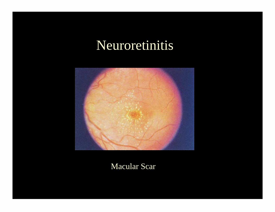
Macular Scar
Neuroretinitis