UVEITIS INVOLVING THE POSTERIOR SEGMENT - LWW …journals.lww.com/retinajournal/Documents/July...
12
Jointly provided by New York Eye and Ear Infirmary of Mount Sinai and MedEdicus LLC This continuing medical education activity is supported through an unrestricted educational grant from Santen Pharmaceutical Co, Ltd. Original Release: July 1, 2015 Last Review: June 9, 2015 Expiration: July 31, 2016 Distributed with NONINFECTIOUS UVEITIS INVOLVING THE POSTERIOR SEGMENT New Frontiers in the Management of CME MONOGRAPH Visit http://tinyurl.com/noninfectiousuveitis for online testing and instant CME certificate. PROgRaM ChaIR aNd MOdERatOR Carlos Pavesio, MD FaCuLtY Bahram Bodaghi, MD, PhD Marc D. de Smet, MD, PhD John Kempen, MD, PhD Sunil K. Srivastava, MD Manfred Zierhut, MD highlights from a roundtable discussion
Transcript of UVEITIS INVOLVING THE POSTERIOR SEGMENT - LWW …journals.lww.com/retinajournal/Documents/July...

Jointly provided by New York Eye and Ear Infirmary of Mount Sinai and MedEdicus LLC
This continuing medical education activity is supported through an unrestricted educational grant from Santen Pharmaceutical Co, Ltd.
Original Release: July 1, 2015 Last Review: June 9, 2015 Expiration: July 31, 2016
Distributed with
NONINFECTIOUS UVEITIS INVOLVING THE POSTERIOR SEGMENT
New Frontiers in theManagement of
CME MONOGRAPH Visit http://tinyurl.com/noninfectiousuveitis for online testing and instant CME certificate.
PROgRaM ChaIR aNd MOdERatORCarlos Pavesio, MD
FaCuLtYBahram Bodaghi, MD, PhD
Marc D. de Smet, MD, PhDJohn Kempen, MD, PhD
Sunil K. Srivastava, MDManfred Zierhut, MD
highlights from a roundtable discussion
2
PROgRaM ChaIR aNd MOdERatORCarlos Pavesio, MDConsultant OphthalmologistMedical Retina Service DirectorMoorfields Eye HospitalLondon, United Kingdom
FaCuLtY Bahram Bodaghi, MD, PhD DHU Vision et HandicapsUniversité Pierre et Marie CurieHôpital Pitié-SalpêtrièreParis, France
Marc D. de Smet, MD, PhD MicroInvasive Ocular Surgery (MIOS) Center Lausanne, Switzerland
John Kempen, MD, PhDChief, Ocular Inflammation ServiceDirector, Ophthalmic EpidemiologyProfessor of Ophthalmology Professor of Epidemiology in Biostatistics and EpidemiologyUniversity of Pennsylvania Health SystemThe Scheie Eye Institute Penn Presbyterian Medical Center Philadelphia, Pennsylvania
Sunil K. Srivastava, MDStaff PhysicianCole Eye InstituteCleveland ClinicCleveland, Ohio
Manfred Zierhut, MDAssociate Professor of OphthalmologyUniversity Eye Clinic Tübingen Tübingen, Germany
CME REVIEwER FORNEw YORk EYE aNd EaR INFIRMaRY OF MOuNt SINaIJohn A. Sorenson, MDSurgical StaffNew York Eye and Ear Infirmary of Mount Sinai Clinical Assistant ProfessorDepartment of Ophthalmology New York University School of MedicineNew York, New York
Learning Method and MediumThis educational activity consists of a supplement and ten (10) study questions. The participant should, in order, read the learning objectives contained at the beginning of this supplement, read the supplement, answer all questions in the post test, and complete the Activity Evaluation/Credit Request form. To receive credit for this activity, please follow the instructions provided on the post test and Activity Evaluation/Credit Request form. This educational activity should take a maximum of 1.5 hours to complete.
Content SourceThis continuing medical education (CME) activity captures content from a roundtable discussion held on January 23, 2015, in Oxford, England.
activity descriptionNoninfectious posterior uveitis is a worldwide challenge for clinicians. Suboptimal treatment can cause irreversible visual impairment, which is particularly concerning because uveitis often strikes people of working age. Local and systemic corticosteroid therapy is the current mainstay of treatment. Significant developments for improving uveitis management are under way that hope to address the drawbacks of current therapy side effects, improve tolerance to therapy, and improve patient outcomes. The purpose of this activity is to provide clinicians with practical insights on current therapy and new data on emerging treatments that can be applied to daily practice.
target audienceThis activity intends to educate US and European retina specialists and other ophthalmologists caring for patients with noninfectious uveitis.
Learning ObjectivesUpon completion of this activity, participants will be better able to:• Review elements of the diagnostic evaluation to differentiate noninfectious from infectious uveitis • Describe the guidelines pertaining to the treatment of noninfectious uveitis• Review the indications for local and systemic therapies in the treatment of noninfectious uveitis • Describe the mechanisms and clinical trial data for emerging local therapy for noninfectious uveitis
accreditation StatementThis activity has been planned and implemented in accordance with the accreditation requirements and policies of the Accreditation Council for Continuing Medical Education (ACCME) through the joint providership of New York Eye and Ear Infirmary of Mount Sinai and MedEdicus LLC. The New York Eye and Ear Infirmary of Mount Sinai is accredited by the ACCME to provide continuing medical education for physicians.
aMa Credit designation StatementThe New York Eye and Ear Infirmary of Mount Sinai designates this enduring material for a maximum of 1.5 AMA PRA Category 1 Credits™. Physicians should claim only the credit commensurate with the extent of their participation in the activity.2
In July 2013, the Accreditation Council for Continuing Medical Education (ACCME) awarded New York Eye and Ear Infirmary of Mount Sinai “Accreditation with Commendation,” for six years as a provider of continuing medical education for physicians, the highest accreditation status awarded by the ACCME.
3
grantor StatementThis continuing medical education activity is supported through an unrestricted educational grant from Santen Pharmaceutical Co, Ltd.
disclosure Policy StatementIt is the policy of New York Eye and Ear Infirmary of Mount Sinai that the faculty and anyone in a position to control activity content disclose any real or apparent conflicts of interest relating to the topics of this educational activity, and also disclose discussions of unlabeled/unapproved uses of drugs or devices during their presentation(s). New York Eye and Ear Infirmary of Mount Sinai has established policies in place that will identify and resolve all conflicts of interest prior to this educational activity. Full disclosure of faculty/planners and their commercial relationships, if any, follows.
disclosuresBahram Bodaghi, MD, PhD, had a financial agreement or affiliation during the past year with the following commercial interests in the form of Consultant/Advisory Board: Allergan, Inc; Santen Pharmaceutical Co, Ltd; and XOMA Corporation; Honoraria from promotional, advertising or non-CME services received directly from commercial interests or their Agents (eg, Speakers Bureaus): Allergan, Inc.
Marc D. de Smet, MD, PhD, had a financial agreement or affiliation during the past year with the following commercial interests in the form of Consultant/Advisory Board: Allergan, Inc; Santen Pharmaceutical Co, Ltd; and ThromboGenics NV; Contracted Research: Bayer, Charles River Laboratories; and ThromboGenics NV; Patent Holder: ThromboGenics NV.
John Kempen MD, PhD, had a financial agreement or affiliation during the past year with the following commercial interests in the form of Consultant/Advisory Board: AbbVie Inc; Roche; Vitae Pharmaceuticals, Inc; and XOMA Corporation; Contracted Research: Eyegate Pharmaceuticals, Inc; and XOMA Corporation.
Carlos Pavesio, MD, had a financial agreement or affiliation during the past year with the following commercial interests in the form of Consultant/Advisory Board: Alcon, Inc; Servier; and XOMA Corporation.
Sunil K. Srivastava, MD, had a financial agreement or affiliation during the past year with the following commercial interests in the form of Consultant/Advisory Board: Santen Pharmaceutical Co, Ltd; Contracted Research: Allergan, Inc.
Manfred Zierhut, MD, has no relevant commercial relationships to disclose.
New York Eye and Ear Infirmary of Mount Sinai Peer Review disclosureJohn Sorenson, MD, has no relevant commercial relationships to disclose.
Editorial Support disclosuresCheryl Guttman Krader; Cynthia Tornallyay, RD, MBA, CCMEP; Diane McArdle, PhD; Kimberly Corbin, CCMEP; Barbara Aubel; and Barbara Lyon have no relevant commercial relationships to disclose.
disclosure attestationThe contributing physicians listed above have attested to thefollowing:1) that the relationships/affiliations noted will not bias or otherwise influence their involvement in this activity;
2) that practice recommendations given relevant to the companies with whom they have relationships/affiliations will be supported by the best available evidence or, absent evidence, will be consistent with generally accepted medical practice; and3) that all reasonable clinical alternatives will be discussed when making practice recommendations.
OFF-LaBEL dISCuSSIONThis CME activity includes discussion of unlabeled and/or investigative uses of drugs. Please refer to the official prescribing information for each drug discussed in this activity for FDA/EMA-approved dosing, indications, and warnings.
For Digital EditionsSystem Requirements:If you are viewing this activity online, please ensure the computer you are using meets the following requirements:• Operating System: Windows or Macintosh• Media Viewing Requirements: Flash Player or Adobe Reader• Supported Browsers: Microsoft Internet Explorer, Firefox, Google Chrome, Safari, and Opera• A good Internet connection
New York Eye and Ear Infirmary of Mount Sinai Privacy & Confidentiality PoliciesCME policies: http://www.nyee.edu/health-professionals/cme/enduring-activities Hospital policies: http://icahn.mssm.edu/about-us/privacy-policy CME Provider Contact InformationFor questions about this activity, call 212-979-4383.
to Obtain aMA PRA Category 1 Credit™To obtain AMA PRA Category 1 Credit™ for this activity, read the material in its entirety and consult referenced sources as necessary. Complete the evaluation form along with the post test answer box within this supplement. Remove the Activity Evaluation/Credit Request page from the printed supplement or print the Activity Evaluation/Credit Request page from the Digital Edition. Return via mail to Kim Corbin, Director, ICME, New York Eye and Ear Infirmary of Mount Sinai, 310 East 14th Street, New York, NY 10003 or fax to (212) 353-5703. Your certificate will be mailed to the address you provide on the Activity Evaluation/Credit Request form. Please allow 3 weeks for Activity Evaluation/Credit Request forms to be processed. There are no fees for participating in and receiving CME credit for this activity.
Alternatively, we offer instant certificate processing and support Green CME. Please take this post test and evaluation online by going to http://tinyurl.com/noninfectiousuveitis. Upon passing, you will receive your certificate immediately. You must score 70% or higher to receive credit for this activity, and may take the test up to 2 times. Upon registering and successfully completing the post test, your certificate will be made available online and you can print it or file it.
disclaimerThe views and opinions expressed in this educational activity are those of the faculty and do not necessarily represent the views of New York Eye and Ear Infirmary of Mount Sinai; MedEdicus LLC; Santen Pharmaceutical Co, Ltd; or RETINA.
This CME activity is copyrighted to MedEdicus LLC ©2015. All rights reserved.
4
IntroductionUveitis represents a large and diverse group of intraocular inflammatory diseases involving the uveal tract. Although it is an uncommon diagnosis, its incidence and prevalence may be higher than previously thought.1 Furthermore, uveitis is an important public health issue because of its potential to cause loss of vision; and, its effect on vision and quality of life also may be greater than often realized. Approximately one-third of patients with uveitis may develop vision impairment during the clinical course of disease,2 approximately 20% may meet criteria for legal blindness,3 and uveitis has been reported to account for up to 10% of cases of blindness in the United States and for 3% to 7% of cases in Europe.4
Permanent vision loss from uveitis is potentially preventable with effective control of the inflammation.5 The approach to management, however, varies depending on the specific condition. Therefore, accurate diagnosis is the first step for determining appropriate care.
General ophthalmologists often refer patients with uveitis involving the posterior segment to a retina specialist. This review aims to assist retina specialists with their clinical decision-making for such cases. It provides guidance on the initial diagnostic evaluation, offers suggestions on when to refer, and discusses treatment with a focus on the use of local corticosteroids for noninfectious uveitis involving the posterior segment. In addition, it reviews some emerging local therapies that may become part of the therapeutic armamentarium used by retina specialists in the future.
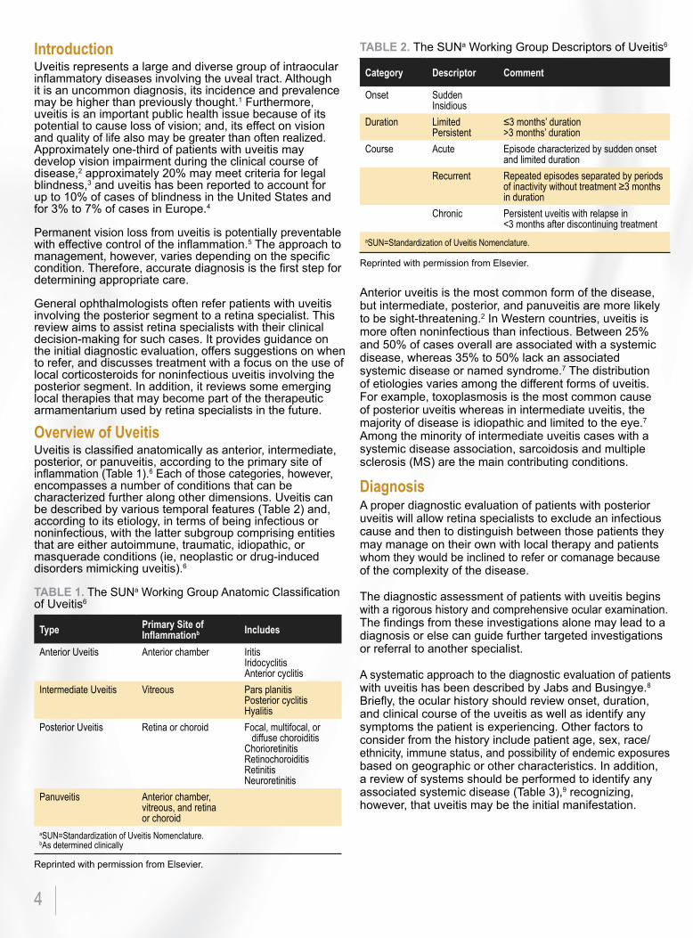
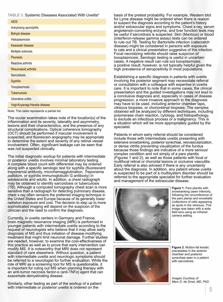
Overview of uveitisUveitis is classified anatomically as anterior, intermediate, posterior, or panuveitis, according to the primary site of inflammation (Table 1).6 Each of those categories, however, encompasses a number of conditions that can be characterized further along other dimensions. Uveitis can be described by various temporal features (Table 2) and, according to its etiology, in terms of being infectious or noninfectious, with the latter subgroup comprising entities that are either autoimmune, traumatic, idiopathic, or masquerade conditions (ie, neoplastic or drug-induced disorders mimicking uveitis).6
TABLE 1. The SUNa Working Group Anatomic Classification of Uveitis6
TABLE 2. The SUNa Working Group Descriptors of Uveitis6
Anterior uveitis is the most common form of the disease, but intermediate, posterior, and panuveitis are more likely to be sight-threatening.2 In Western countries, uveitis is more often noninfectious than infectious. Between 25% and 50% of cases overall are associated with a systemic disease, whereas 35% to 50% lack an associated systemic disease or named syndrome.7 The distribution of etiologies varies among the different forms of uveitis. For example, toxoplasmosis is the most common cause of posterior uveitis whereas in intermediate uveitis, the majority of disease is idiopathic and limited to the eye.7 Among the minority of intermediate uveitis cases with a systemic disease association, sarcoidosis and multiple sclerosis (MS) are the main contributing conditions.
diagnosis A proper diagnostic evaluation of patients with posterior uveitis will allow retina specialists to exclude an infectious cause and then to distinguish between those patients they may manage on their own with local therapy and patients whom they would be inclined to refer or comanage because of the complexity of the disease.
The diagnostic assessment of patients with uveitis begins with a rigorous history and comprehensive ocular examination. The findings from these investigations alone may lead to a diagnosis or else can guide further targeted investigations or referral to another specialist.
A systematic approach to the diagnostic evaluation of patients with uveitis has been described by Jabs and Busingye.8 Briefly, the ocular history should review onset, duration, and clinical course of the uveitis as well as identify any symptoms the patient is experiencing. Other factors to consider from the history include patient age, sex, race/ethnicity, immune status, and possibility of endemic exposures based on geographic or other characteristics. In addition, a review of systems should be performed to identify any associated systemic disease (Table 3),9 recognizing, however, that uveitis may be the initial manifestation.
type Primary Site of Inflammationb Includes
Anterior Uveitis Anterior chamber IritisIridocyclitisAnterior cyclitis
Intermediate Uveitis Vitreous Pars planitisPosterior cyclitisHyalitis
Posterior Uveitis Retina or choroid Focal, multifocal, or diffuse choroiditisChorioretinitisRetinochoroiditisRetinitisNeuroretinitis
Panuveitis Anterior chamber, vitreous, and retina or choroid
aSUN=Standardization of Uveitis Nomenclature.bAs determined clinically
Reprinted with permission from Elsevier.
Reprinted with permission from Elsevier.
Category descriptor Comment
Onset SuddenInsidious
Duration LimitedPersistent
≤3 months’ duration>3 months’ duration
Course Acute Episode characterized by sudden onset and limited duration
Recurrent Repeated episodes separated by periods of inactivity without treatment ≥3 months in duration
Chronic Persistent uveitis with relapse in <3 months after discontinuing treatment
aSUN=Standardization of Uveitis Nomenclature.
5
basis of the pretest probability. For example, Western blot for Lyme disease might be ordered when there is reason to suspect the diagnosis according to the patient’s history and/or extraocular signs and symptoms. Chest x-ray, serum angiotensin-converting enzyme, and liver function tests may be useful if sarcoidosis is suspected. Skin (Mantoux) or blood (interferon-release gamma assay) tests can be ordered to rule out TB. Testing for Bartonella infection (catscratch disease) might be considered in persons with exposure to cats and a clinical presentation suggestive of this infection. Focal necrotizing retinitis should raise suspicion of toxoplasmosis. Serologic testing is useful in uncertain cases. A negative result can rule out toxoplasmosis; a positive result, however, is not typically helpful given the high prevalence of seropositivity in most populations.
Establishing a specific diagnosis in patients with uveitis involving the posterior segment may necessitate referral or consultation with a colleague with expertise in uveitis care. It is important to note that in some cases, the clinical presentation and the guided investigations may not lead to a conclusive diagnosis and, especially in cases of disease progression, a more invasive approach to the diagnosis may have to be used, including anterior chamber taps, vitreous biopsies, or chorioretinal biopsies. The samples obtained will be analyzed by different techniques, including polymerase chain reaction, cytology, and histopathology, to exclude an infectious process or a malignancy. This is a situation which will be more appropriately guided by the specialist.
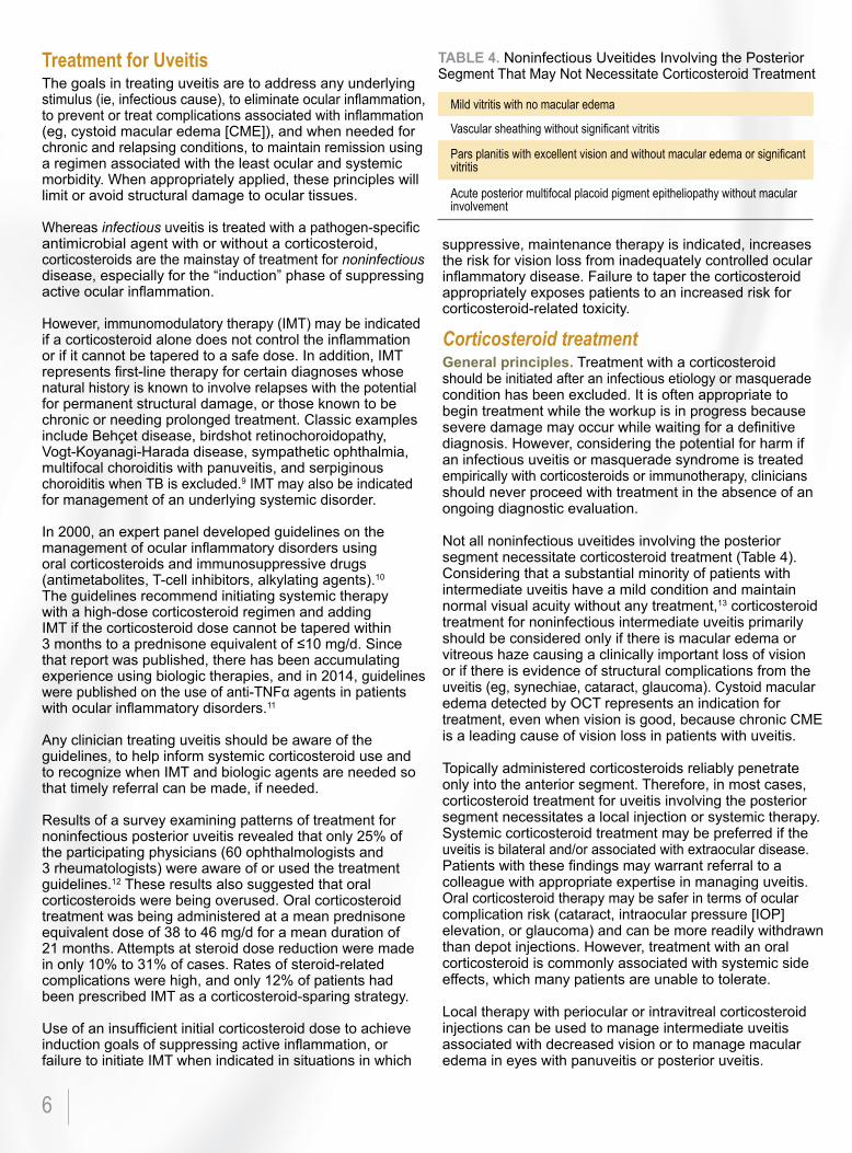
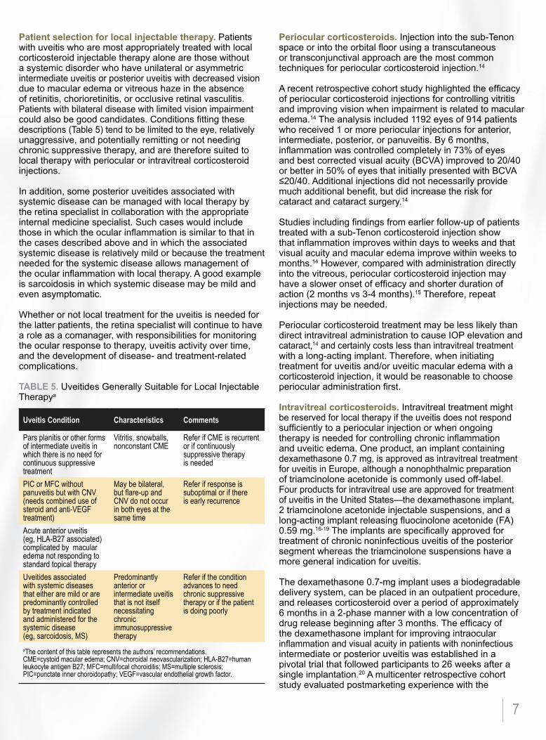
Patients in whom early referral should be considered include those with intermediate uveitis presenting with extensive snowbanking, posterior synechiae, neovascularization, or dense vitritis preventing visualization of the fundus because those findings are indicative of a more serious/complex condition and not simply of a pars planitis (Figures 1 and 2), as well as those patients with focal or multifocal retinal or choroidal lesions or occlusive vasculitis. Early referral is also advised if there is any uncertainty about the diagnosis. In addition, any patient whose uveitis is suspected to be part of a multisystem disorder should be referred to the appropriate specialist for further evaluation and management of the extraocular disease.
TABLE 3. Systemic Diseases Associated With Uveitis9
The ocular examination takes note of the location(s) of the inflammation and its severity, laterality and asymmetry, lesions and their characteristics, and the presence of any structural complications. Optical coherence tomography (OCT) should be performed if macular involvement is suspected, and fluorescein angiography may be appropriate to determine the extent and severity of any retinal vessel involvement. Often, significant leakage can be seen that was not suspected clinically.
The initial diagnostic workup for patients with intermediate or posterior uveitis involves minimal laboratory testing. A complete blood count with differential may be ordered along with a specific serologic test for syphilis (fluorescent treponemal antibody, microhemagglutination, Treponema pallidum, or syphilis immunoglobulin G antibody) in adolescents and adults. In addition, a chest radiograph may be obtained to identify sarcoidosis or tuberculosis (TB). Although a computed tomography chest scan is more sensitive than a radiograph for detecting pulmonary disease, chest x-ray often remains the preferred imaging technique in the United States and Europe because of its generally lower radiation exposure and cost. The decision to step up to more sophisticated imaging will depend on the suspicion of the clinician and the need to confirm the diagnosis.
Currently, in uveitis centers in Germany and France, brain magnetic resonance imaging (MRI) is performed in younger patients with intermediate uveitis according to the request of neurologists who believe that it may allow early diagnosis of MS and thus initiation of disease-modifying treatment that might limit neuronal damage. Further studies are needed, however, to examine the cost-effectiveness of this practice as well as to prove that early intervention can modify MS. It is noteworthy that MRI does not represent standard of care elsewhere. Without question, a patient with intermediate uveitis and neurologic symptoms should be referred to a neurologist for further evaluation. While the role of MRI as a screening tool for MS is questionable, it is important for ruling out MS when planning therapy with an anti-tumor necrosis factor-α (anti-TNFα) agent that can exacerbate demyelinating disease.
Similarly, other testing as part of the workup of a patient with intermediate or posterior uveitis is ordered on the
Note: This table represents a partial list.
AIDSAnkylosing spondylitisBehçet diseaseHistoplasmosisKawasaki diseaseMultiple sclerosisPsoriasisReactive arthritisRheumatoid arthritisSarcoidosisSyphilisToxoplasmosisTuberculosisUlcerative colitisVogt-Koyanagi-Harada disease
Figure 1. Pars planitis with snowbanking (seen inferiorly, following the circumference of the pars plana) and snowballs (collections of cells appearing as spots in the vitreous). This image was taken with a wide field lens using an infrared camera setting.
Figure 2. Mutton-fat keratic precipitates in the anterior chamber and posterior synechiae seen in a patient with sarcoidosis.
Images Courtesy of Marc D. de Smet, MD, PhD
6
treatment for uveitisThe goals in treating uveitis are to address any underlying stimulus (ie, infectious cause), to eliminate ocular inflammation, to prevent or treat complications associated with inflammation (eg, cystoid macular edema [CME]), and when needed for chronic and relapsing conditions, to maintain remission using a regimen associated with the least ocular and systemic morbidity. When appropriately applied, these principles will limit or avoid structural damage to ocular tissues.
Whereas infectious uveitis is treated with a pathogen-specific antimicrobial agent with or without a corticosteroid, corticosteroids are the mainstay of treatment for noninfectious disease, especially for the “induction” phase of suppressing active ocular inflammation.
However, immunomodulatory therapy (IMT) may be indicated if a corticosteroid alone does not control the inflammation or if it cannot be tapered to a safe dose. In addition, IMT represents first-line therapy for certain diagnoses whose natural history is known to involve relapses with the potential for permanent structural damage, or those known to be chronic or needing prolonged treatment. Classic examples include Behçet disease, birdshot retinochoroidopathy, Vogt-Koyanagi-Harada disease, sympathetic ophthalmia, multifocal choroiditis with panuveitis, and serpiginous choroiditis when TB is excluded.9 IMT may also be indicated for management of an underlying systemic disorder.
In 2000, an expert panel developed guidelines on the management of ocular inflammatory disorders using oral corticosteroids and immunosuppressive drugs (antimetabolites, T-cell inhibitors, alkylating agents).10 The guidelines recommend initiating systemic therapy with a high-dose corticosteroid regimen and adding IMT if the corticosteroid dose cannot be tapered within 3 months to a prednisone equivalent of ≤10 mg/d. Since that report was published, there has been accumulating experience using biologic therapies, and in 2014, guidelines were published on the use of anti-TNFα agents in patients with ocular inflammatory disorders.11
Any clinician treating uveitis should be aware of the guidelines, to help inform systemic corticosteroid use and to recognize when IMT and biologic agents are needed so that timely referral can be made, if needed.
Results of a survey examining patterns of treatment for noninfectious posterior uveitis revealed that only 25% of the participating physicians (60 ophthalmologists and 3 rheumatologists) were aware of or used the treatment guidelines.12 These results also suggested that oral corticosteroids were being overused. Oral corticosteroid treatment was being administered at a mean prednisone equivalent dose of 38 to 46 mg/d for a mean duration of 21 months. Attempts at steroid dose reduction were made in only 10% to 31% of cases. Rates of steroid-related complications were high, and only 12% of patients had been prescribed IMT as a corticosteroid-sparing strategy.
Use of an insufficient initial corticosteroid dose to achieve induction goals of suppressing active inflammation, or failure to initiate IMT when indicated in situations in which
suppressive, maintenance therapy is indicated, increases the risk for vision loss from inadequately controlled ocular inflammatory disease. Failure to taper the corticosteroid appropriately exposes patients to an increased risk for corticosteroid-related toxicity. Corticosteroid treatmentGeneral principles. Treatment with a corticosteroid should be initiated after an infectious etiology or masquerade condition has been excluded. It is often appropriate to begin treatment while the workup is in progress because severe damage may occur while waiting for a definitive diagnosis. However, considering the potential for harm if an infectious uveitis or masquerade syndrome is treated empirically with corticosteroids or immunotherapy, clinicians should never proceed with treatment in the absence of an ongoing diagnostic evaluation.
Not all noninfectious uveitides involving the posterior segment necessitate corticosteroid treatment (Table 4). Considering that a substantial minority of patients with intermediate uveitis have a mild condition and maintain normal visual acuity without any treatment,13 corticosteroid treatment for noninfectious intermediate uveitis primarily should be considered only if there is macular edema or vitreous haze causing a clinically important loss of vision or if there is evidence of structural complications from the uveitis (eg, synechiae, cataract, glaucoma). Cystoid macular edema detected by OCT represents an indication for treatment, even when vision is good, because chronic CME is a leading cause of vision loss in patients with uveitis.
Topically administered corticosteroids reliably penetrate only into the anterior segment. Therefore, in most cases, corticosteroid treatment for uveitis involving the posterior segment necessitates a local injection or systemic therapy. Systemic corticosteroid treatment may be preferred if the uveitis is bilateral and/or associated with extraocular disease. Patients with these findings may warrant referral to a colleague with appropriate expertise in managing uveitis. Oral corticosteroid therapy may be safer in terms of ocular complication risk (cataract, intraocular pressure [IOP] elevation, or glaucoma) and can be more readily withdrawn than depot injections. However, treatment with an oral corticosteroid is commonly associated with systemic side effects, which many patients are unable to tolerate.
Local therapy with periocular or intravitreal corticosteroid injections can be used to manage intermediate uveitis associated with decreased vision or to manage macular edema in eyes with panuveitis or posterior uveitis.
Mild vitritis with no macular edema
Vascular sheathing without significant vitritis
Pars planitis with excellent vision and without macular edema or significant vitritis
Acute posterior multifocal placoid pigment epitheliopathy without macular involvement
TABLE 4. Noninfectious Uveitides Involving the Posterior Segment That May Not Necessitate Corticosteroid Treatment
7
Patient selection for local injectable therapy. Patients with uveitis who are most appropriately treated with local corticosteroid injectable therapy alone are those without a systemic disorder who have unilateral or asymmetric intermediate uveitis or posterior uveitis with decreased vision due to macular edema or vitreous haze in the absence of retinitis, chorioretinitis, or occlusive retinal vasculitis. Patients with bilateral disease with limited vision impairment could also be good candidates. Conditions fitting these descriptions (Table 5) tend to be limited to the eye, relatively unaggressive, and potentially remitting or not needing chronic suppressive therapy, and are therefore suited to local therapy with periocular or intravitreal corticosteroid injections.
In addition, some posterior uveitides associated with systemic disease can be managed with local therapy by the retina specialist in collaboration with the appropriate internal medicine specialist. Such cases would include those in which the ocular inflammation is similar to that in the cases described above and in which the associated systemic disease is relatively mild or because the treatment needed for the systemic disease allows management of the ocular inflammation with local therapy. A good example is sarcoidosis in which systemic disease may be mild and even asymptomatic.
Whether or not local treatment for the uveitis is needed for the latter patients, the retina specialist will continue to have a role as a comanager, with responsibilities for monitoring the ocular response to therapy, uveitis activity over time, and the development of disease- and treatment-related complications.
TABLE 5. Uveitides Generally Suitable for Local Injectable Therapya
Periocular corticosteroids. Injection into the sub-Tenon space or into the orbital floor using a transcutaneous or transconjunctival approach are the most common techniques for periocular corticosteroid injection.14
A recent retrospective cohort study highlighted the efficacy of periocular corticosteroid injections for controlling vitritis and improving vision when impairment is related to macular edema.14 The analysis included 1192 eyes of 914 patients who received 1 or more periocular injections for anterior, intermediate, posterior, or panuveitis. By 6 months, inflammation was controlled completely in 73% of eyes and best corrected visual acuity (BCVA) improved to 20/40 or better in 50% of eyes that initially presented with BCVA ≤20/40. Additional injections did not necessarily provide much additional benefit, but did increase the risk for cataract and cataract surgery.14
Studies including findings from earlier follow-up of patients treated with a sub-Tenon corticosteroid injection show that inflammation improves within days to weeks and that visual acuity and macular edema improve within weeks to months.14 However, compared with administration directly into the vitreous, periocular corticosteroid injection may have a slower onset of efficacy and shorter duration of action (2 months vs 3-4 months).15 Therefore, repeat injections may be needed.
Periocular corticosteroid treatment may be less likely than direct intravitreal administration to cause IOP elevation andcataract,14 and certainly costs less than intravitreal treatment with a long-acting implant. Therefore, when initiating treatment for uveitis and/or uveitic macular edema with a corticosteroid injection, it would be reasonable to choose periocular administration first.
Intravitreal corticosteroids. Intravitreal treatment might be reserved for local therapy if the uveitis does not respond sufficiently to a periocular injection or when ongoing therapy is needed for controlling chronic inflammation and uveitic edema. One product, an implant containing dexamethasone 0.7 mg, is approved as intravitreal treatment for uveitis in Europe, although a nonophthalmic preparation of triamcinolone acetonide is commonly used off-label. Four products for intravitreal use are approved for treatment of uveitis in the United States—the dexamethasone implant, 2 triamcinolone acetonide injectable suspensions, and a long-acting implant releasing fluocinolone acetonide (FA) 0.59 mg.16-19 The implants are specifically approved for treatment of chronic noninfectious uveitis of the posterior segment whereas the triamcinolone suspensions have a more general indication for uveitis.
The dexamethasone 0.7-mg implant uses a biodegradable delivery system, can be placed in an outpatient procedure, and releases corticosteroid over a period of approximately 6 months in a 2-phase manner with a low concentration of drug release beginning after 3 months. The efficacy of the dexamethasone implant for improving intraocularinflammation and visual acuity in patients with noninfectious intermediate or posterior uveitis was established in a pivotal trial that followed participants to 26 weeks after a single implantation.20 A multicenter retrospective cohort study evaluated postmarketing experience with the
uveitis Condition Characteristics Comments
Pars planitis or other forms of intermediate uveitis in which there is no need for continuous suppressive treatment
Vitritis, snowballs, nonconstant CME
Refer if CME is recurrent or if continuously suppressive therapy is needed
PIC or MFC without panuveitis but with CNV (needs combined use of steroid and anti-VEGF treatment)
May be bilateral, but flare-up and CNV do not occur in both eyes at the same time
Refer if response is suboptimal or if there is early recurrence
Acute anterior uveitis (eg, HLA-B27 associated) complicated by macular edema not responding to standard topical therapyUveitides associated with systemic diseases that either are mild or are predominantly controlled by treatment indicated and administered for the systemic disease (eg, sarcoidosis, MS)
Predominantly anterior or intermediate uveitis that is not itself necessitating chronic immunosuppressive therapy
Refer if the condition advances to need chronic suppressive therapy or if the patient is doing poorly
aThe content of this table represents the authors’ recommendations.CME=cystoid macular edema; CNV=choroidal neovascularization; HLA-B27=human leukocyte antigen B27; MFC=multifocal choroiditis; MS=multiple sclerosis; PIC=punctate inner choroidopathy; VEGF=vascular endothelial growth factor.
8
dexamethasone implant for treatment of noninfectious uveitis.21 It included 82 eyes of 63 patients who received a total of 142 injections over a period of up to 35 months. The results showed benefit for improving macular edema as well as for vitreous haze and vision. Reinjection was usually needed at approximately 6 months. IOP elevation (>21 mm Hg) occurred in 33 (40.2%) eyes; all but 1 of those eyes required medical treatment and 2 (2.4%) eyes underwent glaucoma surgery. Four (10%) of 40 phakic patients underwent cataract surgery.
The FA 0.59-mg implant, a nonbiodegradable device impregnated with the drug, is placed through a pars plana incision and sutured to the sclera in a surgical procedure, and releases corticosteroid for approximately 2.5 years.18 In the pivotal trial that included patients with posterior uveitis, the FA implant significantly reduced uveitis recurrence compared with sham control and maintained or improved vision during 3 years of follow-up.22 Nearly all phakic patients (93%) underwent cataract surgery over the 3-year study period and the treatment also increased the risk for IOP elevation. During the 3-year study, 78% of eyes receiving an FA implant (0.59 or 2.1 mg) required treatment with an IOP-lowering medication, and 40% required surgery for IOP control.
In the primary end point analysis at 2 years in a study comparing the FA implant with systemic therapy, visual outcomes were similar in the 2 treatment groups, but the implant arm had better control of macular edema and more often had a 2-step improvement in vitreous haze.23 An analysis of data from 3 years of follow-up determined the FA implant was reasonably cost-effective compared with systemic therapy for individuals with noninfectious unilateral uveitis involving the posterior segment.24
Either the sustained-release dexamethasone implant or the intravitreal triamcinolone can be considered a reasonable choice for treating patients with idiopathic intermediate uveitis that is accompanied by macular edema resistant to periocular triamcinolone acetonide injections. The dexamethasone implant is more expensive, and there is no evidence to show it is superior to intravitreal triamcinolone. In Europe, however, intravitreal triamcinolone acetonide is off-label.
Given its longer duration compared with the dexamethasone implant, the FA implant may be a reasonable choice for patients with significant retinal vascular leakage and chronic CME when prolonged therapy is anticipated. Nevertheless, while the benefits can be significant in the right patient, given the substantial risks associated with long-term corticosteroid exposure, it seems prudent that use of the FA implant should be undertaken only after the retina specialist consults with a colleague who is more experienced in management of uveitis to ensure the patient is an appropriate candidate for this long-lasting corticosteroid treatment.
Corticosteroid injections for anterior uveitis-related macular edema. A periocular corticosteroid injection, the
dexamethasone implant, or a systemic corticosteroid may be considered for treatment of persistent macular edema associated with anterior uveitis, but should be used only to treat edema that is truly refractory to topical medication.
Unilateral anterior uveitis that is expected to be self-limiting disease is usually treated initially with intensive topical corticosteroid therapy. Prednisolone acetate, 1%, has been used traditionally and given as often as hourly. Topical difluprednate, 0.05%, available in the United States, is noninferior to prednisolone and can be used on a less frequent dosing schedule as an alternative or as rescue therapy.25 Topical corticosteroid treatment also can be combined with a topical nonsteroidal anti-inflammatory drug (NSAID), although some clinicians believe that topical NSAIDs have little effect in the setting of uveitic macular edema over and beyond that of topical corticosteroids. Anecdotally, topical nepafenac, 0.1%, has been particularly useful, perhaps because of its particularly high ocular penetration,26 although little evidence exists to suggest that any one NSAID is more effective than another, or effective at all in this setting.
When treating anterior uveitis, physicians should note there may be a lag in the onset of macular edema and in its resolution after starting effective anti-inflammatory treatment with a topical corticosteroid. Therefore, in eyes being treated for anterior uveitis, topical anti-inflammatory treatment should be continued for at least 2 to 4 weeks before macular edema is judged refractory.
AcetazolamideAcetazolamide has been reported to be an effective treatment for uveitic macular edema when given in addition to anti-inflammatory drugs and particularly in eyes with acute rather than chronic macular edema.27 A meta-analysis of randomized clinical trials investigating acetazolamide found it was ineffective in improving vision.28 The poor response may be due to late use after chronic edema has caused permanent damage.
Emerging Local therapies for uveitisDrawbacks of existing therapies for uveitis, including toxicity and limited efficacy, have created interest in developing new treatments, and particularly for identifying locally administered agents that would avoid systemic adverse effects and have a better safety profile than corticosteroids.
Anti-TNFα. A few investigators have evaluated off-label intravitreal administration of anti-TNFα agents in a small series of eyes with noninfectious uveitis involving the posterior segment. Infliximab was reported to improve vitreous haze, macular edema, visual acuity, retinal vasculitis, and retinitis.29,30 The agents, however, were poorly tolerated, probably retinotoxic, and even induced panuveitis and vitritis when investigated as treatment for diabetic macular edema (DME) and age-related macular degeneration.31 No safety concerns were identified with the use of intravitreal adalimumab,32 but efficacy data showed mixed outcomes.32,33
9
Methotrexate. A growing number of reports are describing impressive results with intravitreal methotrexate (off-label) for the treatment of uveitis not associated with lymphoma. A multicenter retrospective study reviewing outcomes of 38 eyes (including 16 with intermediate uveitis or pars planitis and 15 with posterior uveitis or panuveitis) treated with methotrexate 400 mcg found improved visual acuity and/or control of intraocular inflammation and/or CME in 79% of eyes.34 Eight eyes relapsed after 1 to 17 months, but the duration of remission in 22 other eyes extended up to 18 months. According to the available evidence, intravitreal methotrexate is a treatment of potential interest as local therapy for a patient with unilateral disease and a contraindication to corticosteroid use.
Anti-vascular endothelial growth factor (VEGF). Intravitreal anti-VEGF therapy also has been used (off-label) in eyes with macular edema associated with uveitis, particularly in the setting of macular edema with neovascularization. Published reports are limited in number, mostly retrospective, and show improvement in macular edema but not in visual acuity.15 Studies comparing anti-VEGF treatment with intravitreal triamcinolone found better outcomes with the corticosteroid.14 In addition, the duration of benefit with anti-VEGF treatment is short-lived, so that frequent repeat injections would be needed. Anecdotally, anti-VEGF treatment may also lead to scarring in inflammatory neovascular membranes. In summary, anti-VEGF therapy is not sufficiently anti-inflammatory to be used as a primary treatment for uveitis. It can, however, be used to manage vascular complications (eg, choroidal neovascularization or retinal neovessels) once inflammation is controlled, or concomitantly with appropriate anti-inflammatory therapy.
Sirolimus. At present, 1 local therapy, intravitreal sirolimus, has completed a phase 3 study for the treatment of noninfectious uveitis involving the posterior segment. Also known as rapamycin, sirolimus acts as an inhibitor of the mammalian target of rapamycin (mTOR) to block T-cell activation, T-cell proliferation, and the production of an array of pro-inflammatory cytokines.35 Sirolimus also has antiangiogenic properties.36 The intravitreal formulation of sirolimus is a clear solution injected with a 30-gauge needle. In situ, it rapidly forms into a drug depot that gradually dissolves, releasing drug for up to 60 days.35
The phase 3 study SAKURA randomized 347 patients to treatment with sirolimus 440 mcg, 880 mcg, or 44 mcg (control).37 In this 2-year study, injections were given every 2 months for the first year and then as-needed.
Percentage of patients with a vitreous haze score of 0 at month 5 was analyzed as the primary efficacy end point. The rates were significantly higher in the sirolimus 440-mcg group compared with the 880-mcg group and the controls (22.5% vs 16.4% and 10.3%, respectively, P=.025). Differences favoring sirolimus 440 mcg over the control group were also achieved in secondary end point analyses of percentage of eyes with inactive disease (0 or 0.5+ vitreous haze) (52.6% vs 35%, P=.008) and success tapering systemic corticosteroids (76.9% vs 63.6%, P=not significant).
There were no unexpected safety signals associated with injection of sirolimus. Among the 3 study arms there were no cases of culture-positive endophthalmitis and 1 case of culture-negative endophthalmitis. Rates of noninfectious endophthalmitis in the 44-mcg, 440-mcg, and 880-mcg arms were 0, 0.9%, and 3.4%, respectively. Rates of glaucoma and progression of cataract were also low, each occurring in 0.9% of patients in the 44-mcg arm. In the 440-mcg arm, 0.9% of patients developed glaucoma and 1.8% of patients had progression of cataract. Should sirolimus become licensed for treatment of uveitis, more details on anatomic outcomes and long-term efficacy would be needed to determine its role in clinical practice. As with any new drug, better understanding of efficacy and limitations will depend on broader experience with its use in clinical practice and additional studies conducted post marketing.
Fluocinolone acetonide intravitreal implant. A third long-acting intravitreal implant containing FA 0.19 mg is currently being investigated as treatment for noninfectious uveitis involving the posterior segment in a multinational phase 3 trial. No outcomes data are available yet.38 This product recently became available in both the United States and Europe with an approval for the treatment of DME. It uses a nonbiodegradable delivery system, as does the FA 0.59-mg implant, and also releases drug over 36 months.39
Summary Appropriate management of uveitis involving the posterior segment is critical for preserving vision as well as for limiting morbidity associated with treatment and extraocular disease, and retina specialists have an important role to play in delivering this care. Corticosteroids remain the most frequently used agent, and while options were initially limited to periocular injections, they have now expanded to the use of intravitreal injections and intraocular devices. Currently, other drugs are emerging and will undoubtedly become therapeutic options, especially considering their potential to reduce the development of local complications, namely cataract and IOP elevation.
As in any indication, however, the safety and efficacy of intervention depends on careful patient selection. Therefore, a proper diagnostic evaluation along with understanding of existing therapeutic guidelines and situations warranting referral will help ensure patients receive the care they require.
10
References 1. Gritz DC, Wong IG. Incidence and prevalence of uveitis in Northern California; the Northern California Epidemiology of Uveitis Study. Ophthalmology. 2004;111(3):491-500. 2. Rothova A, Suttorp-van Schulten MS, Treffers WF, Kijlstra A. Causes and frequency of blindness in patients with intraocular inflammatory disease. Br J Ophthalmol. 1996;80(4):332-336. 3. Durrani OM, Tehrani NN, Marr JE, Moradi P, Stavrou P, Murray PI. Degree, duration, and causes of visual loss in uveitis. Br J Ophthalmol. 2004;88(9):1159-1162. 4. Suttorp-Schulten MS, Rothova A. The possible impact of uveitis in blindness: a literature survey. Br J Ophthalmol. 1996;80(9):844-848. 5. Tomkins-Netzer O, Talat L, Bar A, et al. Long-term clinical outcome and causes of vision loss in patients with uveitis. Ophthalmology. 2014;121(12):2387-2392. 6. Jabs DA, Nussenblatt RB, Rosenbaum JT; Standardization of Uveitis Nomenclature (SUN) Working Group. Standardization of uveitis nomenclature for reporting clinical data. Results of the First International Workshop. Am J Ophthalmol. 2005;140(3):509-516. 7. Michel SS, Foster CS. Definition, classification, etiology, and epidemiology. In: Foster CS, Vitale AT, eds. Diagnosis and Treatment of Uveitis. 2nd ed. New Delhi, India: Jaypee Brothers Medical Publishers Ltd; 2013. 8. Jabs DA, Busingye J. Approach to the diagnosis of the uveitides. Am J Ophthalmol. 2013;156(2):228-236. 9. National Eye Institute. Facts About Uveitis. https://www.nei. nih.gov/health/uveitis/uveitis. Accessed May 10, 2015.10. Jabs DA, Rosenbaum JT, Foster CS, et al. Guidelines for the use of immunosuppressive drugs in patients with ocular inflammatory disorders: recommendations of an expert panel. Am J Ophthalmol. 2000;130(4):492-513.11. Levy-Clarke G, Jabs DA, Read RW, Rosenbaum JT, Vitale A, Van Gelder RN. Expert panel recommendations for the use of anti-tumor necrosis factor biologic agents in patients with ocular inflammatory disorders. Ophthalmology. 2014;121(3):785-796. 12. Nguyen QD, Hatef E, Kayen B, et al. A cross-sectional study of the current treatment patterns in noninfectious uveitis among specialists in the United States. Ophthalmology. 2011;118(1):184-190.13. Donaldson MJ, Pulido JS, Herman DC, Diehl N, Hodge D. Pars planitis: a 20-year study of incidence, clinical features, and outcomes. Am J Ophthalmol. 2007;144(6):812-817.14. Sen HN, Vitale S, Gangaputra SS, et al. Periocular corticosteroid injections in uveitis: effects and complications. Ophthalmology. 2014;121(11):2275-2286.15. Tempest-Roe S, Joshi L, Dick AD, Taylor SR. Local therapies for inflammatory eye disease in translation: past, present, and future. BMC Ophthalmol. 2013;13(1):39. 16. Ozurdex [package insert]. Irvine, CA: Allergan, Inc; 2014.17. Trivaris [package insert]. Irvine, CA: Allergan, Inc; 2008.18. Triesence [package insert]. Fort Worth, TX: Alcon Laboratories, Inc, 2007.19. Retisert [package insert]. Rochester, NY: Bausch + Lomb Incorporated; 2012.20. Lowder C, Belfort R Jr, Lightman S, et al; Ozurdex HURON Study Group. Dexamethasone intravitreal implant for noninfectious intermediate or posterior uveitis. Arch Ophthalmol. 2011;129(5):545-553.21. Zarranz-Ventura J, Carreño E, Johnston RL, et al. Multicenter study of intravitreal dexamethasone implant in noninfectious uveitis: indications, outcomes, and reinjection frequency. Am J Ophthalmol. 2014;158(6):1136-1145.22. Callanan DG, Jaffe GJ, Martin DF, Pearson PA, Comstock TL. Treatment of posterior uveitis with a fluocinolone acetonide implant: three-year clinical trial results. Arch Ophthalmol. 2008;126(9):1191-1201.
23. Multicenter Uveitis Steroid Treatment (MUST) Trial Research Group, Kempen JH, Altaweel MM, Holbrook JT, et al. Randomized comparison of systemic anti-inflammatory therapy versus fluocinolone acetonide implant for intermediate, posterior, and panuveitis: the multicenter uveitis steroid treatment trial. Ophthalmology. 2011;118(10)1916-1926.24. Multicenter Uveitis Steroid Treatment (MUST) Trial Research Group, Sugar EA, Holbrook JT, Kempen JH, et al. Cost-effectiveness of fluocinolone acetonide implant versus systemic therapy for noninfectious intermediate, posterior, and panuveitis. Ophthalmology. 2014;121(10):1855-1862.25. Sheppard JD, Toyos MM, Kempen JH, Kaur P, Foster CS. Difluprednate 0.05% versus prednisolone acetate 1% for endogenous anterior uveitis: a phase III, multicenter, randomized study. Invest Ophthalmol Vis Sci. 2014;55(5):2993-3002.26. Takahashi K, Saishin Y, Saishin Y, et al. Topical nepafenac inhibits ocular neovascularization. Invest Ophthalmol Vis Sci. 2003;44(1):409-415.27. Zierhut M, Thiel HJ, Schlote T. Treatment of uveitic macular edema with acetazolamide. Doc Ophthalmol. 1999;97(3-4): 409-413.28. Karim R, Sykakis E, Lightman S, Fraser-Bell S. Interventions for the treatment of uveitic macula edema: a systematic review and meta-analysis. Clin Ophthalmol. 2013;7:1109-1144.29. Farvardin M, Afarid M, Mehryar M, Hosseini H. Intravitreal infliximab for the treatment of sight-threatening chronic noninfectious uveitis. Retina. 2010;30(9):1530-1535. 30. Markomichelakis N, Delicha E, Masselos S, Sfikakis PP. Intravitreal infliximab for sight-threatening relapsing uveitis in Behçet disease: a pilot study in 15 patients. Am J Ophthalmol. 2012;154(3):534-541.31. Giganti M, Beer PM, Lemanski N, Hartman C, Schartman J, Falk N. Adverse events after intravitreal infliximab (Remicade). Retina. 2010;30(1):71-80. 32. Androudi S, Tsironi E, Kalogeropoulos C, Theodoridou A, Brazitikos P. Intravitreal adalimumab for refractory uveitis-related macular edema. Ophthalmology. 2010;117(8): 1612-1616.33. Hamam RN, Barikian AW, Antonios RS, et al. Intravitreal adalimumab in active noninfectious uveitis: a pilot study. Ocul Immunol Inflamm. 2014 Dec 30:1-8. [Epub ahead of print] 34. Taylor SR, Banker A, Schlaen A, et al. Intraocular methotrexate can induce extended remission in some patients in noninfectious uveitis. Retina. 2013;33(10): 2149-2154.35. Nguyen QD, Ibrahim MA, Watters A, et al. Ocular tolerability and efficacy of intravitreal and subconjunctival injections of sirolimus in patients with non-infectious uveitis: primary 6-month results of the SAVE Study. J Ophthalmic Inflamm Infect. 2013;11(3):32. 36. Dejneka NS, Kuroki AM, Fosnot J, Tang W, Tolentino MJ, Bennett J. Systemic rapamycin inhibits retinal and choroidal neovascularization in mice. Mov Vis. 2004;10: 964-972.37. Srivastava SK. Study Assessing Double-Masked Uveitis Treatment (SAKURA) phase 3 trial. Presented at: American Academy of Ophthalmology Retina Subspecialty Day; October 17-18, 2014; Chicago, IL.38. ClinicalTrials.gov. Safety and efficacy of an injectable fluocinolone acetonide intravitreal insert. NCT01694186. https://clinicaltrials.gov/ct2/show/NCT01694186? term=fluocinolone+acet. Accessed June 3, 2015.39. Iluvien [package insert]. Alpharetta, GA: Alimera Sciences, Inc; 2014.

11
1. Intermediate uveitis: A. Accompanied by neurologic symptoms might prompt brain MRI B. Always necessitates anti-inflammatory treatment because of its sight-threatening potential C. Is most often associated with sarcoidosis D. Refers to disease in which the choroid is the primary site of inflammation
2. A diagnosis of toxoplasmosis: A. Is the most common finding in patients with intermediate uveitis in Western countries B. Should always be confirmed by serologic testing C. Should be suspected in a patient with focal necrotizing retinitis D. Should be suspected in a patient with intermediate uveitis and extensive snowbanking
3. The only diagnostic test recommended as routine in the initial workup of adolescent and adult patients with intermediate uveitis is: A. HLA-B27 typing B. Serology for syphilis C. Brain MRI D. Chest computed tomography scan
4. Uveitides for which immunomodulatory therapy represents first-line therapy include all the following, except: A. Behçet disease B. Birdshot retinochoroidopathy C. Sarcoidosis D. Sympathetic ophthalmia
5. According to expert panel guidelines, IMT is indicated for treating ocular inflammatory disorders when a corticosteroid cannot be tapered to: A. Prednisone equivalent ≤5 mg/d within 3 months B. Prednisone equivalent ≤7.5 mg/d within 6 months C. Prednisone equivalent ≤10 mg/d within 3 months D. Prednisone equivalent ≤10 mg/d within 6 months
6. Treatment of uveitis using a sub-Tenon corticosteroid injection: A. Is appropriate for anterior or intermediate uveitis, but not for posterior or panuveitis B. Is the preferred route for initiating corticosteroid treatment until an infectious etiology is excluded C. Might improve inflammation within days D. Provides cumulative benefits with repeat injections
7. The FA implant that is approved for treatment of chronic noninfectious uveitis: A. Delivers corticosteroid for approximately 6 months B. Has been shown less cost-effective than systemic therapy for treatment of noninfectious unilateral uveitis involving the posterior segment C. Requires suturing to the sclera D. Was associated with a low rate of cataract surgery in the 3-year pivotal trial leading to its approval
8. At month 5 in the phase 3 SAKURA study investigating sirolimus for treatment of noninfectious uveitis involving the posterior segment, differences favoring sirolimus 440 mcg vs control were seen for all the following end points, except: A. Rate of glaucoma formation B. Percentage of eyes with inactive disease C. Percentage of eyes with a vitreous haze score of 0 D. Success in tapering systemic corticosteroids
9. An FA 0.19-mg implant: A. Demonstrated statistically significant efficacy for reducing uveitis recurrence vs sham control in a phase 3 trial B. Has a shorter duration of drug release than the FA 0.59-mg implant C. Is available in the United States and Europe for the treatment of DME D. Uses a biodegradable delivery system and can be delivered in an outpatient procedure
10. Which of the following statements is true about intravitreal infliximab as treatment for noninfectious uveitis involving the posterior segment? A. Infliximab has been investigated as an intravenous infusion, but not as an intravitreal injection for the treatment of uveitis B. Infliximab can be considered as first-line treatment for ocular manifestations of Behçet disease C. Infliximab has been reported to improve vitreous haze, macular edema, and retinal vasculitis D. Infliximab exacerbated vitritis when investigated as treatment for noninfectious uveitis involving the posterior segment
Post test QuestionsTo obtain AMA PRA Category 1 Credit™ for this activity, complete the CME Post Test by writing the best answer to eachquestion in the Answer Box located on the Activity Evaluation/Credit Request form on the following page. Alternatively, you can complete the CME Post Test at http://tinyurl.com/noninfectiousuveitis.
See detailed instructions at To Obtain AMA PRA Category 1 Credit™ on page 3.
activity Evaluation/Credit Request NEW FRONTIERS IN THE MANAGEMENT OF NONINFECTIOUS UVEITIS INVOLVING THE POSTERIOR SEGMENTTo receive AMA PRA Category 1 Credit™, you must complete this Evaluation form and the Post Test. Record your answers to the Post Test in the Answer Box located below. Mail or Fax this completed page to New York Eye and Ear Infirmary of Mount Sinai–ICME, 310 East 14th Street, New York, NY 10003 (Fax: 212-353-5703). Your comments help us to determine the extent to which this educational activity has met its stated objectives, assess future educational needs, and create timely and pertinent future activities. Please provide all the requested information below. This ensures that your certificate is filled out correctly and is mailed to the proper address. It also enables us to contact you about future CME activities. Please print clearly or type. Illegible submissions cannot be processed.
PARTICIPANT INFORMATION (Please Print) ☐ Home ☐ Office
Last Name __________________________________________________ First Name ________________________________________Specialty _________________________Degree ☐ MD ☐ DO ☐ OD ☐ PharmD ☐ RPh ☐ NP ☐ RN ☐ PA ☐ Other___________Institution _____________________________________________________________________________________________________Street Address _________________________________________________________________________________________________City ___________________________State _________________ ZIP Code ________________Country_________________________E-mail _________________________________________ Phone ___________________________Fax __________________________Please note: We do not sell or share e-mail addresses. They are used strictly for conducting post-activity follow-up surveys to assess the impact of this educational activity on your practice.Learner Disclosure: To ensure compliance with the US Centers for Medicare and Medicaid Services regarding gifts to physicians, New York Eye and Ear Infirmary of Mount Sinai Institute for CME requires that you disclose whether or not you have any financial, referral, and/or other relationship with our institution. CME certificates cannot be awarded unless you answer this question. For additional information, please call NYEE ICME at 212-979-4383. Thank you.
☐ Yes ☐ No I and/or my family member have a financial relationship with New York Eye and Ear Infirmary of Mount Sinai and/or refer Medicare/Medicaid patients to it.
☐ I certify that I have participated in the entire activity and claim 1.5 AMA PRA Category 1 Credits™.
Signature Required _______________________________________________________________ Date Completed __________________________OUTCOMES MEASUREMENT☐ Yes ☐ No Did you perceive any commercial bias in any part of this activity? IMPORTANT! If you answered “Yes,” we urge you to be specific about where the bias occurred so we can address the perceived bias with the contributor and/or in the subject matter in future activities._____________________________________________________________________________________________________________Circle the number that best reflects your opinion on the degree to which the following learning objectives were met:5 = Strongly Agree 4 = Agree 3 = Neutral 2 = Disagree 1 = Strongly DisagreeUpon completion of this activity, I am better able to:• Review elements of the diagnostic evaluation to differentiate noninfectious from infectious uveitis 5 4 3 2 1• Describe the guidelines pertaining to the treatment of noninfectious uveitis 5 4 3 2 1• Review the indications for local and systemic therapies in the treatment of noninfectious uveitis 5 4 3 2 1• Describe the mechanisms and clinical trial data for emerging local therapy for noninfectious uveitis 5 4 3 2 1
1. Please list one or more things, if any, you learned from participating in this educational activity that you did not already know. _____________________________________________________________________________________________________________2. As a result of the knowledge gained in this educational activity, how likely are you to implement changes in your practice? 4 = definitely will implement changes 3 = likely will implement changes 2 = likely will not implement any changes 1 = definitely will not make any changes 4 3 2 1Please describe the change(s) you plan to make: __________________________________________________________________________________________________________________________________________________________________________________3. Related to what you learned in this activity, what barriers to implementing these changes or achieving better patient outcomes do you face?__________________________________________________________________________________________________4. Please check the Core Competencies (as defined by the Accreditation Council for Graduate Medical Education) that were enhanced for you through participation in this activity. ☐ Patient Care ☐ Practice-Based Learning and Improvement ☐ Professionalism ☐ Medical Knowledge ☐ Interpersonal and Communication Skills ☐ Systems-Based Practice
5. What other topics would you like to see covered in future CME programs? ________________________________________________
ADDITIONAL COMMENTS ______________________________________________________________________________________________________________________________________________________________________________________________________
POST TEST ANSWER BOX
Original Release: July 1, 2015Last Review: June 9, 2015
Expiration: July 31, 2016
1 2 3 4 5 6 7 8 9 10