UvA-DARE (Digital Academic Repository) The effect of the ... file59 The cholinergic...
23
UvA-DARE is a service provided by the library of the University of Amsterdam (http://dare.uva.nl) UvA-DARE (Digital Academic Repository) The effect of the central nervous system on infection and inflammation : "it takes nerves" Giebelen, I.A.J. Link to publication Citation for published version (APA): Giebelen, I. A. J. (2008). The effect of the central nervous system on infection and inflammation : "it takes nerves". General rights It is not permitted to download or to forward/distribute the text or part of it without the consent of the author(s) and/or copyright holder(s), other than for strictly personal, individual use, unless the work is under an open content license (like Creative Commons). Disclaimer/Complaints regulations If you believe that digital publication of certain material infringes any of your rights or (privacy) interests, please let the Library know, stating your reasons. In case of a legitimate complaint, the Library will make the material inaccessible and/or remove it from the website. Please Ask the Library: http://uba.uva.nl/en/contact, or a letter to: Library of the University of Amsterdam, Secretariat, Singel 425, 1012 WP Amsterdam, The Netherlands. You will be contacted as soon as possible. Download date: 28 Mar 2019
Transcript of UvA-DARE (Digital Academic Repository) The effect of the ... file59 The cholinergic...

UvA-DARE is a service provided by the library of the University of Amsterdam (http://dare.uva.nl)
UvA-DARE (Digital Academic Repository)
The effect of the central nervous system on infection and inflammation : "it takes nerves"Giebelen, I.A.J.
Link to publication
Citation for published version (APA):Giebelen, I. A. J. (2008). The effect of the central nervous system on infection and inflammation : "it takesnerves".
General rightsIt is not permitted to download or to forward/distribute the text or part of it without the consent of the author(s) and/or copyright holder(s),other than for strictly personal, individual use, unless the work is under an open content license (like Creative Commons).
Disclaimer/Complaints regulationsIf you believe that digital publication of certain material infringes any of your rights or (privacy) interests, please let the Library know, statingyour reasons. In case of a legitimate complaint, the Library will make the material inaccessible and/or remove it from the website. Please Askthe Library: http://uba.uva.nl/en/contact, or a letter to: Library of the University of Amsterdam, Secretariat, Singel 425, 1012 WP Amsterdam,The Netherlands. You will be contacted as soon as possible.
Download date: 28 Mar 2019
59
The cholinergic anti-inflammatory pathway regulates the host response during septic
peritonitis
D.J. van Westerloo, I.A.J. Giebelen, S. Florquin, J. Daalhuisen,
M.J. Bruno, A.F. de Vos, K.J. Tracey, T. van der Poll
J Infect Dis. 2005 Jun;191(12):2138-48
�
�

60
Abstract
Background: The nervous system, through the vagus nerve, can down regulate inflammation
in vivo by decreasing the release of tumor necrosis factor-� by endotoxin stimulated
macrophages. This anti-inflammatory effect is mediated by an interaction between
acetylcholine, the principal neurotransmitter of the vagus nerve, and cholinergic nicotinic
acetylcholine receptors on macrophages.
Methods: We determined the role of this “cholinergic anti-inflammatory pathway” during
septic peritonitis in mice induced by intraperitoneal injection of live Escherichia coli.
Peritonitis was preceded by either inhibition of the cholinergic anti-inflammatory pathway
by unilateral cervical vagotomy, stimulation of this pathway by pretreatment of mice with
nicotine or a combination of both interventions.
Results: Initial cytokine release during septic peritonitis was enhanced after previous
vagotomy and was decreased after nicotine pretreatment, independently of the integrity of
the vagus nerve. Further study established that vagotomy before septic peritonitis resulted
in an enhanced influx of neutrophils and a marked increase in proinflammatory cytokine
levels and liver damage. Conversely, nicotine pretreatment strongly decreased cell influx,
proinflammatory cytokine levels, and liver damage, whereas bacterial clearance and survival
were impaired.
Discussion: These data provide the first evidence, to our knowledge, of an important role of
the vagus nerve in regulating the innate immune response to a severe bacterial infection.
Chapter 5
61
Introduction
Innate immunity is the first line of defense against invading pathogens1. The innate
immune system is tightly regulated and consists of a plethora of cell-associated receptors,
cytokines, chemokines, and other mediators that orchestrate the early response to
infection2-4. At the first encounter with pathogens, the host seeks to ensure an adequate
inflammatory reaction to combat infection but at the same time tries to prevent collateral
damage to tissues due to excessive immune activation. Failure to control inflammation
during infection may result in the clinical syndrome of sepsis, characterized by a
damaging systemic inflammatory response and distant organ injury. As such, limiting the
acute inflammatory response to an infection is an important task of the immune system,
and several counter regulatory mechanisms exist to accomplish this, including the release
of anti-inflammatory cytokines, soluble cytokine inhibitors, and stress hormones2-4.
Recently, the cholinergic nervous system was identified as a pathway that reflexively
monitors and modifies the inflammatory response5, 6. The most compelling evidence for a
role of the cholinergic nervous system in the regulation of inflammation is derived from
studies of rodents challenged with endotoxin (lipopolysaccharide [LPS]), the
proinflammatory component of the outer membrane of gram-negative bacteria7, 8. In
studies of experimental endotoxemia in rats, surgical dissection of the vagus nerve led to
enhanced systemic tumor necrosis factor (TNF)–� production and accelerated the
development of shock; in turn, electrical stimulation of the vagus nerve down regulated
TNF-a production and protected the animals from hypotension7. Vagus nerve stimulation
also inhibited the acute inflammatory response to acute hypovolemic hemorrhagic shock9.
The vagus nerve exerts anti-inflammatory effects through its major neurotransmitter
acetylcholine, which interacts with nicotinic acetylcholine receptors on macrophages,
resulting in inhibition of LPS-induced release of TNF-� and other proinflammatory
cytokines7, 8; the acetylcholine receptor �7 subunit is required for this effect8. Hence, this
“cholinergic anti-inflammatory pathway” provides the host with a powerful mechanism
for “sensing” inflammation via sensory pathways that relay information to the brain, as
well as for counteracting excessive inflammation in a very fast, discrete, and localized
way through acetylcholine released by the efferent vagus nerve. Knowledge of the role of
the anti-inflammatory cholinergic pathway during infection is not available. Therefore, in
the present study, we sought to determine whether this anti-inflammatory pathway
Cholinergic Nervous System in Sepsis
62
regulates host responses during experimental abdominal sepsis induced by intraperitoneal
injection with live Escherichia coli. We studied the host response to infection in mice in
which this pathway was disrupted by cervical vagotomy and in mice in which the
peripheral part of this pathway, nicotinic acetylcholine receptors on macrophages, was
stimulated by pre-treatment with nicotine.
Methods
Mice
Female C57BL/6 mice (Harlan; Horst), 8–10 weeks old, were used in all experiments. The
protocol was approved by the Institutional Animal Care and Use Committee of the
Academic Medical Center, University of Amsterdam.
Experimental groups
In a first study (study 1) we evaluated the role of the vagus nerve and nicotinic receptors
in the initial host response during septic peritonitis. Mice were subjected to sham
surgery, unilateral cervical vagotomy, nicotine pretreatment, or a combination of
vagotomy and nicotine pretreatment. To inhibit the cholinergic anti-inflammatory
pathway, we subjected mice to unilateral (left-sided) cervical vagotomy or sham surgery 4
days before induction of septic peritonitis, as described elsewhere7. For this procedure,
mice were anesthetized by intraperitoneal injection of 0.07 mL/g FFM mixture (0.315 mg/
mL fentanyl [Janssen], 10 mg/mL fluanisone, [Janssen], and 5 mg/mL midazolam
[Roche]). A ventral cervical midline incision was used to expose the left cervical vagus
trunk, which was ligated with 4-0 silk sutures and divided. Subsequently, the skin was
closed with 3 sutures. In sham-operated mice, the left vagus nerve was exposed and
isolated from surrounding tissue but was not transected. A unilateral vagotomy was
chosen because early experiments showed that bilateral cervical vagotomy is lethal in
mice (data not shown). In initial experiments, we compared the effect of left-sided versus
right-sided vagotomy and found no major differences (see Results). The peripheral part of
the cholinergic anti-inflammatory pathway (nicotinic acetylcholine receptors on
macrophages) was stimulated by pretreatment of mice with nicotine (Sigma) added to the
drinking water (100 mg/mL), starting 4 days before induction of septic peritonitis10;
control mice received normal drinking water. All mice were killed 6 h after infection.
Chapter 5

63
Hence, 4 groups of mice were studied (n = 8 mice/group): (1) normal drinking water plus
sham surgery, (2) normal drinking water plus vagotomy, (3) nicotine pretreatment plus
sham surgery, and (4) nicotine pretreatment plus vagotomy.
In a separate study (study 2), the effects of vagotomy on host defense and organ damage
were evaluated during more-established sepsis. In this study, mice (n = 8 mice/group)
were subjected to sham surgery or vagotomy as described above and were killed 24 h after
infection. In addition, in separate groups of mice (n = 12 mice/group), survival was
monitored for 3 days.
In another separate study (study 3), the effects of nicotine pretreatment on host defense
and organ damage were evaluated during more-established sepsis. In this study, mice (n =
8 mice/group) were subjected to control or nicotine pretreatment as described above and
were killed 24 h after infection. In addition, in separate groups of mice (n = 12 mice/
group), survival was monitored for 3 days.
Induction of septic peritonitis
Peritonitis was induced as described previously11-13. In brief, E. coli O18:K1 was cultured in
Luria Bertani medium (LB, Difco, Detroit, MI) at 37°C, harvested at mid-log phase and
washed twice before inoculation. Mice were injected intraperitoneally with approximately
1-5 x 10⁴ CFU E. coli in 200μl sterile saline. The inoculum was plated on blood agar plates
to determine the exact number of viable counts (in retrospect 1 x 10⁴ CFU in study 1 and
2 and 5 x 10⁴ CFU in study 3). Mice were euthanized 6 or 24 hours after infection; at this
time point mice were anesthetized by inhalation of isoflurane and peritoneal lavage was
performed with 5ml of sterile isotonic saline using an 18-gauge needle. Peritoneal lavage
fluid was collected in sterile tubes and put on ice. After collection of peritoneal lavage
fluid deeper anesthesia was induced by intraperitoneal injection of 0.07 ml/g FFM (as
described above). After opening of the abdomen blood was drawn from the vena cava
inferior and collected in sterile tubes containing heparin and immediately placed on ice.
Livers were subsequently harvested for histological analysis.
Cell counts and differentials
Cell counts were determined in each peritoneal lavage sample by use of a hemocytometer
(Türck counting chamber). The cells were then diluted to a final concentration of 1 x 10⁵
Cholinergic Nervous System in Sepsis

64
cells/mL, and differential cell counts were performed on cytospin preparations stained
with Giemsa.
Assays
Cytokines and chemokines (TNF-�, IL-1�, IL-6, cytokine-induced neutrophil
chemoattractant (KC) were measured using specific ELISA’s (R&D Systems, Minneapolis,
MN) according to the manufacturer’s instructions. The detection limits were 31 pg/ml for
TNF-�, 16 pg/ml for IL-1�, 16 pg/ml for IL-6, and 12 pg/ml for KC. Alanine
aminotransferase (ALAT) and aspartate aminotransferase (ASAT) were determined with
commercially available kits (Sigma) using a Hitachi analyzer (Boehringer Mannheim,
Mannheim, Germany) according to the manufacturer’s instructions.
Histology
Livers for histology were harvested at 24 hours after infection, fixed in 10% formaline
and embedded in paraffin. 4 �m sections were stained with hematoxylin and eosin (H&E),
and analyzed by a pathologist who was blinded for groups. To score liver injury, the
following parameters were analyzed: formation of thrombi, hepatocellular necrosis and
portal inflammation. Each parameter was graded on a scale of 0 to 4 with 0: absent, 1:
occasional 2: mild, 3: moderate, 4: severe. The total injury score was expressed as the sum
of the score for all parameters, the maximum being 12. Granulocyte staining was
performed as described previously14, 15. Briefly, slides were deparaffinized and endogenous
peroxydase activity was quenched by a solution of methanol/0.03% H2O2 (Merck,
Darmstadt, Germany). After digestion with a solution of pepsin 0.25% (Sigma) in 0.01M
HCl, the sections were incubated in 10% normal goat serum (Dako, Glostrup, Denmark)
and then exposed to FITC-labeled anti-mouse Ly-6-G mAb (Pharmingen, San Diego, CA).
Slides were then incubated with a rabbit anti-FITC antibody (Dako) followed by a further
incubation with a biotinylated swine anti-rabbit antibody (Dako), rinsed again, incubated
in a streptavidin-ABC solution (Dako) and developed using 1% H2O2 and 3.3’-
diaminobenzidin-tetra-hydrochloride (Sigma) in Tris-HCl. After light counterstaining with
methylgreen, the sections were mounted in glycerin gelatin and analyzed. Active caspase
3 staining was used to detect apoptotic bodies as described previously 14. In brief,
deparaffinized slides were boiled 2 x 5 min. in citrate buffer (pH 6.0). Non-specific
binding and endogenous peroxydase activity were blocked, followed by incubation with a
rabbit anti-human active caspase 3 polyclonal antibody (Cell signaling, Beverly, MA),
Chapter 5
65
followed by incubation with a biotinylated swine anti-rabbit antibody (Dako). The slides
were further developed as described above in the Ly-6 protocol. All antibodies were used
in concentrations recommended by the manufacturers. The intensities of the granulocyte
and active caspase 3 staining were scored on a semiquantative scale (0 = absent, 1 = few
positive cells , 2 = moderate staining , 3 = frequent staining, 4 = abundant staining ).
Enumeration of bacteria and monitoring of survival
Liver lobes were harvested and homogenized at 4�C in 4 volumes of sterile saline using a
tissue homogenizer (Biospec Products, Bartlesville, OK). CFU’s were determined from serial
dilutions of peritoneal lavage fluid, liver and blood, plated on blood agar plates and
incubated at 37°C for 16 hours before colonies were counted. All culture plates revealed
pure cultures of E. coli O18:K1 only. Of note, nicotine did not influence the growth of E.
coli in vitro. In survival studies mortality was assessed every 2 hours for 72 hours.
Statistical analysis
Differences between groups were calculated by Mann-Whitney U test or by one way
analysis of variance followed by Tukey’s post test when more than two groups were
compared. For survival analysis, Kaplan-Meier analysis followed by log rank test was
performed. Values are expressed as mean ± SEM unless indicated otherwise. A P-value <
0.05 was considered statistically significant.
Results
Left-sided versus right-sided cervical vagotomy
All mice tolerated unilateral cervical vagotomy well; besides a transient weight loss
(maximal 10-15%) during the first 3 days after the procedure, no sickness behavior or
mortality occurred in any animal up to several weeks thereafter. Sham operated mice also
demonstrated a transient weight loss (5-10%) during the first two days. At the time
peritonitis was induced all mice had regained their original body weight. Left and right
unilateral cervical vagotomy influenced host responses during peritonitis (described
below) in a similar way. To simplify the figures and data presentation, only the results
obtained after left unilateral vagotomy are given.
Cholinergic Nervous System in Sepsis
66
Regulation of initial cytokine release during septic peritonitis by nicotinic receptors
and the vagus nerve
Intraperitoneal injection of E. coli results in a strong and rapid inflammatory response
within the abdominal cavity characterized by the release of inflammatory mediators and
the recruitment of leukocytes into the peritoneal lavage fluid11, 13. First we evaluated
whether the initial inflammatory response during septic peritonitis is mediated by the
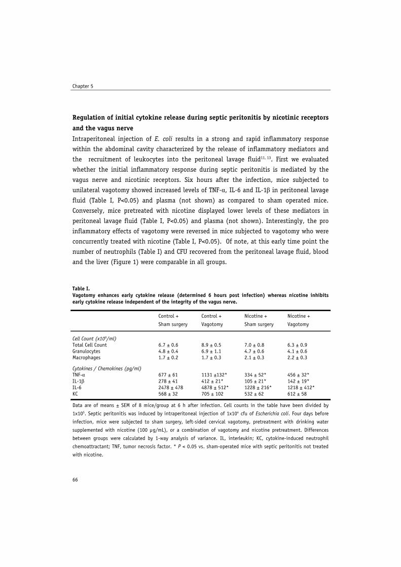
vagus nerve and nicotinic receptors. Six hours after the infection, mice subjected to
unilateral vagotomy showed increased levels of TNF-�, IL-6 and IL-1� in peritoneal lavage
fluid (Table I, P<0.05) and plasma (not shown) as compared to sham operated mice.
Conversely, mice pretreated with nicotine displayed lower levels of these mediators in
peritoneal lavage fluid (Table I, P<0.05) and plasma (not shown). Interestingly, the pro
inflammatory effects of vagotomy were reversed in mice subjected to vagotomy who were
concurrently treated with nicotine (Table I, P<0.05). Of note, at this early time point the
number of neutrophils (Table I) and CFU recovered from the peritoneal lavage fluid, blood
and the liver (Figure 1) were comparable in all groups.
Table I. Vagotomy enhances early cytokine release (determined 6 hours post infection) whereas nicotine inhibits early cytokine release independent of the integrity of the vagus nerve.
Control + Control + Nicotine + Nicotine +
Sham surgery Vagotomy Sham surgery Vagotomy
Cell Count (x105/ml) Total Cell Count 6.7 ± 0.6 8.9 ± 0.5 7.0 ± 0.8 6.3 ± 0.9 Granulocytes 4.8 ± 0.4 6.9 ± 1.1 4.7 ± 0.6 4.1 ± 0.6 Macrophages 1.7 ± 0.2 1.7 ± 0.3 2.1 ± 0.3 2.2 ± 0.3 Cytokines / Chemokines (pg/ml) TNF-� 677 ± 61 1131 ±132* 334 ± 52* 456 ± 32* IL-1��������������������������������������������������������278 ± 41 412 ± 21* 105 ± 21* 142 ± 19* IL-6 2478 ± 478 4878 ± 512* 1228 ± 216* 1218 ± 412* KC 568 ± 32 705 ± 102 532 ± 62 612 ± 58
Data are of means ± SEM of 8 mice/group at 6 h after infection. Cell counts in the table have been divided by
1x10⁵. Septic peritonitis was induced by intraperitoneal injection of 1x10⁴ cfu of Escherichia coli. Four days before
infection, mice were subjected to sham surgery, left-sided cervical vagotomy, pretreatment with drinking water
supplemented with nicotine (100 μg/mL), or a combination of vagotomy and nicotine pretreatment. Differences
between groups were calculated by 1-way analysis of variance. IL, interleukin; KC, cytokine-induced neutrophil
chemoattractant; TNF, tumor necrosis factor. * P < 0.05 vs. sham-operated mice with septic peritonitis not treated
with nicotine.
Chapter 5

67
���
������ ��� �� ����� ������ ��� �� ������
�
�
�
�
�
�������� �������� ��������� ���������
��
���
��
!���"
������ ��� �� ����� ������ ��� �� ������
�
�
�������� �������� ��������� ���������
��
���
��
��#��
������ ��� �� ����� ������ ��� �� ������
�
�
�������� �������� ��������� ���������
��
���
��
Figure 1.
Interference with the cholinergic anti inflammatory
pathway does not influence early bacterial outgrowth.
Peritonitis was induced by intraperitoneal injection of
E. coli. Four days before infection mice were subjected to
left-sided cervical vagotomy, nicotine pretreatment (100
μg/ml added to drinking water) or by a combination of
vagotomy and nicotine pretreatment. Data are means �
SEM of 8 mice per group at 6 hours postinfection.
Differences between groups were calculated by one way
analysis of variance. Differences between groups were
not significant. PLF = peritoneal lavage fluid.
Cholinergic Nervous System in Sepsis
Vagotomy exaggerates, whereas nicotine attenuates the inflammatory response to
established septic peritonitis
Unilateral cervical vagotomy before induction of septic peritonitis was associated with a
significantly enhanced influx of leukocytes into the peritoneal fluid 24 hours
postinfection (P < 0.05 versus sham operated mice), which was almost exclusively caused
by an increased invasion of neutrophils (Table II). In addition, vagotomy resulted in
higher local concentrations of TNF-�, IL-1�, IL-6 and KC during peritonitis when
compared with infected sham operated mice (all P < 0.05 for the difference between
groups; Table II). In contrast, nicotine treatment was associated with a decreased influx
of neutrophils and lower concentrations of cytokines and chemokines in peritoneal lavage
fluid during peritonitis when compared to mice with peritonitis that did not receive
nicotine (all P < 0.05 for the difference between groups; Table III). Plasma cytokine levels
were influenced by vagotomy and nicotine in a similar manner as peritoneal lavage fluid
concentrations (data not shown).

68
Table II. Vagotomy increases cell influx and cytokine and chemokine levels in peritoneal lavage fluid (determined 24 hours post infection).
Sham surgery + Peritonitis Vagotomy + Peritonitis
Cell Count (x106/ml) Total Cell Count 5.5 ± 0.4 27.2 ± 7.3* Granulocytes 4.1 ± 0.3 22.7 ± 1.9* Macrophages 1.3 ± 0.2 4.0 ± 0.9* Cytokines / Chemokines (pg/ml) TNF-� 200 ± 109 685 ± 111* IL-1��������������������������������������������������������309 ± 41 488 ± 43* IL-6 4070 ± 1015 7875 ± 1207* KC 684 ± 92 1057 ± 122*
Data are of means ± SEM of 8 mice/group at 24 h after infection. Cell counts in the table have been divided by
1x10⁶. Septic peritonitis was induced by intraperitoneal injection of 1x10⁴ cfu of Escherichia coli. Four days before
infection, mice were subjected to sham surgery or left-sided cervical vagotomy. Differences between groups were
calculated by Mann-Whitney U test. IL, interleukin; KC, cytokine-induced neutrophil chemoattractant; TNF, tumor
necrosis factor. * P < 0.05 vs. sham-operated mice with septic peritonitis.
Table III. Nicotine reduces cell influx and cytokine and chemokine levels in peritoneal lavage fluid (determined 24 hours post infection).
Control + Peritonitis Nicotine + Peritonitis
Cell Count (x106/ml) Total Cell Count 6.4 ± 0.8 2.9 ± 0.4* Granulocytes 5.1 ± 0.6 1.9 ± 0.3* Macrophages 1.2 ± 0.2 0.9 ± 0.2 Cytokines / Chemokines (pg/ml) TNF-� 687 ± 270 238 ± 128* IL-1��������������������������������������������������������652 ± 132 230 ± 35* IL-6 10719 ± 5413 6643 ± 937* KC 1487 ± 356 953 ± 156*
Data are of means ± SEM of 8 mice/group at 24 h after infection. Cell counts in the table have been divided by
1x10⁶. Septic peritonitis was induced by intraperitoneal injection of 5x10⁴ cfu of Escherichia coli. From four days
before infection, mice received either normal drinking water or drinking water supplemented with nicotine (100 μg/
mL). Differences between groups were calculated by Mann-Whitney U test. IL, interleukin; KC, cytokine-induced
neutrophil chemoattractant; TNF, tumor necrosis factor. * P < 0.05 vs. sham-operated mice with septic peritonitis.
Chapter 5 Chapter 5

69
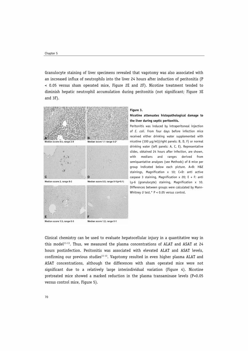
Vagotomy increases, whereas nicotine attenuates, liver injury
Liver injury is one of the hallmarks of distant organ damage in experimental peritonitis
and is related to the extent of systemic inflammation11-13. Mice not injected with E. coli
did not display evidence of necrosis or apoptosis in their livers irrespective of whether
they were subjected to vagotomy or nicotine treatment (not shown). Peritonitis in mice
in which the cholinergic system was not manipulated by either vagotomy or nicotine was
accompanied by histopathological evidence of liver necrosis, inflammation, thrombosis
and apoptosis 24 hours after infection (Figures 2A, 2C, 3A and 3C). In vagotomized mice,
the extent of both liver damage (Figure 2B) and apoptosis (Figure 2D) was markedly
increased during septic peritonitis (P < 0.05 versus sham operated mice).
Cholinergic nervous system in sepsis Cholinergic Nervous System in Sepsis
Figure 2.
Vagotomy increases histopathological damage to
the liver during septic peritonitis.
Peritonitis was induced by intraperitoneal injection
of E. coli. Four days before infection mice were
subjected to left-sided cervical vagotomy (right
panels: B, D, F) or sham surgery (left panels: A, C,
E). Representative liver sections, obtained 24 hours
after infection, are shown, with medians and ranges
derived from semiquantative analyses (see
Methods) of 8 mice per group indicated below each
picture. A+B: H&E stainings, Magnification x 10;
C+D: anti active caspase 3 staining, Magnification x
20; E + F: anti Ly-6 (granulocyte) staining,
Magnification x 10. Differences between groups
were calculated by Mann-Whitney U test.* P < 0.05
versus sham surgery.
Conversely, nicotine treatment prevented liver damage after intraperitoneal infection
with E. coli (P < 0.05 versus control mice, Figure 3B). In addition, the extent of apoptosis
was decreased in nicotine treated mice during peritonitis although the difference with
infected control mice did not reach statistical significance in the semi-quantitative
analysis (P = 0.10; Figure 3D).
70
Granulocyte staining of liver specimens revealed that vagotomy was also associated with
an increased influx of neutrophils into the liver 24 hours after induction of peritonitis (P
< 0.05 versus sham operated mice, Figure 2E and 2F). Nicotine treatment tended to
diminish hepatic neutrophil accumulation during peritonitis (not significant; Figure 3E
and 3F).
Clinical chemistry can be used to evaluate hepatocellular injury in a quantitative way in
this model11-13. Thus, we measured the plasma concentrations of ALAT and ASAT at 24
hours postinfection. Peritonitis was associated with elevated ALAT and ASAT levels,
confirming our previous studies11-13. Vagotomy resulted in even higher plasma ALAT and
ASAT concentrations, although the differences with sham operated mice were not
significant due to a relatively large interindividual variation (Figure 4). Nicotine
pretreated mice showed a marked reduction in the plasma transaminase levels (P<0.05
versus control mice, Figure 5).
Figure 3.
Nicotine attenuates histopathological damage to
the liver during septic peritonitis.
Peritonitis was induced by intraperitoneal injection
of E. coli. From four days before infection mice
received either drinking water supplemented with
nicotine (100 μg/ml)(right panels: B, D, F) or normal
drinking water (left panels: A, C, E). Representative
slides, obtained 24 hours after infection, are shown,
with medians and ranges derived from
semiquantative analyses (see Methods) of 8 mice per
group indicated below each picture. A+B: H&E
stainings, Magnification x 10; C+D: anti active
caspase 3 staining, Magnification x 20; E + F: anti
Ly-6 (granulocyte) staining, Magnification x 10.
Differences between groups were calculated by Mann-
Whitney U test.* P < 0.05 versus control.
Chapter 5

71
Cholinergic Nervous System in Sepsis
Nicotine pretreatment impairs bacterial clearance and survival during septic
peritonitis
An adequate local inflammatory response is important for mounting an effective
antibacterial response during peritonitis11-13. Therefore, we determined the consequences
of the effect of vagotomy and nicotine on the host inflammatory reaction to E. coli
������ ��� �� �����$
%$$$
�$$$
�$$$
�$$$
&�&
'(�
�)
Figure 4.
Effect of vagotomy on plasma transaminase levels.
Peritonitis was induced by intraperitoneal injection of E. coli. Four days before infection mice were subjected to
left-sided cervical vagotomy or sham surgery. Data are means � SEM of 8 mice per group at 24 hours postinfection.
Dotted line represents transaminase levels in uninfected mice. Differences between groups were calculated by
Mann-Whitney U test. Differences between groups were not significant.
������ ��� �� �����$
%$$$
�$$$
�$$$
�$$$
&�&
'(�
�)
Figure 5.
Nicotine reduces plasma transaminase levels.
Peritonitis was induced by intraperitoneal injection of E. coli. From four days before infection mice received either
normal drinking water or drinking water supplemented with nicotine (100 μg/ml). Data are means � SEM of 8 mice
per group at 24 hours postinfection. Dotted line represents transaminase levels in uninfected mice. Differences
between groups were calculated by Mann-Whitney U test.*P < 0.05 vs control mice with peritonitis.
������� *�������$
�$$
%$$$
%�$$
�
&�&
'(�
�)
������� *�������$
%$$$
�$$$
�
&�&
'(�
�)

72
���
������� *��������
�
�
�
+ �
��
���
��
!���"
������� *��������
�
�
� �
��
���
��
��#��
������� *��������
�
� �
��
���
��
Figure 7.
Nicotine impairs bacterial clearance.
Peritonitis was induced by intraperitoneal injection of E. coli. From four days before infection mice received either
normal drinking water of drinking water supplemented with nicotine (100 �g/ml). Data are means � SEM of 8 mice
per group at 24 hours postinfection. Differences between groups were calculated by Mann-Whitney U test.*P < 0.05
vs control mice with peritonitis. PLF = peritoneal lavage fluid.
Chapter 5
infection on the bacterial loads in the peritoneal lavage fluid (the site of the infection),
blood (to evaluate to which extent the infection became systemic), and liver at 24 hours
after infection (i.e. shortly before the first deaths occurred). Vagotomy did not influence
the number of E. coli CFU’s recovered from these three body sites (Figure 6). However,
nicotine treatment facilitated the outgrowth of E. coli in peritoneal fluid, blood and liver
(all P < 0.05 versus control mice, Figure 7).
Figure 6.
Vagotomy does not influence bacterial clearance.
Peritonitis was induced by intraperitoneal injection of E. coli. Four days before infection mice were subjected to
left-sided cervical vagotomy or sham surgery. Data are means � SEM of 8 mice per group at 24 hours postinfection.
Differences between groups were calculated by Mann-Whitney U test. Differences between groups were not
significant. PLF = peritoneal lavage fluid.
���
������ ��� �� ������
�
�
�
!���"
������ ��� �� ������
�
�
�
+
��
���
��
��#��
������ ��� �� ������
�
�
��
���
��
��
���
��

73
Finally, we determined the effect of vagotomy and nicotine treatment on mortality.
Consistent with their influence on the outgrowth of E. coli during the infection,
vagotomy did not alter mortality during septic peritonitis (data not shown), whereas
nicotine significantly accelerated mortality (P < 0.05 versus control mice, Figure 8).
Notably, the relatively modest adverse effect of nicotine on survival in this fulminant
model of sepsis was reproduced in two separate additional experiments (data not shown).
Discussion
The efferent vagus nerve has been implicated as an important anti-inflammatory
pathway through an interaction of its principal neurotransmitter acetylcholine with
nicotinic cholinergic receptors, in particular the �7 subunit, on resident macrophages.
Whereas the function of this cholinergic anti-inflammatory pathway has been well
established in models of sterile inflammation7-9, 16, 17, the present study is the first to
investigate its role in the innate immune response to infection with live bacteria. We here
demonstrate that inhibition of the cholinergic anti-inflammatory pathway by cervical
vagotomy results in an enhanced early and late inflammatory response to septic
peritonitis induced by intraperitoneal injection of E. coli, characterized by increased
cytokine release, an enhanced influx of inflammatory cells to the site of infection and the
occurrence of extensive liver damage. Conversely, activation of the peripheral component
of the pathway by oral administration of nicotine attenuated early and late inflammation,
as reflected by decreased cytokine production and neutrophil recruitment and prevention
of liver damage. These data suggest that the cholinergic anti-inflammatory pathway plays
an essential role in the regulation of inflammatory responses during septic peritonitis.
�,$-$�
� �� �� �� �� ����
��
�� �� ���
���� ���
.���
���#
�#��
Figure 8.
Nicotine accelerates mortality during septic
peritonitis.
Peritonitis was induced by intraperitoneal
injection of E. coli. From four days before
infection mice received either normal drinking
water of drinking water supplemented with
nicotine (100 �g/ml). N = 12 mice per group. P
value indicates the difference between groups by
log-rank test.
Cholinergic Nervous System in Sepsis
74
The currently reported effects of vagus nerve manipulation by vagotomy or nicotine
are in line with earlier investigations that examined the influence of vagus nerve activity
on sterile inflammation. Previous studies have identified an important role for the
afferent vagus nerve in the detection of inflammation by the central nervous system.
Whether humoral or neural pathways are essential in relaying information to the brain is
largely dependent on the magnitude of the inflammatory response. Experimental studies
it have shown that when the level of inflammation is low, such as when a low dose of
endotoxin is injected intraperitoneally, vagotomy inhibits the stimulation of the
hypothalamus-pituitary-adrenal (HPA) axis and the induction of IL-1 in the brain18, 19
whereas high doses of endotoxin induce responses by the brain independent of the vagus
nerve20, 21. This implicates that neural pathways are essential in the relay of localized
inflammation whereas information about severe systemic inflammation reaches the brain
predominantly through humoral pathways. The vagus nerve is not only essential in the
detection of inflammation but also provides an important route for the central nervous
system to respond. In experimental endotoxemia, direct electrical or chemical vagus
nerve stimulation reduced serum TNF-� levels and prevented shock7, 17, whilst cervical
vagotomy augmented serum TNF-� levels and sensitized animals to the lethal effects of
LPS7, 8. In other models of systemic inflammation, induced by either ischemia reperfusion
injury or hypovolemic hemorrhagic shock, stimulation of the vagus nerve decreased
serum TNF-� levels and prevented the development of hypotension9, 16. Previous, as-yet-
unpublished studies of endotoxemic mice by our group have shown that the anti
inflammatory effects of electrical vagus nerve stimulation are relatively short lived and
wean two to four hours after stimulation. On the basis of these results, given the
duration of the septic peritonitis model used here, we decided not to use vagus nerve
stimulation in the present study. It should be noted that nicotine was administered via
the drinking water beginning from four days prior to induction of peritonitis. Due to this
route of administration and due to the acute nature of the model used, we were not able
to examine the effect of postponed treatment with nicotine.
Our data confirm the anti-inflammatory potential of the vagus nerve in a well
established model of abdominal sepsis. We first show that the initial inflammatory
response, which has been shown to be essential for host defense in this model12, 22,
during septic peritonitis is regulated by the vagus nerve and nicotinic receptors. Six
hours after infection mice subjected to unilateral vagotomy showed increased levels of
Chapter 5

75
pro inflammatory cytokines as compared to sham operated mice. Conversely, mice
pretreated with nicotine displayed lower levels of these mediators. Interestingly, the pro
inflammatory effects of vagotomy were overturned in mice subjected to vagotomy
concurrently treated with nicotine confirming that nicotine acts on the peripheral part of
the cholinergic anti inflammatory pathway (which is independent of the integrity of the
vagus nerve). In subsequent studies mice were sacrificed 24 hours postinfection and we
investigated the effects of vagotomy and nicotine pretreatment on host defense and liver
damage during more established septic peritonitis. Interference with the function of the
vagus nerve strongly influenced not only the proinflammatory cytokine response to E.
coli peritonitis but also the migration of leukocytes to the site of the infection, one of
the hallmarks of the early immune reaction to invading pathogens. Moreover, our study
documented a protective role of the intact vagus nerve against liver injury accompanying
experimental E. coli peritonitis. We specifically focused on hepatic injury and
inflammation, since we previously documented hepatocellular damage in this infection
model11-13, and since the liver is richly innervated by the vagus nerve23. We used nicotine
to chemically stimulate the peripheral part of the cholinergic anti-inflammatory pathway.
Previous studies have shown that nicotine inhibits LPS-stimulated TNF-� release by
human, as well as mouse, macrophages in vitro via a specific interaction with the �7
subunit of nicotinic acetylcholine receptors8. These findings were corroborated by in vivo
studies using LPS challenged �7-deficient mice, in which the anti-inflammatory effect of
electrical stimulation of the vagus nerve was abolished8. Together with our finding that
nicotine added to drinking water reduced TNF-� concentrations in mice challenged with
live E. coli in vivo, we consider it likely that nicotine exerts its anti-inflammatory effects
through an interaction with the �7 subunit of nicotinic acetylcholine receptors on
macrophages. Of note, the same scheme and route of nicotine administration has been
reported to reduce colonic damage during spontaneous colitis in IL-10 gene deficient
mice10, 24, 25. Unfortunately, we were not able to confirm and expand our results using �7-
deficient mice, since these mice do not breed well (information by Jackson Laboratories)
and our own prolonged efforts to breed these mice in our institution did not result in a
colony large enough to perform in vivo experiments.
Interruption or stimulation of the vagus nerve had a profound impact on the
recruitment of neutrophils to the infected peritoneal cavity. Since the local release of the
neutrophil attracting chemokine KC during peritonitis was enhanced by vagotomy and
Cholinergic Nervous System in Sepsis
76
decreased by nicotine, it is conceivable that the alterations in neutrophil migration to the
site of infection are, at least in part, mediated by KC. Alternatively, these results can
also be explained by a direct effect of vagotomy or nicotine on neutrophils. Previous
studies have shown that a variety of nicotinic Ach receptors is present on neutrophils and
that stimulation of nicotinic receptors inhibits neutrophil migration which is, at least in
part, mediated by inhibition of adhesion molecule expression on both endothelial cell
surface and neutrophils26.
Whereas the inflammatory response to septic peritonitis was increased after vagotomy
and reciprocally decreased by nicotine treatment, bacterial clearance and survival were
altered by nicotine only. A possible explanation could be that unilateral vagotomy
induces only a partial interference with the cholinergic anti-inflammatory pathway.
Notably, the effect of bilateral vagotomy could not be investigated since this procedure is
lethal in mice. After nicotine pretreatment, bacterial clearance and survival were
significantly reduced. Since host defense in peritonitis is a delicate balance between pro-
inflammatory pathways intended to eliminate bacteria and anti-inflammatory pathways
intended to prevent systemic inflammation, any imbalance in pro- or anti-inflammatory
mediators might prove harmful. Indeed, our laboratory recently demonstrated that
elimination of the anti-inflammatory cytokine IL-10 in septic peritonitis resulted in an
uncontrolled systemic inflammatory response syndrome and lethality in spite of the fact
that IL-10 deficiency facilitated the clearance of bacteria from the peritoneal cavity11. In
the current study, pretreatment with nicotine resulted in a reduction of local and
systemic inflammation but increased lethality due to a decrease in bacterial clearance and
enhanced dissemination of bacteria. Taken together, these findings illustrate the delicacy
of the balance between pro- and anti-inflammation during septic peritonitis.
Excessive activation of coagulation plays an important role in the pathogenesis of
severe sepsis27 and the model used here is associated with profound activation of the
coagulation system28. Of note, in the current studies we did not find consistent effects of
either vagotomy or nicotine on the procoagulant response to abdominal sepsis, as
measured by thrombin-antithrombin levels in peritoneal lavage fluid, and fibrin staining
of liver sections (data not shown).
Chapter 5

77
It should be noted that variation existed in some of the endpoints measured in the
respective control groups of study 2 (sham surgery) and study 3. The exact number of
viable bacteria used to inoculate mice can only be quantified retrospectively; in study 2,
the inoculum contained 1 x 10⁴ CFU whereas in study 3, 5 x 10⁴ E. coli were injected.
Although an effect of sham surgery can not be excluded, we consider it most likely that
the larger bacterial challenge given in study 3 explains the somewhat higher control
values in this experiment. Importantly, in both separate studies control and intervention
groups were injected with exactly the same inoculum at the same time.
Peritonitis is a common cause of sepsis29 and E. coli remains one of the most
frequently isolated pathogens in intraperitoneal infections30. Intraperitoneal
administration of live E. coli results in a paradigm that resembles a clinical condition
commonly associated with septic peritonitis31. By using this model we here demonstrate
for the first time, to our knowledge, that the cholinergic anti-inflammatory pathway is an
essential regulator of the innate immune response to a severe bacterial infection. We
further show that stimulation of the cholinergic anti-inflammatory pathway by nicotine
impairs bacterial clearance and survival during E. coli induced septic peritonitis. The
cholinergic anti-inflammatory pathway may be a future target for the modulation of the
host inflammatory response to sepsis.
Acknowledgements:
We thank A. de Boer for expert technical assistance.
Cholinergic Nervous System in Sepsis

78
Reference List 1. Delves PJ, Roitt IM. The immune system. First of two parts. N Engl J Med 2000;343:37-49 2. van der Poll T, van Deventer SJ. Cytokines and anticytokines in the pathogenesis of sepsis. Infect Dis Clin
North Am 1999;13:413-26, ix. 3. Cohen J. The immunopathogenesis of sepsis. Nature 2002;420:885-891. 4. Riedemann NC, Guo RF, Ward PA. The enigma of sepsis. J Clin Invest 2003;112:460-467. 5. Tracey KJ. The inflammatory reflex. Nature 2002;420:853-9. 6. Blalock JE. Harnessing a neural-immune circuit to control inflammation and shock. J Exp Med 2002;195:
F25-F28. 7. Borovikova LV, Ivanova S, Zhang M, Yang H, Botchkina GI, Watkins LR, Wang H, Abumrad N, Eaton JW,
Tracey KJ. Vagus nerve stimulation attenuates the systemic inflammatory response to endotoxin. Nature 2000;405:458-62.
8. Wang H, Yu M, Ochani M, Amella CA, Tanovic M, Susarla S, Li JH, Yang H, Ulloa L, Al-Abed Y, Czura CJ, Tracey KJ. Nicotinic acetylcholine receptor alpha7 subunit is an essential regulator of inflammation. Nature 2003;421:384-8.
9. Guarini S, Altavilla D, Cainazzo MM, Giuliani D, Bigiani A, Marini H, Squadrito G, Minutoli L, Bertolini A, Marini R, Adamo EB, Venuti FS, Squadrito F. Efferent vagal fibre stimulation blunts nuclear factor-kappaB activation and protects against hypovolemic hemorrhagic shock. Circulation 2003;107:1189-94.
10. Eliakim R, Fan QX, Babyatsky MW. Chronic nicotine administration differentially alters jejunal and colonic inflammation in interleukin-10 deficient mice. Eur J Gastroenterol Hepatol 2002;14:607-614.
11. Sewnath ME, Olszyna DP, Birjmohun R, ten Kate FJ, Gouma DJ, van der PT. IL-10-deficient mice demonstrate multiple organ failure and increased mortality during Escherichia coli peritonitis despite an accelerated bacterial clearance. J Immunol 2001;166:6323-6331.
12. Knapp S, de Vos AF, Florquin S, Golenbock DT, van der Poll T. Lipopolysaccharide binding protein is an essential component of the innate immune response to Escherichia coli peritonitis in mice. Infect Immun 2003;71:6747-6753.
13. Weijer S, Sewnath ME, de Vos AF, Florquin S, van der SK, Gouma DJ, Takeda K, Akira S, van der Poll T. Interleukin-18 facilitates the early antimicrobial host response to Escherichia coli peritonitis. Infect Immun 2003;71:5488-5497.
14. Knapp S, Leemans JC, Florquin S, Branger J, Maris NA, Pater J, Van RN, van der PT. Alveolar macrophages have a protective antiinflammatory role during murine pneumococcal pneumonia. Am J Respir Crit Care Med 2003;167:171-179.
15. Olszyna DP, Florquin S, Sewnath M, Branger J, Speelman P, van Deventer SJ, Strieter RM, van der PT. CXC chemokine receptor 2 contributes to host defense in murine urinary tract infection. J Infect Dis 2001;184:301-307.
16. Bernik TR, Friedman SG, Ochani M, DiRaimo R, Susarla S, Czura CJ, Tracey KJ. Cholinergic antiinflammatory pathway inhibition of tumor necrosis factor during ischemia reperfusion. J Vasc Surg 2002;36:1231-6.
17. Bernik TR, Friedman SG, Ochani M, DiRaimo R, Ulloa L, Yang H, Sudan S, Czura CJ, Ivanova SM, Tracey KJ. Pharmacological stimulation of the cholinergic antiinflammatory pathway. J Exp Med 2002;195:781-8.
18. Fleshner M, Goehler LE, Schwartz BA, McGorry M, Martin D, Maier SF, Watkins LR. Thermogenic and corticosterone responses to intravenous cytokines (IL-1beta and TNF-alpha) are attenuated by subdiaphragmatic vagotomy. J Neuroimmunol 1998;86:134-41.
19. Maier SF, Goehler LE, Fleshner M, Watkins LR. The role of the vagus nerve in cytokine-to-brain communication. Ann N Y Acad Sci 1998;840:289-300.
20. Hansen MK, Daniels S, Goehler LE, Gaykema RP, Maier SF, Watkins LR. Subdiaphragmatic vagotomy does not block intraperitoneal lipopolysaccharide-induced fever. Auton Neurosci 2000;85:83-87.
21. Hansen MK, Nguyen KT, Goehler LE, Gaykema RP, Fleshner M, Maier SF, Watkins LR. Effects of vagotomy on lipopolysaccharide-induced brain interleukin-1beta protein in rats. Auton Neurosci 2000;85:119-126.
22. Cross AS, Sadoff JC, Kelly N, Bernton E, Gemski P. Pretreatment with recombinant murine tumor necrosis factor alpha/cachectin and murine interleukin 1 alpha protects mice from lethal bacterial infection. J Exp Med 1989;169:2021-2027.
23. Kiba T. The role of the autonomic nervous system in liver regeneration and apoptosis--recent developments. Digestion 2002;66:79-88.
24. Sopori ML, Kozak W, Savage SM, Geng Y, Kluger MJ. Nicotine-induced modulation of T Cell function. Implications for inflammation and infection. Adv Exp Med Biol 1998;437:279-289.
Chapter 5

79
25. Kalra R, Singh SP, Kracko D, Matta SG, Sharp BM, Sopori ML. Chronic self-administration of nicotine in rats impairs T cell responsiveness. J Pharmacol Exp Ther 2002;302:935-939.
26. Speer P, Zhang Y, Gu Y, Lucas MJ, Wang Y. Effects of nicotine on intercellular adhesion molecule expression in endothelial cells and integrin expression in neutrophils in vitro. Am J Obstet Gynecol 2002;186:551-6.
27. Levi M, van der Poll T, Buller HR. Bidirectional relation between inflammation and coagulation. Circulation 2004;109:2698-704.
28. Weijer S, Schoenmakers SH, Florquin S, Levi M, Vlasuk GP, Rote WE, Reitsma PH, Spek CA, van der Poll T. Inhibition of the tissue factor/factor VIIa pathway does not influence the inflammatory or antibacterial response to abdominal sepsis induced by Escherichia coli in mice. J Infect Dis 2004;189:2308-2317.
29. Wheeler AP, Bernard GR. Treating patients with severe sepsis. N Engl J Med 1999;340:207-214. 30. McClean KL, Sheehan GJ, Harding GK. Intraabdominal infection: a review. Clin Infect Dis 1994;19:100-116. 31. Fink MP, Heard SO. Laboratory models of sepsis and septic shock. J Surg Res 1990;49:186-196.
Cholinergic Nervous System in Sepsis

80
�