UvA-DARE (Digital Academic Repository) Sickle cell disease ... · (HbSS) and compound heterozygous...
16
UvA-DARE is a service provided by the library of the University of Amsterdam (http://dare.uva.nl) UvA-DARE (Digital Academic Repository) Sickle cell disease Pathogenesis and biomarkers Schimmel, M. Link to publication Citation for published version (APA): Schimmel, M. (2017). Sickle cell disease: Pathogenesis and biomarkers. General rights It is not permitted to download or to forward/distribute the text or part of it without the consent of the author(s) and/or copyright holder(s), other than for strictly personal, individual use, unless the work is under an open content license (like Creative Commons). Disclaimer/Complaints regulations If you believe that digital publication of certain material infringes any of your rights or (privacy) interests, please let the Library know, stating your reasons. In case of a legitimate complaint, the Library will make the material inaccessible and/or remove it from the website. Please Ask the Library: https://uba.uva.nl/en/contact, or a letter to: Library of the University of Amsterdam, Secretariat, Singel 425, 1012 WP Amsterdam, The Netherlands. You will be contacted as soon as possible. Download date: 09 Jan 2020
Transcript of UvA-DARE (Digital Academic Repository) Sickle cell disease ... · (HbSS) and compound heterozygous...

UvA-DARE is a service provided by the library of the University of Amsterdam (http://dare.uva.nl)
UvA-DARE (Digital Academic Repository)
Sickle cell diseasePathogenesis and biomarkersSchimmel, M.
Link to publication
Citation for published version (APA):Schimmel, M. (2017). Sickle cell disease: Pathogenesis and biomarkers.
General rightsIt is not permitted to download or to forward/distribute the text or part of it without the consent of the author(s) and/or copyright holder(s),other than for strictly personal, individual use, unless the work is under an open content license (like Creative Commons).
Disclaimer/Complaints regulationsIf you believe that digital publication of certain material infringes any of your rights or (privacy) interests, please let the Library know, statingyour reasons. In case of a legitimate complaint, the Library will make the material inaccessible and/or remove it from the website. Please Askthe Library: https://uba.uva.nl/en/contact, or a letter to: Library of the University of Amsterdam, Secretariat, Singel 425, 1012 WP Amsterdam,The Netherlands. You will be contacted as soon as possible.
Download date: 09 Jan 2020

Chapter 2
Nucleosomes and neutrophil activation in sickle cell disease painful crisis
Marein Schimmel1,2, Erfan Nur1,2, Bart J Biemond2, Gerard J van Mierlo3, Shabnam Solati3, Dees P Brandjes1, Hans-Martin Otten1, John-John Schnog4,5 and Sacha Zeerleder2,3 ; on behalf of the Curama Study Group
1 Department of Internal Medicine, Slotervaart Hospital, Amsterdam, The Netherlands
2 Department of Clinical Hematology, Academic Medical Center, University of Amsterdam, Amsterdam, The Netherlands
3 Department of Immunopathology, Sanquin Research and Landsteiner Laboratory AMC, University of Amsterdam, Amsterdam, The Netherlands
4 Department of Hematology/Oncology, Sint Elisabeth Hospital, Willemstad, Curaçao
5 Department of Immunology, Red Cross Blood Bank Foundation, Willemstad, Curaçao
Haematologica. 2013 Nov; 98 (11): 1797-803

32 Chapter 2
ABSTR ACT
Activated polymorphonuclear neutrophils play an important role in the pathogenesis
of vaso-occlusive painful sickle cell crisis. Upon activation polymorphonuclear neu-
trophils can form neutrophil extracellular traps. Neutrophil extracellular traps consist
of a meshwork of extracellular DNA, nucleosomes, histones and neutrophil proteases.
Neutrophil extracellular traps have been demonstrated to be toxic to endothelial
and parenchymal cells. This prospective cohort study was conducted to determine
neutrophil extracellular trap formation in sickle cell patients during steady state and
painful crisis. As a measure of neutrophil extracellular traps, plasma nucleosomes
levels were determined and polymorphonuclear neutrophil activation was assessed
measuring plasma levels of elastase-α1-antitrypsin complexes in 74 patients in steady
state, 70 patients during painful crisis, and 24 race-matched controls using Enzyme
Linked Immunosorbent Assay. Nucleosome levels in steady state sickle cell patients
were significantly higher than levels in controls. During painful crisis levels of both
nucleosomes and elastase-α1-antitrypsin complexes increased significantly. Levels of
nucleosomes correlated significantly to elastase-α1-antitrypsin complex levels during
painful crisis, (Sr = 0.654, P < 0.001). This was seen in both HbSS/HbSβ0-thalassemia
(Sr=0.55, P<0.001) and HbSC/HbSβ+-thalassemia patients (Sr=0.90, P=<0.001) during
painful crisis. Levels of nucleosomes showed a correlation with length of hospital
stay and were highest in patients with acute chest syndrome. These data support the
concept that neutrophil extracellular trap formation and neutrophil activation may
play a role in the pathogenesis of painful sickle cell crisis and acute chest syndrome.

Nucleosomes and neutrophil activation in sickle cell disease painful crisis 33
2
INTRODUCTION
Sickle cell disease (SCD) is characterized by recurrent acute painful vaso-occlusive
crisis (VOC), accounting for the vast majority of SCD related hospital admissions [1-3].
VOC related complications, such as acute chest syndrome, stroke and multi-organ
failure are associated with high morbidity and mortality[4]. The exact pathogenesis
of acute painful VOC remains to be elucidated. Alongside the crucial role for sickle
erythrocytes in this, it encompasses an inflammatory response as evidenced by en-
dothelial activation, coagulation activation and enhanced cellular adhesion, finally
all contributing to microvascular occlusion.
Leukocytes play an important role in the development of microvascular obstruction
and sickle cell disease related complications. In steady state sickle cell patients,
leukocytosis is associated with severity of disease[5]. Clinical studies show that
leukocytosis is a risk factor for major sickle cell related complications such as stroke
[6], acute chest syndrome [7] and early death [8]. Additionally, the clinical benefit of
hydroxycarbamide in sickle cell patients has partly been attributed to a reduction
in polymorphonuclear neutrophil (PMN) cell count [9] and reduced PMN adhesion
[10]. In vitro studies have demonstrated that PMN isolated from sickle cell patients
are primed as evidenced by an increased expression of adhesion molecules [11-13],
rendering them more susceptible for inflammatory stimuli as compared to PMN from
healthy controls [14]. Moreover, activation of PMN, e.g. upon interaction with red
blood cells [15], leads to the production of toxic reactive oxygen species (ROS), con-
tributing to oxidative stress [16]. In-vitro studies as well as in-vivo studies in SCD mice
models demonstrate P- and E-selectin interactions with integrins [17-19] to be crucial
for the adherence of leukocytes to endothelial and sickle red blood cells, contribut-
ing to the complex process of vaso-occlusion [20, 21]. This identifies PMN activation
and adhesion as important processes in the pathogenesis of vaso-occlusion in SCD.
Recently, activated PMN have been demonstrated to form neutrophil extracellular
traps (NET) [22]. During NET formation, DNA and DNA-binding proteins are extruded
from the neutrophils exposing a mesh consisting of nucleosomes, histones and neu-
trophil proteases such as elastase. These NET are regarded to be part of the innate
immune response system [23]. However, their function is considered to be a double-
edged sword. On one hand, NET formation is an efficient strategy to kill invading
micro-organisms, like bacteria and fungi. On the other hand, NET can become harm-

34 Chapter 2
ful for the host since its exposed compounds (e.g. the mesh of DNA, histones and
neutrophil proteases) are toxic to endothelial cells and parenchymal tissue [24-26].
NET formation has been reported to be pro-coagulant in inflammatory models and is
thought to contribute to the development of disseminated intravascular coagulation,
and hence to morbidity and mortality in sepsis [27-29]. Circulating nucleosomes and
markers of neutrophil activation have been reported to be suitable markers for NET
formation in plasma in baboons and humans [29-31]. Nucleosomes consist of a core
octamer of two copies each holding the histones H2A, H2B, H3 and H4, around which
a segment of helical DNA of 146 base pairs is wrapped [32]. Nucleosomes can be ac-
tively released into the circulation from dead cells as a result of the activity of factor-
VII activating protease (FSAP) [33]. Circulating cell-free DNA in form of nucleosomes
has been reported to correlate with organ dysfunction, disease severity and mortality
in sepsis patients and children suffering from meningococcal sepsis [34-36].
So far, no data are available on NET formation in sickle cell patients. Since white
blood cell counts have been shown to correlate with morbidity of sickle cell patients
and since PMN activation seems to play an important role in the development of
sickle cell painful vaso-occlusive crisis we hypothesized that NET formation may
be involved in these processes. The aim of this prospective cohort study therefore
was to measure plasma levels of circulating nucleosomes and PMN activation as
evidenced by human neutrophil elastase-α1-antitrypsin (EA) complexes as a measure
of NET formation in plasma in sickle cell patients both during steady state and painful
VOC and to evaluate their correlation with crisis severity.
METHODS
Patients
This study followed a prospective design in which patients with sickle cell anemia
(HbSS) and compound heterozygous states HbSβ0-thalassemia, HbSβ+-thalassemia
and sickle-hemoglobin C (HbSC) patients were eligible for inclusion. Diagnosis of
hemoglobinopathy was confirmed by means of high performance liquid chromatog-
raphy in combination with measurement of erythrocyte mean corpuscular volume.
Consecutive sickle cell patients, ≥ 18 years of age, attending the outpatient clinic
(steady state) or being admitted for a painful crisis to the Academic Medical Center or
the Slotervaart Hospital, Amsterdam, The Netherlands, were approached for partici-

Nucleosomes and neutrophil activation in sickle cell disease painful crisis 35
2
pation. A painful crisis was defined as musculo-skeletal pain not otherwise explained
and recognized as such by the patient and requiring medical treatment. Samples dur-
ing painful crisis were obtained within the first 24 hours of admission. Patients with
painful crisis within four weeks and/or blood transfusion within three months prior to
the evaluation for the present study were excluded from inclusion. Other exclusion
criteria were; pregnancy, inflammatory autoimmune disease or any acute infection
within 3 months prior to study participation. Information concerning complications
during admission was collected from medical records. An acute chest syndrome was
defined as a new infiltrate (on admission or during hospitalization) on chest x-ray
associated with one or more new symptoms of chest pain, fever, tachypnea, wheez-
ing, cough or hypoxemia [37]. Samples from race-matched volunteers were taken for
control reference measurements. Written informed consent was obtained from all
participants before any study procedure was performed. The study protocol was ap-
proved by the local medical ethical committee and conducted in agreement with the
Helsinki declaration of 1975, as revised in 2008.
Blood sample collection and laboratory analysis
Blood samples were taken by venipuncture. Blood vials were centrifuged at 4°C for
15 minutes at 3000 rpm and serum and plasma was stored in small aliquots at -80˚C
until further analysis.
Hematology parameters, nucleosome and EA levels were measured in EDTA-
anticoagulated plasma. Soluble vascular adhesion molecule 1 (sVCAM 1) levels were
determined in serum (R&D Systems; Minneapolis, USA). Lactate hydrogenase (LDH)
and bilirubin levels were measured with spectrophotometry in heparinized plasma
(P800 Modular, Roche, Switzerland). Plasma levels of the long pentraxin-3 (PTX3)
were determined using sandwich ELISA [38]. Antigen levels of von Willebrand factor
(vWFag) were assessed by ELISA using antibodies from Dako (Glostrup, Denmark).
Nucleosome levels were measured using ELISA as described previously [35, 39]. Neu-
trophil activation in form of elastase-α1-antitrypsin (EA) complexes was measured by
an ELISA as previously described [30, 40].
Statistical analysis
For statistical analysis patients were divided primarily in two groups; patients with the
relatively severe genotypes HbSS and HbSβ0-thalassemia grouped together (HbSS/
HbSβ0-thal) and patients with the relatively milder HbSC and HbSβ+-thalassemia

36 Chapter 2
genotypes gathered in the other group (HbSC/HbSβ+-thal) [41, 42]. We used a com-
mercial statistical package (IBM SPSS Statistics 19, SPSS Inc, Hong Kong, PRC) for data
analysis. Since results were not normally distributed, they are expressed as median
with interquartile range. Unless stated otherwise, P<0.05 was considered statistically
significant. Bonferroni’s correction was applied for multiple testing (P<0.004 was
considered statistically significant).
RESULTS
Patients
Seventy-four patients in steady state (49 HbSS/HbSβ0-thal and 25 HbSC/HbSβ+-thal),
70 patients during painful crisis (53 HbSS/HbSβ0-thal and 17 HbSC/HbSβ+-thal)
and 24 healthy race-matched controls (HbAA) were included in the study. Patients’
characteristics are summarized in Table 1. Twenty-four percent of sickle cell patients
during steady state and a similar percentage (23%) of patients during painful crisis
were on hydroxycarbamide treatment. Of the patients included with painful crisis, 1
(HbSS/HbSβ0-thal) patient was admitted with acute chest syndrome (ACS) and 5 (4
HbSS/HbSβ0-thal and 1 HbSC/HbSβ+-thal) patients developed an ACS between 48
and 60 hours after admission. No infection was reported for any of the patients with
an acute chest syndrome.
Nucleosomes and neutrophil activation
Plasma levels of nucleosomes were significantly higher during painful crisis (20.0 U/
ml; IQR 7.9-107.3) as compared to those in steady state (6.4 U/ml; 3.5-9.7, P < 0.001)
(Figure 1A). This was seen in both HbSS/HbSβ0-thal (20.2 U/ml; 8.9-129.0 vs 6.0 U/ml;
3.0-9.8, P < 0.001) and HbSC/HbSβ+-thal (11.7 U/ml; 5.1-67.7 vs 7.1 U/ml; 4.6-9.6, P =
0.045) patients (Figure 1B). Plasma levels of nucleosomes in healthy controls were
just above the detection limit of the assay (5.0 U/ml; 3.0-6.5). In steady state sickle
cell patients plasma levels of nucleosomes were significantly higher compared to
levels in healthy controls (P = 0.031). In the analysis for the two genotype groups
separately, the same was seen for HbSC/HbSβ+-thal patients in steady state (P =
0.020) while plasma nucleosome levels in HbSS/HbSβ0-thal patients in steady state
were comparable with those in healthy controls (P = 0.089).

Nucleosomes and neutrophil activation in sickle cell disease painful crisis 37
2
Table 1. Baseline characteristics
Controls Asymptomatic state Painful crisis
HbAA(n=24)
HbSS/HbSβ0
(n=49)HbSC/HbSβ+
(n=25)HbSS/HbSβ0
(n=53)HbSC/HbSβ+
(n=17)
Age (y) 37 (29-46) 26 (22-40)* 30 (24-42) 28 (23-36)* 27 (24-35)*
Gender (M/F) 9/15 19/30 7/18 31/22 5/12
Hb (g/dl) 12.7 (11.9-14.0) 9.0 (8.1-10.0)* 11.1 (10.5-11.6)* ‡ 8.4 (7.6–9.3)* 11.3 (10.6–11.8)* ‡
Leukocytes (109/l)
5.6 (4.5-7.0) 9.9 (7.5-10.9)* 6.4 (5.0-8.4)‡ 11.8 (9.3-14.0)*† 8.2 (5.9-10.4)* †‡
Neutrophils (109/l)
2.7 (1.7-3.4) 4.7 (3.4-6.0)* 3.7 (2.8-4.3)* ‡ 7.8 (4.9-9.3)* † 5.4 (3.2-6.7)* †‡
LDH (U/l) 181 (152-205) 385 (301-483)* 228 (178-254)* ‡ 462 (337-605)* † 219 (187-320)* ‡
Total bilirubin (mg/dl)
0.5 (0.4-0.8) 3.1 (1.8-4.9)* 1.2 (0.8-1.5)* ‡ 2.9 (1.5-5.7)* 1.5 (0.8-1.6)* ‡
Hospitalization (days)
NA NA NA 3 (2–7) 4 (3–6.5)
Numbers are medians with interquartile range (IQR)
Hb hemoglobin; LDH lactate dehydrogenase; NA Not applicable
* Significantly different as compared to HbAA controls (P < 0.05).
† Significantly different as compared to asymptomatic state (P < 0.05).
‡ Significant difference between HbSS/HbSβ0-thalassemia and HbSC/HbSβ+-thalassemia patients within
steady state or painful crisis (P < 0.05).
Plasma levels of EA were significantly higher during painful crisis (73.6 ng/ml;
54.9-100.8) as compared to those in steady state (46.2 ng/ml; 34.3-65.6, P < 0.001)
(Figure 1C). This was seen in HbSS/HbSβ0-thal patients (75.1 ng/ml; 56.5-102.4 vs. 45.7
ng/ml; 34.7-59.7, P < 0.001), while in HbSC/HbSβ+-thal patients, the increment did
not reach statistical significance (62.0 ng/ml; 48.0-96.7 vs 50.2 ng/ml; 33.3-67.7, P =
0.051) (Figure 1D). Plasma levels of EA in healthy controls (39.9 ng/ml; 31.5-62.2) were
comparable to those in steady state sickle cell patients (P = 0.330).
During painful crisis, levels of nucleosomes correlated significantly with levels of
EA (Sr = 0.654, P < 0.001). This was seen in both HbSS/HbSβ0-thal (Sr = 0.55, P <
0.001) as well as in HbSC/HbSβ+-thal patients (Sr = 0.90, P < 0.001). During steady
state the correlation between levels of nucleosomes and EA was significant but weak
(Sr = 0.236, P = 0.043). The correlation in HbSC/HbSβ+-thal patients in steady state
remained significant (Sr = 0.63, P = 0.001), while no correlation was found between
levels of nucleosomes and EA in HbSS/HbSβ0-thal patients in steady state (Sr = 0.043,
P = 0.77).
38 Chapter 2
nucleosomes
Controls
Steady Stat
eCris
is
Steady Stat
eCris
is1
10
100
1000
10000P = 0.045 P < 0.001
HbSC/HbSb+
HbSS/HbSb0
24 25 17 49 53
U/m
l
nucleosomes
Controls
SCD Steady Stat
e
SCD Crisis
1
10
100
1000
10000
P = 0.031 P < 0.001
24 74 70
U/m
l
elastase-a1-antitrypsin complexes
Controls
SCD Steady Stat
e
SCD Crisis
10
100
1000P = 0.330 P < 0.001
24 74 70
ng/m
l
elastase-a1-antitrypsin complexes
Controls
Steady Stat
eCris
is
Steady Stat
eCris
is10
100
1000P < 0.001P = 0.051
HbSC/HbSb+
HbSS/HbSb0
24 25 17 49 53
ng/m
l
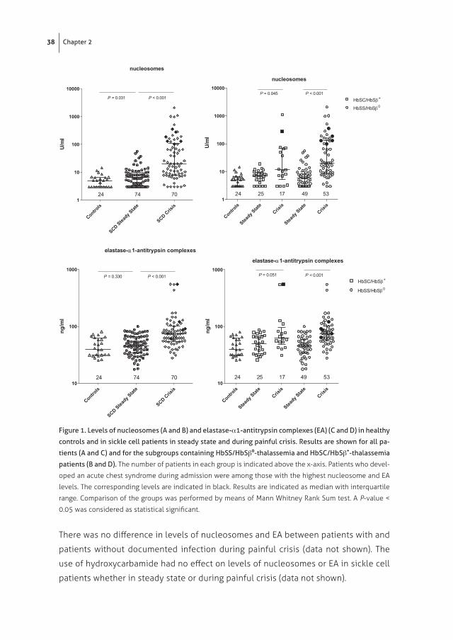
Figure 1. Levels of nucleosomes (A and B) and elastase-α1-antitrypsin complexes (EA) (C and D) in healthy
controls and in sickle cell patients in steady state and during painful crisis. Results are shown for all pa-
tients (A and C) and for the subgroups containing HbSS/HbSβ0-thalassemia and HbSC/HbSβ+-thalassemia
patients (B and D). The number of patients in each group is indicated above the x-axis. Patients who devel-
oped an acute chest syndrome during admission were among those with the highest nucleosome and EA
levels. The corresponding levels are indicated in black. Results are indicated as median with interquartile
range. Comparison of the groups was performed by means of Mann Whitney Rank Sum test. A P-value <
0.05 was considered as statistical significant.
There was no difference in levels of nucleosomes and EA between patients with and
patients without documented infection during painful crisis (data not shown). The
use of hydroxycarbamide had no effect on levels of nucleosomes or EA in sickle cell
patients whether in steady state or during painful crisis (data not shown).

Nucleosomes and neutrophil activation in sickle cell disease painful crisis 39
2
In a paired analysis of 25 patients, accounting for 36 painful crises, significant incre-
ments were observed during painful crisis in plasma levels of both nucleosomes
(from 5.0 U/ml; 3.0-10.8 to 20.2 U/ml; 6.8-94.3, P < 0.001) and EA (47.9 ng/ml; 36.0-
67.6 to 70.6 ng/ml; 55.9-101.4, P < 0.001) as compared to those in steady state
(Figure 2A and 2B).
nucleosomes
0
50
100
150
200
600
1000 P < 0.001
Steady State Crisis
U/m
l
elastase-a1-antitrypsin complexes
Steady State Crisis0
25
50
75
100
125
150500
525
550
ng/m
l
P < 0.001
Figure 2. Paired analysis of levels of nucleosomes (A) and elastase-α1-antitrypsin complexes (EA) (B) in
36 painful crisis in 25 patients included both in steady state and in painful crisis. For comparison between
related samples the Wilcoxon Signed Rank Test was used. A P-value < 0.05 has been considered as statisti-
cally significant. Results are indicated as median with interquartile range.
Nucleosomes and EA in association with markers of endothelial activation,
hemolysis and inflammation
While nucleosome levels in steady state HbSS/HbSβ0-thal patients correlated signifi-
cantly with vWF:Ag (Sr = 0.452, P = 0.001) and sVCAM-1 (Sr = 0.421, P = 0.003) they
only correlated significantly with PTX3 (Sr = 0.623, P = 0.001) during painful crisis. In
the same patient group during painful crisis EA levels just failed to reach a statistical
significant correlation with PTX3 levels (Sr = 0.529, P=0.008).
Leukocyte counts did not correlate with levels of nucleosomes or EA. In addition,
neutrophil count did not correlate with levels of nucleosomes or levels of EA, neither
when results of patients were pooled, nor when they were evaluated separately in

40 Chapter 2
the different subgroups. No association was found between markers of hemolysis
(hemoglobin, LDH and bilirubin) and levels of nucleosomes or EA.
Association with acute chest syndrome and duration of hospitalization
Acute chest syndrome
The 6 patients who developed an ACS were among those with the highest nucleo-
some (359, 274.8, 190, 130, 128 and 100 U/ml, respectively) and EA levels (549.9, 120.8,
91.8, 86.7, 75.1, and 63.9 ng/ml, respectively) (Figure 1A-1D). In these 6 sickle cell
patients with ACS, nucleosome levels were significantly higher than those in patients
during painful crisis without ACS (n= 64; 160.0 U/ml; 121.0-295.9 vs 20.07 U/ml; 7.9-
107.3, P = 0.002).
Hospitalization duration
Nucleosome levels, but not EA levels, correlated significantly with duration of hospi-
tal stay in all sickle cell patients during painful crisis (Sr = 0.441, P <0.001). Excluding
the patients with acute chest syndrome the correlation remained statistically sig-
nificant (Sr = 0.385, P = 0.002). When analyzing the correlation for HbSS/HbSβ0-thal
patients the correlation between nucleosome levels and duration of hospital stay
was stronger (Sr = 0.530, P < 0.001). Figure 3 shows the association between levels of
nucleosomes and duration of hospitalization.
Q1 Q2Q3 Q4
0
5
10
15
P=0.004
P=0.005
nucleosomes
Leng
thof
hosp
italiz
atio
n(in
days
)
Figure 3. Association between levels of nucleo-
somes in all SCD patients and length of hospital
stay (LoS). Median stay is 3 days (IQR 2-7 days).
Display of LOS when nucleosome level is divid-
ed in quartiles. An increasing LOS can be seen
when levels of nucleosomes increase.

Nucleosomes and neutrophil activation in sickle cell disease painful crisis 41
2
DISCUSSION
In the present study we demonstrate that during painful vaso-occlusive crisis
sickle cell patients have significantly higher levels of circulating nucleosomes and
neutrophil activation, as shown by increased EA complexes, as compared to sickle
cell patients in steady state. Results of the paired analyses support the findings
of the between group-analyses. We show that patients developing the severe and
potentially life-threatening complication acute chest syndrome were among those
with the highest nucleosome and EA levels. Moreover, we found that nucleosome
levels correlate with duration of hospitalization. Nucleosome and EA levels correlate
significantly with each other during painful crisis. Together, our data provide indirect
evidence for NET formation in patients with sickle cell disease suffering from VOC.
Our results are in line with a previous study in sickle cell patients demonstrating
significantly increased amounts of circulating cell-free DNA, as determined by quan-
titative PCR amplification, in sickle cell patients during painful crisis as compared to
levels in steady state[43] . Interestingly, in the current study nucleosome levels in
sickle cell patients with acute chest syndrome were comparable to levels measured
in patients with severe sepsis using the same assay [34, 35]. In these patients with
sepsis, circulating cell-free DNA in form of nucleosomes correlated with morbidity
and mortality [34, 35].
This study has several limitations that need to be taken into account when interpret-
ing the data collected. Firstly, we have not performed sequential nucleosome and
EA analysis during admission for painful crisis in our patients, limiting the findings
to a single measurement at presentation with a painful crisis. The observation that
nucleosome and EA levels taken at presentation were highest in patients developing
an ACS during admission are nevertheless in line with previous findings suggesting
neutrophils to be an important player [44, 45] in the pathogenesis of ACS. The obser-
vation that neutrophil count does not correlate with EA or nucleosome levels in this
study in sickle cell patients is supported by observations from studies in patients
with severe sepsis [40]. While EA results reflect general PMN activation, it is likely
that during vaso-occlusive crisis neutrophil count is a measure for circulating (“count-
able”) neutrophils while it does not reflect neutrophils migrated to tissue or adherent
to activated endothelial cells, the latter being observed in vaso-occlusive crisis in
mice models for sickle cell disease [20, 21]. Secondly, the correlation during painful

42 Chapter 2
crisis between nucleosome levels with EA levels and PTX3, both being localized in
NET[46, 47] (while the latter has previously been demonstrated to be increased dur-
ing sickle cell painful vaso-occlusive crisis)[48], support the hypothesis of PMN as
an important nucleosome source via NET formation, at least during vaso-occlusive
complications. This is also in line with the publications reporting circulating nucleo-
somes with or without markers for neutrophil activation to be a good measure for
NET formation in circulation [29, 31]. However, the ELISAs detecting nucleosomes are
not specific for nucleosomes released by PMN, and we can not, therefore, exclude the
possibility that nucleosomes released into the circulation by other cell types, such as
endothelial and parenchymal cells, are detected as well. The statistically significant
correlation between nucleosome levels and markers of endothelial activation, vWFag
and sVCAM-1, in steady state HbSS/HbSβ0-thal patients might be indirect evidence
that damaged endothelial cells contribute to the circulating nucleosomes. Whether
this endothelial cell damage is a consequence of local PMN activation, e.g. in form of
NET formation being cytotoxic to endothelial cells, remains to be established. Thirdly,
results of the analyses performed on the specified genotype groups sometimes di-
verge from the analyses done when pooling data from and considering the patient
population as one group. This may be due to a limited number of patients in the
respective subgroups causing a lack of power to show statistical significant findings.
Moreover, the influence of the interaction of sickle cell erythrocytes with PMN on NET
formation and the role of the genotype in this interaction has to be established yet.
In conclusion, we demonstrate for the first time elevated levels of circulating nu-
cleosomes and neutrophil activation in sickle cell patients with painful crisis sug-
gesting NET formation in these patients. NET, consisting of nucleosomes, proteases
and histones, may promote endothelial activation and contribute to longer and more
severe sickle cell crisis. The role of NET in the prediction of clinical complications in
sickle cell disease painful crisis and as a potential therapeutic target deserves further
study.

Nucleosomes and neutrophil activation in sickle cell disease painful crisis 43
2
REFERENCES
1. Serjeant, G.R., et al., The painful crisis of homozygous sickle cell disease: clinical features. Br. J. Haematol, 1994. 87(3): p. 586-591.
2. Neonato, M.G., et al., Acute clinical events in 299 homozygous sickle cell patients living in France. French Study Group on Sickle Cell Disease. Eur. J. Haematol, 2000. 65(3): p. 155-164.
3. Platt, O.S., et al., Pain in sickle cell disease. Rates and risk factors. N. Engl. J. Med, 1991. 325(1): p. 11-16.
4. Steinberg, M.H., In the clinic. Sickle cell disease. Ann Intern Med, 2011. 155(5): p. ITC31-15; quiz ITC316.
5. Anyaegbu, C.C., et al., Peripheral blood neutrophil count and candidacidal activity correlate with the clinical severity of sickle cell anaemia (SCA). Eur J Haematol, 1998. 60(4): p. 267-8.
6. Ohene-Frempong, K., et al., Cerebrovascular accidents in sickle cell disease: rates and risk factors. Blood, 1998. 91(1): p. 288-294.
7. Castro, O., et al., The acute chest syndrome in sickle cell disease: incidence and risk factors. The Cooperative Study of Sickle Cell Disease. Blood, 1994. 84(2): p. 643-649.
8. Platt, O.S., et al., Mortality in sickle cell disease. Life expectancy and risk factors for early death. N. Engl. J. Med, 1994. 330(23): p. 1639-1644.
9. Charache, S., et al., Effect of hydroxyurea on the frequency of painful crises in sickle cell anemia. Investigators of the Multicenter Study of Hydroxyurea in Sickle Cell Anemia. N. Engl. J. Med, 1995. 332(20): p. 1317-1322.
10. Almeida, C.B., et al., Hydroxyurea and a cGMP-amplifying agent have immediate benefits on acute vaso-occlusive events in sickle cell disease mice. Blood, 2012. 120(14): p. 2879-2888.
11. Okpala, I., The intriguing contribution of white blood cells to sickle cell disease - a red cell disorder. Blood Rev, 2004. 18(1): p. 65-73.
12. Lard, L.R., et al., Neutrophil activation in sickle cell disease. J. Leukoc. Biol, 1999. 66(3): p. 411-415.
13. Fadlon, E., et al., Blood polymorphonuclear leukocytes from the majority of sickle cell pa-tients in the crisis phase of the disease show enhanced adhesion to vascular endothelium and increased expression of CD64. Blood, 1998. 91(1): p. 266-274.
14. Lum, A.F., et al., Inflammatory potential of neutrophils detected in sickle cell disease. Am. J. Hematol, 2004. 76(2): p. 126-133.
15. Hofstra, T.C., et al., Sickle erythrocytes adhere to polymorphonuclear neutrophils and activate the neutrophil respiratory burst. Blood, 1996. 87(10): p. 4440-4447.
16. Nur, E., et al., Oxidative stress in sickle cell disease; pathophysiology and potential implica-tions for disease management. Am. J. Hematol, 2011. 86(6): p. 484-489.
17. Brittain, J.E., et al., Fibronectin bridges monocytes and reticulocytes via integrin alpha-4beta1. Br. J. Haematol, 2008. 141(6): p. 872-881.
18. Hidalgo, A., et al., Heterotypic interactions enabled by polarized neutrophil microdomains mediate thromboinflammatory injury. Nat. Med, 2009. 15(4): p. 384-391.

44 Chapter 2
19. Chang, J., et al., Intravenous immunoglobulins reverse acute vaso-occlusive crises in sickle cell mice through rapid inhibition of neutrophil adhesion. Blood, 2008. 111(2): p. 915-923.
20. Turhan, A., et al., Primary role for adherent leukocytes in sickle cell vascular occlusion: a new paradigm. Proc. Natl. Acad. Sci. U. S. A, 2002. 99(5): p. 3047-3051.
21. Turhan, A., et al., Intravenous immune globulin prevents venular vaso-occlusion in sickle cell mice by inhibiting leukocyte adhesion and the interactions between sickle erythro-cytes and adherent leukocytes. Blood, 2004. 103(6): p. 2397-2400.
22. Brinkmann, V., et al., Neutrophil extracellular traps kill bacteria. Science, 2004. 303(5663): p. 1532-1535.
23. Brinkmann, V. and A. Zychlinsky, Beneficial suicide: why neutrophils die to make NETs. Nat. Rev. Microbiol, 2007. 5(8): p. 577-582.
24. Gupta, A.K., et al., Activated endothelial cells induce neutrophil extracellular traps and are susceptible to NETosis-mediated cell death. FEBS Lett, 2010. 584(14): p. 3193-3197.
25. Xu, J., et al., Extracellular histones are major mediators of death in sepsis. Nat. Med, 2009. 15(11): p. 1318-1321.
26. Saffarzadeh, M., et al., Neutrophil extracellular traps directly induce epithelial and endo-thelial cell death: a predominant role of histones. PLoS. One, 2012. 7(2): p. e32366.
27. Massberg, S., et al., Reciprocal coupling of coagulation and innate immunity via neutrophil serine proteases. Nat. Med, 2010. 16(8): p. 887-896.
28. von Bruhl, M.L., et al., Monocytes, neutrophils, and platelets cooperate to initiate and propagate venous thrombosis in mice in vivo. J. Exp. Med, 2012. 209(4): p. 819-835.
29. Fuchs, T.A., et al., Extracellular DNA traps promote thrombosis. Proc. Natl. Acad. Sci. U. S. A, 2010. 107(36): p. 15880-15885.
30. Van Montfoort, M.L., et al., Circulating nucleosomes and neutrophil activation as risk factors for deep vein thrombosis. Arterioscler. Thromb. Vasc. Biol, 2013. 33(1): p. 147-151.
31. Fuchs, T.A., et al., Circulating DNA and myeloperoxidase indicate disease activity in patients with thrombotic microangiopathies. Blood, 2012. 120(6): p. 1157-1164.
32. Amoura, Z., et al., The key role of nucleosomes in lupus. Arthritis Rheum, 1999. 42(5): p. 833-43.
33. Zeerleder, S., et al., Nucleosome-releasing factor: a new role for factor VII-activating prote-ase (FSAP). FASEB J, 2008. 22(12): p. 4077-4084.
34. Zeerleder, S., et al., Elevated nucleosome levels in systemic inflammation and sepsis. Crit Care Med, 2003. 31(7): p. 1947-1951.
35. Zeerleder, S., et al., Circulating nucleosomes and severity of illness in children suffering from meningococcal sepsis treated with protein C. Crit Care Med, 2012. 40(12): p. 3224-3229.
36. Chen, Q., et al., Circulating nucleosomes as a predictor of sepsis and organ dysfunction in critically ill patients. Int. J. Infect. Dis, 2012. 16(7): p. e558-e564.
37. Miller, S.T., et al., Impact of chronic transfusion on incidence of pain and acute chest syndrome during the Stroke Prevention Trial (STOP) in sickle-cell anemia. J. Pediatr, 2001. 139(6): p. 785-789.

38. Muller, B., et al., Circulating levels of the long pentraxin PTX3 correlate with severity of infection in critically ill patients. Crit Care Med, 2001. 29(7): p. 1404-1407.
39. Van Nieuwenhuijze, A.E., et al., Time between onset of apoptosis and release of nucleo-somes from apoptotic cells: putative implications for systemic lupus erythematosus. Ann. Rheum. Dis, 2003. 62(1): p. 10-14.
40. Nuijens, J.H., et al., Plasma elastase alpha 1-antitrypsin and lactoferrin in sepsis: evidence for neutrophils as mediators in fatal sepsis. J. Lab Clin. Med, 1992. 119(2): p. 159-168.
41. Ballas, S.K., et al., Clinical, hematological, and biochemical features of Hb SC disease. Am. J. Hematol, 1982. 13(1): p. 37-51.
42. Powars, D.R., et al., Outcome in hemoglobin SC disease: a four-decade observational study of clinical, hematologic, and genetic factors. Am. J. Hematol, 2002. 70(3): p. 206-215.
43. Vasavda, N., et al., Circulating DNA: a potential marker of sickle cell crisis. Br J Haematol, 2007. 139(2): p. 331-6.
44. Ball, J.B., et al., A two-event in vitro model of acute chest syndrome: the role of secretory phospholipase A2 and neutrophils. Pediatr Blood Cancer, 2012. 58(3): p. 399-405.
45. Haynes, J., Jr., et al., Activated neutrophil-mediated sickle red blood cell adhesion to lung vascular endothelium: role of phosphatidylserine-exposed sickle red blood cells. Am J Physiol Heart Circ Physiol, 2006. 291(4): p. H1679-85.
46. Savchenko, A.S., et al., Long pentraxin 3 (PTX3) expression and release by neutrophils in vitro and in ulcerative colitis. Pathol Int, 2011. 61(5): p. 290-7.
47. Jaillon, S., et al., The humoral pattern recognition receptor PTX3 is stored in neutrophil granules and localizes in extracellular traps. J. Exp. Med, 2007. 204(4): p. 793-804.
48. Nur, E., et al., Plasma levels of pentraxin-3, an acute phase protein, are increased during sickle cell painful crisis. Blood Cells Mol. Dis, 2011. 46(3): p. 189-194.