Utilizing Plant-Microbial Interactions in Controlling Rice ......
24
International Journal of Agricultural Technology 2017 Vol. 13(7.2): 1597-1620 Available online http://www.ijat-aatsea.com ISSN 1686-9141 Utilizing Plant-Microbial Interactions in Controlling Rice Major Diseases and Increasing Rice Yields Jerome V. Galapon * and Anna Theresa Isabel O. Rebong Philippine Rice Research Institute, Malasin, San Mateo, 3318, Isabela, Philippines. Jerome V. Galapon and Anna Theresa Isabel O. Rebong (2017). Utilizing Plant-Microbial Interactions in Controlling Rice Major Diseases and Increasing Rice Yields. International Journal of Agricultural Technology 13(7.2): 1597-1620. Biological control is an effective and powerful alternative to synthetic chemicals in controlling rice diseases. The rich diversity of the microbial world provides a seemingly endless resource for this purpose. Generally, the study aimed to control major rice pests and diseases using the benefits of plant-microbial interactions. This is through the identification and isolation of beneficial microorganisms, evaluating and determining the antagonistic effect of these microorganisms to major rice diseases, likewise identify their benefits to the growth and yield of the rice plant. The study was conducted in the screenhouse and laboratory at PhilRice Isabela station with four three fungal treatments arranged in a Randomized Complete Block Design (RCBD) with (3) replications. The fungal treatments identified included Vesicular Arbuscular Mycorrhiza (VAM), Trichoderma harzianum, and Metarhizium anisopliae with BLB stopper as positive control. Varieties used in the study included Mestizo 1, NSIC Rc222, IR24 (susceptible Check), and IRBB21 (resistant check). In in-vitro test, the growth of inhibition zones in the positive control and in the three (3) fungal treatments was not observed due to the incompatibility of the culture media. Thus, the result was considered inconclusive. Likewise, in-vivo test was also conducted. Statistical analysis of the disease severity (DS) and disease incidence (DI) of the plants treated separately with the spore suspensions of T. harzianum, M. anisopliae, and VAM were determined to be significantly different to the DS and DI obtained for the negative control. Results imply that the effectiveness of the three fungal treatments is relatively similar to the effect of the commercial BLB stopper when it comes to the inhibition and antagonism of BLB in rice. Furthermore, calculation of percent reduction of DS showed that there is a 5.34% reduction of BLB disease in plants treated with T. harzianum, 1.06% in plants treated with M. anisopliae, and 0.293% in plants treated with VAM. Therefore, the fungi used were confirmed to be effective biocontrol agents against BLB in rice. They are environment-friendly and cheaper substitutes for chemically-based bactericides against BLB, thus limiting the use of harmful chemicals. Keywords: Biological control, antagonistic, Trichoderma harzianum, Metarhizium anisopliae, Vesicular Arbuscular Mycorrhiza (VAM) * Coressponding Author: Jerome V. Galapon; Email: [email protected]
Transcript of Utilizing Plant-Microbial Interactions in Controlling Rice ......

International Journal of Agricultural Technology 2017 Vol. 13(7.2): 1597-1620
Available online http://www.ijat-aatsea.com
ISSN 1686-9141
Utilizing Plant-Microbial Interactions in Controlling Rice
Major Diseases and Increasing Rice Yields
Jerome V. Galapon* and Anna Theresa Isabel O. Rebong
Philippine Rice Research Institute, Malasin, San Mateo, 3318, Isabela, Philippines.
Jerome V. Galapon and Anna Theresa Isabel O. Rebong (2017). Utilizing Plant-Microbial
Interactions in Controlling Rice Major Diseases and Increasing Rice Yields. International
Journal of Agricultural Technology 13(7.2): 1597-1620.
Biological control is an effective and powerful alternative to synthetic chemicals in controlling
rice diseases. The rich diversity of the microbial world provides a seemingly endless resource
for this purpose. Generally, the study aimed to control major rice pests and diseases using the
benefits of plant-microbial interactions. This is through the identification and isolation of
beneficial microorganisms, evaluating and determining the antagonistic effect of these
microorganisms to major rice diseases, likewise identify their benefits to the growth and yield
of the rice plant. The study was conducted in the screenhouse and laboratory at PhilRice Isabela
station with four three fungal treatments arranged in a Randomized Complete Block Design
(RCBD) with (3) replications. The fungal treatments identified included Vesicular Arbuscular
Mycorrhiza (VAM), Trichoderma harzianum, and Metarhizium anisopliae with BLB stopper as
positive control. Varieties used in the study included Mestizo 1, NSIC Rc222, IR24 (susceptible Check), and IRBB21 (resistant check). In in-vitro test, the growth of inhibition zones in the
positive control and in the three (3) fungal treatments was not observed due to the
incompatibility of the culture media. Thus, the result was considered inconclusive. Likewise,
in-vivo test was also conducted. Statistical analysis of the disease severity (DS) and disease
incidence (DI) of the plants treated separately with the spore suspensions of T. harzianum, M.
anisopliae, and VAM were determined to be significantly different to the DS and DI obtained
for the negative control. Results imply that the effectiveness of the three fungal treatments is
relatively similar to the effect of the commercial BLB stopper when it comes to the inhibition
and antagonism of BLB in rice. Furthermore, calculation of percent reduction of DS showed
that there is a 5.34% reduction of BLB disease in plants treated with T. harzianum, 1.06% in
plants treated with M. anisopliae, and 0.293% in plants treated with VAM. Therefore, the fungi used were confirmed to be effective biocontrol agents against BLB in rice. They are
environment-friendly and cheaper substitutes for chemically-based bactericides against BLB,
thus limiting the use of harmful chemicals.
Keywords: Biological control, antagonistic, Trichoderma harzianum, Metarhizium anisopliae,
Vesicular Arbuscular Mycorrhiza (VAM)
* Coressponding Author: Jerome V. Galapon; Email: [email protected]

1598
Introduction
Plant diseases cause a nearly 10-20% decrease in total world food
production annually which lead to loss of billions of dollars. Agriculture is
facing problems regarding the destructive activities of various pests and
pathogens even during the early times. This leads to the reduction of seed
germination and seed quality thus causing a limitation in the potential yield of
crops, loss of large amount of money, and reduction of aesthetic value
(Bhattachargee and Dey, 2013). Bacterial Leaf Blight (BLB) is one of the most
prominent and destructive diseases that affect different species of rice in Asian
countries, especially during the heavy rains of monsoon season (Ashrafuzzman,
1987). In the Philippines, current losses or reduction in yields due to BLB are
of the order of 22.5% in wet seasons to 7.2% in dry seasons in vulnerable crops
and 9.5-1.8%, correspondingly, in resistant crops (Exconde, 1973).
BLB is caused by Xanthomonas oryzae and was first reported in Japan
over a century ago (Mew et al., 1993). X. oryzae enters the plant through
wounds or hydathodes, multiplies in the epitheme, and migrates to the xylem
vessels where multiplication causes blight in leaves (European and
Mediterranean Plant Protection Organization, 2007). X. oryzae interacts with
the plant by secreting proteins to the host cells. These proteins, called effectors,
are injected inside the host cells through type III secretion system, and play
essential roles in pathogenicity in plants. The type III effectors have either
enzymatic or transcription activator-like activities which tend to modify or
degrade host proteins or regulate the gene expression of the host (Kay and
Bonas, 2009). Warm temperatures with high humidity and deep water in rice
fields due to irrigation favour the spread of the disease from infected fields to
adjacent healthy plots (Saleem, 2012). Likewise, hurricanes, storms, and severe
winds can spread the bacteria over many miles in water droplets (British
Society for Plant Pathology, 2014). In addition, over fertilization with nitrogen
and mechanical contact also favour the attack of disease on rice and other
plants (Saleem, 2012).
Objectives: Generally, it aimed to evaluate the potential of plant fungi as
biocontrol agents against bacterial leaf blight (BLB) in rice. Specifically, the
study aimed (1) to determine the antagonistic effects of Trichoderma
harzianum, Metarhizium anisopliae, and Vesicular Arbuscular Mychorrizae
(VAM) against the pathogen Xanthomonas oryzae and (2) to evaluate the
effectiveness of the T. harzianum, M. anisopliae, and VAM against X. oryzae
in-vivo.

International Journal of Agricultural Technology 2017 Vol. 13(7.2): 1597-1620
1599
Materials and methods
Bacterial Culture
The isolates of the fungi Trichoderma harzianum(in Potato Dextrose
Agar plates stored at 30oC), Metarhizium anisopliae (in PDA plates stored at
30oC)and of the pathogen Xanthomonas oryzae (in Nutrient Agar slants stored
at 37oC) were obtained from Philippine Rice Research Institute (PhilRice),
Nueva Ecija. Potato Dextrose Agar (PDA) was used for the mass production of
the plant fungi while Soybean-Casein Digest Agar was used for the mass
production of the pathogen.
Pathogenicity test
Four varieties of rice grains which include the susceptible (IR24),
resistant (IRBB21) and intermediate (NSIC Rc 222 and M1) varieties was
obtained from Philippine Rice Research Institute San Mateo, Isabela and was
grown for 21 days under greenhouse conditions. After 21 days, the seedlings
were transplanted in buckets. They were sprayed with a spore suspension of
Xanthomonas oryzae at 5 x 10⁵
cfu/mL obtained from the isolates of X. oryzae
35 days after the transplantation. The spraying was done prior to the application
of biocontrol agents. Three replicates were used for each isolate. Disease
severity (DS) and disease incidence (DI) were observed during the vegetative
and reproductive phase of the samples. DI was estimated according to the
disease index established by Rafi and company (2013) by measuring the
percentage of infected plants out of the total plants examined.
In vitro experiments
Three hundred μL of a 24-hour bacterial suspension (12 x 108 cfu/ml)
was spread plated in Nutrient Agar (NA) plate and was incubated for two days.
After incubation, 5 mm diameter of a 5-day-old culture of T. harzianum (T1)
was obtained using a sterilized cork borer and was placed at the center of the
NA plate. The same steps were done for M. anisopliae (T2). For the positive
control, 100 μL of bacteriacide (12 x 108 cfu/ml) was placed at the center of
NA plate while the uninoculated NA plate with bacterial suspension was used
as the negative control. Three replicates were made for each treatment. The
plates were incubated at a temperature of 37°C for 2 days and the inward linear
growth was measured. The measurement of the size of inhibition zone and
amount of overgrowth of T. harzianum, M. anisopliae onX. oryzae was used to

1600
evaluate the interaction between the fungi and the bacteria. Larger inhibition
zone indicated a higher biocontrol activity and vice versa.
In-vivo Experiments
Setting-up of plots
Twelve plots were set-up for the three treatments (T. harzianum, M.
anisopliae, and VAM) for the screen house evaluation. The plots were
ploughed and the soil was levelled. Nitrogen fertilizer was added at the rate of
357 kg ha-1
at two doses. After the disease infection (BLB), the first dose was
integrated into the top 15 cm of the soil at day 1 and the second dose was
integrated 30 days after the transplanting of the 21-day-old rice seedlings
infected with BLB. Phosphorus was also be added at the rate of 238 kg ha-1
into the top 15 cm of the soil at day 1(Abdel- Fattah et al., 2007).
Another four other plots were set-up and were sprayed with a
commercial bactericide which served as the positive control. For all treatments
and control set-ups, one species of rice was used which is O. sativa. Each plot
consisted of five seedlings. In order to prevent cross contamination, there was a
physical barrier between plots while they were being sprayed with different
treatments.
Randomized Complete Block design was used to set out the plots.
(Abdel- Fattah et. al., 2007).
Spraying of spore suspensions
Spore suspensions of A (T. harzianum), B (M. anisopliae) and C (VAM)
were sprayed at varying concentrations in 10 x102, cfu/ml for A and 1.2 x 10
11
for B and C. (Abdel- Fattah et. al., 2007).The spraying of the spore suspensions
was done two times at seven day intervals, beginning 14 days after infection of
BLB.
Measuring of disease severity and disease incidence
Disease severity (DS) was measured as percentage of tissue area
infected out of total leaf area examined. For each plot, all leaves were examined
visually to determine the average lesion area percentage and measure the
disease severity in each plot. The following scale was used to score the severity
of BLB (Chaudhry, 1996).

International Journal of Agricultural Technology 2017 Vol. 13(7.2): 1597-1620
1601
Table 1.Disease severity scale for evaluation of Bacterial Blight of rice in the
field
Disease incidence (DI) was measured as percentage of infected plants
out of total plants examined. The formula that was used is outlined below (Rafi
et. al., 2013):
Disease Incidence % =
x 100%
Measuring of DS and DI was done prior to the infection of BLB, and
two times at seven day intervals beginning seven days after the initial spraying
of spore suspensions on the rice seedlings.
Statistical analyses
Data was analyzed using statistical analysis software (SPSS). Data was
subjected to analysis of variance (ANOVA), and the means was compared
using Duncan’s multiple range test at P=0.05.
Results and Discussion
Morphological Observation
According to Central Rice Research Institute (CRRI), the bacteria that
causes BLB is rod-shaped, with typically 1.2 x 0.3- 0.5 um in dimension. They
are usually single but sometimes in pairs, and never in chains. The bacteria do
not possess endospores and capsules. They are considered Gram-negative and
aerobic (Central Rice Research Institute, 2011). Because of these descriptions,
different staining methods were done to confirm if the X. oryzae used satisfies
all these morphological descriptions.
Disease Rating Lesion size (% of leaf length)
0 0
1 >1-10 %
3 >11-30 %
5 >31-50 %
7 >51-75 %
9 >76-100 %
1602
Figure 1.Xanthomonas oryzae subjected to Gram staining procedure. The
bacterial cells are reddish to pinkish in color (Gram-negative) as they retained
safranin instead of crystal violet. Magnification: 400x (under HPO of
Compound Light Microscope- OPTIKA)
Using Gram Staining Method, X. oryzae was observed to be a gram-
negative bacterium (see Figure 1). Its cells do not retain the crystal violet dye.
They appear red or pink as a result of safranin (counterstain).
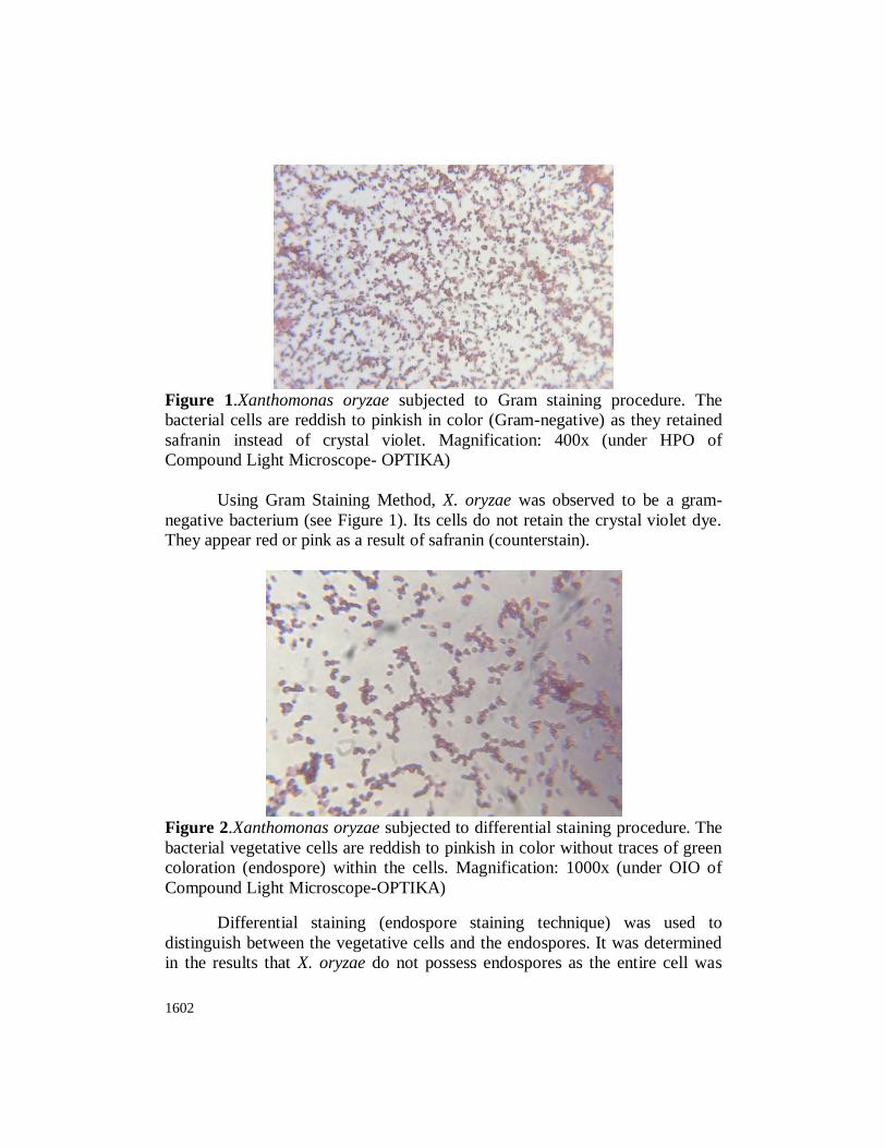
Figure 2.Xanthomonas oryzae subjected to differential staining procedure. The
bacterial vegetative cells are reddish to pinkish in color without traces of green
coloration (endospore) within the cells. Magnification: 1000x (under OIO of
Compound Light Microscope-OPTIKA)
Differential staining (endospore staining technique) was used to
distinguish between the vegetative cells and the endospores. It was determined
in the results that X. oryzae do not possess endospores as the entire cell was
International Journal of Agricultural Technology 2017 Vol. 13(7.2): 1597-1620
1603
pink in color with no dark green stains within the cell that would signify the
presence of endospores (see Figure 2).
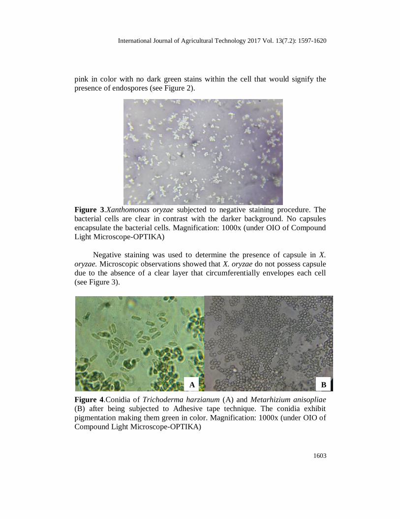
Figure 3.Xanthomonas oryzae subjected to negative staining procedure. The
bacterial cells are clear in contrast with the darker background. No capsules
encapsulate the bacterial cells. Magnification: 1000x (under OIO of Compound
Light Microscope-OPTIKA)
Negative staining was used to determine the presence of capsule in X.
oryzae. Microscopic observations showed that X. oryzae do not possess capsule
due to the absence of a clear layer that circumferentially envelopes each cell
(see Figure 3).
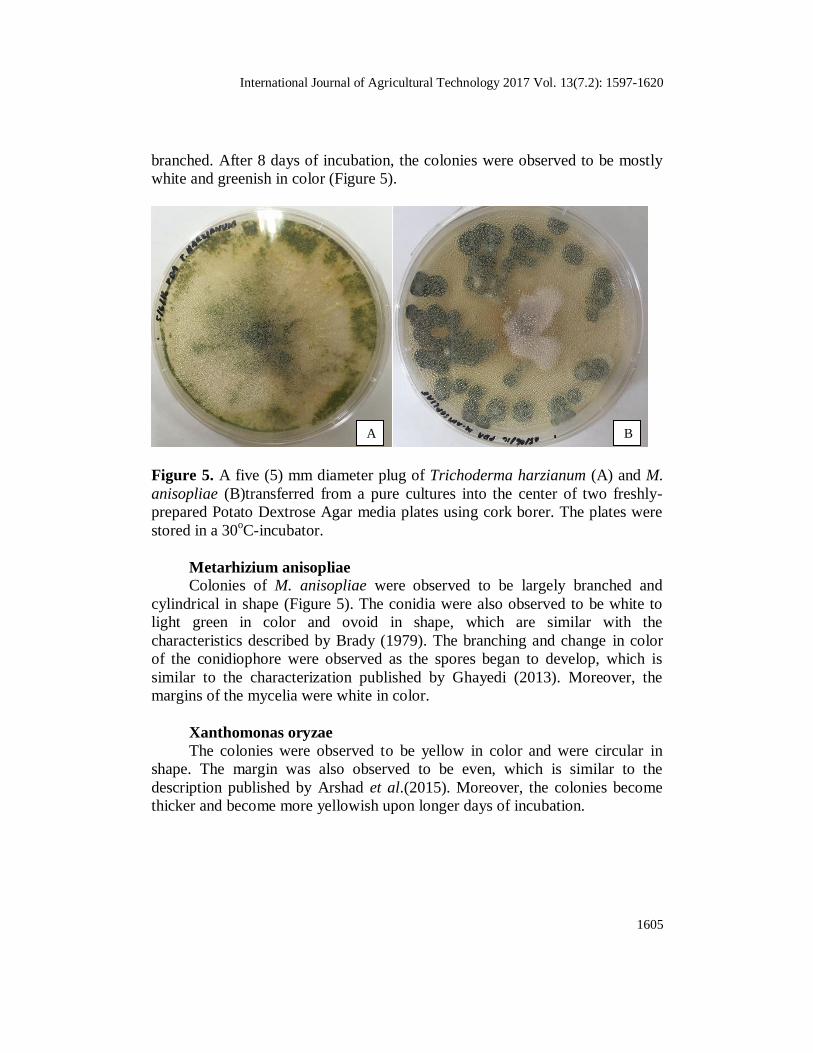
Figure 4.Conidia of Trichoderma harzianum (A) and Metarhizium anisopliae
(B) after being subjected to Adhesive tape technique. The conidia exhibit
pigmentation making them green in color. Magnification: 1000x (under OIO of
Compound Light Microscope-OPTIKA)
A B

1604
As for two fungi (T. harzianum and M. anisopliae), Adhesive Tape
Technique was done to observe the structure of conidia of each of the fungi.
Figure 4 shows the conidia of T. harzianum and M. anisopliae. They were heat-
fixed on a slide prior to microscopic observation. The T. harzianum is circular
in shape while the M. anisopliae is ovular. These conidia are asexually
produced spores which are externally borne to the cells that synthesize them.
Conidia also function for dispersal (“Fungi reproducing asexually by means of
conidia,” N.d.). In addition, the conidia are also involved in the propagation of
the fungi and their infection to the organisms they antagonize. In M. anisopliae,
the proteins found in conidia are those that are involved in protective processes,
appressorium formation, as well as degradation of the cuticle of their host (Su
et al., 2013). These proteins are unlike the proteins found in mycelia which
includes those proteins participating in biosynthetic and energy metabolism,
such as UTP-glucose-1-phosphate uridylyltransferase and heat shock protein 70
(Su et al., 2013). Conversely, in T. harzianum, the proteins found in conidia are
cellobiohydrolases I and II (CBH I and II) and endoglucanase I (EG I) degrade
crystalline cellulose into glucose (Messner et al., 1991) (Dong and Young,
2001). Cellobiohydrolases I and II (CBH I and II) hydrolyze from the chain
ends and predominantly synthesize cellobiose, while endoglucanase I (EG I)
hydrolyze the internal bonds in the cellulose chains (Dong and Young, 2001).
The conidia from both organisms were green in color. This conidial
pigmentation is significant for solar UV radiation tolerance in order not to delay
or not to stop their germination process when exposed to the sun/ solar UV
radiation, making them capable of doing their function (Braga et al., 2006).
These proteins are unlike the proteins found in mycelia which includes
cellulases, which degrade cellulose and chitinases, which degrade chitin (Volk,
2004).
Macroscopic observations (Colony characterization)
Trichoderma harzianum
T. harzianum was inoculated on Potato Dextrose Agar. After four days
of incubation, radial growth of the fungus, as manifested by white colour
spreading all throughout the plate, was observed. Based on the colony
characterization of T. harzianum in several journals (Bhattacharjee et al., 2014;
Jahan, et al., 2013), the observed characteristics were similar in terms of the
margin, texture and hypahal thickness. The colony grown on PDA had a
compact texture, thick hyphae, and had a regular margin. The conidia were
observed to be ovoid in shape, and the conidiophore was found to be highly
International Journal of Agricultural Technology 2017 Vol. 13(7.2): 1597-1620
1605
branched. After 8 days of incubation, the colonies were observed to be mostly
white and greenish in color (Figure 5).
Figure 5. A five (5) mm diameter plug of Trichoderma harzianum (A) and M.
anisopliae (B)transferred from a pure cultures into the center of two freshly-
prepared Potato Dextrose Agar media plates using cork borer. The plates were
stored in a 30oC-incubator.
Metarhizium anisopliae
Colonies of M. anisopliae were observed to be largely branched and
cylindrical in shape (Figure 5). The conidia were also observed to be white to
light green in color and ovoid in shape, which are similar with the
characteristics described by Brady (1979). The branching and change in color
of the conidiophore were observed as the spores began to develop, which is
similar to the characterization published by Ghayedi (2013). Moreover, the
margins of the mycelia were white in color.
Xanthomonas oryzae
The colonies were observed to be yellow in color and were circular in
shape. The margin was also observed to be even, which is similar to the
description published by Arshad et al.(2015). Moreover, the colonies become
thicker and become more yellowish upon longer days of incubation.
B A

1606
Figure 6.Isolates of X. oryzae in soybean agar after 48 hours of incubation
In Vivo
The Disease Severity (DS) and Disease incidence (DI) were measured
on the first and second week after the spraying of spore suspension. One-way
ANOVA and Post-hoc/ Multiple Comparison Test (Tukey HSD) were used to
analyze the DS and DI of the set-ups. The latter test was used to compare the
fungal treatments with control set-ups (positive and negative controls) and/or
other fungal treatments in order to determine if the fungal treatments were
capable of inhibiting the growth of X. oryzae.
As a basis, the positive control was treated with commercial BLB
stopper which surely inhibits BLB in rice, while the negative control is the
which untreated set-up which shows no inhibition against BLB in rice.
While it is true that three varieties of rice (susceptible, resistant, and
intermediate) were tested, statistical analysis showed that there is no significant
difference among the results of the varieties.
The significance values of T. harzianum against the control set-ups
(positive and negative controls) and/or other fungal treatments (M. anisopliae
and VAM). On the first week (Week 1) after the spraying of spore suspension,
comparison between T. harzianum and positive control showed significant
difference which a p-value of 0.038 and 0.014, respectively (Table 2). However,
a significant difference was also obtained for the comparison between T.
harzianum and negative control for all the three replicates having the same p-
value of 0.000. The comparison between T. harzianum and M. anisopliae
showed a p-value higher than 0.05. This indicated that there is no significant
difference on the measured DS and DI between T. harzianum and M. anisopliae.
Since M. anisopliae was found to be capable to inhibit BLB in rice that no
International Journal of Agricultural Technology 2017 Vol. 13(7.2): 1597-1620
1607
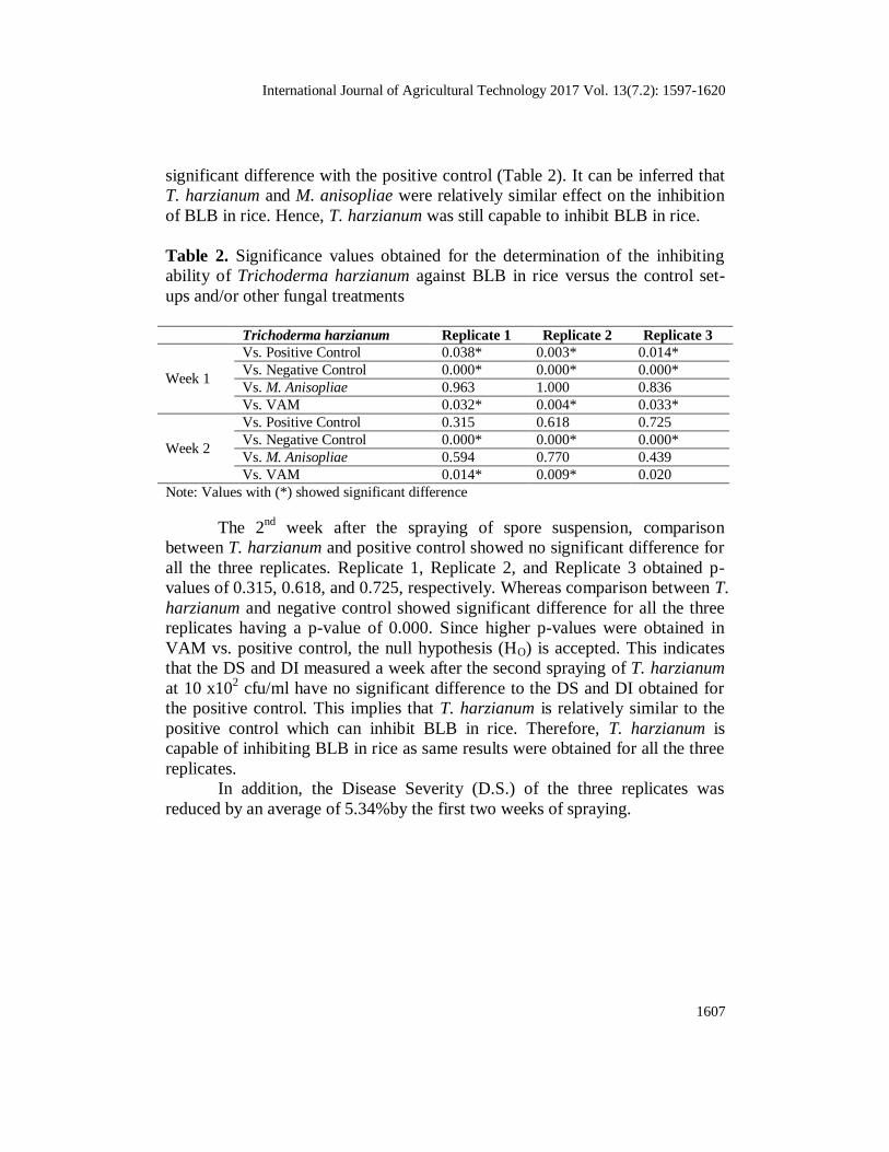
significant difference with the positive control (Table 2). It can be inferred that
T. harzianum and M. anisopliae were relatively similar effect on the inhibition
of BLB in rice. Hence, T. harzianum was still capable to inhibit BLB in rice.
Table 2. Significance values obtained for the determination of the inhibiting
ability of Trichoderma harzianum against BLB in rice versus the control set-
ups and/or other fungal treatments
Trichoderma harzianum Replicate 1 Replicate 2 Replicate 3
Week 1
Vs. Positive Control 0.038* 0.003* 0.014*
Vs. Negative Control 0.000* 0.000* 0.000*
Vs. M. Anisopliae 0.963 1.000 0.836
Vs. VAM 0.032* 0.004* 0.033*
Week 2
Vs. Positive Control 0.315 0.618 0.725
Vs. Negative Control 0.000* 0.000* 0.000*
Vs. M. Anisopliae 0.594 0.770 0.439
Vs. VAM 0.014* 0.009* 0.020
Note: Values with (*) showed significant difference
The 2nd
week after the spraying of spore suspension, comparison
between T. harzianum and positive control showed no significant difference for
all the three replicates. Replicate 1, Replicate 2, and Replicate 3 obtained p-
values of 0.315, 0.618, and 0.725, respectively. Whereas comparison between T.
harzianum and negative control showed significant difference for all the three
replicates having a p-value of 0.000. Since higher p-values were obtained in
VAM vs. positive control, the null hypothesis (HO) is accepted. This indicates
that the DS and DI measured a week after the second spraying of T. harzianum
at 10 x102 cfu/ml have no significant difference to the DS and DI obtained for
the positive control. This implies that T. harzianum is relatively similar to the
positive control which can inhibit BLB in rice. Therefore, T. harzianum is
capable of inhibiting BLB in rice as same results were obtained for all the three
replicates.
In addition, the Disease Severity (D.S.) of the three replicates was
reduced by an average of 5.34%by the first two weeks of spraying.
1608
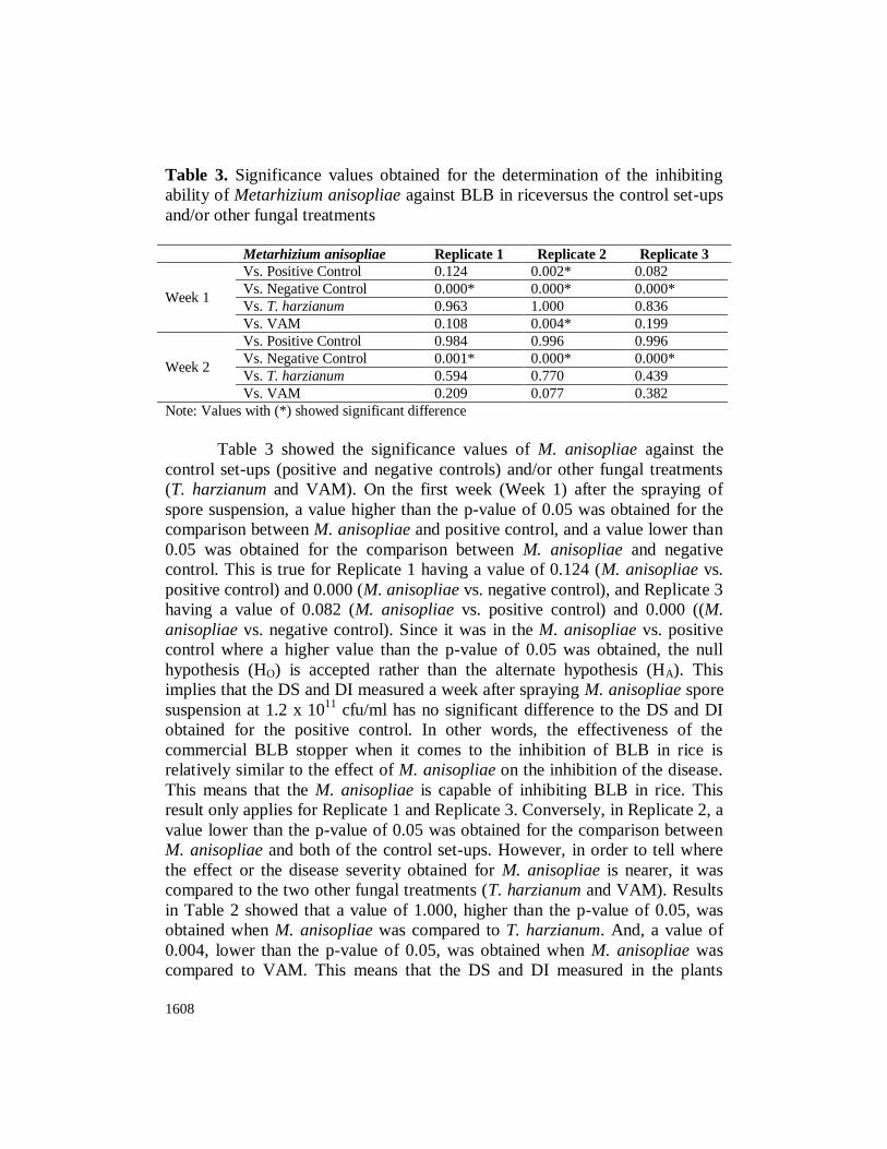
Table 3. Significance values obtained for the determination of the inhibiting
ability of Metarhizium anisopliae against BLB in riceversus the control set-ups
and/or other fungal treatments
Metarhizium anisopliae Replicate 1 Replicate 2 Replicate 3
Week 1
Vs. Positive Control 0.124 0.002* 0.082
Vs. Negative Control 0.000* 0.000* 0.000*
Vs. T. harzianum 0.963 1.000 0.836
Vs. VAM 0.108 0.004* 0.199
Week 2
Vs. Positive Control 0.984 0.996 0.996
Vs. Negative Control 0.001* 0.000* 0.000*
Vs. T. harzianum 0.594 0.770 0.439
Vs. VAM 0.209 0.077 0.382
Note: Values with (*) showed significant difference
Table 3 showed the significance values of M. anisopliae against the
control set-ups (positive and negative controls) and/or other fungal treatments
(T. harzianum and VAM). On the first week (Week 1) after the spraying of
spore suspension, a value higher than the p-value of 0.05 was obtained for the
comparison between M. anisopliae and positive control, and a value lower than
0.05 was obtained for the comparison between M. anisopliae and negative
control. This is true for Replicate 1 having a value of 0.124 (M. anisopliae vs.
positive control) and 0.000 (M. anisopliae vs. negative control), and Replicate 3
having a value of 0.082 (M. anisopliae vs. positive control) and 0.000 ((M.
anisopliae vs. negative control). Since it was in the M. anisopliae vs. positive
control where a higher value than the p-value of 0.05 was obtained, the null
hypothesis (HO) is accepted rather than the alternate hypothesis (HA). This
implies that the DS and DI measured a week after spraying M. anisopliae spore
suspension at 1.2 x 10¹¹
cfu/ml has no significant difference to the DS and DI
obtained for the positive control. In other words, the effectiveness of the
commercial BLB stopper when it comes to the inhibition of BLB in rice is
relatively similar to the effect of M. anisopliae on the inhibition of the disease.
This means that the M. anisopliae is capable of inhibiting BLB in rice. This
result only applies for Replicate 1 and Replicate 3. Conversely, in Replicate 2, a
value lower than the p-value of 0.05 was obtained for the comparison between
M. anisopliae and both of the control set-ups. However, in order to tell where
the effect or the disease severity obtained for M. anisopliae is nearer, it was
compared to the two other fungal treatments (T. harzianum and VAM). Results
in Table 2 showed that a value of 1.000, higher than the p-value of 0.05, was
obtained when M. anisopliae was compared to T. harzianum. And, a value of
0.004, lower than the p-value of 0.05, was obtained when M. anisopliae was
compared to VAM. This means that the DS and DI measured in the plants

International Journal of Agricultural Technology 2017 Vol. 13(7.2): 1597-1620
1609
treated with M. anisopliae had no significant difference to that of the plants
treated with T. harzianum. The effectiveness of the T. harzianum when it comes
to the inhibition of BLB in rice was relatively similar to the effect of M.
anisopliae on the inhibition of the said disease. This means that for Replicate 2,
M. anisopliae is still capable of inhibiting BLB in rice.
On the 2nd
week after the spraying of spore suspension, a value higher
than the p-value of 0.05 was obtained for the comparison of the effect of M.
anisopliae and positive control, and a value lower than the p-value of 0.05 were
obtained for the comparison of the effect of M. anisopliae and negative control.
This is visible on all replicates (Table 3). Because it is in the comparison
between M. anisopliae and positive control where the significance value was
greater than the p-value of 0.05, the alternate hypothesis (HA) was accepted and
the null hypothesis (HO) is rejected. Therefore, the DS and DI of the rice plants
treated with M. anisopliae measured a week after the second spraying had no
significant difference to the DS and DI measured in the positive control. The
effectiveness of the commercial BLB stopper in terms of the growth of
inhibition or eradication of BLB in rice was relatively similar to the effect of M.
anisopliae when it come to the inhibition of BLB. This strongly implies that M.
anisopliae is capable of inhibiting BLB in rice as same results were obtained.
Moreover, the Disease Severity (D.S.) of the three replicates was reduced by
an average of 1.06% by the first two weeks of spraying.
Table 4 showed the significance values of VAM against the control set-
ups (positive and negative controls) and/or other fungal treatments (T.
harzianum and M. anisopliae). On the first week (Week 1), no significant
difference was obtained for the comparison between VAM and positive control
with a p-value higher than 0.05, while a significant difference was obtained for
the comparison between VAM and negative control with a p-value lower than
0.05. This applies for the three replicates. Since p-values of 1.000, 0.970, and
0.953 for Replicate 1, Replicate 2 and Replicate 3, respectively, were obtained
in VAM vs. positive control, the null hypothesis (HO) is accepted. This
indicates that the DS and DI measured a week after spraying spore suspension
of VAM at 1.2 x 10¹¹
cfu/mlhas no significant difference to the DS and DI
obtained for the positive control. Thisfurther implies that VAM is relatively
similar to the positive control which can inhibit BLB in rice. Therefore, VAM
was capable of inhibiting BLB in rice as same results were obtained for all the
three replicates.

1610
Table 4. Significance values obtained for the determination of the inhibiting
ability of Vesicular Arbuscular Mycorrhizae (VAM) against BLB in riceversus
the control set-ups and/or other fungal treatments
VAM Replicate 1 Replicate 2 Replicate 3
Week 1
Vs. Positive Control 1.000 0.970 0.953
Vs. Negative Control 0.024* 0.000* 0.000*
Vs. T. harzianum 0.032* 0.004* 0.033*
Vs. M. Anisopliae 0.108 0.004* 0.199
Week 2
Vs. Positive Control 0.438 0.200 0.278
Vs. Negative Control 0.067 0.000* 0.000*
Vs. T. harzianum 0.014 0.009* 0.020*
Vs. M. Anisopliae 0.209 0.077 0.382
Note: Values with (*) showed significant difference
When compared with the other treatments, VAM showed significant
difference with T. harzianum in the three replicates with p-values of 0.032,
0.004, and 0.033 for Replicate 1, Replicate 2 and Replicate 3, respectively.
Whereas VAM showed no significant difference with M. anisopliae in
Replicate 1 and Replicate 3 with p-values of 0.108 and 0.199, respectively. This
indicates that both VAM and M. anisopliae generally have relatively similar
effect in inhibiting the disease compared to T. harzianum, although VAM
showed a significant difference with M. anisopliae in Replicate 2.
As for the 2nd
week, it was revealed that VAM had no significant
difference with the positive control with a p-value higher than 0.05. This is
visible for the three replicates. On the other hand, VAM had a significant
difference with the negative control with a p-value lower than 0.05. This
applies for Replicate 2 and Replicate 3. A p-value higher than 0.05 was
obtained in VAM vs. positive control, thus the null hypothesis (HO) is accepted.
This indicated that the DS and DI measured the week after the second spraying
of spore suspension of VAM at 1.2 x 10¹¹
cfu/ml has no significant difference
to the DS and DI obtained for the positive control. This further implies that
VAM was relatively similar to the positive control which was capable of
inhibiting BLB in rice. Therefore, VAM was capable of inhibiting the disease
despite having no significant difference with the negative control in Replicate 1.
When compared with the other treatments, VAM showed significant difference
with T. harzianum with p-values of 0.014, 0.009, and 0.020 for Replicate 1,
Replicate 2 and Replicate 3, respectively, while it showed no significant
difference with M. anisopliae with a p-value of 0.209, 0.077 and 0.382 for
Replicate 1, Replicate 2 and Replicate 3, respectively. This implies that both
VAM and M. anisopliae had relatively similar effect in inhibiting the disease
compared to T. harzianum as same results were observed for all the three

International Journal of Agricultural Technology 2017 Vol. 13(7.2): 1597-1620
1611
replicates. The comparison between VAM and M. anisopliae obtained on the
second week of spraying spore suspensions was consistent with the comparison
which obtained in the first week of spraying the spore suspension.Furthermore,
the Disease Severity (D.S.) of the three replicates was reduced by an average of
0.293% by the first two weeks of spraying.
In Vitro
For the antagonistic test, spread plate method was done to evenly
distribute the bacteria all over the Nutrient agar plate. Three hundred microliter
(300 µl) of broth inoculated with bacteria which has a similar turbidity as that
of a 4.0 MacFarland standard was transferred to each of the plates. This
turbidity of the broth is equivalent to an approximate bacterial density of 12 x
108 cfu/ml. Further trials on spread plating showed that 300-µl volume of broth
is the most suitable volume that can occupy and cover the nutrient agar media
without being too runny when tilted or too bare and patchy. The placement of a
five millimeter diameter T. harzianum and M. anisopliae plugs and disk
containing commercial BLB stopper at the center of the bacterial lawn were
done on separate plates after 48 hours of incubation of the bacterial lawn at
37 C. Three replicates were made. The set-ups were initially observed 48 hours
after the placement of the fungal plug.
Figure 7. Antagonistic test against X. oryzae. A 5 mm diameter of T.
harzianum (A) and M. anisopliae (B)plugs and commercial BLB stopper discs
(C) were transferred at the center of two-day old Xanthomonas oryzae lawn.
The X. oryzae (300 µm) was spread all over the Nutrient Agar media in three
separate petri plates using Spread Plate technique prior to the transfer of fungal
plug.

1612
Trichoderma harzianum
The results for the inhibition of X. oryzae against T. harzianum was
negligible because of the incompatibility of medium. X. oryzae and the T.
harzianum failed to grow together in the same medium, which, in this case, was
Nutrient agar. The growth of X. oryzae was only observed on Soybean agar,
while the growth of T. harzianum was only observed in Potato Dextrose Agar.
Although Nutrient Agar is a general media, the components and the conditions
required for the growth of X. oryzae were not supplemented by N.A.
Metarhizium anisopliae
M. anisopliae also showed growth inhibition against X. oryzae with the
production of clear zones after 48 hours of incubation. On replicates 1 and 2,
clear zones of 0.2 cm were observed while on replicate 3, a clear zone of 0.4
cm was observed. However, formations of clear zones are considered
inconclusive.
Commercial BLB Stopper (Positive Control)
Experimental results revealed that BLB Stopper was not able to inhibit X.
oryzae due to the absence of inhibition zone in all replicates. Results are
inconclusive.
Trichoderma harzianum as potential biocontrol for BLB
Based on the results, the spraying of the spore suspension of T.
harzianumat 10 x102 cfu/ml after the first week and second week of spraying
has significantly reduced the DS and DI of the plant leaves infected with BLB
by 5.34%. T. harzianum and positive control also showed no significant
difference for all the three replicates with obtained p-values of 0.315, 0.618,
and 0.725, respectively. According to a study conducted by Gangwar (2013),
the reduction of the severity of the disease is an indication that the T.
harzianum as a fungal bioagent is capable of proliferating and establishing on
the surface of the rice host.
According to Ou (1985), Xanthomonas oryxzae pv oryzae enters
through the hydathodes at the leaf tip and leaf margin of the rice leaf. Curtis
(1943) adds that the cells on the surface of the leaf may be suspended on the
guttation fluid as it exudes at night. The cells can then enter the plant either by
swimming or through the fluid that is withdrawn into the leaf in the morning
(Curtis, 1943). After bacterial multiplication in the intercellular spaces of the
underlying epitheme, they can then enter and spread out through the xylem
(Noda and Kaku, 1999). Aside from this, Ou (1985) says that Xoo can also
enter the xylem through the wounds or openings that resulted from the roots

International Journal of Agricultural Technology 2017 Vol. 13(7.2): 1597-1620
1613
emerging at the base of leaf sheath. Upon entering the xylem, the bacteria can
already interact with the xylem parenchyma cells, moving vertically through
the leaf’s primary veins, and laterally through the commissural veins (Hilaire et
al., 2001). Just after a few days, formation of beads or strands of exudate can
already be observed. According to Mew and company (1993), it is a
characteristic of Bacterial Leaf Blight that resulted from the bacterial cells that
filled the xylem vessels and oozed out from the hydathodes, making it a
possible source of secondary inoculum.
There are different mechanisms by which T. harzianum can serve as a
biocontrol agent against foliar pathogens. Among the most studied mechanisms
include mycoparasitism, competition, and antibiosis (Elad, 2000). In antibiosis,
Trichoderma species are known to be capable of releasing antibiotics and
various chemicals that can harm the pathogens and inhibit their growth
(Leelavathi, et. al., 2014).
The fungi’s ability to colonize and penetrate root tissues, and to induce
series of changes in the plant’s morphology and biochemistry, result to an
Induced Systemic Resistance in the entire plant (ISR). Induced Systemic
Resistance is an important mechanism for biocontrol in vegetative tissues that
is caused by different microorganisms including T. harzianum to protect the
plant from soil or foliar pathogens (Paulitz and Matta, 2000). Aside from this, T.
harzianum is also known because of its ability of producing protease as one of
its biocontrol mechanisms (Elad and Kapat, 1999). Observations from a study
conducted by Elad and Kapat (1999) indicate that T. harzianum secretes
proteolytic enzyme on the leaves infected with a pathogen (B. cinerea), which
then leads to the reduction of the germination and disease development caused
by the pathogen. It was also observed that these proteases secreted by T.
harzianum were able to deactivate the hydrolytic enzymes produced by the
pathogen.
However, results for antagonistic tests of T. harzianum against X. oryzae
in vitro are inconclusive because of the incompatibility of the medium used for
the growth of X. oryzae and T. harzianum. X. oryzae grew best on Soybean
agar, while T. harzianum grew best on Potato Dextrose Agar (PDA). Although
Nutrient Agar (N.A.) is a general media, specific components and conditions
required for the growth of X. oryzae were not supplemented by N.A.
Separate studies by Leelavathi et al. (2014) and Parthasarathy (2014)
revealed positive results wherein Trichoderma was observed to be highly
effective in inhibiting the growth of X. oryzae in vitro using Muller Hinton
medium. However, when the same methodology was used, isolates of X. oryzae
still failed to grow on Muller Hinton medium. In addition, other media like
Saboraud Dextrose Agar (SDA), Muller Hinton Agar (M.H.A.), and Wakimoto

1614
Agar were also used in an attempt to look for a medium where bacteria and the
fungi would grow together for an unbiased antagonistic test, but all of the trials
were unsuccessful. Sivasithamparam and Ghisalberti (1998), strains of
Trichoderma can also produce 40 metabolites that can contribute to their
antibiotic activity, aiding to their effectivity against species of Gram positive
and Gram negative bacteria (Leelavathi, et. al., 2014)
According to Bhatttacharjee and company (2014), Trichoderma species
are known to produce antibiotics and antifungal toxic metabolites. Because of
their ability to secrete enzymes like glucanase, chitinase, cellulose and protease,
they are capable of degrading the cell wall of pathogens, which in turn inhibits
their growth (Bhatttacharjee et al., 2014). These antibiotics, specifically
trichodermin and harzianolide that the Trichoderma strains produce, in
combination with hydrolytic enzymes, lead to a higher level of antagonism
(Howel, 1998).
Aside from this, Trichoderma species also have mycoparasitic ability
which works against economically important plant pathogens, allowing the
development of biocontrol strategies (Harman, et. al., 2004; Motlagh et al.,
2013). As aggressive competitors, Trichoderma species do not only grow very
fast, but they also rapidly colonize substrates to eliminate pathogens (Papavizas,
1985).
All of these mechanisms can act alone or in combination to increase the
efficiency of T. harzianum as a biocontrol agent against pathogens that enter
through leaves (Leelavathi, et. al., 2014). On separate studies conducted by
Gangwar and Sinha (2012) and Kumar and company (2009), the same results
were also observed wherein T. harzianum was able restrict the severity of
Xanthomonas oryzae in rice.
Metarhizium anisopliae as potential biocontrol for BLB
The spraying of the spore suspension of M. anisopliae at 1.2 x 10¹¹
cfu/ml after the first week and second week of spraying was proven to be
efficient in suppressing the DS and DI of the plant leaves infected with BLB
according to the results. Furthermore, there was an average Disease Severity
(D.S.) reduction of 1.06% upon the treatment of M. anisopliae.
Biological control with M. anisopliae offers a long-lasting control to
various insects and plant pathogens (including pathogenic bacteria in rice)
without imparting damage to the environment and to non-target organisms
(Universal Bio-organic and Multiservices Pvt. Ltd. 2008). Some studies
showed that the fungi were internally colonized the plant leaves, petioles and
stem (Batta, 2013). However, the mechanism by which M. anisopliae enters the
plant (for the release of compounds and secondary metabolites) after foliar

International Journal of Agricultural Technology 2017 Vol. 13(7.2): 1597-1620
1615
spray is still unclear. Additionally, M. anisopliae is capable of colonizing plant
roots where it concurrently acts as biofertilizer and biopesticide in order to
boost plant growth (Gao et al., 2011).
The life cycle of M. anisopliae, entomopathogenic fungi, is associated
with both the synthesis and secretion of various active metabolites such as
extracellular enzymes and low-molecular weight compounds (i.e. toxins).
These toxic metabolites or by-products primarily aids the M. anisopliae to resist
and guard themselves against invading pathogens whether bacteria, fungi or
insects. M. anisopliae, and entomopathogenic fungi in general, produce a wide
variety of secondary metabolites with different activities. Their activities
include antibiotic effect, release of cytotoxic substances, insecticidal effect,
release of compounds which induce or inhibit growth of pathogen, attractor,
repellent, and the like. However, the chemical constitution and metabolic fluxes
of the M. anisopliae differ depending on ecological conditions. Particularly, M.
anisopliae secrete a wide array of relatively low molecular weight secondary
metabolites wherein some of which have antibiotic properties whereas others
are vital pathogenicity determinants. Even though these metabolites are
pertained to be toxins, only little is known regarding their properties,
production, as well as spatial distribution (Ravindran et al., 2014).
In particular, M. anisopliae and other entomopathogenic fungi are
considered as unique outstanding microbial entities due to their ingenious
ability to synthesize plethora of bioactive compounds and from the dependence
of their morphological differentiation. For instance, M. anisopliae produces a
cyclodepsipeptide called destruxin that inhibits the growth of various
pathogenic bacteria (i.e. Xanthomonas oryzae) (Male et al., 2009). The
secondary metabolites play an essential role in improving the fungal
adaptability to endure diverse natural habitats. In addition, these metabolites
also act as signaling molecules for the establishment of niche in fungal-plant or
fungal-pathogen interactions, and may also serve as stress protectors.
Nonetheless, the exact function of many secondary metabolites in M. anisopliae
is still unknown. Various chemical classes have been identified in M.
anisopliae and the entire Metarhizium genus. These are cytochalasins C and D,
myroridins, destruxin A,B and E, viridoxin, swainsonine, helvonic acid, 12-
hydroxyovalicin, hydroxy- fungerin, 7-desmethyl analogues of fusarin C and
(8Z)-fusarin C, serinocyclins A and B, as well as aurovertins (Male et al., 2009).
These metabolites are toxic to various insects and pathogenic microbes
(including bacteria like Xanthomonas oryzae) (Gao et al., 2011; Male et al.,
2009). Almost all of these metabolites can be isolated from mycelia or from
fermentation extracts of M. anisopliae. There is lesser information on the
secondary metabolites exclusively present in the conidia of M. anisopliae

1616
(Ravindran et al., 2014). These metabolites, especially destruxin, allows
morphological and cytoskeletal changes in various insects and pathogenic
bacteria. They harmfully affect the pathogen’s cellular immune responses
namely encapsulation and phagocytosis (Male et al., 2009).
Vesicular Arbuscular Mycorrhizae (VAM) as potential biocontrol for
BLB
Based on the results, the spraying of the spore suspension of VAM at 1.2
x 1011
cfu/ml after the first week and second week of spraying has significantly
reduced the DS and DI of the plant leaves infected with BLB by 0.293%. This
indicates the potential of VAM to be utilized as biocontrol agent against BLB.
According to studies, mychorrhiza-induced protection is provided by the
improvement of plant nutrition and the consequent compensation of the
damages caused by the pathogen (Trotta et al. 1996; Fritz 2006; Liu et al.
2007)). With the advancement on the understanding of the physiology and
regulation of the Arbuscular Mycorrhiza (AM) symbiosis, it was further
revealed that symbiosis with AM may involve the activation of plant defense
mechanisms and changes in the plant architecture, root exudation, and even in
the populations of microbes in the rhizosphere (Azcón-Aguilar and Barea 1996;
Whipps 2004). AM symbiosis also confer resistance or tolerance in plants
against biotic stresses. The impact of the symbiotic relationship of VAM with
plants in terms of resistance and tolerance to biotic stresses depends on the AM
fungal isolates and can be modulated by environmental conditions (Pozo and
Azcón-Aguilar, 2007).
Many studies have proven that AM symbiosis had greatly influenced the
inhibition or the reduction of disease severity and incidence caused by soil-
borne pathogens such as fungi, bacteria and oomycetes (Whipps, 2004).
Conversely, fewer studies have focused on the effect of AM symbiosis on
above-ground diseases caused by biotrophic and necrotrophic pathogens and
shoot pathogens.
According to studies, AM symbiosis shows a positive effect on plant
resistance against shoot diseases such as diseases caused by the bacterial
pathogen X. campetris in Medicago (Liu et al., 2007) and by the necrotrophic
fungus Alternaria solani in tomato (Fritz et al., 2006; De La Noval et al. 2007).
Another study also reveals that AM establishment in tomatoes infected with
phytoplasma, specialized obligate parasites of phloem tissue, leads to the
reduction of the symptoms of the disease (Lingua et al., 2002).
In general, two main mechanisms are considered on how AM symbiosis
affects plant pathogens. One is the potential changes in the nutrient levels of the
host plant and alterations of the source-sink relation within the plant which may
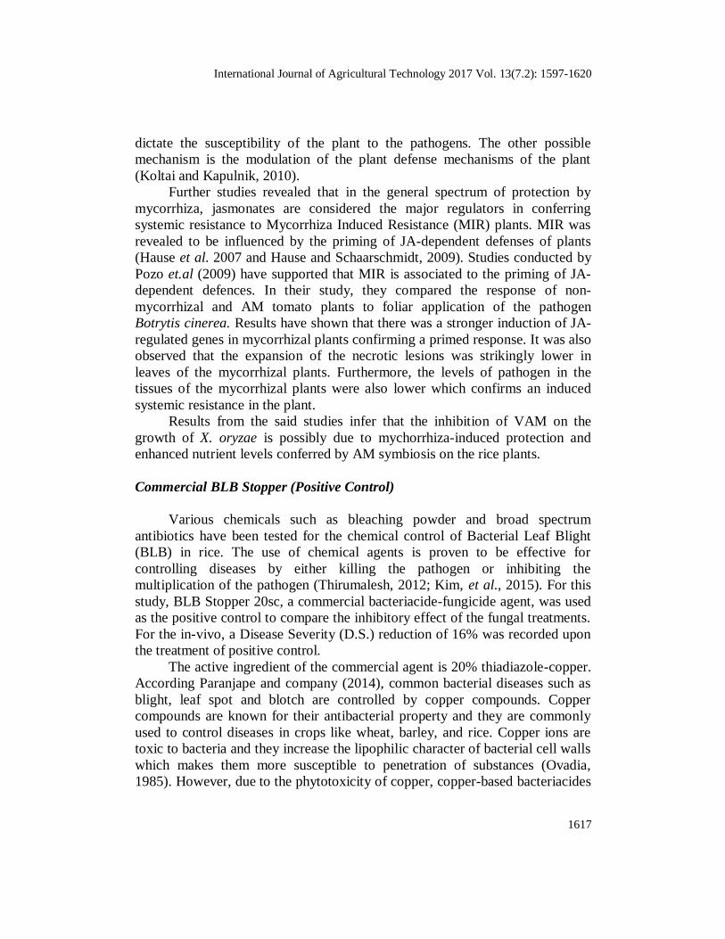
International Journal of Agricultural Technology 2017 Vol. 13(7.2): 1597-1620
1617
dictate the susceptibility of the plant to the pathogens. The other possible
mechanism is the modulation of the plant defense mechanisms of the plant
(Koltai and Kapulnik, 2010).
Further studies revealed that in the general spectrum of protection by
mycorrhiza, jasmonates are considered the major regulators in conferring
systemic resistance to Mycorrhiza Induced Resistance (MIR) plants. MIR was
revealed to be influenced by the priming of JA-dependent defenses of plants
(Hause et al. 2007 and Hause and Schaarschmidt, 2009). Studies conducted by
Pozo et.al (2009) have supported that MIR is associated to the priming of JA-
dependent defences. In their study, they compared the response of non-
mycorrhizal and AM tomato plants to foliar application of the pathogen
Botrytis cinerea. Results have shown that there was a stronger induction of JA-
regulated genes in mycorrhizal plants confirming a primed response. It was also
observed that the expansion of the necrotic lesions was strikingly lower in
leaves of the mycorrhizal plants. Furthermore, the levels of pathogen in the
tissues of the mycorrhizal plants were also lower which confirms an induced
systemic resistance in the plant.
Results from the said studies infer that the inhibition of VAM on the
growth of X. oryzae is possibly due to mychorrhiza-induced protection and
enhanced nutrient levels conferred by AM symbiosis on the rice plants.
Commercial BLB Stopper (Positive Control)
Various chemicals such as bleaching powder and broad spectrum
antibiotics have been tested for the chemical control of Bacterial Leaf Blight
(BLB) in rice. The use of chemical agents is proven to be effective for
controlling diseases by either killing the pathogen or inhibiting the
multiplication of the pathogen (Thirumalesh, 2012; Kim, et al., 2015). For this
study, BLB Stopper 20sc, a commercial bacteriacide-fungicide agent, was used
as the positive control to compare the inhibitory effect of the fungal treatments.
For the in-vivo, a Disease Severity (D.S.) reduction of 16% was recorded upon
the treatment of positive control.
The active ingredient of the commercial agent is 20% thiadiazole-copper.
According Paranjape and company (2014), common bacterial diseases such as
blight, leaf spot and blotch are controlled by copper compounds. Copper
compounds are known for their antibacterial property and they are commonly
used to control diseases in crops like wheat, barley, and rice. Copper ions are
toxic to bacteria and they increase the lipophilic character of bacterial cell walls
which makes them more susceptible to penetration of substances (Ovadia,
1985). However, due to the phytotoxicity of copper, copper-based bacteriacides

1618
are mixed with copper-chelating fungicides such as as manzoceb or maneb or
with Ferric chloride (FeCl3.6H2O). Some of the major bacteriacides are
bronopol, hexachlorophene, acibenzolar-s-meethyl, benzalkonium chloride,
kasugamycin, nitrapyrin, oxylinic acid, probenazole, streptomycin and copper-
containing bacteriacides such as copper hydroxide and thiadiazole-copper
(Paranjapeet et al., 2015). Gleason and company (1993) also verified the
efficiency of copper-containing products in the reduction of foliar leaf and fruit
spotting.
Several studies have revealed that efficiency of copper-containing
bacteriocides, however, this study showed contrasting results in-vitro. This is
may be due to the other components of BLB Stopper Sc20 (not specified in the
label) which may have reacted with the components of the media used that may
have enhanced the growth of the pathogen, rather than suppressing its growth
or there is something lacking in the in-vitro for effective reaction to ensue. The
inhibitory effect of the commercial agent may not also be evident in-vitro but is
more evident when applied in the field due to several factors such as physical
conditions and the interaction of the commercial agent with the infected plant
and with the pathogen in the plant.
Conclusion
Xanthomonas oryzae is the major and sole cause of Bacterial Leaf
Blight (BLB) in rice. Three fungal treatments (Trichoderma harzianum,
Metarhizium anisopliae, and Vesicular Arbuscular Myccrorhizae) were utilized
to determine their probable antagonistic effect to X. oryzae. For in-vitro tests,
incompatibility of media was the primary reason why the inhibition zones
observed in the positive control and in the three (3) fungal treatments are
considered inconclusive. For in-vivo tests, statistical analysis of the DS and DI
of the plants treated separately with the spore suspensions of T. harzianum, M.
anisopliae, and VAM were determined to be relatively/ significantly different
to the DS and DI obtained for the negative control set-up. This is true for all
replicates of plants that were treated with each of the fungal spore suspensions.
Results imply that the effectiveness of the three fungal treatments is relatively
similar to the effect of the commercial BLB stopper when it comes to the
inhibition and antagonism of BLB in rice. Furthermore, calculation of percent
reduction of Disease Severities showed that there is a 5.34% reduction of BLB
disease in plants treated with T. harzianum, 1.06% in plants treated with M.
anisopliae, and 0.293% in plants treated with VAM. Therefore, the fungi used
were confirmed to be effective biocontrol agents against BLB in rice. They are

International Journal of Agricultural Technology 2017 Vol. 13(7.2): 1597-1620
1619
environment-friendly and cheaper substitutes for chemically-based bactericides
against BLB, thus limiting the use of harmful chemicals.
Acknowledgement
The author would like to offer special thanks to the Department of Agriculture, Region
02 for funding the research study. The said research would not be successful without the
funding agency. To Ms. Anna Theresa Isabel O. Rebong for her guidance and support.
Moreover, the author would like also to thank the thesis students from UP Baguio in conducting
the subactivities of the study and lastly, to the field assistants who worked hard for the
completion of the study.
References
Abbott, L. K. and Robson, A. D. (1984). The effect of VA mycorrhizae on plant growth. In:
Kannaiyan, S. Biotechnology of Biofertilizers (pp 312-314.). Narosa Publishing House,
New Delhi, India.
Aryal, S. (2015). Endospore staining- principle, reagents, procedure and result. Retrieved from
http://www.microbiologyinfo.com/endospore-staining-principle-reagents-procedure-
and-result/ on May 15, 2016.
Ashrafuzzaman, H. (1987). Chemical control of bacterial leaf blight of paddy caused by
Xanthomonas campestris pv. Oryzae. Current Plant Science and Biotechnology in
Agriculture, Vol.4, pp 955-958. Barnett, H. L., and Binder, F. L. (1973). The fungal host-parasite relationship. Annu. Rev.
Phytopathol. 11, 273-292. DOI: 10.1146/annurev.py.11.090173.001421.
Batta, Y. (2013). Efficacy of endophytic and applied Metarhizium anisopliae (Metch.) Sorokin
(Ascomycota: Hypocreales) against larvae of Plutella xylostella L. (Yponomeutidae:
Lepidoptera) infesting Brassica napus plants. Crop Protection Journal Vol. 44, Pages
128-134.
Bhattacharjee, R. and Dey, U. (2013). An overview of fungal and bacterial biopesticides to
control plant pathogens/ diseases. African Journal of Microbiology Research, Vol. 8, No.
17. DOI: 10.5897/AJMR2013.6356.
Boyetchko, S.M. and Tewari, J.P. (1988). The effect of VA mycorrhizal fungi on infection by
Bipolaris sorokiniana in barley.In: Kannaiyan, S. Biotechnology of Biofertilizers (pp 312-314.). Narosa Publishing House, New Delhi, India.
Boyetchko, S.M. and Tewari, J.P. (1990). Effect of Phosphorus and VA mycorrhizal fungi on
common root rot of barley. In: Kannaiyan, S. Biotechnology of Biofertilizers (pp 312-
314.). Narosa Publishing House, New Delhi, India.
British Society for Plant Pathology. (2014). Xanthomonas bacteria. Retrieved from
http://www.bsp p.org.uk/downloads/education/BSPP_Xantho_Info.pdf on November 14,
2015.
Central Rice Research Institute (2011). Causal organisms and predisposing factors for bacterial
leaf streak of rice. Retrieved from http://www.rkmp.co.in/content/causal-organism-and-
predisposing-factors-for-bacterial-leaf-streak-of-rice on May 15, 2016.
Dehne, H.W. (1982). Interaction between vesicular-arbuscular mycorrhizal fungi and plant
pathogens. In: Kannaiyan, S. Biotechnology of Biofertilizers (pp 312-314.). Narosa Publishing House, New Delhi, India.

1620
Fritz,M., Jakobsen, I., Lyngkjaer, M.F., Thordal-Christensen, H., and Pons-Kuehnemann, J.
(2006). Arbuscular mycorrhiza reduces susceptibility of tomato to Alternaria solani.
Mycorrhiza 16:413–419.
Fungi reproducing asexually by means of conidia. N.d. Rerieved from
mnb.ca/mycologywebpages/NaturalHistoryOfFungi/Conidia.html on May 15, 2016.
Haus, B., Mrosk, C., Isayenkov, S., and Strack, D. (2007). Jasmonates in arbuscular mycorrhizal interactions. Phytochem 68:101–110.
Kumar, M., R.L. Parate and B.N. Ninawe. (2009). Effect of botanicals, bioagents and some
chemicals against Xanthomonas oryzae pv. oryzae. Journal of Plant Disease Sciences ,
Vol. 4 No. 1 pp. 60-63.
Messner, R., Kubicek- Pranz, E. M., Gsur, A., and Kubicek, C. P. (1991). Cellobiohydrolase II
is the main conidial-bound cellulose in Trichoderma reesei and other Trichoderma
strains. Retrieved from http://www.ncbi.nlm.nih.gov/pubmed/1953300 on May 16, 2016.
Mew, T.W., Alvarez, A.M., Leach, J.E., and Swings, J. (1993). Plant Dis. In: Sonti, R.V. 1998.
Bacterial leaf blight of rice: New insights from molecular genetics. Centre for Cellular
and Molecular Biology; Upper Road, Hyderabad 500 007, India. Current Science, Vol.
74, No. 3. Naher, L., Yusuf, U.K., Ismail, A. and Hossain, K. (2014). Trichoderma spp.: A biocontrol
agent for sustainable management of plant diseases. Pakistan Journal of Botany, Vol. 46,
No. 4. DOI: 1489-1493, 2014.
Sharma, M., Mittal, N., and Mukerji, K.G. (1998). Fungi: Tool for plant disease management.
In: Mukerji, K. G., Chamola, B.P., Upadhyay, R.K. Biotechnological Approaches in
Biocontrol of Plant Pathogens (pp 147). Kluwer Academic / Plenum Publishers 233
Spring Street, New York, N.Y. 10013.
Siddiqui, Z.A. and I. Mahmood, (1995). Some observations on the management of the wilt
disease complex of pigeonpea by treatment with a vesicular arbuscular fungus and
biocontrol agents for nematodes. Biores. Technol., 54: 227-230.
(Received 15 October 2017; accepted 25 November2017)