Uterine myomas: management - Uterine Fibroids - Hope For Fibroids
Upload
ayub-medical-collegeCategory
view
61download
2
Noshairwan Ali KhanRoll. No: 07-128Final Year ( Batch I )

LEIOMYOMA
What is a leiomyoma?It is a benign neoplasm of the muscular wall of
the uterus composed primarily of smooth muscle
What is the incidence of leiomyomas?They are the most common pelvic tumorsIt is found in 25% of white women & 50% of
black women. Age… greater than 30

ETIOLOGY
Unknown Each individual myoma is unicellular in origin Estogens no evidence that it is a causative factor ,
it has been implicated in growth of myomas Myomas contain estrogen receptors in higher
concentration than surrounding myometrium Myomas may increase in size with estrogen therapy
& in pregnancy & decrease after menopause They are not detectable before puberty Progestrone increase mitotic activity & reduce
apoptosis in size There may be genetic predisposition

PATHOLOGY
Frequently multiple
May reach 15 cm in size or larger
Firm
Spherical or irregularly lobulated
Have a false capsule ( pseudocapsule)
Can be easily enucleated from surrounding myometrium

CLASSIFICATION Submucous
leiomyoma Pedunculated
submucous Intramural or
interstitial Subserous or
subperitoneal Pedunculated
abdominal Parasitic Intraligmentary Cervical

MICROSCOPIC STRUCTURE Whorled appearance nonstriated muscle
fibers arranged in bundles running in different directions
Individual cells are spindle shaped uniform Varying amount of connective tissue are
interlaced between muscle fibers Pseudocapsule of areolar tissue & compressed
myometrium Arteries are less dense than myometrium & do
not have a regular pattern of distribution 1-2 major vesseles are found at the base or
pedicle


1-BENIGN DEGENERATION
Atrophic Hyaline yellow, soft gelatinous areas
Cystic liquefaction follows extreme hyalinization
Calcific circulatory deprivation precipitation of ca carbonate & phosphate
Septic circulatory deprivation necrosis infection
Myxomatous (fatty) uncommon, follows hyaline or cystic degenration

1-BENIGN DEGENRATION (cont’d)
Red (carneous) degeneration Commonly occurs during pregnancy Edema & hypertrophy impede blood supply
aseptic degenration & infarction with venous thrombosis & hemorrhage
Painful but self-limiting May result in preterm labor & rarely DIC
2-MALIGNANT TRANSFORMATION Transformation to leiomyosarcomas occurs in
0.1-0.5%


1-SYMPTOMS Symptomatic in only 35-50% of Pt
Symptoms depend on location, size, changes & pregnancy status
1-Abnormal uterine bleeding
The most common 30%
Heavy / prolonged bleeding (menorrhagia) iron deficiency anemia

1-Abnormal uterine bleeding (cont’d)
Submucous myoma produce the most pronounced symptoms of menorrhagia, pre & post-menstrual spotting
Bleeding is due to interruption of blood supply to the endometrium, distortion & congestion of surrounding vessels or ulceration of the overlying endometrium
Pedunculated submucous areas of venouse thrombosis & necrosis on the surface intermenstrtual bleeding

2-PAIN
Vascular occlusion necrosis, infection Torsion of a pedunculated fibroid acute pain Myometrial contractions to expel the myoma Red degenration acute pain Heaviness fullness in the pelvic area Feeling a mass If the tumor gets impacted in the pelvis
pressure on nerves back pain radiating to the lower extremities
Dysparunea if it is protruding to vagina

3-PRESSURE EFFECTS
If large may distort or obstruct other organs like ureters, bladder or rectum urinary symptoms, hydroureter, constipation, pelvic venous congestion & LL edema
Rarely a posterior fundal tumor extreme retroflexion of the uterus distorting the bladder base urinary retention
Parasitic tumor may cause bowel obstruction
Cervical tumors serosanguineous vaginal discharge, bleeding, dyspareunia or infertility

4-INFERTILITY
The relationship is uncertain 27-40% of women with multiple fibroids are
infertile but other causes of infertility are present
Endocavitary tumors affect fertility more
5- SPONTANEOUS ABORTIONS ~2X N incidence before myomectomy 40% after myomectomy 20% More with intracavitary tumors
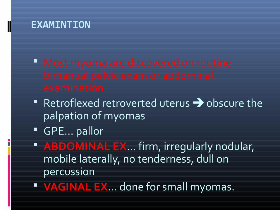
EXAMINTION
Most myoma are discovered on routine bimanual pelvic exam or abdominal examination
Retroflexed retroverted uterus obscure the palpation of myomas
GPE… pallor ABDOMINAL EX… firm, irregularly nodular,
mobile laterally, no tenderness, dull on percussion
VAGINAL EX… done for small myomas.


LABORATORY FINDINGS
Anemia Depletion of iron reserve Rarely erythrocytosis pressure on the
ureters back pressure on the kidneys erythropoietin
Acute degeneration & infection ESR, leucocytosis, & fever

IMAGING
Pelvic U/S is very helpful in confirming the Dx & excluding pregnancy / Particularly in obese Pt
Saline hysterosonography can identify submucous myoma that may be missed on U/S
HSG will show intrauterine leiomyoma MRI highly accurate in delineating the size,
location & no. of myomas , but not always necessary IVP will show ureteral dilatation or deviation &
urinary anomalies
HYSTROSCOPY for identification & removal of submucous myomas


DIFFERENTIAL DIAGNOSIS
Usually easily diagnosed Exclude pregnancy Exclude other pelvic masses -Ovarian Ca -Tubo-ovarian abscess -Endometriosis -Adenexa, omentum or bowel adherent to the uterus Exclude other causes of uterine enlargement: -Adenomyosis -Myometrial hypertrophy -Congenital anomalies -Endometrial Ca

DIFFERENTIAL DIAGNOSIS
Exclude other causes of abnormal bleeding Endometrial hyperplasia Endometrial or tubal Ca Uterine sarcoma Ovarian Ca Polyps Adenomyosis DUB Endometriosis Exogenouse estrogensEndometrial biopsy or D&C is essential in the evaluation of
abnormal bleeding to exclude endometrial Ca

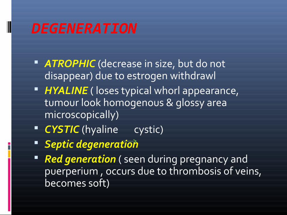
DEGENERATION
ATROPHIC (decrease in size, but do not disappear) due to estrogen withdrawl
HYALINE ( loses typical whorl appearance, tumour look homogenous & glossy area microscopically)
CYSTIC (hyaline cystic) Septic degeneration Red generation ( seen during pregnancy and
puerperium , occurs due to thrombosis of veins, becomes soft)

SARCOMATOUS CHANGE
Very rare 0.1% of cases Starts in the center of tumour Any size or type of myoma can undergo
sarcomatous change Malignant change suspected when: Rapid increase in size Painful tender

INFECTION
Submucous or subserous myoma if lies near an inflammed organ… Infection
More common in the ones that have undergone necrosis
Infection occurs: During puerperium After abortion Inflammed appendix Diverticulum

TORSION
Pedunculated Subserous Myoma…. Torsion Sudden attack of pain Increase in size Tenderness Difficult to differentiate from red
degeneration or torsion of ovarian cyst

Myomas are rarely seen associated with pregnancy (3%) Commonly seen in an elderly primigravida…….

Effects of Myomas on pregnancy
ABORTION (risk is high) PREMATURE LABOUR MALPRESENTATION DURING LABOUR: Abnormal uterine action Cervical dystocia ( interfernce in dilation of
cervix) Obstructed labour Retained placenta

Postpartum haemorrhage DURING PUERPERIUM: Puerperal sepsis Delayed involution of uterus

Effects of Pregnancy on Myomas
INCREASE IN SIZE: Due to congestion oedema of tumour, after this comes back to original size)
CHANGE IN CONSISTENCY: Become Soft due to congestion & oedema
RED DEGENERATION TORSION INFECTION

TREATMENT
DEPENDS ON: Age Parity Pregnancy status Desire for future pregnancy General health Symptoms Size Location

TREATMENT METHODS
EXPECTANT TREATMENT MEDICAL TREATMENT UTERINE ARTERY EMBOLISATION (UAE) MYOMECTOMY HYSTERECTOMY


Adviced to women approaching to menopause
When there are no symptoms Small in size No complications Patient is kept under observation Examined at 6th monthly interval Increase in size seen then surgical
intervention is done


1.GENERAL HEALTH MEASURES
PATIENT MAY BE ANEMIC DUE TO MENORRHAGIA ANEMIA SHOULD BE CORRECTED IN TURN GENERAL HEALTH IS IMPROVED

2. GNRH Analogues
RX results in: 1- size of the myomas 50% maximum 2- This shrinkage is achieved in 3M of RX 3-Amenorrhea & hypoestrogenic side-effects occur 4-Osteopososis may occur if Rx last > 6MIt is indicated for 1- bleeding from myoma except for the polypoid
submucous type 2-Preoperative to size allow for vaginal hysterectomy myomectomy laparoscopic myomectomy

3-6 months Rx … size of myomas (temporary)
Suppression of ovaries ( size reduction of myomas)
Expensive Effects are short lived Side effects include osteoporosis ( temporary
menopause)

3.DANAZOL, ANTIPROGESTERONES (MIFEPRISTONE, RU486)
Temporary relief of symptoms Results are not consistent RU 486 is given for a long period of
time


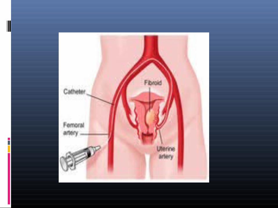
Uterine artery occlusion Particulate emboli (polyvinyl Alcohol PVA) Ischaemic necrosis of fibroids Size reduction Approached by trans-femoral route COMPLICATIONS Failure to canalize uterne arteries Haematoma Pain infection


Removal of myomas Conservation of uterus Abdominal or vaginal route INDICATIONS Less than 40 years than myomectomy is
chosen rather than hysterectomy If she wants conservation of reproductive
function Unexplained infertility

Repeated pregnancy loss Increase in size of tumor CONTRAINDICATIONS Associated carcinoma of endometrium,
treatment is direct to malignancy rather than myomectomy
If suspicion of sarcomatous change.. Pregnancy

Complications of myomectomy
Haemorrhage Sepsis Pelvic vein thrombosis Persistent symptoms (menorrhagia may
persist) Recurrence
Haemorrhage Sepsis Pelvic vein thrombosis Persistent symptoms (menorrhagia may
persist) Recurrence

HYSTERECTOMY

Above 40 years Completed the family TYPES VAGINAL LAPROSCOPIC

VAGINAL HYSTERECTOMY
If size of uterus is less than 12 weeks of gestation
Myomas growing in….. Broad ligament or distorting local anatomy than abdominal hysterectomy is done

ABDOMINAL OR LAPROSCOPIC HYSTERECTOMY Healthy ovaries are preserved under 40 years
or even above Only removed when ovaries are diseased Performed when: More than 12 weeks of gestation Distortion of anatomy Associated disease (endrometriosis, PID)