Uterine and ovarian remnants in an incorrectly spayed ...vri.cz/docs/vetmed/59-2-102.pdf · Uterine...
5
Case Report Veterinarni Medicina, 59, 2014 (2): 102–106 102 Uterine and ovarian remnants in an incorrectly spayed bitch: a case report C.C. Perez-Marin 1 , L. Molina 1 , G. Vizuete 1 , J.M. Sanchez 1 , R. Zafra 2 , M.J. Bautista 1 1 Faculty of Veterinary Science, University of Cordoba, Cordoba, Spain 2 Faculty of Veterinary Science, University of Las Palmas de Gran Canaria, Las Palmas de Gran Canaria, Spain ABSTRACT: A spayed Samoyed bitch, 12 years old, was presented to the Veterinary Clinical Hospital of the University of Cordoba (Spain) with abundant vulvar sanguineous discharge over the previous three days. The clinical examination revealed a remarkable vulvar mass, which protruded through the vulvar lips. Abdominal ultrasonography revealed the presence of structures compatible with uterus and ovary, which had been presum- ably removed eight years previously. An exploratory laparotomy was carried out, which confirmed the presence of the right ovary and a remnant of the uterus. The histological evaluation confirmed a granulosa cell tumour in the ovary, and an enlarged portion of the right uterine horn with brownish contents. The vulvar mass was also surgi- cally removed and fibroma with some fibrosarcoma areas was diagnosed. This case shows the evolution of ovary and uterus into the abdomen, which were incorrectly removed after ovariohysterectomy eight years previously. Keywords: ovariohysterectomy; canine; malpractice; granulosa cell tumour Ovariohysterectomy is one of the most frequent surgical approaches to prevent pregnancy or to treat certain reproductive diseases in bitches. However, diverse problems are associated with surgery, from idiopathic issues to problems related to surgeon malpractice. Thus, surgical intervention can lead to disease associated with reproductive tissue which was properly removed. Granulosa cell tumours, py- ometra, mammary tumours and other diseases as- sociated with the release of steroid hormones from ovary remnants have all been linked with improper removal of reproductive tissue (Sivacolundhu et al. 2001; Ragni 2005; Christensen et al. 2013). Some reports have described the presence of uterine and ovarian remnants in bitches after ovariohysterec- tomy (Ball et al. 2010), usually linked to pyometra. In the present case, the patient did not show clinical signs associated with the uterine remnant over the course of the eight years after the incorrect surgery, although the hormonal effect could be considered as a causal agent involved in the development of the vaginal and ovarian (remnant) tumours. Case description A 12-year-old Samoyed bitch was referred to the Veterinary Clinical Hospital of the University of Cordoba (Spain) presenting with sanguineous vulvar discharge, which had started three days previously, and was accompanied by enlargement of the vulvar region. The owners reported previ- ous parturition (nine years previously) without complications. Eight years previously the bitch was ovariectomised, although problems during the surgery led to the requirement for unilateral castration. After that, the bitch showed oestrous signs and sexual behaviour once a year. At clinical evaluation the animal was apparently normal, al- though blood vulvar discharge was observed. Also, external palpation of the perineal region revealed a large, firm, non-painful vulvar mass protruding through the vulva (Figure 1a,b). Digital palpation of the vagina confirmed the presence of more masses in the vaginal lumen. The largest one measured 5 cm in diameter and was located at the vaginal
Transcript of Uterine and ovarian remnants in an incorrectly spayed ...vri.cz/docs/vetmed/59-2-102.pdf · Uterine...
Case Report Veterinarni Medicina, 59, 2014 (2): 102–106
102
Uterine and ovarian remnants in an incorrectly spayed bitch: a case report
C.C. Perez-Marin1, L. Molina1, G. Vizuete1, J.M. Sanchez1, R. Zafra2, M.J. Bautista1
1Faculty of Veterinary Science, University of Cordoba, Cordoba, Spain2Faculty of Veterinary Science, University of Las Palmas de Gran Canaria,
Las Palmas de Gran Canaria, Spain
ABSTRACT: A spayed Samoyed bitch, 12 years old, was presented to the Veterinary Clinical Hospital of the University of Cordoba (Spain) with abundant vulvar sanguineous discharge over the previous three days. The clinical examination revealed a remarkable vulvar mass, which protruded through the vulvar lips. Abdominal ultrasonography revealed the presence of structures compatible with uterus and ovary, which had been presum-ably removed eight years previously. An exploratory laparotomy was carried out, which confirmed the presence of the right ovary and a remnant of the uterus. The histological evaluation confirmed a granulosa cell tumour in the ovary, and an enlarged portion of the right uterine horn with brownish contents. The vulvar mass was also surgi-cally removed and fibroma with some fibrosarcoma areas was diagnosed. This case shows the evolution of ovary and uterus into the abdomen, which were incorrectly removed after ovariohysterectomy eight years previously.
Keywords: ovariohysterectomy; canine; malpractice; granulosa cell tumour
Ovariohysterectomy is one of the most frequent surgical approaches to prevent pregnancy or to treat certain reproductive diseases in bitches. However, diverse problems are associated with surgery, from idiopathic issues to problems related to surgeon malpractice. Thus, surgical intervention can lead to disease associated with reproductive tissue which was properly removed. Granulosa cell tumours, py-ometra, mammary tumours and other diseases as-sociated with the release of steroid hormones from ovary remnants have all been linked with improper removal of reproductive tissue (Sivacolundhu et al. 2001; Ragni 2005; Christensen et al. 2013). Some reports have described the presence of uterine and ovarian remnants in bitches after ovariohysterec-tomy (Ball et al. 2010), usually linked to pyometra. In the present case, the patient did not show clinical signs associated with the uterine remnant over the course of the eight years after the incorrect surgery, although the hormonal effect could be considered as a causal agent involved in the development of the vaginal and ovarian (remnant) tumours.
Case description
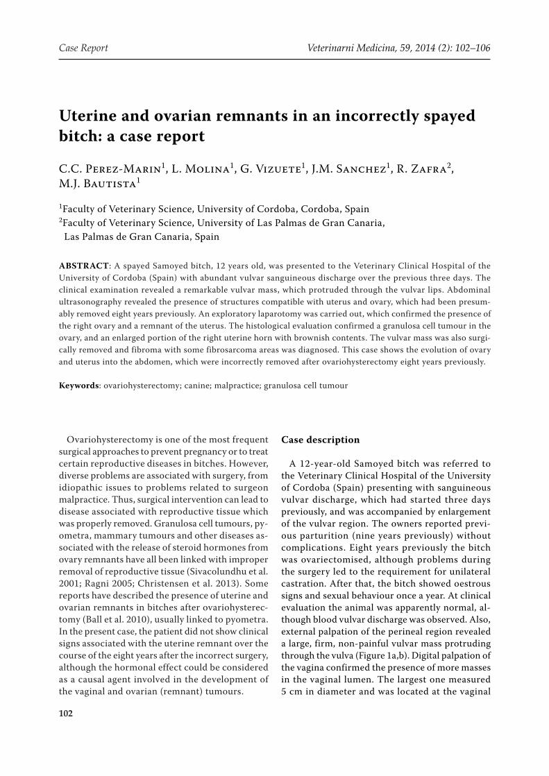
A 12-year-old Samoyed bitch was referred to the Veterinary Clinical Hospital of the University of Cordoba (Spain) presenting with sanguineous vulvar discharge, which had started three days previously, and was accompanied by enlargement of the vulvar region. The owners reported previ-ous parturition (nine years previously) without complications. Eight years previously the bitch was ovariectomised, although problems during the surgery led to the requirement for unilateral castration. After that, the bitch showed oestrous signs and sexual behaviour once a year. At clinical evaluation the animal was apparently normal, al-though blood vulvar discharge was observed. Also, external palpation of the perineal region revealed a large, firm, non-painful vulvar mass protruding through the vulva (Figure 1a,b). Digital palpation of the vagina confirmed the presence of more masses in the vaginal lumen. The largest one measured 5 cm in diameter and was located at the vaginal
Veterinarni Medicina, 59, 2014 (2): 102–106 Case Report
103
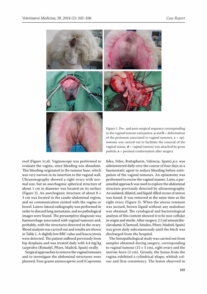
roof (Figure 1c,d). Vaginoscopy was performed to evaluate the vagina, since bleeding was abundant. This bleeding originated in the tumour base, which was very narrow in its insertion to the vaginal wall. Ultrasonography showed a right ovary with nor-mal size, but an anechogenic spherical structure of about 1 cm in diameter was located on its surface (Figure 2). An anechogenic structure of about 8 × 3 cm was located in the caudo-abdominal region, and no communication existed with the vagina or bowel. Latero-lateral radiography was performed in order to discard lung metastasis, and no pathological images were found. The presumptive diagnosis was haemorrhage associated with vaginal tumours and, probably, with the structures detected in the ovary. Blood analysis was carried out and results are shown in Table 1. A slightly low RBC value and leucocytosis were detected. The patient suffered previously from hip dysplasia and was treated daily with 4.4 mg/kg carprofen (Rimadyl, Pfizer, Madrid, Spain) orally.
Surgical approaches to remove the vaginal tumours and to investigate the abdominal structures were planned. Four grams aminocaproic acid (Capromin
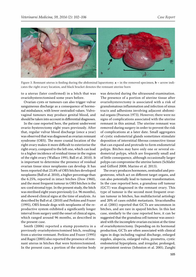
fides, Fides, Rottapharm, Valencia, Spain) p.o. was administered daily over the course of four days as a haemostatic agent to reduce bleeding before extir-pation of the vaginal tumours. An episiotomy was performed to excise the vaginal masses. Later, a par-amedial approach was used to explore the abdominal structure previously detected by ultrasonography. An isolated, dilated, and liquid-filled recess of uterus was found. It was removed at the same time as the right ovary (Figure 3). When the uterus remnant was incised, brown liquid without any malodour was obtained. The cytological and bacteriological analysis of this content showed it to be non-cellular in origin and sterile. After surgery, 2.5 ml amoxicilin-clavulamic (Clamoxil, Sinulox, Pfizer, Madrid, Spain) was given daily subcutaneously until the bitch was discharged from the hospital.
The histopathological study was carried out from samples obtained during surgery, corresponding to vaginal tumour (15 × 5 cm), right ovary and the uterine horn (3 cm). Grossly, the lesion from the vagina exhibited a cylindrical-shape, whitish col-our and firm consistency. The lesion observed in
Figure 1. Pre- and post-surgical sequence corresponding to the vaginal tumour extirpation. a and b = deformation of the perineum associated to vaginal tumours, c = epi-siotomy was carried out to facilitate the removal of the vaginal masse, d = vaginal tumour was attached by gross pedicle, e = perineal conformation after surgery
Case Report Veterinarni Medicina, 59, 2014 (2): 102–106
104
the right ovary exhibited whitish and well-defined circular areas. The uterine horn also showed brown content and numerous cavities.
Microscopically, three corpora lutea were ob-served in the ovary and also a neoplasm composed of a proliferation of granulosa cells. In contrast with the ultrasonographic image, no ovarian cysts were present and the it was presumed that the observed anechogenic spherical structure corresponded to the diagnosed tumour. The ovary neoplasm showed a combined growth pattern (i.e., macrofollicular, tubular and solid, respectively). Firstly, cells were distributed around a cystic cavity, similar to tertiary follicles, with some neoplastic cells. Secondly, neo-plastic cells were well-differentiated, with numerous and relevant tubules. Finally, the solid growth pat-tern presented numerous pleomorphic and vacu-
olated cells, with severe atypias and low mitotic index. The right ovary also showed incipient papil-lary hyperplasia involving the germinal epithelium.
The sample from the uterus exhibited severe endometrial cystic hyperplasia. Numerous endo-metrial glands were found to be frequently dilated and sometimes twisted. As a result, in some areas polypoid hyperplasia of endometrial epithelium was also found. Endometrial oestromal cells also showed activation signs.
A mesenchymal neoplastic proliferation was ob-served in the vagina. This neoplasm is composed of fusiform and well-differentiated cells, embedded in a large amount of collagen. However, small areas of malignancy were observed. The cells from these areas showed severe atypias, pleomorphic nuclei, numerous nucleoli, atypical mitoses, and cells were distributed in an interlacing pattern. An intratumoral infiltrate comprised of numerous well-differentiated mast cells was also observed. The vagina showed ul-ceration affecting the mucosa with a mixed inflamma-tory infiltrate comprised of macrophages, plasma cells neutrophils, lymphocytes and numerous mast cells.
DISCUSSION AND CONCLUSIONS
Vaginal blood discharge is frequently observed in bitches, and multiple diseases and physiological situations may be implicated in this symptom. In healthy bitches, sero-sanguineous secretion, which is maintained for 8–15 days, is observed during oestrous. However, this phenomenon could also be associated with numerous pathologies mainly affecting the ovaries, uterus, vagina, vulva, or uri-nary system. Ultrasonographic assessment revealed the presence of an ovary and a structure similar
Table 1. Biochemical and haematological parameters
Parameter Values Reference valuesRBC 4.81 × 10⁻⁶ μl 5.0–8.0 × 10⁻⁶ μlHGB 10.6 g/dl 11–17 g/dlHCT 29% 37–50%MCV 64 fl 60–77 flMCH 22 pg 20–25 pgMCHC 34.4 g/dl 32–36 g/dlWBC 21.1× 10⁻³ μl 6–12 × 10–3 μlSegmented neutrophils 82% 60–70%Lymphocytes 12% 12–30%Eosinophils 1% 2–10%Basophils 0% 0–1%Monocytes 1% 3–10%Band cells 2% 0–4%PLT 333 × 10–3 μl 200–400 × 10–3 μlUrea 42 mg/dl 20–40 mg/dlCreatinin 1.1 mg/dl 0.5–1.3 mg/dlTotal proteins 7.3 g/dl 6.0–7.5 g/dlFibrinogen 400 mg/dl 100–400 mg/dlGlucose 131 mg/dl 60–115 mg/dlPotasium 4.1 mmol/l 4–5.4 mmol/lALT 41 IU/l 6–70 IU/lProthrombin ratio 6 s 6.25–12.4 sProgesterone 6.3 ng/ml –Estradiol 14.4 pg/ml –
RBC = red blood cells; HGB = haemoglobin; HCT = haema- tocrit; MCV = mean corpuscular volume; MCH = mean corpuscular haemoglobin; MCHC = mean corpuscular hae-moglobin concentration; WBC = white blood cells; PLT = platelets, ALT = alanine transaminase
Figure 2. Ultrasonography showing one of the anechoic structures in the right ovary
Veterinarni Medicina, 59, 2014 (2): 102–106 Case Report
105
to a uterus (later confirmed) in a bitch that was ovariohysterectomised some years before.
Ovarian cysts or tumours can also trigger vulvar sanguineous discharge as a consequence of hormo-nal misbalance, with lower oestradiol values. Vulvo-vaginal tumours may produce genital blood, and should be taken into account in differential diagnoses.
In the case reported here, the patient underwent ovario-hysterectomy eight years previously. After that, regular vulvar blood discharge (once a year) was observed that was diagnosed as ovarian remnant syndrome (ORS). The more cranial location of the right ovary makes it more difficult to exteriorise the right ovary, compared to the left one, which can lead to a higher incidence of residual tissue in the region of the right ovary (Wallace 1991; Ball et al. 2010). It is important to determine the presence of residual ovarian tissue since neoplasms can develop. It has been reported that 23.8% of ORS bitches developed neoplasms (Ball et al. 2010), a higher percentage than the 6.25%, reported in intact bitches (Dow 1960), and the most frequent tumour in ORS bitches is the sex-cord stromal type. In the present study, the bitch was sterilised eight years previously (i.e. 96 months), and showed clinical signs at the time of analysis. As described by Ball et al. (2010) and Perkins and Frazer (1995), ORS female dogs with neoplasms of the re-productive system exhibited a significantly longer interval from surgery until the onset of clinical signs, which ranged around 96 months, as described in the present case.
Smith (2006) reported a stump pyometra in a previously ovariohysterectomised bitch, resulting from a uterine remnant. Perkins and Frazer (1995) and Ragni (2005) also reported the presence of rem-nant uterus in bitches that were hysterectomised. In the present case, a portion of the uterine body
was detected during the ultrasound examination. The presence of a portion of uterine tissue after ovariohysterectomy is associated with a risk of granulomatous inflammation and infection of sinus tracts and adhesions involving adjacent abdomi-nal organs (Pearson 1973). However, there were no signs of complications associated with the uterine remnant in this animal. The uterine remnant was removed during surgery in order to prevent the risk of complications at a later date. Small aggregates of cystic endometrial glands sometimes stimulate deposition of interstitial fibrous connective tissue that can expand and protrude to form endometrial polyps. Bitches may have only one or several en-dometrial polyps, which are frequently small and of little consequence, although occasionally larger polyps can compromise the uterine lumen (Schlafer and Gifford 2008; Marino et al. 2013).
The ovary produces hormones, oestradiol and pro-gesterone, which act on different target organs, and can also potentially lead to tumour transformation. In the case reported here, a granulosa cell tumour (GCT) was diagnosed in the remnant ovary. This type of tumour is the second most frequent ovar-ian tumour in bitches, has multifactorial aetiology and 20% of cases exhibit metastasis. Sivacolundhu et al. (2001) reported that GCTs are uncommon in bitches, and are rare in spayed bitches. In this last case, similarly to the case reported here, it can be suggested that the granulosa cell tumour was associ-ated with the incomplete ovarian excision at the time of ovariohysterectomy. Depending on its hormonal production, GCTs are often associated with clinical signs in dogs including vaginal discharge, enlarged nipples, alopecia, enlarged vulva, pyometra, cystic endometrial hyperplasia, and irregular, prolonged, or persistent oestrus (Johnston et al. 2001; Zanghi
Figure 3. Remnant uterus is finding during the abdominal laparotomy. a = in the removed specimen, b = arrow indi-cates the right ovary location, and black bracket denotes the remnant uterine horn
Case Report Veterinarni Medicina, 59, 2014 (2): 102–106
106
et al. 2007). A higher percentage of canine granu-losa cell tumours are malignant and metastasise to regional lymph nodes and organs, whereas, accord-ing to the microscopic pattern of this tumour, it was diagnosed as a benign granulosa cell tumour. Dogs with nonfunctional granulosa cell tumours usually have no clinical signs related to the reproductive tract (Zanghi et al. 2007); the low mitotic index observed in the GCT in the present case would indicate that this tumour was non-functional and thus that the tumour-induced hormonal disbalance would not be present. In animals with ovarian neoplasms, the residual ovar-ian tissue may not secrete adequate concentrations of hormones to cause clinical signs consistent with oestrogen influence until the tissue undergoes malig-nant transformation. Conversely, the ovarian remnant may secrete low concentrations of hormones that do not result in clinical signs but that do induce neoplasia after a prolonged period (Ball et al. 2010).
Although vaginal and vulvar tumours are not un-common, the most frequently reported tumours are leiomyoma/leoimyosarcome, fibroma, and trans-missible venereal tumours (Radi 2005). Fibroma and some fibrosarcoma areas were diagnosed in the vagina in the present case. Fibrosarcomas can be found in any location of the body but are only rarely described as mesenchymal tumours of bitch vagina (Madewell and Theilen 1987; Gupta and Tiwari 2009).
In conclusion, the present case describes the evo-lution of a remnant ovary and uterus in a bitch, eight years after an incorrect ovariohysterectomy. The clinical signs appeared approximately 96 months later, and were associated with ovarian and vaginal neoplasms, while the uterus did not show evidence of pathology.
REFERENCES
Ball RL, Birchard SJ, May LR, Threlfall WR, Young GS (2010): Ovarian remnant syndrome in dogs and cats: 21 cases (2000–2007). Journal of American Veterinary Medicine Association 236, 548–553.
Christensen NI, Brain PH, Langova V, Flory AB (2013): Vaginal discharge in a spayed dog with multiple distinct malignancies. Australian Veterinary Journal 91, 287–291.
Dow C (1960): Ovarian abnormalities in the bitch. Jour-nal of Comparative Pathology 70, 59–69.
Gupta N, Tiwari SK (2009): Study on incidence, histo-pathological features and surgical managements of neoplasms in canine. Veterinary World 2, 392–395.
Johnston SD, Root Kustritz MV, Olson PNS (2001): Dis-orders of the canine ovary. Ovarian remnant syn-drome. In: Canine and Feline Theriogenology, WB Saunders, Philadelphia. 199–200.
Madewell BR, Theilen GH (1987): Tumours of the uro-genital tract. In: Theilen GH, Madewell BR (eds): Vet-erinary Cancer Medicine. Lea and Febiger, Philadelphia. 567–600.
Marino G, Barna A, Rizzo S, Zanghi A, Catone G (2013): Endometrial polyps in the bitch: a retrospective study of 21 cases. Journal of Comparative Pathology 149, 410–416.
Pearson H (1973): The complications of ovariohysterec-tomy in the bitch. Journal of Small Animal Practice 14, 257–266.
Perkins NR, Frazer GS (1995): Ovarian remnant syn-drome in a toy poodle: a case report. Theriogenology 44, 307–312.
Radi ZA (2005): Vulvar lipoleiomyoma in a dog. Journal of Veterinary Diagnostic Investigation 17, 89–90.
Ragni RA (2005): Pyometra in a bitch following unusual sterilisation. Journal of Small Animal Practice 46, 39–40.
Schlafer DH, Gifford AT (2008): Cystic endometrial hy-perplasia, pseudo-placentational endometrial hyper-plasia, and other cystic conditions of the canine and feline uterus. Theriogenology 70, 349–358.
Sivacolundhu RK, O’Hara AJ, Read RA (2001): Granulosa cell tumour in two speyed bitches. Australian Veteri-nary Journal 79, 173–176.
Smith FO (2006): Canine pyometra. Theriogenology 66, 610–612.
Wallace MS. (1991): The ovarian remnant syndrome in the bitch and queen. Veterinary Clinics of North America: Small Animal Practice 21, 501–507.
Zanghi A, Catone G, Marino G, Quartuccio M, Nicotina PA (2007): Endometrial polypoid adenomyomatosis in a bitch with ovarian granulosa cell tumour and py-ometra. Journal of Comparative Pathology 136, 83–86.
Received: 2014–02–19Accepted after corrections: 2014–03–10
Corresponding Author:
Carlos C. Perez-Marin, University of Cordoba, Veterinary Faculty, Department of Animal Medicine and Surgery, Campus Universitario de Rabanales, Ctra Madrid-Cadiz km 396, 14014, Cordoba, SpainE-mail: [email protected]