Global Cardiac Resynchronization Therapy (CRT) Devices Market 2015-2019
Upload
truongtuongCategory
view
229download
0
Technology Assessment
Technology
Assessment Program Prepared for: Agency for Healthcare Research and Quality 540 Gaither Road Rockville, Maryland 20850
Use of Cardiac Resynchronization
Therapy in the Medicare Population
March 24, 2015 Final Report
Use of Cardiac Resynchronization Therapy in the
Medicare Population
Technology Assessment Report
Project ID: CRDT1013
March 24, 2015
Johns Hopkins University Evidence-based Practice Center
John Rickard, M.D.,M.P.H. Henry Michtalik, M.D., M.P.H., M.H.S.
Ritu Sharma, B.Sc. Zackary Berger, M.D., Ph.D.
Emmanuel Iyoha, M.B.Ch.B., M.P.H. Ariel R. Green, M.D., M.P.H. Nowreen Haq, M.D., M.P.H. Karen A. Robinson, Ph.D.
This report is based on research conducted by the Johns Hopkins University Evidence-based Practice Center under contract to the Agency for Healthcare Research and Quality (AHRQ), Rockville, MD HHSA 290-201-200007-1). The findings and conclusions in this document are those of the author (s) who are responsible for its contents; the findings and conclusions do not necessarily represent the views of AHRQ. No statement in this article should be construed as an official position of the Agency for Healthcare Research and Quality or of the U.S. Department of Health and Human Services. The information in this report is intended to help health care decision-makers; patients and clinicians, health system leaders, and policymakers, make well-informed decisions and thereby improve the quality of health care services. This report is not intended to be a substitute for the application of clinical judgment. Decisions concerning the provision of clinical care should consider this report in the same way as any medical reference and in conjunction with all other pertinent information, i.e., in the context of available resources and circumstances presented by individual patients.
ii

This report may be used, in whole or in part, as the basis for development of clinical practice guidelines and other quality enhancement tools, or as a basis for reimbursement and coverage policies. AHRQ or U.S. Department of Health and Human Services endorsement of such derivative products may not be stated or implied.
None of the investigators has any affiliations or financial involvement related to the material presented in this report. This document is in the public domain and may be used and reprinted without special permission. Citation of the source is appreciated. Persons using assistive technology may not be able to fully access information in this report. For assistance contact [email protected].
Suggested citation: Rickard J, Michtalik H, Sharma R, Berger Z, Iyoha E, Green AR, Haq N, Robinson KA. Use of Cardiac Resynchronization Therapy in the Medicare Population. (Prepared by the Prepared by the Johns Hopkins University Evidence-based Practice Center under Contract No. HHSA 290-201-200007-1) Rockville, MD: Agency for Healthcare Research and Quality. March 24, 2015.
iii

Acknowledgments We would like to thank Allen Zhang and Elisabeth Haberl Nannes for their assistance in data abstraction, Lisa Wilson for conducting meta-analysis and Eric Vohr for copy editing. We would also like to thank the Peer Reviewers, the Task Order Officers, and representatives from CMS. Peer Reviewers We wish to acknowledge individuals listed below for their review of this report. This report was reviewed in draft form by individuals chosen for their expertise and diverse perspectives. The purpose of the review was to provide candid, objective, and critical comments for consideration by the EPC in preparation of the final report. Synthesis of the scientific literature presented here does not necessarily represent the views of individual reviewers. Sana M. Al-Khatib, M.D., M.H.S. Heart Rhythm Specialist and Associate Professor of Medicine Division of Medicine – Cardiology, Department of Medicine Duke University Medical Center Durham, NC Daniel B. Kramer, MD, MPH Cardiac Electrophysiology Cardiac Arrhythmias Specialist Beth Israel Deaconess Medical Center Boston, MA Michael Lauer, M.D. Director, Division of Cardiovascular Sciences National Heart, Lung, and Blood Institute (NHLBI) National Institutes of Health (NIH) Bethesda, MD Win-Kuang Shen, M.D. Mayo Clinic College of Medicine Chair, Division of Cardiovascular Diseases Mayo Clinic Arizona Scottsdale, AZ Lynne Stevenson, M.D. Director, Cardiomyopathy and Heart Failure Program Harvard Medical School Boston, MA
iv

Structured Abstract Objectives: To assess the benefits and harms of cardiac resynchronization with (CRT-D) and compared to an ICD alone, CRT without a defibrillator (CRT-P) compared with optimal medical therapy and CRT-D compared with CRT-P in patients with an EF ≤35% and a QRS duration ≥120 ms. We also sought to assess predictors of response to CRT-D and CRT-P. Data Sources: We searched MEDLINE, Embase®, and the Cochrane Central Register of Controlled Trials (CENTRAL) from January 1, 1995, as this is the date of first article reporting use of CRT through October 20th, 2014. Review Methods: Paired investigators independently screened search results to assess eligibility. Investigators abstracted data sequentially and assessed risk of bias independently. Investigators graded the strength of evidence as a group. Results: CRT-D was found to be effective in reducing heart failure hospitalizations, inducing ventricular reverse remodeling, improving quality of life, and increasing six-minute hall walk distances compared to an ICD alone with a high strength of evidence. In a meta-analysis of minimally symptomatic patients, CRT-D reduced LVESV (ml) (mean difference -22.55, 95% CI -40.66 to -9.56). This analysis was comprised primarily on NYHA class II patients; therefore, the applicability to NYHA class I patients is unclear. In a meta-analysis of patients with advanced heart failure (NYHA class III-IV), CRT-D improved quality of life scores (as measured by the Minnesota Living with Heart Failure Questionnaire) (mean difference -10.91, 95% CI -12.03 to -7.27) compared to an ICD alone. CRT-P was found to be effective in improving all-cause survival and reducing heart failure hospitalizations compared to optimal medal therapy alone with a moderate level of evidence. CRT-P was also found to induce reverse ventricular remodeling and increase six-minute hall walk distances compared to optimal medical therapy alone. These findings were primarily noted in NYHA class III-IV patients. The applicability of these findings to NYHA class I-II patients is unclear. Determining predictors of response to CRT was limited by the likely presence of reporting bias. Nevertheless, a left bundle branch (LBBB) morphology, non-ischemic cardiomyopathy (NICM), and female gender were generally associated with improved outcomes following CRT-D. Sinus rhythm (as compared to atrial fibrillation) and a wider QRS duration were associated with improved outcomes following CRT-D albeit with a lower strength of evidence. There is insufficient evidence to determine predictors of outcomes in patients undergoing CRT-P. There is insufficient evidence to determine the effectiveness of CRT-D versus CRT-P. Compared to CRT-P, device infection was slightly more common in patients receiving CRT-D. Conclusions: There is convincing evidence that CRT-D is effective with regard to improvements in multiple clinical outcomes compared to an ICD alone in patients with an LVEF≤35% and a QRS duration ≥120ms. Similarly, there is convincing evidence that CRT-P is effective in improving multiple clinical endpoints compared to optimal medical therapy alone in the same population. The certainty of these findings varies based on NYHA class. Female gender, LBBB, a wider QRS duration, sinus rhythm, and non-ischemic cardiomyopathy are associated with improved outcomes following CRT although the likely presence of reporting bias qualifies these results. More data are needed for several questions including the efficacy of CRT in patients with a non-LBBB morphology or atrial fibrillation and the comparison of outcomes in patients receiving a CRT-D vs. CRT-P device.
v
Contents Executive summary ..................................................................................................................... ES1 Introduction ......................................................................................................................................1
Scope and Key Questions ........................................................................................................... 2 Methods............................................................................................................................................6
Protocol Development ................................................................................................................ 6 Search Strategy ........................................................................................................................... 6 Study Selection ........................................................................................................................... 6 Data Abstraction and Data Management .................................................................................... 6 Risk of Bias Assessment ........................................................................................................... 10 Data Synthesis ........................................................................................................................... 10 Strength of the Body of Evidence ............................................................................................. 10 Applicability ............................................................................................................................. 11
Results ............................................................................................................................................12 Results of the Search ..................................................................................................................12 Overview of included studies by outcomes ...............................................................................14 Organization of Results Chapter ................................................................................................16 Effectiveness of Cardiac Resynchronization Therapy with Defibrillator (CRT-D) ..................17 Harms of Cardiac Resynchronization Therapy with Defibrillator (CRT-D) .............................40 Effectiveness of Cardiac Resynchronization Therapy with Pacemaker (CRT-P) .....................56 Harms of Cardiac Resynchronization Therapy with Pacemaker (CRT-P) ................................73 Effectiveness of Cardiac Resynchronization Therapy with Pacemaker versus Defibrillator (CRT-P vs CRT-D) ....................................................................................................................83 Harms of Cardiac Resynchronization Therapy with Pacemaker versus Defibrillator (CRT-P vs CRT-D) ..............................................................................................................................90 Predictors of Response to Cardiac Resynchronization Therapy with Defibrillator (CRT-D) .105 Predictors of Response to Cardiac Resynchronization Therapy with Pacemaker (CRT-P). ...131
Discussion ....................................................................................................................................140 Key Findings and the Strength of Evidence ........................................................................... 140 Relationship of Findings to Existing Literature ...................................................................... 143 Applicability ........................................................................................................................... 146 Limitations of the Comparative Effectiveness Review Process ............................................. 146 Limitations of the Evidence Base ........................................................................................... 146 Research Gaps ......................................................................................................................... 146 Conclusion .............................................................................................................................. 148
References ....................................................................................................................................149 List of Abbreviations ...................................................................................................................157 Tables Table 1. PICOTS (population, interventions, comparators, outcomes, timing, setting) for each Key Question ...................................................................................................................................7 Table 2. List of exclusion criteria at the abstract and article screening level ..................................8 Table 3. List of device manufacturers ..............................................................................................9 Table 4. Strength of evidence grades and definitions ....................................................................11 Table 5. List of included studies by outcomes ...............................................................................14 Table 6. List of trials included in the review .................................................................................15
vi
Table 7. Evidence addressing effectiveness and harms of CRT-D ................................................17 Table 8. Study characteristics of trials assessing effectiveness of CRT-D ....................................19 Table 9. Summary of risk of bias for trials assessing effectiveness of CRT-D ............................22 Table 10. Outcomes reported in the trials assessing effectiveness of CRT-D ..............................25 Table 11. Summary of CRT-D effectiveness outcomes reported by subgroup .............................37 Table 12. Strength of evidence for key effectiveness outcomes of CRT-D ..................................38 Table 13. Summary of risk of bias for trials assessing harms of CRT-D ......................................42 Table 14. Summary of risk of bias for cohort studies assessing harms of CRT-D ........................44 Table 15. List of harms reported in studies assessing harms of CRT-D........................................46 Table 16.Characteristics of studies of CRT-D reporting on the incidence of pneumothorax........49 Table 17. Characteristics of studies of CRT-D reporting on the incidence of pocket hematoma .50 Table 18. Characteristics of studies of CRT-D reporting on the incidence of cardiac device infection .........................................................................................................................................51 Table 19. Characteristics of studies of CRT-D reporting on the incidence of cardiac perforation/tamponade ...................................................................................................................52 Table 20. Characteristics of studies of CRT-D reporting on the incidence of lead dislodgement 53 Table 21. Characteristics of studies of CRT-D reporting inappropriate ICD shocks ....................55 Table 22. Evidence addressing effectiveness and harms of CRT-P ..............................................57 Table 23. Study characteristics of trials assessing effectiveness of CRT-P ..................................59 Table 24. Summary of risk of bias for trials assessing effectiveness of CRT-P ............................63 Table 25. Outcomes reported in the trials assessing effectiveness of CRT-P ...............................65 Table 26. Summary of CRT-P effectiveness outcomes reported by subgroup ..............................72 Table 27. Strength of evidence for key effectiveness outcomes of CRT-P ...................................73 Table 28. Summary of risk of bias for trials assessing harms of CRT-P .......................................75 Table 29. Summary of risk of bias for cohort studies assessing harms of CRT-P ........................77 Table 30. List of harms reported in the studies assessing harms of CRT-P ..................................78 Table 31. Characteristics of studies of CRT-P reporting on the procedure-related complications80 Table 32. Characteristics of studies of CRT-P reporting on the incidence of pneumothorax .......80 Table 33. Characteristics of studies of CRT-P reporting on the incidence of pocket hematoma ..81 Table 34. Characteristics of studies of CRT-P reporting on the incidence of cardiac device infection .........................................................................................................................................81 Table 35. Characteristics of studies of CRT-P reporting on the incidence of cardiac perforation/tamponade ...................................................................................................................82 Table 36. Characteristics of studies of CRT-P reporting on the incidence of lead dislodgement .82 Table 37. Characteristics of studies of CRT-P reporting on the death within one week ...............83 Table 38. Evidence addressing effectiveness and harms of CRT-P vs CRT-D .............................84 Table 39. Study characteristics of trials assessing effectiveness of CRT-P vs CRT-D .................85 Table 40. Summary of risk of bias for trials assessing effectiveness of CRT-P vs CRT-D ..........87 Table 41. Summary of effectiveness outcomes reported in the trial of CRT-P vs CRT-D, by subgroup .........................................................................................................................................90 Table 42. Strength of evidence for key effectiveness outcomes of CRT-P vs CRT-D .................90 Table 43. Summary of risk of bias for trials assessing harms of CRT-P vs CRT-D .....................93 Table 44. Summary of risk of bias for cohort studies assessing harms of CRT-P vs CRT-D .......94 Table 45. List of harms reported in the studies assessing harms of CRT-P vs CRT-D .................96 Table 46. Summary of effectiveness outcomes by comparator ...................................................102 Table 47. Summary of harms by comparator ...............................................................................104 Table 48. Study characteristics of studies assessing predictors of response to CRT-D .............108 Table 49. Summary of risk of bias for studies assessing predictor of response to CRT-D .........110
vii

Table 50. Included predictors of response for CRT-D ................................................................112 Table 51. Definitions of response for CRT-D predictors ............................................................114 Table 52. Evidence addressing predictors of response to CRT-D, by predictor ..........................115 Table 53. Summary of findings of predictors of response to CRT-D, by outcome .....................127 Table 54. Study characteristics of studies assessing predictor of response to CRT-P .................131 Table 55. Summary of risk of bias for studies assessing predictor of response to CRT-P ..........132 Table 56. Included predictors of response for CRT-P .................................................................133 Table 57. Definitions of response for CRT-P predictors .............................................................133 Table 58. Evidence addressing predictors of response to CRT-P, by predictor ..........................134 Table 59. Summary of findings of predictors of response to CRT-P, by outcome......................139 Table 60. Summary of the strength of evidence for key effectiveness outcomes........................142 Table 61. Prior systematic reviews of cardiac resynchronization therapy ...................................144 Table 62. Characteristics of an ideal study to compare the effectiveness of CRT-D vs. CRT-P 147 Table 63. Characteristics of an ideal study to compare the effectiveness of CRT-D in Patients with a non-Left Bundle Branch Block morphology ....................................................................148 Table 64. Characteristics of an ideal study to compare the effectiveness of CRT-D in Patients with Atrial Fibrillation .................................................................................................................148
Figures Figure 1. Analytic framework for use of cardiac resynchronization therapy with defibrillator (CRT-D) in the Medicare population ...............................................................................................3 Figure 2. Analytic framework for use of cardiac resynchronization therapy without defibrillator capacity (CRT-P) in the Medicare population .................................................................................4 Figure 3. Analytic framework for use of cardiac resynchronization therapy with defibrillator capacity (CRT-D) versus cardiac resynchronization therapy without defibrillator capacity (CRT-P) in the Medicare population ..........................................................................................................5 Figure 4. Summary of the literature search and screen ..................................................................13 Figure 5. Summary of risk of bias for trials assessing effectiveness of CRT-D ............................23 Figure 6. Meta-analysis of left ventricular end-diastolic volume in trials including minimally symptomatic patients CRT- D Effectiveness .................................................................................30 Figure 7a and b. Meta-analysis of trials including minimally symptomatic patients in terms of improvement in quality of scores via the Minnesota Living with Heart Failure Questionnaire CRT- D Effectiveness ....................................................................................................................32 Figure 8. Meta-analysis of trials including minimally symptomatic patients in terms of left ventricular ejection fraction improvement CRT- D Effectiveness ................................................34 Figure 9. Meta-analysis of patients with NYHA class I-II heart failure in terms of improvement in 6-minute hall walk distance CRT- D Effectiveness ...................................................................36 Figure 10. Summary of risk of bias for trials assessing harms of CRT-D .....................................43 Figure 11. Summary of risk of bias for cohort studies assessing harms of CRT-D ......................45 Figure 12. Summary of risk of bias for trials assessing effectiveness of CRT-P ..........................64 Figure 13. Summary of risk of bias for trials assessing harms of CRT-P .....................................75 Figure 14. Summary of risk of bias for cohort studies assessing harms of CRT-P .......................78 Figure 15. Summary of risk of bias for cohort studies assessing harms of CRT-P vs CRT-D......95 Figure 16. Summary of risk of bias for studies assessing predictor of response to CRT-D ........112
viii

Appendixes Appendix A. Detailed Electronic Database Search Strategies Appendix B. Forms Appendix C. List of Excluded Studies Appendix D. Evidence Tables Appendix E. SIP
ix

Executive Summary Background
Chronic heart failure (CHF) is a major public health problem in the United States affecting an estimated 4.9 million Americans, causing high rates of hospitalization, poor quality of life, and 300,000 deaths each year.1
Cardiac resynchronization (CRT) is a pacing modality utilizing a left ventricular (LV) pacing lead with the goal of re-synchronizing myocardial contraction in patients with CHF. CRT was originally indicated in patients with significant LV dysfunction, defined as a left ventricular ejection fraction (LVEF) ≤ 35%, New York Heart Association (NYHA) class III-IV heart failure symptoms, and a QRS duration ≥120ms on optimal medical therapy.2-4More recently, the indications for CRT expanded to include patients with minimally symptomatic heart failure (NYHA class II).5 ,6 The appropriateness of CRT in patients with NYHA class I symptoms is unclear.1 The vast majority of candidates for CRT devices also have an indication for an implantable cardiac defibrillator (ICD), therefore, the large majority of patients receiving CRT in the United States receive a CRT device with a defibrillator (CRT-D) as opposed to a CRT pacemaker (CRT-P). CRT-P devices are occasionally placed in patients who wish to avoid ICD shocks or in patients with an indication for frequent ventricular pacing due to conduction disease who have an LVEF between 36-50 percent.
Scope and Key Questions We conducted a systematic review on the efficacy for both CRT-D and CRT-P. The
questions were nominated by the Centers for Medicare and Medicaid (CMS).We sought to address the following questions for patients with an LVEF ≤35% and a QRS duration≥120ms:
• What is the effectiveness and safety of CRT-D compared to an ICD alone? • What is the effectiveness and safety of CRT-P compared to optimal medical therapy
alone? • What is the comparative effectiveness and safety of CRT-D versus CRT-P? • What are the clinical predictors of response in patients deemed appropriate candidates for
CRT-D devices? • What are the clinical predictors of response in patients deemed appropriate candidates for
CRT-P devices?
ES-1

Methods With input from key informants, we refined the questions, including eligibility criteria, and
developed a protocol (PROSPERO registration CRD42014009981). We searched MEDLINE, Embase®, and the Cochrane Central Register of Controlled Trials
(CENTRAL) from January 1, 1995, as this is the date of first article reporting use of CRT through October 20th, 2014. We also reviewed the reference lists of all included articles, requested information from device manufacturers and searched Clinicaltrials.gov.
Citations were screened independently by two reviewers using predefined eligibility criteria. One reviewer completed data abstraction and a second reviewer checked abstraction for completeness and accuracy. Data when available by subgroups (females, QRS duration >150 ms, left bundle branch block, atrial fibrillation and non-ischemic cardiac conditions) were also abstracted. Two reviewers independently assessed risk of bias for individual studies. We used the Cochrane Collaboration’s tool for assessing the risk of bias of controlled studies.7 For nonrandomized studies, we used the Newcastle Ottawa Scale 8 and for predictor studies, we used Quality In Prognosis Studies (QUIPS) tool.9 Differences between reviewers were resolved through consensus.
All studies were summarized qualitatively. We conducted meta-analyses for an outcome when there were sufficient data (at least 3 studies of the same design) and studies were sufficiently homogenous with respect to key variables (population characteristics, intervention, and outcome measurement) using profile likelihood estimate for random effects model. We identified substantial statistical heterogeneity in the trials as an I-squared statistic with a value greater than 50 percent. All meta-analyses were conducted using STATA 12.1 (College Station, TX).
We graded the strength of evidence using the scheme recommended by the AHRQ EPC Methods Guide for Conducting Comparative Effectiveness Reviews.10 For this report, we graded the strength of evidence for the outcomes we classified during protocol development as the most important or critical outcomes, including quality of life as assessed by the Minnesota Living with Heart Failure Inventory Score (MLHFQ), left ventricular end systolic volume, hospitalizations for heart failure and, all- cause mortality.
ES-2
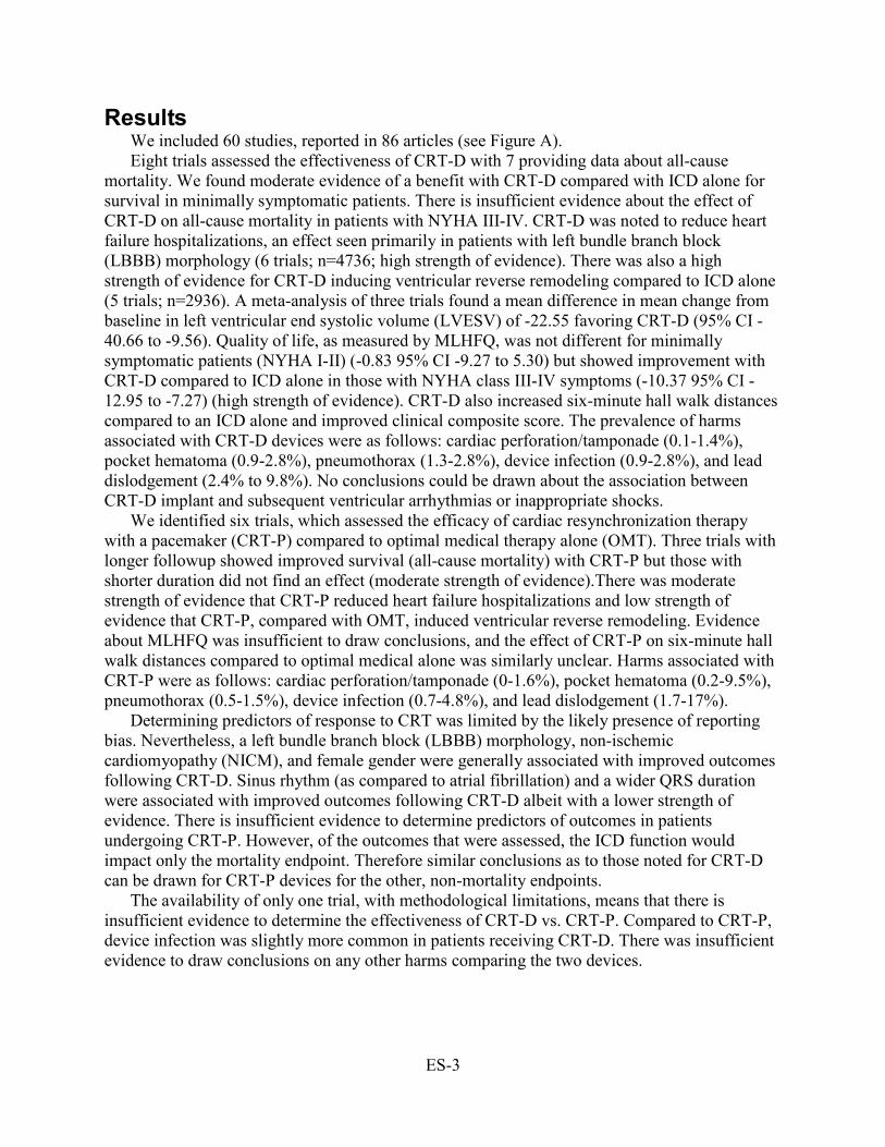
Results We included 60 studies, reported in 86 articles (see Figure A). Eight trials assessed the effectiveness of CRT-D with 7 providing data about all-cause
mortality. We found moderate evidence of a benefit with CRT-D compared with ICD alone for survival in minimally symptomatic patients. There is insufficient evidence about the effect of CRT-D on all-cause mortality in patients with NYHA III-IV. CRT-D was noted to reduce heart failure hospitalizations, an effect seen primarily in patients with left bundle branch block (LBBB) morphology (6 trials; n=4736; high strength of evidence). There was also a high strength of evidence for CRT-D inducing ventricular reverse remodeling compared to ICD alone (5 trials; n=2936). A meta-analysis of three trials found a mean difference in mean change from baseline in left ventricular end systolic volume (LVESV) of -22.55 favoring CRT-D (95% CI -40.66 to -9.56). Quality of life, as measured by MLHFQ, was not different for minimally symptomatic patients (NYHA I-II) (-0.83 95% CI -9.27 to 5.30) but showed improvement with CRT-D compared to ICD alone in those with NYHA class III-IV symptoms (-10.37 95% CI -12.95 to -7.27) (high strength of evidence). CRT-D also increased six-minute hall walk distances compared to an ICD alone and improved clinical composite score. The prevalence of harms associated with CRT-D devices were as follows: cardiac perforation/tamponade (0.1-1.4%), pocket hematoma (0.9-2.8%), pneumothorax (1.3-2.8%), device infection (0.9-2.8%), and lead dislodgement (2.4% to 9.8%). No conclusions could be drawn about the association between CRT-D implant and subsequent ventricular arrhythmias or inappropriate shocks.
We identified six trials, which assessed the efficacy of cardiac resynchronization therapy with a pacemaker (CRT-P) compared to optimal medical therapy alone (OMT). Three trials with longer followup showed improved survival (all-cause mortality) with CRT-P but those with shorter duration did not find an effect (moderate strength of evidence).There was moderate strength of evidence that CRT-P reduced heart failure hospitalizations and low strength of evidence that CRT-P, compared with OMT, induced ventricular reverse remodeling. Evidence about MLHFQ was insufficient to draw conclusions, and the effect of CRT-P on six-minute hall walk distances compared to optimal medical alone was similarly unclear. Harms associated with CRT-P were as follows: cardiac perforation/tamponade (0-1.6%), pocket hematoma (0.2-9.5%), pneumothorax (0.5-1.5%), device infection (0.7-4.8%), and lead dislodgement (1.7-17%).
Determining predictors of response to CRT was limited by the likely presence of reporting bias. Nevertheless, a left bundle branch block (LBBB) morphology, non-ischemic cardiomyopathy (NICM), and female gender were generally associated with improved outcomes following CRT-D. Sinus rhythm (as compared to atrial fibrillation) and a wider QRS duration were associated with improved outcomes following CRT-D albeit with a lower strength of evidence. There is insufficient evidence to determine predictors of outcomes in patients undergoing CRT-P. However, of the outcomes that were assessed, the ICD function would impact only the mortality endpoint. Therefore similar conclusions as to those noted for CRT-D can be drawn for CRT-P devices for the other, non-mortality endpoints.
The availability of only one trial, with methodological limitations, means that there is insufficient evidence to determine the effectiveness of CRT-D vs. CRT-P. Compared to CRT-P, device infection was slightly more common in patients receiving CRT-D. There was insufficient evidence to draw conclusions on any other harms comparing the two devices.
ES-3
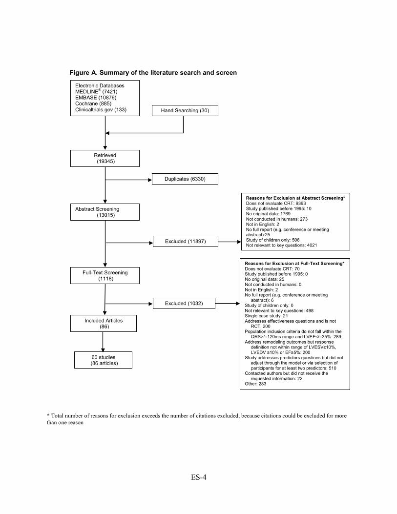
* Total number of reasons for exclusion exceeds the number of citations excluded, because citations could be excluded for more than one reason
Electronic Databases MEDLINE® (7421) EMBASE (10876) Cochrane (885) Clinicaltrials.gov (133)
Retrieved (19345)
Abstract Screening (13015)
Duplicates (6330)
Full-Text Screening (1118)
Excluded (11897)
Included Articles (86)
Excluded (1032)
Reasons for Exclusion at Full-Text Screening* Does not evaluate CRT: 70 Study published before 1995: 0 No original data: 25 Not conducted in humans: 0 Not in English: 2 No full report (e.g. conference or meeting
abstract): 6 Study of children only: 0 Not relevant to key questions: 498 Single case study: 21 Addresses effectiveness questions and is not
RCT: 200 Population inclusion criteria do not fall within the
QRS>/=120ms range and LVEF</=35%: 289 Address remodeling outcomes but response
definition not within range of LVESV≥10%, LVEDV ≥10% or EF≥5%: 200
Study addresses predictors questions but did not adjust through the model or via selection of participants for at least two predictors: 510
Contacted authors but did not receive the requested information: 22
Other: 283
Reasons for Exclusion at Abstract Screening* Does not evaluate CRT: 9393 Study published before 1995: 10 No original data: 1769 Not conducted in humans: 273 Not in English: 2 No full report (e.g. conference or meeting abstract):25 Study of children only: 506 Not relevant to key questions: 4021
Hand Searching (30)
Figure A. Summary of the literature search and screen
60 studies (86 articles)
ES-4

Discussion Key Findings and the Strength of Evidence Efficacy and Safety of CRT-D (KQ1a, KQ2)
There is convincing evidence that CRT-D devices are effective in reducing heart failure symptoms, improving myocardial function, and reducing hospitalizations for heart failure in patients with an LVEF≤35% and a QRS duration ≥120 ms compared to therapy with an ICD alone. Specifically, we found moderate strength of evidence for benefit of CRT-D versus ICD alone for all-cause mortality in minimally symptomatic patients. This statement is derived from data looking primarily at NYHA class II patients. The applicability of this finding to NYHA class I patients, a population significantly under-represented in studies, is unclear. There is insufficient evidence to determine whether CRT-D devices are effective in improving survival compared to an ICD alone in an advanced heart failure population (NYHA III-IV).
In terms of pre-specified subgroups, there is generally strong evidence that in CRT-D patients (compared to an ICD alone), female gender, a left bundle branch block, and non-ischemic cardiomyopathy are associated with superior outcomes. Sinus rhythm (as opposed to a history of atrial fibrillation) and a wider QRS complex and also associated with superior outcomes in patients undergoing CRT-D implant compared to an ICD alone although the data for this are less compelling. (Table A.) Efficacy and Safety of CRT-P (KQ3a, KQ4)
There is moderate evidence that CRT-P, compared to optimal medical therapy, is effective in improving survival, reducing LVESV, and reducing hospitalizations for heart failure in patients with an LVEF≤35% and a QRS duration ≥120 ms compared to optimal medical therapy alone. These data are largely derived from patients with NYHA class III-IV heart failure. The applicability of these findings to patient with NYHA class I-II heart failure is unclear. We found insufficient evidence about the effect of CRT-P on quality of life, as measured with the MLHFQ, compared to optimal medical therapy alone. Efficacy and Safety of CRT-D versus CRT-P (KQ5, KQ6)
Only one included trial contained both CRT-D and CRT-P arms, and direct comparisons between those arms were lacking. Therefore, there is insufficient evidence to determine the effectiveness of CRT-D compared to CRT-P. In comparing harms between CRT-D and CRT-P devices, there was also insufficient evidence to draw any conclusions except for device infections, which appear to be slightly more common for CRT-D devices. Predictors of Response: CRT-D and CRT-P (KQ1b, KQ3b)
The evidence regarding predictors of a favorable response following CRT varied considerably based on outcome. In addition, the high likelihood of reporting bias qualifies these results. Age was not an important predictor of outcomes in patients receiving CRT-D devices. However, data for very elderly patients (> 75 years of age) were limited. Non-ischemic cardiomyopathy, female gender, and a left bundle branch block morphology were strongly associated with improved outcomes. A history of atrial fibrillation and a narrower QRS duration were predictive of poorer outcomes although the evidence for this was less robust. There was inadequate evidence to determine the predictive nature of chronic kidney disease, left atrial volume, baseline LVEF, body mass index, and left ventricular end-diastolic volume on outcomes following CRT-D implant. There was also insufficient evidence to draw conclusions as to the predictive nature of baseline characteristics in patients receiving a CRT-P device. However, of
ES-5

the outcomes that were assessed, the ICD function would impact only the mortality endpoint. Therefore similar conclusions as to those noted for CRT-D can likely be drawn for CRT-P devices for the other, non-mortality endpoints.
ES-6
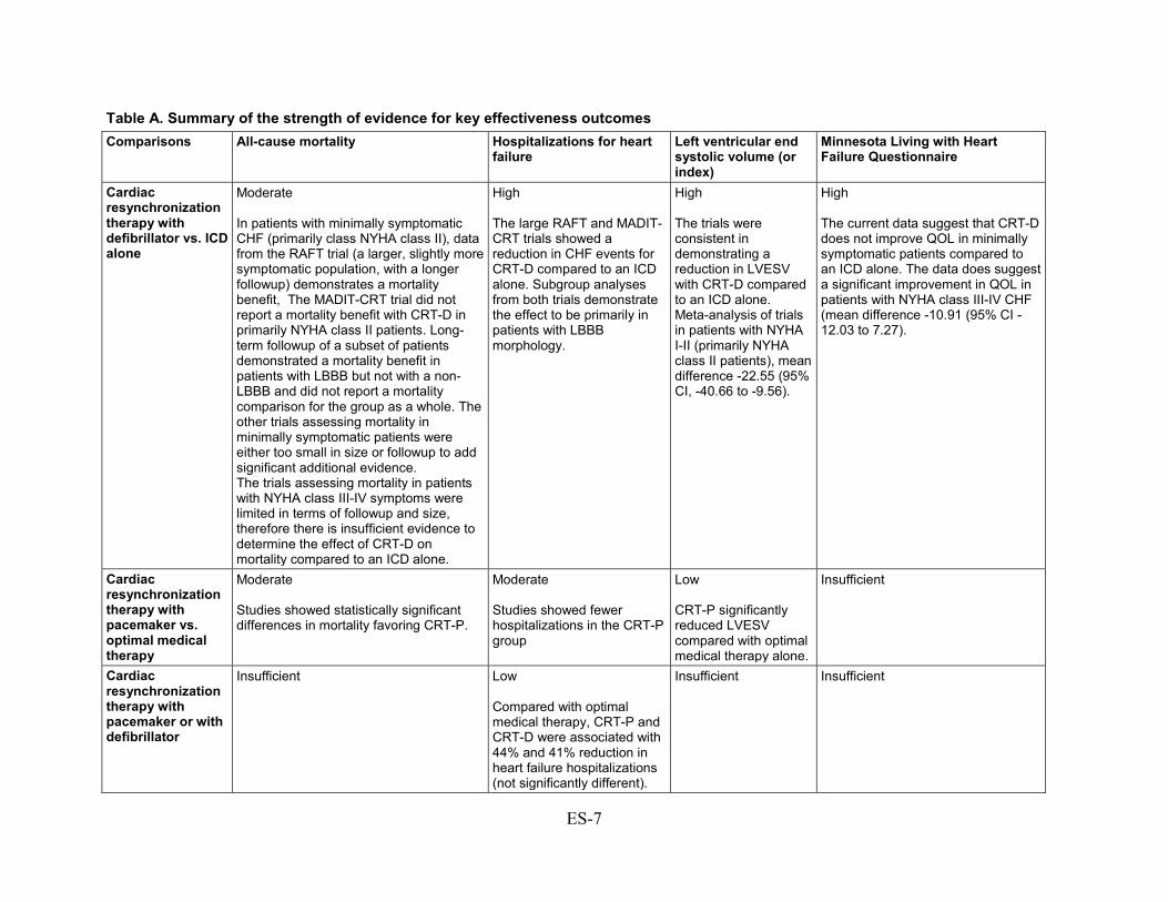
Table A. Summary of the strength of evidence for key effectiveness outcomes Comparisons All-cause mortality Hospitalizations for heart
failure Left ventricular end systolic volume (or index)
Minnesota Living with Heart Failure Questionnaire
Cardiac resynchronization therapy with defibrillator vs. ICD alone
Moderate In patients with minimally symptomatic CHF (primarily class NYHA class II), data from the RAFT trial (a larger, slightly more symptomatic population, with a longer followup) demonstrates a mortality benefit, The MADIT-CRT trial did not report a mortality benefit with CRT-D in primarily NYHA class II patients. Long-term followup of a subset of patients demonstrated a mortality benefit in patients with LBBB but not with a non-LBBB and did not report a mortality comparison for the group as a whole. The other trials assessing mortality in minimally symptomatic patients were either too small in size or followup to add significant additional evidence. The trials assessing mortality in patients with NYHA class III-IV symptoms were limited in terms of followup and size, therefore there is insufficient evidence to determine the effect of CRT-D on mortality compared to an ICD alone.
High The large RAFT and MADIT-CRT trials showed a reduction in CHF events for CRT-D compared to an ICD alone. Subgroup analyses from both trials demonstrate the effect to be primarily in patients with LBBB morphology.
High The trials were consistent in demonstrating a reduction in LVESV with CRT-D compared to an ICD alone. Meta-analysis of trials in patients with NYHA I-II (primarily NYHA class II patients), mean difference -22.55 (95% CI, -40.66 to -9.56).
High The current data suggest that CRT-D does not improve QOL in minimally symptomatic patients compared to an ICD alone. The data does suggest a significant improvement in QOL in patients with NYHA class III-IV CHF (mean difference -10.91 (95% CI -12.03 to 7.27).
Cardiac resynchronization therapy with pacemaker vs. optimal medical therapy
Moderate Studies showed statistically significant differences in mortality favoring CRT-P.
Moderate Studies showed fewer hospitalizations in the CRT-P group
Low CRT-P significantly reduced LVESV compared with optimal medical therapy alone.
Insufficient
Cardiac resynchronization therapy with pacemaker or with defibrillator
Insufficient Low Compared with optimal medical therapy, CRT-P and CRT-D were associated with 44% and 41% reduction in heart failure hospitalizations (not significantly different).
Insufficient Insufficient
ES-7
Relationship of Findings to Existing Literature Our current review differs from prior reviews in that only studies with patients with an
LVEF≤35% and a baseline QRS duration≥120 ms undergoing biventricular pacing were included. These criteria were developed in consultation with our key informants and largely mirror the current appropriate use criteria for CRT from United States guidelines.1 This eliminated the REVERSE, BLOCK CHF, and HOBIPACE trials which included patients with an LVEF>35%.11-13 The criteria also excluded all trials looking at the effects of CRT in a narrow QRS duration population,14-16 and studies of LV only pacing.17 ,18 We considered the appropriate control for the CRT-D effectiveness question to be an ICD alone given the compelling data demonstrating improvements in mortality with an ICD that evolved concomitantly with studies of CRT effectiveness. We considered the appropriate control for CRT-P to be optimal medical therapy alone to assess the impact of cardiac resynchronization. We did not assess the comparison of CRT-D to optimal medical therapy as we determined this to be an inappropriate comparison, given the known improvements in mortality by defibrillation. Also, in contrast to several previous reviews, we included only RCTs to assess the questions regarding effectiveness.
In terms of minimally symptomatic patients, the results of our review largely agree with those of prior reviews, which focused on the same population. Similarly, the current review is in agreement with the systematic review performed in 2007 by Mcallister et al., which included studies primarily involving an advanced heart failure population.19 Our review arrived at somewhat different conclusions in terms of the efficacy of CRT-D vs. CRT-P compared to that by Jiang et. al.20 Given that we considered only RCTs for determination of effectiveness, only the COMPANION trial was included in our review, which likely explains the discrepancy in conclusions.3
In our systematic analysis of predictors of outcomes following CRT, many studies assessing the capacity for baseline characteristics to predict responses (defined in many different ways) were identified. The large majority of studies were small (<100 patients) and not properly controlled. At a minimum, we pre-specified that a cohort study addressing our questions about predictors of response to CRT had to include at least gender and either QRS duration or morphology in a multivariate model to address confounding factors. Such criteria eliminated many studies. Despite this, the positive predictive effect noted with LBBB, female gender, non-ischemic cardiomyopathy, a wider QRS duration, and normal sinus rhythm on multiple outcomes was supportive of the similar findings noted from the pre-specified subgroup analyses of the RCTs. There are other potential predictors we did not consider (e.g. lead position). Given the large number of potential predictors in the literature, a review of all predictors was not practical. Our predictors were chosen based on prevailing knowledge of the most important predictors, identified in consultation with our key informants.
Finally, we did not conduct individual patient data meta-analysis to assess predictors meaning that our analyses may suggest that clinically relevant subgroup effects exist, but we are unable to quantify the effects reliably or precisely.
Applicability The generalizability of these results is slightly limited. Race was reported very infrequently,
prohibiting an assessment of applicability based on racial differences. The majority of patients included in the RCTs were male, although a large focus in sub-studies has been given to the role of CRT in women, given the heightened response to therapy seen in this population. The average age in the RCTs and cohort studies was in the mid 60s although many patients included were in
ES-8

the age range of the elderly Medicare population. There has not been an RCT that specifically enrolled Medicare eligible patients. Also, data for very elderly patients (> 75 years of age) are limited. In cohort studies and subgroup analyses from the RCTs, age was not found to be an important predictor of outcomes. Taken together, the results of our review are generalizable to the Medicare population although given the absence of dedicated RCTs, a definitive statement of generalizability to this population is not possible.
Limitations of the Comparative Effectiveness Review Process In addressing the questions of efficacy of CRT-D and CRT-P, several studies potentially of
interest were excluded since they were non-randomized. For the questions about the predictors of response to CRT, many retrospective cohort studies were excluded because of a mixed population of CRT-D and CRT-P devices. Attempts were made to contact the authors of such studies to obtain the device-specific data but, in many cases, this was unsuccessful. In addition, many cohorts that contained outcomes of interest were excluded due to failure to control for gender and QRS duration and/or morphology, important baseline confounders. Finally, we did not include prior or conduct new individual patient data meta-analyses to assess predictors. Therefore, our analyses may suggest that clinically relevant subgroup effects exist, but we are unable to quantify the effects reliably or precisely.
Limitations of the Evidence Base Multiple, well-conducted RCTs were identified addressing the questions about the efficacy of
CRT-D and CRT-P. The majority of patients enrolled in the clinical trials had NYHA class II-IV heart failure symptoms. The applicability of the current findings to class I patients is less clear. In contrast, only the COMPANION trial contained both CRT-D and CRT-P arms.3 However, a direct comparison of the CRT-D to CRT-P arms was not reported for several outcomes. For the questions examining predictors of response to CRT many of the included cohort studies were relatively small. While all studies controlled for gender and either QRS duration or morphology based on our pre-specified inclusion criteria, the remaining variables in the model varied widely between studies. Similarly, many studies used statistical criteria to create their multivariate adjustments, rather than pre-specifying clinical factors known to be important.
Research Gaps There is convincing evidence that CRT-D results in reverse ventricular remodeling and
improvements in quality of life compared to an ICD alone. However, only two trials showed a mortality benefit of CRT-D over ICD alone. The RAFT trial primarily contained patients with NYHA class II symptoms.6 Long term follow up of the MADIT-CRT trial suggested a mortality benefit in the LBBB subgroup alone but did not report mortality for the cohort as a whole.21 Whether CRT-D results in improved survival compared to an ICD alone in patients with advanced heart failure is unclear.
Several subgroup analyses from RCTs as well as cohort studies demonstrate superior outcomes in patients with a native LBBB compared to a non-LBBB. Subgroup analysis from the MADIT-CRT trial suggested possible harm for CRT-D versus an ICD alone in non-LBBB patients.5 ,21Subgroup analyses from other RCTs suggested little benefit of CRT in non-LBBB patients (but no convincing trend towards harm).6 One important issue with the assessment of CRT efficacy according to QRS morphology is the interaction between QRS duration, another
ES-9

variable with impact on outcome, and morphology. Patients with a LBBB tend to have wider QRS durations than patients with non-LBBBs. Several retrospective studies have attempted to determine the relative impact on outcomes of QRS duration within various QRS morphology groups in patients receiving a CRT device with mixed results.22 ,23 There has not been an RCT, which compares CRT to a control in patients with a non-LBBB morphology. Given the lack of such a trial, the ability to conclude definitively that CRT is ineffective or, in fact harmful, in patients with non-LBBB morphology is limited.
Similarly, subgroup analyses from RCTs suggest limited benefit of CRT in patients with atrial fibrillation. Outside the small MUSTIC-AF study, data focused on the AF population are lacking.24 Therefore, the ability to definitively conclude a lack of benefit in patients with AF receiving CRT is not possible.
The effectiveness of CRT-D versus CRT-P in patients with an LVEF≤35% has not been adequately addressed. The COMPANION trial, which included both arms, did not directly compare the CRT types and is therefore inadequate to answer this question definitively.3
Conclusion We performed a systematic review to evaluate the efficacy and safety of CRT-D and CRT-P
devices as well as predictors of outcomes following implant of such devices. There is convincing evidence that CRT-D is effective with regard to improvements in multiple outcomes compared to an ICD alone in patients with an LVEF≤35% and a QRS duration ≥120ms. These findings are based on patients primarily with NYHA class II-IV heart failure. The applicability of these findings to patients with NYHA class I symptoms is unclear. Similarly, there is convincing evidence that CRT-P is effective in improving multiple endpoints compared to optimal medical therapy alone in the same population. These data are primarily derived from NYHA class III-IV and the applicability to patients with NYHA class I and II is less clear. Female gender, LBBB, a widened QRS duration, sinus rhythm, and non-ischemic cardiomyopathy are associated with improved outcomes following CRT.
ES-10

Reference List
1. Heart Failure Society of America (HFSA) practice guidelines. HFSA guidelines for management of patients with heart failure caused by left ventricular systolic dysfunction--pharmacological approaches. J Card Fail 1999; 5(4):357-82. PMID: 10634677
2. Abraham WT, Fisher WG, Smith AL et al. Cardiac resynchronization in chronic heart failure. N Engl J Med 2002; 346(24):1845-53. PMID: 12063368
3. Bristow MR, Saxon LA, Boehmer J et al. Cardiac-resynchronization therapy with or without an implantable defibrillator in advanced chronic heart failure. N Engl J Med 2004; 350(21):2140-50. PMID: 15152059
4. Cleland JG, Daubert JC, Erdmann E et al. The effect of cardiac resynchronization on morbidity and mortality in heart failure. N Engl J Med 2005; 352(15):1539-49. PMID: 15753115
5. Moss AJ, Hall WJ, Cannom DS et al. Cardiac-resynchronization therapy for the prevention of heart-failure events. N Engl J Med 2009; 361(14):1329-38. PMID: 19723701
6. Tang AS, Wells GA, Talajic M et al. Cardiac-resynchronization therapy for mild-to-moderate heart failure. N Engl J Med 2010; 363(25):2385-95. PMID: 21073365
7. Higgins JPT. Cochrane handbook for systemic reviews of interventions Version 5.1.0. The Cochrane Collaboration. 2011;Oxford, England. Available at: http://handbook.cochrane.org. PMID:
8. Wells GA . The Newcastle-Ottawa Scale (NOS) for assessing the quality of nonrandomised studies in meta-analyses. 2011. www.ohri.ca/programs/clinical_epidemiology/oxford.asp. 2011.
9. Hayden JA. Assessing bias in studies of prognostic factors. Ann Intern Med. 2013; 158(4):280-6. PMID: 23420236
10. Agency for Healthcare Research and Quality. Grading the Strength of a Body of Evidence When Assessing Health Care Interventions - AHRQ and the Effective Health Care Program:
An Update. Rockville, MD: Agency for Healthcare Research and Quality. 2013: Available at: http://effectivehealthcare.ahrq.gov/ehc/products/457/1163/GradingTheStrengthofEvidence_DraftMethodsChapter_20120625.pdf. 2013. PMID:
11. Daubert C, Gold MR, Abraham WT et al. Prevention of disease progression by cardiac resynchronization therapy in patients with asymptomatic or mildly symptomatic left ventricular dysfunction: insights from the European cohort of the REVERSE (Resynchronization Reverses Remodeling in Systolic Left Ventricular Dysfunction) trial. J Am Coll Cardiol 2009; 54(20):1837-46. PMID: 19800193
12. Kindermann M, Hennen B, Jung J, Geisel J, Bohm M, Frohlig G. Biventricular versus conventional right ventricular stimulation for patients with standard pacing indication and left ventricular dysfunction: the Homburg Biventricular Pacing Evaluation (HOBIPACE). J Am Coll Cardiol 2006; 47(10):1927-37. PMID: 16697307
13. Curtis AB, Worley SJ, Adamson PB et al. Biventricular pacing for atrioventricular block and systolic dysfunction. N Engl J Med 2013; 368(17):1585-93. PMID: 23614585
14. Beshai JF, Grimm RA, Nagueh SF et al. Cardiac-resynchronization therapy in heart failure with narrow QRS complexes. N Engl J Med 2007; 357(24):2461-71. PMID: 17986493
15. Thibault B, Harel F, Ducharme A et al. Cardiac resynchronization therapy in patients with heart failure and a QRS complex <120 milliseconds: the Evaluation of Resynchronization Therapy for Heart Failure (LESSER-EARTH) trial. Circulation 2013; 127(8):873-81. PMID: 23388213
16. Ruschitzka F, Abraham WT, Singh JP et al. Cardiac-resynchronization therapy in heart failure with a narrow QRS complex. N Engl J Med 2013; 369(15):1395-405. PMID: 23998714
17. Boriani G, Kranig W, Donal E et al. A randomized double-blind comparison of biventricular versus left ventricular stimulation
ES-11
for cardiac resynchronization therapy: the Biventricular versus Left Univentricular Pacing with ICD Back-up in Heart Failure Patients (B-LEFT HF) trial. Am Heart J 2010; 159(6):1052-8.e1. PMID: 20569719
18. Thibault B, Ducharme A, Harel F et al. Left ventricular versus simultaneous biventricular pacing in patients with heart failure and a QRS complex >/=120 milliseconds. Circulation 2011; 124(25):2874-81. PMID: 22104549
19. McAlister FA, Ezekowitz J, Hooton N et al. Cardiac resynchronization therapy for patients with left ventricular systolic dysfunction: a systematic review. JAMA 2007; 297(22):2502-14. PMID: 17565085
20. Jiang M, He B, Zhang Q. Comparison of CRT and CRT-D in heart failure: systematic review of controlled trials. Int J Cardiol 2012; 158(1):39-45. PMID: 21262545
21. Goldenberg I, Kutyifa V, Klein HU et al.
Survival with cardiac-resynchronization therapy in mild heart failure. N Engl J Med 2014; 370(18):1694-701. PMID: 24678999
22. Dupont M, Rickard J, Baranowski B et al. Differential response to cardiac resynchronization therapy and clinical outcomes according to QRS morphology and QRS duration. J Am Coll Cardiol 2012; 60(7):592-8. PMID: 22796255
23. Cleland JG, Abraham WT, Linde C et al. An individual patient meta-analysis of five randomized trials assessing the effects of cardiac resynchronization therapy on morbidity and mortality in patients with symptomatic heart failure. Eur Heart J 2013; 34(46):3547-56. PMID: 23900696
24. Leclercq C, Walker S, Linde C et al. Comparative effects of permanent biventricular and right-univentricular pacing in heart failure patients with chronic atrial fibrillation. Eur Heart J 2002; 23(22):1780-7. PMID: 12419298
ES-12

Introduction Chronic heart failure (CHF) is a major public health problem in the United States affecting
an estimated 4.9 million Americans, with 550,000 new cases diagnosed annually.1 CHF patients have high rates of hospitalization, poor quality of life, and account for 300,000 deaths in the United States each year.1 The annual cost of CHF in 2010 was estimated at $39.2 billion or approximately 2 percent of the total United States healthcare budget.2 Targeted interventions for this commonly encountered condition are needed, aimed at improving quality of life, reducing hospitalizations, and decreasing mortality.
Left ventricular (LV) activation delay, as approximated by widening of the QRS complex on a twelve lead electrocardiogram, is present in approximately one-quarter to one-third of heart failure patients. Such delay leads to inefficient myocardial hemodynamics, which may impair cardiac function further. Cardiac resynchronization (CRT) is a pacing modality utilizing an LV pacing lead with the goal of re-synchronizing myocardial contraction in patients with CHF, depressed systolic function, and significant LV activation delay. CRT is thought to produce a reduction in intraventricular dyssynchrony and more favorable hemodynamics by placement of a pacing lead, either endovascularly via a coronary sinus tributary, or epicardially with direct placement on the lateral LV wall via a thoracotomy. CRT was originally indicated in patients with significant LV dysfunction, defined as a left ventricular ejection fraction (LVEF) ≤ 35%, with New York Heart Association (NYHA) class III-IV heart failure symptoms, and with a QRS duration ≥120ms on optimal medical therapy.3-5More recently, the indications for CRT expanded to include patients with less advanced heart failure.6 ,7 In addition, the most recent guidelines for CRT implantation have called to attention the importance of both QRS duration and morphology.1
Multiple large scale clinical trials have been conducted to assess the effects of CRT. Early trials of CRT compared CRT pacemakers with optimal medical therapy alone in patients with advanced CHF.3 ,5 ,8With the concomitant development of the implantable cardiac defibrillator (ICD), comparisons used in the large clinical trials changed to compare patients with ICDs with and without CRT.6 ,7 Currently, the vast majority of candidates for CRT devices also have an indication for an ICD; therefore, the large majority of patients receiving CRT in the United States receive a CRT defibrillator (CRT-D) as opposed to a CRT pacemaker (CRT-P). CRT-P devices are occasionally placed in patients who wish to avoid ICD shocks (such as in the geriatric population who may be more interested in quality of life compared to life prolongation) or in patients with an indication for frequent right ventricular pacing due to conduction disease who have an LVEF between 36-50 percent.
The early trials of CRT focused on “softer” or more subjective endpoints including changes in quality of life scores, NYHA functional class, and six minute hall walk distances.3 ,8 More objective or “harder” endpoints were included in subsequent studies including ventricular remodeling, myocardial oxygen consumption, CHF hospital admissions, and all-cause mortality.4
,7 CRT has been one of the most important therapeutics for the treatment of CHF over the past 20 years, but not every patient who meets the guideline criteria for CRT appears to respond to this therapy. While the percentage of “non-responders” to CRT fluctuates greatly primarily based on how one defines “response”, it is generally estimated that between 30-40 percent of patients receiving CRT derive what may appear to be little benefit.9 Prediction of response to CRT is an important goal in order to tailor this therapy to patients most apt to derive benefit.9 In addition, the specter of patient harm in certain subgroups has been raised.10
1
The impact of bundle branch block morphology and duration on patient outcomes receiving CRT devices continues to be an important area of research.11-13 The new 2013 United States guidelines for the implantation of CRT capable devices take both bundle branch block morphology and QRS duration into consideration in determining appropriateness for CRT device implantation.14 ,15 It is not yet clear how these new guidelines may impact response rates, but any improvement is expected to be incremental, with the issue of non-responders not completely resolved. Not all potential causes of non-response were considered in the new guidelines.16
Scope and Key Questions We conducted a systematic review on the efficacy for both CRT-D and CRT-P. The
questions were nominated for the Agency for Healthcare Research and Quality (AHRQ) Evidence-based Practice Center (EPC) Program by the Centers for Medicare and Medicaid (CMS) and thus focus on the Medicare population. We developed analytic frameworks to illustrate the different questions and outcomes we considered (Figures 1, 2 and 3). We sought to address the following Key Questions (KQ): KQ1a: Is cardiac resynchronization therapy with defibrillator (CRT-D) effective in reducing heart failure symptoms, improving myocardial function, reducing hospitalization and/or improving survival in patients with an LVEF≤35% and a QRS duration≥120ms? KQ1b: What are the clinical predictors of response in Medicare eligible patients who are deemed appropriate candidates for CRT-D devices? KQ2: What are the adverse effects or complications associated with CRT-D implantation in the Medicare population? KQ3a: Is cardiac resynchronization therapy in the absence of defibrillator capacity (CRT-P) effective in reducing heart failure symptoms, improving myocardial function, reducing hospitalization and/or improving survival in patients with LVEF≤35% and a QRS duration≥120ms? KQ3b: What are the clinical predictors of response in Medicare eligible patients who are deemed appropriate candidates for CRT-P devices? KQ4: What are the adverse effects or complications associated with CRT-P implantation in the Medicare population? KQ5: What is the effectiveness of CRT-D versus CRT-P in reducing heart failure symptoms, improving myocardial function, reducing hospitalization and/or improving survival in patients with LVEF≤35% and a QRS duration≥120ms? KQ6: What are the adverse effects or complications associated with CRT-D versus CRT-P implantation in the Medicare population?
2

CRT-D
Intermediate outcomes • 6 minute hall walk distance • Left ventricular end diastolic volume/volume index • Left ventricular end systolic volume/volume index • Left ventricular ejection fraction • Clinical composite score (Packer Score)
(KQ1)
Health outcomes
• Heart failure hospitalizations
• All-cause mortality • Minnesota Living with
Heart Failure Questionnaire
• SF-36
Adverse effects of intervention • Procedure related complications • Length of hospital stay • Pneumothorax • Pocket hematoma • Device Infection • Cardiac perforation/ tamponade • Lead dislodgement • Ventricular arrhythmias • Inappropriate ICD shocks
(KQ2)
Figure 1. Analytic framework for use of cardiac resynchronization therapy with defibrillator (CRT-D) in the Medicare population
(KQ1)
3

CRT-P
Intermediate outcomes • 6 minute hall walk distance • Left ventricular end diastolic volume/ volume index • Left ventricular end systolic volume/volume index • Left ventricular ejection fraction • Clinical composite score (Packer Score)
(KQ3) (KQ3)
Health outcomes
• Heart failure hospitalizations
• All-cause mortality • Minnesota Living with
Heart Failure Questionnaire
• SF-36
Adverse effects of intervention
• Procedure related complications • Length of hospital stay • Pneumothorax • Pocket hematoma • Device Infection • Cardiac perforation/ tamponade • Lead dislodgement • Ventricular arrhythmias
(KQ4)
Figure 2. Analytic framework for use of cardiac resynchronization therapy without defibrillator capacity (CRT-P) in the Medicare population
4

CRT-D vs.
CRT-P
Intermediate outcomes • 6 minute hall walk distance • Left ventricular end diastolic volume/volume index • Left ventricular end systolic volume/ volume index • Left ventricular ejection fraction • Clinical composite score (Packer Score)
(KQ5) (KQ5)
Health outcomes
• Heart failure hospitalizations
• All-cause mortality • Minnesota Living with
Heart Failure Questionnaire
• SF-36
Adverse effects of intervention • Procedure related complications • Length of hospital stay • Pneumothorax • Pocket hematoma • Device Infection • Cardiac perforation/ tamponade • Lead dislodgement • Ventricular arrhythmias • Inappropriate ICD shocks
(KQ6)
Figure 3. Analytic framework for use of cardiac resynchronization therapy with defibrillator capacity (CRT-D) versus cardiac resynchronization therapy without
defibrillator capacity (CRT-P) in the Medicare population
5

Methods The methods for this Technology Assessment follow the AHRQ Methods Guide for
Effectiveness and Comparative Effectiveness Reviews.17
Protocol Development Representatives from the Coverage and Analysis Group at CMS posed the questions for this review. With feedback from these representatives, from AHRQ representatives and from our key informants, we refined these questions and developed a protocol for this systematic review. Our protocol was registered on PROSPERO (CRD42014009981).
Search Strategy We searched MEDLINE, Embase®, and the Cochrane Central Register of Controlled Trials
(CENTRAL) from January 1, 1995, as this is the date of first article reporting use of CRT through October 20th, 2014. We developed a search strategy based on medical subject headings (MeSH®) terms and text words of key articles (Appendix B). We also reviewed the reference lists of all included articles. In addition, we requested Scientific Information Packets from device manufacturers (Table 3). We had no language restrictions in the search strategies. Additionally, on January 22nd, 2015, we searched Clinicaltrials.gov to identify relevant registered trials. Study Selection
Study selection was based on predefined eligibility criteria of patient populations, interventions, outcome measures, and study design (see Table 1 and 2). Abstracts were screened independently by two reviewers, and were excluded if both reviewers agreed that one or more of the exclusion criteria was met (see Appendix C Abstract Screen Form). Differences between reviewers regarding abstract eligibility were resolved through consensus. We used DistillerSR (Evidence Partners, 2010) to manage the screening process.
Citations promoted on the basis of the abstract screen underwent another independent screen using the full-text of the articles. Additional exclusion criteria were applied at this level (see Table 2 and Appendix C Article Screen Form). Differences regarding citation eligibility were resolved through consensus.
Data Abstraction and Data Management We created and pilot tested data extraction forms in Excel (Microsoft, Redmond, WA)
(Appendix C). Reviewers extracted information on general study characteristics (e.g., study design, study period, and followup), study participants (e.g., age, gender, race/ethnicity, etc.), eligibility criteria, interventions, outcome measures and the method of ascertainment, and the results of each outcome, including measures of variability. Data when available by subgroups (females, QRS duration >150 ms, left bundle branch block, atrial fibrillation and non-ischemic cardiac conditions) were also abstracted. For studies reporting patient data, including outcomes, undifferentiated as to CRT-D or CRT-P we contacted the authors for clarification and data (see Data Synthesis).
One reviewer completed data abstraction and a second reviewer checked the first reviewer’s abstraction for completeness and accuracy. We resolved differences between reviewer pairs through discussion and, as needed, through consensus among our team.
6

Table 1. PICOTS (population, interventions, comparators, outcomes, timing, setting) for each Key Question Effectiveness
KQ1a: CRT-D KQ3a: CRT-P KQ5: CRT-D vs CRT-P
Harms KQ2: CRT-D KQ4: CRT-P KQ6: CRT-D vs CRT-P
Clinical predictors KQ1b: CRT-D KQ 2b: CRT-P
Population • Age ≥ 18
• Subjects with a left ventricular ejection fraction ≤35% and a QRS duration ≥120 ms.
Interventions • Cardiac resynchronization therapy with a defibrillator (CRT-D)
• Cardiac resynchronization without a defibrillator (CRT-P)
• Cardiac resynchronization therapy with a defibrillator (CRT-D)
• Cardiac resynchronization without a defibrillator (CRT-P)
• Cardiac resynchronization therapy with a defibrillator (CRT-D)
• Cardiac resynchronization without a defibrillator (CRT-P)
Comparisons • CRT-D: Implantable Cardioverter Defibrillator (ICD)
• CRT-P: Optimal medical therapy • CRT-D versus CRT-P
• CRT-D: Implantable Cardioverter Defibrillator (ICD)
• CRT-P: Optimal medical therapy • CRT-D versus CRT-P
• CRT-D: Implantable Cardioverter Defibrillator (ICD)
• CRT-P: Optimal medical therapy
Outcomes • 6 minute hall walk distance • Minnesota Living with Heart Failure
Questionnaire • SF-36 • Left ventricular end systolic
volume/volume index • Left ventricular end diastolic
volume/volume index • Left ventricular ejection fraction • Clinical composite score (Packer
Score) • Hospitalizations for heart failure • All- cause mortality
• Procedure related complications • Length of hospital stay • Pneumothorax • Pocket hematoma • Device Infection • Cardiac perforation/ tamponade • Lead dislodgement • Ventricular arrhythmias • Inappropriate ICD shocks (CRT-D
only) • Death (within a week)
• Age • Gender • Cardiomyopathy subtype • History of atrial fibrillation • QRS duration • QRS morphology • Chronic kidney disease • Left atrial volume • Left ventricular ejection fraction • Body mass index • Baseline left ventricular end diastolic
volume
Type of study Randomized controlled trials
Any study design except case report Any study design except case report
Timing and setting
CRT-D and CRT-P at 3-6 months, 1 year, and ≥2 year end-points KQ2,4 and 6 (harms) Outcomes (above) from CRT-D and CRT-P at any time point
Any time point Any time point
7

Table 2. List of exclusion criteria at the abstract and article screening level Abstract screening level Article screening level
Exclusion criteria • Does not evaluate CRT • Study published before 1995 • No original data (systematic reviews,
meta-analysis, editorial, commentary) • Not conducted in humans • Not in English • No full report (e.g. conference or
meeting abstract) • Study of children only • Not relevant to key questions
• Does not evaluate CRT • Study published before 1995 • No original data (systematic reviews, meta-
analysis, editorial, commentary) • Not conducted in humans • Not in English • No full report (e.g. conference or meeting
abstract) • Study of children only • Not relevant to key questions.
Additional exclusion criteria at the article screening level
• Single case study • Addresses effectiveness questions (KQ1a,
3a, and 5) only and is not RCT • Population inclusion criteria do not fall
within the QRS duration ≥120ms range and LVEF≤35%
• Address remodeling outcomes but response definition not within range of LVESV≥10%, LVEDV ≥10% or EF≥5%
• Study does not adjust via model or participant selection for at least 2 predictor of gender, QRS duration and/or morphology (LBB or not ) and does not include at least 30 patients
8

Table 3. List of CRT device manufacturers Manufacturer Device
Boston Scientific COGNIS® CRT-D
ENERGEN™ CRT-D
INCEPTA™ CRT-D
PUNCTUA™ CRT-D
INVIVE™ CRT-P
BIOTRONIK Lumax 300 HF-T CRT-D
Lumax 340 HF-T CRT-D
Lumax 540 HF-T CRT-D
Stratos LV-T CRT-P
Evia HFT-T CRT-P
Medtronic Viva™ XT CRT-D
Viva™ S CRT-D
Protecta® XT CRT-D
Protecta® CRT-D
Consulta® CRT-D
Concerto® II CRT-D
Maximo® II CRT-D
InSync Sentry® CRT-D
InSync II™ Marquis CRT-D
InSync III® CRT-P
Consulta® CRT-P
Syncra™ CRT-P
St. Jude Medical Promote™ Plus CRT-D
Quadra Assura™ CRT-D
Unify Assura™ CRT-D
Unify Quadra™ CRT-D
Unify™ CRT-D
Anthem™ CRT-P
SORIN GROUP PARADYM™ CRT
Paradym™ RF SonR® CRT-D
Paradym™ RF CRT-D
9

Risk of Bias Assessment Two reviewers independently assessed risk of bias for individual studies. We used the
Cochrane Collaboration’s tool for assessing the risk of bias of controlled studies.18 For nonrandomized studies, we used the Newcastle Ottawa Scale 19 and for predictor studies, we used Quality In Prognosis Studies (QUIPS) tool.20 Differences between reviewers were resolved through consensus. Data Synthesis
For each key question, we created a detailed set of evidence tables containing all information abstracted from eligible studies. We followed these steps for studies which reported data for both devices (CRT-D and CRT-P) in one arm or group or for which the type of device was unclear:
1. If the type of device is not specified, we contacted the study authors to request information about type of device.
2. If the number of patients receiving each device is not specified, we contacted the study authors to request information about the number of patients receiving each device.
3. If the number of patients receiving each device is not specified, and the outcomes are not presented separately, we contacted the study author to request device-specific outcome data.
4. If the number of patients receiving each device is specified, but the outcomes are not presented separately, we attributed the reported outcomes to the device received by ≥ 90 percent of the patients.
5. If the number of patients receiving each device is specified and the outcomes are not presented separately and no more than 90 percent of the patients received any one type of device or all devices were received by an equal number of patients, we contacted the study authors to request device-specific outcome data.
All studies were summarized qualitatively. We conducted meta-analyses for an outcome when there were sufficient data (at least 3 studies of the same design) and studies were sufficiently homogenous with respect to key variables (population characteristics, intervention, and outcome measurement) using profile likelihood estimate for random effects model. We identified substantial statistical heterogeneity in the trials as an I-squared statistic with a value greater than 50 percent. We planned to assess publication bias using Begg‘s and Eggers tests (with alpha of 0.10) including evaluation of the asymmetry of funnel plots for each comparison of interest for the outcomes where meta-analyses are conducted. Criteria for testing for funnel plot asymmetry was at least 10 studies of unequal sizes contributing quantitative data for which there is no apparent relationship between study size and between study clinical or methodological diversity. All meta-analyses were conducted using STATA 12.1 (College Station, TX). Strength of the Body of Evidence
We graded the strength of evidence using the scheme recommended by the AHRQ EPC Methods Guide for Conducting Comparative Effectiveness Reviews.21 For this report, we graded the strength of evidence for the outcomes we classified during protocol development as the most important or critical outcomes, including quality of life as assessed by the Minnesota Living with Heart Failure Questionnaire, left ventricular end systolic volume, hospitalizations for heart failure and, all- cause mortality. We considered five domains: study limitations, directness,
10
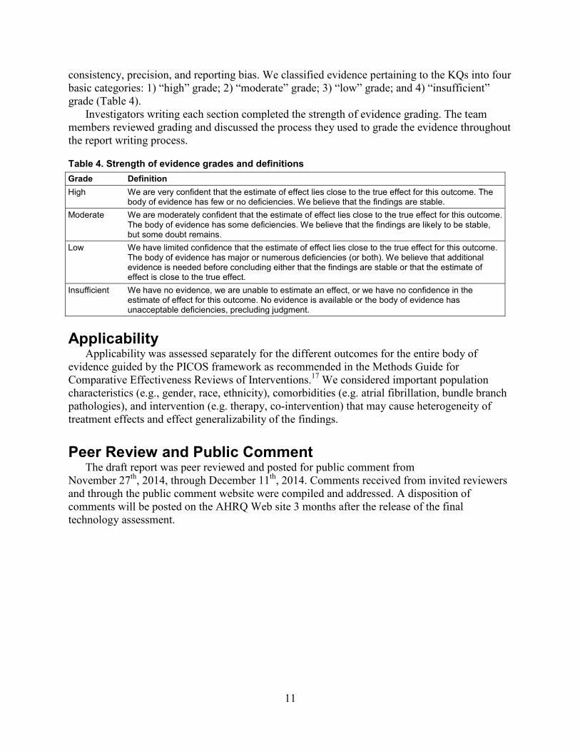
consistency, precision, and reporting bias. We classified evidence pertaining to the KQs into four basic categories: 1) “high” grade; 2) “moderate” grade; 3) “low” grade; and 4) “insufficient” grade (Table 4).
Investigators writing each section completed the strength of evidence grading. The team members reviewed grading and discussed the process they used to grade the evidence throughout the report writing process.
Table 4. Strength of evidence grades and definitions Grade Definition High We are very confident that the estimate of effect lies close to the true effect for this outcome. The
body of evidence has few or no deficiencies. We believe that the findings are stable. Moderate We are moderately confident that the estimate of effect lies close to the true effect for this outcome.
The body of evidence has some deficiencies. We believe that the findings are likely to be stable, but some doubt remains.
Low We have limited confidence that the estimate of effect lies close to the true effect for this outcome. The body of evidence has major or numerous deficiencies (or both). We believe that additional evidence is needed before concluding either that the findings are stable or that the estimate of effect is close to the true effect.
Insufficient We have no evidence, we are unable to estimate an effect, or we have no confidence in the estimate of effect for this outcome. No evidence is available or the body of evidence has unacceptable deficiencies, precluding judgment.
Applicability
Applicability was assessed separately for the different outcomes for the entire body of evidence guided by the PICOS framework as recommended in the Methods Guide for Comparative Effectiveness Reviews of Interventions.17 We considered important population characteristics (e.g., gender, race, ethnicity), comorbidities (e.g. atrial fibrillation, bundle branch pathologies), and intervention (e.g. therapy, co-intervention) that may cause heterogeneity of treatment effects and effect generalizability of the findings.
Peer Review and Public Comment
The draft report was peer reviewed and posted for public comment from November 27th, 2014, through December 11th, 2014. Comments received from invited reviewers and through the public comment website were compiled and addressed. A disposition of comments will be posted on the AHRQ Web site 3 months after the release of the final technology assessment.
11

Results Results of the Search
Figure 4 summarizes the results of our searching and screening. We identified 13,015 unique citations and excluded 10,805 of these during the abstract screen. During the full-text screening, we excluded 1032 citations (See Appendix D for list of excluded articles with reason (s) for exclusion). Sixty studies reported in eighty six articles are included in this review. Scientific information packets (SIPs)
As part of the grey literature search, device manufacturer companies were asked to provide information about pertinent studies conducted with their products (published, unpublished, and ongoing clinical trials). One company responded that no relevant studies had been conducted. Two companies provided scientific information packets, with potentially relevant studies; these citations were checked against our existing citation database, yielding ten new citations, none of which met our eligibility criteria. (Appendix F)
12
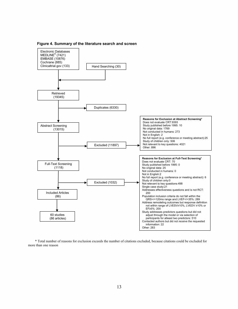
* Total number of reasons for exclusion exceeds the number of citations excluded, because citations could be excluded for
more than one reason
Electronic Databases MEDLINE® (7421) EMBASE (10876) Cochrane (885) Clinicaltrial.gov (133)
Retrieved (19345)
Abstract Screening (13015)
Duplicates (6330)
Full-Text Screening (1118)
Excluded (11897)
Included Articles (86)
Excluded (1032)
Reasons for Exclusion at Full-Text Screening* Does not evaluate CRT: 70 Study published before 1995: 0 No original data: 25 Not conducted in humans: 0 Not in English:2 No full report (e.g. conference or meeting abstract): 6 Study of children only:0 Not relevant to key questions:498 Single case study:21 Addresses effectiveness questions and is not RCT:
200 Population inclusion criteria do not fall within the
QRS>/=120ms range and LVEF</=35%: 289 Address remodeling outcomes but response definition
not within range of LVESV≥10%, LVEDV ≥10% or EF≥5%: 200
Study addresses predictors questions but did not adjust through the model or via selection of participants for atleast two predictors: 510
Contacted authors but did not receive the requested information: 22
Other: 283
Reasons for Exclusion at Abstract Screening* Does not evaluate CRT:9393 Study published before 1995: 10 No original data: 1769 Not conducted in humans: 273 Not in English: 2 No full report (e.g. conference or meeting abstract):25 Study of children only: 506 Not relevant to key questions: 4021 Other: 896
Hand Searching (30)
60 studies (86 articles)
Figure 4. Summary of the literature search and screen
13

Overview of included studies by outcomes We list the number and type of studies identified as addressing each comparison of interest,
by type of outcome assessed. List of RCTs, and other analyses and reports from these trials, are shown in Table 6.
Table 5. List of included studies by outcomes
Comparison Effectiveness Harms Clinical predictors CRT–D vs. Implantable cardioverter defibrillator alone
8 trials (reported in 16 articles)
24 studies (reported in 27 articles) 6 trials and 18 cohort
11 studies (reported in 14 articles) 2 trials and 9 cohort
CRT–P vs. Optimal medical therapy
6 trials (reported in 16 articles)
11 studies (reported in 13 articles) 5 trials and 6 cohort
2 studies 2 cohort
CRT-D vs CRT-P 1 trial (reported in 3articles)
9 studies 2 trials and 7 cohort
NA
CRT-D: Cardiac resynchronization therapy with defibrillator, CRT-P: Cardiac resynchronization therapy with pacemaker, NA: not applicable
14

Table 6. List of trials included in the review Trial with Primary Publication Secondary Analyses and Other
Reports from Trials
CARE HF (Cardiac Resynchronization-Heart Failure)- Cleland,20045
Cleland,200622 Cleland,200723 Cleland,200924 Ghio,200925 Gras,200726 Wikstrom,200927 Cleland,201228
COMPANION (Comparison of Medical Therapy, Pacing, and Defibrillation in Heart Failure) -Bristow,20044
Anand, 200929 Carson,200530
MADIT CRT (Multicenter Automatic Defibrillator Implantation Trial–Cardiac Resynchronization Therapy) trial- Moss,20096
Arshad,201131 Barsheshet,201132 Goldenberg,201133 Hsu,201234 Ouellet,201235 Penn, 201036 Solomon,201037 Tompkins, 201338 Zareba,201110 Jamerson,201439 Ruwald,201440 Goldenberg,201441
MIRACLE (Multicenter InSync Randomized Clinical Evaluation)-
Sutton,200342 Abraham,20023
MIRACLE-ICD (Multicenter InSync ICD Randomized Clinical Evaluation) -Young,200343
-
MIRACLE-ICD II (Multicenter InSync ICD Randomized Clinical Evaluation II)- Abraham,200444
-
MUSTIC (Multisite Stimulation in Cardiomyopathy)- Cazeau,20018
Leclercq,200245
MASCOT (Management of Atrial fibrillation Suppression in AF-HF Comorbidity Therapy)
Schuchert,201346
RAFT (Resynchronization–Defibrillation for Ambulatory Heart Failure Trial) -Tang,20107
Birnie,201347 Gilis,201448 Healey,201249
SMART AV (Smart Delay Determined AV Optimization) Cheng, 201250 Gold,201151
Other trials
Diab, 201152
Garikipati,201453
Higgins,200354
Leclercq,200755
Lozanzo,200056
Pinter,200957
Pokushalov,201058
15

Organization of Results Chapter We present our results by outcomes. Each section follows the format listed below: Study Characteristics Population Characteristics Outcomes
A. Effectiveness • All- cause mortality (Key outcome) • Heart failure hospitalizations (Key outcome) • Left ventricular end systolic volume/volume index (Key outcome) • Minnesota Living with Heart Failure Questionnaire (Key outcome) • SF-36 • Left ventricular ejection fraction • Left ventricular end diastolic volume/volume index • Clinical composite score (Packer Score) • 6 minute hall walk distance
B. Harms
• Procedure related complications • Length of hospital stay • Pneumothorax • Pocket hematoma • Device Infection • Cardiac perforation/ tamponade • Lead dislodgement • Ventricular arrhythmias • Inappropriate ICD shocks (CRT-D only) • Death (within a week)
C. Clinical predictors
• Age • Gender • Cardiomyopathy subtype • History of atrial fibrillation • QRS duration • QRS morphology • Chronic kidney disease • Left atrial volume • Left ventricular ejection fraction • Body mass index • Baseline left ventricular end diastolic volume
16
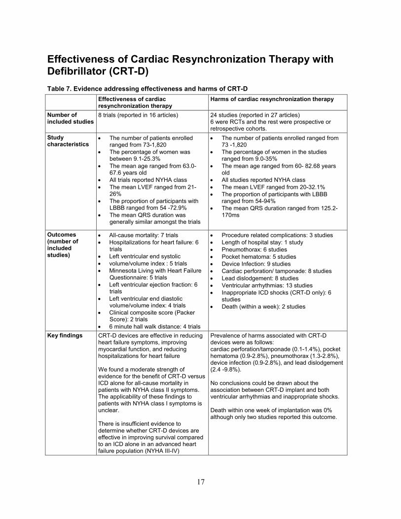
Effectiveness of Cardiac Resynchronization Therapy with Defibrillator (CRT-D) Table 7. Evidence addressing effectiveness and harms of CRT-D Effectiveness of cardiac
resynchronization therapy Harms of cardiac resynchronization therapy
Number of included studies
8 trials (reported in 16 articles) 24 studies (reported in 27 articles) 6 were RCTs and the rest were prospective or retrospective cohorts.
Study characteristics
• The number of patients enrolled ranged from 73-1,820
• The percentage of women was between 9.1-25.3%
• The mean age ranged from 63.0-67.6 years old
• All trials reported NYHA class • The mean LVEF ranged from 21-
26% • The proportion of participants with
LBBB ranged from 54 -72.9% • The mean QRS duration was
generally similar amongst the trials
• The number of patients enrolled ranged from 73 -1,820
• The percentage of women in the studies ranged from 9.0-35%
• The mean age ranged from 60- 82.68 years old
• All studies reported NYHA class • The mean LVEF ranged from 20-32.1% • The proportion of participants with LBBB
ranged from 54-94% • The mean QRS duration ranged from 125.2-
170ms
Outcomes (number of included studies)
• All-cause mortality: 7 trials • Hospitalizations for heart failure: 6
trials • Left ventricular end systolic • volume/volume index : 5 trials • Minnesota Living with Heart Failure
Questionnaire: 5 trials • Left ventricular ejection fraction: 6
trials • Left ventricular end diastolic
volume/volume index: 4 trials • Clinical composite score (Packer
Score): 2 trials • 6 minute hall walk distance: 4 trials
• Procedure related complications: 3 studies • Length of hospital stay: 1 study • Pneumothorax: 6 studies • Pocket hematoma: 5 studies • Device Infection: 9 studies • Cardiac perforation/ tamponade: 8 studies • Lead dislodgement: 8 studies • Ventricular arrhythmias: 13 studies • Inappropriate ICD shocks (CRT-D only): 6
studies • Death (within a week): 2 studies
Key findings CRT-D devices are effective in reducing heart failure symptoms, improving myocardial function, and reducing hospitalizations for heart failure We found a moderate strength of evidence for the benefit of CRT-D versus ICD alone for all-cause mortality in patients with NYHA class II symptoms. The applicability of these findings to patients with NYHA class I symptoms is unclear. There is insufficient evidence to determine whether CRT-D devices are effective in improving survival compared to an ICD alone in an advanced heart failure population (NYHA III-IV)
Prevalence of harms associated with CRT-D devices were as follows: cardiac perforation/tamponade (0.1-1.4%), pocket hematoma (0.9-2.8%), pneumothorax (1.3-2.8%), device infection (0.9-2.8%), and lead dislodgement (2.4 -9.8%). No conclusions could be drawn about the association between CRT-D implant and both ventricular arrhythmias and inappropriate shocks. Death within one week of implantation was 0% although only two studies reported this outcome.
17

Study Characteristics Eight trials (reported in 16 articles) addressed the effectiveness of CRT-D.6 ,7 ,10 ,31 ,32 ,37 ,38 ,41
,43 ,44 ,47 ,49 ,52 ,54 ,56 ,57 The studies used largely consistent comparators when assessing patients with an implantable cardioverter defibrillator (ICD) and active biventricular pacing versus an ICD and no active biventricular pacing (Evidence Table 1).
One trial separated patients into two groups based on the presence or absence of baseline ventricular dyssynchrony.52 Patients with dyssynchrony all received a CRT-D device. The trial randomized patients without dyssynchrony to a CRT-D device or an ICD alone. Another trial started as a crossover design with patients crossing over between active CRT-on versus CRT-off.54 The study changed protocol after the trial started to a parallel arm design. Two studies did not report funding status (Table 8).52 ,56
18
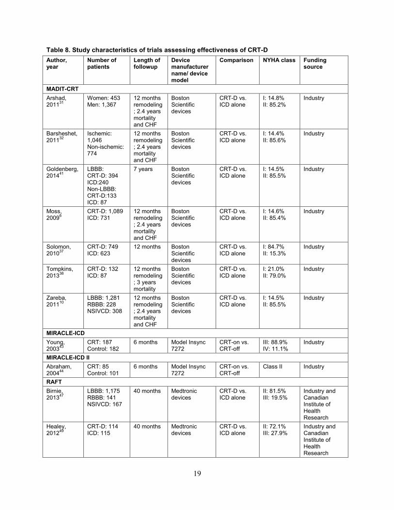
Table 8. Study characteristics of trials assessing effectiveness of CRT-D Author, year
Number of patients
Length of followup
Device manufacturer name/ device model
Comparison NYHA class Funding source
MADIT-CRT Arshad, 201131
Women: 453 Men: 1,367
12 months remodeling; 2.4 years mortality and CHF
Boston Scientific devices
CRT-D vs. ICD alone
I: 14.8% II: 85.2%
Industry
Barsheshet,201132
Ischemic: 1,046 Non-ischemic: 774
12 months remodeling; 2.4 years mortality and CHF
Boston Scientific devices
CRT-D vs. ICD alone
I: 14.4% II: 85.6%
Industry
Goldenberg, 201441
LBBB: CRT-D: 394 ICD:240 Non-LBBB: CRT-D:133 ICD: 87
7 years Boston Scientific devices
CRT-D vs. ICD alone
I: 14.5% II: 85.5%
Industry
Moss, 20096
CRT-D: 1,089 ICD: 731
12 months remodeling; 2.4 years mortality and CHF
Boston Scientific devices
CRT-D vs. ICD alone
I: 14.6% II: 85.4%
Industry
Solomon, 201037
CRT-D: 749 ICD: 623
12 months Boston Scientific devices
CRT-D vs. ICD alone
I: 84.7% II: 15.3%
Industry
Tompkins, 201338
CRT-D: 132 ICD: 87
12 months remodeling; 3 years mortality
Boston Scientific devices
CRT-D vs. ICD alone
I: 21.0% II: 79.0%
Industry
Zareba, 201110
LBBB: 1,281 RBBB: 228 NSIVCD: 308
12 months remodeling; 2.4 years mortality and CHF
Boston Scientific devices
CRT-D vs. ICD alone
I: 14.5% II: 85.5%
Industry
MIRACLE-ICD Young, 200343
CRT: 187 Control: 182
6 months Model Insync 7272
CRT-on vs. CRT-off
III: 88.9% IV: 11.1%
Industry
MIRACLE-ICD II Abraham, 200444
CRT: 85 Control: 101
6 months Model Insync 7272
CRT-on vs. CRT-off
Class II Industry
RAFT Birnie, 201347
LBBB: 1,175 RBBB: 141 NSIVCD: 167
40 months Medtronic devices
CRT-D vs. ICD alone
II: 81.5% III: 19.5%
Industry and Canadian Institute of Health Research
Healey, 201249
CRT-D: 114 ICD: 115
40 months Medtronic devices
CRT-D vs. ICD alone
II: 72.1% III: 27.9%
Industry and Canadian Institute of Health Research
19
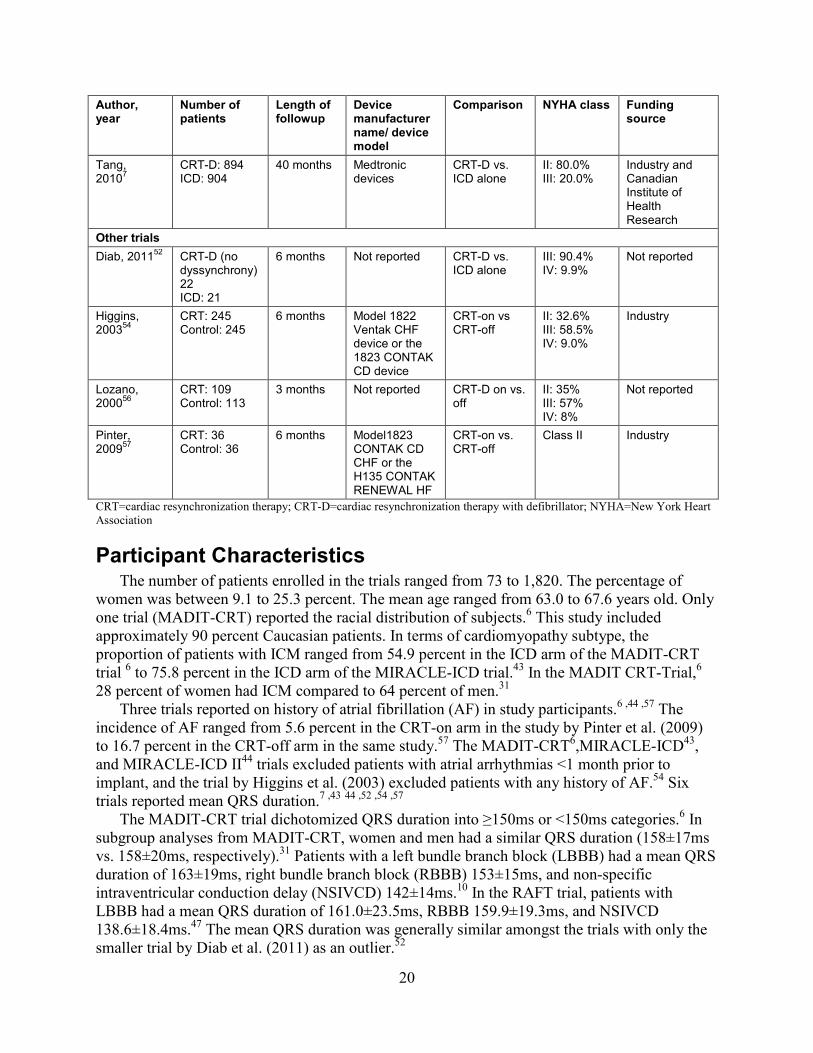
Author, year
Number of patients
Length of followup
Device manufacturer name/ device model
Comparison NYHA class Funding source
Tang, 20107
CRT-D: 894 ICD: 904
40 months Medtronic devices
CRT-D vs. ICD alone
II: 80.0% III: 20.0%
Industry and Canadian Institute of Health Research
Other trials Diab, 201152 CRT-D (no
dyssynchrony) 22 ICD: 21
6 months Not reported CRT-D vs. ICD alone
III: 90.4% IV: 9.9%
Not reported
Higgins, 200354
CRT: 245 Control: 245
6 months Model 1822 Ventak CHF device or the 1823 CONTAK CD device
CRT-on vs CRT-off
II: 32.6% III: 58.5% IV: 9.0%
Industry
Lozano, 200056
CRT: 109 Control: 113
3 months Not reported CRT-D on vs. off
II: 35% III: 57% IV: 8%
Not reported
Pinter, 200957
CRT: 36 Control: 36
6 months Model1823 CONTAK CD CHF or the H135 CONTAK RENEWAL HF
CRT-on vs. CRT-off
Class II Industry
CRT=cardiac resynchronization therapy; CRT-D=cardiac resynchronization therapy with defibrillator; NYHA=New York Heart Association
Participant Characteristics The number of patients enrolled in the trials ranged from 73 to 1,820. The percentage of
women was between 9.1 to 25.3 percent. The mean age ranged from 63.0 to 67.6 years old. Only one trial (MADIT-CRT) reported the racial distribution of subjects.6 This study included approximately 90 percent Caucasian patients. In terms of cardiomyopathy subtype, the proportion of patients with ICM ranged from 54.9 percent in the ICD arm of the MADIT-CRT trial 6 to 75.8 percent in the ICD arm of the MIRACLE-ICD trial.43 In the MADIT CRT-Trial,6 28 percent of women had ICM compared to 64 percent of men.31
Three trials reported on history of atrial fibrillation (AF) in study participants.6 ,44 ,57 The incidence of AF ranged from 5.6 percent in the CRT-on arm in the study by Pinter et al. (2009) to 16.7 percent in the CRT-off arm in the same study.57 The MADIT-CRT6,MIRACLE-ICD43, and MIRACLE-ICD II44 trials excluded patients with atrial arrhythmias <1 month prior to implant, and the trial by Higgins et al. (2003) excluded patients with any history of AF.54 Six trials reported mean QRS duration.7 ,43 44 ,52 ,54 ,57
The MADIT-CRT trial dichotomized QRS duration into ≥150ms or <150ms categories.6 In subgroup analyses from MADIT-CRT, women and men had a similar QRS duration (158±17ms vs. 158±20ms, respectively).31 Patients with a left bundle branch block (LBBB) had a mean QRS duration of 163±19ms, right bundle branch block (RBBB) 153±15ms, and non-specific intraventricular conduction delay (NSIVCD) 142±14ms.10 In the RAFT trial, patients with LBBB had a mean QRS duration of 161.0±23.5ms, RBBB 159.9±19.3ms, and NSIVCD 138.6±18.4ms.47 The mean QRS duration was generally similar amongst the trials with only the smaller trial by Diab et al. (2011) as an outlier.52
20

Four trials reported the incidence of LBBB ranging from 54 to 72.9 percent.6 ,7 ,54 ,57 One study reported no QRS morphology data.52 Two trials43 ,44 reported on the incidence of RBBB. While these two studies excluded paced patients, given a lack of data on the number of NSIVCD patients, we could not determine the number of LBBB patients from these two studies. Six trials reported the number of RBBB patients, ranging from 7.6 percent in the CRT arm of the RAFT trial7 to 20.8 percent in the CRT-off arm from the MIRACLE-ICD II trial.44 Three trials reported patients with NSIVCD ranging from roughly 11 to 32 percent.7 ,10 ,54
Only the RAFT trial included patients with a paced rhythm prior to CRT.7 These patients represented 7.4 to 7.6 percent of the patients in this trial. All paced patients in the RAFT trial had a QRS duration >200ms.7
The NYHA class was a key inclusion criteria in all trials. Three trials included only patients with NYHA class III-IV symptoms.43 ,52 ,57 One trial included primarily class III patients, however roughly one-third of the patients in this study were NYHA class II.54 The trial by Lozano et al. (2000) included patients with class II, III, and IV symptoms.56 The RAFT trial included primarily NYHA class II patients, although roughly 20 percent were NYHA class III.7 The MIRACLE-ICD trial included only NYHA class II patients.43 The MADIT-CRT study enrolled only patients with NYHA class I or II symptoms, of which NYHA class II represented roughly 85 percent.6
All trials reported the mean left ventricular ejection fraction (LVEF) and it was similar across studies ranging from 21-26 percent. Only two trials reported serum creatinine.6 ,57 Mean serum creatinine ranged from 1.1-1.2 mg/dl. These trials were homogeneous in patient population with the major exception of NYHA class (Evidence Table 4).
Risk of Bias The majority of trials did not report whether they performed random sequence generation;
therefore, we cannot rule out selection bias. In the MADIT-CRT trial, the treating physicians were aware of study-group assignments introducing possible performance bias.6 ,10 ,31 ,32 ,37 ,38 The RAFT trial conducted 6-minute hall walk tests and administered QOL questionnaires 7. However, this outcome was only reported as a secondary analysis limited to patients with permanent AF.49 Despite these limitations, overall, the included RCTs have a low risk of bias (Table 9 and Figure 5).
21
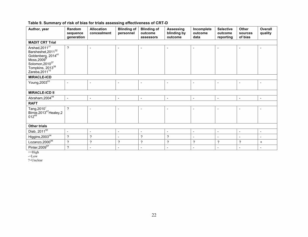
Table 9. Summary of risk of bias for trials assessing effectiveness of CRT-D Author, year Random
sequence generation
Allocation concealment
Blinding of personnel
Blinding of outcome assessors
Assessing blinding by outcome
Incomplete outcome data
Selective outcome reporting
Other sources of bias
Overall quality
MADIT CRT Trial Arshad,201131 Barsheshet,201132 Goldenberg, 201441 Moss,20096 Solomon,201037 Tompkins, 201338 Zareba,201110
? - - - - - - - -
MIRACLE-ICD Young,200343 - - - - - - - - -
MIRACLE-ICD II Abraham,200444 - - - - - - - - - RAFT Tang,20107 Birnie,201347Healey,201249
? - - - - - - - -
Other trials Diab, 201152 - - - - - - - - - Higgins,200354 ? ? - ? ? - - - - Lozanzo,200056 ? ? ? ? ? ? ? ? + Pinter,200957 ? - - - - - - - - +=High -=Low ?=Unclear
22
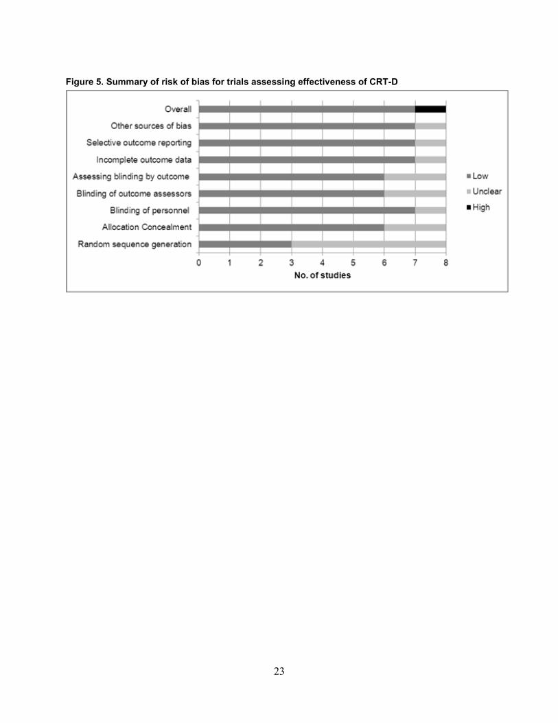
Figure 5. Summary of risk of bias for trials assessing effectiveness of CRT-D
23

Effectiveness Outcomes The most common outcomes assessed were all-cause mortality and heart failure
hospitalizations (Table 9). Multiple studies also measured indices of reverse ventricular remodeling changes including LVEF, LVESV, LVEDV, left ventricular end-systolic volume indexed to body surface areas (LVESVi), and left ventricular end-diastolic volume indexed to body surface areas (LVEDVi). Six studies assessed quality of life (QOL) as measured by the MLHFQ. Five reported functional capacity changes by noting variations in 6-minute hall walk distances, and two studies measured symptomatic improvement via the same clinical composite score which assigned patients to one of three groups (worsened, improved, or unchanged) (Table 10).
24

Table 10. Outcomes reported in the trials assessing effectiveness of CRT-D Author, year All-cause
mortality HF hospitalization
LVESV LVESVi MLHFQ LVEF LVEDV LVEDVi Clinical composite score
6MHWD
MADIT-CRT Moss,20096 X X X X X
Goldenberg, 201441 X
Tompkins, 201338 X X X
Solomon, 201037 (other endpoints are redundant from MADIT-CRT6
X X
Arshad,2 01131
X X X X X
Zareba, 201110
X X X X X
Barsheshet, 201132
X X X X X
MIRACLE-ICD Young,200343 X X (part of
combined endpoint)
X X X X X X
MIRACLE-ICD II Abraham,200444 X X X X X X X X
RAFT Tang, 20107
X X
Birnie, 201347 X X
Healey, 201249 X X X X Other trials
Diab, 201152 X X X X X
Higgins, 200354 X X X X X
25

Author, year All-cause mortality
HF hospitalization
LVESV LVESVi MLHFQ LVEF LVEDV LVEDVi Clinical composite score
6MHWD
Pinter, 200957 X X X X X Lozano, 200056 X
HF= heart failure; LVESV=left-ventricular end-systolic volume; LVEVi=left-ventricular end-systolic volume indexed to body surface area; MLHFQ = Minnesota Living with Heart Failure Questionnaire; LVEF=left-ventricular ejection fraction; LVEDV=left ventricular end-diastolic volume; LVEDVI=left-ventricular end-diastolic volume indexed to body surface area; 6MHWD=6-minute hall walk distance
26
All-cause Mortality Seven trials (reported in 14 articles) assessed the all-cause mortality outcome.6 ,7 ,10 ,31 ,32 ,38 ,41
,43 ,44 ,47 ,49 ,52 ,54 ,56hree trials included patients with primarily minimally symptomatic CHF 6 ,7 ,44, two included patients with primarily advanced CHF 43 ,52 and two included both populations.54 ,56 In the MADIT-CRT trial over a mean followup of 2.4 years, 3.3 percent of the CRT group died compared with 2.5 percent in the ICD alone arm (HR:1.0, 95% CI, 0.69 to 1.44, p=0.99).6 In long term follow up of 854 patients from MADIT-CRT (1,818 were originally followed) patients with a LBBB undergoing CRT derived a significant improvement in mortality compared to an ICD alone (log rank p=0.002) but those with a non-LBBB did not (log rank p=0.2).41 Long term mortality data comparing CRT-D to an ICD alone in the cohort as a whole were not reported. In another analysis from MADIT-CRT trial, women derived a significant benefit in survival from CRT-D compared to an ICD alone (log rank p=0.02), whereas men did not (log rank p=0.83) (Table 11).31
In a separate analysis from MADIT-CRT, in patients with RBBB, with or without a left anterior fascicular block [LAFB]), there was no difference in survival in patients receiving CRT-D compared with an ICD alone (log rank p=0.374).38
Lastly, in one analysis from MADIT-CRT trial (stratified by cardiomyopathy subtype), patients with ICM had no statistically significant difference in survival with CRT-D versus ICD (HR: 0.99, 95% CI, 0.65 to1.52, p=0.984).32 Patients with non-ischemic cardiomyopathy (NICM) had no statistically significant difference in survival with CRT-D versus ICD (HR: 0.87, 95% CI, 0.45 to 1.67, p=0.669).32 In the RAFT trial (primarily NYHA class II patients), the 5-year actuarial rate of death in the CRT-D arm was 28.6 versus 34.6 percent in the ICD alone arm (HR: 0.75, 95% CI, 0.562 to 0.91, p=0.0003).7
In patients with permanent AF, there was no difference in survival between patients in the CRT-D arm and ICD alone arm (HR: 1.04, 95% CI, 0.66 to 1.62, p=0.88).49 RAFT reported other pre-defined subgroups of interest, but they were not broken down by survival alone and thus precluded analysis. Grouped by bundle branch block morphology, patients with a LBBB had improved survival with CRT-D compared to an ICD alone (HR: 0.664, 95% CI, 0.516 to 0.853, p=0.0013).47 There was no statistically significant difference between the CRT-D and ICD arms for patients with RBBB (HR: 0.544, 95% CI, 0.264 to1.121, p=0.095) or NSIVCD (HR: 0.930, 95% CI, 0.491 to 1.1761, p=0.0825).47 In MIRACLE-ICD II (enrolling patients with NYHA class II), two patients in each group died at 6-month followup.44
In terms of the trials of patients with more advanced CHF symptoms, in the MIRACLE-ICD trial the cumulative survival at 6 months was 92.2 percent in the ICD arm and 92.4 percent in the CRT-D arm (log rank p=0.96).43 In the trial by Diab et al. (2011) (enrolling NYHA class III-IV), there were two deaths in the ICD arm and none in the CRT arm at a followup of 6 months.52 In the trial by Higgins et al. (2003) (which contained mixed NYHA class population), there were 11 deaths in the CRT-D arm compared with 16 deaths in the ICD arm at 6 months followup.54 In the trial by Lozano et al. (2000) enrolling a mixed NYHA population (the majority of which were NYHA class III), the survival rate at 3 months in the CRT-on cohort was 93 percent versus 86 percent in the CRT-off cohort—a result that was not statistically significant (p=0.18).56
In summary, in patients with less symptomatic CHF, data from the RAFT trial demonstrate a mortality benefit, which conflicts with the originally reported median 2.4 year followup of the MADIT-CRT trial, which did not. Long term followup of the MADIT-CRT trial demonstrated a mortality benefit in LBBB but not in non-LBBB. Whether CRT-D produced a mortality
27
improvement in the cohort as a whole was not reported and many patients in the original trial were lost to followup. The other trials assessing mortality in minimally symptomatic patients were either too small in size or followup to add significant additional evidence. Given this, the strength of evidence that CRT-D improves mortality in patients with minimally symptomatic CHF is moderate (Table 12). The trials assessing mortality in patients with NYHA class III-IV symptoms were limited in terms of followup and size, therefore there is insufficient evidence to determine the effect of CRT-D on mortality compared to an ICD alone. Three reasonably homogenous trials (RAFT, MADIT-CRT, and MIRACLE-ICD II) reported mortality, however the 6-month followup in MIRACLE-ICD II is limiting. Therefore, there are too few trials to perform a meta-analysis. Women appear to have improved survival compared to men with CRT-D compared to an ICD alone, although we need more data to confirm this finding.
The evidence on bundle branch block morphology and survival in patients receiving CRT-D is conflicted and limited to patients with less symptomatic CHF.
Heart Failure (HF) Hospitalizations Six trials (reported in 11 articles) assessed HF hospitalization outcomes.6 ,7 ,10 ,31 ,32 ,43 ,44 ,47 ,49
,52 ,54 In the RAFT trial, over the duration of followup, 19.5 percent in the CRT arm were hospitalized for HF compared to 26.1 percent in the ICD arm (HR: 0.68, 95% CI, 0.56 to 0.83, p<0.001).7 In patients with permanent AF from this trial, 19.3 percent of patients in the CRT-D arm were hospitalized for HF versus 27.8 percent in the ICD arm, a result of borderline significance (p=0.052).49 The RAFT trial reported other pre-defined subgroups of interest, but they were not broken down by HF hospitalization alone, thus precluding analysis. Grouped by bundle branch block morphology, patients with a LBBB had fewer HF hospitalizations with CRT-D compared to an ICD alone (HR: 0.603, 95% CI, 0.469 to 0.774, p<0.001]).47 There was no statistically significant difference in HF hospitalizations between the CRT-D and ICD arm for patients with RBBB (HR: 1.142, 95% CI, 0.580 to 2.249, p=0.705, 95% CI, 264 to 1.121, p=0.095) or NSIVCD (HR: 1.021, 95% CI, 0.574 to 1.81, p=0.944).47
In the MADIT CRT trial, over the duration of followup, there were 151 HF events (13.9 %) in the CRT arm and 167 HF events (22.8 %) in the ICD arm (HR: 0.59, 95% CI, 0.47 to 0.74, p<0.001).6 In an analysis from MADIT-CRT trial, both women and men derived fewer heart hospitalizations with CRT-D compared to an ICD alone (HR: 0.30, 95% CI, 0.18 to 0.50, p<0.001 and HR: 0.65, 95% CI, 0.50 to 0.84, respectively p=0.001).31 In another analysis from the MADIT-CRT trial, stratified by QRS morphology, patients with a LBBB morphology had fewer HF events with CRT-D versus an ICD alone (HR: 0.41, 95% CI, 0.31to 0.54, p<0.001).10 Patients with a RBBB had no statistically significant difference in CHF events with CRT-D versus ICD (HR: 0.88, 95% CI, 0.46 to 1.67, p=0.690).10 Patients with a NSIVCD morphology had no statistically significant difference in HF events with CRT-D versus ICD (HR: 1.31, 95% CI, 0.78 to 2.16, p<0.306).10 In another analysis from the MADIT-CRT trial (stratified by cardiomyopathy subtype), patients with both ICM from NICM had fewer HF events with CRT-D compared to an ICD alone (HR: 0.58, 95% CI,0.45 to 0.77, p=<0.001 and HR: 0.50, 95% CI, 0.35 to 0.75, p=0.001, respectively).32
The MIRACLE-ICD trial did not report HF hospitalizations alone (without combination with all-cause survival).43 A study by Diab et al. (2011) hospitalized two patients in the CRT arm (both in the dyssynchrony present arm) and two in the ICD arm for HF over 6 months of followup.52 In the study by Pinter et al. (2009), the authors reported percentages only for all-cause hospitalizations and stated that the reasons for hospitalization were the same in both
28

groups.57 In the study by Higgins et al. (2003) there was a non-statistically significant 15 percent reduction in HF progression in patients receiving CRT versus control (p=0.35).54
Both the large RAFT and MADIT-CRT trials showed a reduction in HF events for CRT-D compared to an ICD alone. Subgroup analyses from both trials demonstrate the effect to be primarily in patients with LBBB morphology. The definition for HF events in MADIT-CRT incorporated both inpatient and outpatient CHF management. There are too few trials to perform a meta-analysis.
The overall the strength of evidence that CRT-D results in fewer hospitalizations for CHF compared to an ICD alone is high (Table 12).
Left Ventricular End Systolic Volume/Volume Index Five trials (reported in 8 articles) reported change in LVESV.6 ,10 ,32 ,38 ,43 ,44 ,52 ,54 In the
MADIT-CRT trial there was a significant decrease in LVESV in the CRT-D arm compared to the ICD arm alone (57ml vs. 18ml, p<0.001), respectively.6
In an analysis from the RAFT trial (grouped by bundle branch block morphology), patients with a LBBB had a significant reduction in LVESV compared to those receiving an ICD alone (62.1±31.5ml vs. 18.3±16.5ml, p<0.01).47 Patients with a non-LBBB morphology (RBBB and NSIVCD grouped together), derived a significant reduction in LVESV with CRT-D compared to ICD alone (45.7±27.3ml vs. 17.5±16.1, p<0.01).47
In an analysis from MADIT-CRT (stratified by cardiomyopathy subtype), patients with both ICM and NICM demonstrated reductions in LVESV with CRT-D versus an ICD alone compared to baseline (-29%±14 vs. -10%±9 and -37%±16 vs. -11%±9 ).32 This effect was greater in patients with NICM compared with ICM.
In the MIRACLE-ICD trial the median change in LVESV in the control group was -8.2ml (-19.1 to 0.6) compared with -22.2ml (-32.8 to -10.7) in the CRT-D arm (p=0.06).43 In the MIRACLE-ICD II trial the mean change in LVESV in the CRT arm compared with control was (-42ml vs. -14ml, p=0.01, respectively).44
In the trial by Pinter et al. (2009) the mean change in the CRT-on arm was -21±45ml versus -5+22ml in the CRT-off arm—a change which was not statistically significant.57 In the study by Diab et al. (2011) the change in LVESV was dichotomized into reduction ≥15 percent from baseline.52 Amongst patients with no dyssynchrony at baseline, patients with CRT were more likely to have a reduction in LVESV from baseline of at least 15 percent compared to patients receiving an ICD alone (57% vs. 11%, p=0.002).
The MADIT-CRT trial also reported changes in LVESVi.6 ,37 At 12-month followup, patients receiving CRT-D derived a greater improvement in LVESVi compared to patients receiving an ICD alone (-28.7±15.5 vs. -9.1±8.2, p=0.0001).37 In an analysis from MADIT-CRT, both women and men derived improvements in LVESVI compared to an ICD alone (ml/body surface area, -31 vs. -10 and -27 vs. -8). This reduction was greater in women compared to men (p<0.001).31
The trials were generally consistent in demonstrating a reduction in LVESV with CRT-D compared to an ICD alone. This effect was noted across multiple subgroups including patients with non- LBBB block morphologies. We performed a meta-analysis incorporating the three trials, which enrolled minimally symptomatic CHF. This meta-analysis demonstrated a clear benefit in terms of LVESV reduction favoring CRT-D compared to an ICD alone (Mean difference: -22.55, 95% CI, -40.66 to -9.56) (Figure 6).
There were not enough trials enrolling patients NYHA class III-IV CHF to perform meta-analysis. Nevertheless, the data are consistent in favoring CRT-D over an ICD alone in terms of
29

LVESV reduction this population. The strength of evidence is high favoring CRT-D over an ICD alone in terms of LVESV reduction (Table 12). Figure 6. Meta-analysis of left ventricular end-diastolic volume comparing CRT-D with ICD alone in minimally symptomatic patients (NYHA I-II)
Minnesota Living with Heart Failure Questionnaire
Five trials (reported in 6 articles) reported quality of life via the MLHFQ.43 ,44 ,49 ,52 ,54 ,57 In the trial by Higgins et al. (2003) there was a non-significant improvement in MLHFQ score with CRT-D versus an ICD alone (-7±2 vs. 5±2, p=0.43).54 When divided by NYHA class I/II vs. II/IV subgroups, patients with advanced CHF (NYHA class III/IV) had a significant improvement in MLHFQ score with CRT-D versus an ICD alone (-16±3 vs. -5±3, p=0.017) whereas patients with less symptomatic CHF (NYHA class I and II) had no significant difference (-1±2 vs. -4±2, p=0.26).
In the MIRACLE-ICD trial, there was a significant improvement in MLHFQ scores in the CRT-D arm compared to the control arm (-17.5 [-21 to -14] vs. -11 [-16 to -7], p=0.02).43
In the trial by Diab et al. (2011) there was an improvement in MLHFQ score in patients receiving CRT-D with dyssynchrony present at baseline (-29), without dyssynchrony at baseline (-16), and in patients without dyssynchrony receiving an ICD alone (-8).52 While a global p-value was presented comparing all three arms, the impact of baseline dyssynchrony makes this difficult to interpret.
In the trial by Pinter et al. (2009) there was no statistically significant change in MLHFQ scores between patients with CRT-on vs. -off (-7.8±20.1 vs. -0.2±13.5, p=non-significant).57
In the MIRACLE-ICD II trial there was no significant change in MLHFQ scores between patients in the control arm and those receiving CRT (-10.7±21.7 vs. -13.3±25.1, p=0.49).44
Moss, 2009
Abraham, 2004
Pinter, 2009
Overall (I-squared=0%)
year
Author,
746
68
36
followup
CRT-D at
N
620
85
36
followup
Control at
N
-39.00 (-74.58, -3.42)
-28.00 (-49.91, -6.09)
-16.00 (-32.39, 0.39)
-22.55 (-40.66, -9.56)
ES (95% CI)
-39.00 (-74.58, -3.42)
-28.00 (-49.91, -6.09)
-16.00 (-32.39, 0.39)
-22.55 (-40.66, -9.56)
ES (95% CI)
CRT-D decreases LVESV CRT-D increases LVESV 0 -50 -40 -30 -20 -10 0 10
Pooled mean difference between groups in mean change from baseline
30
In the permanent AF subgroup from the RAFT trial there was a non-significant improvement in MLHFQ scores with CRT-D compared to an ICD alone (-11±18 vs. -5±21, p=0.057).49
The trial by Pinter et al. (2009) also reported data from the SF-36 health survey. Of 10 metrics incorporating subscales of physical and mental function, only changes in general health scores were different between patients with CRT-on and -off (-5.8±14.9 vs. -5.8±13.9, p=0.02).57
The current data suggest that CRT-D does not improve quality of life in minimally symptomatic patients compared to an ICD alone, though the meta-analysis should be interpreted with caution given the substantial heterogeneity (0.83, 95% CI, -9.27 to 5.30) (Figure 7a). The data does suggest a significant improvement in quality of life in patients with NYHA class III-IV CHF with a high strength of evidence supporting this conclusion (mean difference-10.91, 95% CI, -12.03 to -7.27) (Figure 7b and Table 12).
31
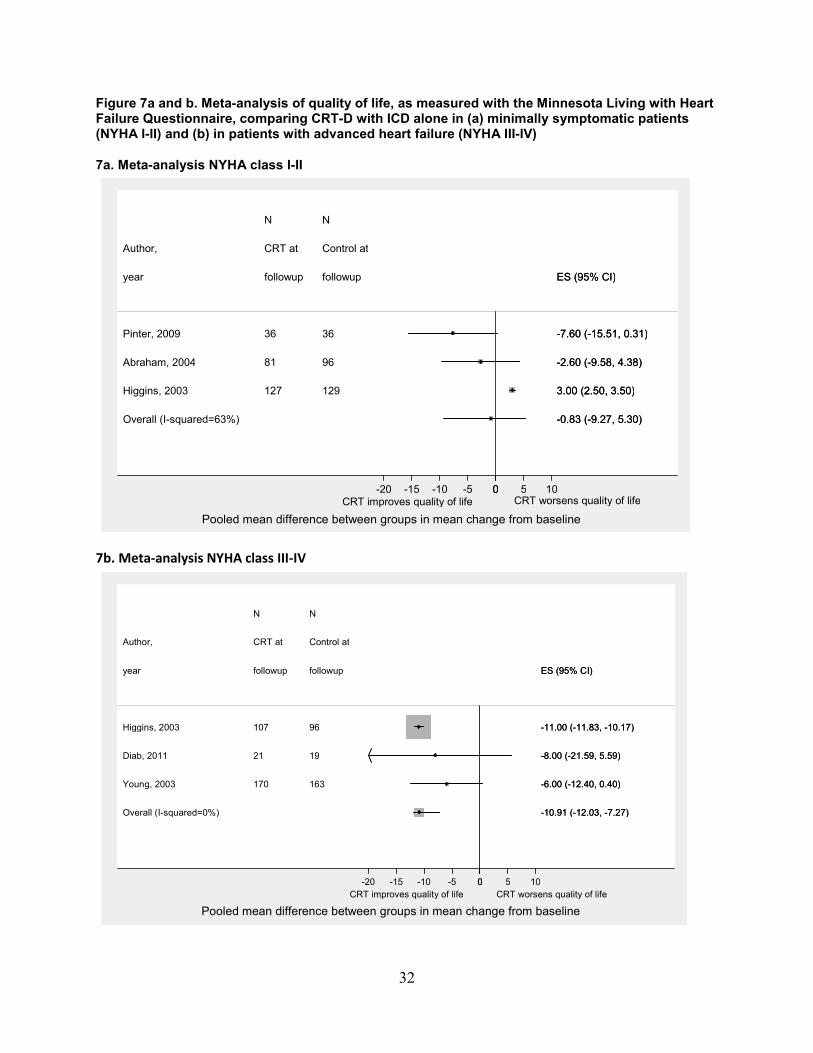
Figure 7a and b. Meta-analysis of quality of life, as measured with the Minnesota Living with Heart Failure Questionnaire, comparing CRT-D with ICD alone in (a) minimally symptomatic patients (NYHA I-II) and (b) in patients with advanced heart failure (NYHA III-IV)
7a. Meta-analysis NYHA class I-II
7b. Meta-analysis NYHA class III-IV
Pinter, 2009
Abraham, 2004
Higgins, 2003
Overall (I-squared=63%)
year
Author,
36
81
127
followup
CRT at
N
36
96
129
followup
Control at
N
-7.60 (-15.51, 0.31)
-2.60 (-9.58, 4.38)
3.00 (2.50, 3.50)
-0.83 (-9.27, 5.30)
ES (95% CI)
-7.60 (-15.51, 0.31)
-2.60 (-9.58, 4.38)
3.00 (2.50, 3.50)
-0.83 (-9.27, 5.30)
ES (95% CI)
CRT improves quality of life CRT worsens quality of life 0 -20 -15 -10 -5 0 5 10
Pooled mean difference between groups in mean change from baseline
Higgins, 2003
Diab, 2011
Young, 2003
Overall (I-squared=0%)
year
Author,
107
21
170
followup
CRT at
N
96
19
163
followup
Control at
N
-11.00 (-11.83, -10.17)
-8.00 (-21.59, 5.59)
-6.00 (-12.40, 0.40)
-10.91 (-12.03, -7.27)
ES (95% CI)
-11.00 (-11.83, -10.17)
-8.00 (-21.59, 5.59)
-6.00 (-12.40, 0.40)
-10.91 (-12.03, -7.27)
ES (95% CI)
CRT improves quality of life CRT worsens quality of life 0 -20 -15 -10 -5 0 5 10
Pooled mean difference between groups in mean change from baseline
32

Left Ventricular Ejection Fraction Six trials (reported in 9 articles) reported change in LVEF.6 ,10 ,31 ,32 ,43 ,44 ,52 ,54 ,57 In the
MADIT-CRT trial, there was a significant improvement in LVEF in the CRT arm compared to the ICD arm (11% vs. 3%, p<0.001).6 In an analysis from MADIT-CRT, both women and men had improvement in LVEF with CRT-D compared to an ICD alone, although the magnitude was significantly greater in women.31
In another analysis from MADIT-CRT (stratified by QRS morphology), patients with an LBBB morphology had significant improvement in LVEF (3.4%±3.0 in the ICD arm compared to 11.9%±5.1 in the CRT-D arm, p<0.01).10 The analysis grouped RBBB and NSIVCD patients as “non- LBBB”. This cohort similarly showed an improvement in LVEF with CRT-D versus ICD alone (8.8%±4.9 vs. 3.4%±3.1, p<0.01).10 The improvement in LVEF was larger in patients with LBBB compared with non- LBBB. In subgroup analyses (stratified by cardiomyopathy subtype), patients with both ICM and NICM showed improvement in LVEF with CRT-D compared to an ICD alone (10.5%±5 vs. 3%±3 and 12%±5 vs. 3%±3).32 The effect was greater in patients with NICM.
In the MIRACLE-ICD trial, the median change in LVEF in the CRT arm was 2.1 percent (95% CI, 0.12 to 4.1) compared to 1.7 percent (95% CI, 0.7 to 2.4, p=0.12).43 In the MIRACLE-ICD II trial, the mean change in LVEF in the CRT arm was 3.8 percent vs. 0.8 percent in the ICD arm (p=0.02).44
In the trial by Higgins et al. (2003) the mean change in LVEF in CRT patients was 5.1 percent ±0.7 versus 2.8 percent ±0.7 in the ICD arm (p=0.02).54 In the trial by Pinter et al. (2009) the change in LVEF in the CRT-on arm was 3.9 percent ±8.9 vs. 1.9 percent ±6.8 in the CRT-off arm, which was not statistically different.57
In the study by Diab et al. (2011) the change in LVEF was dichotomized into improvement ≥15 percent from baseline rather than used as a continuous variable.52 The study made a comparison between CRT-D vs. ICD alone in patients who lacked baseline dyssynchrony. The proportion of patients demonstrating >15 percent improvement in LVEF was greater in the CRT arm than the ICD alone arm (p=0.007).
The majority of studies, including the very large MADIT-CRT trial, were consistent in demonstrating an improvement in LVEF with CRT compared to ICD alone.6 This effect existed across multiple subgroups including patients with non-LBBB morphologies. The study by Higgins et al. (2003) reported separately the changes in LVEF in the NYHA class I-II cohort and class III-IV cohorts.54 We performed a meta-analysis incorporating the four trials enrolling patients with minimally symptomatic CHF. In pooled analysis a clear benefit in terms of LVEF improvement existed favoring CRT-D over an ICD alone (1.82, 95% CI, 1.51 to 2.65) (Figure 8). There were not enough trials enrolling patients NYHA class III-IV to perform meta-analysis.
33

Figure 8. Meta-analysis of left ventricular ejection fraction comparing CRT-D with ICD alone in minimally symptomatic patients (NYHA I-II)
Left Ventricular End Diastolic Volume/Volume Index Four trials (reported in 7 articles) reported change in LVEDV.6 ,10 ,32 ,38 ,43 ,44 ,57 In the
MADIT-CRT trial, there was a significant decrease in LVEDV in the CRT-D arm compared to the ICD arm alone (52ml vs. 15ml, p<0.001).6
In an analysis from the RAFT trial, grouped by bundle branch block morphology,47 patients with a LBBB had a significant reduction in LVEDV compared to an ICD alone (56.7±34.1ml vs. 14.8±14.5ml, p<0.01). Patients with a non- LBBB morphology (RBBB and NSIVCD grouped together), derived a significant reduction in LVEDV with CRT-D compared to ICD alone (41.0±28.13ml vs. 14.4±14.2ml, p<0.01).
In an analysis from MADIT-CRT stratified by cardiomyopathy subtype, patients with both ischemic cardiomyopathy and NICM demonstrated reductions in LVEDV with CRT-D versus an ICD alone compared to baseline (-18%±10 vs. -5%±5 and -24%±12 vs. -7%±6).32 This effect was greater in patients with NICM compared with ICM.
In the MIRACLE-ICD trial the median change in LVEDV in the control group was -5.7ml (-16.2 to 1.8) compared with -19.9m (-39.7 to -6.3) in the CRT-D arm (p=0.06).43 In the MIRACLE-ICD II trial the mean change in LVEDV in the CRT arm compared with control was (-42ml vs. -16ml, p=0.04).44
In the study by Pinter et al. (2009) the mean change in the CRT-on arm was -16±44ml vs. -13±47ml in the CRT-off arm, a change, which was not statistically significant.57
The MADIT-CRT trial also reported changes in LVEDV indexed to body surface area (LVESDi).6 ,37 At 12-month followup, patients receiving CRT-D derived a greater improvement in LVEDVi compared to patients receiving an ICD alone (-26.2±16.5 vs. -7.4±7.2, p=0.0001).37
Moss, 2009
Abraham, 2004
Pinter, 2009
Higgins, 2003
Overall (I-squared=0%)
year
Author,
746
68
36
123
followup
CRT at
N
620
85
36
125
followup
Control at
N
8.00 (1.22, 14.78)
3.00 (0.65, 5.34)
2.00 (-1.68, 5.68)
1.80 (1.58, 2.02)
1.82 (1.51, 2.65)
ES (95% CI)
8.00 (1.22, 14.78)
3.00 (0.65, 5.34)
2.00 (-1.68, 5.68)
1.80 (1.58, 2.02)
1.82 (1.51, 2.65)
ES (95% CI)
CRT decreases LVEF CRT increases LVEF 0 -2 0 2 4 6 8 10
Pooled mean difference between groups in mean change from baseline
34
In an analysis from MADIT-CRT, both women and men derived improvements in LVEDVI compared to an ICD alone (ml/body surface area) (-29 vs. -9 and -22 vs. -7). This reduction was greater in women compared to men (p<0.001).31
The trials were consistent in demonstrating a reduction in LVEDV with CRT-D compared to an ICD alone. This effect existed across multiple subgroups including patients with non-LBBB morphologies. However, given differences in NYHA class amongst patients included in these trials, there were not enough trials enrolling patients of similar NYHA class to perform a meta-analysis.
Clinical Composite Score Two trials reported a clinical composite score.43 ,44 In each trial, the score categorized
patients as improved, worsened, or unchanged following CRT. In the MIRACLE-ICD trial, 42.9, 23.6, and 33.5 percent of patients in the control arm were improved, unchanged, or worsened respectively compared with 52.4, 15.0, and 32.6 percent in the CRT-D arm (p=0.06).43 In the MIRACLE-ICD II trial, 36, 34, and 31 percent of patients in the control arm were improved, unchanged, or worsened respectively compared with 58, 22, and 17 percent in the CRT-D arm (p=0.06).44
The current data suggest that CRT-D likely results in greater improvement in clinical composite score compared with an ICD alone. Still more data are needed to confirm this finding.
6-Minute Hall Walk Distance Four trials (reported in 5 articles) reported changes in 6-minute hall walk distance
(6MHWD).43 ,44 ,49 ,54 ,57 In the MIRACLE-ICD trial there was no statistically significant change in median 6MHWD
in the control arm compared to the CRT-D arm (53m (43-75) versus 55m (44-79), p=0.36).43 ,43 In the trial by Pinter et al. (2009) there was no statistically change in 6MHWD between the
CRT-on and -off arms (53.3±113.3m vs. 27.3±71.1m, p=non-significant).57 In the trial by Higgins et al. (2003) there was a significant improvement in 6MHWD in the CRT-D arm compared to the ICD alone arm (35±7m vs. 15±7m, p=0.043).54 This effect was limited to the patients with advanced CHF symptoms (NYHA class III-IV).
In the MIRACLE-ICD II trial, there was no difference in change in 6MHWD in the CRT arm compared to the control arm (38±109m vs. 33m±98m, p=0.59).44
In the permanent AF cohort from the RAFT trial there was no difference in change in 6MHWD between patients receiving CRT-D versus an ICD alone (19±84m vs. 16±76m, p=0.88).49
We performed a meta-analysis incorporating the three trials which included minimally symptomatic patients and reported changes in 6MHWD.44 ,54 ,57 In this analysis, CRT-D resulted in a significant improvement in 6MHWD compared to an ICD alone (7.04, 95% CI, 3.12 to 11.84) (Figure 9). Not enough studies of patients with NYHA class III-IV symptoms reporting 6MWWD were available for meta-analysis. The two trials of this population reported opposite conclusions.
In conclusion, the data suggest that CRT-D is effective in improving 6MHWD in patients with minimally symptomatic CHF compared to those receiving an ICD alone. We need more data to determine the impact of CRT-D versus an ICD alone in terms of changes in 6MHWD in patients with advanced CHF.
35

Figure 9. Meta-analysis of 6-minute hall walk distance comparing CRT- D with ICD alone in minimally symptomatic patients (NYHA I-II)
Pinter, 2009
Higgins, 2003
Abraham, 2004
Overall (I-squared=0%)
year
Author,
36
125
78
followup
CRT at
N
36
130
93
followup
Control at
N
26.00 (-17.77, 69.78)
7.00 (4.78, 9.22)
5.00 (-26.47, 36.47)
7.04 (3.12, 11.84)
ES (95% CI)
26.00 (-17.77, 69.78)
7.00 (4.78, 9.22)
5.00 (-26.47, 36.47)
7.04 (3.12, 11.84)
ES (95% CI)
CRT decreases walk distance CRT increases walk distance 0 -20 -10 0 10 20 30 40
Pooled mean difference between groups in mean change from baseline
36
Table 11. Summary of CRT-D effectiveness outcomes reported by subgroup Female (no. of trial)
LBBB (no. of trial)
QRS duration >150ms (no. of trial)
Non ischemic cardiac conditions (no. of trial)
Atrial fibrillation (no. of trial)
All-cause mortality 1 Trial Beneficial in women
2 trials Beneficial in LBBB
NR 1 trial No difference in survival
1 trial No difference in survival
Hospitalizations for heart failure
1 Trial Beneficial in women
2 trials Beneficial in LBBB patients
NR 1 trials Beneficial in NICM patients
1 trials Beneficial in patients with AF
Minnesota Living with Heart Failure Questionnaire NR NR NR NR 1 trial
No difference in outcome 6-minute hall walk distance NR NR NR NR 1 trial
No difference in outcome Left ventricular end systolic volume/volume index 1 trial Beneficial in women
1 trial Beneficial in LBBB patients
NR 1 trials Beneficial in NICM patients
1 trial Non-significant improvement with CRT-D
Left ventricular ejection fraction
1 trial Beneficial in women
1 trial Beneficial in LBBB patients
NR 1 trials Beneficial in NICM patients
NR
Left ventricular end diastolic volume/volume index 1 trial Beneficial in women
1 trial Beneficial in LBBB patients
NR 1 trials Beneficial in NICM patients
NR
CRT-D- Cardiac Resynchronization Therapy with Defibrillator, LBBB-Left bundle branch block, NR-Not Reported, NICM-Non Ischemic Cardiomyopathy
37

Table 12. Strength of evidence for key effectiveness outcomes of CRT-D Key outcomes No. Studies
(number of patients)
Study limitations
Directness Consistency Precision Reporting bias Strength of evidence Finding
All-cause mortality
7 (5812)
Low
Direct Inconsistent Precise Undetected Moderate In patients with minimally symptomatic CHF (primarily class NYHA class II), data from the RAFT trial (a larger, slightly more symptomatic population, with a longer followup) demonstrates a mortality benefit, The MADIT-CRT trial did not report a mortality benefit with CRT-D in primarily NYHA class II patients. Long-term followup of a subset of patients demonstrated a mortality benefit in patients with LBBB but not with a non-LBBB and did not report a mortality comparison for the group as a whole. The other trials assessing mortality in minimally symptomatic patients were either too small in size or followup to add significant additional evidence. The trials assessing mortality in patients with NYHA class III-IV symptoms were limited in terms of followup and size, therefore there is insufficient evidence to determine the effect of CRT-D on mortality compared to an ICD alone.
Heart failure hospitalizations
6 (4736)
Low
Direct Consistent Precise Undetected High The large RAFT and MADIT-CRT trials showed a reduction in HF events for CRT-D compared to an ICD alone. Subgroup analyses from both trials demonstrate the effect to be primarily in patients
38

Key outcomes No. Studies (number of patients)
Study limitations
Directness Consistency Precision Reporting bias Strength of evidence Finding with an LBBB morphology
Left ventricular end systolic volume (or index)
5 (2938) Low
Direct Consistent Precise Undetected High The trials were consistent in demonstrating a reduction in LVESV with CRT-D compared to an ICD alone. Meta-analysis of trials in patients with NYHA I-II (primarily NYHA class II patients), mean difference -22.55 (95% CI, -40.66 to -9.56).
Minnesota Living with Heart Failure Questionnaire
5 (2895)
Low
Direct Inconsistent Precise Selective outcome reporting (not reported in main RAFT cohort)
High The current data suggest that CRT-D does not improve QOL in minimally symptomatic patients compared to an ICD alone. The data do suggest a significant improvement in QOL in patients with NYHA class III-IV CHF (mean difference -10.91 (95% CI, -12.03 to 7.27)).
39
Harms of Cardiac Resynchronization Therapy with Defibrillator (CRT-D)
Study Characteristics Twenty-four studies (reported in 27 articles) reported on the harms of CRT-D.6 ,35 ,39 ,40 ,43 ,44
,48 ,54 ,57 ,59-76 There were eight RCTs (reported in 9 articles) 6 ,35 ,39 ,40 ,43 ,44 ,48 ,54 ,57 and the rest were prospective or retrospective cohorts. The largest study included 7090 patients62 and the smallest included 45,60 both of which were cohort studies. Followup ranged from 30 days6 ,39 to 3 years64. Ten studies were industry-supported.6 ,35 ,43 ,44 ,48 ,57 ,59 ,62 ,66 ,75 One study was supported by grant from the AHRQ.72The remaining studies did not report their funding source or reported that they received no external funding (Evidence Table 1).
Population Characteristics The percentage of women in the studies ranged from 9.0 percent in the study by Kuehlkamp
et al. (2002)69 to 35 percent in the study by Nian-Sang et al. (2010)63 The mean age ranged from 60 years in the primary prophylaxis arm of the study by Theuns et al.65 to 82.68 years old in the study by Strimel et al. (2011)64 (a study in an octogenarian population). One study did not report the mean age.63 Only two studies reported racial distribution of participants, and 90 percent were white in both.6 ,35
Eighteen studies (reported in 21 articles) reported the proportion of patients with ICM ranged from 26 to 79 percent.6 ,39 ,40 ,43 ,44 ,48 ,54 ,57 ,60 ,62-64 ,67-69 ,71-76Five studies did not report on the proportion with ICM.35 ,59 ,61 ,65 ,66 The proportion of patients with AF ranged from 11.16 to 42 percent.65 Eleven studies did not report the proportion of patients with AF.35 ,43 ,44 ,54 ,60 ,63 ,64 ,67 ,71
,73 ,74 The proportion of patients in NYHA Class IV ranged from 1.668 to 11.143 percent. The
proportion of patients in NYHA Class III ranged from 19.248 to 8965 percent. The proportion of patients in NYHA Class II ranged from 659 to 10044 percent. The proportion of patients in NYHA Class I ranged from 0.0 to 7.1 percent.64 Six studies did not report the breakdown of NYHA class of participants.6 ,35 ,62 ,63 ,71 ,73 The MADIT-CRT trial included only patients with NYHA class I-II heat failure.6 ,39 ,40
The mean LVEF ranged from 2070 to 32.163 percent. Four studies did not report the mean LVEF.48 ,54 ,60 ,67 The proportion of participants with LBBB ranged from 52.6%54 to 9460 percent. Thirteen studies did not report on the proportion of participants with LBBB.43 ,44 ,57 ,62-67 71 ,73 ,75 ,76
The mean QRS interval ranged from 125.2ms63 to 170ms.69 Eight studies did not report the mean QRS interval.6 ,35 ,57 ,64 ,65 ,67 ,68 ,70 ,73 The mean creatinine ranged from 1.05mg/dl to 1.4mg/dl. Eighteen studies did not report on renal function.43 ,44 ,48 ,54 ,60-67 ,69 ,71 ,73-76 In general, the patient populations were generally homogenous with the major exception being NYHA class, which varied considerably across studies (Evidence Table 5).
Risk of Bias The majority of the RCTs did not report whether they performed random sequence
generation; therefore, selection bias cannot be ruled out. In the MADIT-CRT trial and all of its sub-studies 6 ,10 ,31 ,32 ,37-40 the treating physicians were aware of study-group assignments
40

introducing possible performance bias. Despite these limitations, overall, the included RCTs had a low risk of bias (Figure 10 and Table 13).
In the cohort studies, there was some heterogeneity in terms of comparability of the cohorts to a typical CHF population receiving CRT-D devices. The study by Strimel et al. (2011) focused on an elderly population with a mean age of 82.68 years old.64 In the study by Nian-sang et al. (2010) the average age was 57 years old, the mean LVEF was 32.1 percent, and the mean QRS duration was 125.2ms.63 In the study by Bossard et al. (2014) NICM represented 73 percent of the cohort.60 Almost all studies ascertained exposure via medical record review. Overall, the risk of bias in the included cohort studies is moderate (Figure 11 and Table 14).
Given the heterogeneity of study designs, population characteristics, and followup times, we could not conduct meta-analyses.
41

Table 13. Summary of risk of bias for trials assessing harms of CRT-D Author, year Random
sequence generation
Allocation concealment
Blinding of personnel
Blinding of outcome assessors
Assessing blinding by outcome
Incomplete outcome data
Selective outcome reporting
Other sources of bias
Overall quality
MADIT CRT Ouellet,201235 Moss,20096 Jamerson,201439 Ruwald,201440
? ? + + - - - - -
MIRACLE-ICD Young,200343 - - - - - - - - -
MIRACLE-ICD II Abraham,200444 ? - - - - - - - - RAFT Trial Gilis,201448 ? - - - - - - - - Other trials Higgins,200354 ? - - - - - - - - Pinter,200957 ? - - - - - - - -
+High -Low ?Unclear
42

Figure 10. Summary of risk of bias for trials assessing harms of CRT-D
43
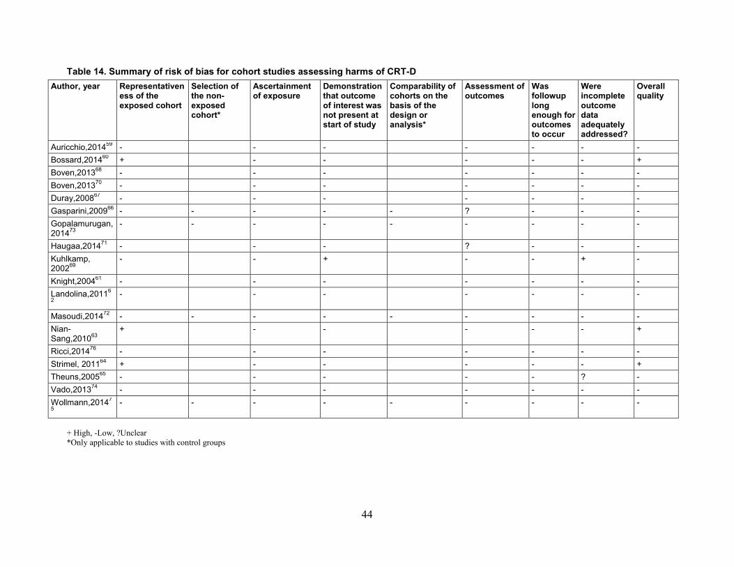
Table 14. Summary of risk of bias for cohort studies assessing harms of CRT-D Author, year Representativen
ess of the exposed cohort
Selection of the non-exposed cohort*
Ascertainment of exposure
Demonstration that outcome of interest was not present at start of study
Comparability of cohorts on the basis of the design or analysis*
Assessment of outcomes
Was followup long enough for outcomes to occur
Were incomplete outcome data adequately addressed?
Overall quality
Auricchio,201459 - - - - - - - Bossard,201460 + - - - - - + Boven,201368 - - - - - - - Boven,201370 - - - - - - - Duray,200867 - - - - - - - Gasparini,200966 - - - - - ? - - - Gopalamurugan,201473
- - - - - - - - -
Haugaa,201471 - - - ? - - - Kuhlkamp, 200269
- - + - - + -
Knight,200461 - - - - - - - Landolina,20116
2 - - - - - - -
Masoudi,201472 - - - - - - - - - Nian-Sang,201063
+ - - - - - +
Ricci,201476 - - - - - - - Strimel, 201164 + - - - - - + Theuns,200565 - - - - - ? - Vado,201374 - - - - - - - Wollmann,20147
5 - - - - - - - - -
+ High, -Low, ?Unclear *Only applicable to studies with control groups
44
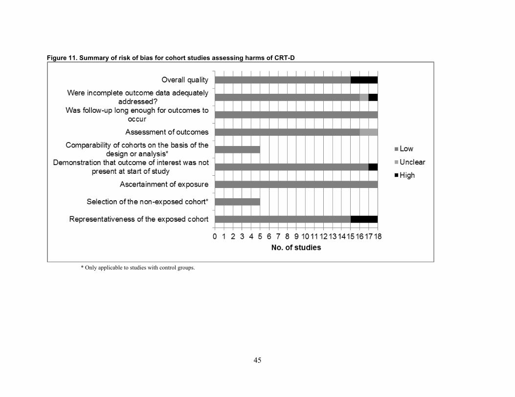
Figure 11. Summary of risk of bias for cohort studies assessing harms of CRT-D
* Only applicable to studies with control groups.
45

Table 15. List of harms reported in studies assessing harms of CRT-D Author, year Procedure
related complications (type not-specified)
Length of hospital stay
Pneumothorax Pocket hematoma
Device Infection
Cardiac perforation/ tamponade
Lead dislodgement
Ventricular arrhythmias
Inappropriate ICD shocks (CRT-D only)
Death within a week
RCTs
MADIT CRT Moss,20096 X X X Ouellet,201235 X
Jamerson, 201439
X X X X X X
Ruwald, 201440
X X
MIRACLE-ICD Young, 200343 X X X X MIRACLE-ICD II Abraham,200444
X X X X
RAFT Gilis,201448 X X X X X X
Other trials Pinter,200957 X
Higgins,200354 X Prospective cohorts Auricchio,201459
X X X
Wollman, 201475
X X X X X
Duray,200867 X X X X X Gasparini,200966
X
Knight,200461 X Kuelkamp, 200269
X X X X X
46

Author, year Procedure related complications (type not-specified)
Length of hospital stay
Pneumothorax Pocket hematoma
Device Infection
Cardiac perforation/ tamponade
Lead dislodgement
Ventricular arrhythmias
Inappropriate ICD shocks (CRT-D only)
Death within a week
Landolina,201162
X
Theuns,200565 X Retrospective cohorts Bossard,20146
0 X X
Massoudi,201472
X X
Gopalamurugan,201473
X
Ricci, 201476 X X Haugaa,201471 X Vado,201474 X Nian-Sang,201063
X
Strimel, 201164 X X X X
Van Boven,201270
X X
Van Boven,201368
X
47

Harms Outcomes List of harms reported in the included studies is shown in Table 15.
Procedure-related Complications Three studies reported on procedure-related harms in general48 ,72 ,75 In the RAFT trial, 147
out of 904 (16.4 %) patients in the ICD group and 126 out of 894 (13.9 %) patients in the CRT-D group were hospitalized for “device-related events” over the course of the study (mean follow up in the ICD group was 39.2+19.4 months and mean follow up in the CRT-D group was 41.2+19.6 months, p=0.031).48 This difference between the two groups was not statistically significant (p=0.148). In the study by Massoudi et al, mechanical complications occurred in 1.7 percent of ICD patients and 2.3 percent of CRT-D patients (p=0.049) at 3years of follow up.72In the study by Wollman et al. from the PainFree SmartShockTM Technology study, peri-procedural complications occurred in 13 of 114 (11.4%) patients in the ICD arm and 20 of 132 (15.2%) patients in the CRT-D arm at one month post implant.75 In general, complications are slightly more common with a CRT-D device compared to an ICD alone.
Length of Hospital Stay Only one study reported length of hospital stay.48 Gillis et al. (2014) reported from the RAFT
trial that the total number of days hospitalized was less in the group randomized to CRT-D as compared with the ICD-only group (12,783 vs. 14,896 days).48 The average length of hospital stay (per stay) was significantly less in the CRT-D group versus the ICD-only group (8.83+13.30 vs. 9.59+14.40, p= 0.005).
Pneumothorax Six studies in seven articles reported on the incidence of pneumothorax of which four
reported on a cohort of CRT-D patients only, and two compared patients receiving a CRT-D device to an ICD alone (Table 7).6 ,39 ,43 ,59 ,67 ,69 ,75
The incidence of pneumothorax among patients receiving a CRT-D device ranged from 1.3-2.8 percent. In the large MADIT-CRT trial, pneumothorax was slightly more common in patients receiving a CRT-D device (1.7 %) compared to an ICD alone (0.85 %).6 Men had a lower incidence of pnemo-or hemothorax compared with women(0.9% vs. 3.3%, p<0.001)39 In the Wollman et al. study, there was one pneumothorax in each the CRT-D and ICD arms.75 Lastly, the moderate-sized MIRACLE-ICD trial reported an incidence of pneumothorax or hemothorax of 0.8 percent but did not break down the incidence of pneumothorax further.
Pneumothorax is an uncommon complication of CRT-D device implant. The incidence of pneumothorax appears to be slightly more common in patients receiving a CRT-D device compared to an ICD alone although we need more data to confirm this finding (Table 16).
48

Table 16. Characteristics of studies of CRT-D reporting on the incidence of pneumothorax Author, year Arm name N for
analysis Time Point (s)
N of patients with outcomes
% of patients with outcomes
Auricchio, 201459
Overall 457 17.0+8.7 months (mean)
12.8 2.8
Duray, 200867 Overall 79 6 months 1 1.3
Kuelkamp, 200269 Overall 84 185 days (median)
2 2.4
Wollmann, 201475 ICD 114 1 month 1 0.9%
Wollmann, 201475 CRT-D 132 1 month 1 0.8%
Moss, 20096 ICD 731 30 days 6 0.8
Moss, 20096 CRT-D 1,089 30 days 19 1.7
Young, 200343 MIRACLE-ICD
Overall 369 6 months 3 0.8 (pneumothorax and hemothorax combined)
CRT=cardiac resynchronization therapy; ICD=implantable cardioverter defibrillator
Pocket Hematoma Six articles from five studies reported on the incidence of pocket hematoma 6 ,39 ,48 ,59 ,67 ,69
(Table 6). Two studies reported a comparison of CRT-D with ICD alone6 ,48 and three reported the incidence in cohorts of CRT-D only.59 ,67 ,69 Overall, the incidence of pocket hematoma in patients receiving a CRT-D device ranged from 0.9 to 2.8 percent.
In the studies by Gills et al. (2014)48 and Moss et al. (2009)6, the incidence of pocket hematoma in CRT-D patients was slightly higher than in patients receiving an ICD alone (0.6-0.8 % higher). In the MADIT-CRT trial, there was no difference in the incidence of pocket hematoma between men and women receiving CRT-D devices (3.9% vs 3.6% p=0.75).
Pocket hematoma is an uncommon but well-reported complication of CRT-D device implantation. Compared to patients receiving an ICD alone, pocket hematoma appears to be slightly more common in patients receiving a CRT-D device (Table 17).
49

Table 17. Characteristics of studies of CRT-D reporting on the incidence of pocket hematoma Author, year Arm
name N for analysis
Time point (s) N of patients with outcomes
% of patients with outcomes
Auricchio, 201459
Overall 457 17.0+8.7 months (mean)
13 2.8
Duray, 200867 Overall 79 6 months 1 1.3
Gillis, 201448 ICD 904 30 days 3 0.3
Gillis, 201448 CRT-D 894 30 days 8 0.9
Kuelkamp, 200269 Overall 84 185 days (median)
3 3.6
Moss, 20096 ICD 731 30 days 18 2.5
Moss, 20096 CRT-D 1089 30 days 36 3.3 CRT=cardiac resynchronization therapy; ICD=implantable cardioverter defibrillator
Device Infection Nine studies in 10 articles reported on the incidence of device infections.6 ,39 ,48 ,59 ,61 ,62 ,67 ,69 ,72
,75 Four studies in five articles compared infection rates between a CRT-D and an ICD alone arm.6 ,39 ,39 ,48 ,72
The other five studies were cohorts of CRT-D patients only. The range of CRT-D device infections ranged from 0.9 to 2.8 percent over a highly variable followup time. At 30 days, both the RAFT48 and MADIT-CRT6 trials showed a slightly higher incidence of device infection with CRT-D compared to an ICD alone (0.6 to 0.9 % higher). In the MADIT-CRT trial, the incidence of device infection amongst CRT-D devices was higher in women compared to men (2.0% vs. 0.8%, p=0.019).39In the study by Massoudi et al., there was a higher incidence of device infection in the CRT-D arm(1.9%) vs. the ICD arm (1.0%), p=0.002. In analysis from the Wollman et al. study, there were four infections in the ICD arm and 2 in the CRT-D arm at 1 month followup.75
Device infection is an uncommon complication of CRT-D device implant. The incidence of device infection is slightly more common in patients receiving a CRT-D device compared to an ICD alone (Table 18).
50

Table 18. Characteristics of studies of CRT-D reporting on the incidence of cardiac device infection Author, year Arm name N for
analysis Time point (s) N of patients
with outcomes
% of patients with outcomes
Auricchio, 201459
Overall 457 17.0+8.7 months (mean)
13 2.8
Duray, 200867 Overall 79 6 months 1 1.3
Massoudi, 201472 ICD 3545 3 years 32 1.0
Massoudi, 201472 CRT-D 3545 3 years 62 1.9 Wollman,201475 ICD 114 1 month 4 3.5
Wollman,201475 CRT-D 132 1 month 2 1.5
Gillis, 201448 ICD 904 30 days 20 2.2
Gillis, 201448 CRT-D 894 30 days 25 2.8
Knight, 200461 Overall 443 329 + 180 days
5 1.1
Kuelkamp, 200269 Overall 84 185 days (median)
2 2.4
Landolina, 201162
Overall 3253 18 months (median)
30 0.92
Moss, 20096 ICD 731 30 days 6 0.8
Moss, 20096 CRT-D 1089 30 days 19 1.7
CRT=cardiac resynchronization therapy; ICD=implantable cardioverter defibrillator
Cardiac Perforation/Tamponade Eight studies reported on cardiac perforation/tamponade (Table 5).39 ,43 ,44 ,48 ,64 ,67 ,69 ,75 Four
studies compared the incidence of cardiac perforation/tamponade in patients receiving CRT-D versus and ICD alone.39 ,48 ,64 ,75
In the study by Gillis et al. (2014), the incidence of cardiac perforation/tamponade was the same (0.1 %) regardless of receiving a CRT-D versus an ICD alone.48 In the study by Strimel et al. (2011) one patient had perforation/tamponade in the CRT-D cohort compared to no patients in the ICD alone cohorts.64 In the PainFree SST Study, there was one perforation in the CRT-D arm and none in the ICD arm. In subgroup analysis from MADIT-CRT, the incidence of tamponade between men and women receiving CRT_D devices were similar. The range of cardiac perforation/tamponade for patients receiving CRT-D across all reported cohorts was between 0.1-1.4 percent. The study by Kuelkamp et al. (2002) reported four cases of cardiac perforation or coronary sinus dissection but did not break the complication down any further.69
Cardiac perforation/tamponade existed in multiple trials but appears to be a rare event that does not appear to be more frequent in patients receiving a CRT-D device compared to an ICD alone (Table 19).
51
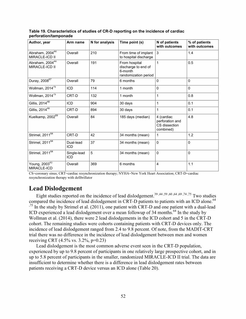
Table 19. Characteristics of studies of CR-D reporting on the incidence of cardiac perforation/tamponade Author, year Arm name N for analysis Time point (s) N of patients
with outcomes % of patients with outcomes
Abraham, 200444 MIRACLE-ICD II
Overall 210 From time of implant to hospital discharge
3 1.4
Abraham, 200444 MIRACLE-ICD II
Overall 191 From hospital discharge to end of 6-month randomization period
1 0.5
Duray, 200867 Overall 79 6 months 0 0
Wollman, 201475 ICD 114 1 month 0 0
Wollman, 201475 CRT-D 132 1 month 1 0.8
Gillis, 201448 ICD 904 30 days 1 0.1
Gillis, 201448 CRT-D 894 30 days 1 0.1
Kuelkamp, 200269 Overall 84 185 days (median) 4 (cardiac perforation and CS dissection combined)
4.8
Strimel, 201164 CRT-D 42 34 months (mean) 1 1.2
Strimel, 201164 Dual-lead ICD
37 34 months (mean) 0 0
Strimel, 201164 Single-lead ICD
5 34 months (mean) 0 0
Young, 200343 MIRACLE-ICD
Overall 369 6 months 4 1.1
CS=coronary sinus; CRT=cardiac resynchronization therapy; NYHA=New York Heart Association; CRT-D=cardiac resynchronization therapy with defibrillator
Lead Dislodgement Eight studies reported on the incidence of lead dislodgement.39 ,44 ,59 ,60 ,64 ,69 ,74 ,75 Two studies
compared the incidence of lead dislodgement in CRT-D patients to patients with an ICD alone.64
,75 In the study by Strimel et al. (2011), one patient with CRT-D and one patient with a dual-lead ICD experienced a lead dislodgement over a mean followup of 34 months.64 In the study by Wollman et al. (2014), there were 2 lead dislodgements in the ICD cohort and 5 in the CRT-D cohort. The remaining studies were cohorts containing patients with CRT-D devices only. The incidence of lead dislodgement ranged from 2.4 to 9.8 percent. Of note, from the MADIT-CRT trial there was no difference in the incidence of lead dislodgement between men and women receiving CRT (4.5% vs. 3.2%, p=0.23)
Lead dislodgement is the most common adverse event seen in the CRT-D population, experienced by up to 9.8 percent of participants in one relatively large prospective cohort, and in up to 5.8 percent of participants in the smaller, randomized MIRACLE-ICD II trial. The data are insufficient to determine whether there is a difference in lead dislodgement rates between patients receiving a CRT-D device versus an ICD alone (Table 20).
52
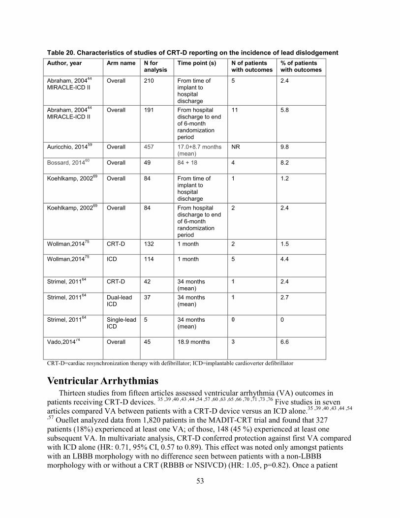
Table 20. Characteristics of studies of CRT-D reporting on the incidence of lead dislodgement Author, year Arm name N for
analysis Time point (s) N of patients
with outcomes % of patients with outcomes
Abraham, 200444 MIRACLE-ICD II
Overall 210 From time of implant to hospital discharge
5 2.4
Abraham, 200444 MIRACLE-ICD II
Overall 191 From hospital discharge to end of 6-month randomization period
11 5.8
Auricchio, 201459
Overall 457 17.0+8.7 months (mean)
NR 9.8
Bossard, 201460
Overall 49 84 + 18
4 8.2
Koehlkamp, 200269 Overall 84 From time of implant to hospital discharge
1 1.2
Koehlkamp, 200269 Overall 84 From hospital discharge to end of 6-month randomization period
2 2.4
Wollman,201475 CRT-D 132 1 month 2 1.5
Wollman,201475 ICD 114 1 month 5 4.4
Strimel, 201164 CRT-D 42 34 months
(mean) 1 2.4
Strimel, 201164 Dual-lead ICD
37 34 months (mean)
1 2.7
Strimel, 201164 Single-lead ICD
5 34 months (mean)
0 0
Vado,201474 Overall 45 18.9 months 3 6.6
CRT-D=cardiac resynchronization therapy with defibrillator; ICD=implantable cardioverter defibrillator
Ventricular Arrhythmias Thirteen studies from fifteen articles assessed ventricular arrhythmia (VA) outcomes in
patients receiving CRT-D devices. 35 ,39 ,40 ,43 ,44 ,54 ,57 ,60 ,63 ,65 ,66 ,70 ,71 ,73 ,76 Five studies in seven articles compared VA between patients with a CRT-D device versus an ICD alone.35 ,39 ,40 ,43 ,44 ,54
,57 Ouellet analyzed data from 1,820 patients in the MADIT-CRT trial and found that 327 patients (18%) experienced at least one VA; of those, 148 (45 %) experienced at least one subsequent VA. In multivariate analysis, CRT-D conferred protection against first VA compared with ICD alone (HR: 0.71, 95% CI, 0.57 to 0.89). This effect was noted only amongst patients with an LBBB morphology with no difference seen between patients with a non-LBBB morphology with or without a CRT (RBBB or NSIVCD) (HR: 1.05, p=0.82). Once a patient
53

experienced an arrhythmic event, CRT-D was not protective against subsequent VA compared with ICD alone (HR: 1.58, 95% CI, 0.99 to 2.53).35 In addition, acute procedure related VA’s were similar among men and women receiving both CRT-D and ICD devices alone from this trial.39 Ruwald et al. reported on the incidence of VAs based on EF response from MADIT-CRT 5 percent of patients in whom the LVEF improved to greater than 50 percent experienced a VA following CRT over a mean follow up of 2.2±0.8 years (following a 1 year post implant period) compared to 13 percent in the 36-50 percent LVEF group and 30 percent in the LVEF<35 percent group.40
Higgins et al. (2003) randomized 490 patients with symptomatic CHF and VA to have their CRT-D devices programmed with CRT-on versus off.54 Of the 245 patients randomized to CRT-on, 36 (15%) received appropriate treatment of VA versus 16% with CRT-off.
In the study by Abraham et al. (2004) over a 6-month followup, 26 percent in the control group (ICD only) and 22 percent in the CRT group experienced >1 appropriately detected, spontaneous episode of ventricular tachycardia and ventricular fibrillation (p= 0.61).44
In the study by Young et al. (2003) 26 percent in the control group versus 22 percent in the CRT group experienced at least one VA (p=0.47).43
In the study by Pinter et al. (2009) over a 6-month followup, 19.4 percent of patients had a VA requiring therapy in the CRT-on arm versus 16.7 percent in the CRT-off arm, a difference that was non- significant.57 In the study by Gopalamurugan et al., there was no difference in the incidence of VAs in patients receiving a CRT-D device compared to an ICD alone over a mean follow up of 23.9±9.8 months.73 Theuns et al. (2005) compared CRT-D patients with primary or secondary ICD indications and found that VA occurred in only seven out of 38 patients with a primary prophylactic indication, compared with 29 out of 48 patients with a secondary prophylactic indication (p<0.001).65
Five studies reported the incidence of VA in a cohort of CRT-D patients alone.60 ,63 ,66 ,71 ,76 The study by Gasparini et al. (2009) found that 126 patients had 621 appropriately detected VAs over a mean followup period of 14 months.66 The study by Bossard et al. (2014) evaluated outcomes of 49 patients in a CRT-D registry who survived at least 5 years after implant.60 Fourteen patients (28.6%) experienced VA.60 Nian-sang et al. (2010) examined the potential pro-arrhythmic effect of CRT during the perioperative period in 54 patients newly implanted with CRT-D devices.63 Except for one patient with a history of frequent premature ventricular contractions but without paroxysmal or sustained ventricular tachycardia before implantation, the others had no previous history of VA. In total, four patients (7.4%) experienced ventricular tachycardia/ventricular fibrillation within 3 days of implantation. They did not experience any additional VA over the 12 months of followup.63 Ricci et al. followed 1,404 CRT-D patients over a median follow up of 31 months; 36 percent experienced a VA. Haugaa and colleagues followed 201 patients who had received a CRT-D device of whom 14 percent experienced a VA over a followup of 2 years.
van Boven et al. (2013) followed a cohort of patients primarily receiving CRT-D devices (96.5 %) and separated patients into responders and non-responders (response was defined as an LVEF≥35percent on followup echocardiogram).70 Over a 3-year followup period, 12 percent of patients experienced ≥1 appropriate shock all of whom were deemed non-responders by echocardiography.
Overall, there is conflicting evidence as to whether CRT-D is protective from VAs compared to an ICD alone, with four trials showing no difference and one large trial showing CRT to be
54

protective from VAs. More data are needed to confirm this latter finding. The data, however, are consistent that CRT-D does not appear to increase the rate of VAs compared to an ICD alone.
Inappropriate Implantable Cardioverter Defibrillator Shocks Eight studies reported on inappropriate ICD shocks.40 ,43 ,44 ,48 ,64 ,68 ,70 ,76 Three studies
compared the incidence of inappropriate shocks in patients receiving a CRT-D device versus an ICD alone. Abraham et al. (2004) found no difference in the rate of inappropriate shocks during the 6 month followup period in patients receiving a CRT-D device compared to those receiving an ICD alone 44; however they did not report the numbers or percentages of participants experiencing inappropriate shocks (p=0.78).
In the RAFT trial, 2.2 percent of patients in the CRT-D group were hospitalized for inappropriate shocks versus 3.3 percent in the ICD-only group.48 The trial did not report the incidence of inappropriate shocks not resulting in hospitalization.
In the study by Young et al. (2003) there was no difference in the incidence of inappropriate shocks between patients in the CRT group versus the control arm over a 6-month followup (4.2 vs. 7.2%, p=0.26). These data were not sufficient to serve as the basis for a meta-analysis because the duration of followup varied from 30 days to 3 years.
Strimel et al. (2011) reported that two (2.4%) of octogenarians with ICDs (with or without CRT) experienced inappropriate shocks over a mean followup of 34 months.64
In the retrospective cohort study by Van Boven et al. (2013), 33 participants (6.1 %) experienced an IS over a mean follow up time of 3.2 + 1.8 years.68 In a second study by van Boven et al. (2013)70, the incidence of IS was 8.5 percent (Table 21). Ricci et al. reported an incidence of 7% of inappropriate shocks in a cohort of 1404 CRT-D patients over a median follow up of 31 months.76 Ruwald et al, in analysis from MADIT-CRT, demonstrated no significant difference in inappropriate shocks amongst patients based on level of LVEF improvement. 40
In conclusion, there is no apparent difference in the incidence of inappropriate ICD shocks in patients receiving a CRT-D device compared to an ICD alone. Table 21. Characteristics of studies of CRT-D reporting inappropriate ICD shocks Author, year Arm name N for
analysis Time point (s) N of patients
with outcomes
% of patients with outcomes
Gillis, 201448 ICD 904 30 days 30 3.3
Gillis, 201448 CRT-D 894 30 days 20 2.2
Strimel, 201164 Overall 84 34 months (mean)
2 2.4
Ricci, 201476 Overall 1404 31 months (median)
101 7
Van Boven, 201368
CRT-D 543 3.2 + 1.8 years (mean followup)
33 6.1
55

Author, year Arm name N for analysis
Time point (s) N of patients with outcomes
% of patients with outcomes
Van Boven, 201370
Overall 179 Median 3.0 years 12 8.5
Young, 200343 CRT-on 180 6 months 8 4.2
Young, 200343 MIRACLE-ICD
CRT-off 189 6months 13 7.2
CRT=cardiac resynchronization therapy; CRT-D=cardiac resynchronization therapy with defibrillator; ICD=implantable cardioverter defibrillator
Death Within One Week Only two studies reported on death within 1 week of implantation.64 ,67 Both of these cohort
studies reported zero deaths.
56
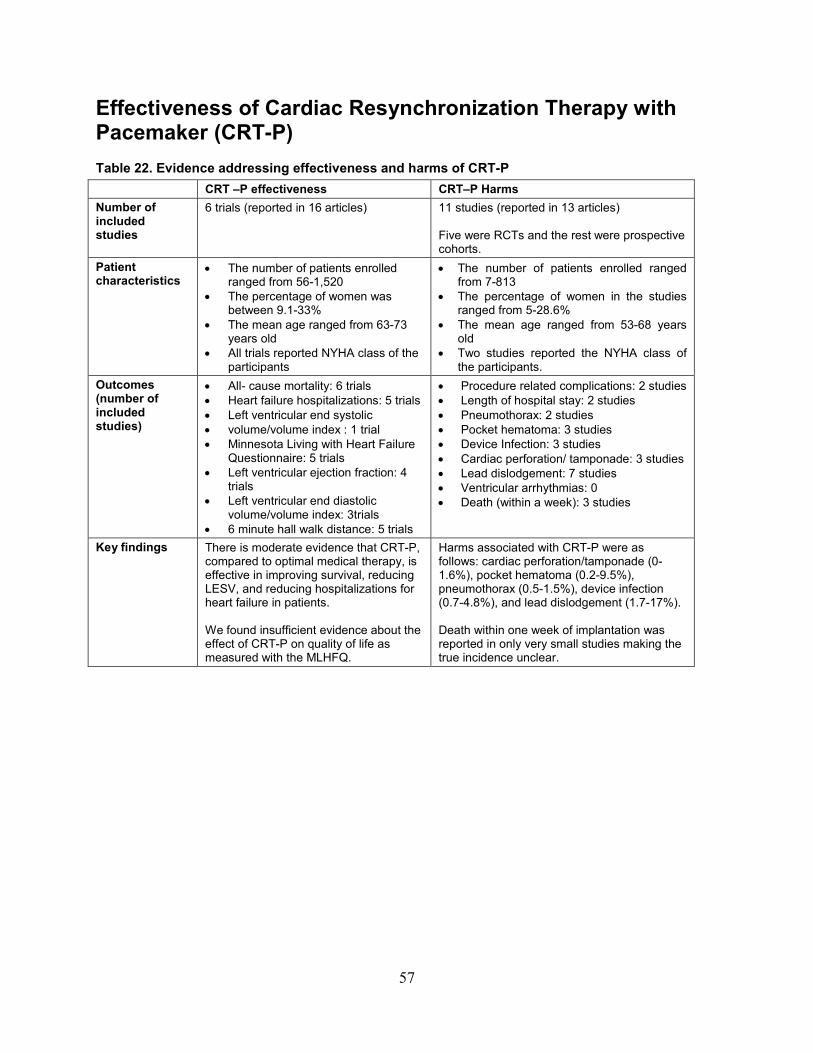
Effectiveness of Cardiac Resynchronization Therapy with Pacemaker (CRT-P) Table 22. Evidence addressing effectiveness and harms of CRT-P CRT –P effectiveness CRT–P Harms Number of included studies
6 trials (reported in 16 articles) 11 studies (reported in 13 articles) Five were RCTs and the rest were prospective cohorts.
Patient characteristics
• The number of patients enrolled ranged from 56-1,520
• The percentage of women was between 9.1-33%
• The mean age ranged from 63-73 years old
• All trials reported NYHA class of the participants
• The number of patients enrolled ranged from 7-813
• The percentage of women in the studies ranged from 5-28.6%
• The mean age ranged from 53-68 years old
• Two studies reported the NYHA class of the participants.
Outcomes (number of included studies)
• All- cause mortality: 6 trials • Heart failure hospitalizations: 5 trials • Left ventricular end systolic • volume/volume index : 1 trial • Minnesota Living with Heart Failure
Questionnaire: 5 trials • Left ventricular ejection fraction: 4
trials • Left ventricular end diastolic
volume/volume index: 3trials • 6 minute hall walk distance: 5 trials
• Procedure related complications: 2 studies • Length of hospital stay: 2 studies • Pneumothorax: 2 studies • Pocket hematoma: 3 studies • Device Infection: 3 studies • Cardiac perforation/ tamponade: 3 studies • Lead dislodgement: 7 studies • Ventricular arrhythmias: 0 • Death (within a week): 3 studies
Key findings There is moderate evidence that CRT-P, compared to optimal medical therapy, is effective in improving survival, reducing LESV, and reducing hospitalizations for heart failure in patients. We found insufficient evidence about the effect of CRT-P on quality of life as measured with the MLHFQ.
Harms associated with CRT-P were as follows: cardiac perforation/tamponade (0-1.6%), pocket hematoma (0.2-9.5%), pneumothorax (0.5-1.5%), device infection (0.7-4.8%), and lead dislodgement (1.7-17%). Death within one week of implantation was reported in only very small studies making the true incidence unclear.
57
Study Characteristics Six trials3-5 ,8 ,55 ,58 addressed the effectiveness of CRT-P (reported in 16 articles).3-5 ,8 ,22-25 ,27-
30 ,42 ,45 ,55 ,58 Six of the articles reported re-analyses from the CARE-HF clinical trial,22-25 ,27 ,28 two reported secondary analyses of the COMPANION trial,29 ,30 and one presented a secondary analysis of the MIRACLE trial.42 Four trials were RCTs 3-5 ,58 and two were randomized crossover trials.8 ,55 All six trials reported the device name (Table 20). The planned length of followup ranged from 3 to 18 months. One study did not report followup time.23 One study also assessed effectiveness at the end of study (followup at 29 months) (Evidence Table 1).25
In general, the trials were heterogeneous in the optimal medical therapy used as the comparison group. Three trials compared CRT-P to medical therapy.3-5 One trial compared CRT-P and coronary artery bypass grafting (CABG) to CABG alone.58 One trial compared biventricular (BiV) pacing to no pacing, using a crossover design.8 Another trial compared BiV pacing to right ventricular (RV) pacing alone, using a crossover model.55 Three trials (published in 12 articles) were industry funded (Table 23).3-5 One trial, published in two articles, was partially industry funded. 8 ,45
58

Table 23. Study characteristics of trials assessing effectiveness of CRT Author, year
Length of followup (months)
Study design
Number of patients
Comparison Device model name
NYHA class
Funding source
CARE-HF
Cleland, 20045
18 RCT 809 CRT-P vs. OMT
InSync or InSync III, Medtronic
III-IV Industry
Cleland, 200924
18 RCT 809 CRT-P vs. OMT
InSync or InSync III, Medtronic
III-IV Industry
Ghio, 200925
18 RCT 735 CRT-P vs. OMT
InSync or InSync III, Medtronic
III-IV Industry
Wikstrom,200927
18 RCT 813 CRT-P vs. OMT
InSync or InSync III, Medtronic
III-IV Industry
Cleland, 200622
18 RCT 812
CRT-P vs. OMT
InSync or InSync III, Medtronic
III-IV Industry
Cleland, 201228
18 RCT 809 (309 with re-consent)
CRT-P vs. OMT
InSync or InSync III, Medtronic
III-IV Industry
Cleland, 200723
NR Post Hoc Analysis
813 CRT-P vs. OMT
InSync or InSync III, Medtronic
I-IV
Industry
COMPANION
Bristow, 20044
Medical therapy arm: 14.8 months CRT-P arm 16.5 months
RCT 1,520 CRT-P vs. OMT
Contak TR 1241
III-IV Industry
Carson 200530
Mortality endpoint OMT arm: 14.8 months CRT-P arm 16.5 months
RCT 1,510 CRT-P vs. OMT
Contak TR 1241
III-IV Industry
Anand 200929
Hospitalization endpoint OPT arm:11.9 months CRT-P arm: 16.2 months
RCT 1,520 CRT-P vs. OMT
Contak TR 1241
III-IV Industry
MIRACLE Abraham, 20023
6 RCT 453 CRT-P vs. OMT
InSync 8040, Medtronic
III-IV Industry
59

Author, year
Length of followup (months)
Study design
Number of patients
Comparison Device model name
NYHA class
Funding source
St. John Sutton, 200342
6 RCT 323 CRT-P vs. OMT
InSync 8040, Medtronic
III-IV Industry
MUSTIC
Cazeau, 20018
6 RCT cross-over
67 CRT-P on vs. off
Chorum MSP 7336 and Insync 8040
III-IV Industry; Swedish Heart and Lung Association; Swedish Medical Research Council
Leclercq,2002,45
6 RCT cross-over
45 CRT-P on vs. off
Chorum 7336 MSP, ELA Medical, Montrouge, France, and InSync 8040, Medtronic
III-IV Industry
Other trials
RD-CHF Leclercq, 200755
6 RCT cross- over
56 CRT-P vs. RV pacing
Chorum MSP 7336, Ela Medical
III-IV Not Reported
Pokushalov, 201058
18 RCT 178 CABG vs. CABG+CRT-P
Insync III, Medtronic
III-IV Not Reported
OMT=optimal medical therapy; RCT=randomized controlled trial; CRT-P=cardiac resynchronization therapy with pacemaker; CABG=coronary artery bypass grafting; RV=right ventricle; vs.=versus
60

Participant Characteristics The number of patients in the trials ranged from 56 to 1,520. The percentage of women in the
trials ranged from 9.1 percent in Leclerq et al. (2007)55 (this study, the RD-CHF trial, included no women in one of its comparison arms) to 33 percent in the CRT-P arm of the COMPANION trial.4 The mean age in the trials ranged from 63 to 73 years old. Two trials reported median rather than mean age (median ranging from 66-69 years old).5 ,8 One study reported racial distribution of subjects (90 percent of the subjects were white).3 ,42 The proportion of patients with ischemic cardiomyopathy (ICM) ranged from 32 to 59 percent. One trial did not report on the proportion of ICM.42 Only one trial reported the prevalence of atrial fibrillation (AF) (21%).55 Three trials excluded patients with any history of AF.4 ,5 ,8 One trial excluded patients with a history of AF within 1 month3 of enrollment, and another did not exclude patients with AF, but also did not report any history of AF.58
All six trials reported NYHA class of study participants, with five of six enrolling patients having class III-IV symptoms. Enrollment of class IV patients ranged from 6 percent in the CRT-P arm of CARE-HF5 to 18 percent in the optimal medical therapy arm of the COMPANION trial.4 Of note, in the large CARE-HF trial, 21.5 percent of patients assessed themselves to be class I-II (in contradiction to physician assessment).23 The trial by Cazeau et al. (2001) only enrolled patients with class III symptoms.8
Four of the primary trials reported the mean left ventricle ejection fraction (LVEF),3 ,8 ,55 ,58 ranging from 21.6 percent in the optimal medical therapy (OMT) arm of the MIRACLE trial3 to 30 percent in the CABG alone arm in the trial by Pokushalov et al. (2010).58 Two large trials reported median LVEF, ranging from 20 percent in the optimal medical therapy arm of the COMPANION trial4 to 25 percent in both arms of the CARE-HF trial.5
Four of the primary trials reported the mean QRS duration,3 ,8 ,55 ,58 ranging from 137ms in the CABG/CRT arm of the trial by Pokushalov et al. (2010)58 to 20ms in the trial by Leclercq et al. (2007)55 Of note, all patients had permanent RV pacing prior to CRT-P upgrade in this study.55 Two large trials reported median QRS duration, ranging from 158ms in the OMT arm of the COMPANION trial4 to 160ms in both arms of the CARE-HF trial.5 Four of the primary trials reported the incidence of native left bundle branch block prior to CRT-P. The study by Leclercq et al. (2007)55 enrolled only patients with permanent RV pacing and the MIRACLE3 and CARE-HF5 trials did not report on QRS morphology. The incidence of left bundle branch block ranged from 69 percent in the CRT-P arm of the COMPANION trial4 to 87 percent in the MUSTIC trial.8 Only the COMPANION trial reported the incidence of right bundle branch block4 (9 percent in the optimal medical therapy arm and 12 percent in the CRT-P arm). No studies reported on patients with non-specific intraventricular conduction delay. The study by Leclercq et al. (2007) was the only trial to evaluate paced patients (100% in this trial).55
In general, these studies comprised homogeneous patient populations with regard to physiological criteria, such as NYHA class and QRS duration. The proportions of female patients varied between studies, and only one reported race; thus gender and racial makeup of these populations might not be generalizable (Evidence Table 2).
61

Risk of Bias Several types of risk of bias were present in these studies. The most common potential cause
of bias was lack of allocation concealment and blinding; details of allocation and blinding were not reported in the majority of studies. In the majority of studies, criteria or protocols for outcome assessment were also not assessed, making outcome reporting bias a potential concern (Table 24 and Figure 12).
62

Table 24. Summary of risk of bias for trials assessing effectiveness of CRT-P Author, year Random
sequence generation
Allocation concealment
Blinding of personnel
Blinding of outcome assessors
Assessing blinding by outcome
Incomplete outcome data
Selective outcome reporting
Other sources of bias
Overall quality
CARE HF Cleland,20045 Cleland,200723 Cleland,201228 Cleland,200622 Cleland,200924 Wikstrom,200927 Ghio,200925
-
? ? ? ? - + ? +
COMPANION Bristow,20044 Anand, 200929 Carson,200530
? ? + - - - - + +
MIRACLE Abraham,20023 Sutton,200342
- - - - - - - - -
MUSTIC Cazeau, 20018 Leclercq,2002,45
? ? - ? ? ? ? - +
Other Trials Leclercq,200755 - ? ? ? ? - - - - Pokushalov,201058 ? ? ? ? ? - - - + +=High -=Low ?=Unclear
63

Figure 12. Summary of risk of bias for trials assessing effectiveness of CRT-P
Effectiveness Outcomes The studies addressed various outcomes, the most common of which were all-cause mortality
and changes in quality of life as measured by MLHFQ (in 8 studies) (Table 25). Five trials assessed changes in 6-minute hall walk distance and heart failure hospitalizations, four studies assessed changes in LVEF, left ventricle end systolic volume (LVESV), and left ventricle end diastolic volume (LVEDV). No studies assessed changes in clinical composite score. No study reported results for these outcomes by the pre-specified subgroups of interest.
64
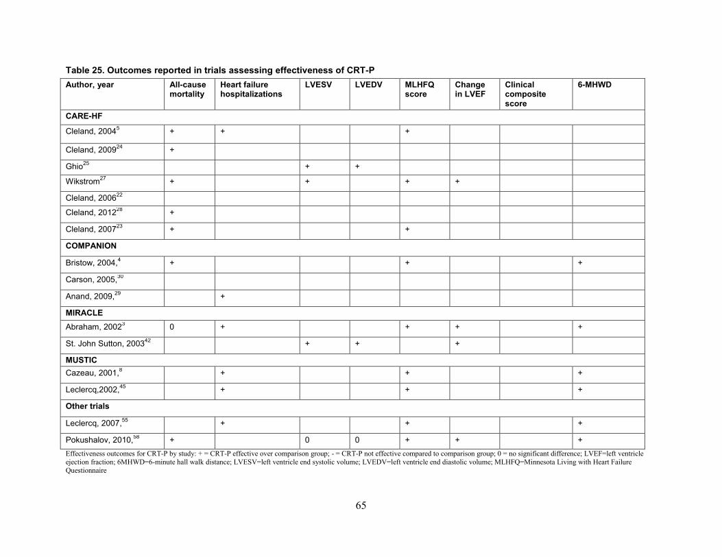
Table 25. Outcomes reported in trials assessing effectiveness of CRT-P Author, year All-cause
mortality Heart failure hospitalizations
LVESV LVEDV MLHFQ score
Change in LVEF
Clinical composite score
6-MHWD
CARE-HF
Cleland, 20045 + + +
Cleland, 200924 +
Ghio25 + +
Wikstrom27 + + + +
Cleland, 200622
Cleland, 201228 +
Cleland, 200723 + +
COMPANION
Bristow, 2004,4 + + +
Carson, 2005,30
Anand, 2009,29 +
MIRACLE Abraham, 20023 0 + + + +
St. John Sutton, 200342 + + +
MUSTIC Cazeau, 2001,8 + + +
Leclercq,2002,45 + + +
Other trials
Leclercq, 2007,55 + + +
Pokushalov, 2010,58 + 0 0 + + + Effectiveness outcomes for CRT-P by study: + = CRT-P effective over comparison group; - = CRT-P not effective compared to comparison group; 0 = no significant difference; LVEF=left ventricle ejection fraction; 6MHWD=6-minute hall walk distance; LVESV=left ventricle end systolic volume; LVEDV=left ventricle end diastolic volume; MLHFQ=Minnesota Living with Heart Failure Questionnaire
65
All-cause Mortality Six trials (published in 10 articles) assessed all-cause mortality.3-5 ,22-24 ,27 ,28 ,55 ,58 Pokushalov et al. (2010) assessed the outcome at 6, 12, and 18 months, comparing those
receiving CABG alone to those receiving CABG and CRT-P.58 At 6 months of followup, 10 CABG patients (11.4%) and four CABG + CRT-P patients (4.4%) died. At 12 months of followup, eight CABG patients (9.1%) and three CABG + CRT-P patients (3.3%) died. At 18 months of followup, in the CABG group, 23 patients (26.4%) died, compared with nine (9.9%) in the CABG + CRT-P group (log-rank test, p=0.006). Fourteen of the observed deaths were due to sudden cardiac death, and 18 deaths were due to “pump failure”.
In the study by Cleland et al. (2004) comparing CRT-P to medical therapy, 120 of 404 patients (30%) died of any cause in the medical therapy group, compared to 82 of 409 patients (20%) in the CRT-P group (HR: 0.64, 95% CI, 0.48 to 0.85, p<0.002).5 Cleland et al. (2009) also reported these results.24 In Cleland et al. (2006) after 1,600 days of followup (mean-followup of 36.4 months), the mortality was 154 of 404 (38%) in the medical therapy group, compared to 101 of 409 (25%), in the CRT-P group (HR: 0.6, 95% CI, 0.47 to 0.77, p<0.001).22 Cleland et al. (2012)28 also reported these results, as did Cleland et al. (2007).23
Abraham et al. (2002) compared a control group to a CRT-P group.3 At 6 months, the number of patients who died from any cause was 16 of 225 (7.1%) in the control group, compared to 12 of 228 (5.3%) in the CRT-P group (HR: 0.73, 95% CI, 0.34 to 1.54, p=0.4).
Leclerq et al. (2007) assessed all-cause mortality at 6 months.55 During the crossover phase, two patients died from sudden cardiac death during the biventricular phase and two patients died from CHF during the right ventricular phase. Two other patients died from a pulmonary embolism and respiratory failure while in the right ventricular phase. The overall mortality in the study was 13.5 percent at 6 months of followup. The study did not report analyses of comparisons of mortality between groups.
Wikstrom et al. (2009) compared all-cause mortality between CRT-P and medical therapy in two separate groups, those with and without ischaemic heart disease (IHD), at 18 months. CRT-P had a significant effect on all-cause mortality (HR: 0.60, 95% CI, 0.42 to 0.86 and HR: 0.59, 95% CI, 0.37 to 0.92 for IHD and no IHD, respectively).27
In the COMPANION trial, 77 of 308 patients in the optimal medical therapy group (25%) died during the entire study period, for a mortality rate of 19 percent. The mortality rate in the CRT-P group was 21 percent (131 of 617 patients) during the entire study period. The study reported an association between CRT-P implementation and a reduction in the risk of death from any cause (HR: 0.76, 95% CI, 0.58 to 1.01, p=0.059).4 ,30 The study also reported that pump failure was the predominant mode of death and that it was reduced by CRT-P, but that cardiac death was not reduced by CRT-P compared to OMT.
Three trials with longer followup times (reported in 4 articles) showed statistically significant differences in mortality favoring CRT-P.4 ,5 ,27 ,58 Two additional trials that reported on all-cause mortality had shorter followup times (3 or 6 months), which might partially explain the lack of statistically significant results showing difference between CRT-P and optimal medical therapy.3
,55There is moderate strength of evidence favoring CRT-P versus optimal medical therapy in mortality (Table 27).
The results are derived from NYHA class III-IV patients and applicability to NYHA class I and II is unclear. Future studies should seek to reproduce this mortality finding in CRT-P with consistent comparators and methodology.
66
Heart Failure Hospitalizations Five trials, reported in six articles, assessed heart failure hospitalization outcome.3 ,5 ,8 ,29 ,45 ,55
Cleland et al., assessed hospitalizations for heart failure at 18 months.5 Of the 404 patients in the medical therapy group, 184 (46%) had been hospitalized by the end of followup, compared to 125 (31%), of the 409 patients in the CRT-P group (HR: 0.61, 95% CI, 0.49 to 0.77, p<0.001).
Abraham et al. (2002) compared a control group, N=225, to a CRT-P group, N=228, at 6 months.3 In the control group there were 34 hospitalizations (15.1%), and in the CRT-P group 18 hospitalizations (7.9%) (HR: 0.5, 95% CI, 0.28 to 0.88, p<0.02).
Leclerq et al. (2007) assessed heart failure hospitalization at 3 months in the crossover study.55 At 3 months, there was one hospitalization in the BiV-first group versus nine hospitalizations in the RV first group. Compared to the RV first group, the BiV-first group had significantly fewer hospitalizations (p=0.01).
The MUSTIC trial8 using a crossover model, randomized patients to active or inactive pacing first. Three hospitalizations for heart failure occurred during active pacing, and nine during inactive pacing (p<0.05). Among one of the subgroups specified for the current review, patients with atrial fibrillation, Leclercq et al. (2002) conducted a secondary analysis of the MUSTIC trial, finding a total of three hospitalizations during the first three-months of the crossover study (the number of hospitalizations in each group was not reported).45 During the entire 6 months of this crossover study, 10 of 44 patients (23%) were hospitalized for heart failure during the univentricular period, for a total of 11 hospitalizations, compared to 3 (7%) during the biventricular period.
In a secondary analysis of the COMPANION trial, Anand et al. (2009) showed (after adjustment for length of followup) an association between CRT-P and a 44 percent reduction in heart failure hospital admissions per patient-year compared with the optimal medical therapy group (from 0.7 to 0.4, no p-value specified).29
In summary, the five trials addressing hospitalization outcome reported fewer hospitalizations in the CRT-P group compared to optimal medical therapy. One study found fewer hospitalizations in a subgroup of patients with AF.45 There is moderate strength of evidence indicating fewer hospitalizations for CRT-P compared with optimal medical therapy (Table 27).
These results are derived from NYHA class III-IV patients and applicability to NYHA class I and II is unclear.
Left Ventricular End Systolic Volume Four trials assessed LVESV in comparing CRT-P with optimal medical therapy. 25 ,27 ,42 ,58 Pokushalov et al. (2010) assessed the outcome at 6, 12, and 18 months, comparing those
receiving CABG alone to those receiving CABG and CRT-P.58 At baseline, 6, 12, and 18 months, the difference in LVESV between CABG and CABG + CRT-P over time was not statistically significant (p=0.06). In addition, the differences intragroup and between groups at the individual time points were not statistically significant.
St John Sutton et al. (2003) compared CRT-P to control at 3 and 6 months and found a statistically significant decrease in LVESV in the CRT-P group but not in the control group.42 In within-arm comparisons, the LVESV decreased a median of 21.8mL in the CRT-P group (95% CI, -29.7 to -13.9), compared to a median increase of 0.6mL in the control group (95% CI, -8.7 to 8.7, p<0.05). Similar changes were reported at 6 months, with the median decrease of 25.6mL in the intervention group (95% CI, -37.4 to -17.7, p<0.05).
67
Ghio et al. (2009) compared CRT-P to medical therapy in the CARE-HF trial.25 The decrease in LVESV at 18 months from baseline was 55.1mL more, that is, a greater decrease, in the CRT-P group than in the medical therapy group (95% CI, -67.2 to -42.9. p<0.0001). Wikstrom et al. (2009) compared LVESV between CRT-P and medical therapy in two separate groups, those with and without ICM, at 3 months.27 In those with ICM, the mean LVESV was 193.99cm3 (SD 69.36) in the CRT-P group and 231.54cm3 (SD 86.05) in the medical therapy group; in those without ICM, the mean LVESV was 194.01cm3 (SD 104.74) in the CRT-P group and 233.18cm3 (SD 98.36) in the medical therapy group (p=0.0354).
In summary, these trials provide moderate evidence that CRT-P improves LVESV (Table 27). These results are derived from NYHA class III-IV patients and applicability to NYHA class I and II is unclear.
Left Ventricular End Diastolic Volume Three trials assessed LVEDV in comparing CRT-P with optimal medical therapy.25 ,42 ,58 Pokushalov et al. (2010) assessed the outcome at 6, 12, and 18 months, comparing those
receiving CABG alone to those receiving CABG and CRT-P.58 At baseline, 6, 12, and 18 months, the difference in LVEDV over time was not statistically significant (p=0.65). In addition, the differences intragroup and between groups at the individual time points were not statistically significant.
St John Sutton et al. (2003) compared CRT-P to control at 3 and 6 months and found a statistically significant decrease in LVEDV in the CRT-P group but not in the control group.42 In within-arm comparisons, the LVEDV decreased a median of 22.6mL in the CRT-P group (95% CI, -33.3 to -5.8), compared to a median increase of 2.8mL in the control group (95% CI, -3.8 to 12.3, p<0.05). The study reported similar changes at 6 months, with a median decrease of 27.2mL in the intervention group (95% CI, -37.1 to -16.9, p<0.05
Ghio et al. (2009) compared CRT-P to medical therapy in the CARE-HF trial.25 For LVEDV at 18 months, the change from baseline was greater in the CRT-P group than the medical therapy group, -57.6mL (95% CI, -71.8 to -43.4, p<0.0001).
The fact that only two of three studies showed statistically significant differences in LVEDV, can likely be explained by the difference in comparisons. St John Sutton et al. (2003) together with the CARE-HF trial (Ghio et al. (2009)) compared CRT-P to medical therapy25 ,27 ,42 whereas Pokushalov et al. (2010)58 compared CABG therapy alone to CABG + CRT-P.
In summary, it is unclear whether LVEDV is improved by CRT-P compared with optimal medical therapy (Table 27).
Minnesota Living with Heart Failure Questionnaire Score Five trials (published in 8 articles) assessed quality of life using MLHFQ in comparing CRT-
P with optimal medical therapy.3-5 ,8 ,23 ,27 ,45 ,58 Pokushalov et al. (2010) assessed the outcome at 6, 12, and 18 months, comparing those
receiving CABG alone to those receiving CABG and CRT-P.58 At baseline, 6, 12, and 18 months, the MLHFQ score in the CABG group versus the CABG + CRT-P group was 63.2+/-19 versus 64.9+/-20; 51.9+/-22 versus 39.8+/-16 (p<0.05 for difference between the two groups and p<0.01 for difference from baseline); 41.7+/-8 versus 32.9+/-7 (p<0.05 for difference between the two groups and p<0.01 for difference from baseline); and 46.4+/-11 versus 22.9+/- 5 (p<0.05 for difference between the two groups and p<0.01 for difference from baseline), respectively. The difference between CABG and CABG + CRT-P over time was statistically significant
68

(p<0.001). Of note, the MLHFQ score at 18 months decreased from 87 at baseline to 64 in the CABG-only group and from 91 at baseline to 82 in the CABG + CRT-P group.
Abraham et al. (2002) compared a control group to a CRT-P group.3 At 6 months, the median change in the MLHFQ score from baseline was -9 in the control group (N=193, 95% CI, -12 to -5), and -18 in the CRT-P group (95% CI, -22 to -12). The difference between these intragroup changes was statistically significant (p=0.001).
Cleland et al. (2004) comparing CRT-P to medical therapy, found at 90 days that the mean difference between groups in MLHFQ score was -10 (95% CI, -8 to -12, p<0.001).5 Wikstrom et al. (2009) also from the CARE-HF trial compared MLHFQ between CRT-P and medical therapy at 3 months in two separate groups, those with and without ICM.27 CRT-P had no significant effect on MLHFQ. In those with ICM, the mean difference was 31.29 (SD 19.74) in the CRT-P group and 30.25 (SD 22.00) in the medical therapy group. In those without ICM, the mean difference was 41.50 (SD 20.49) in the CRT-P group and 35.56 (SD 21.68) in the medical therapy group, p=0.1542. Another report from the CARE-HF trial, Cleland et al. (2007) found that the proportion of subjects with an MLHFQ score <= 35 was 166 (41%) in the control group, compared to 213 (52%) in the CRT-P group (HR: 0.64, 95% CI, 0.48 to 0.86, p=0.002).23
Bristow et al. (2004) compared MLHFQ between CRT-P and optimal medical therapy groups. At 3 months, the change compared to baseline was -24+/-27 in the CRT-P group versus -9+/-21 in the OMT group (p<0.001). A similar, statistically significant difference existed at 6 months (p<0.001).4
In the MUSTIC trial those with active pacing had a MLHFQ score of 29.6+/-21.3, while those on inactive pacing had a score of 43.2+/22.8 (p<0.001).8
Among one of the subgroups specified for the current review, patients with atrial fibrillation, Leclercq et al. (2002) conducted a secondary analysis of the MUSTIC trial.45 For the group receiving biventricular pacing first, the score was 40 (SD 23) at randomization. For the RV pacing first group, the score was 50 (SD 20) at randomization. At six months, the score was 38.5 (SD 21.4) in the univentricular group, and 34.1 (SD 20.6) in the biventricular group, showing no statistically significant difference.
These trials assessed this outcome at different endpoints and with different comparisons, which might explain the inconsistency in the results comparing CRT-P to other therapy. There was insufficient evidence to draw any conclusions about the effect of CRT-P on MLHFQ compared with optimal medical therapy (Table 27).
Left Ventricular Ejection Fraction Four trials assessed the change in LVEF, including two reports of the MIRACLE trial.3 ,27 ,42
,55 ,58 Pokushalov et al. (2010) assessed the outcome at baseline, 6, 12, and 18 months, comparing
those receiving CABG alone to those receiving CABG and CRT-P.58 At baseline, 6, 12, and 18 months, the LVEF in the CABG group versus the CABG + CRT-P group was 30+/-2.2 versus 28+/-2.7; 34+/-3.4 versus 39+/-3.7; 32+/-3.4 versus 41+/-2.5 (p<0.05 for difference between the two groups and p<0.01 for difference from baseline); 29+/-2.6 versus 42+/-1.4 (p<0.05 for difference between the two groups and p<0.01 for difference from baseline); and 28+/2.7 versus 42+/-1.9 (p<0.05 for difference between the two groups and p<0.01 for difference from baseline), respectively. The difference between CABG and CABG + CRT-P over time was significant (p<0.001). Of note, the number in the CABG-only group decreased over time from 87
69
at baseline to 64 at 18 months; and, in the CABG + CRT-P group, from 91 at baseline to 82 at 18 months.
St John Sutton et al. (2003) compared CRT-P to control at 3 and 6 months.42 In within-arm comparisons, the LVEF increased a median of 0.6 in the CRT-P group (95% CI, -0.4 to 1.8), compared to a median increase of 2.3 in the control group (95% CI, 1.5to 3.2), which was significant (p<0.05). Similar changes were noticed at 6 months, with the median increase of 3.6 in the CRT-P group (95% CI, 2.5 to 5.8, p<0.05).
Abraham et al. (2002) compared a control group to a CRT-P group.3 At 6 months, median change in the LVEF from baseline was -0.2 in the control group (95% CI, -1 to 1.5), and in the CRT-P group, 4.6 (95% CI, -3.2 to 6.4). This difference was statistically significant (p<0.001).
It should be noted that St. John Sutton et al. (2003)42 and Abraham et al. (2002)3 represent two reports of LVEF from the same trial, MIRACLE, with differing results.
In Leclerq et al. (2007) for all patients, the LVEF was 29.5 (SD 11) at baseline and 29 (SD 11) at 3 months.55 For the group receiving biventricular pacing first, the LVEF was 32 (SD 11) at baseline and 34 (SD 12) at 3 months; for the group receiving right ventricular pacing first the LVEF was 32 (SD 13) at baseline and 37 (SD 11) at 3 months. The difference in the right ventricular- first group was not statistically significant from that of the biventricular-first group (p=0.1). Leclerq et al. (2007) conducted their crossover study differently from the other trials making it difficult to compare.
Wikstrom et al. (2009) compared LVEF between CRT-P and medical therapy in two separate groups, those with and without ICM, at 3 months and found no significant difference between CRT-P and medical therapy.27 In those with ICM, the mean LVEF at 3 months was 29.08 (SD 6.90) in the CRT-P group and 26.31 (SD 6.50) in the medical therapy group; in those without ICM, the LVEF was 30.59 (SD 8.19) in the CRT-P group and 26.56 (SD 6.92) in the medical therapy group (p=0.3550).
In summary, three of these four trials showed improved LVEF with CRT-P compared to optimal medical therapy, though comparisons were different between trials, as was length of followup. Thus, the absolute difference in LVEF is not comparable from study to study.
6-Minute Hall Walk Distance Five trials assessed 6-minute hall walk distance (6MHWD) in comparing CRT-P with
optimal medical therapy. Pokushalov et al. (2010) assessed the outcome at 6, 12, and 18 months, comparing those receiving CABG alone to those receiving CABG and CRT-P.58 At baseline, 6, 12, and 18 months, the distance walked by the CABG group versus the CABG + CRT-P group was: 265+/-32m versus 248+/-51m, 379+/-127m versus 432+/-129m (p<0.05 for difference between the two groups and p<0.01 for difference from baseline), 317+/- 67m versus 448 +/- 79m (p<0.05 for difference between the two groups and p<0.01 for difference from baseline), and 289+/-72m versus 452+/- 65m (p<0.05 for difference between the two groups and p<0.01 for difference from baseline).
Abraham et al. (2002) compared a control group to a CRT-P group.3 At 6 months, the median change of walk distance from baseline was +10m in the control group (N=198, 95% CI, 0 to +25), and +39m in the CRT-P group (95% CI, +26 to +54). The difference between these groups was statistically significant (p=0.005).
Leclerq et al. (2007) assessed 6MHWD at baseline and 3 months in three groups: patients receiving biventricular pacing first, patients receiving right ventricular pacing first, and all patients.55 For the group receiving biventricular pacing first, the walk distance was 316m (SD
70
25) at baseline and 358m (SD 88) at 3 months. For the right ventricular pacing first group, the distance was 332m (SD 173) at baseline and 414m (SD 110) at 3 months. For all patients, the distance was 324m (SD 149) at baseline and 386m (SD 99) at 3 months. The difference between the right ventricular-first group and the biventricular-first group was statistically significant (p=0.002).
Bristow et al. (2004) compared optimal medical therapy with CRT-P.4 At 3 months, the change in 6MHWD was 33±99m in the pacemaker group, compared to 9±84m in the optimal medical therapy group (p<0.001). Results were similar at 6 months.
In the MUSTIC trial the mean distance walked was 375±83m during the inactive period, compared to 424±83m during the active period (p<0.004). 8
Among one of the subgroups specified for the current review, patients with atrial fibrillation, Leclercq et al. (2002) conducted a secondary analysis of the MUSTIC trial.45 For the group receiving biventricular pacing first, the walk distance was 338m (SD 95) at randomization. For the RV pacing first group, the distance was 317m (SD 71) at randomization. At six months, the walk distance was 341m (SD 100) in the univentricular group, and 359 (SD 121) in the biventricular group, which was not statistically significantly different.
In summary, though these five trials considered different comparisons, they all showed effectiveness in improving 6MHWD in comparing CRT-P to another treatment.
These results are derived from NYHA class III-IV patients and applicability to NYHA class I and II is unclear. One study showed no difference in the AF subgroup of the MUSTIC trial.45 It is unclear whether the improvements in 6MHWD translate to differences that are significant for patients or clinically.
71
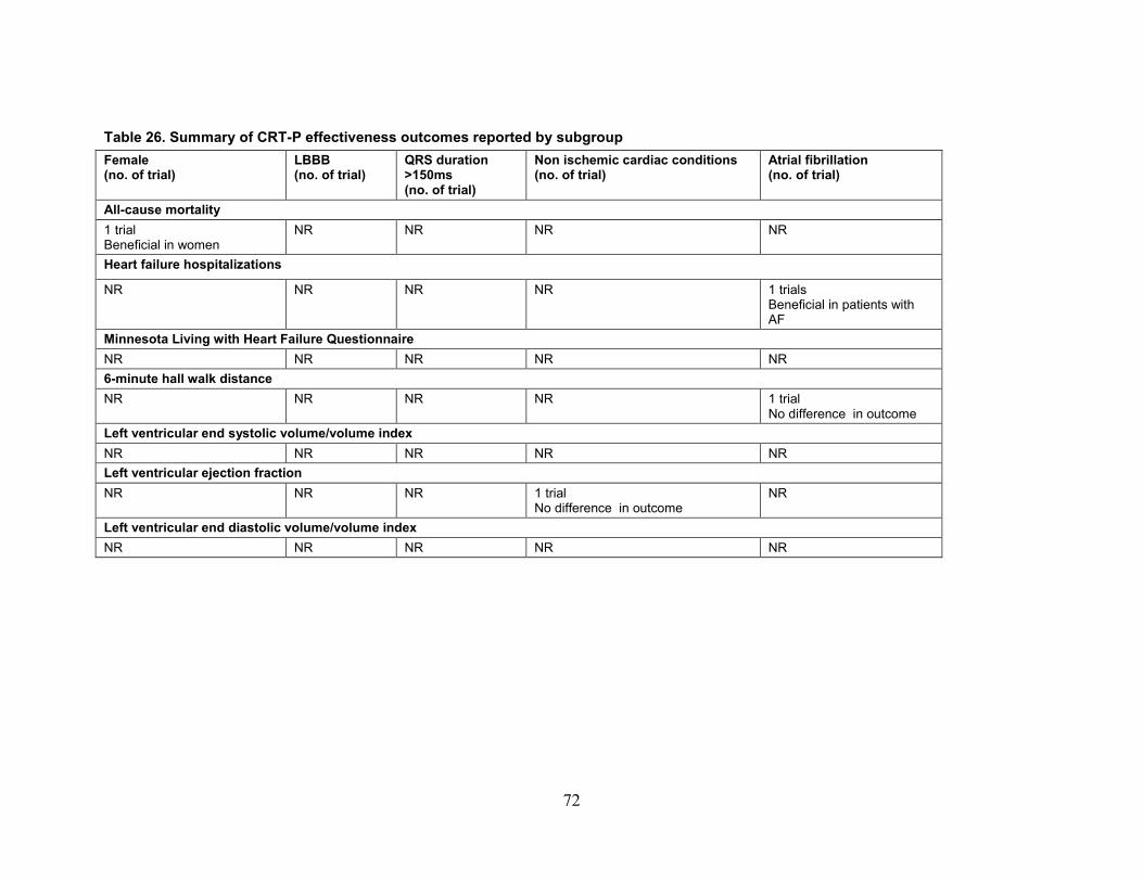
Table 26. Summary of CRT-P effectiveness outcomes reported by subgroup Female (no. of trial)
LBBB (no. of trial)
QRS duration >150ms (no. of trial)
Non ischemic cardiac conditions (no. of trial)
Atrial fibrillation (no. of trial)
All-cause mortality 1 trial Beneficial in women
NR NR NR NR
Heart failure hospitalizations
NR
NR
NR NR
1 trials Beneficial in patients with AF
Minnesota Living with Heart Failure Questionnaire NR NR NR NR NR 6-minute hall walk distance NR NR NR NR 1 trial
No difference in outcome Left ventricular end systolic volume/volume index NR NR NR NR NR Left ventricular ejection fraction NR NR
NR 1 trial
No difference in outcome NR
Left ventricular end diastolic volume/volume index NR NR NR NR NR
72

Table 27. Strength of evidence for key effectiveness outcomes of CRT-P Key Outcomes No. Studies
(number of patients)
Study limitation Directness Consistency Precision Reporting bias
Strength of evidence Finding
All- cause mortality 6 (2,635) Low Direct Inconsistent Precise Undetected Moderate Studies showed statistically significant differences in mortality favoring CRT-P
Hospitalizations for heart failure
5 (1,666) Low Direct Consistent Precise Undetected High Studies showed fewer hospitalizations in the CRT-P group
Left ventricular end systolic volume (LVESV)
3 (1,236) Low Direct Consistent Precise Undetected Moderate CRT-P significantly reduced LESV compared with optimal medical therapy
Minnesota Living with Heart Failure Questionnaire (MLHFQ)
5 (2,445) Low Direct Inconsistent Precise NA Insufficient These studies assessed this outcome at different endpoints and with different comparisons, which might explain the inconsistency in the results comparing CRT-P to other therapy
CRT-P=cardiac resynchronization therapy with pacemaker; 6MHWD=6-minute hall walk distance; NA=not applicable
73

Harms of Cardiac Resynchronization Therapy with Pacemaker (CRT-P)
Study Characteristics Eleven studies (reported in 13 articles) assessed harms associated with CRT-P.4 ,5 ,8 ,24 ,26 ,53 ,58
,77-82 Five were RCTs 4 ,5 ,24 ,26 ,53 ,58, three were secondary analyses of CARE-HF trial5 ,24 ,26 one was a crossover study,8 and the rest were prospective cohort studies. Followup ranged from 185 days to 36 months.
The studies used various devices. Three studies reported the use of only a single type of CRT-P device.58 ,77 ,79 Three studies used the InSync model 8040,77 ,78 ,81 three studies used the InSync III,58 ,79 ,81 one study used the InSync 7272,78 and four studies used other devices.78 ,80-82 Two studies did not report the device type they used.5 ,53
Two studies explicitly reported funding from industry.5 ,77 One study had non-profit organization funding.78 The other studies did not report their sources of support (Evidence Table 1).
Participant Characteristics The number of participants in the trials at baseline ranged from seven to 813. The percentage
of women among participants ranged from 580 to 28.6 percent.82 One study did not report the mean age.5 Mean age in other studies ranged from 53 to 68 years old. No studies assessing harms reported the racial makeup of their participants.
The proportion of patients with ICM ranged from 36 to 48 percent. Three studies did not report on the proportion of ICM.58 ,81 ,82 Two studies reported the prevalence of AF among their participants, which ranged from 6 to 33 percent78 ,80 Two studies reported the NYHA class of the participants78 ,79 and included participants in all NYHA classes.
Three studies did not report either the mean or the median LVEF.53 ,81 ,82 Of those studies reporting this characteristic, the mean LVEF ranged from 19 to 30 percent.
In general, these studies were heterogeneous in patient population, and frequently did not report proportions of female patients or racial categories of participants (Evidence Table 3).
Risk of Bias There were limitations in the reporting of harms in the studies. The studies did not report at
what time point the harms were assessed, making it impossible to calculate an incidence for these harms. In addition, the studies did not report confidence intervals for the proportions of patients with these harms. For these reasons (implying statistical imprecision), as well as other issues reflecting possible bias (including lack of clarity regarding outcome reporting and outcome assessment) the risk of bias was generally high (Table 28 and 29) (Figure 13 and 14).
74

Table 28. Summary of risk of bias for trials assessing harms of CRT-P Author, year Random
sequence generation
Allocation concealment
Blinding of personnel
Blinding of outcome assessors
Assessing blinding by outcome
Incomplete outcome data
Selective outcome reporting
Other sources of bias
Overall quality
COMPANION Bristow,20044 ? ? + - - - - + + CARE HF Cleland,200924 Gras,200726 Cleland,20045
-
? ? ? ? - + ? +
MUSTIC TRIAL Cazeau,20018 ? ? - ? ? ? ? - + Other trials Pokushalov,201058
? ? ? ? ? - - - +
Garikipati,201453
- ? + - - ? ? ? -
+High -Low ?Unclear
75

Figure 13. Summary of risk of bias for trials assessing harms of CRT-P
76
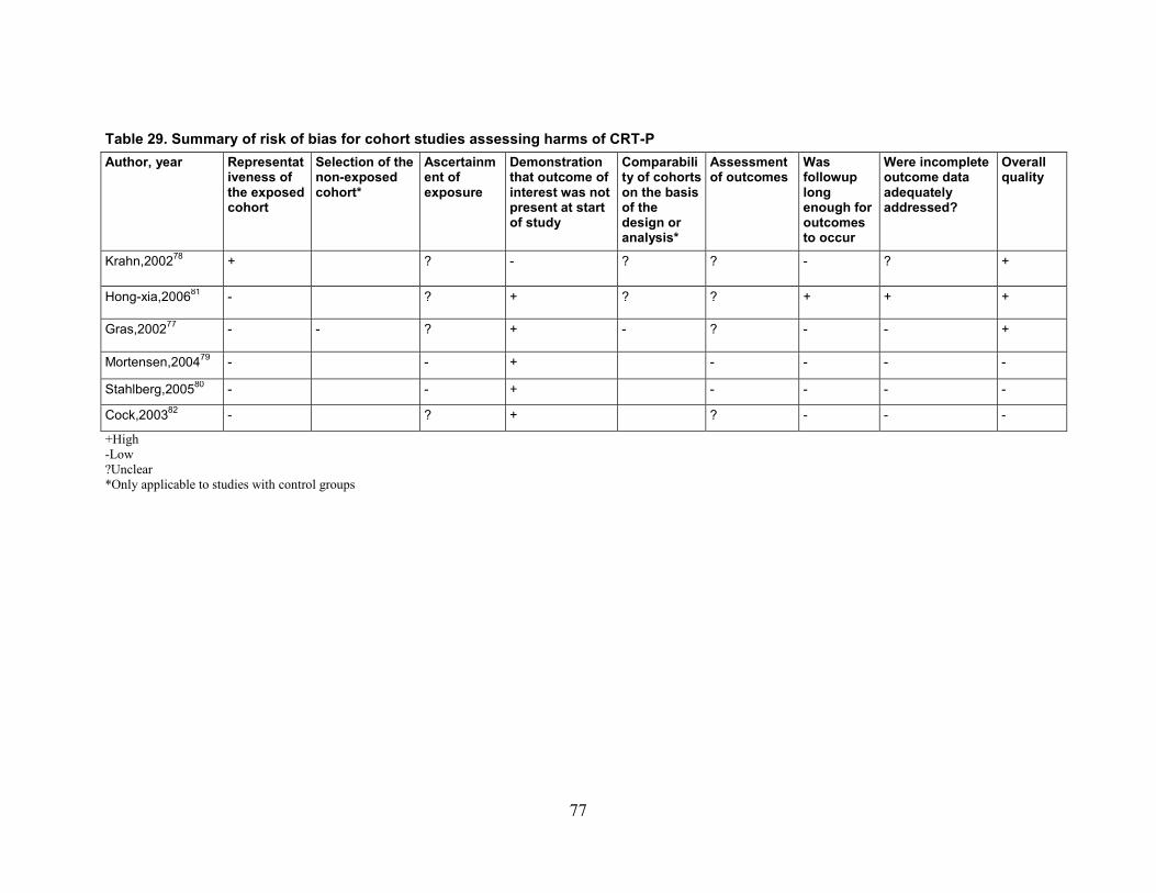
Table 29. Summary of risk of bias for cohort studies assessing harms of CRT-P Author, year Representat
iveness of the exposed cohort
Selection of the non-exposed cohort*
Ascertainment of exposure
Demonstration that outcome of interest was not present at start of study
Comparability of cohorts on the basis of the design or analysis*
Assessment of outcomes
Was followup long enough for outcomes to occur
Were incomplete outcome data adequately addressed?
Overall quality
Krahn,200278 + ? - ? ? - ? +
Hong-xia,200681 - ? + ? ? + + +
Gras,200277 - - ? + - ? - - +
Mortensen,200479 - - + - - - -
Stahlberg,200580 - - + - - - -
Cock,200382 - ? + ? - - -
+High -Low ?Unclear *Only applicable to studies with control groups
77
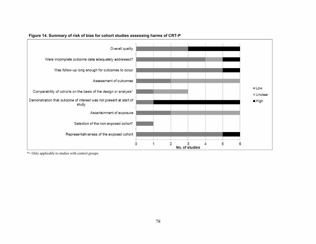
Figure 14. Summary of risk of bias for cohort studies assessing harms of CRT-P
*= Only applicable to studies with control groups.
78

Table 30. List of harms reported in the studies assessing harms of CRT-P
RCT=randomized controlled trail
Author, year Study design Procedure related complications
Length of hospital stay
Pneumothorax Pocket hematoma
Device Infection
Cardiac perforation/ tamponade
Lead dislodge-ment
Death within a week
COMPANION Bristow, 2004,4 RCT X
CARE-HF Cleland, 20045 RCT X X X Cleland, 2009 24 RCT X
Gras, 200726 RCT X X X X
MUSTIC Cazeau,2001 8 Randomized
crossover X
Other trials Garikipati, 201453 RCT X X X X Pokushalov ,201058
RCT X
Cock, 200382 Prospective cohort
X
Gras, 200277 Prospective cohort
X X X
Hong-xia, 200681 Prospective cohort
X
Krahn, 200278 Prospective cohort
X X X
Mortensen, 200479 Prospective cohort
X X
Stahlberg, 200580 Prospective cohort
X
79

Harms Outcomes Procedure-related Complications
Two studies reported miscellaneous procedure-related complications, both reporting a 33.3 percent proportion of patients with this outcome (Table 31). The small sample sizes and the small number of studies make it difficult to draw a conclusion other than that more data are needed. Table 31. Characteristics of studies of CRT-P reporting on the procedure-related complications
Author, year N for analysis N of patients with outcomes % of patients with outcomes
Garikipati, 201453 21 4 33.3
Krahn, 200278 45 15 33.3 Length of Hospital Stay
Garikipati et al. (2014) reported that among 21 patients, the patients undergoing transvenous placement of the CRT-P (N=12) had a shorter hospital stay than those in the epicardial arm (N=9), though the difference was not statistically significant (3.4 +/-2.6 vs. 5.4 +/- 4.6 days, p=0.22).53
Cleland et al. (2009) reported from the CARE-HF trial that as a result of the implantation procedure, patients receiving CRT-P initially spent more days in the hospital by 3 months followup (mean 7.5 days, median 4, IQR 2-8), versus 3.4 days (median 0, IQR 0-1).24 Afterwards, patients with CRT-P spent fewer days in the hospital (384 in the control group versus 222 in the CRT-P group). The overall number of days spent in the hospital per patient was similar in the CRT-P and control groups (20.7, median 9, IQR 4-26, compared to 22.4, median 9, IQR 0-31, respectively).
These two studies indicate that length of hospital stay might not be significantly different in those receiving CRT-P. However, as with other harms, few studies, with small sample sizes, address this harm.
Pneumothorax The CARE-HF trial (reported in two articles) assessed pneumothorax.5 ,26 At 24 hours, the
proportion of pneumothorax was higher in the medical therapy group than in the CRT-P group. At 18 months, only the proportion on the CRT-P group was reported (Table 32).5 As is the case with other harms, it is difficult to draw a conclusion based on limited data.
Table 32. Characteristics of studies of CRT-P reporting on the incidence of pneumothorax Author, year Arm name N for
analysis Time Point (s) N of patients
with outcomes % of patients with outcomes
Cleland, 20045 CRT-P 409 18 month 6 1.5
Gras, 200726 CRT-P 404 24 hrs. 2 0.5
CRT-P=cardiac resynchronization therapy with pacemaker
80
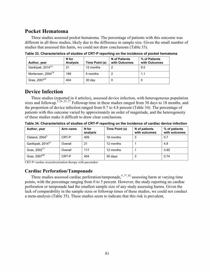
Pocket Hematoma Three studies assessed pocket hematoma. The percentage of patients with this outcome was
different in all three studies, likely due to the difference in sample size. Given the small number of studies that assessed this harm, we could not draw conclusions (Table 33). Table 33. Characteristics of studies of CRT-P reporting on the incidence of pocket hematoma
Author, year N for Analysis Time Point (s)
N of Patients with Outcomes
% of Patients with Outcomes
Garikipati, 201453 21 12 months 2 9.5
Mortensen, 200479 189 6 months 2 1.1
Gras, 200726 404 30 day 0 0
Device Infection Three studies (reported in 4 articles), assessed device infection, with heterogeneous population
sizes and followup.5 ,26 ,53 ,77 Followup time in these studies ranged from 30 days to 18 months, and the proportion of device infection ranged from 0.7 to 4.8 percent (Table 34). The percentage of patients with this outcome varied by approximately an order of magnitude, and the heterogeneity of these studies make it difficult to draw clear conclusions. Table 34. Characteristics of studies of CRT-P reporting on the incidence of cardiac device infection
Author, year Arm name N for analysis
Time Point (s) N of patients with outcomes
% of patients with outcomes
Cleland, 20045 CRT-P 409 18 months 3 0.7
Garikipati, 201453 Overall 21 12 months 1 4.8
Gras, 200277 Overall 117 12 months 1 0.85
Gras, 200726 CRT-P 404 30 days 3 0.74
CRT-P=cardiac resynchronization therapy with pacemaker
Cardiac Perforation/Tamponade Three studies assessed cardiac perforation/tamponade,4 ,77 ,82 assessing harm at varying time
points, with the percentage ranging from 0 to 5 percent. However, the study reporting no cardiac perforation or tamponade had the smallest sample size of any study assessing harms. Given the lack of comparability in the sample sizes or followup times of these studies, we could not conduct a meta-analysis (Table 35). These studies seem to indicate that this risk is prevalent,
81

Table 35. Characteristics of studies of CRT-P reporting on the incidence of cardiac perforation/tamponade
Author, year N for analysis Time Point (s) N of patients with outcomes
% of patients with outcomes
Bristow, 2004,4 617 During procedure
NR 1.6
Cock, 200382 7 2-3 months 0 0
Gras, 200277 117 During procedure
1 0.85
Lead Dislodgement Seven studies assessed lead dislodgement5 ,26 ,77-81. The proportion of patients experiencing
this harm ranged from 1.71 to 17 percent for those studies that assessed the proportion over the entire population, which comprised all except one study.5 Two studies reported lead dislodgement rates for only part of their study population. Cleland et al. (2004)5 reported the proportion of patients with lead dislodgement only in the CRT-P arm as 5.9 percent; and Gras et al. (2007) reported a proportion of patients with lead dislodgement in the CRT-P arm as 2.7 percent (Table 36).26 It is difficult to interpret these results since the studies did not report the recorded time point of the dislodgement, and the studies followed their populations for different lengths of time. Table 36. Characteristics of studies of CRT-P reporting on the incidence of lead dislodgement
Author, year Arm N for analysis
N of patients with outcomes
% of patients with outcomes
Cleland, 20045
CRT-P 409 24 5.9
Gras, 200277 Overall 117 16 13.7
Gras, 200726
CRT-P 404 11 2.7
Hong-xia, 200681
Overall 117 2 1.71
Krahn, 200278 Overall 45 3 6.7
Mortensen, 200479
Overall 189 12 6.4
Stahlberg, 200580
Overall 35 6 17
CRT-P=cardiac resynchronization therapy with pacemaker
Death within One Week Three studies assessed death within a week.8 ,58 ,78 Krahn et al. (2002) found one death within
1 week, due to sequelae from stroke, among 45 patients, for a prevalence of 2.2 percent.78 Pokushalov et al. (2010) assessed the harm in two arms, one of patients undergoing CABG (4 of 87 patients, or 4.6%), at 4 days, the other in patients undergoing CABG and CRT-P (1 of 91 patients, or 1.1%), at 4 days.58 In the MUSTIC trial using a crossover design, one patient died from myocardial infarction a few hours after a premature switch from inactive to active pacing; another patient died suddenly two hours after switching from inactive to active pacing (Table 37).8 As for the other harms, the studies are heterogeneous in their assessment time points and in
82
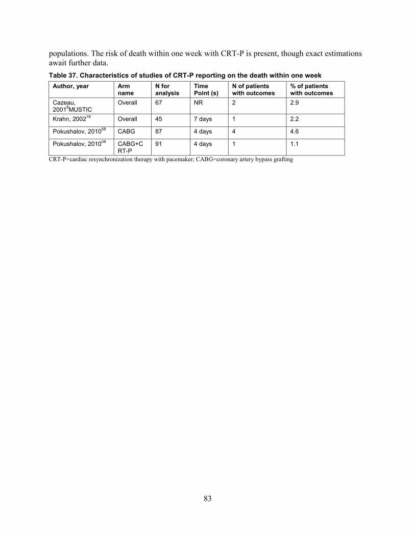
populations. The risk of death within one week with CRT-P is present, though exact estimations await further data. Table 37. Characteristics of studies of CRT-P reporting on the death within one week
Author, year Arm name
N for analysis
Time Point (s)
N of patients with outcomes
% of patients with outcomes
Cazeau, 20018MUSTIC
Overall 67 NR 2 2.9
Krahn, 200278 Overall 45 7 days 1 2.2
Pokushalov, 201058 CABG 87 4 days 4 4.6
Pokushalov, 201058 CABG+CRT-P
91 4 days 1 1.1
CRT-P=cardiac resynchronization therapy with pacemaker; CABG=coronary artery bypass grafting
83

Effectiveness of Cardiac Resynchronization Therapy with Pacemaker versus Defibrillator (CRT-P vs CRT-D) Table 38. Evidence addressing effectiveness and harms of CRT–P vs CRT-D
CRT–P vs CRT-D effectiveness CRT–P vs CRT-D Harms Number of included studies
1 trial (reported in 3 articles) 9 studies (reported in 11 articles) 2 were RCTs, 3 were prospective cohorts and four were retrospective cohorts.
Patient characteristics
• The percentage of women was between 33-31%
• The median age ranged from 66-68 years old
• The trial reported NYHA class of the participants
• The median LVEF ranged from 20-22%
• The percentage of women in the studies ranged from 16-44%
• The mean age ranged from 58-74 years old • Four studies reported the NYHA class of the
participants. • The mean LVEF ranged from 20-31%
Outcomes (number of included studies)
• All- cause mortality: 1 trial ( reported 2 articles)
• Hospitalizations for heart failure: 1trial
• Left ventricular end systolic • volume/volume index : 0 • Minnesota Living with Heart Failure
Questionnaire: 1 trial • Left ventricular ejection fraction: 0
study • Left ventricular end diastolic
volume/volume index: 0 study • 6 minute hall walk distance: 1trial
• Procedure related complications: 2 studies • Length of hospital stay: 2 studies • Pneumothorax: 4 studies • Pocket hematoma: 2 studies • Device Infection: 5 studies • Cardiac perforation/ tamponade: 4 studies • Lead dislodgement: 4 studies • Ventricular arrhythmias: 1 study • Inappropriate shocks: 2 studies • Death (within a week): 2 studies
Key findings There was insufficient evidence to determine the effectiveness of CRT-D vs. CRT-P
Compared to CRT-P, device infection was slightly more common in patients receiving CRT-D. There was insufficient evidence to draw conclusions on any other harms comparing the two devices
84

Study Characteristics One RCT (reported in three articles) compared the effectiveness of CRT-P versus CRT-D.4 ,29
,30 The COMPANION trial reported initial results in 2004,4 with subsequent additional analyses for mortality30 and hospitalizations.29 The trial included 1,520 subjects with NYHA Class III or IV heart failure and ICM or dilated cardiomyopathy with a QRS duration of greater than 120ms, randomized in a 1:2:2 ratio to optimized pharmacological therapy alone, or in combination with CRT-P (Contak TR model 1241, Guidant) or CRT-D (Contak CD model 1823, Guidant). The planned length of followup was 12 months. The study was industry sponsored (Guidant) (Table 39). Participant Characteristics
The COMPANION trial included 308 participants in the optimal pharmacologic therapy (OPT) alone arm, 617 in the CRT-P plus OPT arm and 595 in the CRT-D plus OPT arm.4 The median age of participants in the trial arm ranged from 66-68 years old, and the majority of participants were male (67-69%). Most participants were NYHA class III (67-68%), had ICM (54-59%), and a median left ventricle ejection fraction ranging from 20-22 percent. Median QRS duration was 160ms in the CRT-P and CRT-D arms, and 158ms in the OPT arm. Over two-thirds of participants had a left bundle branch block (range 69-73%). None of the articles reported racial distribution, history of atrial disease, or glomerular filtration rates of the participants (Evidence Table 6). Table 39. Study characteristics of trials assessing effectiveness of CRT-P vs CRT-D
Author, year Number of patients
Length of followup
Device manufacturer name/ device model
Comparison Funding source
COMPANION Bristow,20044 Anand, 200929 Carson,200530
OMPT : 308 CRT-P: 617 CRT-D: 595
12 months CRT-P (Contak TR model 1241, Guidant) or CRT-D (Contak CD model 1823, Guidant)
OPT CRT-P CRT-D
Industry
85

Risk of bias The primary endpoint of the COMPANION trial was a composite of all-cause mortality and
hospitalization, with a secondary endpoint of all-cause mortality. Additional outcomes assessed included the 6-minute hall walk distance and MLHFQ. However, while the study masked the steering and endpoints committee to treatment assignment, it did not mask physicians, patients, and members of the data management and analysis team, raising concerns for potential bias. Similarly, the randomization technique and allocation concealment are unclear. Despite having both CRT-P and CRT-D arms, direct comparisons between the two arms were lacking (Table 40).
86

Table 40. Summary of risk of bias for trials assessing effectiveness of CRT-P vs CRT-D Author, year Random
sequence generation
Allocation Concealment
Blinding of personnel
Blinding of outcome assessors
Assessing blinding by outcome
Incomplete outcome data
Selective outcome reporting
Other sources of bias
Overall quality
COMPANION-Bristow,20044 Anand, 200929 Carson,200530
? ? + - - - - + +
+=High -=Low ?=Unclear
87

Effectiveness Outcomes The COMPANION trial did not report change in left ventricle ejection fraction, left ventricle
end systolic volume, left ventricle end diastolic volume, or clinical composite score (Packer score). The trial reported composite outcomes of hospitalization with death and separate outcomes by cardiovascular diagnoses. Subgroup analyses were presented for the hazards of all-cause mortality for CRT-D.
All-cause Mortality Two articles reported all-cause mortality from the COMPANION trial.4 ,30 Carson et al.
(2005) examined the time to cause-specific death, including sudden cardiac death and “pump failure” (progressive heart failure).30 The study did not specify followup time, but provided 3 years of data. Overall, 313 patients died, 78 percent from a cardiac cause, of which pump failure (44.4%) and sudden cardiac death (26.5%) were most common. Only CRT-D resulted in statistically significant fewer cardiac deaths (p=0.006). In regards to non-cardiac mortality, there was no significant difference between the treatment groups. The all-cause mortality for the OPT, CRT-P, and CRT-D arms were 25, 21.2, and 17.6 percent, respectively, suggesting the benefit in mortality is from CRT-D in attenuating death from cardiac causes. Compared to OPT, CRT-D reduced cardiac deaths by 38 percent (p=0.006) whereas CRT-P reduced cardiac deaths by only 14.5 percent (p=0.33).
Bristow et al. (2004) reported the secondary outcome from COMPANION, mortality at 12 months, classified according to cardiac and non-cardiac causes.4 The 1-year mortality rate in the OPT group was 19 percent. Compared to OPT, CRT-P resulted in a mortality reduction of 24 percent (HR: 0.76, 95% CI, 0.58 to 1.01, p=0.059) whereas CRT-D resulted in a 36 percent reduction (HR: 0.64, 95% CI, 0.48 to 0.86, p=0.003). For CRT-D, subgroup analyses for all-cause mortality showed that subjects with non-ischemic cardiomyopathy (NICM) had a greater reduction in mortality (HR: 0.50, 95% CI, 0.29 to 0.88; p=0.015). The study reported no significant reduction in mortality for CRT-D for participants with ICM (HR: 0.73, 95% CI, 0.52 to 1.04, p=0.082). The trial found a reverse trend for CRT-P. Compared to OPT, subjects with NICM had a 9 percent reduction in mortality with CRT-P (HR: 0.91, 95% CI, 0.55 to 1.49, p=0.70) in contrast to 28 percent for those with ICM (HR: 0.72, 95% CI, 0.51 to 1.01, p=0.058). The study, however, reported no direct comparisons of CRT-P versus CRT-D.
In conclusion, the trial reported that CRT-D significantly decreased all-cause mortality by 36 percent (p=0.003) (likely driven by cardiac causes). The reduction in mortality by CRT-P was more moderate (24%; p=0.059). Primarily due to high risk of bias, and lack of direct comparisons, there is insufficient strength of evidence for this outcome (Table 42). Interestingly, subgroup analyses suggest that those with NICM benefit more with CRT-D whereas those with ICM benefit more from CRT-P.
Hospitalization for Heart Failure One article reported the impact of CRT-P and CRT-D on hospitalization from the
COMPANION trial.29 Median followup ranged from 11.9 to 16.2 months. Overall, of the 1,520 patients, 959 were hospitalized at least once. Of the total 2,428 hospitalizations, 1,596 (66%) were for cardiac causes. OPT, CRT-P, and CRT-D arms accounted for 388, 628, and 580 cardiac hospitalizations respectively. CRT therapy resulted in a lower number of cardiac hospital admissions per patient-year: 1.2 for OPT, 0.8 for CRT-P, 0.8 for CRT-D. Also, almost half as many subjects with CRT had greater than two cardiac hospital admissions per patient-year, 27
88

percent for the OPT arm versus 16 percent for the CRT-P and CRT-D arms. Hospital admissions specific to heart failure were also higher in the OPT arm (46%) than either the CRT-P (33%) or CRT-D (36%) arms. The study found a 44 percent and 41 percent reduction in heart failure hospital admissions per patient-year for CRT-P and CRT-D, respectively, compared to the OPT arm (OPT: 0.7 admissions per patient-year; CRT-P: 0.4; CRT-D: 0.4, no p-value specified).
In summary, the study found that when compared with OPT, CRT-P and CRT-D resulted in a 44 and 41 percent reduction, respectively, in heart failure hospitalizations. No statistically significant differences were found when CRT-P was directly compared to CRT-D for the hospitalization endpoints. We graded this finding as low strength of evidence primarily due to the high risk of bias (Table 42).
Left Ventricular End Systolic Volume and Left Ventricular End Diastolic Volume
The COMPANION trial did not report on change in left ventricular end systolic volume and left ventricular end diastolic volume. There is insufficient evidence to draw conclusions about CRT-D versus CRT-P for this outcome.
Minnesota Living with Heart Failure Questionnaire One article reported the change MLHFQ score for CRT-P and CRT-D at 3 and 6 months with
the OPT arm as reference.4 For the OPT arm, the MLHFQ median score decreased by -9±21 and -12±23 at 3 and 6 months respectively. In contrast, compared to OPT, the MLHFQ score decreased by over 2-fold for both the CRT-P and CRT-D arms: -24±27 and -25±26 versus -24±28 and -26±28 at 3 and 6 months for CRT-P versus CRT-D arms respectively (p <0.001). The study made no direct comparison between the CRT-P versus CRT-D groups, but the results appear similar. Because of this indirect comparison of CRT-P and CRT-D and the high risk of bias, there is insufficient evidence to draw conclusions about CRT-D versus CRT-P for MLHFQ (Table 42).
Change in Left Ventricular Ejection Fraction The COMPANION trial did not report on change in left ventricular ejection fraction.
6-minute Hall Walk Distance One article reported 6-minute hall walk distance outcome for the COMPANION trial.4 At
baseline, there was no statistically significant difference in median distance walked between the OPT, CRT-P, and CRT-D arms (244m, 274m, 258m, respectively). The outcome was assessed at 3 and 6 months post-intervention with the OPT arm as reference. For the OPT arm, the distance walked increased by 9±84m and 1±93m at 3 and 6 months respectively. In contrast, compared to OPT, the median distance walked significantly increased: 33±99m and 40±96m versus 44±109m and 46±98m at 3 and 6 months for CRT-P versus CRT-D (p<0.001). The study made no direct comparison between the CRT-P versus CRT-D arms, but the results appear similar.
89

Table 41. Summary of effectiveness outcomes reported in the trial of CRT-P versus CRT-D, by subgroup Female LBBB QRS duration
>150ms Non ischemic cardiac conditions
Atrial fibrillation
No Sub group analysis done LBBB-Left bundle branch block Table 42. Strength of evidence for key effectiveness outcomes of CRT-P vs CRT-D
Key outcomes
No. Studies (number of patients)
Risk of bias
Directness Consistency Precision Reporting bias
Strength of evidence Finding
All- cause mortality
1 (1520) High Indirect Unknown (Single study)
Precise NA Insufficient
Hospitalizations for heart failure
1 (1,520) High Direct Unknown (Single study)
Precise Undetected Low Compared with optimal medical therapy, CRT-P and CRT-D were associated with 44% and 41% reduction in heart failure hospitalizations (not significantly different).
Left ventricular end systolic volume
0 NA NA NA NA NA Insufficient
Minnesota Living with Heart Failure Score
1 (1,520) High Direct Unknown (Single study)
Imprecise NA Insufficient
NA = not applicable
90

Harms of Cardiac Resynchronization Therapy with Pacemaker versus Defibrillator (CRT-P vs CRT-D)
Study Characteristics Nine studies (reported in eleven articles) assessed harms comparing CRT-P with CRT-D,
including two RCTs.4 ,29 ,30 ,46 ,83-89 The Management of Atrial Fibrillation Suppression in Atrial Fibrillation-Heart Failure Comorbidity Therapy (MASCOT) trial was a multicenter, single-blinded, randomized parallel trial that examined the safety and efficacy of an atrial overdrive pacing algorithm in CRT patients.46 However, treating clinicians determined the selection of CRT-P versus CRT-D and the harms assessment was a post-hoc analysis (Evidence Table 1).
The second RCT, COMPANION, was a single-blinded trial that assigned patients in a 1:2:2 ratio to treatment with protocol-mandated optimal pharmacologic therapy alone, optimal pharmacologic therapy plus CRT-P, or optimal pharmacologic therapy plus CRT-D. 4 ,29 ,30The primary outcome was a composite of all-cause mortality and all-cause hospitalization, and the secondary endpoint was all-cause mortality. The other included studies were cohort studies.
Of the non-randomized studies, three were prospective84 ,87 ,89 and four were retrospective.83
,85 ,86 ,88 Followup ranged from approximately 6 months84 to 5 years.86 Only three studies specified the device names.4 ,29 ,30
Three studies (reported in five articles) reported funding.4 ,29 ,30 ,46 ,88 The two RCTs were industry funded.4 ,29 ,30 ,46 Two studies explicitly specified no funding.85 ,87
Population Characteristics The number of participants in the studies ranged from 4084 to 26,887.88 The percentage of
women among participants in the study arms ranged from 16 85 to 44 percent.85 Only one study reported the racial makeup of its participants.88 Mean age in the study arms ranged from 58 to 74 years old.
Seven studies (reported in nine articles) reported the percentage of participants with ICM,4 ,29
,30 ,84-89 ranging from 1984 to 70 percent86 per study arm. All but two studies reported mean ejection fraction.85 ,88 ranging from 20 to 31 percent.87 All but two studies reported history of atrial fibrillation,4 ,29 ,30 ,88 ranging from 11 to 41 percent per study arm.85
Mean QRS duration ranged from 147ms to 185ms but four studies did not report it.83-85 ,88 Only two studies specified QRS morphology, with the predominance being left bundle branch block, ranging from 69 to 84 percent per arm. Only four studies (reported in six articles) specified NYHA classification,4 ,29 ,30 ,46 ,83 ,87 with the majority of participants being Class III, ranging from 54 to 87 percent. Four studies reported baseline renal function.85-87 ,89 Two studies reported eGFR,85 ,87 with a mean range of 52 to 72ml/min/1.73m2 per arm.87 Two studies reported creatinine, ranging from a median of 1.2 mg/dL89 to a mean level of 1.6mg/dL.86
No studies reported baseline left atrial volume. However, four studies reported left atrial diameter,46 ,85-87 ranging from a mean of 4.2cm85 ,87 to 6.2cm.86 Only one study reported baseline left ventricular end diastolic volume, the mean volume ranging from 236ml to 251ml per arm.84 Five additional studies reported baseline left ventricular end diastolic diameter,4 ,46 ,85-87 ranging from a mean diameter of 6.1cm to 7.1cm. Only one study reported body mass index, ranging from 27 kg/m2 to 28kg/m2 in the study arms (Evidence Table 7).85
91

Risk of Bias The COMPANION trial had high risk of bias as it did not mask patients, physicians,
independent statisticians, and members of the data-management group and the data safety and monitoring board to the treatment assignments (although the steering committee, the end-points committee, and the sponsor were unaware of the treatment assignments). Similarly, a very high percentage (26%) of participants changed from medical therapy to receive implants. Random sequence generation and allocation concealment for the trial were also unclear. Finally, reporting bias is suggested, as articles rarely reported direct comparisons of CRT-P versus CRT-D. The MASCOT trial was also at high risk of bias as device type was not randomized but determined by the treating clinicians. The harms assessment in this trial was a post-hoc analysis. Risk of bias was introduced in this analysis, as the randomization no longer preserved the distribution of measured and unmeasured confounders. The authors noted that compared to CRT-P participants, CRT-D recipients were more likely to be male (p <0.0001), have ICM (p <0.001) and shorter QRS duration (p <0.0005), and less likely to receive spironolactone (p <0.0001) and anti-arrhythmic medications (p <0.0222). The study made no adjustment for these factors.
With respect to the cohort studies83-89, the main concerns for bias included unclear description of the cohort,83 self-reported outcomes in the main study,87 and no standardized followup time.88 However, ascertainment of exposure, pre-specified outcomes, and followup were adequate. Four of the seven studies had low risk of bias (Table 43 and 44) (Figure 15).
92
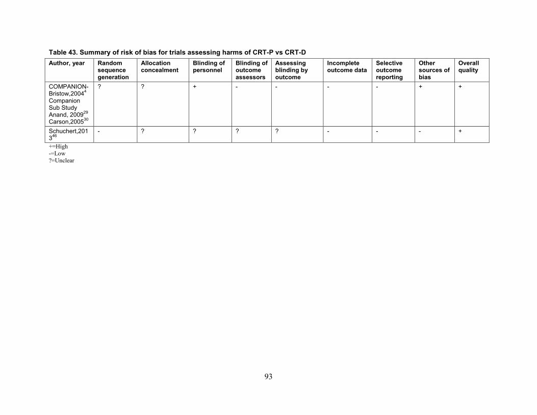
Table 43. Summary of risk of bias for trials assessing harms of CRT-P vs CRT-D Author, year Random
sequence generation
Allocation concealment
Blinding of personnel
Blinding of outcome assessors
Assessing blinding by outcome
Incomplete outcome data
Selective outcome reporting
Other sources of bias
Overall quality
COMPANION-Bristow,20044 Companion Sub Study Anand, 200929 Carson,200530
? ? + - - - - + +
Schuchert,201346
- ? ? ? ? - - - +
+=High -=Low ?=Unclear
93
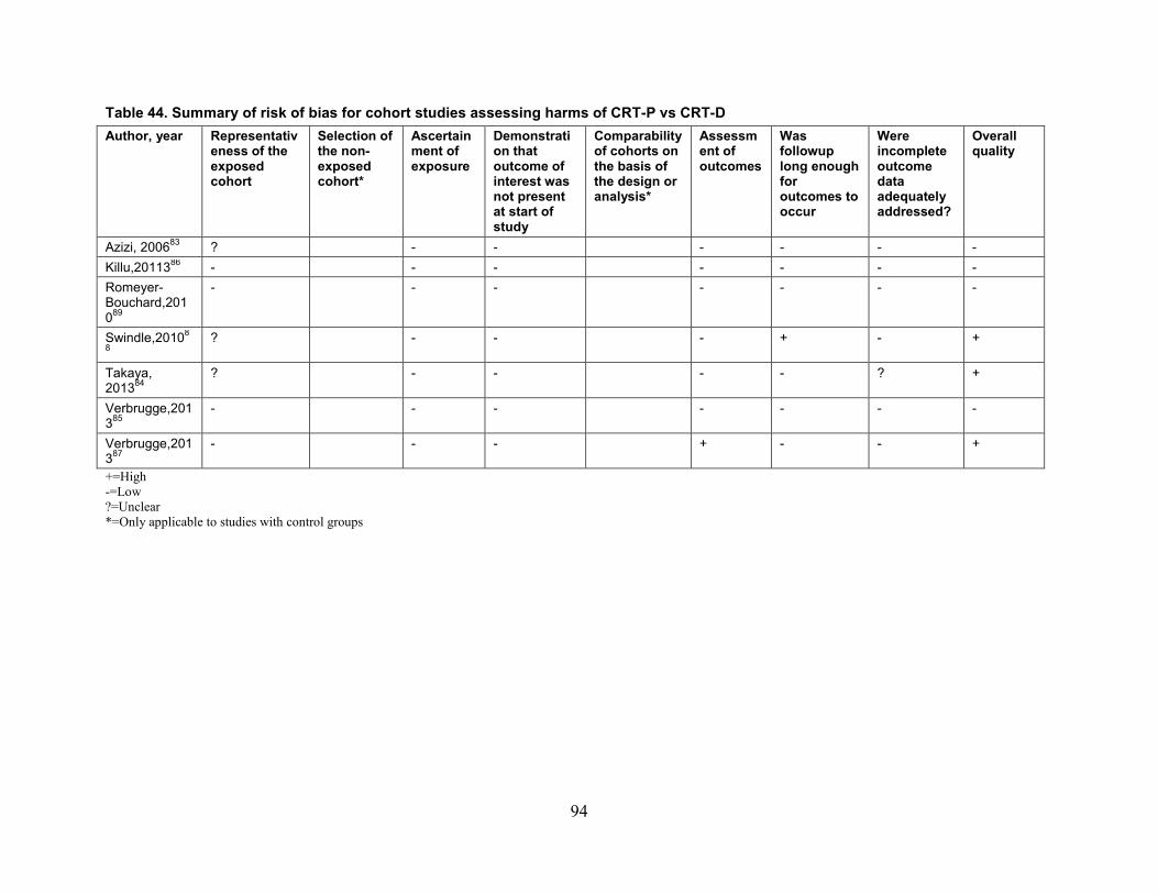
Table 44. Summary of risk of bias for cohort studies assessing harms of CRT-P vs CRT-D Author, year Representativ
eness of the exposed cohort
Selection of the non-exposed cohort*
Ascertainment of exposure
Demonstration that outcome of interest was not present at start of study
Comparability of cohorts on the basis of the design or analysis*
Assessment of outcomes
Was followup long enough for outcomes to occur
Were incomplete outcome data adequately addressed?
Overall quality
Azizi, 200683 ? - - - - - - Killu,2011386 - - - - - - - Romeyer-Bouchard,201089
- - - - - - -
Swindle,20108
8 ? - - - + - +
Takaya, 201384
? - - - - ? +
Verbrugge,201385
- - - - - - -
Verbrugge,201387
- - - + - - +
+=High -=Low ?=Unclear *=Only applicable to studies with control groups
94

Figure 15. Summary of risk of bias for cohort studies assessing harms of CRT-P vs CRT-D
*= Only applicable to studies with control groups
95

Table 45. List of harms reported in the studies assessing harms of CRT-P vs CRT-D Author, year
Procedure related complications
Length of hospital stay
Pneumothorax
Pocket hematoma
Cardiac perforation/ tamponade
Device infection
Lead dislodgement
Death within 1 week
Ventricular arrhythmia
Inappropriate shocks (CRT-D Only)
RCTs
COMPANION Anand,200929
X1
MASCOT Schuchert,201046*
X
Bristow, 20044 X X Carson,200530 X5 Prospective Cohorts
Romeyer-Boucherd, 200789
X X X X
Takaya, 201084 X Verbrugge, 201187 X X X X2 X Verbrugge, 201185 X Retrospective Studies Azizi, 200583 X X X X X X Killu, 200886 X X X X X X4 X Swindle, 200588 X X X3
*Post-hoc analysis 1. Index hospitalization of the device implantation was excluded from length of stay analyses for the COMPANION Trial. 2. Device infections were classified as “long term complications.” 3. Mortality was reported as in-hospital mortality for patients undergoing device procedures. 4. 30-Day Mortality was reported. 5. Exact timing of death was not specified.
CRT-D=cardiac resynchronization therapy with defibrillator
96
Harms Outcomes Harms most commonly reported were pneumothorax83 ,86 ,87 ,89 and device infections.46 ,83 ,86
,87 ,89 Studies rarely reported pocket hematoma, ventricular arrhythmia, or inappropriate shocks (Table 45). For most adverse events, the studies did not report the exact timing and did not specify the differential experience amongst CRT-P and CRT-D participants. Instead, the studies often reported events for the entire CRT cohort. Procedure-related Complications
Two studies reported procedure-related complications.4 ,88 Swindle et al. (2005) utilized a definition of complications which included, but was not limited to, pneumothorax, cardiac perforation with pericardial effusion or tamponade, mechanical complications of the device, implant infection, hemorrhage, and acute renal failure requiring dialysis.88
For CRT-P compared to CRT-D, 94.2 versus 95.0 percent of participants had no complications, 5.2 versus 4.6 percent had one complication, and 0.6 versus 0.4 percent had greater than one complication.
In comparing CRT-P, CRT-D, and ICD, the study found no difference in frequency of complications by device type (p=0.29), although it was lower for younger patients, under the age of 80 years old (p=0.03). The second study, COMPANION, reported 10 percent of CRT-P and 8 percent of CRT-D patients experienced moderate or severe adverse events related to the implantation procedure.4 There was no statistically significant difference in complications between CRT-P and CRT-D (p=0.42), consistent with the first study.
Length of Hospital Stay Two studies reported the length of hospital stay,87 ,88 finding that the presence of a device
complication or an elevated comorbidity score resulted in increased length of stay and total cost of hospitalization.88
Swindle et al. (2005) showed that advanced age was associated with increased length of stay and total cost of hospitalization, but this was only consistent among patients undergoing a CRT-D procedure. The study found an association between elective admission coding or by implantation of the device on day 1 or 2, and shorter lengths of stay (median 2.0 vs. 6.0 days) and lower mortality (by 80.0%) (The study defined elective procedures as those that used the Uniform/Universal Billing Form 92 Managed Care, the official Centers for Medicare and Medicaid Services form used by hospitals when submitting bills for reimbursement).
The study also compared the length of stay within the three device groups (CRT-P, CRT-D, and ICD) across the number of device complications and participant comorbidity scores, but made no direct comparisons between the CRT-P and CRT-D groups. Verbrugge et al. (2011) reported an overall median length of stay of 3 days (IQR: 3, 5) for CRT for all age groups but did not specify length of stay by CRT type 87.
The COMPANION trial excluded initial implantation and elective implantation of CRT-P and CRT-D devices from its hospitalization and length of stay analyses.29
No studies provided direct comparison of CRT-P and CRT-D for length of hospital stay for the initial device implantation.
97
Pneumothorax Four studies reported on pneumothorax.83 ,86 ,87 ,89 These studies reported a pneumothorax
prevalence of 0.3, 0.6, 1, and 1.4 percent, respectively.83 ,86 ,87 ,89 One study also noted no difference by age, comparing participants age ≤80 (1.3%) versus >80 years old (2.2%) (p=0.36).86 No study directly compared pneumothorax by CRT type.
Pocket Hematoma Two studies reported on pocket hematoma,86 ,89 each with a clear definition of the event. Killu et al. (2008) defined a pocket hematoma as “clotted blood in the device pocket” with a
severe classification when it “resulted in refractory pain, threatened the integrity of the incision, or required pocket evacuation or transfusion”.86 Among those participants age ≤80 versus >80 years old, 0.5 and 1.1 percent, respectively, experienced a hematoma requiring intervention (p=0.41). The study did not report CRT type.86
The other study, by Romeyer-Bouchard et al. (2007),89 defined a pocket hematoma as requiring two investigators to agree on a “palpable mass that protruded 2cm anterior to the pulse generator and lead (s)”.89 Twenty-nine patients (9.5%) experienced a large hematoma, with five (1.66%) requiring re-intervention due to the size of the hematoma. The presence of hematoma was significantly correlated with re-intervention (r=0.2; p<0.001). As in the prior study by Killu et al. (2008) this study did not report the CRT type, although it did suggest larger pockets from the larger leads, connectors, and size of the CRT-D devices as a predisposing factor for hematoma.89
Device Infection Five studies reported on device infection.46 ,83 ,86 ,87 ,89. Killu et al. (2008) reported a prevalence of 0.3 percent, with no statistically significant
difference among participants above versus below the age of 80 years (p >0.99).86 Verbrugge et al. (2011) report a prevalence of 0.5 percent secondary to infection.87 Azizi et al. (2005) reported infection and prevalence in only two of 244 participants (0.8%).83 However, none of these three studies distinguished infection by CRT type or provide specific definitions of or timing criteria for infections.
In contrast, Schuchert et al. (2010) did report a CRT-specific rate of infection: 1.7 percent for CRT-P and 2.1 percent for CRT-D (p=0.88).46 However, only one study clearly defined infection and adjusted for device type.89 Romeyer-Bouchard et al. (2007) defined device-related infection (DRI) as “local signs of inflammation at the generator pocket (e.g. erythema, warmth, fluctuance, wound dehiscence, tenderness, purulent drainage, or frank erosion by generator or lead puncturing the skin)”.89 This was further categorized as “early”, “late”, or “delayed” when occurring within 30 days, after 30 but less than 365 days, and over 364 days, respectively. The overall prevalence of DRI was 4.3 percent (1.7 percent incidence per year; 7 early and 6 late infections of 303 participants with a mean followup of 31 months). With respect to devices, 1.6 percent of CRT-P and 8.6 percent of CRT-D participants experienced a DRI. Infections were predominantly among participants with CRT-D (77%), followed by CRT-P (15.4%) and a device upgrade (7.6%). After adjusting for procedure time, dialysis, and re-intervention, a CRT-D device had a hazard ratio of 10.45 (95% CI, 1.75 to 62.45, p=0.01) for a DRI compared to CRT-P. Procedure time, dialysis, re-intervention, and a CRT-D device were independent predictors of DRI. This study would suggest that CRT-D is associated with higher risk of device infections.
98
The authors point out that technical factor, such as larger leads, connectors, size, and pocket size may predispose to infection and stretch the skin relatively thinner. Similarly, lead materials and size may affect bacterial adhesion.
Cardiac Perforation/Tamponade Four studies reported on cardiac perforation (including coronary sinus perforation) or cardiac
tamponade.4 ,83 ,86 ,87 In the COMPANION trial, coronary venous perforation and tamponade were 1.1 and 0.5 percent respectively for the CRT-P and 0.8 and 0.3 percent respectively for the CRT-D groups (no p-value specified).4 Azizi et al. (2005) only reported one coronary sinus perforation for 244 participants (0.4%).83 Verbrugge et al. (2011) reported cardiac tamponade in only one case of 220 participants (0.5%), occurring in the 70-79 year old cohort.87 Killu et al. (2008) compared coronary sinus perforation among those participants age ≤80 (0.2%) versus >80 years old (0%) and found no significant difference (p >0.99) 86; however, all three studies did not report which CRT type experienced the event.83 86 ,87 No direct comparison of cardiac perforation/tamponade was made for CRT-P versus CRT-D.
Lead Dislodgement Four studies reported on lead dislodgement.83 ,86 ,87 ,89 However, only one study explicitly
defined lead dislodgement as: “a radiographic finding of lead dislocation”, “a significant increase in capture threshold or loss of capture”, “inadequate lead sensing necessitating a lead revision”, or a combination of these.86 Verbrugge et al. (2011) reported multiple reasons for lead replacement (5.4%), including dislocation, microperforation, and diaphragmatic stimulation.87 However, the study reported this complication by age category and not separated by device type. Similarly, Romeyer-Bouchard et al. (2007) reported dislodgement in 6.9 percent of cases but did not specify by device type.89 The study also correlated lead dislodgement with re-intervention (r =0.8; p<0.001).89 Azizi et al. (2005) reported seven of 244 cases with dislodged leads (2.9%)83 and Kilu et al. (2008) reported a lead revision rate of 7.1 percent,86 but neither study specified dislodgement by device type. Consequently, lead dislodgement could not be compared for CRT-P versus CRT-D. Interestingly, two studies found no difference in the prevalence of lead replacement with increasing age (age ≤80 vs. >80 years old; 7.3% vs. 5.6%, p=0.66)86 or dislodgment (age <70, 70-79 and ≥80 years old; 3% vs. 1% vs. 2% respectively; no p-value).87
Ventricular Arrhythmia Only one study reported on ventricular arrhythmia perioperatively.83 In the study by Azizi et
al. (2005) ventricular tachyarrhythmia’s requiring defibrillation occurred in four of 285 procedures (1.3%).83 However, the study did not specify the prevalence for CRT-P versus CRT-D procedures. No direct comparison of ventricular arrhythmia was made for CRT-P versus CRT-D.
Inappropriate Shocks Cardiac Resynchronization Therapy with Defibrillator Only
Two studies reported inappropriate shocks.85 ,86 Killu et al. (2008) reported 31 of 728 (4.3%) participants received inappropriate shocks over the duration of the study (median 3.1 years).86 Younger participants (age ≤80 years old) received a higher percentage of inappropriate therapy (5.1%) as compared to those over the age of 80 years old (1.4%) (no p-value specified).
99
However, time to first inappropriate shock did not significantly vary by age group (p=0.21). Verbrugge et al. (2011) reported three inappropriate shocks in their study but did not specify among how many participants the events occurred.85 The variation in followup and reporting of number of inappropriate shocks versus number of participants with inappropriate shocks limits the interpretation of this data.
Death Within a Week Two studies reported death within 1 week.83 ,84 In the study by Takaya et al. (2010) no events
occurred during the 6 month followup for either the five CRT-P or 35 CRT-D participants.84 Similarly, the study by Azizi et al. (2005) reported no peri-operative mortality.83 The study by Killu et al. (2008) specified a 30-day mortality, with six deaths among those participants age ≤80 (1.0%) versus zero among those >80 years old (0%) (p >0.99).86 The study did not report mortality by CRT type, however. Although Swindle et al. (2005) did report odds of in-hospital mortality by device type (CRT-D, CRT-P, ICD), they did not make any direct comparisons.88 The authors did find that among patients undergoing CRT-D placement, the odds of death were over 30-fold greater for those using ionotrope and at least one complication versus those with none (OR: 35.51, 95% CI, 14.44 to 87.32, p<0.001).88 However, the study made no direct CRT comparisons. The COMPANION trial did not specify the timing of deaths in its study nor directly compare mortality between CRT-P and CRT-D.30 Consequently, although five studies reported on mortality, the studies rarely specified the exact timing of death, and they used Kaplan-Meyer survival curves for mortality.30 ,86 These included studies are not comparable in their followup and definition of mortality, and no definitive conclusion can be made.
100

Table 46. Summary of effectiveness outcomes by comparator Outcomes CRT-D vs. Implantable Cardioverter
Defibrillator (ICD) (no. of studies and result)
CRT-P vs Optimal medical therapy (no. of studies and result)
CRT D vs. CRT P (no. of studies and result)
All- cause mortality 7 trials (reported in 14 articles) The data offer modest corroboration that CRT-D improves mortality in patients with minimally symptomatic CHF. Data are inconclusive as to the effect on this outcome in patients with NYHA class III-IV
6 trials (reported in 10 articles) The data offer moderate support favoring CRT-P versus OMT in reducing mortality.
1 trial (reported in 2 articles) Data are inconclusive as to the effect on this outcome
Heart failure hospitalizations
6trials (reported in 11 articles) The data strongly favor CRT-D over an ICD alone in terms of reduced hospitalizations
5 trials, (reported in 6 articles) The data moderately support fewer hospitalizations for CRT-P compared with OMT
1 trial (reported in one article) Data are inconclusive as to the effect on this outcome
Left ventricular end systolic volume/volume index
5 trials (reported in 8 articles) The data strongly favors CRT-D over an ICD alone in terms of LVESV reduction
3 trials The data from the studies suggest that CRT-P significantly improves LVESV
NR
Minnesota Living with Heart Failure Questionnaire
5 trials (reported in 6 articles) The current data suggest that CRT-D does not improve QOL in minimally symptomatic patients compared to an ICD alone. The data do suggest a significant improvement in QOL in patients with NYHA class III-IV CHF
5 trials (reported in 8 articles) Data are inconclusive as to the effect on this outcome
1 trial (reported in one article) Data are inconclusive as to the effect on this outcome
Left ventricular ejection fraction
6 trials (reported in 9 articles) The majority of studies were consistent in demonstrating an improvement in LVEF with CRT compared to ICD alone
4 trials Three of these four showed improved LVEF with CRT-P, though comparisons were different between studies, as was length of followup. Thus, the absolute difference in LVEF is not comparable from study to study.
NR
Left ventricular end diastolic volume/volume index
4 trials (reported in 7 articles) The trials were consistent in demonstrating a reduction in LVEDV with CRT-D compared to an ICD alone
3 trials It is unclear whether LVEDV is improved by CRT-P
NR
101

Outcomes CRT-D vs. Implantable Cardioverter Defibrillator (ICD) (no. of studies and result)
CRT-P vs Optimal medical therapy (no. of studies and result)
CRT D vs. CRT P (no. of studies and result)
Clinical composite score
2 trials The current data suggest that CRT-D likely results in greater improvement in clinical composite score compared with an ICD alone
NR NR
6-Minute Hall Walk Distance
4 trials (reported in 5 articles) Data suggest that CRT-D is effective in improving 6MHWD in patients with minimally symptomatic CHF compared to those receiving an ICD alone. It is unclear for patients with advanced CHF
5 trials All showed effectiveness in improving 6MHWD in comparing CRT-P to another treatment
1 trial (reported in one article) The study made no direct comparison between the CRT-P versus CRT-D arms, but the results appear similar.
102
Table 47. Summary of harms by comparator Harms CRT-D vs. Implantable Cardioverter
Defibrillator (no. of studies and result)
CRT-P vs Optimal medical therapy (no. of studies and result)
CRT D vs. CRT P (no. of studies and result)
Procedure-related complications
1 study No statistically significant difference between the two groups
2 studies The small sample sizes and the small number of studies make it difficult to draw a conclusion other than that more data are needed
2 studies There was no statistically significant difference in complications between CRT-P and CRT-D
Length of hospital stay
1 study The average length of hospital stay (per stay) was significantly less in the CRT-D group vs. the ICD-only group
2 studies These two studies indicate that length of hospital stay might not be significantly different in those receiving CRT-P. However, as with other harms, few studies, with small sample sizes, address this harm
2 studies No studies provided direct comparison of CRT-P and CRT-D for length of hospital stay for the initial device implantation
Pneumothorax
5 studies The incidence of pneumothorax appears to be slightly more common in patients receiving a CRT-D device compared to an ICD
1 study It is difficult to summarize a conclusion based on limited data.
4 studies No study directly compared pneumothorax by CRT type
Pocket hematoma
5 studies Compared to patients receiving an ICD alone, pocket hematoma appears to be slightly more common in patients receiving a CRT-D device
3 studies The heterogeneity of these studies make it difficult to draw clear conclusions
2 studies Studies did not report incidence of pocket hematoma by CRT type
Device infection
7 studies The incidence of device infection is slightly more common in patients receiving a CRT-D device compared to an ICD alone
3 studies The heterogeneity of these studies make it difficult to draw clear conclusions
5 studies Only one study clearly defined infection and adjusted for device type. As such, the conclusion is unclear.
Cardiac perforation/tamponade
3 studies Cardiac Perforation/Tamponade appears to be a rare event that does not appear to be more frequent in patients receiving a CRT-D device compared to an ICD
3 studies These studies seem to indicate that the risk of this outcome is prevalent.
4 studies All three studies did not report which CRT type experienced the outcome
Lead dislodgement
5 studies The data are insufficient to determine whether there is a difference in lead dislodgement rates between patients receiving a CRT-D device versus an ICD
7 studies It is difficult to interpret these results since the studies did not report the recorded time point of the dislodgement, and the studies
4 studies No study specified dislodgement by device type
103
Harms CRT-D vs. Implantable Cardioverter Defibrillator (no. of studies and result)
CRT-P vs Optimal medical therapy (no. of studies and result)
CRT D vs. CRT P (no. of studies and result)
alone followed their populations for different lengths
Ventricular arrhythmias
10 studies There is conflicting evidence as to whether CRT-D is protective from VAs compared to an ICD alone. . The data, however, are consistent that CRT-D does not appear to increase the rate of VAs compared to an ICD alone
NR 1 study The study did not specify the prevalence for CRT-P versus CRT-D
Inappropriate implantable cardioverter defibrillator shocks
6 studies There is no apparent difference in the incidence of inappropriate ICD shocks in patients receiving a CRT-D device compared to an ICD alone
NA 2 studies The variation in followup and reporting of number of inappropriate shocks versus number of participants with inappropriate shocks limits the interpretation of this data
Death within one week
2 studies Both of these cohort studies reported zero deaths
3 studies The risk of death within one week with CRT-P is present, though exact estimations await further data
2 studies The studies rarely specified the exact timing of death and are not comparable in their followup and definition of mortality
104

Predictors of Response to Cardiac Resynchronization Therapy with Defibrillator (CRT-D)
Study Characteristics We identified secondary analyses from two RCTs: three were from the MADIT-CRT trial33
,34 ,36 and two from the SMART-AV trial.50 ,51 We also identified a study analyzing the Medicare Implantable Cardioverter-Defibrillator Registry 90 and eight cohort studies. Of the cohort studies, one included multiple centers91, five included single centers92-96, and two didn’t specify the number of centers.97 ,98
Five studies (reported in 8 articles) used CRT-D devices only,33 ,34 ,36 ,50 ,51 ,90 ,92 ,97 and six used a mixed CRT-D and cardiac resynchronization therapy with pacemaker (CRT-P) population where the CRT-D cohort represented ≥90 percent of the total cohort.91 ,93-96 ,98 Only three studies (reported in 4 articles) identified the specific device name.50 ,51 ,95 ,98
Two such studies were secondary analyses from the SMART-AV trial, which used Boston Scientific CRT-D models (including H220, H225, H227, H229, N119, and N118).50 ,51 One cohort study used the following devices: Boston Scientific Contak Renewal 4RF, TR, or CD; Medtronic InSync Sentry or III; and Biotronik Lumax 340 HF-T.98 In the trial by Leong et al. (2013), implanted devices included the Guidant Contak Renewal, Contak TR or CD, Medtronic Insync III or Sentry, Insync Protecta, St. Jude Atlas HF, and Biotronik Lumax.95 The MADIT-CRT and SMART-AV trials were both industry funded (Evidence Table 1).
105
Population Characteristics The average patient was in their 60s and the percentage of women in the cohorts ranged from
21.4 percent in the study by Shen et al. (2009b)92 to 57.1 percent in the study by Niebauer et al.96Ischemic cardiomyopathy (ICM) was slightly more common than non-ischemic cardiomyopathy (NICM) in the majority of studies, ranging from 38 percent in the less than 65-year-old arm from the analysis by Penn et al.36 to 77.8 percent in the right bundle branch block (RBBB) cohort from the study by Bilchick et al. (2010)90 History of atrial fibrillation ranged from 8 percent in the analysis of the MADIT-CRT trial by Penn et al. (2010)36 to 70.5 percent in the study by Rickard et al.94 The MADIT-CRT trial excluded patients with any form of AF present less than 1 month prior to enrollment.33 ,34 ,36
Bundle branch block morphology and pacing varied amongst the studies. The studies by Mascioli et al. (2012)91 and Niebauer et al. included only patients with native LBBB.91 ,96 In the study by Shen at al. (2009b), one arm contained only patients with a native LBBB, and the other arm contained only patients with a paced LBBB pattern.92 The incidence of native LBBB in the remaining studies ranged from 67.7 to 76.9 percent. Three articles reported on the incidence of RBBB (range 6.7 to 13 %).34 ,51 ,98 Three articles also reported on the incidence of non-specific intraventricular conduction delay (NSIVCD) (range 12 to 23.6%).51 ,90 ,98 Two studies reported data on patients with paced-LBBB morphology.91 ,95 In the study by Leong et al. (2013), the incidence of a paced rhythm prior to CRT was 5 percent.95 The study by Mascioli et al. (2012)91 contained one arm that was 100 percent patients with paced RBBB. One study included only patients with near 100 percent right ventricle pacing, although it did not report the exact number of patients or from where they were recruited (Evidence Table 9).94
The studies were heterogeneous in their inclusion of varied NYHA class patients. Analyses using the MADIT-CRT trial contained only patients with NYHA class I-II CHF.33 ,34 ,36 The two secondary studies from the SMART-AV trial contained primarily NYHA class III-IV patients.50
,51 Four cohort studies included only patients with NYHA class III-IV symptoms91 ,93 ,96 ,97 while two others included patients with NYHA class II, III, and IV symptoms.92 ,95 The inclusion criteria were not clear in one study that reported 6.2 percent NYHA class IV patients out of a baseline of 581 patients.98
The studies were homogenous in terms of study design and many key variables including baseline ejection fraction, age, cardiomyopathy subtype, QRS duration, incidence of native LBBB, and gender. However, there were several important heterogeneities. NYHA class varied significantly with some studies including only NYHA class I-II patients, some including only class III and IV patients, and others including class II, III, and IV patients. Only a minority of studies reported the incidence of a RBBB, NSIVCD, and paced QRS pattern prior to CRT, and it varied modestly. The six studies that reported renal function did so in three different ways (baseline creatinine, glomerular filtration rate, creatinine clearance), making a general comparison across studies difficult. Only one analysis reported the race of the included patients (Table 48).
106

Table 48. Study characteristics of studies assessing predictors of response to CRT-D Author, year CRT-D (%) Funder Total number of patients Age (mean,
years) Female gender (%)
NYHA class Race
Analyses of RCTs
MADIT-CRT
Goldenberg,201133
100% Industry 718 (derivation)
NR NR I-II* NR
Hsu,201234
100% Industry Hypo: 190 63.6
33
I-II* 88% Caucasian/9% AA
Resp: 371 64.9 77 I-II* 92% Caucasian/6% AA
Super Resp: 191 64.2 75 I-II* 93%Caucasian/5%AA
Penn, 201036
100% Industry Arm 1 (Age<60):548 53 24 I-II* NR
Arm 2 (Age 60-74):941 67 27 I-II* NR
Arm 3 (Age >75):331 78 22 I-II* NR
SMART-AV Cheng, 201250
100% Industry 846 Female:65.2 Male:66.5
32.7 III-IV* NR
Gold,201151
100% Industry 426 66 34 III-IV* NR
Cohort Studies
Bilchick ,201090 100% Institutional grand 14946 73.02 27.3 I:1.21% II:10.99% III:74.07% IV:13.73%
NR
Niebauer, 201496 98.8% NR 170 Responders:64.4 Non-responders:53.8
Responders:52.1 Non-responders:31.6
III-IV NR
Leong,201395 92% NR 848 67 22 II:23% NR
107
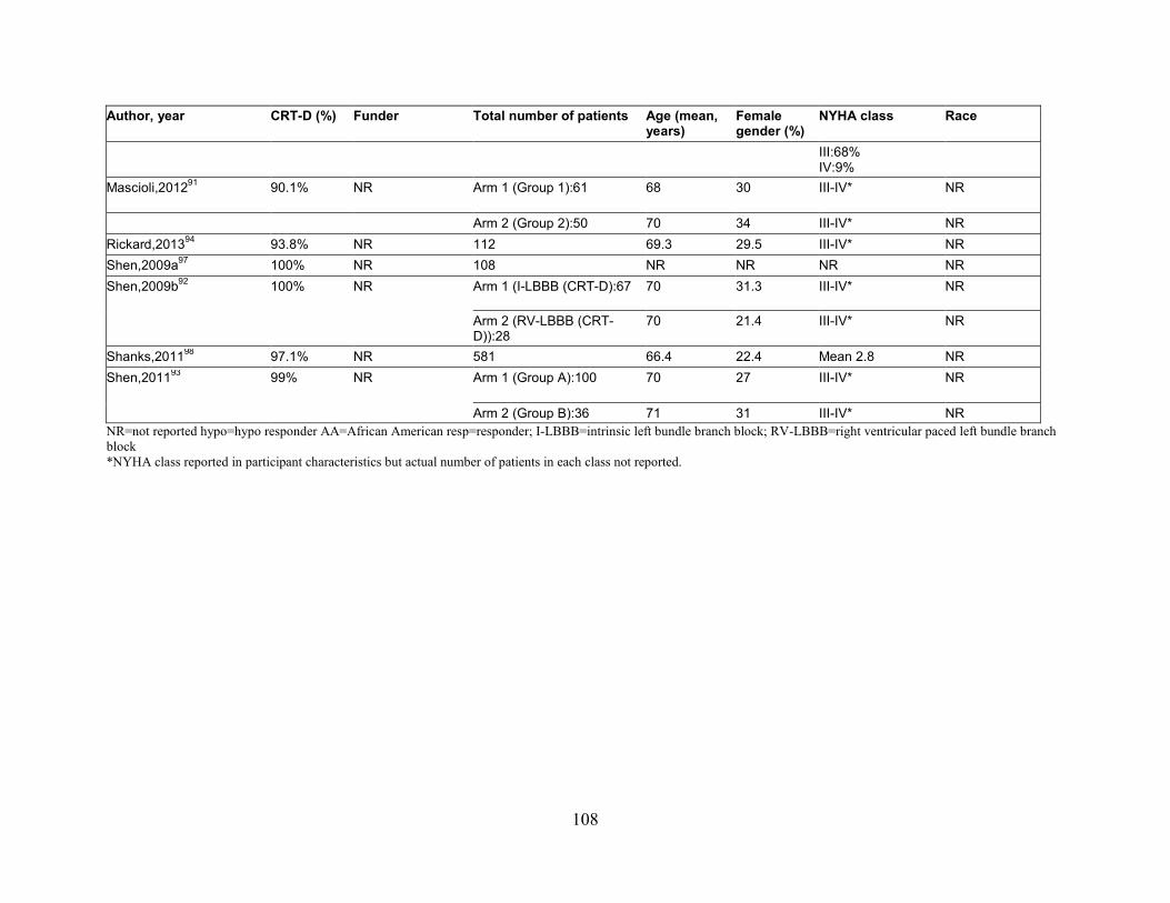
Author, year CRT-D (%) Funder Total number of patients Age (mean, years)
Female gender (%)
NYHA class Race
III:68% IV:9%
Mascioli,201291 90.1% NR Arm 1 (Group 1):61 68
30
III-IV* NR
Arm 2 (Group 2):50 70 34 III-IV* NR Rickard,201394 93.8% NR 112 69.3 29.5 III-IV* NR Shen,2009a97 100% NR 108 NR NR NR NR Shen,2009b92 100% NR Arm 1 (I-LBBB (CRT-D):67 70 31.3 III-IV* NR
Arm 2 (RV-LBBB (CRT-D)):28
70 21.4 III-IV* NR
Shanks,201198 97.1% NR 581 66.4 22.4 Mean 2.8 NR Shen,201193 99% NR Arm 1 (Group A):100 70
27
III-IV* NR
Arm 2 (Group B):36 71 31 III-IV* NR NR=not reported hypo=hypo responder AA=African American resp=responder; I-LBBB=intrinsic left bundle branch block; RV-LBBB=right ventricular paced left bundle branch block *NYHA class reported in participant characteristics but actual number of patients in each class not reported.
108

Risk of Bias Given a strong predilection towards the publishing of positive results, the findings from this
section must be qualified by the high likelihood of reporting bias. The studies, including analyses from the RCTs, had several potential sources of bias as assessed using QUIPS: study participation, study attrition, prognostic factor measurement, outcome measurement, confounding, and statistical analysis. In terms of study participation, the majority of studies were adequate in their description of the source population, recruitment period and place, and exclusion/inclusion criteria. We assessed two studies as having potential for bias from poor description of study participation.97 ,98 Three studies had very specific populations. One study included only patients with near 100 percent right ventricular pacing94, and the other two had only patients with either a native or paced LBBB.92 ,96 These factors limit the generalizability of these three studies.
In terms of attrition rates, the majority of studies did not report dropout rates or reasons for dropout, making the risk of bias from this unclear. The measurement of the prognostic indicators and outcomes across the studies was, in general, adequate. In terms of confounding, for inclusion we required all studies to control for at least gender and either QRS duration or morphology. A significant limitation of the modeling for many studies was not including a priori known clinically significant factors for adjustment, but rather relying solely on statistical criteria for model building. Otherwise, statistical analysis across the studies was generally of adequate quality. Overall, the risk of bias was high due to the high likelihood of reporting bias (Table 49 and Figure 16).
109
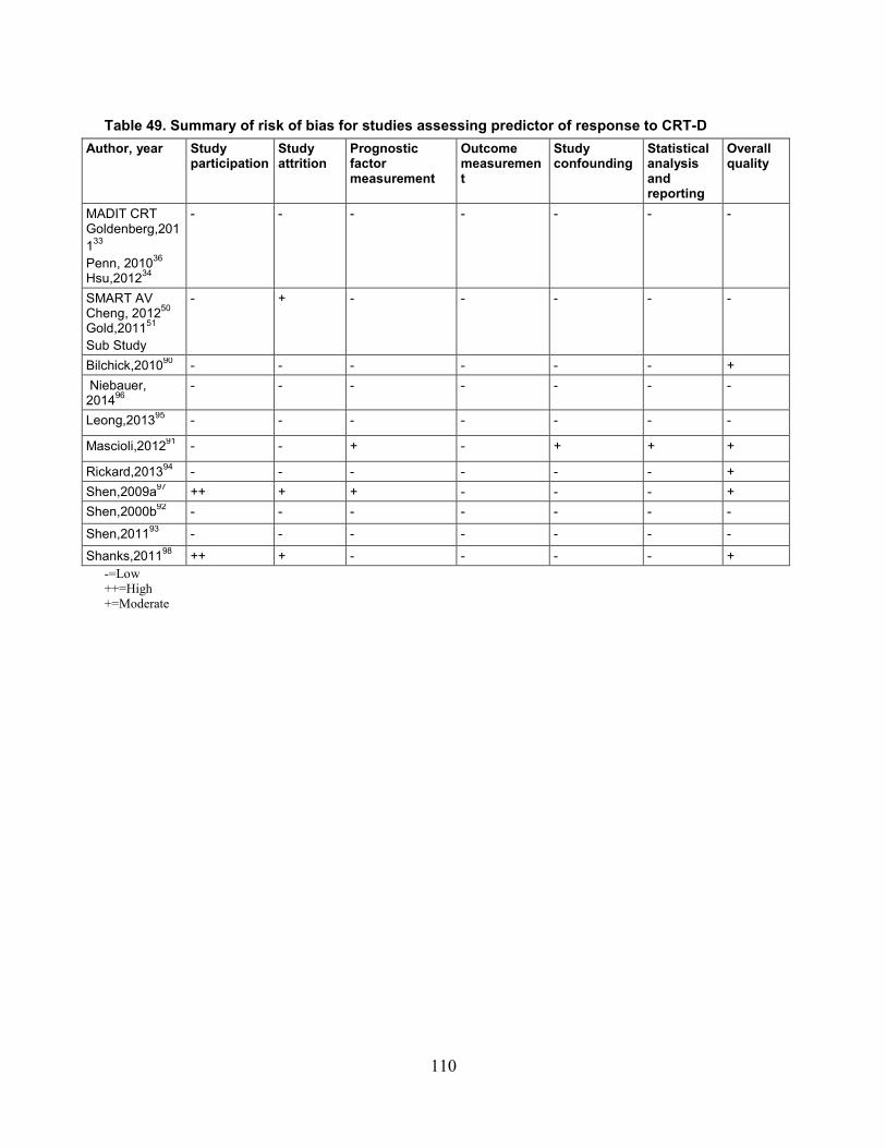
Table 49. Summary of risk of bias for studies assessing predictor of response to CRT-D Author, year Study
participation Study attrition
Prognostic factor measurement
Outcome measurement
Study confounding
Statistical analysis and reporting
Overall quality
MADIT CRT Goldenberg,201133 Penn, 201036 Hsu,201234
- - - - - - -
SMART AV Cheng, 201250 Gold,201151 Sub Study
- + - - - - -
Bilchick,201090 - - - - - - + Niebauer, 201496
- - - - - - -
Leong,201395 - - - - - - -
Mascioli,201291 - - + - + + +
Rickard,201394 - - - - - - + Shen,2009a97 ++ + + - - - + Shen,2000b92 - - - - - - -
Shen,201193 - - - - - - -
Shanks,201198 ++ + - - - - + -=Low ++=High +=Moderate
110
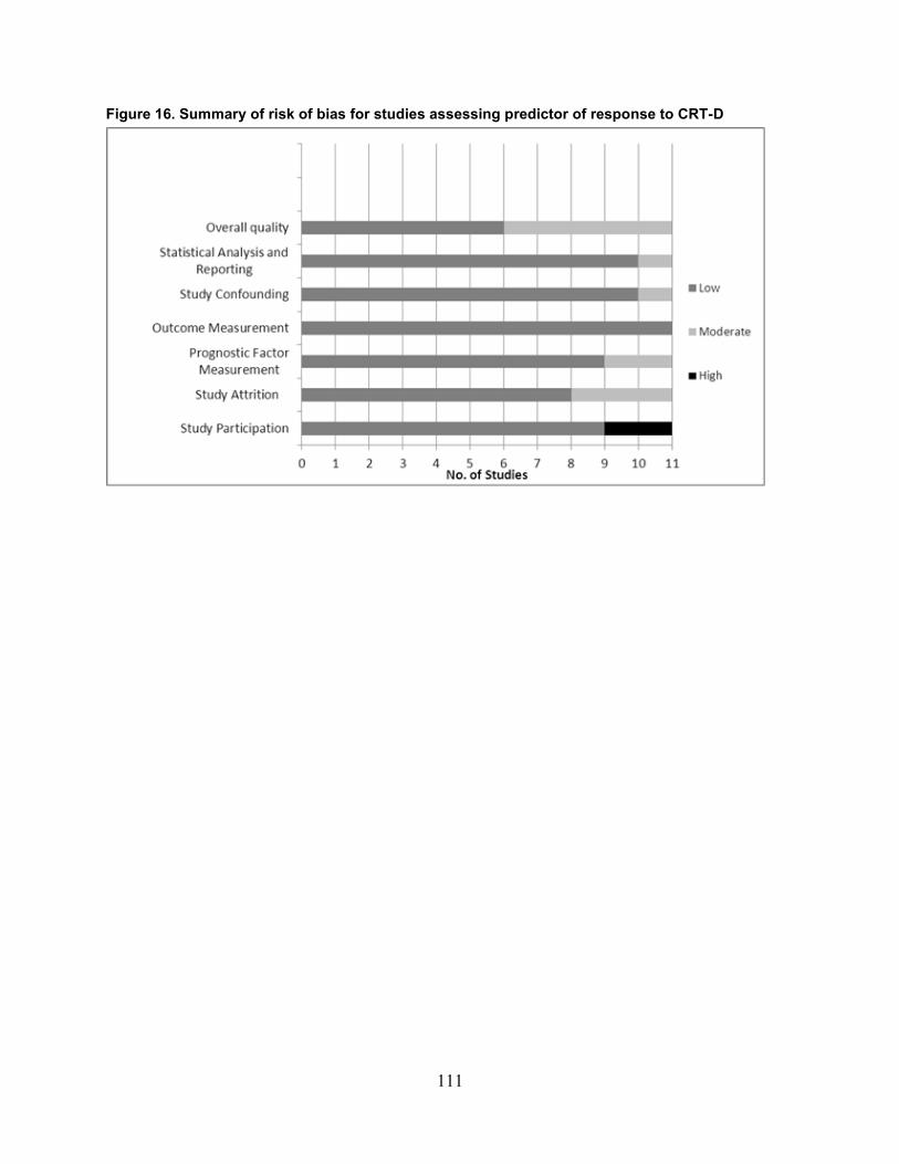
Figure 16. Summary of risk of bias for studies assessing predictor of response to CRT-D
111

Table 50. Included predictors of response for CRT-D Author, year Age Gender CM
type AF QRSd QRS
morphology CKD LA
volume LVEF BMI LVEDV
MADIT-CRT Goldenberg,201133
X X X X X X
Hsu,201234 X X X X X X
Penn, 201036 X Y Y Y Y Y Y Y
SMART-AV Cheng, 201250 X X X X X X X X Gold,201151 X X X X X X
Niebauer, 201496 X X X Z X
Bilchick ,201090 X X X X X X X
Leong,201395 X X X X X X X
Mascioli,201291 Y X X Rickard,201394 Y Y Y Y Y
Shanks,201198 X X X X Shen,2009a97 X X X X X
Shen,2009b92 X X X X X X
Shen,201193 X X X X X X X X= controlled for and effect size reported Y=controlled for but effect size not reported Z=included only patients with LBBB CM=cardiomyopathy; AF=atrial fibrillation; QRSd=QRS duration; CKD=chronic kidney disease; LA=left atrial; LVEF=left ventricular ejection fraction BMI=body mass index; LVESV=left ventricular end systolic volume
112
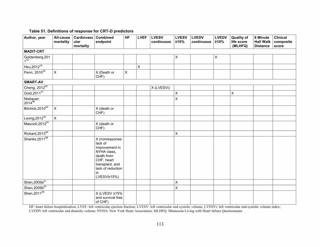
Table 51. Definitions of response for CRT-D predictors Author, year All-cause
mortality Cardiovascular mortality
Combined endpoint
HF LVEF LVESV continuous
LVESV ≥10%
LVEDV continuous
LVEDV ≥10%
Quality of life score (MLHFQ)
6 Minute Hall Walk Distance
Clinical composite score
MADIT-CRT Goldenberg,201133
X X
Hsu,201234 X Penn, 201036 X X (Death or
CHF) X
SMART-AV Cheng, 201250 X (LVESVi) Gold,201151 X X Niebauer, 201496
X
Bilchick,201090 X X (death or CHF)
Leong,201395 X Mascioli,201291 X (death or
CHF)
Rickard,201394 X Shanks,201198 X (nonresponse:
lack of improvement in NYHA class, death from CHF, heart transplant, and lack of reduction in LVESVi≥15%)
Shen,2009a97 X Shen,2009b92 X Shen,201193 X (LVESV ≥15%
and survival free of CHF)
HF: heart failure hospitalization; LVEF: left ventricular ejection fraction; LVESV: left ventricular end-systolic volume; LVESVi: left ventricular end-systolic volume index; LVEDV left ventricular end diastolic volume; NYHA: New York Heart Association; MLHFQ: Minnesota Living with Heart failure Questionnaire
113
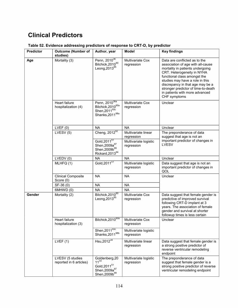
Clinical Predictors Table 52. Evidence addressing predictors of response to CRT-D, by predictor
Predictor Outcome (Number of studies)
Author, year Model Key findings
Age Mortality (3) Penn, 201036 Bilchick,201090 Leong,201395
Multivariate Cox regression
Data are conflicted as to the association of age with all-cause mortality in patients undergoing CRT. Heterogeneity in NYHA functional class amongst the studies may have a role in this discrepancy in that age may be a stronger predictor of time-to-death in patients with more advanced CHF symptoms
Heart failure hospitalization (4)
Penn, 201036a
Bilchick,201090a
Shen,201193c
Shanks,201198a
Multivariate Cox regression
Unclear
LVEF (0) NA NA Unclear LVESV (5) Cheng, 201250 Multivariate linear
regression The preponderance of data suggest that age is not an important predictor of changes in LVESV
Gold,201151 Shen,2009a97 Shen,2009b99 Rickard,201394
Multivariate logistic regression
LVEDV (0) NA NA Unclear MLHFQ (1) Gold,201151 Multivariate logistic
regression Data suggest that age is not an important predictor of changes in QOL
Clinical Composite Score (0)
NA NA Unclear
SF-36 (0) NA NA 6MHWD (0) NA NA
Gender Mortality (2) Bilchick,201090 Leong,201395
Multivariate Cox regression
Data suggest that female gender is predictive of improved survival following CRT-D implant at 3 years. The association of female gender and survival at shorter followup times is less certain
Heart failure hospitalization (3)
Bilchick,201090a Multivariate Cox regression
Unclear
Shen,201193c
Shanks,201198b Multivariate logistic regression
LVEF (1) Hsu,201234 Multivariate linear regression
Data suggest that female gender is a strong positive predictor of reverse ventricular remodeling endpoint
LVESV (5 studies reported in 6 articles)
Goldenberg,201133 Gold,201151 Shen,2009a97 Shen,2009b99
Multivariate logistic regression
The preponderance of data suggest that female gender is a strong positive predictor of reverse ventricular remodeling endpoint
114

Predictor Outcome (Number of studies)
Author, year Model Key findings
Rickard,201394
Cheng, 201250 Multivariate linear regression
LVEDV (1) Goldenberg,201133
Multivariate logistic regression
Data suggest that female gender is a strong positive predictor of reverse ventricular remodeling endpoint
MLHFQ (1) Gold,201151 Multivariate logistic regression
Female gender may be predictive of improved MLHFQ scores but we need more data to confirm this finding
Clinical Composite Score (0)
NA NA Unclear
SF-36 (0) NA NA 6MHWD (0) NA NA
Cardiomyopathy subtype
Mortality (2) Bilchick,201090 Leong,201395
Multivariate Cox regression
NICM is predictive of improved survival at early, 1-year, and 3-year followup after CRT-D implantation
Heart failure hospitalization (3)
Bilchick,201090a Multivariate Cox regression
Unclear
Shen,201193c
Shanks,201198b Multivariate logistic regression
LVEF (1) Hsu,201234 Multivariate linear regression
Data suggest that NICM is a strong positive predictor of reverse ventricular remodeling endpoint
LVESV (4 studies in 5 articles)
Goldenberg,201133 Cheng, 201250 Gold,201151 Rickard,201394 Niebauer, 201496
Multivariate logistic regression
The preponderance of data suggest that NICM is a strong positive predictor of reverse ventricular remodeling endpoints
LVEDV (1) Goldenberg,201133
Multivariate logistic regression
Data suggest that NICM is a strong positive predictor of reverse ventricular remodeling endpoints
MLHFQ (1) Gold,201151 Multivariate logistic regression
Cardiomyopathy subtype is not predictive of improved QOL scores, although we need more data to confirm this finding
Clinical Composite Score (0)
NA NA Unclear
SF-36 (0) NA NA Unclear 6MHWD (0) NA NA Unclear
Atrial fibrillation
Mortality (2) Bilchick,201090 Leong,201395
Multivariate Cox regression
Data suggest that a history of AF is predictive of increased all-cause mortality at early, 1-year, and 3-year followup after CRT-D
Heart failure hospitalization (1)
Bilchick,201090a Multivariate Cox regression
Unclear
LVEF (0) NA NA LVESV (1) Cheng, 201250 Multivariate linear
regression
115
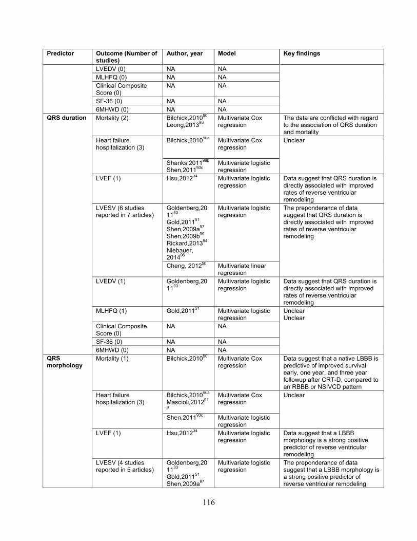
Predictor Outcome (Number of studies)
Author, year Model Key findings
LVEDV (0) NA NA MLHFQ (0) NA NA Clinical Composite Score (0)
NA NA
SF-36 (0) NA NA 6MHWD (0) NA NA
QRS duration
Mortality (2) Bilchick,201090 Leong,201395
Multivariate Cox regression
The data are conflicted with regard to the association of QRS duration and mortality
Heart failure hospitalization (3)
Bilchick,201090a Multivariate Cox regression
Unclear
Shanks,201198b
Shen,201193c Multivariate logistic regression
LVEF (1) Hsu,201234 Multivariate logistic regression
Data suggest that QRS duration is directly associated with improved rates of reverse ventricular remodeling
LVESV (6 studies reported in 7 articles)
Goldenberg,201133 Gold,201151 Shen,2009a97 Shen,2009b99 Rickard,201394 Niebauer, 201496
Multivariate logistic regression
The preponderance of data suggest that QRS duration is directly associated with improved rates of reverse ventricular remodeling
Cheng, 201250
Multivariate linear regression
LVEDV (1) Goldenberg,201133
Multivariate logistic regression
Data suggest that QRS duration is directly associated with improved rates of reverse ventricular remodeling
MLHFQ (1) Gold,201151 Multivariate logistic regression
Unclear Unclear Clinical Composite
Score (0) NA NA
SF-36 (0) NA NA 6MHWD (0) NA NA
QRS morphology
Mortality (1) Bilchick,201090
Multivariate Cox regression
Data suggest that a native LBBB is predictive of improved survival early, one year, and three year followup after CRT-D, compared to an RBBB or NSIVCD pattern
Heart failure hospitalization (3)
Bilchick,201090a
Mascioli,201291
a
Multivariate Cox regression
Unclear
Shen,201193c Multivariate logistic regression
LVEF (1) Hsu,201234 Multivariate logistic regression
Data suggest that a LBBB morphology is a strong positive predictor of reverse ventricular remodeling
LVESV (4 studies reported in 5 articles)
Goldenberg,201133 Gold,201151 Shen,2009a97
Multivariate logistic regression
The preponderance of data suggest that a LBBB morphology is a strong positive predictor of reverse ventricular remodeling
116
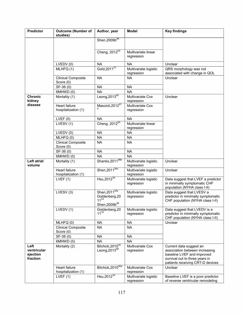
Predictor Outcome (Number of studies)
Author, year Model Key findings
Shen,2009b99
Cheng, 201250
Multivariate linear regression
LVEDV (0) NA NA Unclear MLHFQ (1) Gold,201151 Multivariate logistic
regression QRS morphology was not associated with change in QOL
Clinical Composite Score (0)
NA NA Unclear
SF-36 (0) NA NA 6MHWD (0) NA NA
Chronic kidney disease
Mortality (1) Leong,201395 Multivariate Cox regression
Unclear
Heart failure hospitalization (1)
Mascioli,201291
a Multivariate Cox regression
LVEF (0) NA NA LVESV (1) Cheng, 201250
Multivariate linear regression
LVEDV (0) NA NA MLHFQ (0) NA NA Clinical Composite Score (0)
NA NA
SF-36 (0) NA NA 6MHWD (0) NA NA
Left atrial volume
Mortality (1) Shanks,201198b Multivariate logistic regression
Unclear
Heart failure hospitalization (1)
Shen,201193c Multivariate logistic regression
Unclear
LVEF (1) Hsu,201234 Multivariate logistic regression
Data suggest that LVEF a predictor in minimally symptomatic CHF population (NYHA class I-II)
LVESV (3) Shen,201193c
Goldenberg,201133 Shen,2009b99
Multivariate logistic regression
Data suggest that LVESV a predictor in minimally symptomatic CHF population (NYHA class I-II)
LVEDV (1) Goldenberg,201133
Multivariate logistic regression
Data suggest that LVEDV is a predictor in minimally symptomatic CHF population (NYHA class I-II)
MLHFQ (0) NA NA Unclear Clinical Composite
Score (0) NA NA
SF-36 (0) NA NA 6MHWD (0) NA NA
Left ventricular ejection fraction
Mortality (2) Bilchick,201090 Leong,201395
Multivariate Cox regression
Current data suggest an association between increasing baseline LVEF and improved survival out to three years in patients receiving CRT-D devices
Heart failure hospitalization (1)
Bilchick,201090a Multivariate Cox regression
Unclear
LVEF (1) Hsu,201234 Multivariate logistic regression
Baseline LVEF is a poor predictor of reverse ventricular remodeling
117
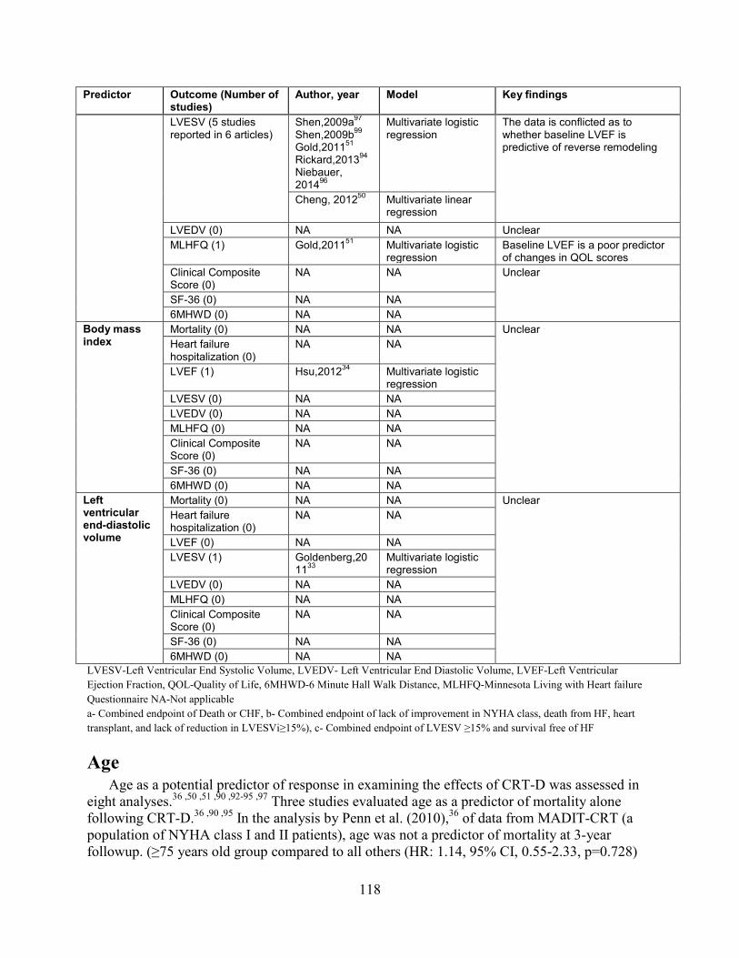
Predictor Outcome (Number of studies)
Author, year Model Key findings
LVESV (5 studies reported in 6 articles)
Shen,2009a97 Shen,2009b99 Gold,201151 Rickard,201394 Niebauer, 201496
Multivariate logistic regression
The data is conflicted as to whether baseline LVEF is predictive of reverse remodeling
Cheng, 201250
Multivariate linear regression
LVEDV (0) NA NA Unclear MLHFQ (1) Gold,201151 Multivariate logistic
regression Baseline LVEF is a poor predictor of changes in QOL scores
Clinical Composite Score (0)
NA NA Unclear
SF-36 (0) NA NA 6MHWD (0) NA NA
Body mass index
Mortality (0) NA NA Unclear Heart failure hospitalization (0)
NA NA
LVEF (1) Hsu,201234 Multivariate logistic regression
LVESV (0) NA NA LVEDV (0) NA NA MLHFQ (0) NA NA Clinical Composite Score (0)
NA NA
SF-36 (0) NA NA 6MHWD (0) NA NA
Left ventricular end-diastolic volume
Mortality (0) NA NA Unclear Heart failure hospitalization (0)
NA NA
LVEF (0) NA NA LVESV (1) Goldenberg,20
1133 Multivariate logistic regression
LVEDV (0) NA NA MLHFQ (0) NA NA Clinical Composite Score (0)
NA NA
SF-36 (0) NA NA 6MHWD (0) NA NA
LVESV-Left Ventricular End Systolic Volume, LVEDV- Left Ventricular End Diastolic Volume, LVEF-Left Ventricular Ejection Fraction, QOL-Quality of Life, 6MHWD-6 Minute Hall Walk Distance, MLHFQ-Minnesota Living with Heart failure Questionnaire NA-Not applicable a- Combined endpoint of Death or CHF, b- Combined endpoint of lack of improvement in NYHA class, death from HF, heart transplant, and lack of reduction in LVESVi≥15%), c- Combined endpoint of LVESV ≥15% and survival free of HF
Age Age as a potential predictor of response in examining the effects of CRT-D was assessed in
eight analyses.36 ,50 ,51 ,90 ,92-95 ,97 Three studies evaluated age as a predictor of mortality alone following CRT-D.36 ,90 ,95 In the analysis by Penn et al. (2010),36 of data from MADIT-CRT (a population of NYHA class I and II patients), age was not a predictor of mortality at 3-year followup. (≥75 years old group compared to all others (HR: 1.14, 95% CI, 0.55-2.33, p=0.728)
118
In the very large Medicare registry study by Bilchick et al. (2010), patients 70-79 years old had higher 3-year mortality rates compared to patients 50-59 years old (HR: 1.75, 95% CI, 1.51 to 2.04).90 In the study by Leong et al. (2013), age was not a predictor of worsened survival at a median of 44 months followup (HR: 0.991, 95% CI, 0.973 to 1.01, p=0.3).95
Four analyses evaluated age as a predictor of heart failure hospitalizations, one looked at HF hospitalization alone36 and the others combined it with another endpoint.36 ,90 ,93 ,98 In the MADIT-CRT analysis by Penn et al. (2010) age (broken into 3 groups: ≥75, 60-74, and <60 years old) was a predictor of CHF admissions at 3-year followup (the oldest group derived the most benefit) (HR: 0.46, 95%CI,0.29 to 0.74, p=0.001).36 Two studies combined time-to-death or first hospitalization into a single endpoint.36 ,90 Also in this analysis, age was a predictor of survival without CHF admissions in patients 60-74 and ≥75 but not in patients <60 years old. In contrast, the study by Bilchick et al. (2010) reported an association between age and the composite outcome of survival without CHF admissions at 3 years in patients ≥80 compared to patients 50-59 years old (HR:1.24, 95% CI, 1.10 to 1.38). Patients in the 60-69 year old cohort in this study had improved survival without CHF admissions compared to the 50-59 year old reference group (HR:0.84, 95% CI, 0.75 to 0.94).
One study used combined endpoints incorporating both CHF admissions and evidence of change in left ventricular end-systolic volume (LVESV) from baseline.93 Shen et al. (2011) reported no statistically significant association between age and response (defined as a reduction in LVESV >15 percent and survival without CHF admissions) (OR 0.97 (0.88-1.08), p=0.60).93 No studies evaluate age as a predictor of change in LVEF. Five studies assessed age as a predictor of changes in LVESV from baseline following CRT.50 ,51 ,92 ,94 ,97 Cheng et al. (2012), in analysis of data from the SMART-AV trial, reported that increasing age was associated with a greater reduction in LVESV.50 Gold and colleagues, using data from the same trial but using a dichotomous definition of response (reduction in LVESV ≥15%), found no association between age and response (OR: 1.00, 95% CI, 0.98 to 1.02, p=0.801).51 The other three studies (two by Shen, and one by Rickard), did not report an association between age and changes in left LVESV.92 ,94 ,97 No studies looked at age as a predictor of changes in LVEDV. Only one study looked at age as a predictor of change in quality of life (QOL) (as assessed by the MLHFQ. Gold et al. (2011) found that age was not associated with changes in QOL score (OR: 0.99, 95% CI, 0.97 to 1.01, p=0.209).51 No studies examined age as a predictor of changes in clinical composite score, SF-36, or 6-minute hall walk distance (6MHWD).
To summarize, the data are conflicted as to the association of age with all-cause mortality in patients undergoing CRT. Heterogeneity in NYHA functional class amongst the studies may have a role in this discrepancy in that age may be a stronger predictor of time-to-death in patients with more advanced CHF symptoms. Given a paucity of data on CHF admissions alone, it’s unclear whether age is predictive of CHF admissions. The preponderance of data suggests that age is not an important predictor of changes in LVESV and changes in QOL scores following CRT (Table 50 and 51).
Gender Ten (reported in 12 articles) included gender as a potential predictor of response.33 ,34 ,50 ,51 ,90
,92-98 Two studies evaluated gender as a predictor of mortality alone following CRT.90 ,95 The study by Bilchick et al. (2010)90, associated female gender with improved mortality at 3 years (HR:0.87, 95% CI, 0.81to 0.94, p <0.001) but not at 1 year (HR:0.92, 95% CI, 0.82 to 1.02,
119
p=0.155). In the study by Leong et al. (2013) gender was not a significant predictor of survival at a median of 44 months followup (HR: 0.991, 95% CI, 0.973 to 1.01, p=0.3).95
Three studies addressed gender in terms of predicting CHF admissions: all utilized CHF as part of a combined endpoint.90 ,93 ,98 The study by Bilchick et al. (2010) reported non-statistically significant association of female gender with the endpoint of survival without CHF admissions at 1 year (1.08 95% CI, 1 to 1.16, p=0.065) or at 3 years (1.00 95% CI, 0.94-1.05, p=0.872).90 In the study by Shanks et al. (2011), gender was not a predictor of non-response defined as lack of improvement in NYHA class ≥1, death from worsening CHF, heart transplant, or lack of reduction in LVESV indexed to body surface areas (LVESV index >15 %) (OR: 1.504 [male gender], 95% CI, 0.814 to 2.777, p=0.192).98 Shen et al. (2011) reported a non-statistically significant association between gender and response (defined as a reduction in LVESV >15 percent and survival without CHF admissions).93 Only one study reported the association of gender and change in ejection fraction. This analysis of MADIT-CRT trial by Hsu et al. (2012), reported an association between female gender and an improvement in LVEF (≥14.5 percent from baseline) (OR: 1.96,95% CI, 1.32 to 2.90, p<0.001).34
Six studies (reported in 7 articles) assessed the relationship between gender and changes in LVESV from baseline following CRT.33 ,50 ,51 ,92 ,94 ,96 ,97 In an analysis from MADIT-CRT, Goldenberg et al. (2011) reported an association between female gender and improved rates of rates of reduction in LVESV ≥15 percent from baseline.33 Cheng et al. (2012) reported a positive association between male gender and an increase in LVESV index from baseline with data from the SMART-AV trial.50 Gold and colleagues reported a negative association between male gender and reduction in LVESV ≥15 percent from baseline (OR: 0.53, 95% CI, 0.33 to 0.85, p=0.008) also from the SMART-AV trial.51 The study by Shen et al. (2009a) reported no association between gender with response (OR: 0.51, 95% CI, 0.11 to 2.34, p=0.38).97 Three studies reported no association between gender and change in LVESV.94 ,96 ,97 These studies included highly specialized populations. Rickard et al. (2013) was a cohort of patients with paced rhythms undergoing upgrade to CRT and Shen et al. (2009b) and Niebauer et al. 96 included populations of patients all with native or paced LBBB.
One study reported an association between female gender and improved rates of reduction in LVEDV ≥10 percent from baseline.33 Only one study reported gender as a predictor of change in QOL. This analysis from the SMART-AV trial reported that male gender was significantly associated with inferior MLHFQ scores following CRT-D (OR: 0.56, 95% CI, 0.34 to 0.91, p=0.018).51 No studies examined gender as a predictor of changes in clinical composite score, SF-36, or 6MHWD.
Overall, the data suggest that female gender is predictive of improved survival following CRT-D implant at 3 years. The association of female gender and survival at shorter followup times is less certain. Given the lack of data on CHF admissions alone, it is unclear whether gender is predictive of CHF admissions. The preponderance of data suggest that female gender is a strong positive predictor of reverse ventricular remodeling endpoint (changes in LVEF, LVESV indexed to body surface areas [LVESVi] , and LVEDV indexed to body surface areas [LVEDVi]) following CRT. The studies that failed to show this association were limited by substantial heterogeneities in the population studied. Female gender may be predictive of improved MLHFQ scores but we need more data to confirm this finding (Table 50 and 51).
120

Cardiomyopathy Subtype Eight studies (reported in 9 articles) included cardiomyopathy subtype as a potential
predictor of response in examining the effects of CRT-D.33 ,34 ,50 ,51 ,90 ,93-96 ,98 Two CRT-D studies evaluated cardiomyopathy subtype as a predictor of mortality alone following CRT.90 ,95 The study by Bilchick et al. (2010), reported an association between ICM and impaired survival at 1 year (HR: 1.39, 95% CI, 1.21 to 1.59, p<0.001) and 3 years (HR: 1.44, 95% CI, 1.33 to 1.57, p<0.001) compared to NICM.90 The study by Leong et al. (2013) found an association between NICM and improved survival at a median of 44 months followup (HR: 0.730, 95% CI, 0.535-0.996, p=0.047) compared to ICM.95 Three studies assessed cardiomyopathy subtype as a predictor of CHF admissions, all of which utilized CHF as part of a combined endpoint.90 ,93 ,98 The study by Bilchick et al. (2010) reported an association between ICM and impaired survival without CHF admissions both at 1 year (HR: 1.24, 95% CI, 1.13 to 1.35, p =<0.001) and at 3 years (HR: 1.32, 95% CI, 1.22-1.40, p<0.001). In the study by Shanks et al. (2011), ICM was predictive of non-response (defined as lack of improvement in NYHA class ≥1, death from worsening CHF, heart transplant, or lack of reduction in LSESV index >15 %) (OR: 2.264, 95% CI, 1.272 to 4.031, p=0.005). The study by Shen et al. (2011) did not report an association between ICM and response (OR: 0.74, 95% CI, 0.04 to 12.87, p=0.84) (defined as a reduction in LVESV >15 percent and survival without CHF admissions).93
Only one study examined the association of cardiomyopathy subtype and change in LVEF. In an analysis of MADIT-CRT trial data, Hsu et al. (2012) reported a strong association between NICM and a large improvement in LVEF (≥14.5% from baseline) (OR: 1.80, 95% CI, 1.20 to 2.71, p=0.005).34 Four studies (reported in 5 articles) examined whether cardiomyopathy subtype would predict changes in LVESV from baseline following CRT.33 ,50 ,51 ,94 ,96 In an analysis from MADIT-CRT, Goldenberg et al. (2011) reported an association between NICM and improved rates of reduction in LVESV ≥15 percent from baseline.33 Cheng et al. (2012) reported a positive association between ICM and increase in LVESV index from baseline from the SMART-AV trial.50 Gold et al. (2011) reported a negative association between ICM and reduction in LVESV ≥15 percent from baseline (OR: 0.58, 95% CI, 0.37 to 0.91, p=0.019) also from the SMART-AV trial.51 The study by Niebauer et al. reported no association between cardiomyopathy subtype and response in a highly selected population of patients with LBBB.96 Similarly, the study by Rickard et al. (2013) reported no association between cardiomyopathy subtype and response in a highly selected population of patients with frequent right ventricular pacing.94
One study examined the association of cardiomyopathy type and change in left ventricular end diastolic volume (LVEDV). In an analysis from MADIT-CRT, Goldenberg et al. (2011) reported an association between NICM and improved rates of rates of reduction in LVEDV ≥10 percent from baseline.33 Only one study assessed cardiomyopathy subtype as a predictor of change in quality of life. In an analysis from SMART-AV, Gold et al. (2011) found that cardiomyopathy subtype was not associated with change in quality of life following CRT.51 There were no studies that examined cardiomyopathy subtype as a predictor of changes in clinical composite score, SF-36, or 6MHWD.
To summarize, our findings suggest that NICM is predictive of improved survival at early, 1-year, and 3-year followup after CRT-D implantation. Given the lack of data on CHF admissions alone, it’s unclear whether cardiomyopathy subtype is predictive of CHF admissions. The preponderance of data suggests that NICM is a strong positive predictor of reverse ventricular remodeling (changes in LVEF, LVESVi, and LVEDVi following CRT. Cardiomyopathy subtype
121
is not predictive of improved quality of life scores, although we need more data to confirm this finding (Table 50 and 51).
Atrial Fibrillation Three studies examining the effects of CRT-D included a history of AF as a potential
predictor of response.50 ,90 ,95 Two studies evaluated a history of AF as a predictor of mortality alone following CRT-D.90 ,95 The study by Bilchick et al. (2010) reported an association between a history of AF and impaired survival at 1 year (HR:1.27, 95% CI, 1.14 to 1.41, p <0.001) and 3 years (HR:1.21, 95% CI, 1.14 to 1.30, p<0.001).90 The study by Leong et al. (2013) reported an association between a history of AF and worsened survival at a median of 44 months followup (HR:1.4, 95% CI, 1.0 to 2.0, p=0.045).95 No study assessed AF as a predictor of CHF admissions alone. One study assessed history of AF in terms of predicting a combined endpoint of CHF admissions and death.90 This study reported an association between a history of AF and impaired survival without CHF admissions both at 1 year (HR:1.27, 95% CI, 1.18 to 1.37, p=<0.001) and at 3 years (HR:1.22, 95% CI, 1.15 to 1.28, p <0.001). No studies reported the history of AF and change in ejection fraction or LVEDV. In an analysis from SMART-AV, Cheng et al. (2012) did not report an association between a history of AF and a change in LVESV index (p=0.945).50 No studies examined a history of AF as a predictor of changes in QOL score, clinical composite score, or 6MHWD.
Overall the data suggest that a history of AF is predictive of increased all-cause mortality at early, 1-year, and 3-year followup after CRT-D. Given the lack of data on CHF admissions alone, it’s unclear whether a history of AF is predictive of CHF admissions. Similarly there are not sufficient data to determine the predictive value of AF in terms of remodeling endpoints, changes in quality of life, and 6MHWD (Table 50 and 51).
QRS Duration Nine studies (reported in 11 articles) included QRS duration as a potential predictor of
response in examining the effects of CRT-D.33 ,34 ,50 ,51 ,90 ,92 ,93 ,95-98 Two studies evaluated QRS duration as a predictor of mortality alone.90 ,95
The study by Bilchick et al. (2010) reported an association between a QRS duration ≥150ms and improved survival at one year (HR: 0.77, 95% CI, 0.70 to 0.84, p <0.001) and three years (HR: 0.86, 95% CI, 0.81 to 0.91, p <0.001) compared to a QRS duration of 120-149ms.90 The study by Leong et al. (2013) reported a non-statistically significant association of QRS duration with survival (HR: 0.997, 95% CI, 0.991 to 1.00, p=0.4).95 Three studies assessed the QRS duration in terms of predicting CHF admissions, all of which utilized CHF as part of a combined endpoint.90 ,93 ,98
The study by Bilchick et al. (2010) reported an association between a QRS duration ≥150ms and improved survival without CHF admissions at one year (HR:0.78, 95% CI, 0.73 to 0.83, p<0.001) and three years (HR:0.83, 95% CI, 0.79 to 0.87, p<0.001), compared to a QRS duration 120-149ms.90 The study by Shanks et al. (2011) reported that QRS duration was not predictive of non-response (defined as lack of improvement in NYHA class ≥1, death from worsening CHF, heart transplant, or lack of reduction in LVESV index >15 %) (OR: 0.994, 95% CI, 0.986 to 1.002, p=0.119).98 The study by Shen et al. (2011) reported no association between QRS duration and response (OR: 1.03, 95% CI, 0.995 to 1.07, p=0.09) (defined as a reduction in LVESV >15 percent and survival without CHF admissions).93
122
An analysis from MADIT-CRT, Hsu et al. (2012), reported a strong association between a QRS duration >150ms and a large improvement in LVEF (≥14.5% from baseline) (OR: 1.79, 95% CI, 1.17 to 2.73, p=0.007).34
Five studies (reported in 6 articles) assessed association of QRS duration subtype with changes in LVESV from baseline following CRT.33 ,50 ,51 ,92 ,94 ,97 In an analysis from MADIT-CRT, Goldenberg et al. (2011) reported an association between QRS duration ≥150ms and improved rates of rates of reduction in LVESV ≥15 percent from baseline.33 In analysis from SMART-AV Cheng et al. (2012) reported an association between increasing QRS duration and reduction in LVESV index from baseline.50 In another analysis from SMART-AV Gold et al. (2011) did not report an association between QRS duration and reduction in LVESV ≥15 percent from baseline.51 The study by Shen et al. did not associate increasing QRS duration with increased rates of response (OR: 1.02, 95% CI, 1.00 to 1.04, p<0.13).97The study by Niebauer found no association with QRS duration and reduction in LVESV≥15% in a highly selected population all with native LBBB. 96Finally two other studies reported no association between QRS duration and response.94 ,99
In an analysis from MADIT-CRT, Goldenberg et al. (2011) reported an association between QRS duration ≥150ms and improved rates of rates of reduction in LVEDV ≥10 percent from baseline.33 In an analysis from SMART-AV, Gold et al. (2011) reported no association between QRS duration and change in QOL following CRT.51 No studies examined QRS duration as a predictor of changes in clinical composite score or 6MHWD.
To summarize QRS duration is intimately linked with QRS morphology and the relative contribution of each in predicting outcomes is unclear. The data are conflicted with regard to the association of QRS duration and mortality in patients undergoing CRT-D implantation. Given the lack of data on CHF admissions, it is unclear whether QRS duration is predictive of CHF admissions. The preponderance of data suggests that QRS duration is directly associated with improved rates of reverse ventricular remodeling (changes in LVEF, LVESVi, and LVEDVi) following CRT (Table 50 and 51).
QRS Morphology Seven studies (reported in 9 articles) included QRS morphology as a potential predictor of
response in examining the effects of CRT-D 33 ,34 ,50 ,51 ,90-93 ,97 One study assessed QRS morphology as a predictor of mortality.90 This study reported an association between an RBBB pattern and worsened survival at both 1 year (HR:1.44, 95% CI, 1.26 to 1.65, p<0.001) and 3 years (HR:1.37, 95% CI, 1.26 to 1.49, p<0.001), compared to patients with a LBBB morphology.90 In addition, it reported an association between an NSIVCD pattern and worsened survival at 1 year (HR: 1.18, 95% CI, 1.05 to 1.32, p<0.001) and 3 years (HR: 1.08, 95% CI, 1.00 to 1.16, p<0.001), compared to patients with a LBBB morphology.
Three studies assessed QRS morphology as a predictor of CHF admissions as part of a composite endpoint.90 ,91 ,93 The study by Bilchick et al. (2010) reported an association between an RBBB and worsened survival without CHF admissions at one year (HR:1.32, 95% CI, 1.20 to 1.45, p<0.001) and three years (HR:1.28,95% CI, 1.20 to 1.38, p<0.001) compared to patients with a LBBB morphology.90 In addition, it reported an association between an NSIVCD pattern and worsened survival without CHF admissions at one year (HR:1.08, 95% CI, 1.00 to 1.17, p<0.001) and three years (HR:1.05, 95% CI, 0.99 to 1.12, p<0.001), compared to patients with a LBBB morphology. The study by Shen et al. (2011) reported an association between right ventricle paced LBBB pattern and response (OR: 6.83, 95% CI, 1.11 to 42.12, p=0.04) (defined
123

as a reduction in LVESV >15 percent and freedom from CHF admissions).93 The study by Mascioli et al. (2012) reported an association between the presence of a “false” LBBB pattern and worsened survival free of CHF admissions, compared to patients with a “true” LBBB (defined as a QRS morphology in V1-V2, duration ≥140ms for men and ≥130ms for women and mid-QRS notching or slurring in ≥2 leads among I, AVL, V1,V2,V5, and V6) (OR: 3.98, 95% CI, 1.51to 10.48, p=0.005).91
One study assessed the association of QRS morphology and change in LVEF.34 This study reported a strong association between a LBBB pattern and a large improvement in LVEF (≥14.5%from baseline) (OR: 2.05, 95% CI, 1.24 to 3.40, p=0.006). Four studies (reported in 5 articles) assessed whether QRS morphology would predict changes in LVESV from baseline following CRT.33 ,50 ,51 ,92 ,97 In an analysis from MADIT-CRT, Goldenberg et al. (2011) reported an association between a LBBB pattern and improved rates of reduction in LVESV ≥15 percent from baseline.33 In an analysis from SMART-AV, Cheng et al. (2012) reported a strong association between LBBB and reduction in LVESV index from baseline.50 In another analysis from SMART-AV, Gold et al. (2011) reported no association between QRS morphology and reduction in LVESV ≥15 percent from baseline.51 The study by Shen et al. (2009a) found no association between a QRS morphology and increased rates of response (OR: 0.53, 95% CI 0.06 to 4.89, p<0.58).97 Another study by Shen et al. (2009b) (including only patients with native or pacing induced LBBB morphologies) reported an association between an right ventricle paced LBBB pattern and response (OR: not reported, , 95% CI, 2.00 to 111.85, p<0.008).92
In an analysis from MADIT-CRT, Goldenberg et al. (2011) reported an association between a LBBB pattern and improved rates of rates of reduction in LVESV ≥10 percent from baseline.33 One study examined QRS morphology subtype as a predictor of change in quality of life. Gold et al. (2011) found that QRS morphology was not associated with change in quality of life following CRT.51 No studies examined QRS morphology as a predictor of changes in clinical composite score or 6MHWD.
Overall, QRS morphology is intimately linked with QRS duration, and the relative contribution of each in predicting outcomes is unclear. Our analysis suggests that a native LBBB is predictive of improved survival early, one year, and three year followup after CRT-D, compared to an RBBB or NSIVCD pattern. Given the lack of data on CHF admissions alone, it’s unclear whether QRS morphology is predictive of CHF admissions. The preponderance of data suggest that a LBBB morphology is a strong positive predictor of reverse ventricular remodeling (changes in LVEF, LVESVi, and LVEDVi) following CRT, compared with RBBB or NSIVCD. Due to a paucity of data, the association of paced ventricular rhythms and response is unclear (Table 50 and 51).
Chronic Kidney Disease Three studies included chronic kidney disease (CKD) as a potential predictor of response in
patients receiving a CRT-D device.50 ,91 ,95 Leong et al. (2013) reported a direct association between improving renal function (measured via GFR) and improved all-cause mortality (HR (per 10ml/min increase in GFR): 0.83, 95% CI, 0.77 to 0.89, p<0.001).95 Mascioli et al. (2012) reported no significant association between the presence of class IV CKD and survival free of CHF admissions.91 Cheng et al. (2012) reported no statistically significant association between CKD and change in LVESV index.50 Overall, there are not sufficient data to determine the predictive nature of CKD in patients receiving CRT-D devices (Table 50 and 51).
124

Left Atrial Volume Four studies (reported in 5 articles) included left atrial volume (LAV) alone or indexed to
body surface area (LAVi) as a potential predictor of response in patients receiving a CRT-D device.33 ,34 ,92 ,93 ,98 In the study by Shanks et al. (2011) there was a non-statistically significant association of a greater LAVi and non-response to CRT (defined as lack of improvement in NYHA class ≥1, death from worsening CHF, heart transplant, or lack of reduction in LVESV index >15 %) (OR: 1.013, 95% CI, 1.000 to 1.026, p =0.058).98 A study by Shen et al. (2011) did not find an association between LAVi and response (defined as a reduction in LVESV ≥15 percent from baseline and freedom from CHF admissions over the duration of followup).93 In an analysis from MADIT-CRT, Goldenberg et al. (2011) reported an association between baseline LAV <40ml/m2 and a higher rate of reduction in LVESV ≥15 percent from baseline.33 Another study by Shen et al. (2009b) (in a highly selected population containing only patients with native or paced LBBB patterns) reported no association between LAV and response defined as a ≥15% reduction in LVESV from baseline (OR: not reported, , 95% CI, 0.99 to 1.02, p=NS).92 Hsu et al. (2012), analyzing data from MADIT-CRT, reported LAVi as a predictor of a dramatic improvement in LVEF (≥14.5 percent from baseline) (OR: 1.47, 95% CI, 1.21 to 1.79, p<0.001). In another analysis from MADIT-CRT, Goldenberg et al. (2011) reported an association between a baseline LAV <40ml/m2 and improved rates of rates of reduction in LVEDV ≥10 percent from baseline.33
To summarize there are insufficient data to determine the predictive nature of LAV in terms of mortality, CHF hospitalizations, quality of life, 6MHWD, or change in clinical composite score for patients undergoing CRT-D implantation. LAV at baseline does seem to be a predictor of reverse remodeling endpoints (changes in LVEF, LVESVi, and LVEDVi) in a minimally symptomatic CHF population (NYHA class I-II). There is not sufficient evidence to conclude that LAV is a predictor of reverse ventricular remodeling in patients with NYHA class III and IV CHF (Table 50 and 51).
Left Ventricular Ejection Fraction Seven studies (reported in 8 articles) included baseline LVEF as a potential predictor of
response in examining the effects of CRT-D.50 ,51 ,90 ,92 ,94-97 Two studies evaluated LVEF as a predictor of mortality alone following CRT-D.90 ,95 The study by Bilchick et al. (2010), reported an association between an increased LVEF and improved survival at both one year (HR: 0.97, 95% CI, 0.96 to 0.98, p<0.001) and three years (HR: 0.98, 95% CI, 0.98 to 0.99, p<0.001).90 The smaller study by Leong et al. (2013) reported no association between LVEF and improved survival (HR: 0.95, 95% CI, 0.84 to 1.1, p=0.4). 95 One study assessed LVEF as a predictor of a composite endpoint including CHF admissions.90
The study by Bilchick et al. (2010) reported an association between a higher baseline LVEF and improved survival without CHF admissions at one year (HR: 0.97, 95% CI, 0.96 to 0.98, p<0.001) and three years (HR:0.98, 95% CI, 0.98 to 0.99, p<0.001).90 Five studies (reported in 6 articles) assessed baseline LVEF as a predictor of changes in LVESV from baseline following CRT 50 ,51 ,92 ,94 ,96 ,97 The study by Niebauer and colleague was the only study to demonstrate an association between baseline LVEF and reduction in LVESV from baseline (OR 1.08 (1.03-1.14), p=<0.005).96
Cheng et al. (2012) reported no association between baseline LVEF and change in LVESV index from baseline in the SMART AV trial.50 Similarly Gold et al. (2011) reported no
125
association between baseline LVEF and reduction in LVESV ≥15 percent from baseline also from the SMART-AV trial.51 In the study by Shen et al. (2009a) baseline LVEF was not predictive of improved rates of response (≥15% reduction in LVESV).97 Two studies reported that there was no association between baseline LVEF and response.92 ,94 No studies assessed baseline LVEF and change in LVEDV.
One study assessed baseline LVEF and change in quality of life. In an analysis from SMART-AV, Gold et al. (2011) found that baseline LVEF was not associated with change in quality of life following CRT.51 No studies examined baseline LVEF as a predictor of changes in clinical composite score or 6MHWD.
In summary, the current data suggest an association between increasing baseline LVEF and improved survival out to three years in patients receiving CRT-D devices. Baseline LVEF is a poor predictor of reverse ventricular remodeling and changes in QOL scores following CRT-D. There are not sufficient data, however, to determine the predictive nature of baseline LVEF in terms of CHF hospitalizations, 6MHWD, or change in clinical composite score (Table 50 and 51).
Body Mass Index In an analysis from MADIT-CRT, Hsu et al. (2012) included body mass index (BMI) as a
potential predictor of response to CRT-D.34 This study reported a direct association between a BMI <30 kg/m2 and a dramatic improvement in LVEF (≥14.5% from baseline) following CRT (OR: 1.51, 95% CI, 1.03 to 2.20, p<0.035). Overall, there are not sufficient data to determine the predictive nature of BMI and response to CRT-D (Table 50 and 51).
Left Ventricular End-diastolic Volume In an analysis from MADIT-CRT, Goldenberg et al. (2011) assessed the LVEDV at baseline
as a potential predictor of response to CRT-D.33 This study reported an association between baseline LVEDV ≥125ml/m2 and improved rates of reduction in LVESV from baseline ≥15 percent and baseline ≥10 percent. Overall, there are insufficient data to determine the predictive nature of LVEDV and response to CRT-D (Table 50 and 51).
126
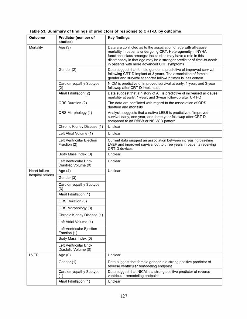
Table 53. Summary of findings of predictors of response to CRT-D, by outcome Outcome Predictor (number of
studies) Key findings
Mortality Age (3) Data are conflicted as to the association of age with all-cause mortality in patients undergoing CRT. Heterogeneity in NYHA functional class amongst the studies may have a role in this discrepancy in that age may be a stronger predictor of time-to-death in patients with more advanced CHF symptoms
Gender (2) Data suggest that female gender is predictive of improved survival following CRT-D implant at 3 years. The association of female gender and survival at shorter followup times is less certain
Cardiomyopathy Subtype (2)
NICM is predictive of improved survival at early, 1-year, and 3-year followup after CRT-D implantation
Atrial Fibrillation (2) Data suggest that a history of AF is predictive of increased all-cause mortality at early, 1-year, and 3-year followup after CRT-D
QRS Duration (2) The data are conflicted with regard to the association of QRS duration and mortality
QRS Morphology (1) Analysis suggests that a native LBBB is predictive of improved survival early, one year, and three year followup after CRT-D, compared to an RBBB or NSIVCD pattern
Chronic Kidney Disease (1) Unclear
Left Atrial Volume (1) Unclear
Left Ventricular Ejection Fraction (2)
Current data suggest an association between increasing baseline LVEF and improved survival out to three years in patients receiving CRT-D devices
Body Mass Index (0) Unclear
Left Ventricular End-Diastolic Volume (0)
Unclear
Heart failure hospitalizations
Age (4) Unclear
Gender (3)
Cardiomyopathy Subtype (3) Atrial Fibrillation (1)
QRS Duration (3)
QRS Morphology (3)
Chronic Kidney Disease (1)
Left Atrial Volume (4)
Left Ventricular Ejection Fraction (1) Body Mass Index (0)
Left Ventricular End-Diastolic Volume (0)
LVEF Age (0) Unclear
Gender (1) Data suggest that female gender is a strong positive predictor of reverse ventricular remodeling endpoint
Cardiomyopathy Subtype (1)
Data suggest that NICM is a strong positive predictor of reverse ventricular remodeling endpoint
Atrial Fibrillation (1) Unclear
127

Outcome Predictor (number of studies)
Key findings
QRS Duration (1) Data suggest that QRS duration is directly associated with improved rates of reverse ventricular remodeling
QRS Morphology (1) Data suggest that a LBBB morphology is a strong positive predictor of reverse ventricular remodeling
Chronic Kidney Disease (0) Unclear
Left Atrial Volume (1) Data suggest that left atrial volume is a predictor in minimally symptomatic CHF population (NYHA class I-II)
Left Ventricular Ejection Fraction (1)
Baseline LVEF is a poor predictor of reverse ventricular remodeling
Body Mass Index (1) Unclear
Left Ventricular End-Diastolic Volume (0)
Unclear
LVESV Age (5) The preponderance of data suggests that age is not an important predictor of changes in LVESV
Gender (6 studies reported in 7 articles)
The preponderance of data suggest that female gender is a strong positive predictor of reverse ventricular remodeling endpoints
Cardiomyopathy subtype (5 studies in 5 articles)
The preponderance of data suggest that NICM is a strong positive predictor of reverse ventricular remodeling endpoints
Atrial Fibrillation (1) Unclear
QRS Duration (6 studies reported in 7 articles)
The preponderance of data suggest that QRS duration is directly associated with improved rates of reverse ventricular remodeling
QRS Morphology (4 studies reported in 5 articles)
The preponderance of data suggest that a LBBB morphology is a strong positive predictor of reverse ventricular remodeling
Chronic Kidney Disease (1) Unclear
Left Atrial Volume (3) Data suggest that left atrial volume is a predictor in minimally symptomatic CHF population (NYHA class I-II)
Left Ventricular Ejection Fraction (5 studies reported in 6 articles)
The data are conflicted as to the association between baseline LVEF and reverse ventricular remodeling
Body Mass Index (0) Unclear
Left Ventricular End-Diastolic Volume (1)
Unclear
LVEDV Age (0) Unclear
Gender (1) The preponderance of data suggest that female gender is a strong positive predictor of reverse ventricular remodeling endpoint
Cardiomyopathy Subtype (1)
The preponderance of data suggest that NICM is a strong positive predictor of reverse ventricular remodeling endpoint
Atrial Fibrillation (0) Unclear
QRS Duration (1) The preponderance of data suggest that QRS duration is directly associated with improved rates of reverse ventricular remodeling
QRS Morphology (0) Unclear
Chronic Kidney Disease (0) Unclear
Left Atrial Volume (1) Seem to be a predictor in minimally symptomatic CHF population (NYHA class I-II)
Left Ventricular Ejection Fraction (0)
Unclear
128
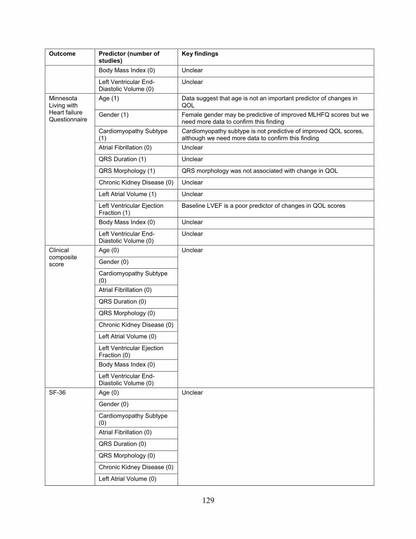
Outcome Predictor (number of studies)
Key findings
Body Mass Index (0) Unclear
Left Ventricular End-Diastolic Volume (0)
Unclear
Minnesota Living with Heart failure Questionnaire
Age (1) Data suggest that age is not an important predictor of changes in QOL
Gender (1) Female gender may be predictive of improved MLHFQ scores but we need more data to confirm this finding
Cardiomyopathy Subtype (1)
Cardiomyopathy subtype is not predictive of improved QOL scores, although we need more data to confirm this finding
Atrial Fibrillation (0) Unclear
QRS Duration (1) Unclear
QRS Morphology (1) QRS morphology was not associated with change in QOL
Chronic Kidney Disease (0) Unclear
Left Atrial Volume (1) Unclear
Left Ventricular Ejection Fraction (1)
Baseline LVEF is a poor predictor of changes in QOL scores
Body Mass Index (0) Unclear
Left Ventricular End-Diastolic Volume (0)
Unclear
Clinical composite score
Age (0) Unclear
Gender (0)
Cardiomyopathy Subtype (0) Atrial Fibrillation (0)
QRS Duration (0)
QRS Morphology (0)
Chronic Kidney Disease (0)
Left Atrial Volume (0)
Left Ventricular Ejection Fraction (0) Body Mass Index (0)
Left Ventricular End-Diastolic Volume (0)
SF-36 Age (0) Unclear
Gender (0)
Cardiomyopathy Subtype (0) Atrial Fibrillation (0)
QRS Duration (0)
QRS Morphology (0)
Chronic Kidney Disease (0)
Left Atrial Volume (0)
129
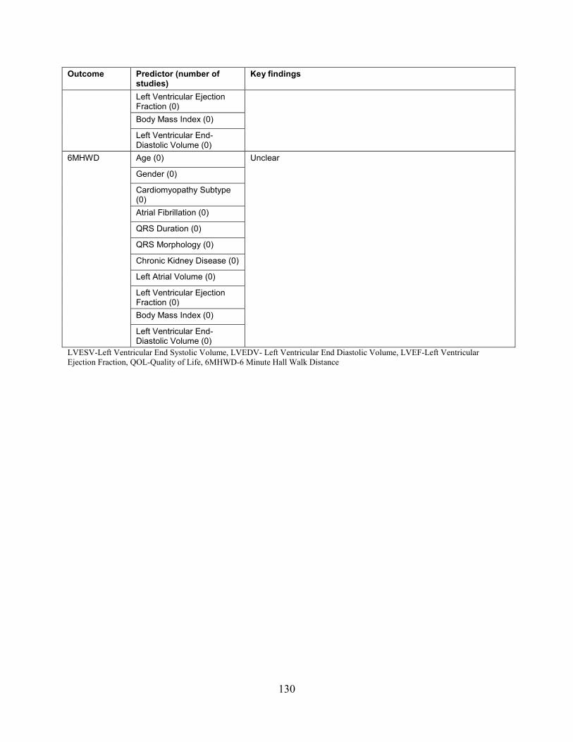
Outcome Predictor (number of studies)
Key findings
Left Ventricular Ejection Fraction (0) Body Mass Index (0)
Left Ventricular End-Diastolic Volume (0)
6MHWD Age (0) Unclear
Gender (0)
Cardiomyopathy Subtype (0) Atrial Fibrillation (0)
QRS Duration (0)
QRS Morphology (0)
Chronic Kidney Disease (0)
Left Atrial Volume (0)
Left Ventricular Ejection Fraction (0) Body Mass Index (0)
Left Ventricular End-Diastolic Volume (0)
LVESV-Left Ventricular End Systolic Volume, LVEDV- Left Ventricular End Diastolic Volume, LVEF-Left Ventricular Ejection Fraction, QOL-Quality of Life, 6MHWD-6 Minute Hall Walk Distance
130

Predictors of Response to Cardiac Resynchronization Therapy with Pacemaker (CRT-P)
Study Characteristics Two studies evaluated predictors of response in patients undergoing CRT-P implantation
(Table 54).100 ,101 Both studies were multiple-center cohort studies and included a mixed CRT-D and CRT-P population. The studies used a multivariate model that controlled the presence of a concomitant defibrillator (CRT-D implanted) with CRT-P as the reference group. One of the studies reported the specific names of the devices implanted (Boston Scientific TR/TR2, Contak CD or Renewal I/II, and Medtronic Insync I/III).100 None of these studies reported the source of funding (Evidence Table 1).
Population Characteristics The percentage of women in the included studies ranged from 23 to 24 percent. The mean
age of participants ranged from 65 to 69.3 years old. No studies reported the race of participants. Only one study reported incidence of AF.100 In this study, 22 percent of the participants had a history of AF. Only one study reported the mean QRS duration (148.5ms).101 None of the studies reported the incidences of any type of QRS morphology. Both studies included data on NYHA class. The study by Stabile et al. (2009) included patients with NYHA II, III, and IV CHF, of whom class III represented 69 percent.100 The study by Zhang et al. (2009) included patients with NYHA class III-IV CHF, of whom 87 percent were NYHA class III.101 Both studies reported baseline LVEF, which ranged from 24.5 percent in the study by Zhang et al. (2009)101 to 28.2 percent in the CRT-P arm in the study by Stabile et al. (2009)100 Neither study reported data on renal function. Both studies were homogenous in terms of baseline patient population (Table 54). Table 54. Study characteristics of studies assessing predictor of response to CRT-P Author, year CRT-P (%) Funder Total
number of patients
Age (mean, years)
Female gender (%)
NYHA class
Race Ischemic cardiomyopathy (%)
Stabile, 2009100
50.2% NR 233 68.8 23 III-IV NR 49
Zhang, 2009101
49% NR Survivors: 175
65
25
III-IV NR 50
Non-survivors:64
68 19 III-IV NR 67
NR = not reported
131
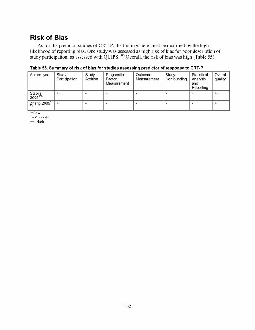
Risk of Bias As for the predictor studies of CRT-P, the findings here must be qualified by the high
likelihood of reporting bias. One study was assessed as high risk of bias for poor description of study participation, as assessed with QUIPS.100 Overall, the risk of bias was high (Table 55).
Table 55. Summary of risk of bias for studies assessing predictor of response to CRT-P Author, year Study
Participation Study Attrition
Prognostic Factor Measurement
Outcome Measurement
Study Confounding
Statistical Analysis and Reporting
Overall quality
Stabile, 2009100
++ - + - - + ++
Zhang,20091
01 + - - - - - +
-=Low +=Moderate ++=High
132

Table 56. Included predictors of response for CRT-P Author, year Age Gender CM
type AF QRSd QRS
morphology CKD LA
volume LVEF BMI LVEDV
Stabile, 2009100 X X X X X
Zhang, 2009101 X X X X X X= controlled for and effect size reported Y=controlled for but effect size not reported CM=cardiomyopathy; AF=atrial fibrillation; QRSd=QRS duration; CKD=chronic kidney disease; LA=left atrial; LVEF=left ventricular ejection fraction BMI=body mass index; LVESV=left ventricular end systolic volume
Table 57. Definitions of response for CRT-P predictors
Author, year
All-cause mortality
Cardiovascular mortality
Combined endpoint
HF hospitalizations
LVEF LVESV continuous
LVESV ≥10%
LVEDV continuous
LVEDV ≥10%
Quality of life score (MLHFQ)
6 Minute Hall Walk Distance
Clinical Composite Score
Stabile, 2009100
X
Zhang, 2009101
X
LVEF: left ventricular ejection fraction; LVESV: left ventricular end-systolic volume; LVESVi: left ventricular end-systolic volume index; LVEDV left ventricular end diastolic volume; HF: heart failure; NYHA: New York Heart Association; MLHFQ: Minnesota Living with Heart failure Questionnaire
133
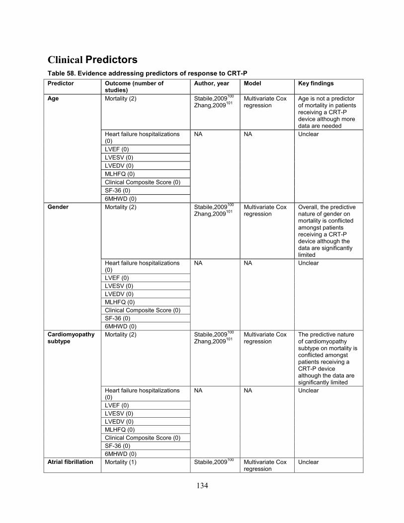
Clinical Predictors Table 58. Evidence addressing predictors of response to CRT-P Predictor Outcome (number of
studies) Author, year Model Key findings
Age Mortality (2) Stabile,2009100 Zhang,2009101
Multivariate Cox regression
Age is not a predictor of mortality in patients receiving a CRT-P device although more data are needed
Heart failure hospitalizations (0)
NA
NA
Unclear
LVEF (0) LVESV (0) LVEDV (0) MLHFQ (0) Clinical Composite Score (0) SF-36 (0) 6MHWD (0)
Gender Mortality (2) Stabile,2009100 Zhang,2009101
Multivariate Cox regression
Overall, the predictive nature of gender on mortality is conflicted amongst patients receiving a CRT-P device although the data are significantly limited
Heart failure hospitalizations (0)
NA
NA
Unclear
LVEF (0) LVESV (0) LVEDV (0) MLHFQ (0) Clinical Composite Score (0) SF-36 (0) 6MHWD (0)
Cardiomyopathy subtype
Mortality (2) Stabile,2009100 Zhang,2009101
Multivariate Cox regression
The predictive nature of cardiomyopathy subtype on mortality is conflicted amongst patients receiving a CRT-P device although the data are significantly limited
Heart failure hospitalizations (0)
NA
NA
Unclear
LVEF (0) LVESV (0) LVEDV (0) MLHFQ (0) Clinical Composite Score (0) SF-36 (0) 6MHWD (0)
Atrial fibrillation
Mortality (1) Stabile,2009100 Multivariate Cox regression
Unclear
134

Predictor Outcome (number of studies)
Author, year Model Key findings
Heart failure hospitalizations (0)
NA
NA
LVEF (0) LVESV (0) LVEDV (0) MLHFQ (0) Clinical Composite Score (0) SF-36 (0) 6MHWD (0)
QRS duration
Mortality (2) Stabile,2009100 Zhang,2009101
Multivariate Cox regression
QRS duration is not a predictor of mortality in patients receiving a CRT-P device.
Heart failure hospitalizations (0)
NA
NA
Unclear
LVEF (0) LVESV (0) LVEDV (0) MLHFQ (0) Clinical Composite Score (0) SF-36 (0) 6MHWD (0)
QRS morphology
Mortality (0) NA
NA
Unclear Heart failure hospitalizations
(0) LVEF (0) LVESV (0) LVEDV (0) MLHFQ (0) Clinical Composite Score (0) SF-36 (0) 6MHWD (0)
Chronic kidney disease
Mortality (0) NA
NA
Unclear Heart failure hospitalizations
(0) LVEF (0) LVESV (0) LVEDV (0) MLHFQ (0) Clinical Composite Score (0) SF-36 (0) 6MHWD (0)
Left atrial volume
Mortality (0) NA
NA
Unclear Heart failure hospitalizations
(0) LVEF (0) LVESV (0) LVEDV (0) MLHFQ (0) Clinical Composite Score (0) SF-36 (0) 6MHWD (0)
135
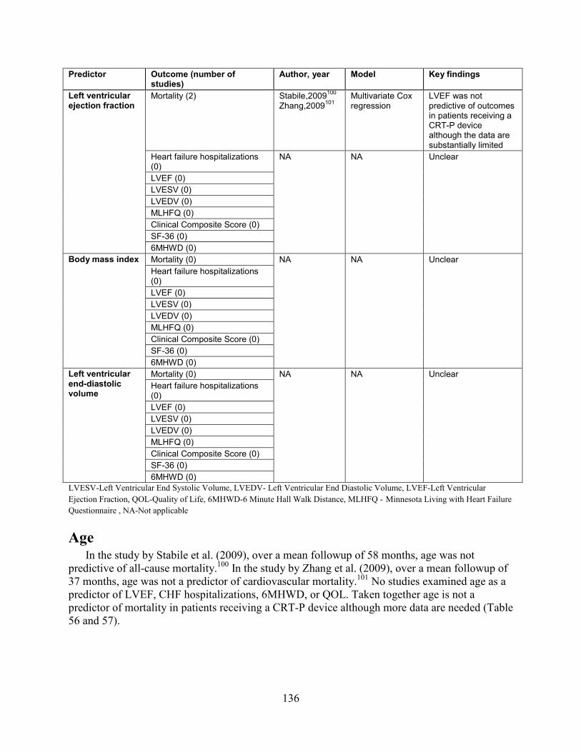
Predictor Outcome (number of studies)
Author, year Model Key findings
Left ventricular ejection fraction
Mortality (2) Stabile,2009100 Zhang,2009101
Multivariate Cox regression
LVEF was not predictive of outcomes in patients receiving a CRT-P device although the data are substantially limited
Heart failure hospitalizations (0)
NA
NA
Unclear
LVEF (0) LVESV (0) LVEDV (0) MLHFQ (0) Clinical Composite Score (0) SF-36 (0) 6MHWD (0)
Body mass index
Mortality (0) NA
NA
Unclear Heart failure hospitalizations (0) LVEF (0) LVESV (0) LVEDV (0) MLHFQ (0) Clinical Composite Score (0) SF-36 (0) 6MHWD (0)
Left ventricular end-diastolic volume
Mortality (0) NA
NA
Unclear Heart failure hospitalizations (0) LVEF (0) LVESV (0) LVEDV (0) MLHFQ (0) Clinical Composite Score (0) SF-36 (0) 6MHWD (0)
LVESV-Left Ventricular End Systolic Volume, LVEDV- Left Ventricular End Diastolic Volume, LVEF-Left Ventricular Ejection Fraction, QOL-Quality of Life, 6MHWD-6 Minute Hall Walk Distance, MLHFQ - Minnesota Living with Heart Failure Questionnaire , NA-Not applicable
Age In the study by Stabile et al. (2009), over a mean followup of 58 months, age was not
predictive of all-cause mortality.100 In the study by Zhang et al. (2009), over a mean followup of 37 months, age was not a predictor of cardiovascular mortality.101 No studies examined age as a predictor of LVEF, CHF hospitalizations, 6MHWD, or QOL. Taken together age is not a predictor of mortality in patients receiving a CRT-P device although more data are needed (Table 56 and 57).
136
Gender In the study by Stabile et al. (2009), over a mean followup of 58 months, male gender was
predictive of worsened survival (HR: 3.62, 95% CI, 1.88 to 6.99, p=0.006).100 In the study by Zhang et al. (2009), over a mean followup of 37 months, gender was not a predictor of cardiovascular mortality (HR: 0.679, 95% CI, 0.329 to 1.399, p=0.294).101 No studies examined gender as a predictor of LVEF, CHF hospitalizations, 6MHWD, or QOL. Overall, the predictive nature of gender on mortality is conflicted amongst patients receiving a CRT-P device although the data are significantly limited (Table 56 and 57).
Cardiomyopathy Subtype In the study by Stabile et al. (2009), over a mean followup of 58 months, cardiomyopathy
subtype was not predictive of worsened survival (for ICM HR: 1.08, 95% CI, 0.63 to 1.85, p=0.405).100 In the study by Zhang et al. (2009), over a mean followup of 37 months, ICM was a predictor of cardiovascular mortality (HR: 2.696, 95% CI, 1.487 to 4.889, p=0.001).101 No studies examined cardiomyopathy subtype as a predictor of LVEF, CHF hospitalizations, 6MHWD, or QOL. Taken together, the predictive nature of cardiomyopathy subtype on mortality is conflicted amongst patients receiving a CRT-P device although the data are significantly limited (Table 56 and 57).
Atrial Fibrillation One study reported history of AF as a predictor of survival in patients receiving a CRT-P
device.100 This study reported a mean followup of 58 months; a history of AF was predictive of worsened survival (HR: 1.96, 95% CI, 1.01 to 3.94, p=0.038). No studies examined AF as a predictor of LVEF, CHF hospitalizations, 6MHWD, or QOL. Therefore, there is insufficient evidence to determine the predictive nature of AF on outcomes following CRT-P (Table 56 and 57).
QRS Duration Both studies assessed QRS duration as a predictor of survival in patients receiving a CRT-P
device.100 ,101 In the study by Stabile et al. (2009), over a mean followup of 58 months, QRS duration ≥160ms was not predictive of worsened survival.100 In the study by Zhang et al. (2009), over a mean followup of 37 months, QRS duration was not a predictor of cardiovascular mortality.101 No studies examined QRS duration as a predictor of LVEF, CHF hospitalizations, 6MHWD, or QOL. Taken together QRS was not predictive of outcomes in patients receiving a CRT-P device although the data are substantially limited (Table 56 and 57).
QRS Morphology No studies included QRS morphology as a predictor of any outcome in patients receiving CRT-P (Table 56 and 57).
Chronic Kidney Disease No studies included CKD as a predictor of any outcome in patients receiving CRT-P (Table 56 and 57).
137
Left Atrial Volume No studies included LAV as a predictor of any outcome in patients receiving CRT-P (Table 56 and 57).
Left Ventricular Ejection Fraction Two studies examined baseline LVEF as a predictor of survival in patients receiving a CRT-
P device.100 ,101 In the study by Stabile et al. (2009), over a mean followup of 58 months, baseline LVEF was not predictive of worsened survival.100 In the study by Zhang et al. (2009), over a mean followup of 37 months, baseline LVEF was not a predictor of cardiovascular mortality.101 No studies examined baseline as a predictor of LVEF response, CHF hospitalizations, 6MHWD, or QOL. Taken together LVEF was not predictive of outcomes in patients receiving a CRT-P device although the data are substantially limited (Table 56 and 57).
Body Mass Index No studies of CRT-P included BMI as a potential predictor of response (Table 56 and 57).
Left Ventricular End-Diastolic Volume No studies of CRT-P included LVEDV as a potential predictor of response (Table 56 and 57).
138

Table 59. Summary of findings of predictors of response to CRT-P, by outcomes Outcome Predictor (number of
studies) Key findings
Mortality Age (2) Age is not a predictor of mortality in patients receiving a CRT-P device although more data are needed
Gender (2) Overall, the predictive nature of gender on mortality is conflicted amongst patients receiving a CRT-P device although the data are significantly limited
Cardiomyopathy subtype (2)
The predictive nature of cardiomyopathy subtype on mortality is conflicted amongst patients receiving a CRT-P device although the data are significantly limited
Atrial fibrillation (1) Unclear
QRS duration (2) QRS duration was not a significant predictor of mortality QRS morphology (0) Analysis suggests that a native LBBB is predictive of improved survival
early, one year, and three year followup after CRT-D, compared to an RBBB or NSIVCD pattern
Chronic kidney disease (0)
Unclear
Left atrial volume (0) Unclear Left ventricular ejection fraction (2)
LVEF was not predictive of outcomes in patients receiving a CRT-P device although the data are substantially limited
Body mass index (0) Unclear Left ventricular end-diastolic volume (0)
Unclear
Heart failure hospitalizations, LVEF, LVESV, LVEDV, MLHFQ, Clinical composite score, SF-36, 6MHWD
Age (0) Unclear Gender (0)
Cardiomyopathy subtype (0) Atrial fibrillation (0)
QRS duration (0) QRS morphology (0) Chronic kidney disease (0) Left atrial volume (0) Left ventricular ejection fraction (0) Body mass index (0) Left ventricular end-diastolic volume (0) Left atrial volume (0) Left ventricular ejection fraction (0) Body mass index (0) Left ventricular end-diastolic volume (0)
LVESV-Left Ventricular End Systolic Volume, LVEDV- Left Ventricular End Diastolic Volume, LVEF-Left Ventricular Ejection Fraction, MLHFQ-Minnesota Living with Heart Failure Questionnaire, 6MHWD-6 Minute Hall Walk Distance
139
Discussion
Key Findings and the Strength of Evidence Efficacy and Safety of CRT-D (KQ1a, KQ2)
There is convincing evidence that CRT-D devices are effective in reducing heart failure symptoms, improving myocardial function, and reducing hospitalizations for heart failure in patients with an LVEF≤35% and a QRS duration ≥120 ms compared to therapy with an ICD alone. Specifically, we found moderate strength of evidence for benefit of CRT-D versus ICD alone for all-cause mortality in minimally symptomatic patients. This statement is derived from data looking primarily at NYHA class II patients. The applicability of this finding to NYHA class I patients, a population significantly under-represented in studies, is unclear. There is insufficient evidence to determine whether CRT-D devices are effective in improving survival compared to an ICD alone in an advanced heart failure population (NYHA III-IV).(Table 60)
In terms of pre-specified subgroups, there is compelling evidence that in CRT-D patients (compared to an ICD alone), female gender, a left bundle branch block, and non-ischemic cardiomyopathy are associated with superior outcomes. Sinus rhythm (as opposed to a history of atrial fibrillation) and a wider QRS complex and also associated with superior outcomes in patients undergoing CRT-D implant compared to an ICD alone although the data for this are less compelling.
Prevalence of harms associated with CRT-D devices were as follows: cardiac perforation/tamponade (0.1-1.4%), pocket hematoma (0.9-2.8%), pneumothorax (1.3-2.8%), device infection (0.9-2.8%), and lead dislodgement (2.4% to 9.8%). No conclusions could be drawn about the association between CRT-D implant and both ventricular arrhythmias and inappropriate shocks. Death within one week of implantation was 0 percent although only two studies reported this outcome.
Efficacy and Safety of CRT-P (KQ3a, KQ4) There is moderate evidence that CRT-P, compared to optimal medical therapy, is effective in
improving survival, reducing LESV, and reducing hospitalizations for heart failure in patients with an LVEF≤35% and a QRS duration ≥120 ms compared to optimal medical therapy alone. These data are largely derived from patients with NYHA class III-IV heart failure. The applicability of these findings to patient with NYHA class I-II heart failure is unclear. We found insufficient evidence about the effect of CRT-P on quality of life as measured with the MLHFQ.
Harms associated with CRT-P were as follows: cardiac perforation/tamponade (0-1.6%), pocket hematoma (0.2-9.5%), pneumothorax (0.5-1.5%), device infection (0.7-4.8%), and lead dislodgement (1.7-17%). Death within one week of implantation was reported in only very small studies making the true incidence unclear.
Efficacy and Safety of CRT-D versus CRT-P (KQ5, KQ6) Only one included trial contained both CRT-D and CRT-P arms, and direct comparisons
between those arms were lacking. Therefore there is insufficient evidence to determine the effectiveness of CRT-D compared to CRT-P. In comparing harms between CRT-D and CRT-P devices, there was also insufficient evidence to draw any conclusions except for device infections, which appear to be slightly more common for CRT-D devices.
140

Predictors of Response: CRT-D and CRT-P (KQ1b, KQ3b) The evidence regarding predictors of a favorable response following CRT varied
considerably based on outcome. In addition, the high likelihood of reporting bias qualifies these results. Age was not an important predictor of outcomes in patients receiving CRT-D devices. However, data for very elderly patients (> 75 years of age) was limited. Non-ischemic cardiomyopathy, female gender, and a left bundle branch block morphology were strongly associated with improved outcomes. A history of atrial fibrillation and a narrower QRS duration were associated with poorer outcomes although the evidence for this was less robust. There was insufficient data to determine the predictive nature of chronic kidney disease, left atrial volume, baseline LVEF, body mass index, and left ventricular end-diastolic volume on outcomes following CRT-D implant. There was also insufficient evidence to draw conclusions as to the predictive nature of baseline characteristics and outcomes in patients receiving a CRT-P device. However, of the outcomes that were assessed, the ICD function would impact only the mortality endpoint. Therefore similar conclusions as to those noted for CRT-D can likely be drawn for CRT-P devices for the other, non-mortality endpoints (Table 60).
141
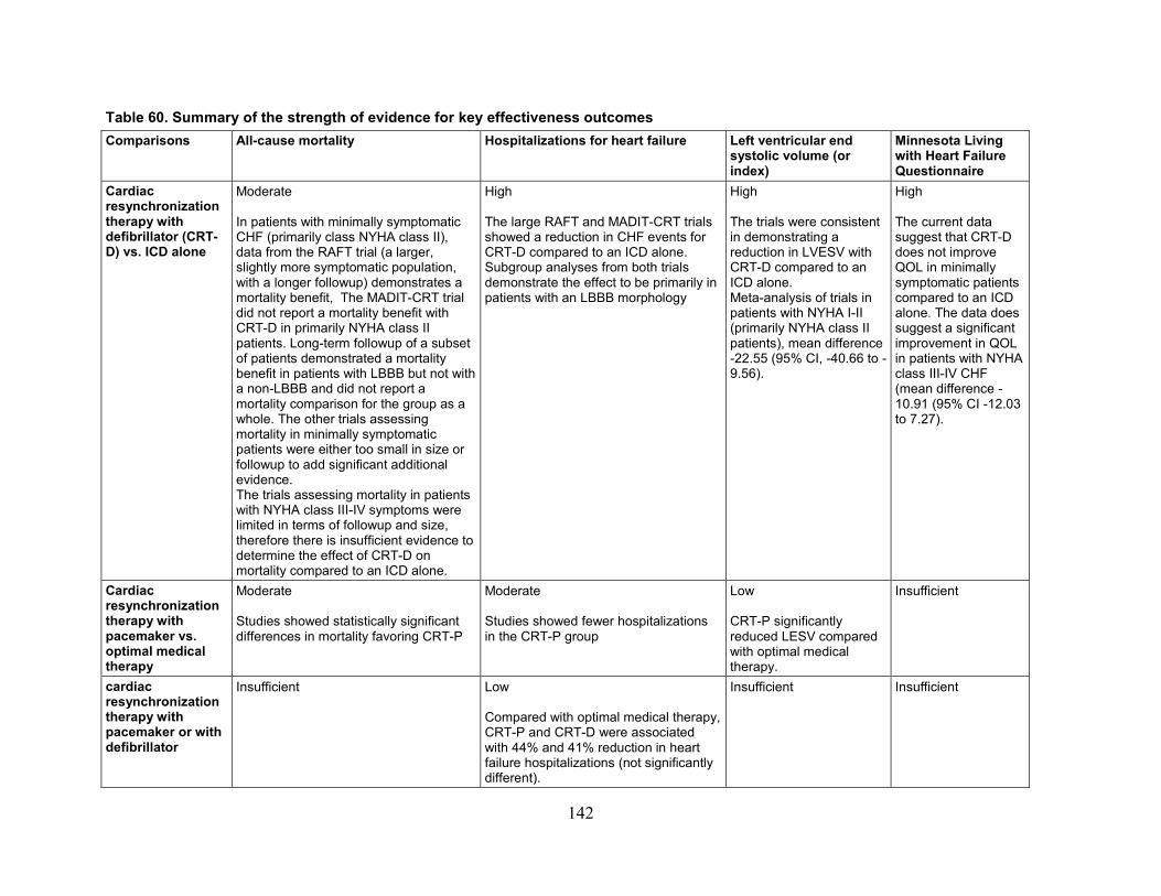
Table 60. Summary of the strength of evidence for key effectiveness outcomes Comparisons All-cause mortality Hospitalizations for heart failure Left ventricular end
systolic volume (or index)
Minnesota Living with Heart Failure Questionnaire
Cardiac resynchronization therapy with defibrillator (CRT-D) vs. ICD alone
Moderate In patients with minimally symptomatic CHF (primarily class NYHA class II), data from the RAFT trial (a larger, slightly more symptomatic population, with a longer followup) demonstrates a mortality benefit, The MADIT-CRT trial did not report a mortality benefit with CRT-D in primarily NYHA class II patients. Long-term followup of a subset of patients demonstrated a mortality benefit in patients with LBBB but not with a non-LBBB and did not report a mortality comparison for the group as a whole. The other trials assessing mortality in minimally symptomatic patients were either too small in size or followup to add significant additional evidence. The trials assessing mortality in patients with NYHA class III-IV symptoms were limited in terms of followup and size, therefore there is insufficient evidence to determine the effect of CRT-D on mortality compared to an ICD alone.
High The large RAFT and MADIT-CRT trials showed a reduction in CHF events for CRT-D compared to an ICD alone. Subgroup analyses from both trials demonstrate the effect to be primarily in patients with an LBBB morphology
High The trials were consistent in demonstrating a reduction in LVESV with CRT-D compared to an ICD alone. Meta-analysis of trials in patients with NYHA I-II (primarily NYHA class II patients), mean difference -22.55 (95% CI, -40.66 to -9.56).
High The current data suggest that CRT-D does not improve QOL in minimally symptomatic patients compared to an ICD alone. The data does suggest a significant improvement in QOL in patients with NYHA class III-IV CHF (mean difference -10.91 (95% CI -12.03 to 7.27).
Cardiac resynchronization therapy with pacemaker vs. optimal medical therapy
Moderate Studies showed statistically significant differences in mortality favoring CRT-P
Moderate Studies showed fewer hospitalizations in the CRT-P group
Low CRT-P significantly reduced LESV compared with optimal medical therapy.
Insufficient
cardiac resynchronization therapy with pacemaker or with defibrillator
Insufficient Low Compared with optimal medical therapy, CRT-P and CRT-D were associated with 44% and 41% reduction in heart failure hospitalizations (not significantly different).
Insufficient Insufficient
142
Relationship of Findings to Existing Literature Several systematic reviews have focused on CRT but none has performed a comprehensive
assessment of predictors of outcomes following CRT (see Table 61). Our current review also differs from prior reviews in that only studies with patients with an LVEF≤35% and a baseline QRS duration≥120 ms undergoing biventricular pacing were included. These criteria were developed in consultation with our key informants and largely mirror the current appropriate use criteria for CRT based on guidelines.1 This eliminated the REVERSE, BLOCK-HF, and HOBIPACE trials which included patients with LVEFs >35%.102-104 In addition, all trials looking at the effects of CRT in a narrow QRS population,105-107 and studies of LV only pacing were excluded.108 ,109 We considered the appropriate control for the CRT-D effectiveness question to be an ICD alone given the compelling data demonstrating improvements in mortality with an ICD that evolved concomitantly with studies of CRT effectiveness. We considered the appropriate control and for CRT-P to be optimal medical therapy alone to assess the impact of cardiac resynchronization. We did not assess the comparison of CRT-D to optimal medical therapy as we determined this to be an inappropriate comparison, given the known improvements in mortality by defibrillation. Also, in contrast to several previous reviews, we included only RCTs to assess the key questions regarding effectiveness.
In terms of minimally symptomatic patients, the results of our review largely agree with those of prior reviews, which focused on the same population. Similarly, the current review is in agreement with the systematic review performed in 2007 by Mcallister et al., which included studies primarily involving an advanced heart failure population.110 Our review arrived at somewhat different conclusions in terms of the efficacy of CRT-D vs. CRT-P compared to that by Jiang et al.111 Given that we considered only RCTs for determination of effectiveness, only the COMPANION trial was included in our review, which likely explains the discrepancy in conclusions.4
In our systematic analysis of predictors of outcomes following CRT, many studies assessing the capacity for baseline characteristics to predict responses (defined in many different ways) were identified. The large majority of studies were small (<100 patients) and not properly controlled. At a minimum, we pre-specified that a cohort study address our questions about predictors of response to CRT had to include at least gender and either QRS duration or morphology in a multivariate model to address confounding. Such criteria eliminated many studies. Despite this, the positive predictive effect noted with LBBB, female gender, non-ischemic cardiomyopathy, a wider QRS duration, and normal sinus rhythm on multiple outcomes was supportive of the similar findings noted from the pre-specified subgroup analyses of the RCTs. There are other potential predictors we did not consider (e.g. lead position). Given the large number of potential predictors in the literature, a review of all predictors was not practical. Our predictors were chosen based on prevailing knowledge of the most important predictors, identified in consultation with our key informants.
Finally, we did not conduct individual patient data meta-analysis to assess predictors meaning that our analyses may suggest that clinically relevant subgroup effects exist, but we are unable to quantify the effects reliably or precisely.
143
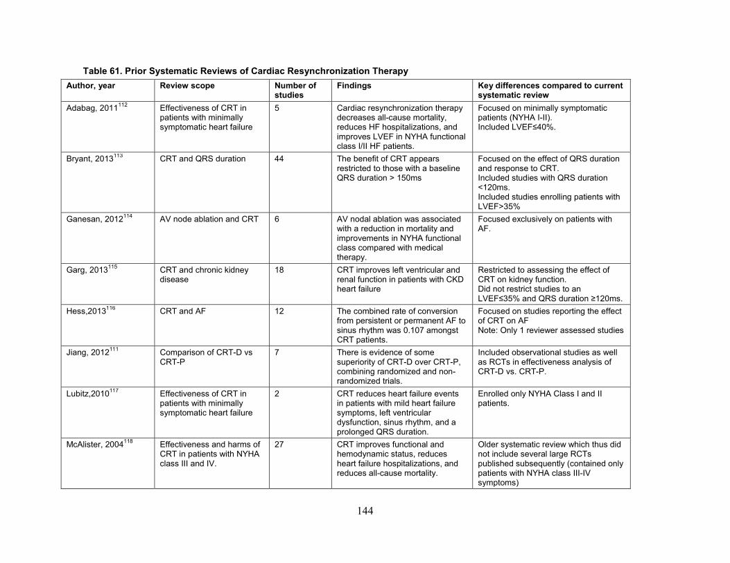
Table 61. Prior Systematic Reviews of Cardiac Resynchronization Therapy Author, year Review scope Number of
studies Findings Key differences compared to current
systematic review Adabag, 2011112 Effectiveness of CRT in
patients with minimally symptomatic heart failure
5 Cardiac resynchronization therapy decreases all-cause mortality, reduces HF hospitalizations, and improves LVEF in NYHA functional class I/II HF patients.
Focused on minimally symptomatic patients (NYHA I-II). Included LVEF≤40%.
Bryant, 2013113 CRT and QRS duration 44 The benefit of CRT appears restricted to those with a baseline QRS duration > 150ms
Focused on the effect of QRS duration and response to CRT. Included studies with QRS duration <120ms. Included studies enrolling patients with LVEF>35%
Ganesan, 2012114 AV node ablation and CRT 6 AV nodal ablation was associated with a reduction in mortality and improvements in NYHA functional class compared with medical therapy.
Focused exclusively on patients with AF.
Garg, 2013115 CRT and chronic kidney disease
18 CRT improves left ventricular and renal function in patients with CKD heart failure
Restricted to assessing the effect of CRT on kidney function. Did not restrict studies to an LVEF≤35% and QRS duration ≥120ms.
Hess,2013116 CRT and AF 12 The combined rate of conversion from persistent or permanent AF to sinus rhythm was 0.107 amongst CRT patients.
Focused on studies reporting the effect of CRT on AF Note: Only 1 reviewer assessed studies
Jiang, 2012111 Comparison of CRT-D vs CRT-P
7 There is evidence of some superiority of CRT-D over CRT-P, combining randomized and non-randomized trials.
Included observational studies as well as RCTs in effectiveness analysis of CRT-D vs. CRT-P.
Lubitz,2010117 Effectiveness of CRT in patients with minimally symptomatic heart failure
2 CRT reduces heart failure events in patients with mild heart failure symptoms, left ventricular dysfunction, sinus rhythm, and a prolonged QRS duration.
Enrolled only NYHA Class I and II patients.
McAlister, 2004118 Effectiveness and harms of CRT in patients with NYHA class III and IV.
27 CRT improves functional and hemodynamic status, reduces heart failure hospitalizations, and reduces all-cause mortality.
Older systematic review which thus did not include several large RCTs published subsequently (contained only patients with NYHA class III-IV symptoms)
144
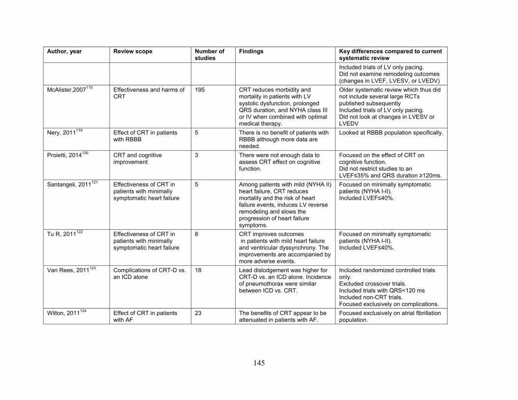
Author, year Review scope Number of studies
Findings Key differences compared to current systematic review Included trials of LV only pacing. Did not examine remodeling outcomes (changes in LVEF, LVESV, or LVEDV)
McAlister,2007110 Effectiveness and harms of CRT
195 CRT reduces morbidity and mortality in patients with LV systolic dysfunction, prolonged QRS duration, and NYHA class III or IV when combined with optimal medical therapy.
Older systematic review which thus did not include several large RCTs published subsequently Included trials of LV only pacing. Did not look at changes in LVESV or LVEDV
Nery, 2011119
Effect of CRT in patients with RBBB
5 There is no benefit of patients with RBBB although more data are needed.
Looked at RBBB population specifically.
Proietti, 2014120 CRT and cognitive improvement
3 There were not enough data to assess CRT effect on cognitive function.
Focused on the effect of CRT on cognitive function. Did not restrict studies to an LVEF≤35% and QRS duration ≥120ms.
Santangeli, 2011121
Effectiveness of CRT in patients with minimally symptomatic heart failure
5 Among patients with mild (NYHA II) heart failure, CRT reduces mortality and the risk of heart failure events, induces LV reverse remodeling and slows the progression of heart failure symptoms.
Focused on minimally symptomatic patients (NYHA I-II). Included LVEF≤40%.
Tu R, 2011122 Effectiveness of CRT in patients with minimally symptomatic heart failure
8 CRT improves outcomes in patients with mild heart failure and ventricular dyssynchrony. The improvements are accompanied by more adverse events.
Focused on minimally symptomatic patients (NYHA I-II). Included LVEF≤40%.
Van Rees, 2011123 Complications of CRT-D vs. an ICD alone
18 Lead dislodgement was higher for CRT-D vs. an ICD alone. Incidence of pneumothorax were similar between ICD vs. CRT.
Included randomized controlled trials only. Excluded crossover trials. Included trials with QRS<120 ms Included non-CRT trials. Focused exclusively on complications.
Wilton, 2011124 Effect of CRT in patients with AF
23 The benefits of CRT appear to be attenuated in patients with AF.
Focused exclusively on atrial fibrillation population.
145
Applicability The generalizability of these results is slightly limited. Race was reported very infrequently,
prohibiting an assessment of applicability based on racial differences. The majority of patients included in the RCTs were male, although a large focus in sub-studies has been given to the role of CRT in women, given the heightened response to therapy seen in this population. The average age in the RCTs and cohort studies was in the mid 60s although many patients included were in the age range of the elderly Medicare population. There has not been an RCT that specifically enrolled Medicare eligible patients. Also, data for very elderly patients (> 75 years of age) are limited. In cohort studies and subgroup analyses from the RCTs, age was not found to be an important predictor of outcomes. Taken together, the results of our review are fairly generalizable to the Medicare population although given the absence of dedicated RCTs, a definitive statement of generalizability to this population is not possible.
Limitations of the Comparative Effectiveness Review Process
In addressing the questions of efficacy of CRT-D and CRT-P (KQs 1A, 3A, and 5), several studies potentially of interest were excluded since they were non-randomized. For the questions about the predictors of response to CRT (KQs 1b and 3b), many retrospective cohort studies were excluded because of a mixed population of patients who received CRT-D or CRT-P devices. We attempted to contact the authors of such studies to obtain the device-specific data. We contacted the authors of 24 studies, of which we received responses from 2 authors with sufficient information to include in our review. In addition, many cohorts, which contained outcomes of interest were excluded due to failure to control for gender and QRS duration and/or morphology, important baseline confounders. Finally, we did not include prior or conduct new individual patient data meta-analyses to assess predictors. Therefore, our analyses may suggest that clinically relevant subgroup effects exist, but we are unable to quantify the effects reliably or precisely.
Limitations of the Evidence Base Multiple well-conducted RCTs were identified addressing the questions about the efficacy or
CRT-D and CRT-P (KQs 1a and 3a). The majority of patients enrolled in the clinical trials had NYHA class II-IV heart failure symptoms. The applicability of the current findings to class I patients is less clear. In contrast, for the comparison of CRT-D with CRT-P (KQ 5), only the COMPANION trial was found to include both CRT-D and CRT-P arms.4 However, a direct comparison of the CRT-D to CRT-P arms was not reported for several outcomes. For the questions examining predictors of response to CRT (KQs 1b and 3b), many of the included cohort studies were relatively small. While all studies controlled for gender and either QRS duration or morphology based on our pre-specified inclusion criteria, the remaining variables in the model varied widely between studies. Similarly, many studies used statistical criteria to create their multivariate adjustments, not including important a priori clinical factors.
146
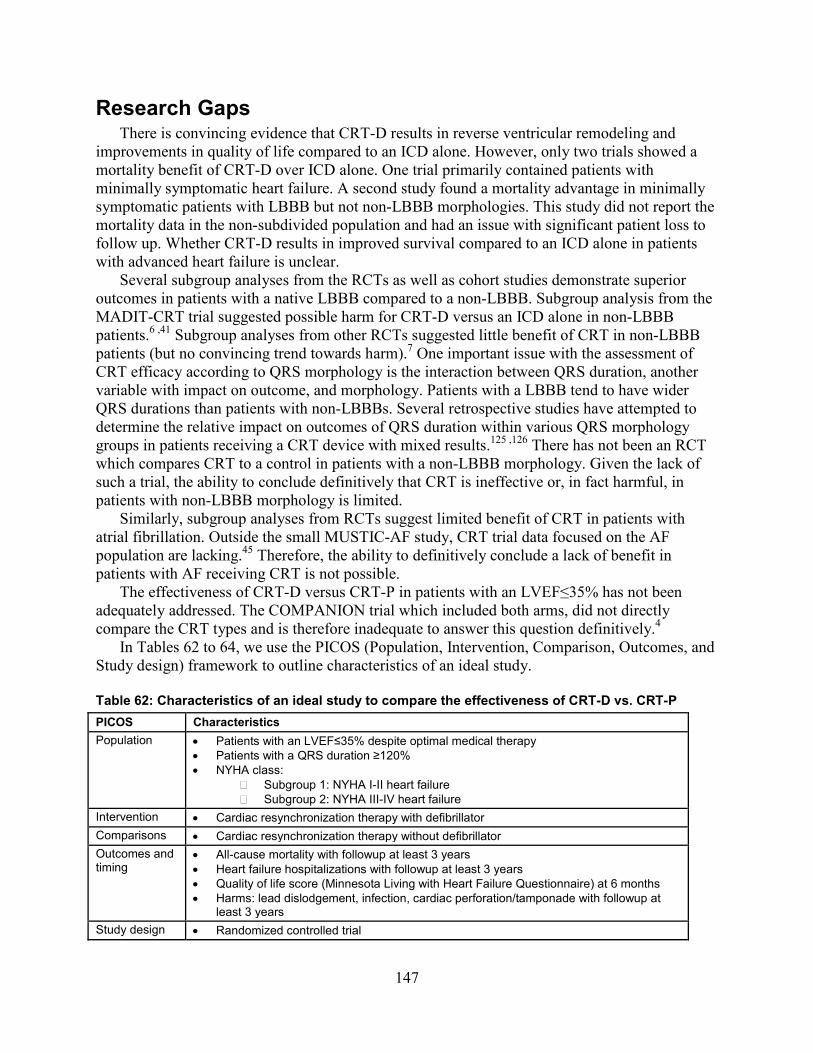
Research Gaps There is convincing evidence that CRT-D results in reverse ventricular remodeling and
improvements in quality of life compared to an ICD alone. However, only two trials showed a mortality benefit of CRT-D over ICD alone. One trial primarily contained patients with minimally symptomatic heart failure. A second study found a mortality advantage in minimally symptomatic patients with LBBB but not non-LBBB morphologies. This study did not report the mortality data in the non-subdivided population and had an issue with significant patient loss to follow up. Whether CRT-D results in improved survival compared to an ICD alone in patients with advanced heart failure is unclear.
Several subgroup analyses from the RCTs as well as cohort studies demonstrate superior outcomes in patients with a native LBBB compared to a non-LBBB. Subgroup analysis from the MADIT-CRT trial suggested possible harm for CRT-D versus an ICD alone in non-LBBB patients.6 ,41 Subgroup analyses from other RCTs suggested little benefit of CRT in non-LBBB patients (but no convincing trend towards harm).7 One important issue with the assessment of CRT efficacy according to QRS morphology is the interaction between QRS duration, another variable with impact on outcome, and morphology. Patients with a LBBB tend to have wider QRS durations than patients with non-LBBBs. Several retrospective studies have attempted to determine the relative impact on outcomes of QRS duration within various QRS morphology groups in patients receiving a CRT device with mixed results.125 ,126 There has not been an RCT which compares CRT to a control in patients with a non-LBBB morphology. Given the lack of such a trial, the ability to conclude definitively that CRT is ineffective or, in fact harmful, in patients with non-LBBB morphology is limited.
Similarly, subgroup analyses from RCTs suggest limited benefit of CRT in patients with atrial fibrillation. Outside the small MUSTIC-AF study, CRT trial data focused on the AF population are lacking.45 Therefore, the ability to definitively conclude a lack of benefit in patients with AF receiving CRT is not possible.
The effectiveness of CRT-D versus CRT-P in patients with an LVEF≤35% has not been adequately addressed. The COMPANION trial which included both arms, did not directly compare the CRT types and is therefore inadequate to answer this question definitively.4
In Tables 62 to 64, we use the PICOS (Population, Intervention, Comparison, Outcomes, and Study design) framework to outline characteristics of an ideal study. Table 62: Characteristics of an ideal study to compare the effectiveness of CRT-D vs. CRT-P PICOS Characteristics Population • Patients with an LVEF≤35% despite optimal medical therapy
• Patients with a QRS duration ≥120% • NYHA class:
� Subgroup 1: NYHA I-II heart failure � Subgroup 2: NYHA III-IV heart failure
Intervention • Cardiac resynchronization therapy with defibrillator Comparisons • Cardiac resynchronization therapy without defibrillator Outcomes and timing
• All-cause mortality with followup at least 3 years • Heart failure hospitalizations with followup at least 3 years • Quality of life score (Minnesota Living with Heart Failure Questionnaire) at 6 months • Harms: lead dislodgement, infection, cardiac perforation/tamponade with followup at
least 3 years Study design • Randomized controlled trial
147
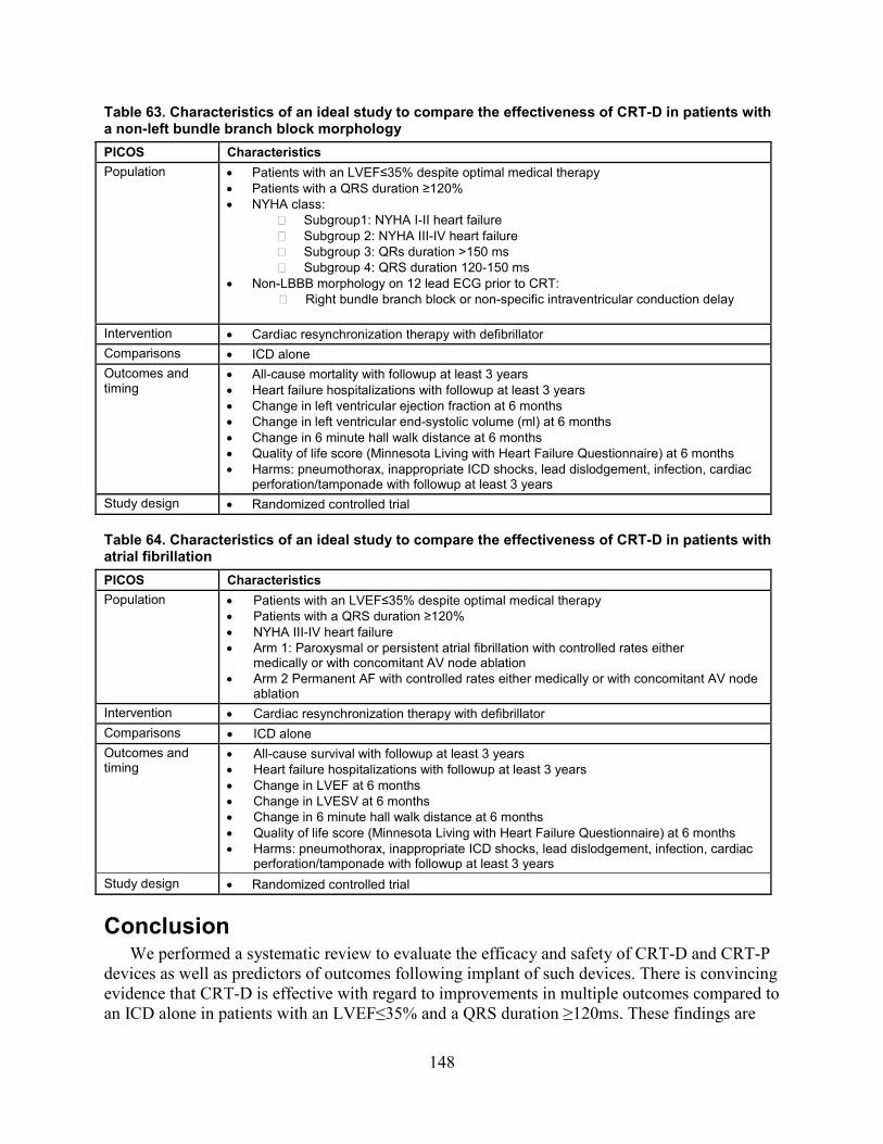
Table 63. Characteristics of an ideal study to compare the effectiveness of CRT-D in patients with a non-left bundle branch block morphology PICOS Characteristics Population • Patients with an LVEF≤35% despite optimal medical therapy
• Patients with a QRS duration ≥120% • NYHA class:
� Subgroup1: NYHA I-II heart failure � Subgroup 2: NYHA III-IV heart failure � Subgroup 3: QRs duration >150 ms � Subgroup 4: QRS duration 120-150 ms
• Non-LBBB morphology on 12 lead ECG prior to CRT: � Right bundle branch block or non-specific intraventricular conduction delay
Intervention • Cardiac resynchronization therapy with defibrillator Comparisons • ICD alone Outcomes and timing
• All-cause mortality with followup at least 3 years • Heart failure hospitalizations with followup at least 3 years • Change in left ventricular ejection fraction at 6 months • Change in left ventricular end-systolic volume (ml) at 6 months • Change in 6 minute hall walk distance at 6 months • Quality of life score (Minnesota Living with Heart Failure Questionnaire) at 6 months • Harms: pneumothorax, inappropriate ICD shocks, lead dislodgement, infection, cardiac
perforation/tamponade with followup at least 3 years Study design • Randomized controlled trial Table 64. Characteristics of an ideal study to compare the effectiveness of CRT-D in patients with atrial fibrillation PICOS Characteristics Population • Patients with an LVEF≤35% despite optimal medical therapy
• Patients with a QRS duration ≥120% • NYHA III-IV heart failure • Arm 1: Paroxysmal or persistent atrial fibrillation with controlled rates either
medically or with concomitant AV node ablation • Arm 2 Permanent AF with controlled rates either medically or with concomitant AV node
ablation Intervention • Cardiac resynchronization therapy with defibrillator Comparisons • ICD alone Outcomes and timing
• All-cause survival with followup at least 3 years • Heart failure hospitalizations with followup at least 3 years • Change in LVEF at 6 months • Change in LVESV at 6 months • Change in 6 minute hall walk distance at 6 months • Quality of life score (Minnesota Living with Heart Failure Questionnaire) at 6 months • Harms: pneumothorax, inappropriate ICD shocks, lead dislodgement, infection, cardiac
perforation/tamponade with followup at least 3 years Study design • Randomized controlled trial
Conclusion We performed a systematic review to evaluate the efficacy and safety of CRT-D and CRT-P
devices as well as predictors of outcomes following implant of such devices. There is convincing evidence that CRT-D is effective with regard to improvements in multiple outcomes compared to an ICD alone in patients with an LVEF≤35% and a QRS duration ≥120ms. These findings are
148

based on patients primarily with NYHA class II-IV heart failure. The applicability of these findings to patients with NYHA class I symptoms is unclear. Similarly, there is convincing evidence that CRT-P is effective in improving multiple endpoints compared to optimal medical therapy alone in the same population. These data are primarily derived from NYHA class III-IV and the applicability to patients with NYHA class I and II is less clear. Female gender, LBBB, a widened QRS duration, sinus rhythm, and non-ischemic cardiomyopathy are associated with improved outcomes following CRT.
149

Reference List
1. Heart Failure Society of America (HFSA) practice
guidelines. HFSA guidelines for management of patients with heart failure caused by left ventricular systolic dysfunction--pharmacological approaches. J Card Fail 1999; 5(4):357-82. PMID: 10634677
2. Lloyd-Jones D, Adams RJ, Brown TM et al. Heart disease and stroke statistics--2010 update: a report from the American Heart Association. Circulation 2010; 121(7):e46-e215. PMID: 20019324
3. Abraham WT, Fisher WG, Smith AL et al. Cardiac resynchronization in chronic heart failure. N Engl J Med 2002; 346(24):1845-53. PMID: 12063368
4. Bristow MR, Saxon LA, Boehmer J et al. Cardiac-resynchronization therapy with or without an implantable defibrillator in advanced chronic heart failure. N Engl J Med 2004; 350(21):2140-50. PMID: 15152059
5. Cleland JG, Daubert JC, Erdmann E et al. The effect of cardiac resynchronization on morbidity and mortality in heart failure. N Engl J Med 2005; 352(15):1539-49. PMID: 15753115
6. Moss AJ, Hall WJ, Cannom DS et al. Cardiac-resynchronization therapy for the prevention of heart-failure events. N Engl J Med 2009; 361(14):1329-38. PMID: 19723701
7. Tang AS, Wells GA, Talajic M et al. Cardiac-resynchronization therapy for mild-to-moderate heart failure. N Engl J Med 2010; 363(25):2385-95. PMID: 21073365
8. Cazeau S, Leclercq C, Lavergne T et al. Effects of multisite biventricular pacing in patients with heart failure and intraventricular conduction delay. N Engl J Med 2001; 344(12):873-80. PMID: 11259720
9. Birnie DH, Tang AS. The problem of non-response to cardiac resynchronization therapy. Curr Opin Cardiol 2006; 21(1):20-6. PMID: 16355025
10. Zareba W, Klein H, Cygankiewicz I et al. Effectiveness of Cardiac Resynchronization Therapy by QRS Morphology in the Multicenter Automatic Defibrillator Implantation Trial-Cardiac Resynchronization Therapy (MADIT-CRT). Circulation 2011; 123(10):1061-72. PMID: 21357819
11. Adelstein EC, Saba S. Usefulness of baseline electrocardiographic QRS complex pattern to predict response to cardiac resynchronization. Am J Cardiol 2009; 103(2):238-42. PMID: 19121443
12. Rickard J, Kumbhani DJ, Gorodeski EZ et al. Cardiac resynchronization therapy in non-left bundle branch block morphologies. Pacing Clin Electrophysiol 2010; 33(5):590-5. PMID: 20025703
13. Peterson PN, Greiner MA, Qualls LG et al. QRS duration, bundle-branch block morphology, and outcomes among older patients with heart failure receiving cardiac resynchronization therapy. JAMA 2013; 310(6):617-26. PMID: 23942680
14. Russo AM, Stainback RF, Bailey SR et al. ACCF/HRS/AHA/ASE/HFSA/SCAI/SCCT/SCMR 2013 appropriate use criteria for implantable cardioverter-defibrillators and cardiac resynchronization therapy: a report of the American College of Cardiology Foundation appropriate use criteria task force, Heart Rhythm Society, American Heart Association, American Society of Echocardiography, Heart Failure Society of America, Society for Cardiovascular Angiography and Interventions, Society of Cardiovascular Computed Tomography, and Society for Cardiovascular Magnetic Resonance. J Am Coll Cardiol 2013; 61(12):1318-68. PMID: 23453819
15. Epstein AE, DiMarco JP, Ellenbogen KA et al. 2012 ACCF/AHA/HRS focused update incorporated into the ACCF/AHA/HRS 2008 guidelines for device-based therapy of cardiac rhythm abnormalities: a report of the American College of Cardiology Foundation/American Heart Association Task Force on Practice Guidelines and the Heart Rhythm Society. J Am Coll Cardiol 2013; 61(3):e6-75. PMID: 23265327
16. Linde C, Ellenbogen K, McAlister FA. Cardiac resynchronization therapy (CRT): clinical trials, guidelines, and target populations. Heart Rhythm 2012; 9(8 Suppl):S3-S13. PMID: 22521934
17. Methods Guide for Effectiveness and Comparative Effectiveness Reviews. AHRQ Publication No. 10(14)-EHC063-EF. Rockville, MD: Agency for Healthcare Research and Quality. January 2014. Chapters available at:
150

www.effectivehealthcare.ahrq.gov. PMID:
18. Higgins JPT. Cochrane handbook for systemic reviews of interventions Version 5.1.0. The Cochrane Collaboration. 2011;Oxford, England. Available at: http://handbook.cochrane.org. PMID:
19. Wells GA. The Newcastle-Ottawa Scale (NOS) for assessing the quality of nonrandomised studies in meta-analyses. 2011. www.ohri.ca/programs/clinical_epidemiology/oxford.asp. 2011. PMID:
20. Hayden JA . Assessing bias in studies of prognostic factors. Ann Intern Med. 2013; 158(4):280-6. PMID: 23420236
21. Agency for Healthcare Research and Quality. Grading the Strength of a Body of Evidence When Assessing Health Care Interventions - AHRQ and the Effective Health Care Program: An Update. Rockville, MD: Agency for Healthcare Research and Quality. 2013: Available at: http://effectivehealthcare.ahrq.gov/ehc/products/457/1163/GradingTheStrengthofEvidence_DraftMethodsChapter_20120625.pdf. 2013. PMID:
22. Cleland JG, Daubert JC, Erdmann E et al. Longer-term effects of cardiac resynchronization therapy on mortality in heart failure. Eur Heart J 2006; 27(16):1928-32. PMID: 16782715
23. Cleland JG, Freemantle N, Daubert JC, Toff WD, Leisch F, Tavazzi L. Long-term effect of cardiac resynchronisation in patients reporting mild symptoms of heart failure: a report from the CARE-HF study. Heart 2008; 94(3):278-83. PMID: 17984215
24. Cleland JG, Calvert MJ, Verboven Y, Freemantle N. Effects of cardiac resynchronization therapy on long-term quality of life: an analysis from the CArdiac Resynchronisation-Heart Failure (CARE-HF) study. Am Heart J 2009; 157(3):457-66. PMID: 19249415
25. Ghio S, Freemantle N, Scelsi L et al. Long-term left ventricular reverse remodelling with cardiac resynchronization therapy: results from the CARE-HF trial. Eur J Heart Fail 2009; 11(5):480-8. PMID: 19287017
26. Gras D, Bocker D, Lunati M et al. Implantation of cardiac resynchronization therapy systems in the CARE-HF trial: procedural success rate and safety.
Europace 2007; 9(7):516-22. PMID: 17540662
27. Wikstrom G, Blomstrom-Lundqvist C, Andren B et al. The effects of aetiology on outcome in patients treated with cardiac resynchronization therapy in the CARE-HF trial. Eur Heart J 2009; 30(7):782-8. PMID: 19168870
28. Cleland JG, Freemantle N, Erdmann E et al. Long-term mortality with cardiac resynchronization therapy in the Cardiac Resynchronization-Heart Failure (CARE-HF) trial. Eur J Heart Fail 2012; 14(6):628-34. PMID: 22552183
29. Anand IS, Carson P, Galle E et al. Cardiac resynchronization therapy reduces the risk of hospitalizations in patients with advanced heart failure: results from the Comparison of Medical Therapy, Pacing and Defibrillation in Heart Failure (COMPANION) trial. Circulation 2009; 119(7):969-77. PMID: 19204305
30. Carson P, Anand I, O'Connor C et al. Mode of death in advanced heart failure: the Comparison of Medical, Pacing, and Defibrillation Therapies in Heart Failure (COMPANION) trial. J Am Coll Cardiol 2005; 46(12):2329-34. PMID: 16360067
31. Arshad A, Moss AJ, Foster E et al. Cardiac resynchronization therapy is more effective in women than in men: the MADIT-CRT (Multicenter Automatic Defibrillator Implantation Trial with Cardiac Resynchronization Therapy) trial. J Am Coll Cardiol 2011; 57(7):813-20. PMID: 21310317
32. Barsheshet A, Goldenberg I, Moss AJ et al. Response to preventive cardiac resynchronization therapy in patients with ischaemic and nonischaemic cardiomyopathy in MADIT-CRT. Eur Heart J 2011; 32(13):1622-30. PMID: 21075774
33. Goldenberg I, Moss AJ, Hall WJ et al. Predictors of response to cardiac resynchronization therapy in the Multicenter Automatic Defibrillator Implantation Trial with Cardiac Resynchronization Therapy (MADIT-CRT). Circulation 2011; 124(14):1527-36. PMID: 21900084
34. Hsu JC, Solomon SD, Bourgoun M et al. Predictors of super-response to cardiac resynchronization therapy and associated improvement in clinical outcome: the MADIT-CRT (multicenter automatic defibrillator implantation trial with cardiac resynchronization
151
therapy) study. J Am Coll Cardiol 2012; 59(25):2366-73. PMID: 22698490
35. Ouellet G, Huang DT, Moss AJ et al. Effect of cardiac resynchronization therapy on the risk of first and recurrent ventricular tachyarrhythmic events in MADIT-CRT. J Am Coll Cardiol 2012; 60(18):1809-16. PMID: 23040580
36. Penn J, Goldenberg I, Moss AJ et al. Improved outcome with preventive cardiac resynchronization therapy in the elderly: a MADIT-CRT substudy. J Cardiovasc Electrophysiol 2011; 22(8):892-7. PMID: 21831163
37. Solomon SD, Foster E, Bourgoun M et al. Effect of cardiac resynchronization therapy on reverse remodeling and relation to outcome: multicenter automatic defibrillator implantation trial: cardiac resynchronization therapy. Circulation 2010; 122(10):985-92. PMID: 20733097
38. Tompkins C, Kutyifa V, McNitt S et al. Effect on cardiac function of cardiac resynchronization therapy in patients with right bundle branch block (from the Multicenter Automatic Defibrillator Implantation Trial With Cardiac Resynchronization Therapy. Am J Cardiol 2013; 112(4):525-9. PMID: 23725995
39. Jamerson D, McNitt S, Polonsky S, Zareba W, Moss A, Tompkins C. Early Procedure-Related Adverse Events by Gender in MADIT-CRT. J Cardiovasc Electrophysiol 2014; 25(9):985-9. PMID: 24758374
40. Ruwald MH, Solomon SD, Foster E et al. Left Ventricular Ejection Fraction Normalization in Cardiac Resynchronization Therapy and Risk of Ventricular Arrhythmias and Clinical Outcomes: Results from the MADIT-CRT Trial. Circulation 2014. PMID: 25301831
41. Goldenberg I, Kutyifa V, Klein HU et al. Survival with cardiac-resynchronization therapy in mild heart failure. N Engl J Med 2014; 370(18):1694-701. PMID: 24678999
42. St John Sutton MG, Plappert T, Abraham WT et al. Effect of cardiac resynchronization therapy on left ventricular size and function in chronic heart failure. Circulation 2003; 107(15):1985-90. PMID: 12668512
43. Young JB, Abraham WT, Smith AL et al.
Combined cardiac resynchronization and implantable cardioversion defibrillation in advanced chronic heart failure: the MIRACLE ICD Trial. JAMA 2003; 289(20):2685-94. PMID: 12771115
44. Abraham WT, Young JB, Leon AR et al. Effects of cardiac resynchronization on disease progression in patients with left ventricular systolic dysfunction, an indication for an implantable cardioverter-defibrillator, and mildly symptomatic chronic heart failure. Circulation 2004; 110(18):2864-8. PMID: 15505095
45. Leclercq C, Walker S, Linde C et al. Comparative effects of permanent biventricular and right-univentricular pacing in heart failure patients with chronic atrial fibrillation. Eur Heart J 2002; 23(22):1780-7. PMID: 12419298
46. Schuchert A, Muto C, Maounis T et al. Lead complications, device infections, and clinical outcomes in the first year after implantation of cardiac resynchronization therapy-defibrillator and cardiac resynchronization therapy-pacemaker. Europace 2013; 15(1):71-6. PMID: 22927665
47. Birnie DH, Ha A, Higginson L et al. Impact of QRS morphology and duration on outcomes after cardiac resynchronization therapy: Results from the Resynchronization-Defibrillation for Ambulatory Heart Failure Trial (RAFT). Circ Heart Fail 2013; 6(6):1190-8. PMID: 23995437
48. Gillis AM, Kerr CR, Philippon F et al. Impact of Cardiac Resynchronization Therapy on Hospitalizations in the Resynchronization-Defibrillation for Ambulatory Heart Failure Trial. Circulation 2014. PMID: 24610807
49. Healey JS, Hohnloser SH, Exner DV et al. Cardiac resynchronization therapy in patients with permanent atrial fibrillation: results from the Resynchronization for Ambulatory Heart Failure Trial (RAFT). Circ Heart Fail 2012; 5(5):566-70. PMID: 22896584
50. Cheng A, Gold MR, Waggoner AD et al. Potential mechanisms underlying the effect of gender on response to cardiac resynchronization therapy: insights from the SMART-AV multicenter trial. Heart Rhythm 2012; 9(5):736-41. PMID: 22182496
51. Gold MR, Birgersdotter-Green U, Singh JP et al. The relationship between ventricular electrical
152

delay and left ventricular remodelling with cardiac resynchronization therapy. Eur Heart J 2011; 32(20):2516-24. PMID: 21875862
52. Diab IG, Hunter RJ, Kamdar R et al. Does ventricular dyssynchrony on echocardiography predict response to cardiac resynchronisation therapy? A randomised controlled study. Heart 2011; 97(17):1410-6. PMID: 21700757
53. Garikipati NV, Mittal S, Chaudhry F et al. Comparison of endovascular versus epicardial lead placement for resynchronization therapy. Am J Cardiol 2014; 113(5):840-4. PMID: 24406108
54. Higgins SL, Hummel JD, Niazi IK et al. Cardiac resynchronization therapy for the treatment of heart failure in patients with intraventricular conduction delay and malignant ventricular tachyarrhythmias. J Am Coll Cardiol 2003; 42(8):1454-9. PMID: 14563591
55. Leclercq C, Cazeau S, Lellouche D et al. Upgrading from single chamber right ventricular to biventricular pacing in permanently paced patients with worsening heart failure: The RD-CHF Study. Pacing Clin Electrophysiol 2007; 30 Suppl 1:S23-30. PMID: 17302711
56. Lozano I, Bocchiardo M, Achtelik M et al. Impact of biventricular pacing on mortality in a randomized crossover study of patients with heart failure and ventricular arrhythmias. Pacing Clin Electrophysiol 2000; 23(11 Pt 2):1711-2. PMID: 11139906
57. Pinter A, Mangat I, Korley V et al. Assessment of resynchronization therapy on functional status and quality of life in patients requiring an implantable defibrillator. Pacing Clin Electrophysiol 2009; 32(12):1509-19. PMID: 19765233
58. Pokushalov E, Romanov A, Prohorova D et al. Coronary artery bypass grafting with concomitant cardiac resynchronisation therapy in patients with ischaemic heart failure and left ventricular dyssynchrony. Eur J Cardiothorac Surg 2010; 38(6):773-80. PMID: 20447832
59. Auricchio A, Gold MR, Brugada J et al. Long-term effectiveness of the combined minute ventilation and patient activity sensors as predictor of heart failure events in patients treated with cardiac resynchronization therapy: Results of the Clinical Evaluation of the Physiological Diagnosis Function in the PARADYM CRT device Trial
(CLEPSYDRA) study. Eur J Heart Fail 2014. PMID: 24639140
60. Bossard M, Sticherling C, Kuhne M, Frey S, Osswald S, Schaer B. Outcome of patients with cardiac resynchronisation defibrillator therapy and a follow-up of at least five years after implant. Swiss Med Wkly 2014; 144:w13938. PMID: 24573612
61. Knight BP, Desai A, Coman J, Faddis M, Yong P. Long-term retention of cardiac resynchronization therapy. J Am Coll Cardiol 2004; 44(1):72-7. PMID: 15234410
62. Landolina M, Gasparini M, Lunati M et al. Long-term complications related to biventricular defibrillator implantation: rate of surgical revisions and impact on survival: insights from the Italian Clinical Service Database. Circulation 2011; 123(22):2526-35. PMID: 21576653
63. Luo NS, Yuan WL, Lin YQ et al. Potential proarrhythmic effect of cardiac resynchronization therapy during perioperative period: data from a single cardiac center. Chin Med J (Engl) 2010; 123(17):2295-8. PMID: 21034537
64. Strimel W, Koplik S, Chen HR, Song J, Huang SK. Safety and effectiveness of primary prevention cardioverter defibrillators in octogenarians. Pacing Clin Electrophysiol 2011; 34(7):900-6. PMID: 21438896
65. Theuns DA, Thornton AS, Klootwijk AP et al. Outcome in patients with an ICD incorporating cardiac resynchronisation therapy: differences between primary and secondary prophylaxis. Eur J Heart Fail 2005; 7(6):1027-32. PMID: 16109500
66. Gasparini M, Menozzi C, Proclemer A et al. A simplified biventricular defibrillator with fixed long detection intervals reduces implantable cardioverter defibrillator (ICD) interventions and heart failure hospitalizations in patients with non-ischaemic cardiomyopathy implanted for primary prevention: the RELEVANT. Eur Heart J 2009; 30(22):2758-67. PMID: 19567380
67. Duray GZ, Israel CW, Pajitnev D, Hohnloser SH. Upgrading to biventricular pacing/defibrillation systems in right ventricular paced congestive heart failure patients: prospective assessment of procedural parameters and response rate. Europace 2008; 10(1):48-52. PMID: 18077483
153

68. Boven NV, Theuns D, Bogaard K et al. Atrial Fibrillation in Cardiac Resynchronization Therapy with a Defibrillator: A Risk Factor for Mortality, Appropriate and Inappropriate Shocks. J Cardiovasc Electrophysiol 2013. PMID: 23889733
69. Kuhlkamp V. Initial experience with an implantable cardioverter-defibrillator incorporating cardiac resynchronization therapy. J Am Coll Cardiol 2002; 39(5):790-7. PMID: 11869843
70. Van Boven N, Bogaard K, Ruiter J et al. Functional response to cardiac resynchronization therapy is associated with improved clinical outcome and absence of appropriate shocks. J Cardiovasc Electrophysiol 2013; 24(3):316-22. PMID: 23210664
71. Haugaa KH, Marek JJ, Ahmed M et al. Mechanical dyssynchrony after cardiac resynchronization therapy for severely symptomatic heart failure is associated with risk for ventricular arrhythmias. J Am Soc Echocardiogr 2014; 27 (8):872-9. PMID: 24798865
72. Masoudi FA, Mi X, Curtis LH et al. Comparative effectiveness of cardiac resynchronization therapy with an implantable cardioverter-defibrillator versus defibrillator therapy alone: a cohort study. Ann Intern Med 2014; 160(9):603-11. PMID: 24798523
73. Gopalamurugan AB, Ganesha Babu G, Rogers DP et al. Is CRT pro-arrhythmic? A comparative analysis of the occurrence of ventricular arrhythmias between patients implanted with CRTs and ICDs. Front Physiol 2014; 5:334. PMID: 25278901
74. Vado A, Menardi E, Rossetti G, Ballari G, Feola M, Bobbio M. Single-center experience of a quadripolar pacing lead for cardiac resynchronization therapy. J Interv Card Electrophysiol 2014; 39(2):161-5. PMID: 24293178
75. Wollmann CG, Lawo T, Kuhlkamp V et al. Implantable defibrillators with enhanced detection algorithms: detection performance and safety results from the PainFree SST study. Pacing Clin Electrophysiol 2014; 37(9):1198-209. PMID: 24665992
76. Ricci RP, Pignalberi C, Landolina M et al. Ventricular rate monitoring as a tool to predict and prevent atrial fibrillation-related inappropriate
shocks in heart failure patients treated with cardiac resynchronisation therapy defibrillators. Heart 2014; 100(11):848-54. PMID: 24682569
77. Gras D, Leclercq C, Tang AS, Bucknall C, Luttikhuis HO, Kirstein-Pedersen A. Cardiac resynchronization therapy in advanced heart failure the multicenter InSync clinical study. Eur J Heart Fail 2002; 4(3):311-20. PMID: 12034157
78. Krahn AD, Snell L, Yee R, Finan J, Skanes AC, Klein GJ. Biventricular pacing improves quality of life and exercise tolerance in patients with heart failure and intraventricular conduction delay. Can J Cardiol 2002; 18(4):380-7. PMID: 11992131
79. Mortensen PT, Sogaard P, Mansour H et al. Sequential biventricular pacing: evaluation of safety and efficacy . Pacing Clin Electrophysiol 2004; 27 (3):339-45. PMID: 15009860
80. Stahlberg M, Braunschweig F, Gadler F, Karlsson H, Linde C. Three year outcome of cardiac resynchronization therapy: a single center evaluation. Pacing Clin Electrophysiol 2005; 28(10):1013-7. PMID: 16221256
81. Niu HX, Hua W, Wang FZ, Zhang S, Chen KP, Chen X. Complications of cardiac resynchronization therapy in patients with congestive heart failure. Chin Med J (Engl) 2006; 119(6):449-53. PMID: 16584641
82. de Cock CC, van Campen CM, Visser CA. Major dissection of the coronary sinus and its tributaries during lead implantation for biventricular stimulation: angiographic follow-up. Europace 2004; 6(1):43-7. PMID: 14697725
83. Azizi M, Castel MA, Behrens S, Rodiger W, Nagele H. Experience with coronary sinus lead implantations for cardiac resynchronization therapy in 244 patients. Herzschrittmacherther Elektrophysiol 2006; 17(1):13-8. PMID: 16547655
84. Takaya Y, Noda T, Nakajima I et al. Electrocardiographic predictors of response to cardiac resynchronization therapy in patients with intraventricular conduction delay. Circ. J. 2014; 78(1):71-7.
85. Verbrugge FH, De Vusser P, Rivero-Ayerza M et al. Cardiac resynchronization therapy with or without defibrillator: experience from a high-volume Belgian implantation centre. Acta Cardiol
154
2013; 68(1):37-45. PMID: 23457908
86. Killu AM, Wu JH, Friedman PA et al. Outcomes of cardiac resynchronization therapy in the elderly. Pacing Clin Electrophysiol 2013; 36(6):664-72. PMID: 23252710
87. Verbrugge FH, Dupont M, De Vusser P et al. Response to cardiac resynchronization therapy in elderly patients (>/=70 years) and octogenarians. Eur J Heart Fail 2013; 15(2):203-10. PMID: 23002114
88. Swindle JP, Rich MW, McCann P, Burroughs TE, Hauptman PJ. Implantable cardiac device procedures in older patients: use and in-hospital outcomes. Arch Intern Med 2010; 170(7):631-7. PMID: 20386008
89. Romeyer-Bouchard C, Da Costa A, Dauphinot V et al. Prevalence and risk factors related to infections of cardiac resynchronization therapy devices. Eur Heart J 2010; 31(2):203-10. PMID: 19875388
90. Bilchick KC, Kamath S, DiMarco JP, Stukenborg GJ. Bundle-branch block morphology and other predictors of outcome after cardiac resynchronization therapy in Medicare patients. Circulation 2010; 122(20):2022-30. PMID: 21041691
91. Mascioli G, Padeletti L, Sassone B et al. Electrocardiographic criteria of true left bundle branch block: a simple sign to predict a better clinical and instrumental response to CRT. Pacing Clin Electrophysiol 2012; 35(8):927-34. PMID: 22651702
92. Shen X, Aronow WS, Holmberg MJ et al. Cardiac resynchronization therapy in patients with intrinsic and right ventricular pacing-induced left bundle branch block pattern. Am J Ther 2009 ; 16(6):e44-50. PMID: 19940605
93. Shen X, Nair CK, Aronow WS et al. A patient selection score for cardiac resynchronization therapy. Echocardiography 2011; 28(2):188-95. PMID: 21276075
94. Rickard J, Cheng A, Spragg D et al. QRS narrowing is associated with reverse remodeling in patients with chronic right ventricular pacing upgraded to cardiac resynchronization therapy. Heart Rhythm 2013; 10(1):55-60. PMID: 23000040
95. Leong DP, Hoke U, Delgado V et al. Right ventricular function and survival following cardiac resynchronisation therapy. Heart 2013; 99(10):722-8. PMID: 23315608
96. Niebauer MJ, Rickard J, Polakof L, Tchou PJ, Varma N. QRS frequency characteristics help predict response to cardiac resynchronization in left bundle branch block less than 150 milliseconds. Heart Rhythm 2014. PMID: 25068573
97. Shen X, Aronow WS, Nair CK et al. Are the extent, location, and score of segmental wall motion abnormalities related to cardiac resynchronization therapy response? Echocardiography 2009; 26(10):1136-45. PMID: 19725853
98. Shanks M, Delgado V, Ng AC et al. Clinical and echocardiographic predictors of nonresponse to cardiac resynchronization therapy. Am Heart J 2011; 161(3):552-7. PMID: 21392611
99. Shen X, Aronow WS, Holmberg MJ et al. Cardiac resynchronization therapy in patients with intrinsic and right ventricular pacing-induced left bundle branch block pattern. Am J Ther 2009 ; 16(6):e44-50. PMID: 19940605
100. Stabile G, Solimene F, Bertaglia E et al. Long-term outcomes of CRT-PM versus CRT-D recipients. Pacing Clin Electrophysiol 2009; 32 Suppl 1:S141-5. PMID: 19250079
101. Zhang Q, van Bommel RJ, Fung JW et al. Tissue Doppler velocity is superior to strain imaging in predicting long-term cardiovascular events after cardiac resynchronisation therapy. Heart 2009; 95(13):1085-90. PMID: 19363024
102. Daubert C, Gold MR, Abraham WT et al. Prevention of disease progression by cardiac resynchronization therapy in patients with asymptomatic or mildly symptomatic left ventricular dysfunction: insights from the European cohort of the REVERSE (Resynchronization Reverses Remodeling in Systolic Left Ventricular Dysfunction) trial. J Am Coll Cardiol 2009; 54(20):1837-46. PMID: 19800193
103. Kindermann M, Hennen B, Jung J, Geisel J, Bohm M, Frohlig G. Biventricular versus conventional right ventricular stimulation for patients with standard pacing indication and left ventricular dysfunction: the Homburg Biventricular Pacing
155
Evaluation (HOBIPACE). J Am Coll Cardiol 2006; 47(10):1927-37. PMID: 16697307
104. Curtis AB, Worley SJ, Adamson PB et al. Biventricular pacing for atrioventricular block and systolic dysfunction. N Engl J Med 2013; 368(17):1585-93. PMID: 23614585
105. Beshai JF, Grimm RA, Nagueh SF et al. Cardiac-resynchronization therapy in heart failure with narrow QRS complexes. N Engl J Med 2007; 357(24):2461-71. PMID: 17986493
106. Thibault B, Harel F, Ducharme A et al. Cardiac resynchronization therapy in patients with heart failure and a QRS complex <120 milliseconds: the Evaluation of Resynchronization Therapy for Heart Failure (LESSER-EARTH) trial. Circulation 2013; 127(8):873-81. PMID: 23388213
107. Ruschitzka F, Abraham WT, Singh JP et al. Cardiac-resynchronization therapy in heart failure with a narrow QRS complex. N Engl J Med 2013; 369(15):1395-405. PMID: 23998714
108. Boriani G, Kranig W, Donal E et al. A randomized double-blind comparison of biventricular versus left ventricular stimulation for cardiac resynchronization therapy: the Biventricular versus Left Univentricular Pacing with ICD Back-up in Heart Failure Patients (B-LEFT HF) trial. Am Heart J 2010; 159(6):1052-8.e1. PMID: 20569719
109. Thibault B, Ducharme A, Harel F et al. Left ventricular versus simultaneous biventricular pacing in patients with heart failure and a QRS complex >/=120 milliseconds. Circulation 2011; 124(25):2874-81. PMID: 22104549
110. McAlister FA, Ezekowitz J, Hooton N et al. Cardiac resynchronization therapy for patients with left ventricular systolic dysfunction: a systematic review. JAMA 2007; 297(22):2502-14. PMID: 17565085
111. Jiang M, He B, Zhang Q. Comparison of CRT and CRT-D in heart failure: systematic review of controlled trials. Int J Cardiol 2012; 158(1):39-45. PMID: 21262545
112. Adabag S, Roukoz H, Anand IS, Moss AJ. Cardiac resynchronization therapy in patients with minimal heart failure: a systematic review and meta-analysis. J Am Coll Cardiol 2011; 58(9):935-41. PMID: 21851882
113. Bryant AR, Wilton SB, Lai MP, Exner DV. Association between QRS duration and outcome with cardiac resynchronization therapy: a systematic review and meta-analysis. J Electrocardiol 2013; 46(2):147-55. PMID: 23394690
114. Ganesan AN, Brooks AG, Roberts-Thomson KC, Lau DH, Kalman JM, Sanders P. Role of AV nodal ablation in cardiac resynchronization in patients with coexistent atrial fibrillation and heart failure a systematic review. J Am Coll Cardiol 2012; 59(8):719-26. PMID: 22340263
115. Garg N, Thomas G, Jackson G et al. Cardiac resynchronization therapy in CKD: a systematic review. Clin J Am Soc Nephrol 2013; 8(8):1293-303. PMID: 23660183
116. Hess PL, Jackson KP, Hasselblad V, Al-Khatib SM. Is cardiac resynchronization therapy an antiarrhythmic therapy for atrial fibrillation? A systematic review and meta-analysis. Curr Cardiol Rep 2013; 15(2):330. PMID: 23299710
117. Lubitz SA, Leong-Sit P, Fine N, Kramer DB, Singh J, Ellinor PT. Effectiveness of cardiac resynchronization therapy in mild congestive heart failure: systematic review and meta-analysis of randomized trials. Eur J Heart Fail 2010; 12(4):360-6. PMID: 20335354
118. McAlister FA, Ezekowitz JA, Wiebe N et al. Systematic review: cardiac resynchronization in patients with symptomatic heart failure. Ann Intern Med 2004; 141(5):381-90. PMID: 15353430
119. Nery PB, Ha AC, Keren A, Birnie DH. Cardiac resynchronization therapy in patients with left ventricular systolic dysfunction and right bundle branch block: a systematic review. Heart Rhythm 2011; 8(7):1083-7. PMID: 21300176
120. Proietti R, Manzoni GM, Cravello L, Castelnuovo G, Bernier ML, Essebag V. Can cardiac resynchronization therapy improve cognitive function? A systematic review. Pacing Clin Electrophysiol 2014; 37(4):520-30. PMID: 24372257
121. Santangeli P, Di Biase L, Pelargonio G et al. Cardiac resynchronization therapy in patients with mild heart failure: a systematic review and meta-analysis. J Interv Card Electrophysiol 2011; 32(2):125-35. PMID: 21594629
156
122. Tu R, Zhong G, Zeng Z et al. Cardiac resynchronization therapy in patients with mild heart failure: a systematic review and meta-analysis of randomized controlled trials. Cardiovasc Drugs Ther 2011; 25(4):331-40. PMID: 21750900
123. van Rees JB, de Bie MK, Thijssen J, Borleffs CJ, Schalij MJ, van Erven L. Implantation-related complications of implantable cardioverter-defibrillators and cardiac resynchronization therapy devices: a systematic review of randomized clinical trials. J Am Coll Cardiol 2011; 58(10):995-1000. PMID: 21867832
124. Wilton SB, Leung AA, Ghali WA, Faris P, Exner DV. Outcomes of cardiac resynchronization therapy in patients with versus those without atrial
fibrillation: a systematic review and meta-analysis. Heart Rhythm 2011; 8(7):1088-94. PMID: 21338711
125. Dupont M, Rickard J, Baranowski B et al. Differential response to cardiac resynchronization therapy and clinical outcomes according to QRS morphology and QRS duration. J Am Coll Cardiol 2012; 60(7):592-8. PMID: 22796255
126. Cleland JG, Abraham WT, Linde C et al. An individual patient meta-analysis of five randomized trials assessing the effects of cardiac resynchronization therapy on morbidity and mortality in patients with symptomatic heart failure. Eur Heart J 2013; 34(46):3547-56. PMID: 23900696
157

List of Abbreviations Abbreviations Definitions 6MHWD 6 Minute Hall Walk Distance AF Atrial fibrillation
AHRQ Agency for Healthcare Research and Quality
CABG Coronary Artery Bypass Grafting CAG Coverage and Analysis Group
CARE HF Cardiac Resynchronization-Heart Failure CENTRAL Cochrane Central Register of Controlled Trials
CI Confidence Interval CINAHL Cumulative Index to Nursing and Allied Health Literature
CMS Centers for Medicare and Medicaid services
COMPANION Comparison of Medical Therapy, Pacing, and Defibrillation in Heart Failure CRT Cardiac resynchronization
CRT-D Cardiac resynchronization defibrillator
CRT-P Cardiac resynchronization pacemaker
GFR Glomerular Filtration Rate HR Hazard Ratio ICD Implantable Cardiac Defibrillator
ICM Ischemic Cardiomyopathy IHD Ischemic Heart Disease IS Inappropriate shocks IVCD Intra Ventricular Conduction Delay KQ Key question
LAV Left Atrial Volume LBBB Left Bundle Branch Block LV Left Ventricle
LVEDV Left Ventricular End Systolic Volume Index LVEDVi Left Ventricular End Systolic Volume Index LVEF Left Ventricular Ejection Fraction
LVESV Left Ventricular End Systolic Volume LVESVi Left Ventricular End Systolic Volume Index MADIT Multicenter Automatic Defibrillator Implantation Trial–Cardiac Resynchronization Therapy MASCOT Management of Atrial fibrillation Suppression in AF-HF Comorbidity Therapy MeSH Medical subject headings
MLHFQ Minnesota Living With Heart Failure Questionnaire MUSTIC Multisite Stimulation in Cardiomyopathy NA Not Applicable NICM Non Ischemic Cardiomyopathy NR Not reported
NSIVCD Non-Specific Intra Ventricular conduction defect NYHA New York Heart Association OMT Optimal Medical Therapy OPT Optimal Pharmacological Therapy
158

OR Odds Ratio PBBBlock Paced Bundle Branch Block PICOTS Population , intervention, comparison, outcome, timing, setting
QOL Quality of life QUIPS Quality In Prognosis Studies
RAFT Resynchronization–Defibrillation for Ambulatory Heart Failure Trial RBBB Right Bundle Branch Block
RCT Randomized controlled trial
SIP Scientific Information Packets SD Standard deviation
SMART AV Smart Delay Determined AV Optimization U.S. United States
VA Ventricular Arrhythmia
159

160