URINARY TRACT OBSTRUCTION - Barbados Underground
41
URINARY TRACT OBSTRUCTION Obstructive Uropathy
Transcript of URINARY TRACT OBSTRUCTION - Barbados Underground

URINARY TRACT OBSTRUCTIONObstructive Uropathy
• Recognition of urinary obstruction is important because :– obstruction increases susceptibility to infection – obstruction increases susceptibility to stone formation– unrelieved obstruction almost always leads to permanent
renal atrophy, termed hydronephrosis or obstructive uropathy
Obstruction may be sudden or insidious, partial or complete, unilateral or bilateral; Obstruction may occur at any level of the urinary tract
from the urethra to the renal pelvis. It can be caused by lesions that are intrinsic to the urinary
tract or extrinsic lesions that compress the ureter. The common causes are as follows:

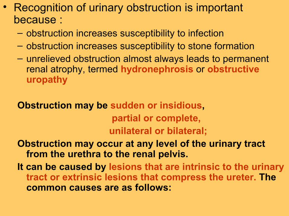
Causes of Urinary Tract Obstruction• Congenital anomalies
– Posterior urethral valves and urethral strictures– Meatal stenosis– Bladder neck obstruction– Ureteropelvic junction narrowing or obstruction– Severe vesicoureteral reflux
• Urinary calculi• Benign prostatic hypertrophy• Tumors
– Carcinoma of the prostate– Bladder tumors– Contiguous malignant disease (retroperitoneal lymphoma)– Carcinoma of the cervix or uterus
• Inflammation– Prostatitis– Ureteritis– UrethritisProstatitis– Ureteritis– Urethritis– Retroperitoneal fibrosis
• Sloughed papillae or blood clots• Normal pregnancy• Uterine prolapse and cystocele• Functional disorders:
– Neurogenic (spinal cord damage) – Other functional abnormalities of the ureter or bladder (often termed
dysfunctional obstruction)


HYDRONEPHROSIS• Hydronephrosis is the dilatation of the renal pelvis and calyces associated
with progressive atrophy of the kidney due to obstruction to the outflow of urine.
• Even with complete obstruction, glomerular filtration persists for some time because the filtrate subsequently diffuses back into the renal interstitium and perirenal spaces, where it ultimately returns to the lymphatic and venous systems.
• Because of this continued filtration, the affected calyces and pelvis become dilated, often markedly so.
• The high pressure in the pelvis is transmitted back through the collecting ducts into the cortex, causing renal atrophy, but it also compresses the renal vasculature of the medulla, causing a diminution in inner medullary plasma flow.
• The medullary vascular defects are reversible, but if protracted, obstruction will lead to medullary functional disturbances.
• Accordingly, the initial functional alterations are largely tubular, manifested primarily by impaired concentrating ability.
• Only later does the GFR begin to diminish. • Obstruction also triggers an interstitial inflammatory reaction, mediated by
activated tubular cells and leukocytes, leading eventually to interstitial fibrosis.

In hydronephrosis, the kidney is distended because the flow of urine is obstructed and urine backs up in the kidney's small tubes and
central collecting area (renal pelvis).
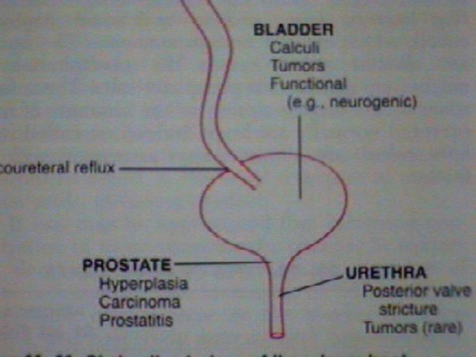
• MORPHOLOGY. • ACUTE When the obstruction is sudden and complete. the
reduction of glomerular filtration usually leads to mild dilation of the pelvis and calyces but sometimes to atrophy of the renal parenchyma.
• When the obstruction is subtotal or intermittent, glomerular filtration is not suppressed, and progressive dilation ensues.
• Depending on the level of urinary block, the dilation may affect first the bladder or ureter and then the kidney.
• In gross appearance, the kidney may have slight to massive enlargement. The earlier features are those of simple dilation of the pelvis and calyces.
• In addition there is often significant interstitial inflammation, even in the absence of infection.
• IN CHRONIC CASES, the picture is one of cortical tubular atrophy with marked diffuse interstitial fibrosis. Progressive blunting of the apices of the pyramids occurs, and eventually these become cupped.
• IN FAR ADVANCED CASES, the kidney may become transformed into a thin-walled cystic structure having a diameter of up to 15 to 20 cm with striking parenchymal atrophy, total obliteration of the pyramids, and thinning of the cortex.
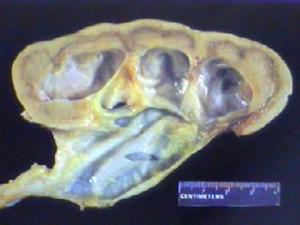
There was a large renal calculus (stone) that obstructed the calyces of the lower pole of this kidney, leading to a focal hydronephrosis (dilation of the
collecting system). The stasis from the obstruction and dilation led to infection. The infection with inflammation is characterized by the pale
yellowish-tan areas next to the dilated calyces with hyperemic mucosal surfaces. The upper pole is normal and shows good corticomedullary
demarcations.
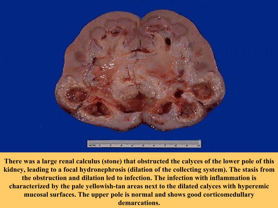
There was a large renal calculus (stone) that obstructed the calyces of the lower pole of this kidney, leading to a focal hydronephrosis (dilation of the collecting system). The stasis from
the obstruction and dilation led to infection. The infection with inflammation is characterized by the pale yellowish-tan areas next to the dilated calyces with hyperemic
mucosal surfaces. The upper pole is normal and shows good corticomedullary demarcations.

This kidney shows much more advanced
hydronephrosis.Note that there is only a thin rim of remaining renal cortex. Such a kidney is non-
functional and a source for ongoing infection.
The problem may originate from the ureteral orifice up to
the pelvis. In this case, a large
"staghorn" calculus was present that filled up the
pelvis and calyceal system. In bilateral cases, the problem
originates in the bladder trigone or urethra (or the
prostate around the urethra) or may be due to a large
neoplasm impinging on both ureters.

This abdominal CT scan with contrast demonstrates right hydronephrosis and hydroureter as a consequence of ureteral obstruction.
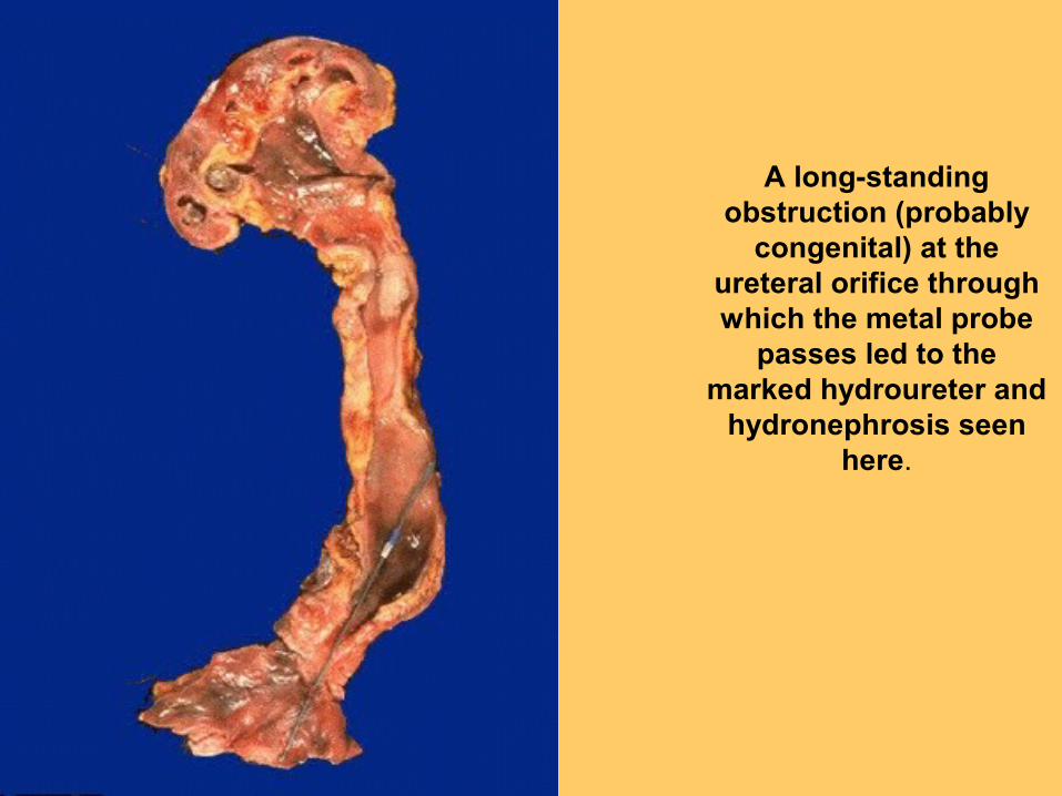
A long-standing obstruction (probably
congenital) at the ureteral orifice through which the metal probe
passes led to the marked hydroureter and
hydronephrosis seen here.

This radiograph of the lower abdomen
and pelvis demonstrates a
stent that has been placed in the
urinary tract from the right renal pelvis to the
bladder to relieve obstruction. (The bright radiopaque
clips at the patient's upper
right quadrant are left from previous cholecystectomy).

The markedly enlarged prostate seen here has not only large lateral lobes, but a very large median lobe as
well that obstructs the prostatic urethra and led to
chronic urinary tract obstruction.
As a result, the bladder became both enlarged and hypertrophied as it had to
work against the obstruction with every episode of urination.
That is why the surface of the bladder appears
trabeculated. Note also that a yellowish-brown calculus formed in
the bladder.

Clinical CourseAcute obstruction• May provoke pain attributed to distention of the collecting system or renal
capsule. • Most of the early symptoms are produced by the basic cause of the
hydronephrosis. • Thus, calculi lodged in the ureters may give rise to renal colic, and prostatic
enlargements to bladder symptoms.Unilateral, complete, or partial hydronephrosis • May remain silent for long periods of time, since the unaffected kidney can
maintain adequate renal function. In bilateral partial obstruction• Earliest manifestation is that of inability to concentrate the urine, reflected by
polyuria and nocturia. • Some patients will have acquired distal tubular acidosis, renal salt wasting,
and secondary renal calculi, and a typical picture of tubulointerstitial nephritis with scarring and atrophy of the papilla and medulla.
• Hypertension is common in such patients.Complete bilateral obstruction • Results in oliguria or anuria • Is incompatible with long survival unless the obstruction is relieved.

• Clinical Course. • Acute obstruction:• pain • calculi lodged in the ureters may give rise to renal colic, • bladder symptoms due to and prostatic hypertrophy/
NB: Unilateral, complete, or partial hydronephrosis may re main silent for long periods, since the unaffected kidney can maintain adequate renal function. Sometimes its exis tence first becomes apparent in the course of intravenous pyelography.
It is regrettable that this disease tends to remain asymptomatic, because it has been shown that in its early stages, perhaps the first few weeks, relief of such obstruction is compatible with reversion to normal func tion.
Ultrasonography is a useful noninvasive technique in the diagnosis of obstructive uropathy.
• In bilateral partial obstruction, the earliest manifestation is that of inability to concentrate the urine, reflected by polyuria and nocturia.
• Hypertension is common in such patients.
• Complete bilateral obstruction results in oliguria or anuna and is incompatible with long survival unless the obstruction is relieved.
• Curiously, after relief of complete urinary tract obstruction, postobstructive diuresis occurs. This can often be massive, with the kidney excreting large amounts of urine rich in sodium chloride.

• Here is an example of urinary tract obstruction in the urethra. On the left is the hypertrophied urinary bladder, hypertrophied because of chronic obstruction. On the right is the urethra running through the penis. At the point indicated by the arrow, is an obstruction of the urethra at the base of the penis.


• The passage of a calculus (stone) through the urinary tract is diagrammed here. Calculi form when there is increased excretion of solutes such as calcium and when urine alkalinity, acidity, stasis, and/or concentration are favorable. The most common varieties of calculi are:
• Type of Stone Frequency Calcium oxalate (or phosphate) 75% Magnesium ammonium phosphate (struvite, or "triple phosphate") 12% Uric acid 6% Cystine 1% Other 6%

• Stones containing calcium are far more frequent than other types, and about half the time occur when there is hypercalciuria. Only about 10% of the time do they appear as a consequence of hypercalcemia. The struvite stones are also known as "infection" stones because bacteria such as Proteus that split urea to ammonia favor their formation. Uric acid stones may be seen in association with gout, but often are not, and may just reflect increased precipitation of urates in an acid urine. Rare cystine stones also form in acid urine.
• Urinary tract calculi are usually unilateral and about 1 to 3 mm in size. Their passage is marked by intense abdominal or back or flank pain. This pain can be paroxysmal, known as renal or ureteral "colic". Hematuria may also be present. Larger stones that cannot pass may produce hydronephrosis or hydroureter.

Urolithiasis (Renal Calculi, Stones)• Stones may form at any level in the urinary tract, but
most arise in the kidney • Urolithiasis is a frequent clinical problem, affecting 5
to 10% of Americans in their lifetime• Males are affected more often than females• Peak age at onset is between 20 and 30 years.• Familial and hereditary predisposition to stone
formation has long been known• Many of the inborn errors of metabolism, such as gout,
cystinuria, and primary hyperoxaluria, provide good examples of hereditary disease characterized by excessive production and excretion of stone-forming substances.

Cause and Pathogenesis• There are four main types of calculi:• 75% are calcium containing: composed mostly of calcium
oxalate, or calcium oxalate mixed with calcium phosphate.• 15% are “Triple stones” or struvite stones, composed of
magnesium ammonium phosphate.• 6% are uric acid stones• 1-2% contain cystine• An organic matrix of mucoprotein, making up 1 to 5% of
the stone by weight, is present in all calculi. • Although there are many causes for the initiation and
propagation of stones, the most important determinant is an increased urinary concentration of the stones’ constituents, such that it exceeds their solubility in urine (supersaturation).
• A low urine volume in some metabolically normal patients may also favor supersaturation.


Urolithiasis (Renal Calculi, Stones)
• Calcium Oxalate stones – Hypercalciuria– Hypercalcemia– Hyperuricosuria– Hyperoxalauria– Hypocitraturia

• Calcium oxalate stones are associated • With both hypercalcemia and hypercalciuria (5% of patients)
– due to Hyperparathyroidism, diffuse bone disease, sarcoidosis, and other hypercalcemic states.
• Hypercalciuria without hypercalcemia (~ 55%) – Absorptive hypercalciuria - hyperabsorption of calcium from the
intestine– Renal hypercalciuria - an intrinsic impairment in renal tubular
reabsorption of calcium – Idiopathic fasting hypercalciuria with normal parathyroid function.
• ~ 20% are associated with increased uric acid secretion (hyperuricosuric calcium nephrolithiasis), with or without hypercalciuria. – The mechanism of stone formation in this setting involves
“nucleation” of calcium oxalate by uric acid crystals in the collecting ducts.
• ~ 5% associated with hyperoxaluria, either hereditary (primary oxaluria) or, more commonly, acquired by intestinal overabsorption in patients with enteric diseases.
• The latter, so-called “enteric hyperoxaluria,” also occurs in vegetarians, because much of their diet is rich in oxalates.
• Hypocitraturia associated with acidosis and chronic diarrhea of unknown cause may produce calcium stones.
• In a variable proportion of patients with calcium stones no cause can be found (idiopathic calcium stone disease).

• Magnesium ammonium phosphate stones:– Are formed largely following infections by urea-
splitting bacteria (e.g., proteus and some staphylococci), which convert urea to ammonia.
– The resultant alkaline urine causes the precipitation of magnesium ammonium phosphate salts.
– These form some of the largest stones, as the amounts of urea excreted normally are huge.
– Staghorn calculi are almost always associated with infection.
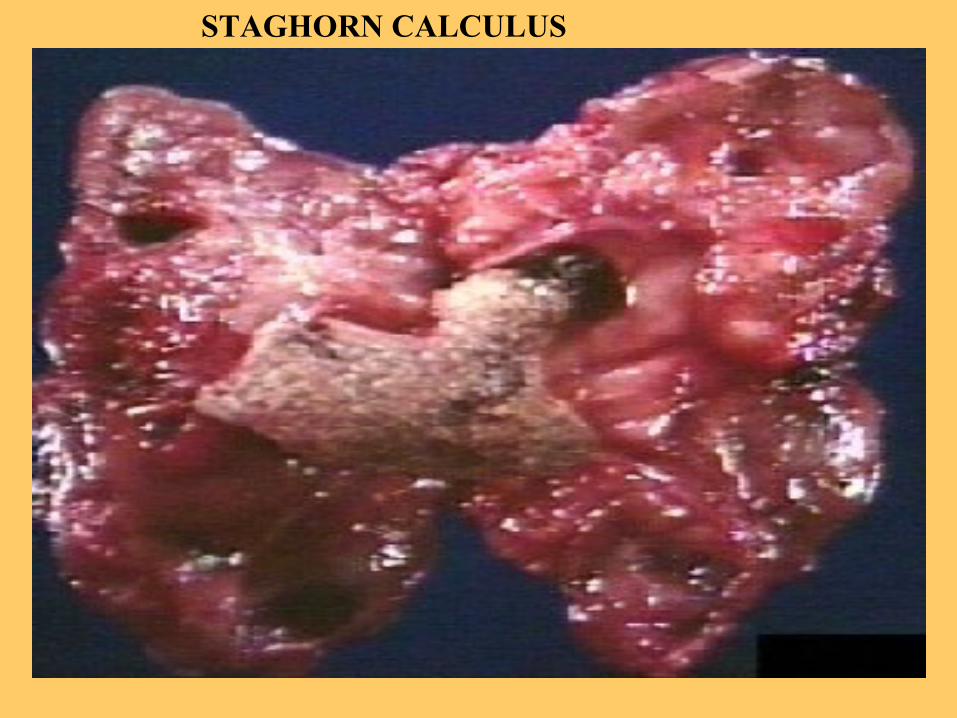
STAGHORN CALCULUS

Nephrolitihiasis
• Large stone impacted in the renal pelvis

A large staghorn calculus is seen cortical areas. obstructing the renal pelvi-calyceal system. The lower pole of the kidney shows areas of hemorrhage and necrosis with collapse of cortical areas

Uric acid stones: • Are common in patients with hyperuricemia,
such as gout, and diseases involving rapid cell turnover, such as the leukemias.
• In contrast to the radiopaque calcium stones, uric acid stones are radiolucent.
Cystine stones:• Are associated with a genetically determined
defect in the renal transport of certain amino acids, including cystine, which leads to cystinuria. These stones form at low pH.

• It is noteworthy that the following factors favor the formation of calculi:
• increased concentration of stone constituents• changes in urinary pH• decreased urine volume• Presence of bacteria• However, many calculi occur in the absence of
these factors, and conversely patients with hypercalciuria, hyperoxaluria, and hyperuricosuria often do not form stones.
• It has, therefore, been postulated that stone forma tion is enhanced by a deficiency in inhibitors of crystal formation in urine.
• The list of such inhibitors is long, including pyrophosphate, diphosphonate, citrate, glycosaminoglycans, and a glycoprotein called nephrocalcin.

• Stones are unilateral in about 80% of patients. • The favored sites for their formation are
– Within the renal calyces and pelves – In the bladder
• If formed in the renal pelvis:– They tend to remain small– Have an average diameter of 2 to 3 mm– May have smooth contours or – may take the form of an irregular, jagged mass of spicules. – Often many stones are found in the same kidney.
• Occasionally, progressive accretion of salts leads to the development of branching structures known as staghorn stones, which create a cast of the pelvic and calyceal system.

CLINICAL COURSE• Stones are of importance when they obstruct urinary flow or
produce ulceration and bleeding. • They may be present without producing any symptoms or
significant renal damage. • In general, smaller stones are most hazardous, as they may
pass into the ureters, producing pain referred to as colic (one of the most intense forms of pain) as well as ureteral obstruction.
• Larger stones cannot enter the ureters and are more likely to remain silent within the renal pelvis.
• Commonly, these larger stones first manifest themselves by hematuria.
• Stones also predispose to superimposed infection, both by their obstructive nature and by the trauma they produce.