Urinalysis and body fluid crystals
61
Mercury Y. Lin, MD FCAP LCDR MC USN Staff Pathologist NHCP
-
Upload
mercury-lin -
Category
Health & Medicine
-
view
910 -
download
0
Transcript of Urinalysis and body fluid crystals
Mercury Y. Lin, MD FCAP LCDR MC USN
Staff Pathologist NHCP

Overview of laboratory examination for urine and body fluid crystals with emphasis on morphologic properties.
“Piss for path”: helpful information for pathologist-clinical lab consultants. Inherent limitation of urine examination.
Most importantly, enable board exam candidates to rock the clinical path portion of ABP examination. (continue our tradition of near 100% passing rate for 1st time exam takers).


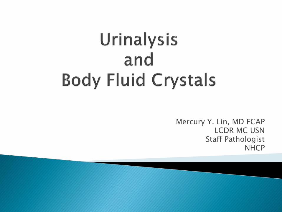
5th century BC: Hippocrates described fluid intake and urine output. DM urines taste sweet.
Byzantium and Middle Ages: Uroscopy, “piss prophets” and pseudo-science (“Uromancy”)
1820’s: Richard Bright noted relationship between edema and proteinuria. (archaic term “Bright’s Disease”)
1837: Medical urine microscopy being taught in Paris by Alfred Donné.
1844: Golding Bird publishes “Urinary Deposits” and described urine casts in pts with “Bright’s Dz.”
Sweet urine
I agree, it is sweet with a fruity odor, which may present ketone bodies. Let’s pass it around so everyone can get a sip.
pathology consults in Byzantium
Urine Wheel from “Epiphaniae Medicorum”

NCCLS definition: “testing of urine with procedures commonly performed in an expeditious, reliable, and cost effective manner…”
So routine UA protocols look for
- Physical : (color, transparency, odor, foam, spec gravity)
- Chemical: (pH, protein, blood, nitrite, leuk esterase, glucose, ketone, bilirubin, urobilinogen)
- Microscopic: (RBC, WBC, Epithelial cells, casts, microrganisms, crystals)
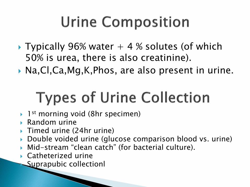
Typically 96% water + 4 % solutes (of which 50% is urea, there is also creatinine).
Na,Cl,Ca,Mg,K,Phos, are also present in urine.
1st morning void (8hr specimen) Random urine Timed urine (24hr urine) Double voided urine (glucose comparison blood vs. urine) Mid-stream “clean catch” (for bacterial culture). Catheterized urine Suprapubic collectionl

Routine UA (room temp specimens) shall be performed within 2 hrs of voiding. (CAP and NCCLS requirement).
Refrigeration may reduce bacterial growth, but may result in precipitation of amorphous urates and phosphates (interfere with microscopy).
For 24hr urine collection, preservative maybe required, depending on analyte of interest.
UA specimen in room temp >2hr should be rejected. (refer to CAP checklist and local lab policy for exceptions).
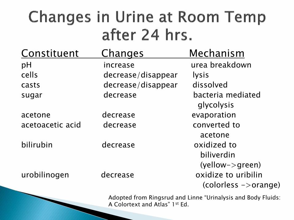
Constituent Changes Mechanism pH increase urea breakdown
cells decrease/disappear lysis
casts decrease/disappear dissolved
sugar decrease bacteria mediated
glycolysis
acetone decrease evaporation
acetoacetic acid decrease converted to
acetone
bilirubin decrease oxidized to
biliverdin
(yellow->green)
urobilinogen decrease oxidize to uribilin
(colorless ->orange)
Adopted from Ringsrud and Linne “Urinalysis and Body Fluids: A Colortext and Atlas” 1st Ed.

Color: normal is pale yellow to straw colored. if clear redhemoglobin cloudy redRBCs brownmyoglobin, hemoglobin, bilirubin blackmelanin, homogentisic acid.
Foam: normal is small amt of white foam, if shaken. if abundant foam proteinuria yellow foambilirubin.
Specific Gravity: normal is 1.001 – 1.035 (water is 1.000) spec grav is dependent on mass and amount of particles in
urine. “spec grav” reported by reagent strips is actually a gestimation
by measures ionic concention (since most urine solutes are ionized).

A refractometer can be used for more accurate measurement of urine specific gravity.
Usually, it doesn’t matter, but if there is prominent glucosuria.
Spec grav by refractometer will be >1.035
yet
Spec grav by reagent strip will be WNL (b/c glucose is not ionized).
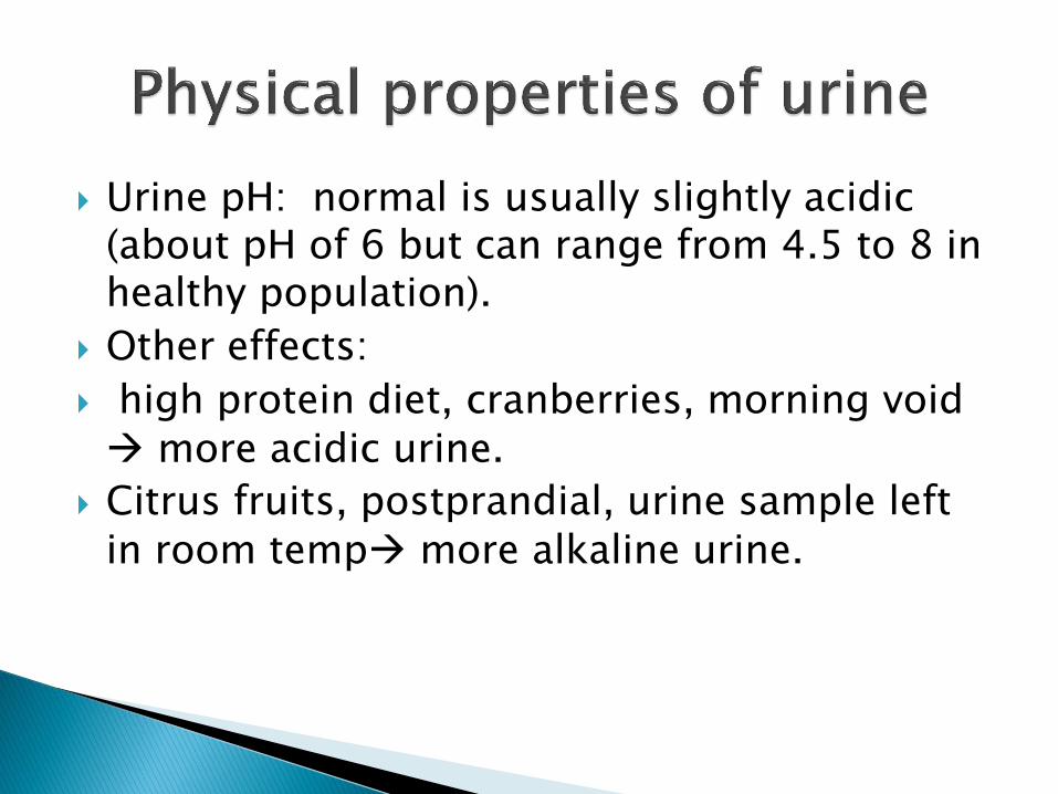
Urine pH: normal is usually slightly acidic (about pH of 6 but can range from 4.5 to 8 in healthy population).
Other effects:
high protein diet, cranberries, morning void more acidic urine.
Citrus fruits, postprandial, urine sample left in room temp more alkaline urine.
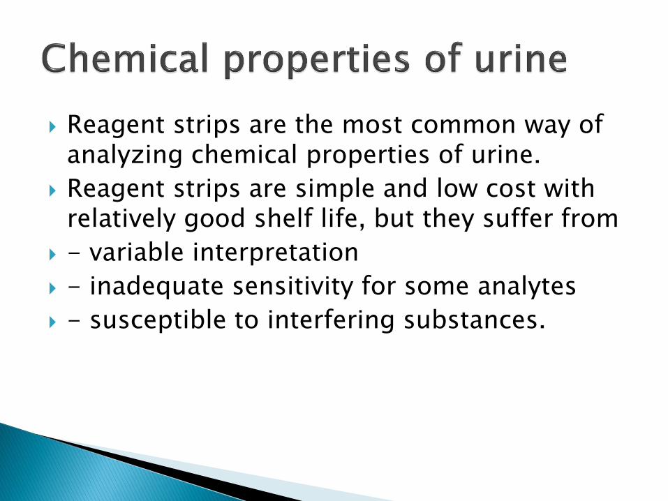
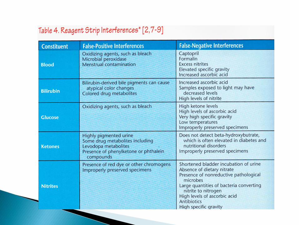
Reagent strips are the most common way of analyzing chemical properties of urine.
Reagent strips are simple and low cost with relatively good shelf life, but they suffer from
- variable interpretation
- inadequate sensitivity for some analytes
- susceptible to interfering substances.


Urine is also been used to screen for metabolic disease in babies.
But reagent strip (glucose oxidase reaction) will only detects glucose.
Clinitest will detect reducing substances (including glucose, galactose, fructose, lactose, and pentose).

If regagent strip is negative (for glucose) but Clinitest is positive. Additonal testing is needed to identify the specific non-glucose reducing sugars.
Ascorbic acid, nalidixic acid, cephalosporins and probenecid in large quantities may cause false positive results with Clinitest.

Most of the time, examination is performed by lab instruments
This is an Iris iQ®200SPRINT™ Automated Urine Microscopy Analyzer. By flow cell digital imaging, it can analye 101 samples/hr and reports RBC WBC, WBC clumps, squamous epithelial cells, renal epithelial cells, urothelial cells, hyaline casts, other casts, crystals (9 subtype), yeasts, bacteria, sperm mucus, oval fat bodies, trichomans
But remember, without an astute human operator, even the most advanced lab instruments is no more than a machine. Auto “TAR” by lab techs will report out false results.

Urine microscopy are often unstained, so it will look slightly different to us (anatomic focus pathologists).
RBCs
Crenulated RBCs due to urine hypertonicity

These pale RBCs have “ghost cell” appearance. This is due to loss of Hb which occurs in hypotonic urine specimen. This doesn’t necessarily mean patient is anemic.
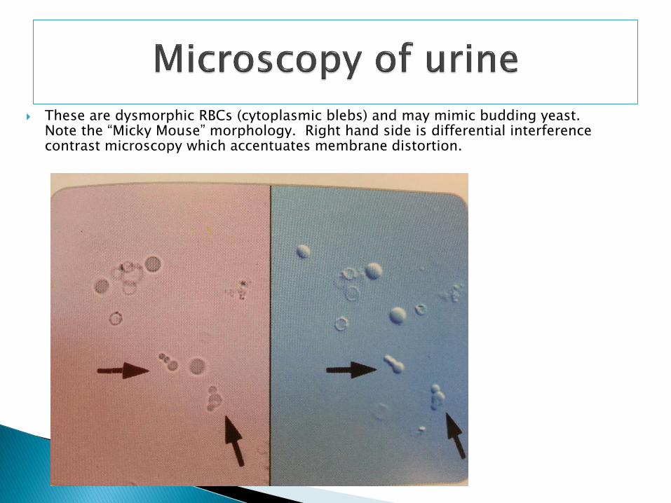
These are dysmorphic RBCs (cytoplasmic blebs) and may mimic budding yeast. Note the “Micky Mouse” morphology. Right hand side is differential interference contrast microscopy which accentuates membrane distortion.
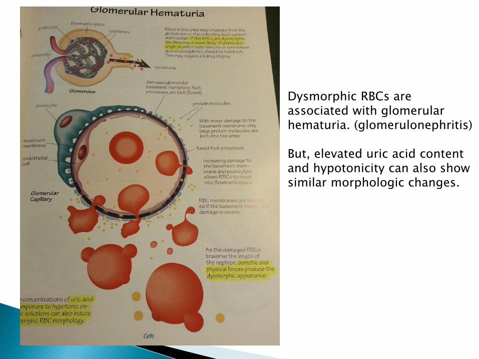
Dysmorphic RBCs are associated with glomerular hematuria. (glomerulonephritis) But, elevated uric acid content and hypotonicity can also show similar morphologic changes.
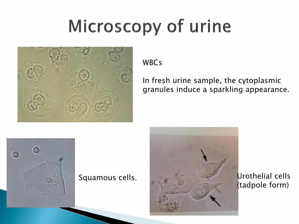
WBCs In fresh urine sample, the cytoplasmic granules induce a sparkling appearance.
Squamous cells. Urothelial cells (tadpole form)

Urine eosinophils (to identify eos, the urine needs to be stained with either Wright Giemsa or Hansel stain).
Eosinophiluria: Top 3 drug inducted acute intersitital nephritis, hypersensitivity, parasitic infections. Also transplant rejection, RPGN, Post infectious GN, eosphilic cystitis…etc. The reagent strip will show ++WBC, but negative leukocyte esterase.
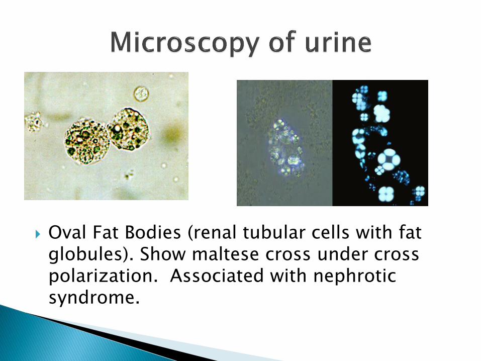
Oval Fat Bodies (renal tubular cells with fat globules). Show maltese cross under cross polarization. Associated with nephrotic syndrome.

True maltese cross (arms of equal length): lipids (e.g. oval fat bodies)
vs. Pseudo maltese cross(arms of unequal length): starch, leucine,
tiamterene diuretics, adenine phosphoribosyltransferase [APRT] deficiency
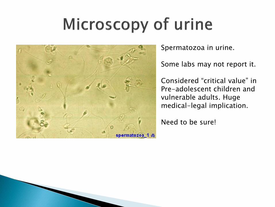
Spermatozoa in urine. Some labs may not report it. Considered “critical value” in Pre-adolescent children and vulnerable adults. Huge medical-legal implication. Need to be sure!

Now we turn our attention towards urinary casts (cylinders). Casts in the urine mirrors that physiologic state of the kidney. Casts are classified into 2 broad groups. Acellular Casts Cellular Casts - Hyaline - RBC - Granular - WBC - Waxy - RTE - Fatty - Bacterial

Hyaline casts and granular casts are the only 2 types of cast that should be seen in normal population.
Hyaline casts can be difficult to visualize by bright field microscopy, but (fortunately) are also clinically least important.
Vigorous exercise can increase hyaline cast in urine. Granular casts are less commonly seen.
Hyaline casts are composed of Tamm-Horsfall proteins. The matrix contains few granular material. Hyaline casts do not polarize.
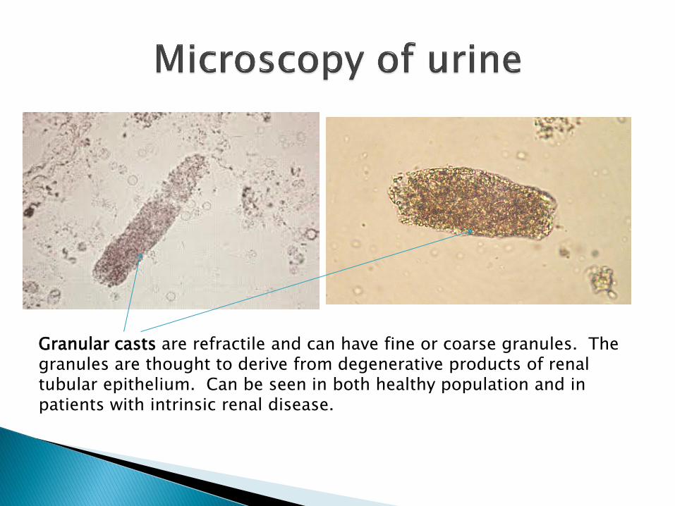
Granular casts are refractile and can have fine or coarse granules. The granules are thought to derive from degenerative products of renal tubular epithelium. Can be seen in both healthy population and in patients with intrinsic renal disease.
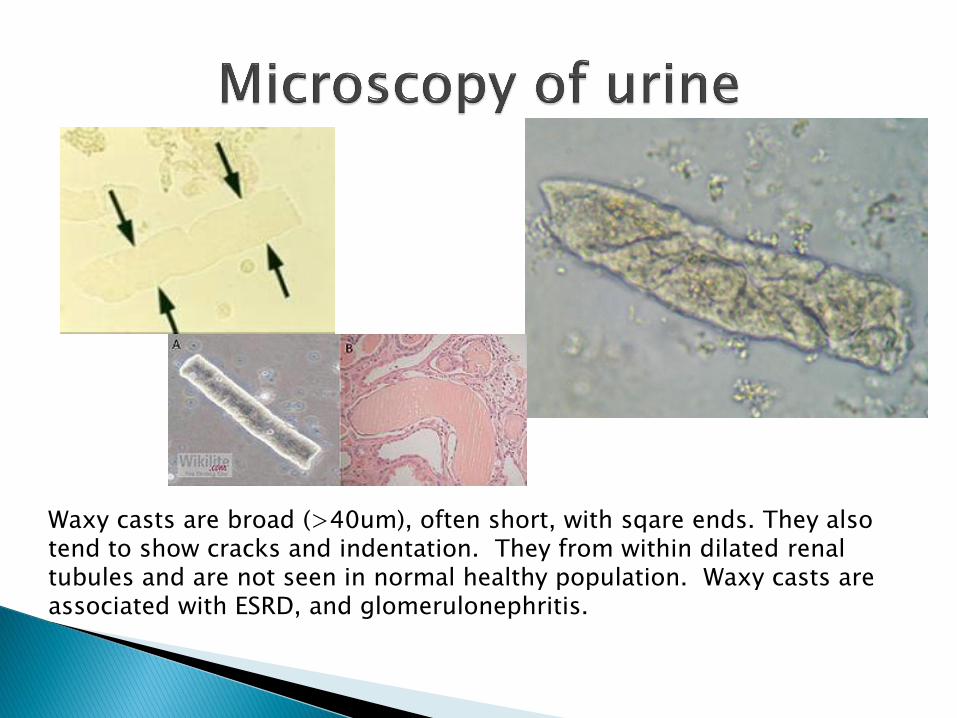
Waxy casts are broad (>40um), often short, with sqare ends. They also tend to show cracks and indentation. They from within dilated renal tubules and are not seen in normal healthy population. Waxy casts are associated with ESRD, and glomerulonephritis.
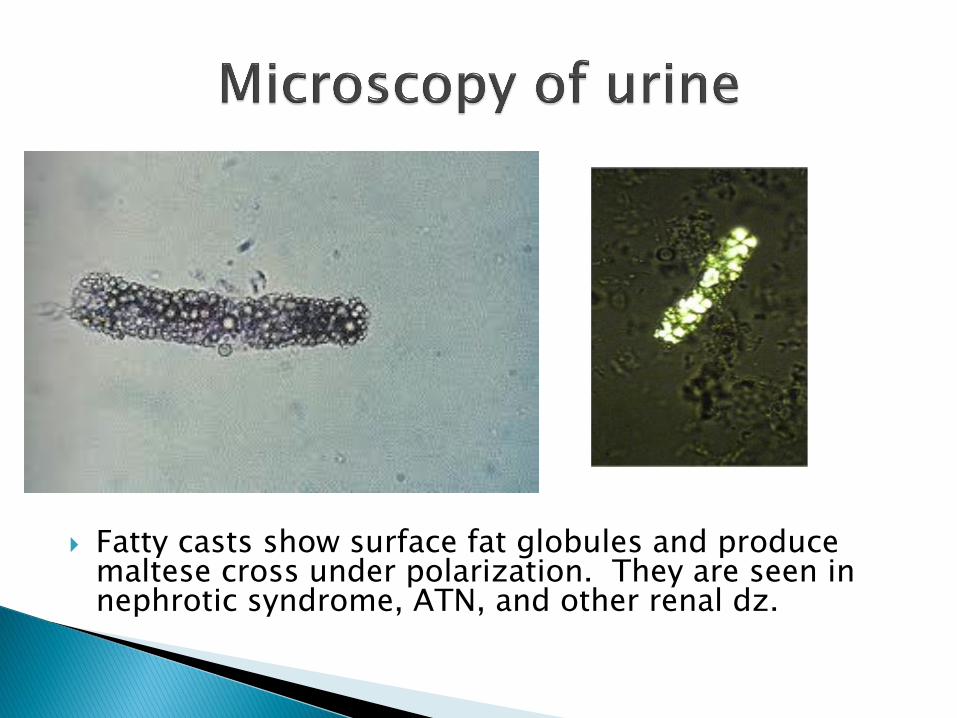
Fatty casts show surface fat globules and produce maltese cross under polarization. They are seen in nephrotic syndrome, ATN, and other renal dz.

RBC casts are rarely seen. RBCs are stuck together in a matrix of Tamm-Horsfall protein. But RBC casts are fragile and will degenerate after voiding. Reddish appearance due to Hb. Seen in acute glomerulonephritis, renal neoplasms, and malignant HTN.
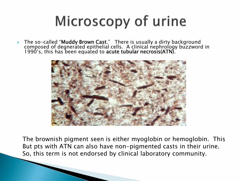
The so-called “Muddy Brown Cast.” There is usually a dirty background composed of degnerated epithelial cells. A clinical nephrology buzzword in 1990’s, this has been equated to acute tubular necrosis(ATN).
The brownish pigment seen is either myoglobin or hemoglobin. This But pts with ATN can also have non-pigmented casts in their urine. So, this term is not endorsed by clinical laboratory community.

WBC casts: highly refractive, look for multilobed nuclei and single WBCs in surrounding area. WBC casts are seen pyelonephritis, interstitial nephritis, renal transplant rejection, sepsis, lupus nephritis…etc.

Renal Tubular Epithelial (RTE) Casts: Mimics WBC cast and at times can be difficult to differentiated. So the term “Cellular Cast” can be used to include both RTE and WBC casts.
RTE casts are seen in ATN, renal transplant reject, heavy metal poisoning…etc.

Renal stones form because of undesired phase change of a substance.
Normal urine is in a shifting state of oversaturation. Small crystals are constantly forming and dissolving.
Stones develop when there is an unstable oversaturation which leads to precipitation.
Many different crystals have been described in urine. We will mainly focused on ones that are either clinically important or IMHO, morphologically peculiar

Amorphous urate and phosphate are similar/essentially identical under light microscopy.
Difference are: Amorphus urate: seen in pH <5.8, show birefringence in large clumps, associated with dehyration, fever. But usually no significant clinical implication. Amorphus phosphate: seen in pH >6.3, no birefringence, clinically insignificant.

Alternatively, urine can also be spun and the pellet is examined macroscopically.
Urate is pink.
Phosphate is white.

Triple phosphate/Struvite (Ammonium magnesium phosphate) crystals. Left is the prototypical coffin lid morphology. But, note that triple phosphate crystals can be seen (less commonly) in “fern” form. These crystals develop via biofilm produced by urease producing bacteria.
Associated with staghorn calculi, UTI, vericoureteral reflux.
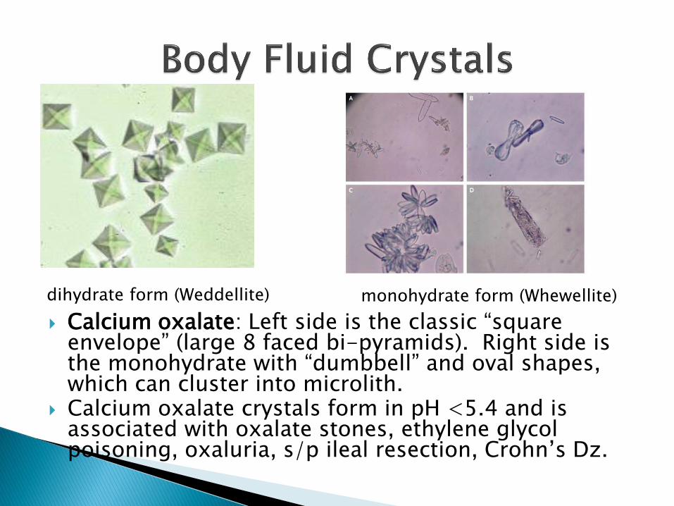
Calcium oxalate: Left side is the classic “square envelope” (large 8 faced bi-pyramids). Right side is the monohydrate with “dumbbell” and oval shapes, which can cluster into microlith.
Calcium oxalate crystals form in pH <5.4 and is associated with oxalate stones, ethylene glycol poisoning, oxaluria, s/p ileal resection, Crohn’s Dz.
dihydrate form (Weddellite) monohydrate form (Whewellite)

2/3 of all renal calculi are calcium oxalate stones.
S/P small bowel resection leads to malabsorption of fat. Ca selectively binds fat and leave free oxalate in the colon to be absorbedoxalate goes to the kindey and stones develop.
Ethylene glycol ingestionmetabolized to glycoaldehydeglycolic acidglyoxylic acidoxalic acidadd Ca to form calcium oxalate (more crystals in monohydrate form).
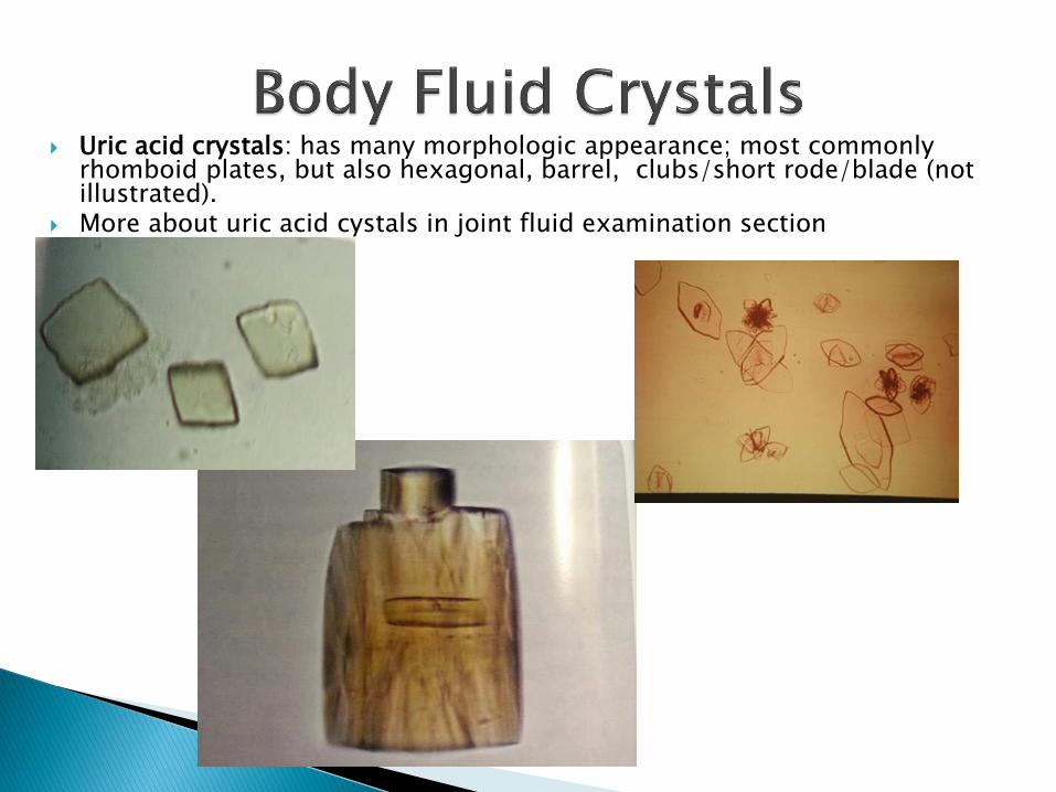
Uric acid crystals: has many morphologic appearance; most commonly rhomboid plates, but also hexagonal, barrel, clubs/short rode/blade (not illustrated).
More about uric acid cystals in joint fluid examination section

Cystine: variably sized hexagonal plates.
Associated with cystinuria.
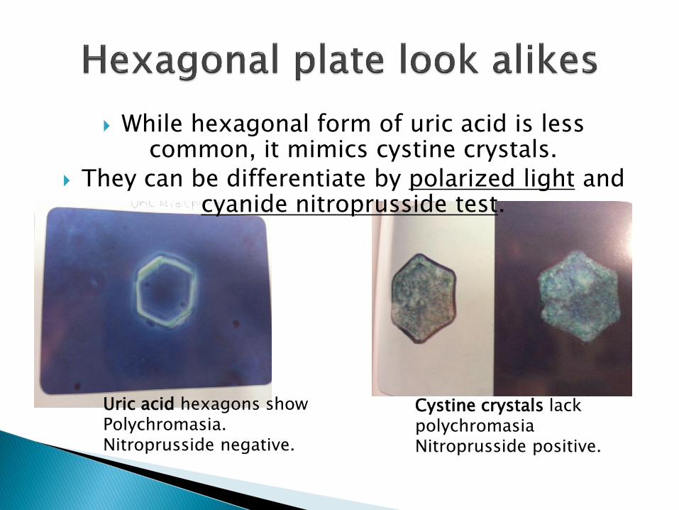
While hexagonal form of uric acid is less common, it mimics cystine crystals.
They can be differentiate by polarized light and cyanide nitroprusside test.
Uric acid hexagons show Polychromasia. Nitroprusside negative.
Cystine crystals lack polychromasia Nitroprusside positive.
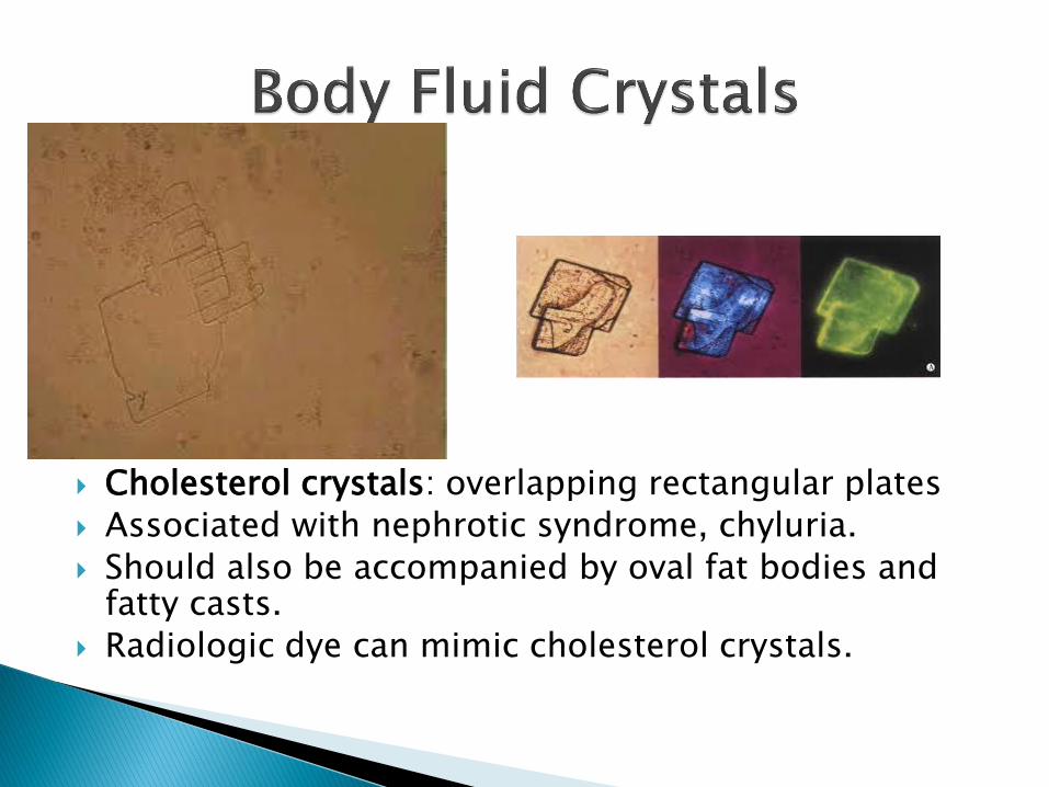
Cholesterol crystals: overlapping rectangular plates
Associated with nephrotic syndrome, chyluria.
Should also be accompanied by oval fat bodies and fatty casts.
Radiologic dye can mimic cholesterol crystals.
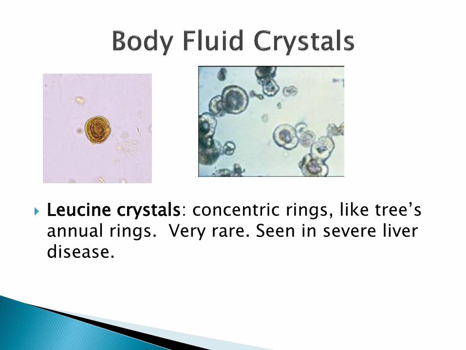
Leucine crystals: concentric rings, like tree’s annual rings. Very rare. Seen in severe liver disease.

Tyrosine crystals: tufts of fine silky needles.
Seen in severe liver dz, tyrosinemia.
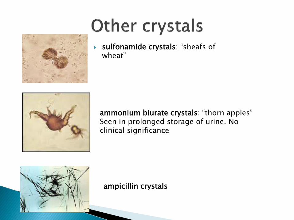
sulfonamide crystals: “sheafs of wheat”
ammonium biurate crystals: “thorn apples” Seen in prolonged storage of urine. No clinical significance
ampicillin crystals

Should be examined with compensated polarized light microscopy.
Also consider other clinical laboratory values (e.g. cell count)
First, lets review compensated polarized light microscopy.
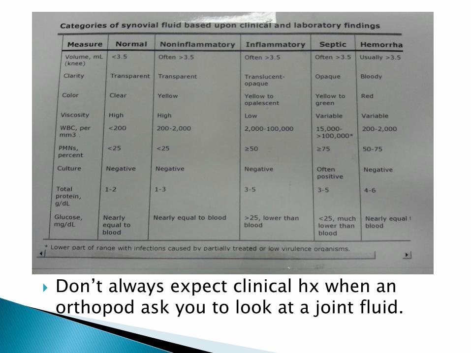
Don’t always expect clinical hx when an orthopod ask you to look at a joint fluid.

Since I barely recall being awake during undergrad physics, this is my fraudulent technician’s version of polarized microscopy.
Most illumination transmits light waves whose electric field vectors vibrate in all perpendicular planes with respect to the direction of propagation. With polarized light, filtration is used to restrict vibration to a single plane.
http://www.olympusmicro.com

Crystals can be either
- isotropic: index of refraction is equal in all directions.
- anisotropic: refraction differs when light enters at non-equivalent axis.
http://www.olympusmicro.com

When placing anisotropic crystals between cross polarization. From the left, polarized light (P) enter the anisotropic crystal and is refracted into 2 component waves. This light then passes through the analyzer (A). One component wave is now “retarded.”
After passing through the analyzer, light interference between 2 component waves acquire a spectrum of color under usual bright field microscopy.
http://www.olympusmicro.com

In this case, light reflections from outer- and inner- surfaces of the bubble (separated by a few micros) interfere with each other and result in this colorful display.
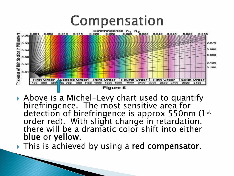
Above is a Michel-Levy chart used to quantify birefringence. The most sensitive area for detection of birefringence is approx 550nm (1st order red). With slight change in retardation, there will be a dramatic color shift into either blue or yellow.
This is achieved by using a red compensator.

From: Bullough- Orthopedic Pathology 5th ed.
arrow is the “slow wave”
“U PAY PEB” Urate PArallel Yellow, PErpendicular Blue
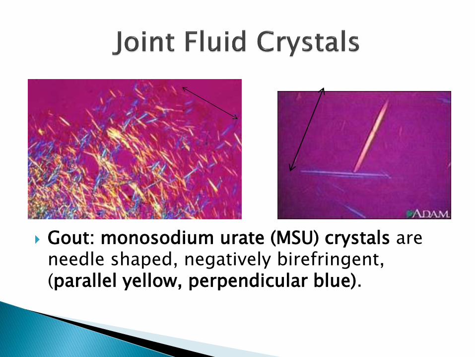
Gout: monosodium urate (MSU) crystals are needle shaped, negatively birefringent, (parallel yellow, perpendicular blue).


Calcium pyrophosphate crystals are rhomboid, positively birefringent (parallel blue, perpendicular yellow).
Now the fun parts, lets walk over to the core lab and get some hands on experience with operating a compensated polarized light microscope.
Questions?