Uptake,ltranslocationlandkbiotransformationl ...
15
Wang et al. J Nanobiotechnol (2020) 18:103 https://doi.org/10.1186/s12951-020-00659-6 RESEARCH Uptake, translocation and biotransformation of selenium nanoparticles in rice seedlings (Oryza sativa L.) Kang Wang 1 , Yaqi Wang 1 , Kui Li 2 , Yanan Wan 1* , Qi Wang 1 , Zhong Zhuang 1 , Yanbin Guo 2 and Huafen Li 1* Abstract Background: Selenium (Se) in soil mainly consists of selenite, selenate, and elemental Se. However, little is known about the mechanism involved in the uptake and biotransformation of elemental Se by plants. Results: In this study, the uptake, translocation, subcellular distribution and biotransformation of selenium nanopar- ticles (SeNPs) in rice (Oryza sativa L.), and a comparison with selenite and selenate, were investigated through hydro- ponic experiments. The study revealed that SeNPs could be absorbed by rice plants; and aquaporin inhibitor was responsible for a 60.4% inhibition of SeNP influx, while metabolic inhibitor was ineffective. However, the SeNPs uptake rate of rice roots was approximately 1.7 times slower than that of selenite or selenate. Under the SeNPs or selenite treatment, Se was primarily accumulated in roots rather than in shoots, whereas an opposite trend was observed with selenate treatment. Additionally, most of the absorbed Se was distributed in cell wall of the SeNPs or selenite treated- rice plants, while its proportion was the highest in soluble cytosol of the selenate treated-rice plants. The absorbed SeNPs or selenite was rapidly assimilated to organic forms, with SeMet being the most predominant species in both shoots and roots of the rice plants. However, following selenate treatment, Se(VI) remained as the most predominant species, and only a small amount of it was converted to organic forms. Conclusion: Therefore, this study provides a deeper understanding of the mechanisms associated SeNPs uptake and biotransformation within plants. Keywords: Se nanoparticles, Inorganic Se, Uptake, Subcellular distribution, Assimilation, Plants © The Author(s) 2020. This article is licensed under a Creative Commons Attribution 4.0 International License, which permits use, sharing, adaptation, distribution and reproduction in any medium or format, as long as you give appropriate credit to the original author(s) and the source, provide a link to the Creative Commons licence, and indicate if changes were made. The images or other third party material in this article are included in the article’s Creative Commons licence, unless indicated otherwise in a credit line to the material. If material is not included in the article’s Creative Commons licence and your intended use is not permitted by statutory regulation or exceeds the permitted use, you will need to obtain permission directly from the copyright holder. To view a copy of this licence, visit http://creativeco mmons.org/licenses/by/4.0/. The Creative Commons Public Domain Dedication waiver (http://creativecommons.org/publicdomain/ zero/1.0/) applies to the data made available in this article, unless otherwise stated in a credit line to the data. Background Selenium (Se) is an essential microelement for humans and animals [1], and when its intake as a dietary compo- nent is appropriate, it can be beneficial to human health, particularly taking into consideration its antioxidation and anticarcinogenic effects, immune function, and many other roles it plays in biological processes [2, 3]. How- ever, between 500 and 1000 million individuals in the world are estimated to be Se-deficient, and these num- bers include persons living in New Zealand, Europe, and parts of China [4–6]. Plants, especially cereals and cereal products, are the principal sources of dietary Se; never- theless, their Se content is generally low [7]. us, agro- nomic biofortification with Se fertilization is considered as an effective way to produce Se-rich crops, and improve the Se intake of target populations [8–10]. Although no previous studies have conclusively dem- onstrated that Se is an essential trace element for plants, its benefits if applied at proper doses have been reported [11–14]. erefore, understanding the mechanism of Se uptake and metabolism within plants is of great impor- tance. In soil environments, selenite and selenate are the Open Access Journal of Nanobiotechnology *Correspondence: [email protected]; [email protected] 1 Beijing Key Laboratory of Farmland Soil Pollution Prevention and Remediation, Key Laboratory of Plant-Soil Interactions of the Ministry of Education, College of Resources and Environmental Sciences, China Agricultural University, Beijing 100193, The People’s Republic of China Full list of author information is available at the end of the article
Transcript of Uptake,ltranslocationlandkbiotransformationl ...
Wang et al. J Nanobiotechnol (2020) 18:103 https://doi.org/10.1186/s12951-020-00659-6
RESEARCH
Uptake, translocation and biotransformation of selenium nanoparticles in rice seedlings (Oryza sativa L.)Kang Wang1, Yaqi Wang1, Kui Li2, Yanan Wan1*, Qi Wang1, Zhong Zhuang1, Yanbin Guo2 and Huafen Li1*
Abstract
Background: Selenium (Se) in soil mainly consists of selenite, selenate, and elemental Se. However, little is known about the mechanism involved in the uptake and biotransformation of elemental Se by plants.
Results: In this study, the uptake, translocation, subcellular distribution and biotransformation of selenium nanopar-ticles (SeNPs) in rice (Oryza sativa L.), and a comparison with selenite and selenate, were investigated through hydro-ponic experiments. The study revealed that SeNPs could be absorbed by rice plants; and aquaporin inhibitor was responsible for a 60.4% inhibition of SeNP influx, while metabolic inhibitor was ineffective. However, the SeNPs uptake rate of rice roots was approximately 1.7 times slower than that of selenite or selenate. Under the SeNPs or selenite treatment, Se was primarily accumulated in roots rather than in shoots, whereas an opposite trend was observed with selenate treatment. Additionally, most of the absorbed Se was distributed in cell wall of the SeNPs or selenite treated-rice plants, while its proportion was the highest in soluble cytosol of the selenate treated-rice plants. The absorbed SeNPs or selenite was rapidly assimilated to organic forms, with SeMet being the most predominant species in both shoots and roots of the rice plants. However, following selenate treatment, Se(VI) remained as the most predominant species, and only a small amount of it was converted to organic forms.
Conclusion: Therefore, this study provides a deeper understanding of the mechanisms associated SeNPs uptake and biotransformation within plants.
Keywords: Se nanoparticles, Inorganic Se, Uptake, Subcellular distribution, Assimilation, Plants
© The Author(s) 2020. This article is licensed under a Creative Commons Attribution 4.0 International License, which permits use, sharing, adaptation, distribution and reproduction in any medium or format, as long as you give appropriate credit to the original author(s) and the source, provide a link to the Creative Commons licence, and indicate if changes were made. The images or other third party material in this article are included in the article’s Creative Commons licence, unless indicated otherwise in a credit line to the material. If material is not included in the article’s Creative Commons licence and your intended use is not permitted by statutory regulation or exceeds the permitted use, you will need to obtain permission directly from the copyright holder. To view a copy of this licence, visit http://creat iveco mmons .org/licen ses/by/4.0/. The Creative Commons Public Domain Dedication waiver (http://creat iveco mmons .org/publi cdoma in/zero/1.0/) applies to the data made available in this article, unless otherwise stated in a credit line to the data.
BackgroundSelenium (Se) is an essential microelement for humans and animals [1], and when its intake as a dietary compo-nent is appropriate, it can be beneficial to human health, particularly taking into consideration its antioxidation and anticarcinogenic effects, immune function, and many other roles it plays in biological processes [2, 3]. How-ever, between 500 and 1000 million individuals in the
world are estimated to be Se-deficient, and these num-bers include persons living in New Zealand, Europe, and parts of China [4–6]. Plants, especially cereals and cereal products, are the principal sources of dietary Se; never-theless, their Se content is generally low [7]. Thus, agro-nomic biofortification with Se fertilization is considered as an effective way to produce Se-rich crops, and improve the Se intake of target populations [8–10].
Although no previous studies have conclusively dem-onstrated that Se is an essential trace element for plants, its benefits if applied at proper doses have been reported [11–14]. Therefore, understanding the mechanism of Se uptake and metabolism within plants is of great impor-tance. In soil environments, selenite and selenate are the
Open Access
Journal of Nanobiotechnology
*Correspondence: [email protected]; [email protected] Beijing Key Laboratory of Farmland Soil Pollution Prevention and Remediation, Key Laboratory of Plant-Soil Interactions of the Ministry of Education, College of Resources and Environmental Sciences, China Agricultural University, Beijing 100193, The People’s Republic of ChinaFull list of author information is available at the end of the article
Page 2 of 15Wang et al. J Nanobiotechnol (2020) 18:103
two major Se species that can be absorbed via the roots of plant through different mechanisms [15, 16]. Selenite can be taken up by plants through silicon influx transport-ers [17], or possibly via phosphate transporters through a metabolically-dependent active process [18, 19]; and thereafter, readily assimilated to organic Se forms that accumulated in roots but seldom transported to above-ground parts [18, 20]. Contrarily, selenate enters root cells via high-affinity sulfate transporters in the plasma membrane [15, 16], and are easily transported from roots to shoots through the xylem, with a relatively small pro-portion assimilated to organic forms [18, 20]. Under certain environmental conditions, these two Se species may exist in the rhizosphere [21]; however, elemental Se, which constitutes 26–66% of total Se reserves can also be found in soil environments [22], and its absorption mechanism and assimilation process within plants has not been clarified.
As an elemental form of Se, red selenium nanoparti-cles (SeNPs), which have a nano size, can be synthesized using chemical methods [23]. It has been demonstrated that in mammals, SeNPs show excellent bioactivity, high bioavailability, and low cytotoxicity compared to other Se species [24]; thus, they are always applied in medi-cine as antimicrobial, antioxidant, and anticancer agents [25–28]. More importantly, SeNPs can be absorbed and assimilated to other Se species in plant tissues [29], thereby offering the possibility for their application in fertilization and plant nutrition in agricultural fields. However, the uptake mechanism of SeNPs by plants is still unclear so far and needs further investigation. As described above, the fate of inorganic Se in plants has been widely reported in the past decades [18, 30, 31], but few studies focused on comparing the biological differ-ence of SeNPs from selenite and selenate in higher plants, with respect to their phytouptake, translocation, accu-mulation, and assimilation, are available. Furthermore, it is commonly believed that the subcellular distribution of elements is associated with some biological processes, such as the detoxification mechanism for toxic elements [32, 33]. Hence, research on the subcellular distribution of Se in plant tissues is also necessary for a proper under-standing of the processes associated with its transporta-tion and metabolism within plants.
Rice (Oryza sativa L.) is the most widely consumed cereal in Southeast and East Asia. Regrettably, a global survey showed that 75% of the rice that is consumed has a Se content which is insufficient for human requirements [7]. It has been generally believed that in rice grains, the predominant Se species are the organic forms (> 80%) [34, 35], which are more bioavailable and efficient for humans compared with the inorganic forms [36]. There-fore, in this study, with rice seedlings as the experimental
plant, the uptake mechanism of SeNPs and their fate in plants, were investigated by performing a series of experiments under hydroponic conditions in a green-house. These experiments aimed to (1) explore whether rice plants can uptake SeNPs; and if they do (2) ascertain the SeNPs absorption mechanism; (3) investigate the bio-transformation characteristics of SeNPs in plants; and (4) compare the difference of SeNPs from selenite and selenate that related to their uptake, translocation, accu-mulation, subcellular distribution as well as transforma-tion in rice seedlings. Therefore, it is expected that the findings of this study will lead to a deeper understanding of the fate of the engineered SeNPs in higher plants, and also provide more insight regarding the potential applica-tion of the engineered SeNPs in the agricultural produc-tion of Se-enriched products.
Materials and methodsRice seedlings preparation and culture conditionRice (Oryza sativa L., Zhunliangyou 608) seeds were surface sterilized using 30% (v/v) H2O2 for 15 min, and thereafter thoroughly rinsed at least three times using deionized water. The prepared seeds were then immersed in a saturated CaSO4 solution overnight, and subsequently germinated on the pre-sterilized float-ing plastic sheet nets moistened with deionized water at 25 ± 2 °C in the dark. After 10 days of pre-culture, the uniform rice seedlings were selected and trans-ferred to 2.5 L polyvinyl chloride pots (four plants per pot), containing 1/2-strength Kimura solution. The composition of the nutrient solution was: 91 μM KNO3, 183 μM Ca(NO3)2·4H2O, 274 μM MgSO4·7H2O, 100 μM KH2PO4, 183 μM (NH4)2SO4, 1 μM MnSO4·H2O, 3 μM H3BO3, 1 μM (NH4)6Mo7O24·4H2O, 1 μM ZnSO4·7H2O, 0.2 μM CuSO4·5H2O, 60 μM Fe(III)-EDTA. The pH of this solution was buffered at 5.5 with 2 mM 2-(N-mor-pholino) ethanesulfonic acid monohydrate (MES) and adjusted using KOH or HCl. The nutrient solution was renewed twice per week. Rice seedlings were grown in a greenhouse with controlled conditions as follows: 14 h photoperiod with a light intensity of 240–350 μmol (m2 s)−1, 25 ± 4 °C/20 ± 2 °C day/night temperatures, and a relative humidity maintained at 60–70%.
SeNPs preparation and characterizationChemosynthesized SeNPs were prepared as previously described [23]. Basically, SeNPs were synthesized via sodium selenite (Na2SeO3) reduction using sodium thio-sulfate pentahydrate (Na2S2O3·5H2O) as the reducing agent and sodium dodecyl sulfate (SDS) as a stabilizer. The prepared SeNPs were collected by centrifugation at 10,000g for 6–7 min, and resuspended using 0.5% polyvi-nylpyrrolidone K30 (PVP K30) for further use.
Page 3 of 15Wang et al. J Nanobiotechnol (2020) 18:103
The elemental composition of SeNPs was analyzed using an energy dispersive X-ray (EDX) detector (Hitachi HT7700, Tokyo, Japan), in combination with a transmis-sion electron microscope (TEM) (Hitachi H7500, Tokyo, Japan). The analyses were performed at the National Center for Nanoscience and Technology in Beijing, China, following the manufacturer’s instruction. Addi-tionally, a dynamic light scattering and particle size ana-lyzer (SZ-100, Horiba, Japan) were used to measure the hydrodynamic diameter and zeta potential of SeNPs.
Experimental design and implementationExperiment 1. Whether plants can uptake SeNPs?To investigate SeNPs uptake, 2.5 L portions of the test solution (pH 5.5) containing 2 mM MES and 10 μM SeNPs with or without the rice seedlings (30-day-old) was used. Two milliliter aliquots of the culture solution were obtained at 0, 1, 5, 12, 24, 48, 72, and 120 h dur-ing the cultivation period. Subsequently, the collected solutions were filtered through 0.22-μm cellulose nitrate filters (Millipore, Billerica, MA, USA), and stored in the centrifuge tubes at –80 °C for subsequent Se species analysis. Moreover, the rice seedlings were also sampled at the corresponding time set as above to determine the Se content in shoots and roots. Each treatment was repli-cated three times (two plants per pot).
Experiment 2. How SeNPs is taken up by plants?The aquaporin inhibition experiment was performed with minor modifications as previously described [37]. The 30-day-old uniform rice seedlings were transferred into 200 mL portions of the test solution (pH 5.5) con-taining 2 mM MES with two treatments: T1, 10 μM SeNPs; T2, 10 μM SeNPs + 0.1 mM AgNO3. Each treat-ment was replicated three times (two plants per beaker).
The metabolic inhibition experiment was conducted with slight modifications from that previously described by Li et al. [18]. Given that the metabolic inhibitor car-bonyl cyanide 3-chlorophenylhydrazone (CCCP) was initially dissolved in ethanol and added to the test solu-tion with a final ethanol concentration of 0.01% (v/v), an additional control treatment was included. The 30-day-old uniform rice seedlings were transferred into 200 mL portions of the test solution (pH 5.5) containing 2 mM MES with three treatments: T1, 10 μM SeNPs; T2, 10 μM SeNPs + 0.01% (v/v) Ethanol; T3, 10 μM SeNPs + 1 μM CCCP. Each treatment was replicated three times (two plants per beaker).
After 60 min of absorption, the rice seedlings were immersed in an ice-cold desorption solution (pH 5.5) containing 1 mM CaSO4 and 2 mM MES for 15 min to remove the ions adsorbed on root surfaces. The sepa-rated roots were then washed, oven-dried, weighted,
powdered, and stored in the polyethylene bags for subse-quent Se content analysis.
Experiment 3. Comparison of uptake and transformation between SeNPs and inorganic Se in plantsThe 30-day-old rice seedlings were transferred into 2.5 L portions of 1/2-strength Kimura solution (pH 5.5) containing 2 mM MES with four treatments: T1, 10 μM SeNPs; T2, 30 μM SeNPs; T3, 10 μM selenite; T4, 10 μM selenate. Each treatment was replicated three times (two plants per pot). In this experiment, selenite and selenate were supplied as Na2SeO3 and Na2SeO4, respectively.
After exposure for 72 h, the treated rice seedlings were harvested. Then, they were dipped in an ice-cold desorp-tion solution for 15 min, and subsequently rinsed with deionized water for three times. The separated shoots and roots of fresh rice plants were weighed, frozen in liquid nitrogen, pulverized, and stored at –80 °C for sub-sequent analysis of Se species as well as other determina-tion. Simultaneously, five milliliter aliquots of the culture solution were sampled at 0 and 72 h to monitor the Se species.
Separation of subcellular fractionThe separation of subcellular fractions of rice tissues was conducted with slight modifications as previously described [32, 38]. Briefly, 0.4000 g of the fresh tis-sue sample was homogenized in 10 mL of a pre-cooled extracted solution containing 1 mM dithioerythritol, 250 mM sucrose, and 50 mM Tris buffer (pH 7.5). The homogenate obtained was centrifuged at 300g for 10 min, and the residue was collected as the cell wall fraction (F1). Subsequently, the supernatant was centrifuged at 20,000g for 30 min; and the residue of this step was sam-pled as the organelle fraction (F2), while the resultant supernatant was considered as the soluble cytosol frac-tion (F3). Afterward, the soluble cytosol was diluted to 50 mL with 5% HNO3 (GR), and the three fractions were stored in the centrifuge tubes for subsequent Se analysis. All these processes were performed at a constant temper-ature of 4 °C.
Determination of Se content and speciation in rice plantsThe samples of rice tissues were digested using 8 mL of concentrated HNO3 (GR) in a microwave oven digestion system (MARS5, CEC Corp., USA). The digestion solu-tions were pre-reduced using 6 M HCl (GR) in a water bath at 95–99 °C for 2 h. The Se content was then deter-mined using a hydride generation atomic fluorescence spectrometer (HG-AFS, Jitian Instruments Co., Bei-jing, China). The blank and a standard reference mate-rial GBW10045 (GSB-23, rice flour; purchased from the Center for Standard Reference of China), were both
Page 4 of 15Wang et al. J Nanobiotechnol (2020) 18:103
included in the digestion process to verify the accuracy and precision of the sample analysis, and the recovery for GBW10045 was between 87% and 109%.
The Se speciation in rice tissues were extracted with slight modifications as previously described [35]. Basi-cally, 0.4000 g of the fresh rice tissue sample was hydro-lyzed with five milliliters of 8 mg mL−1 protease XIV (Sigma Aldrich, USA) in the 15 mL centrifugal tube. The mixture was shaken continually in an oscillation box (37 °C) at 125 rpm for 24 h. Subsequently, the extracts were centrifuged at 12,000 rpm for 15 min. Finally, the supernatant was filtered through 0.22-μm cellulose nitrate filters (Millipore, Billerica, MA, USA) and stored in the centrifuge tubes at –80 °C for subsequent Se spe-cies analysis, which was performed by high performance liquid chromatography-ultraviolet treatment-hydride generation-atomic fluorescence spectrometry (HPLC–UV-HG-AFS; SA-50, Jitian Instruments Co., Beijing, China).
The identification of Se species in the rice samples was achieved using anion exchange chromatography, in which the column was connected to a HG-AFS detec-tion system. The HPLC system consisted of an anion exchange column (4.1 mm × 250 mm × 10 μm; PRP-X100; Hamilton, Switzerland) fitted in a guard column
using the HPLC workstation based on the peak areas of the calibration curves.
Data analysisSe content (CShoot−Se , CRoot−Se , CShoot F1/F2/F3−Se , CRoot F1/F2/F3−Se
) of rice plants were calculated based on dry weight (DW) or fresh weight (FW). Total Se content (TShoot−Se, TRoot−Se, TRice−Se) , Se uptake, Se proportion (Shoot − Se%,
Root − Se%, ShootF1/F2/F3 − Se%, RootF1/F2/F3 − Se%)
, and Se transfer factor (TF) were calculated using the fol-lowing Eqs. (1)–(9):
Statistical analysisAll results were presented as mean ± SE (n = 3). One-way ANOVA with multi-comparisons using Duncan’s test was employed. All statistical analyses were performed using SPSS v19.0 software (SPSS, Inc., Chicago, IL, USA) and p < 0.05 were considered significant.
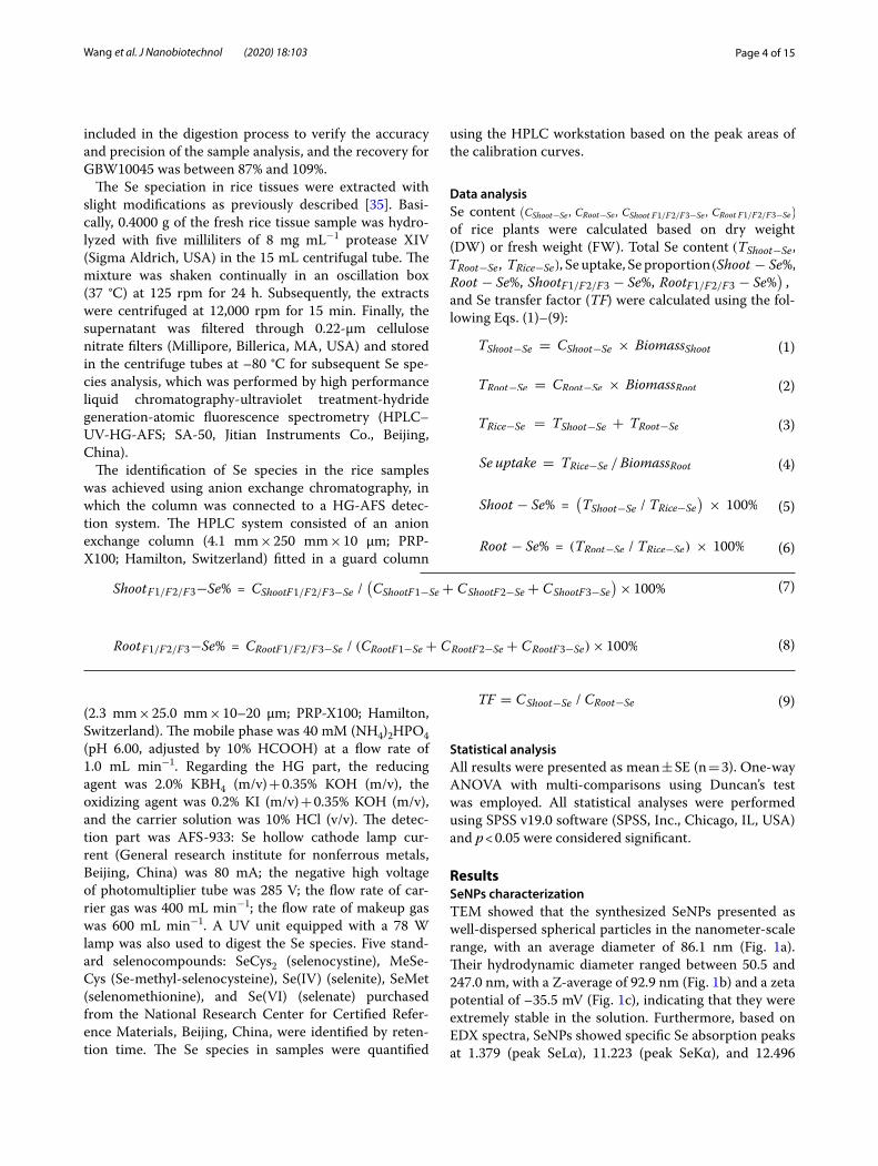
ResultsSeNPs characterizationTEM showed that the synthesized SeNPs presented as well-dispersed spherical particles in the nanometer-scale range, with an average diameter of 86.1 nm (Fig. 1a). Their hydrodynamic diameter ranged between 50.5 and 247.0 nm, with a Z-average of 92.9 nm (Fig. 1b) and a zeta potential of –35.5 mV (Fig. 1c), indicating that they were extremely stable in the solution. Furthermore, based on EDX spectra, SeNPs showed specific Se absorption peaks at 1.379 (peak SeLα), 11.223 (peak SeKα), and 12.496
(1)TShoot−Se = CShoot−Se × BiomassShoot
(2)TRoot−Se = CRoot−Se × BiomassRoot
(3)TRice−Se = TShoot−Se + TRoot−Se
(4)Se uptake = TRice−Se /BiomassRoot
(5)Shoot − Se% =(
TShoot−Se / TRice−Se
)
× 100%
(6)Root − Se% = (TRoot−Se / TRice−Se) × 100%
(7)ShootF1/F2/F3−Se% = CShootF1/F2/F3−Se /(
CShootF1−Se + CShootF2−Se + CShootF3−Se
)
× 100%
(8)RootF1/F2/F3−Se% = CRootF1/F2/F3−Se / (CRootF1−Se + CRootF2−Se + CRootF3−Se)× 100%
(9)TF = CShoot−Se / CRoot−Se(2.3 mm × 25.0 mm × 10–20 μm; PRP-X100; Hamilton, Switzerland). The mobile phase was 40 mM (NH4)2HPO4 (pH 6.00, adjusted by 10% HCOOH) at a flow rate of 1.0 mL min−1. Regarding the HG part, the reducing agent was 2.0% KBH4 (m/v) + 0.35% KOH (m/v), the oxidizing agent was 0.2% KI (m/v) + 0.35% KOH (m/v), and the carrier solution was 10% HCl (v/v). The detec-tion part was AFS-933: Se hollow cathode lamp cur-rent (General research institute for nonferrous metals, Beijing, China) was 80 mA; the negative high voltage of photomultiplier tube was 285 V; the flow rate of car-rier gas was 400 mL min−1; the flow rate of makeup gas was 600 mL min−1. A UV unit equipped with a 78 W lamp was also used to digest the Se species. Five stand-ard selenocompounds: SeCys2 (selenocystine), MeSe-Cys (Se-methyl-selenocysteine), Se(IV) (selenite), SeMet (selenomethionine), and Se(VI) (selenate) purchased from the National Research Center for Certified Refer-ence Materials, Beijing, China, were identified by reten-tion time. The Se species in samples were quantified
Page 5 of 15Wang et al. J Nanobiotechnol (2020) 18:103
0.1 1 10 100 10000
2
4
6
8
10
12
14
Freq
uenc
y (%
)
Hydrodynamic diameter (nm)
b
a1 a2
-200 -100 0 100 2000.0
0.2
0.4
0.6
0.8
1.0
1.2
1.4c
Inte
nsity
(a.u
.)
Zeta potential (mV)
0 3 6 9 12 150
500
1000
1500
2000
2500
3000
3500
4000
Na Cu
Cu
Se
Se
Cps
(eV
)
Enegy (keV)
d
Se
O
Fig. 1 a TEM images, b hydrodynamic diameter, c zeta potential, and d EDX spectra of SeNPs
Page 6 of 15Wang et al. J Nanobiotechnol (2020) 18:103
(peak SeKβ) keV (Fig. 1d), indicating that they were actu-ally synthesized via a chemical method.
Rice seedlings can uptake SeNPsNo Se(VI) or any other organic Se was detected; however, throughout the exposure time (120 h), only a small amount of Se(IV) was present in the 10 μM SeNPs treated culture solution, regardless of the presence or absence of rice seedlings (Additional file 1: Figure S1). Under the SeNPs treatment without rice seedlings, the concentration of Se(IV) ranged from 5.20 to 7.26 μg L−1 and accounted for approximately 0.8% of the SeNPs concentration, indicat-ing that the SeNPs remained stable during the treatment period. Moreover, Se(IV) concentration in the SeNPs with rice seedlings treatment was not changed obviously either (varied from 2.95 to 10.08 μg L−1) even when the exposure time was extended, suggesting that the rice plants or their growth had no significant effect on SeNPs.
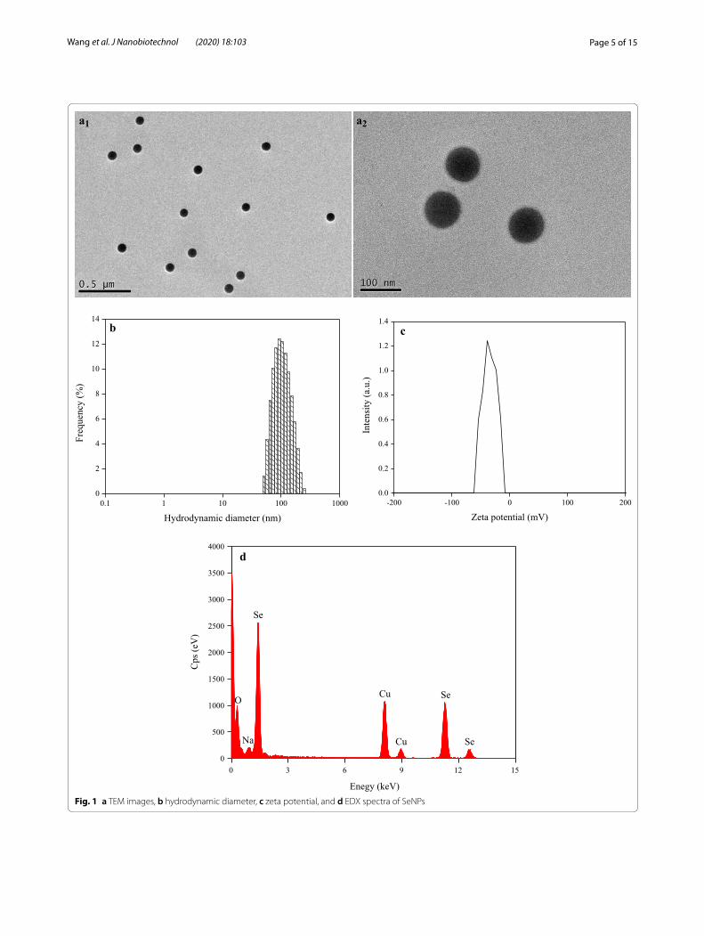
The Se content in rice seedlings increased significantly with an increase in the exposure time (Fig. 2). Its content in roots ranged between 16.16 and 303.43 μg g−1 DW all through the 120 h cultivation period. However, no Se was detected in the 1 h treated-shoot samples owing to the short exposure time; but in the 120 h treated-shoot samples, Se content reached 13.48 μg g−1 DW, indicating that the rice plants could uptake SeNPs and then transport to the aerial parts successfully.
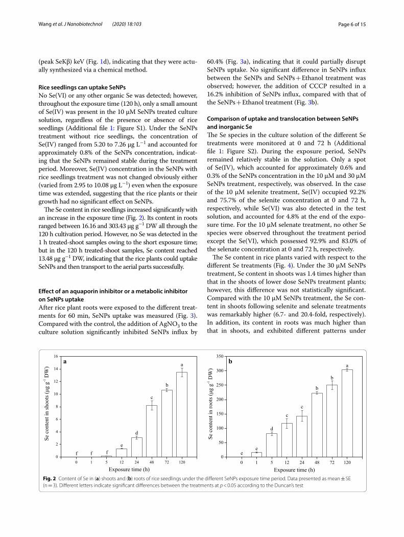
Effect of an aquaporin inhibitor or a metabolic inhibitor on SeNPs uptakeAfter rice plant roots were exposed to the different treat-ments for 60 min, SeNPs uptake was measured (Fig. 3). Compared with the control, the addition of AgNO3 to the culture solution significantly inhibited SeNPs influx by
60.4% (Fig. 3a), indicating that it could partially disrupt SeNPs uptake. No significant difference in SeNPs influx between the SeNPs and SeNPs + Ethanol treatment was observed; however, the addition of CCCP resulted in a 16.2% inhibition of SeNPs influx, compared with that of the SeNPs + Ethanol treatment (Fig. 3b).
Comparison of uptake and translocation between SeNPs and inorganic SeThe Se species in the culture solution of the different Se treatments were monitored at 0 and 72 h (Additional file 1: Figure S2). During the exposure period, SeNPs remained relatively stable in the solution. Only a spot of Se(IV), which accounted for approximately 0.6% and 0.3% of the SeNPs concentration in the 10 μM and 30 μM SeNPs treatment, respectively, was observed. In the case of the 10 μM selenite treatment, Se(IV) occupied 92.2% and 75.7% of the selenite concentration at 0 and 72 h, respectively, while Se(VI) was also detected in the test solution, and accounted for 4.8% at the end of the expo-sure time. For the 10 μM selenate treatment, no other Se species were observed throughout the treatment period except the Se(VI), which possessed 92.9% and 83.0% of the selenate concentration at 0 and 72 h, respectively.
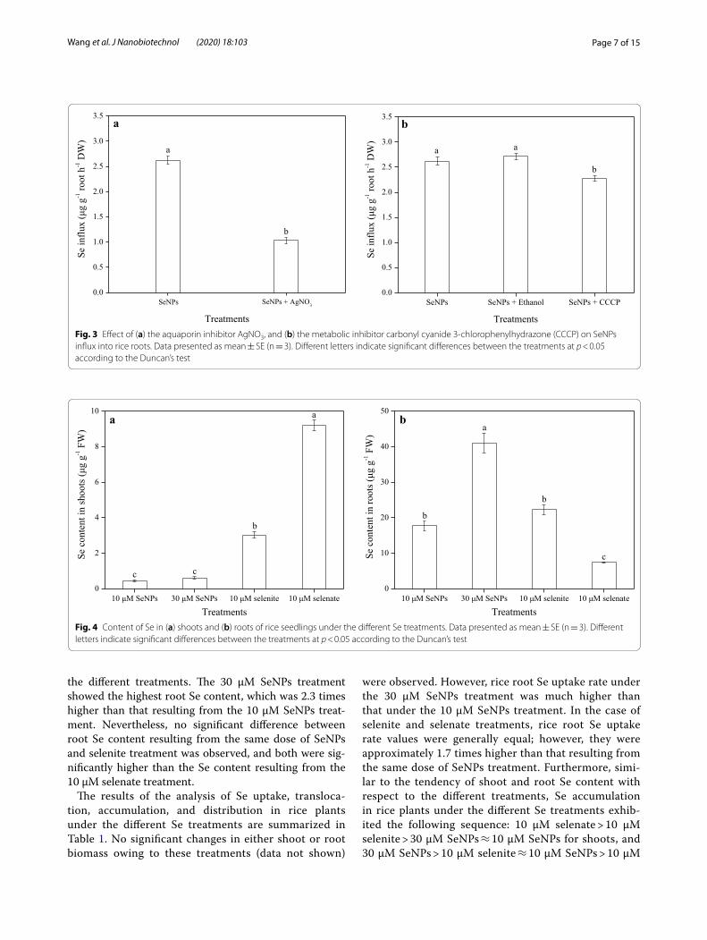
The Se content in rice plants varied with respect to the different Se treatments (Fig. 4). Under the 30 μM SeNPs treatment, Se content in shoots was 1.4 times higher than that in the shoots of lower dose SeNPs treatment plants; however, this difference was not statistically significant. Compared with the 10 μM SeNPs treatment, the Se con-tent in shoots following selenite and selenate treatments was remarkably higher (6.7- and 20.4-fold, respectively). In addition, its content in roots was much higher than that in shoots, and exhibited different patterns under
0 1 5 12 24 48 72 1200
2
4
6
8
10
12
14
16
a
b
c
d
eff
stoohsnitnetnoc
eS(μ
g g-1
DW
)
Exposure time (h)
a
f
0 1 5 12 24 48 72 1200
50
100
150
200
250
300
350
a
b
b
cc
d
e
stoornitnetnoc
eS(μ
g g-1
DW
)
Exposure time (h)
b
e
Fig. 2 Content of Se in (a) shoots and (b) roots of rice seedlings under the different SeNPs exposure time period. Data presented as mean ± SE (n = 3). Different letters indicate significant differences between the treatments at p < 0.05 according to the Duncan’s test
Page 7 of 15Wang et al. J Nanobiotechnol (2020) 18:103
the different treatments. The 30 μM SeNPs treatment showed the highest root Se content, which was 2.3 times higher than that resulting from the 10 μM SeNPs treat-ment. Nevertheless, no significant difference between root Se content resulting from the same dose of SeNPs and selenite treatment was observed, and both were sig-nificantly higher than the Se content resulting from the 10 μM selenate treatment.
The results of the analysis of Se uptake, transloca-tion, accumulation, and distribution in rice plants under the different Se treatments are summarized in Table 1. No significant changes in either shoot or root biomass owing to these treatments (data not shown)
were observed. However, rice root Se uptake rate under the 30 μM SeNPs treatment was much higher than that under the 10 μM SeNPs treatment. In the case of selenite and selenate treatments, rice root Se uptake rate values were generally equal; however, they were approximately 1.7 times higher than that resulting from the same dose of SeNPs treatment. Furthermore, simi-lar to the tendency of shoot and root Se content with respect to the different treatments, Se accumulation in rice plants under the different Se treatments exhib-ited the following sequence: 10 μM selenate > 10 μM selenite > 30 μM SeNPs ≈ 10 μM SeNPs for shoots, and 30 μM SeNPs > 10 μM selenite ≈ 10 μM SeNPs > 10 μM
0.0
0.5
1.0
1.5
2.0
2.5
3.0
3.5
b
a
SeNPs + AgNO3
ggμ(
xulfnieS
-1 ro
ot h
-1 D
W)
Treatments
SeNPs
a
SeNPs SeNPs + Ethanol SeNPs + CCCP0.0
0.5
1.0
1.5
2.0
2.5
3.0
3.5
b
a
ggμ(
xulfnieS
-1 ro
ot h
-1 D
W)
Treatments
a
b
Fig. 3 Effect of (a) the aquaporin inhibitor AgNO3, and (b) the metabolic inhibitor carbonyl cyanide 3-chlorophenylhydrazone (CCCP) on SeNPs influx into rice roots. Data presented as mean ± SE (n = 3). Different letters indicate significant differences between the treatments at p < 0.05 according to the Duncan’s test
10 μM SeNPs 30 μM SeNPs 10 μM selenite 10 μM selenate0
2
4
6
8
10 a
b
c
stoohsnitnetnoc
eS(μ
g g-1
FW
)
Treatments
c
a
10 μM SeNPs 30 μM SeNPs 10 μM selenite 10 μM selenate0
10
20
30
40
50
c
a
b
b
stoornitnetnoc
eS(μ
g g-1
FW
)
Treatments
b
Fig. 4 Content of Se in (a) shoots and (b) roots of rice seedlings under the different Se treatments. Data presented as mean ± SE (n = 3). Different letters indicate significant differences between the treatments at p < 0.05 according to the Duncan’s test
Page 8 of 15Wang et al. J Nanobiotechnol (2020) 18:103
selenate for roots. Furthermore, for the two doses of SeNPs treatment, the absorbed Se was principally dis-tributed in the roots of rice plants (> 90%) and was sel-dom transported to the shoots, owing to an extremely low transfer factor (TF). The Se distribution in shoots following the selenate treatment was significantly higher than that resulting from the 10 μM selenite and SeNPs treatment, while the opposite phenomenon was observed in roots. Therefore, the Se TF value in rice plants of the 10 μM selenate treatment was the highest among the same dose treatment plants. It was 9.1- and 48.6-fold higher than that obtained under selenite and SeNPs treatment, respectively.
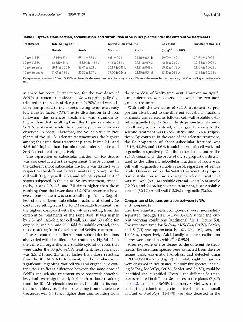
The separation of subcellular fraction of rice tissues was also conducted in this experiment. The Se content in the different shoot subcellular fractions was distinct with respect to the different Se treatments (Fig. 5a–c). In the cell wall (F1), organelle (F2), and soluble cytosol (F3) of shoots subjected to the 30 μM SeNPs treatment, respec-tively, it was 1.9, 4.3, and 2.0 times higher than those resulting from the lower dose of SeNPs treatment; how-ever, none of them was statistically significant. Regard-less of the different subcellular fractions of shoots, Se content resulting from the 10 μM selenate treatment was the highest compared with the values resulting from the different Se treatments of the same dose. It was higher by 2.3- and 14.8-fold for cell wall, 2.6- and 60.1-fold for organelle, and 8.4- and 98.8-fold for soluble cytosol, than those resulting from the selenite and SeNPs treatment.
The Se content in different root subcellular fractions also varied with the different Se treatments (Fig. 5d–f). In the cell wall, organelle, and soluble cytosol of roots that were under the 30 μM SeNPs treatment, respectively, it was 2.5, 2.1, and 2.1 times higher than those resulting from the 10 μM SeNPs treatment, and both values were significant. Regarding root cell wall and organelle Se con-tent, no significant difference between the same dose of SeNPs and selenite treatment were observed; nonethe-less, both were significantly higher than those resulting from the 10 μM selenate treatment. In addition, its con-tent in soluble cytosol of roots resulting from the selenate treatment was 4.4 times higher than that resulting from
the same dose of SeNPs treatment. However, no signifi-cant differences were observed between the two inor-ganic Se treatments.
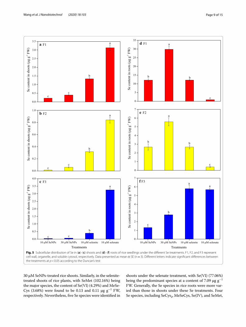
With both the two doses of SeNPs treatment, Se pro-portion distributed to the different subcellular fractions of shoots was ranked as follows: cell wall > soluble cyto-sol > organelle (Fig. 6). Similarly, its proportion of shoots in cell wall, soluble cytosol, and organelle owing to the selenite treatment was 65.5%, 18.9%, and 15.6%, respec-tively. By contrast, in the case of the selenate treatment, the Se proportion of shoot subcellular fractions was 45.1%, 43.3%, and 11.6%, in soluble cytosol, cell wall, and organelle, respectively. On the other hand, under the SeNPs treatments, the order of the Se proportion distrib-uted to the different subcellular fractions of roots was: cell wall > organelle > soluble cytosol, regardless of SeNPs levels. However, unlike the SeNPs treatment, its propor-tion distribution in roots owing to selenite treatment was: cell wall (59.1%) > soluble cytosol (28.0%) > organelle (12.9%), and following selenate treatment, it was: soluble cytosol (82.1%) ≫ cell wall (12.3%) > organelle (5.6%).
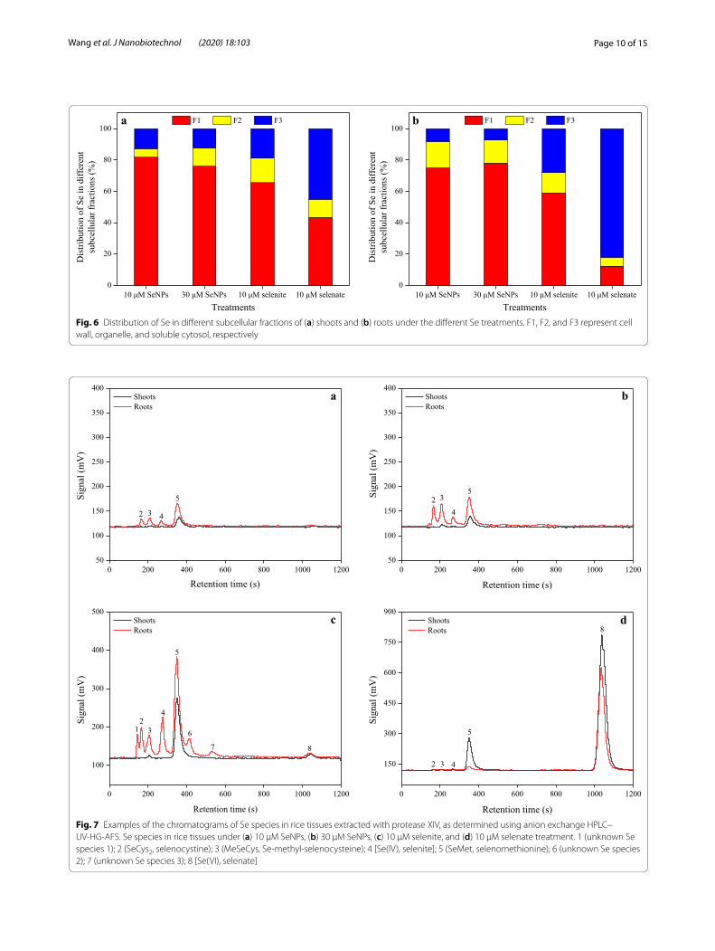
Comparison of biotransformation between SeNPs and inorganic SeThe five standard selenocompounds were successfully separated through HPLC–UV-HG-AFS under the cur-rent working conditions (Additional file 1: Figure S3). The retention time for SeCys2, MeSeCys, Se(IV), SeMet, and Se(VI) was approximately 167, 204, 269, 359, and 1 008 s, respectively. Additionally, all their calibration curves were excellent, with R2 ≥ 0.9984.
After exposure of rice tissues to the different Se treat-ments, the selenium species were extracted from the rice tissues using enzymatic hydrolysis, and detected using HPLC–UV-HG-AFS (Fig. 7). In total, eight Se species were observed in rice tissues, but only five species, includ-ing SeCys2, MeSeCys, Se(IV), SeMet, and Se(VI), could be identified and quantified. Overall, the different Se treat-ments resulted in different Se species in rice plants (Fig. 7; Table 2). Under the SeNPs treatment, SeMet was identi-fied as the predominant species in rice shoots; and a small amount of MeSeCys (15.69%) was also detected in the
Table 1 Uptake, translocation, accumulation, and distribution of Se in rice plants under the different Se treatments
Data presented as mean ± SE (n = 3). Different letters in the same column indicate significant differences between the treatments at p < 0.05 according to the Duncan’s test
Treatments Total Se (μg pot−1) Distribution of Se (%) Se uptake Transfer factor (TF)
Shoots Roots Shoots Roots (μg g−1 root FW)
10 μM SeNPs 4.84 ± 0.37 c 68.14 ± 5.33 b 6.64 ± 0.21 c 93.36 ± 0.21 b 19.00 ± 1.49 c 0.0254 ± 0.0005 c
30 μM SeNPs 6.64 ± 0.88 c 153.50 ± 14.84 a 4.16 ± 0.54 d 95.81 ± 0.54 a 42.86 ± 2.62 a 0.0153 ± 0.0024 c
10 μM selenite 29.47 ± 2.26 b 83.44 ± 8.23 b 26.19 ± 0.48 b 73.81 ± 0.48 c 30.26 ± 1.73 b 0.1357 ± 0.0005 b
10 μM selenate 91.61 ± 7.99 a 26.36 ± 1.77 c 77.60 ± 0.34 a 22.40 ± 0.34 d 33.30 ± 0.83 b 1.2333 ± 0.0298 a
Page 9 of 15Wang et al. J Nanobiotechnol (2020) 18:103
30 μM SeNPs-treated rice shoots. Similarly, in the selenite-treated shoots of rice plants, with SeMet (102.16%) being the major species, the content of Se(VI) (4.29%) and MeSe-Cys (3.68%) were found to be 0.13 and 0.11 μg g−1 FW, respectively. Nevertheless, five Se species were identified in
shoots under the selenate treatment, with Se(VI) (77.06%) being the predominant species at a content of 7.09 μg g−1 FW. Generally, the Se species in rice roots were more var-ied than those in shoots under these Se treatments. Four Se species, including SeCys2, MeSeCys, Se(IV), and SeMet,
10 μM SeNPs 30 μM SeNPs 10 μM selenite 10 μM selenate0.0
0.5
1.0
1.5
2.0
2.5
3.0
3.5
4.0
ggμ(stoohs
nitnetnoceS
-1 F
W) a
b
cc
c F3
Treatments
0.0
0.2
0.4
0.6
0.8
1.0
ggμ(stoohs
nitnetnoceS
-1 F
W) a
b
cc
b F2
0.0
0.5
1.0
1.5
2.0
2.5
3.0
3.5 a
b
c
ggμ(stoohs
nitnetnoceS
-1 F
W)
a F1
c
0
1
2
3
4
5
6
7
ggμ(stoor
nitnetnoceS
-1 F
W)
c
b
a
b
e F2
10 μM SeNPs 30 μM SeNPs 10 μM selenite 10 μM selenate0
1
2
3
4
5
6
7
ggμ(stoor
nitnetnoceS
-1 F
W) a
a
b
f F3
Treatments
c
0
5
10
15
20
25
30
35
ggμ(stoor
nitnetnoceS
-1 F
W)
c
a
b
d F1
b
Fig. 5 Subcellular distribution of Se in (a)–(c) shoots and (d)–(f) roots of rice seedlings under the different Se treatments. F1, F2, and F3 represent cell wall, organelle, and soluble cytosol, respectively. Data presented as mean ± SE (n = 3). Different letters indicate significant differences between the treatments at p < 0.05 according to the Duncan’s test
Page 10 of 15Wang et al. J Nanobiotechnol (2020) 18:103
10 μM SeNPs 30 μM SeNPs 10 μM selenite 10 μM selenate0
20
40
60
80
100
Treatments
F1 F2 F3a
10 μM SeNPs 30 μM SeNPs 10 μM selenite 10 μM selenate0
20
40
60
80
100b
Dis
tribu
tion
of S
e in
diff
eren
tsu
bcel
lula
r fra
ctio
ns (%
)
Dis
tribu
tion
of S
e in
diff
eren
tsu
bcel
lula
r fra
ctio
ns (%
)
Treatments
F1 F2 F3
Fig. 6 Distribution of Se in different subcellular fractions of (a) shoots and (b) roots under the different Se treatments. F1, F2, and F3 represent cell wall, organelle, and soluble cytosol, respectively
0 200 400 600 800 1000 120050
100
150
200
250
300
350
400
5
43
)V
m(langiS
Retention time (s)
ShootsRoots
a
2
0 200 400 600 800 1000 120050
100
150
200
250
300
350
400
5
4
3
Retention time (s)
)V
m(langiS
ShootsRoots
b
2
0 200 400 600 800 1000 1200
100
200
300
400
500
87
6
5
4
32
)V
m(langiS
Retention time (s)
ShootsRoots
c
1
0 200 400 600 800 1000 1200
150
300
450
600
750
900
8
5
43
)V
m(langiS
Retention time (s)
ShootsRoots
d
2
Fig. 7 Examples of the chromatograms of Se species in rice tissues extracted with protease XIV, as determined using anion exchange HPLC–UV-HG-AFS. Se species in rice tissues under (a) 10 μM SeNPs, (b) 30 μM SeNPs, (c) 10 μM selenite, and (d) 10 μM selenate treatment. 1 (unknown Se species 1); 2 (SeCys2, selenocystine); 3 (MeSeCys, Se-methyl-selenocysteine); 4 [Se(IV), selenite]; 5 (SeMet, selenomethionine); 6 (unknown Se species 2); 7 (unknown Se species 3); 8 [Se(VI), selenate]
Page 11 of 15Wang et al. J Nanobiotechnol (2020) 18:103
were identified in roots; and SeMet was the dominant spe-cies following the 10 μM SeNPs or 30 μM SeNPs treat-ment. Concerning to the selenite treatment, excluding the five identifiable Se species, three unknown Se species, referred to as unknown species 1, 2, and 3, at retention times of 145 s (RT145), 414 s (RT414), and 531 s (RT531), respectively, were also identified in the roots. The main Se species was still SeMet, which accounted for 26.58% of the total Se at a content of 5.94 μg g−1 FW. With regard to the selenate treatment, only SeCys2, SeMet, and Se(VI) were monitored in the roots; however, the most abundant species was Se(VI), which occupied 65.94% of the total Se with its content of 4.91 μg g−1 FW.
DiscussionMechanisms of SeNPs uptake by rice seedlingsLiterature has confirmed that nanoparticles (NPs) with size below 100 nm possess unique physico-chemical, electrical, optical, and biological activities, that distin-guish them from their bulkier counterparts [39]; and only NPs or NPs aggregates with diameters less than those of the pores on cell walls can readily pass through and reach the plasma membrane [40, 41]. Given that SeNPs are much larger than cell wall pore size, their passage through the plasma membrane seems impossible. How-ever, in this study, it was found that the rice roots can absorb SeNPs, and transport to the aerial parts (Fig. 2). Therefore, it was reasonable to consider that the SeNPs uptake by rice roots is not solely determined by cell wall pore diameter; other mechanisms may be involved in the process.
Based on existing literature, this study is the first time to investigate the exact mechanism of SeNPs entering into the roots of rice. The results provided evidence that the SeNP uptake was sensitive to the aquaporin inhibitor, AgNO3, but was unaffected by the metabolic inhibitor, CCCP (Fig. 3). It has been established that AgNO3 is a potential inhibitor of aquaporins originating from plants, and the mechanism by which it inhibits aquaporin func-tion is attributed to the ability of silver to react with the sulfhydryl group of a cysteine and a histidine, resulting in gating by the targeted aquaporin [42]. In present work, it was found that the presence of AgNO3 in the culture solution significantly decreased rice root SeNPs absorp-tion by 60.4% (Fig. 3a), a finding that is consistent with those of our previous studies involving wheat, in which a decrease in SeNPs uptake of 92.5% was observed follow-ing SeNPs + AgNO3 treatment [29]. Hence, based on the results of this study, it can be suggested that rice seed-lings absorb SeNPs partially via aquaporin. On the other hand, as an uncoupler of oxidative phosphorylation and protonophores, CCCP can reduce the operation of ATP synthase and concurrently, eliminate the effect of root respiration [43]. Our results showed that this respiratory inhibitor, CCCP, suppressed SeNPs uptake only by 16.2% (Fig. 3b); thus, it is reasonable to assume that SeNPs enter rice roots through an energy-independent passive diffusion. Actually, this downtrend could be attributed to a small amount of Se(IV) present in the test solution, whose uptake is an active process that consumes energy [18, 19]. However, further investigations are still essential to develop a more comprehensive understanding of the uptake mechanism of elemental Se (especially red SeNPs)
Table 2 Content and percentage of Se species in rice tissues
Data presented as mean ± SE (n = 3). Different letters in the same line indicate significant differences between the treatments at p < 0.05 according to the Duncan’s test
SeCys2 selenocystine, MeSeCys Se-methyl-selenocysteine, Se(IV) selenite, SeMet selenomethionine, Se(VI) selenate, ND not detected
*Values in parentheses represent Se species percentage in rice shoots or roots calculated using the expression: Se species content / total tissue content × 100%
Se species 10 μM SeNPs (μg g−1 FW) 30 μM SeNPs (μg g−1 FW) 10 μM selenite (μg g−1 FW) 10 μM selenate (μg g−1 FW)
Shoots
SeCys2 ND ND ND 0.10 ± 0.00 a (1.10%)
MeSeCys ND 0.10 ± 0.01 a (15.69%) 0.11 ± 0.01 a (3.68%) 0.09 ± 0.00 a (0.98%)
Se(IV) ND ND ND 0.05 ± 0.00 a (0.56%)
SeMet 0.43 ± 0.02 c (95.18%)* 0.39 ± 0.02 c (63.19%) 3.10 ± 0.15 b (102.16%) 3.39 ± 0.09 a (36.83%)
Se(VI) ND ND 0.13 ± 0.01 b (4.29%) 7.09 ± 0.06 a (77.06%)
Roots
SeCys2 0.16 ± 0.00 c (0.93%) 0.33 ± 0.03 b (0.80%) 0.49 ± 0.02 a (2.17%) 0.13 ± 0.01 c (1.72%)
MeSeCys 0.17 ± 0.02 c (0.97%) 0.32 ± 0.02 b (0.79%) 0.48 ± 0.02 a (2.15%) ND
Se(IV) 0.07 ± 0.01 b (0.38%) 0.07 ± 0.00 b (0.18%) 0.36 ± 0.01 a (1.61%) ND
SeMet 1.12 ± 0.03 b (6.31%) 1.05 ± 0.06 b (2.56%) 5.94 ± 0.06 a (26.58%) 0.42 ± 0.00 c (5.59%)
Se(VI) ND ND 0.13 ± 0.00 b (0.59%) 4.91 ± 0.18 a (65.94%)
Page 12 of 15Wang et al. J Nanobiotechnol (2020) 18:103
in plants, so as to improve the Se content in crops more efficiently.
Difference between SeNPs and inorganic Se that related to their fate in rice seedlingsIn this current research, the uptake characteristics of SeNPs by rice plants were various from those of selenite and selenate. During the 72 h treatment period, selenite uptake rate was generally equal to that of selenate; how-ever, both were much higher than of SeNPs (Table 1). This finding is partly supported by Hu et al., [29], who reported the influx rate for selenite was 2.5-fold higher than that for SeNPs. However, a comparison of selenite and selenate uptake rates in previous studies showed inconsistencies e.g., a similar uptake rate for selenite and selenate by wheat plants during a 24 h treatment period has been reported [18]. Contrary to these find-ings, in experiments involving Astragalus crotolariae and soybean (Glycin max), Broyer et al. [44] and Zhang et al. [45] found that selenite uptake rate and accumula-tion were higher than those of selenate, whereas de Souza et al. [46] observed a selenate uptake rate that was much higher than that of selenite in experiments using Indian mustard. These discrepancies are possibly caused by the different experimental conditions employed as well as the different plant species studied.
Another important difference between SeNPs and the two inorganic Se forms revealed by this study is their translocation and accumulation in plants. After 72 h of exposure, Se was mainly accumulated in the roots of plants that received SeNPs or selenite treatment, and was seldom transported to the above-ground parts, owing to low TF values. By contrast, most of Se rather accu-mulated in shoots than roots in the case of the selenate-treated rice, in which the Se TF value was extremely high (1.2333) as shown in Table 1. This study also revealed that the Se content in the shoots of selenite and selenate-treated rice plants were, respectively, 6.7- and 20.4-fold higher than that in the shoots of 10 μM SeNPs-treated plants (Fig. 4). It has been generally accepted that Se transportation in plants depends primarily on the initial Se form e.g., Hu et al. [29] reasoned that little SeNPs and selenite were transported to the aerial parts of wheat, while greater portions were retained in roots. Another study showed that the shoot/root ratio of total Se content in all plant species ranged from 0.6 to 1.0 when supplied with SeMet and was less than 0.5 for those supplied with selenite, while it varied from 1.4 for rice to 17.2 for Indian mustard when exposed to selenate [47]. Moreover, xylem is responsible for the longitudinal translocation of ions absorbed by plant roots, and it acts as an ultimate decisive factor in the accumulation of these ions in shoots [48]. Evidence that selenate is extremely mobile during xylem
transport has been reported. Its concentration in xylem sap was found to be 5.7–43.2 times higher than its con-centration in the supplied nutrient solution; while only small amounts of selenite was transported in the xylem of selenite-treated plants [18]. However, Se xylem trans-portation in plants following SeNPs treatment remains unclear, and thus should be further investigated. Until recently, the most accepted explanation for the transloca-tion engineered NPs was that they can move intra- and/or extra-cellularly through tissues until reach the xylem. The mechanism of the passage of these NPs through the Casparian strip into the xylem has not yet been studied in-depth [49]; however, the meristematic zone or root apex are possible routes. Once the engineered NPs reach the vascular system, they are then transported to the aer-ial parts of plant at the pace of water transpiration and nutrient flow [50].
The distribution of toxic elements in different subcellular fractions is closely related to the detoxification mechanism of plants [33, 51]. A general hypothesis is that the cell wall and vacuole (the most important compartment to solu-ble cytosol) in roots are the major sites for sequestrating toxic elements; thereby alleviating their toxicity to plants [51]. However, this hypothesis still leaves one question unanswered: Is there a hidden link between the subcellu-lar distribution of elements and their fate in plants? It has been established that owing to their substantial mechanical rigidity and strength, plant cell walls act as a barrier to the movement of heavy metal ions across the cell membrane [52]; thus inhibiting their translocation from roots to shoots. This study revealed that Se was mainly distributed in cell wall fraction in both shoots and roots under the SeNPs and selenite treatment (Figs. 5 and 6), a finding that is partially corroborated by other observations, especially when the selenite concentration in the culture solution was high [53]. On the contrary, for the selenate treatment, a minor portion of Se was distributed in cell wall of roots (12.3%), whereas the most significant proportion (82.1%) was distributed in soluble cytosol (Figs. 5 and 6). Correspondingly, consider-ing the Se transfer ability of rice plants under the different Se treatments (Table 1), it seems reasonable to consider that the subcellular distribution of Se in the different plant sub-cellular fractions, is responsible for Se transportation within plants. Namely, its transportation is limited within plants if Se mainly distributed in cell wall of tissues, whereas unim-peded if most of Se distributed in soluble cytosol.
To advance the understanding of Se metabolism mech-anism in plants as well as the consequent impacts on human health, it is very important to explore Se trans-formation in rice seedlings after Se fertilization. In this study, only five Se species: SeCys2, MeSeCys, Se(IV), SeMet, and Se(VI) were identified and quantified owing to the limited availability of standard Se compounds
Page 13 of 15Wang et al. J Nanobiotechnol (2020) 18:103
(Additional file 1: Figure S3). The results demonstrated that in rice tissues, the absorbed SeNPs could be con-verted to Se(IV) and organic Se species, demonstrat-ing that the bioavailability of SeNPs for plants. Analysis of the 10 μM SeNPs-treated rice tissue samples showed that SeMet was the predominant species in both shoots (95.18%) and roots (6.31%), while SeCys2, MeSeCys, and Se(IV) were also identified in roots (Fig. 7a; Table 2). Additionally, a small amount of MeSeCys (15.69%) was detected in the shoots of 30 μM SeNPs-treated rice plants (Fig. 7b; Table 2), suggesting that the Se speciation in rice plants was affected by SeNPs dosages. Under the 10 μM selenite treatment, MeSeCys (3.68%), SeMet (102.16%), and Se(VI) (4.29%) were observed in shoots; while sum of eight Se species, including three unidentified Se spe-cies at RT145, RT414, RT531, were found in roots, and SeMet was the most abundant species, which accounted for 26.58% of the total Se (Fig. 7c; Table 2). Among the three unidentified Se species, unknown Se species 2 at RT414 could be SeOMet (selenomethionine Se-oxide), which is the only unknown species whose identity could be postulated, given that its retention time was similar to that of SeMet, coupled with the fact that it could be easily formed through SeMet oxidation [54]. A previous study reported that several other unidentified Se species as well as SeMet, SeOMet, and MeSeCys could be identi-fied in the root extracts of selenite-treated wheat plants [18]. However, given that chloroplasts are the primary site for Se(VI) metabolization [15], all the five identifiable Se species were detected in shoots, whereas only SeCys2, SeMet, and Se(VI) were discovered in the roots of 10 μM selenate-treated plants; and the most abundant species, Se(VI), had a proportion of 77.06% and 65.94% in shoots and roots, respectively (Fig. 7d; Table 2). These results are very consistent with the findings of a previous study on wheat, which demonstrated that when wheat plants were supplied with selenate, Se(VI) remained by far the most predominant species in the shoots and roots, while only small amounts were converted to Se(IV) and orga-noselenium species such as MeSeCys [30]. It is commonly believed that Se speciation varies with the different exog-enous forms of Se e.g., following the selenate treatment of Indian mustard (Brassica juncea), the main species iden-tified was Se(VI), whereas SeMet and SeOMet were pre-dominate in selenite-treated plants [55]. Similar results have also been reported by Pedrero et al. [56] on radish and Kápolna et al. [57] on carrot.
In this study, multiple Se species were identified in sel-enite-treated plants, with organic Se being the predomi-nant species, whereas Se(VI) remained the major species following selenate treatment (Fig. 7; Table 2). This phe-nomenon could be interpreted by considering two expla-nations: (1) Once selenate is absorbed by plants, it can be
metabolized via the sulfur assimilation pathway [15, 16], in which the first step leading to Se(VI) assimilation is its reduction to Se(IV) by ATP sulfurylase, followed by the second step, which is its subsequent conversion to SeCys by selenocysteine methytransferase. Contrarily, once sel-enite was taken up by plants, it is rapidly converted to organic forms and accumulated in roots [18, 30, 46, 47]. Therefore, the reduction of Se(VI) to Se(IV) is the rate-limiting step in selenate metabolism within plants [16, 46]. (2) It has demonstrated that Se(IV) reduction does not require sulfite reductase [16]. This non-enzymatic Se(IV) reduction may also explain why the assimila-tion of Se(IV) to selenoamino acids is easier than that of Se(VI) [46]. Unquestionably, this study showed that the absorbed SeNPs can be converted to organic and oxidative Se species in plants; however, SeNPs assimila-tion mechanism within plants has not yet been studied in-depth, and therefore needs to be investigated in the future studies.
ConclusionsThis study provides physiological evidence that SeNPs can be taken up by rice seedlings; and the SeNPs influx into rice roots is partially through aquaporin, owing to a non-energy consuming passive process, even though the SeNPs uptake rate was much lower than that of selenite or selenate. The absorbed SeNPs or selenite were seldom transported to shoots, but were primarily accumulated in root cell walls. Additionally, they were rapidly assimilated to organic forms, including SeMet, which was the pre-dominant species in both shoots and roots. Contrarily, selenate was readily transported from roots to shoots, and mainly accumulated in the soluble cytosol of shoots; Se(VI) was found to be the most predominant species in the rice plants, and only a relatively small proportion of it was assimilated to organic forms. Based on these find-ings, it is reasonable to consider that SeNPs could be used as a new fertilizer to produce Se-biofortified crops, and eventually meeting the Se requirements of humans and domestic animals.
Supplementary informationSupplementary information accompanies this paper at https ://doi.org/10.1186/s1295 1-020-00659 -6.
Additional file 1: Figure S1. Concentration and proportion of Se(IV) in the culture solution during the exposure period. Data presented as mean ± SE (n = 3). Figure S2. Proportion of different Se species in the culture solution under the different Se treatments during the exposure period. Figure S3. Chromatogram of five standard selenocompounds through HPLC-UV-HG-AFS. SeCys2 selenocystine, MeSeCys Se-methyl-selenocyst-eine, Se(IV) selenite, SeMet selenomethionine, Se(VI) selenate.
Page 14 of 15Wang et al. J Nanobiotechnol (2020) 18:103
AcknowledgementsWe would like to thank Editage (www.edita ge.cn) for English language editing.
Authors’ contributionsKW, YQW, and HFL conceived and designed the experiments. KW and YQW performed the experiments. KW analyzed the data and wrote the paper with the help of QW and ZZ. KL and YBG provided technical support for the SeNPs preparation and characterization. YNW and HFL reviewed and edited the manuscript. All authors read and approved the final manuscript.
FundingThis work was supported by the National Natural Science Foundation of China (No. 41,907,146).
Availability of data and materialsAll data generated or analyzed during this study are included in this published article and its additional files.
Ethics approval and consent to participateNot applicable.
Consent for publicationNot applicable.
Competing interestsThe authors declare that they have no competing interests.
Author details1 Beijing Key Laboratory of Farmland Soil Pollution Prevention and Remedia-tion, Key Laboratory of Plant-Soil Interactions of the Ministry of Education, College of Resources and Environmental Sciences, China Agricultural Univer-sity, Beijing 100193, The People’s Republic of China. 2 Beijing Key Laboratory of Biodiversity and Organic Farming, College of Resources and Environmental Sciences, China Agricultural University, Beijing 100193, The People’s Republic of China.
Received: 31 January 2020 Accepted: 13 July 2020
References 1. Rayman MP. Selenium and human health. Lancet.
2012;379(9822):1256–68. 2. Fairweather-Tait SJ, Bao Y, Broadley MR, Collings R, Ford D, Hesketh
JE, et al. Selenium in human health and disease. Antioxid Redox Sign. 2011;14(7):1337–83.
3. Rayman MP. The importance of selenium to human health. Lancet. 2000;356(9225):233–41.
4. Lv Y, Yu T, Yang Z, Zhao W, Zhang M, Wang Q. Constraint on selenium bioavailability caused by its geochemical behavior in typical Kaschin-Beck disease areas in Aba, Sichuan Province of China. Sci Total Environ. 2014;493:737–49.
5. Combs GF. Selenium in global food systems. Brit J Nutr. 2001;85(5):517–47.
6. Chen L, Yang F, Xu J, Hu Y, Hu Q, Zhang Y, et al. Determination of selenium concentration of rice in China and effect of fertilization of selenite and selenate on selenium content of rice. J Agr Food Chem. 2002;50(18):5128–30.
7. Williams PN, Lombi E, Sun G, Scheckel K, Zhu Y, Feng X, et al. Selenium characterization in the global rice supply chain. Environ Sci Technol. 2009;43(15):6024–30.
8. Huang G, Ding C, Yu X, Yang Z, Zhang T, Wang X. Characteristics of time-dependent selenium biofortification of rice (Oryza sativa L.). J Agr Food Chem. 2018;66(47):12490–7.
9. D’Amato R, Fontanella MC, Falcinelli B, Beone GM, Bravi E, Marconi O, et al. Selenium biofortification in rice (Oryza sativa L.) sprouting: effects on Se yield and nutritional traits with focus on phenolic acid profile. J Agr Food Chem. 2018;66(16):4082–90.
10. Carey A, Scheckel KG, Lombi E, Newville M, Choi Y, Norton GJ, et al. Grain accumulation of selenium species in rice (Oryza sativa L). Environ Sci Technol. 2012;46(10):5557–64.
11. Malik JA, Goel S, Kaur N, Sharma S, Singh I, Nayyar H. Selenium antago-nises the toxic effects of arsenic on mungbean (Phaseolus aureus Roxb.) plants by restricting its uptake and enhancing the antioxidative and detoxification mechanisms. Environ Exp Bot. 2012;77:242–8.
12. Feng R, Wei C, Tu S. The roles of selenium in protecting plants against abiotic stresses. Environ Exp Bot. 2013;87:58–68.
13. El-Ramady H, Abdalla N, Taha HS, Alshaal T, El-Henawy A, Faizy SEDA, et al. Selenium and nano-selenium in plant nutrition. Environ Chem Lett. 2016;14(1):123–47.
14. Han D, Li X, Xiong S, Tu S, Chen Z, Li J, et al. Selenium uptake, specia-tion and stressed response of Nicotiana tabacum L. Environ Exp Bot. 2013;95:6–14.
15. Terry N, Zayed A, De Souza M, Tarun A. Selenium in higher plants. Annu Rev Plant Physiol Plant Mol Biol. 2000;51:401–32.
16. Sors T, Ellis D, Salt D. Selenium uptake, translocation, assimilation and metabolic fate in plants. Photosynth Res. 2005;86(3):373–89.
17. Zhao XQ, Mitani N, Yamaji N, Shen RF, Ma JF. Involvement of silicon influx transporter OsNIP2;1 in selenite uptake in rice. Plant Physiol. 2010;153(4):1871–7.
18. Li H, McGrath SP, Zhao F. Selenium uptake, translocation and spe-ciation in wheat supplied with selenate or selenite. New Phytol. 2008;178(1):92–102.
19. Zhang L, Hu B, Li W, Che R, Deng K, Li H, et al. OsPT2, a phosphate transporter, is involved in the active uptake of selenite in rice. New Phytol. 2014;201(4):1183–91.
20. Wang P, Menzies NW, Lombi E, McKenna BA, James S, Tang C, et al. Synchrotron-based X-ray absorption near-edge spectroscopy imaging for laterally resolved speciation of selenium in fresh roots and leaves of wheat and rice. J Exp Bot. 2015;66(15):4795–806.
21. Elrashidi M, Adriano D, Workman S, Lindsay W. Chemical equilibria of selenium in soils: a theoretical development. Soil Sci. 1987;144(2):141–52.
22. Fox PM, LeDuc DL, Hussein H, Lin Z, Terry N. Selenium speciation in soils and plants. Acs Symposium. 2002;835:339–54.
23. Lin Z, Wang CRC. Evidence on the size-dependent absorption spectral evolution of selenium nanoparticles. Mater Chem Phys. 2005;92(2):591–4.
24. Benko I, Nagy G, Tanczos B, Ungvari E, Sztrik A, Eszenyi P, et al. Subacute toxicity of nano-selenium compared to other selenium species in mice. Environ Toxicol Chem. 2012;31(12):2812–20.
25. Torres SK, Campos VL, León CG, Rodríguez-Llamazares SM, Rojas SM, González M, et al. Biosynthesis of selenium nanoparticles by Pantoea agglomerans and their antioxidant activity. J Nanopart Res. 2012;14(11):1236.
26. Forootanfar H, Adeli-Sardou M, Nikkhoo M, Mehrabani M, Amir-Heidari B, Shahverdi AR, et al. Antioxidant and cytotoxic effect of biologically synthesized selenium nanoparticles in comparison to selenium dioxide. J Trace Elem Med Bio. 2014;28(1):75–9.
27. Zhai X, Zhang C, Zhao G, Stoll S, Ren F, Leng X. Antioxidant capacities of the selenium nanoparticles stabilized by chitosan. J Nanobiotechnol. 2017;15(1).
28. Bai K, Hong B, Hong Z, Sun J, Wang C. Selenium nanoparticles-loaded chitosan/citrate complex and its protection against oxidative stress in d-galactose-induced aging mice. J Nanobiotechnol. 2017;15(1).
29. Hu T, Li H, Li J, Zhao G, Wu W, Liu L, et al. Absorption and bio-transforma-tion of selenium nanoparticles by wheat seedlings (Triticum aestivum L.). Front Plant Sci. 2018;9:597.
30. Huang QQ, Wang Q, Wan YN, Yu Y, Jiang RF, Li HF. Application of X-ray absorption near edge spectroscopy to the study of the effect of sulphur on selenium uptake and assimilation in wheat seedlings. Biol Plantarum. 2017;61(4):726–32.
31. Hawrylak-Nowak B. Comparative effects of selenite and selenate on growth and selenium accumulation in lettuce plants under hydroponic conditions. Plant Growth Regul. 2013;70(2):149–57.
32. Wan Y, Wang K, Liu Z, Yu Y, Wang Q, Li H. Effect of selenium on the subcel-lular distribution of cadmium and oxidative stress induced by cadmium in rice (Oryza sativa L.). Environ Sci Pollut R. 2019;26(16):16220–8.
33. Yu Y, Fu P, Huang Q, Zhang J, Li H. Accumulation, subcellular distribu-tion, and oxidative stress of cadmium in Brassica chinensis supplied
Page 15 of 15Wang et al. J Nanobiotechnol (2020) 18:103
• fast, convenient online submission
•
thorough peer review by experienced researchers in your field
• rapid publication on acceptance
• support for research data, including large and complex data types
•
gold Open Access which fosters wider collaboration and increased citations
maximum visibility for your research: over 100M website views per year •
At BMC, research is always in progress.
Learn more biomedcentral.com/submissions
Ready to submit your research ? Choose BMC and benefit from:
with selenite and selenate at different growth stages. Chemosphere. 2019;216:331–40.
34. Guzmán Mar JL, Hinojosa Reyes L, Mizanur Rahman GM, Kingston HMS. Simultaneous extraction of arsenic and selenium species from rice products by microwave-assisted enzymatic extraction and analysis by ion chromatography-inductively coupled plasma-mass spectrometry. J Agr Food Chem. 2009;57(8):3005–13.
35. Li H, Lombi E, Stroud JL, McGrath SP, Zhao F. Selenium speciation in soil and rice: Influence of water management and Se fertilization. J Agr Food Chem. 2010;58(22):11837–43.
36. Rayman MP, Infante HG, Sargent M. Food-chain selenium and human health: spotlight on speciation. Brit J Nutr. 2008;100(2):238–53.
37. Zhang L, Shi W, Wang X. Difference in selenite absorption between high- and low-selenium rice cultivars and its mechanism. Plant Soil. 2006;282(1–2):183–93.
38. Weigel HJ, Jäger HJ. Subcellular distribution and chemical form of cad-mium in bean plants. Plant Physiol. 1980;65(3):480–2.
39. Ingle AP, Duran N, Rai M. Bioactivity, mechanism of action, and cyto-toxicity of copper-based nanoparticles: a review. Appl Microbiol Biot. 2014;98(3):1001–9.
40. Moore MN. Do nanoparticles present ecotoxicological risks for the health of the aquatic environment? Environ Int. 2006;32(8):967–76.
41. Dietz K, Herth S. Plant nanotoxicology. Trends Plant Sci. 2011;16(11):582–9.
42. Niemietz CM, Tyerman SD. New potent inhibitors of aquaporins: silver and gold compounds inhibit aquaporins of plant and human origin. Febs Lett. 2002;531(3):443–7.
43. Shioi J, Taylor BL. Oxygen taxis and proton motive force in salmonella typhimurium. J Biol Chem. 1984;259(17):10983–8.
44. Broyer T, Johnson C, Huston R. Selenium and nutrition of Astragalus. 1. Effects of selenite or selenate supply on growth and selenium content. Plant Soil. 1972;36(3):635–49.
45. Zhang Y, Pan G, Chen J, Hu Q. Uptake and transport of selenite and selenate by soybean seedlings of two genotypes. Plant Soil. 2003;253(2):437–43.
46. de Souza MP, Pilon-Smits EAH, Lytle CM, Hwang S, Tai J, Honma TSU, et al. Rate-limiting steps in selenium assimilation and volatilization by Indian mustard. Plant Physiol. 1998;117(4):1487–94.
47. Zayed A, Lytle CM, Terry N. Accumulation and volatilization of different chemical species of selenium by plants. Planta. 1998;206(2):284–92.
48. Uraguchi S, Mori S, Kuramata M, Kawasaki A, Arao T, Ishikawa S. Root-to-shoot Cd translocation via the xylem is the major process determining shoot and grain cadmium accumulation in rice. J Exp Bot. 2009;60(9):2677–88.
49. Su Y, Ashworth V, Kim C, Adeleye AS, Rolshausen P, Roper C, et al. Delivery, uptake, fate, and transport of engineered nanoparticles in plants: a critical review and data analysis. Environ Sci Nano. 2019;6(8):2311–31.
50. Zhang L, Feng C, Chen Z, Liu L, Jiang K, Li Q, et al. Superaligned carbon nanotube grid for high resolution transmission electron microscopy of nanomaterials. Nano Lett. 2008;8(8):2564–9.
51. Hall JL. Cellular mechanisms for heavy metal detoxification and tolerance. J Exp Bot. 2002;53(366):1–11.
52. Fry SC. Cross-linking of matrix polymers in the growing cell walls of angiosperms. Ann Rev Plant Physiol. 1986;37:165–86.
53. Ding Y, Wang R, Guo J, Wu F, Xu Y, Feng R. The effect of selenium on the subcellular distribution of antimony to regulate the toxicity of antimony in paddy rice. Environ Sci Pollut R. 2015;22(7):5111–23.
54. Larsen E, Hansen M, Paulin H, Moesgaard S, Reid M, Rayman M. Speciation and bioavailability of selenium in yeast-based intervention agents used in cancer chemoprevention studies. J Aoac Int. 2004;87(1):225–32.
55. Kahakachchi C, Boakye HT, Uden PC, Tyson JF. Chromatographic specia-tion of anionic and neutral selenium compounds in Se-accumulating Brassica juncea (Indian mustard) and in selenized yeast. J Chromatogr a. 2004;1054(1–2):303–12.
56. Pedrero Z, Madrid Y, Cámara C. Selenium species bioaccessibility in enriched radish (Raphanus sativus): A potential dietary source of sele-nium. J Agr Food Chem. 2006;54(6):2412–7.
57. Kápolna E, Hillestrøm PR, Laursen KH, Husted S, Larsen EH. Effect of foliar application of selenium on its uptake and speciation in carrot. Food Chem. 2009;115(4):1357–63.
Publisher’s NoteSpringer Nature remains neutral with regard to jurisdictional claims in pub-lished maps and institutional affiliations.