Uptake of neutrophils and neutrophilic components by macrophages in the inflamed peritoneal cavity...
20
Fish & Shellfish Immunology (1998) 8, 319–338 Article ID: fi980139 Uptake of neutrophils and neutrophilic components by macrophages in the inflamed peritoneal cavity of rainbow trout (Oncorhynchus mykiss) ANTO u NIO AFONSO* 1,2 ,JOANA SILVA 1 ,SUSANA LOUSADA 1 ,ANTHONY E. ELLIS 3 AND MANUEL T. SILVA 1 Institute of Molecular and Cellular Biology and 2 CIMAR, Abel Salazar Biomedical Institute, University of Porto, Portugal 3 Marine Laboratory, Aberdeen, U.K. (Received 24 September 1997, accepted 16 February 1998) The interaction between neutrophils and macrophages in peritoneal inflam- matory exudates in rainbow trout injected intraperitoneally with several bacterial and non-bacterial phlogistic agents was studied by light and electron microscopic histochemistry. The transfer of neutrophilic components to macrophages was demonstrated by the presence in macrophages of myelo- peroxidase, an enzyme of the neutrophil not produced by rainbow trout monocytes and macrophages. Two mechanisms observed for the transfer were phagocytosis of intact neutrophils and phagocytosis of apoptotic bodies released from inflammatory neutrophils. Neutrophils with phagocytosed bacteria were also found to be ingested by macrophages, with the consequent transfer of neutrophilic components and bacteria to the macrophages. Two other mechanisms of transfer are considered. One involves the intake by the macrophage of neutrophilic granules or granular molecules released by lysing or degranulating neutrophils, respectively. The other may be through gap junctions which were observed between adhering inflammatory neutrophils and macrophages. ? 1998 Academic Press Key words: rainbow trout, peritoneal leucocytes, neutrophil/macrophage interaction, phagocytes in fish infection. I. Introduction A crucial mechanism of defence in vertebrates is phagocytosis, killing and degradation of invading microorganisms by neutrophils and macrophages (Suzuki, 1986; Lehrer, 1988; Ainsworth, 1992; Secombes & Fletcher, 1992; Densen et al., 1995). Studies carried out on mammals indicate that phagocytes display di#erent patterns of localisation in the normal host and mobilisation into inflamed territories (Lehrer, 1988; Haslett et al., 1989; Silva et al., 1989; van Furth, 1992; Densen et al., 1995). Such patterns can be summarised as follows. Neutrophils are present in large numbers in blood and haemato- poietic organs as reserve pools, and, under normal conditions, are rare in the *To whom correspondence should be sent at Institute of Molecular and Cellular Biology, Rua do Campo Alegre, 823, 4150-Porto, Portugal. 319 1050–4648/98/050319+20 $30.00/0 ? 1998 Academic Press
-
Upload
antonio-afonso -
Category
Documents
-
view
215 -
download
2
Transcript of Uptake of neutrophils and neutrophilic components by macrophages in the inflamed peritoneal cavity...

Fish & Shellfish Immunology (1998) 8, 319–338Article ID: fi980139
Uptake of neutrophils and neutrophilic components bymacrophages in the inflamed peritoneal cavity of rainbow
trout (Oncorhynchus mykiss)
ANTOuNIO AFONSO*1,2, JOANA SILVA1, SUSANA LOUSADA
1, ANTHONY E. ELLIS3
AND
MANUEL T. SILVA
1Institute of Molecular and Cellular Biology and 2CIMAR, Abel SalazarBiomedical Institute, University of Porto, Portugal
3Marine Laboratory, Aberdeen, U.K.
(Received 24 September 1997, accepted 16 February 1998)
The interaction between neutrophils and macrophages in peritoneal inflam-matory exudates in rainbow trout injected intraperitoneally with severalbacterial and non-bacterial phlogistic agents was studied by light and electronmicroscopic histochemistry. The transfer of neutrophilic components tomacrophages was demonstrated by the presence in macrophages of myelo-peroxidase, an enzyme of the neutrophil not produced by rainbow troutmonocytes and macrophages. Two mechanisms observed for the transfer werephagocytosis of intact neutrophils and phagocytosis of apoptotic bodiesreleased from inflammatory neutrophils. Neutrophils with phagocytosedbacteria were also found to be ingested by macrophages, with the consequenttransfer of neutrophilic components and bacteria to the macrophages. Twoother mechanisms of transfer are considered. One involves the intake by themacrophage of neutrophilic granules or granular molecules released by lysingor degranulating neutrophils, respectively. The other may be through gapjunctions which were observed between adhering inflammatory neutrophilsand macrophages. ? 1998 Academic Press
Key words: rainbow trout, peritoneal leucocytes, neutrophil/macrophageinteraction, phagocytes in fish infection.
*To whom correspondence should be sent at Institute of Molecular and Cellular Biology,Rua do Campo Alegre, 823, 4150-Porto, Portugal.
I. Introduction
A crucial mechanism of defence in vertebrates is phagocytosis, killing anddegradation of invading microorganisms by neutrophils and macrophages(Suzuki, 1986; Lehrer, 1988; Ainsworth, 1992; Secombes & Fletcher, 1992;Densen et al., 1995). Studies carried out on mammals indicate that phagocytesdisplay di#erent patterns of localisation in the normal host and mobilisationinto inflamed territories (Lehrer, 1988; Haslett et al., 1989; Silva et al., 1989;van Furth, 1992; Densen et al., 1995). Such patterns can be summarised asfollows. Neutrophils are present in large numbers in blood and haemato-poietic organs as reserve pools, and, under normal conditions, are rare in the
3191050–4648/98/050319+20 $30.00/0 ? 1998 Academic Press

320 A. AFONSO ET AL.
tissues and body cavities. In contrast, macrophages are present in all bodycompartments and constitute the first phagocytes which invading micro-organisms encounter. Neutrophils from the reserve pools are quickly andextensively attracted to the infectious foci by microbial products and bycytokines produced by immune cells including macrophages (Lehrer, 1988;Appelberg, 1992a,b,c). Monocytes, the blood circulating precursors of tissuemacrophages, are also recruited to infectious sites, although at a slower ratethan neutrophils, where they increase the initial macrophage population;cytokines produced by neutrophils are known to attract monocytes (Taubet al., 1996). It follows, therefore, that each of the two phagocytes has thecapacity to attract the other. Shortly after the onset of an infection, thephagocyte population at the infectious foci is enlarged and composed of bothmacrophages and neutrophils. Previous studies have dealt with the inter-actions between the two phagocytes in those sites in mammals, using theperitoneal cavity of mice (Silva et al., 1989; Sousa, 1994).
The peritoneal model of infection, both in mammals (Silva et al., 1989;Appelberg et al., 1991; Sousa, 1994) and fish (Afonso et al., 1997, 1998), providesa clear example of the distinct patterns of distribution and mobilisation of thetwo phagocytes referred to above. In the case of fish, it has been shown thatthe sterile peritoneal cavity of rainbow trout contains a significant populationof resident macrophages but very few neutrophils (Afonso et al., 1997),though the latter are quickly and extensively attracted to the cavity whenmicroorganisms are injected intraperitoneally (Afonso et al., 1998).
Classically, macrophages have been reported as the only phagocytes impli-cated in the killing of so-called intracellular parasites, because these phago-cytes are the host cell for such pathogens. On the other hand, neutrophils areconsidered as e#ector phagocytes for extracellular parasites only, their rolebeing viewed as irrelevant against the parasites growing intracellularly inmacrophages because in such a residence these parasites are not accessibleto phagocytosis and killing by neutrophils (Mackaness, 1969; Adams &Hamilton, 1984; Edwards & Kirkpatrick, 1986).
Since the initial finding of a persistent presence of significant numbers ofneutrophils in the peritoneal cavity of mice intraperitoneally infected withmycobacteria (Silva et al., 1989; Appelberg et al., 1991), an intracellularparasite growing inside macrophages has been of interest to researchersinvestigating the possible roles of neutrophils in defence mechanisms againstintramacrophagic pathogens. In particular the possibility was proposed of aneutrophil participation in the defence against intramacrophagic infection byan indirect mechanism alternative to the classical direct activity via phago-cytosis and killing of infectious agents. Such a mechanism would involve theinteraction between neutrophils and macrophages in the infectious foci, withthe phagocytosis of the neutrophil, or of neutrophilic components, by theinfected macrophage (Silva et al., 1989; Sousa, 1994).
In fish, both types of phagocytes are known to be present and to operate ininfectious situations through processes essentially identical to those reportedfor mammals (Ellis, 1977; Ainsworth, 1992; Hine, 1992; Secombes & Fletcher,1992; Afonso et al., 1998). The present paper describes and discusses somefacets of the interaction between neutrophils and macrophages in rainbow

UPTAKE OF NEUTROPHILS BY MACROPHAGES 321
trout (Oncorhynchus mykiss) using the peritoneal model of inflammation(Silva et al., 1989; Sousa, 1994; Afonso et al., 1997) which produces exudateswhere the two professional phagocytes co-exist (Afonso et al., 1998).
II. Materials and Methods
EXPERIMENTAL FISH
Sexually immature rainbow trout weighing between 100 and 200 g werepurchased from a commercial farm. The animals were maintained in recircu-lating aerated fresh water, at 15) C, in 7000 litre fibreglass tanks. Waterquality was maintained with mechanical and biological filtration and fishwere fed ad libitum on commercial pellets. The fish to be injected weretransferred to 600 litre tanks where they were maintained for at least 15 daysin the same conditions as above before the study. Only healthy fish, asconcluded from their activity and exterior appearance, were used for the i.p.injections.
GROWTH, HARVESTING AND TREATMENT OF BACTERIAL AGENTS
The strain FP-3 of Yersinia ruckeri type 1 was kindly supplied by Prof. J.Barja (University of Santiago de Compostela, Spain). To prepare the suspen-sions, the bacteria were grown in Tryptic Soy Broth (TSB) (Difco) at 18) Cuntil the middle of the exponential phase, using as inoculum bacteria repeat-edly passaged on Tryptic Soy Agar. The bacteria were collected from the brothby centrifugation and suspended in TSB with 15% glycerol to the indicatedconcentrations, divided in aliquots and frozen at "70) C. Final suspensions ofbacteria in phosphate bu#ered saline (PBS) with osmolality adjusted to310 mOsm l"1 were adjusted to 5#108 bacterial ml"1 by counting in a HelberBacteria Counting Chamber (Fisons Scientific Equipment, Loughborough,U.K.). Preliminary infectivity tests showed that, for the conditions used in thiswork, this inoculum is not pathogenic for trout.
Renibacterium salmoninarum (strain FT-10) was kindly supplied by Prof. J.Barja (University of Santiago de Compostela, Spain). To prepare the suspen-sions, bacteria grown on KDM 2 medium (Evelyn, 1977) for 15 days at 15) Cwere suspended in L-15 medium (Gibco BRL) with 5% foetal bovine serum,divided in aliquots and frozen at "70) C. The inocula to be injected wereprepared by washing and resuspending the above aliquots in PBS, at 1#109
bacteria ml"1. Bacteria were counted in a chamber as described above forY. ruckeri. Under the conditions described, this inoculum of R. salmoninarumwas found to induce a progressive, fatal, infection.
INTRAPERITONEAL INJECTION OF PHLOGISTIC AGENTS
The following agents were used: 12% casein in water, undiluted IncompleteFreund’s Adjuvant (IFA), and suspensions of bacteria in PBS. All agents were
322 A. AFONSO ET AL.
injected into the peritoneal cavity of fish under anaesthesia (0·3 ml ethyleneglycol monophenyl ether l"1, Merck), in volumes of 100 ìl using a 25 G needle.Immediately after the i.p. injection the fish were returned to their tank.Control fish were injected i.p. with PBS.
COLLECTION OF PERITONEAL LEUCOCYTES
The peritoneal cells were collected at intervals (see Results) after the i.p.injections by a procedure previously described in Afonso et al. (1997).Briefly, after killing the fish by anaesthetic overdose and exsanguination,5 ml of PBS containing 20 units ml"1 heparin (Choay) was injected i.p.in the ventral midline followed by injection through the same needle of about1 ml of air. The abdominal area was massaged for about 30 s to dispersethe peritoneal cells in the injected PBS and the cells in suspension werethen collected with a syringe fitted with a 19 G needle and placed on iceuntil processed.
STUDY OF PERITONEAL LEUCOCYTES
The peritoneal cell suspension was analysed within minutes after collectionfor several morphological and cytochemical parameters as follows.
Light microscopy
Total cell counts were performed with a haemocytometer. Cytospin prep-arations were made with a Shandon Cytospin 2 using a volume of peritoneallavage depending on the cell concentration, in order to obtain adequatenumbers of cells per slide. The cytospins were stained with Wright’s stain(Haemacolor, Merck) after fixation for 1 min with formol–ethanol (10 ml of37% formaldehyde and 90 ml of absolute ethanol) (Kaplow, 1965), andleucocyte di#erential counts were performed as described (Afonso et al., 1997).
Electron microscopy
The peritoneal leucocyte suspension in PBS was centrifuged at 1000 g andthe pellet was fixed with 4% paraformaldehyde–1·25% glutaraldehyde–10 mM
calcium fixative (Silva & Macedo, 1983; Silva et al., 1987) at 4) C. After aminimum of 4 h the cells were washed twice with 50 mM cacodylate bu#ersupplemented with 10 mM calcium chloride (pH 6·4) and then fixed at roomtemperature for 2 h with 1% OsO4 in veronal–acetate bu#er supplementedwith 10 mM calcium chloride (Silva & Macedo, 1983) and postfixed at roomtemperature with 1% aqueous uranyl acetate for 1 h (Silva et al., 1971). Afterdehydration in ethanol the samples were embedded in Epon and ultrathinsections were cut with an LKB Ultratome III microtome. Routinely, thesections were contrasted with uranyl acetate followed by lead citrate (Silvaet al., 1987). Observations and micrographs were done with a Zeiss EM 10 Celectron microscope.

UPTAKE OF NEUTROPHILS BY MACROPHAGES 323
Cytochemistry
Peroxidase. For light microscopy the method of Kaplow (1965) or a modifi-cation of it (Antonow’s technique) were used. Antonow’s method was carriedout as follows (Afonso et al., 1998):
(i) Fixation of the cytospin preparations was carried out with formol–ethanol using the Kaplow method (Kaplow, 1965).
(ii) After rinsing with running tap water for a few seconds, the prepara-tions were treated with a 1/30 dilution (in water) of the Kaplow’s reagent(without safranin) for 5–10 min at room temperature. The diluted reagent wasprepared fresh or kept frozen.
(iii) To counterstain the preparations the Wright or the Gram stain wasused.
In the case of fish injected i.p. with Y. ruckeri, the number of peritonealmacrophages positive for peroxidase was counted. For this purpose, peritonealcells from groups of six fish were collected at intervals (see Fig. 11) until 16days after the bacterial inoculation. The cells were processed, as describedabove, for leucocyte counts and staining of peroxidase. The numbers of totalmacrophages, peroxidase-positive macrophages and neutrophils were countedin at least 200 leucocytes per fish.
For ultrastructural studies the method of Robbins et al. (1971) was used.Controls involved omitting hydrogen peroxide from the incubation mixtures.
Esterase. Ultrastructural detection of alpha-naphthyl butyrate (ANB)esterase was carried out as described by Bozdech and Bainton (1981). In somesamples this was followed by the peroxidase technique carried out as indicatedabove (Robbins et al., 1971).
Polysaccharide staining. The ultrastructural staining of cytoplasmicpolysaccharides was carried out using Thiéry’s method (Silva & Macedo,1987; Thiéry, 1967) applied to ultrathin sections of samples processed asdescribed above for conventional electron microscopy or for ultrastructuralcytochemistry for peroxidase.
III. Results
INFLAMMATORY PERITONEAL EXUDATE
The i.p. injection of the phlogistic agents used in the present study inducesneutrophilic and monocytic/macrophagic responses of variable magnitudewhich were previously described in detail (Afonso et al., 1998). Therefore, theperitoneal exudates analysed contained predominantly phagocytes whichwere of three types: resident macrophages, incoming exudate macrophages(monocytes) and neutrophils (mostly incoming).
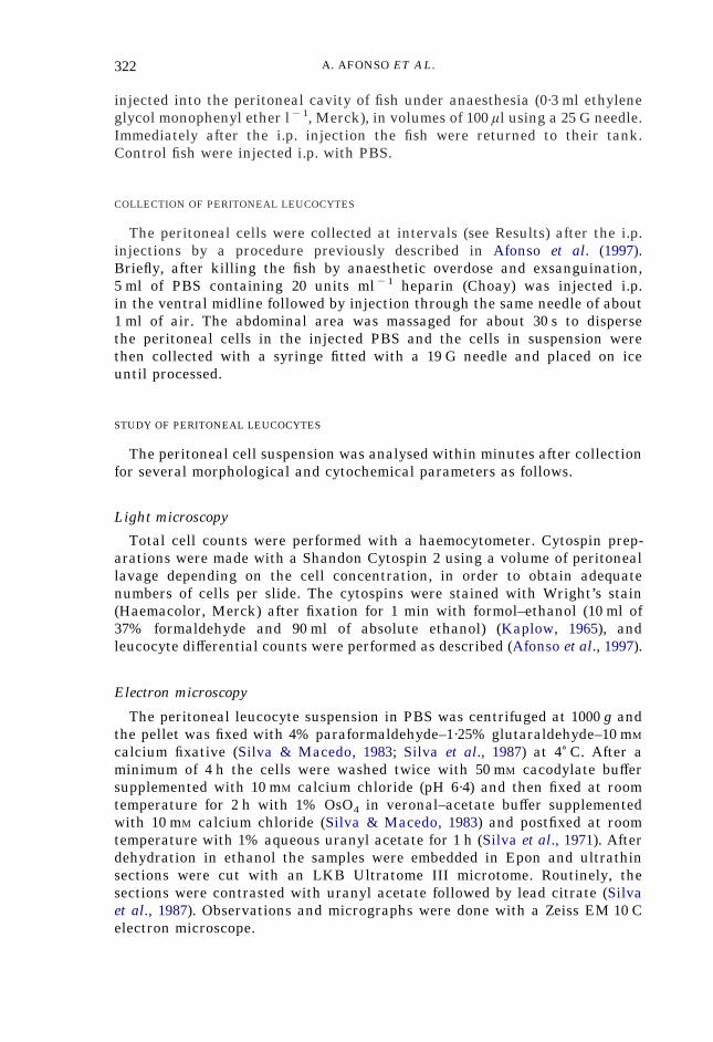
As previously reported in detail (Afonso et al., 1997, 1998) the rainbow troutneutrophils were peroxidase-positive (Figs 1–9), ANB esterase-negative(Fig. 10) and contained large amounts of intracytoplasmic glycogen. On theother hand, blood monocytes and peritoneal resident macrophages were ANBesterase-positive, peroxidase-negative and contained few glycogen granules(Afonso et al., 1997, 1998).

324 A. AFONSO ET AL.
Figs 1–9. Cytospin preparation of leucocytes in the peritoneal exudate of a rainbowtrout 48 h after the intraperitoneal inoculation of 5#107 Y. ruckeri. Preparationstained for peroxidase using Kaplow’s (Figs 1, 4, 6, 9) and Antonow’s (Figs 2, 3, 5, 7,8) technique. Fig. 1. Note the abundant peroxidase-positive (blue) neutrophilgranules. A macrophage contains an intact neutrophil and a few small peroxidase-positive cytoplasmic granules. Fig. 2. Note the peroxidase-positive (green) cyto-plasm of the neutrophils. A macrophage contains an apoptotic neutrophil. Fig. 3.Macrophages with large areas containing peroxidase activity. Fig. 4. Three neu-trophil apoptotic bodies including one inside a macrophage. Fig. 5. Macrophagewith a neutrophil apoptotic body. Fig. 6. Macrophage containing several smallperoxidase-positive granules. Fig. 7. Neutrophil apoptotic body in contact with aneutrophil. Fig. 8. One neutrophil has the nuclear characteristics of apoptosis.Fig. 9. Note the neutrophil embracing a macrophage. All magnifications #1000.

UPTAKE OF NEUTROPHILS BY MACROPHAGES 325
Contrary to the resident macrophages of the unstimulated peritoneal cavityof rainbow trout (Afonso et al., 1997), some large mononuclear leucocytes ofthe cavity inflamed by the injection of all the phlogistic agents studiedwith morphology of macrophages contained peroxidase-positive material(Figs 1–6, 10). That these peroxidase-positive leucocytes were macrophageswas confirmed by using the simultaneous staining for esterase/peroxidase(Fig. 10) or peroxidase/glycogen, as described in detail elsewhere (Afonsoet al., 1998).
The patterns of peroxidase positivity inside macrophages found in lightmicroscopy preparations can be grouped as follows: type 1—positivity due tothe granules of intact neutrophils (Fig. 1), including apoptotic neutrophils(Fig. 2), phagocytosed by macrophages (see below); type 2—clumps ofperoxidase-positive material in large vacuoles (Fig. 3); type 3—clumps ofpositive granules inside small vacuoles (Figs 4 and 5); and type 4—dispersedand usually scarce small granules (Fig. 6). Ultrastructural images correspond-ing to the above patterns were also observed; some examples are shown inFigs 12, 13 and 16.
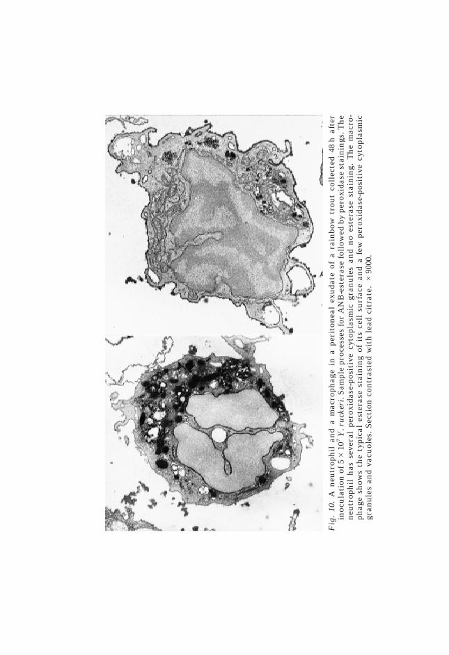
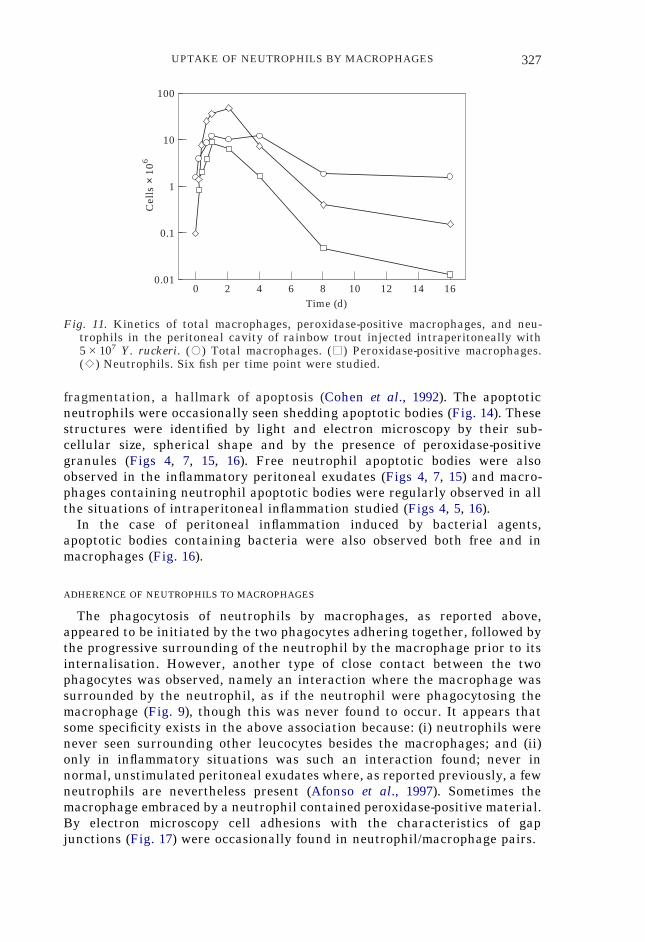
Quantification of the peroxidase-positive peritoneal macrophages was car-ried out in light microscopy preparations stained by the Antonow’s method inthe case of i.p. inoculation of Y. ruckeri (Fig. 11). This quantification showsthat the kinetics of peroxidase-positive macrophages followed that of incom-ing neutrophils, and that at 24 h post-inoculation most of the macrophagescontained peroxidase activity. Throughout the period of time studied(Fig. 11), most of the peroxidase-positive macrophages were of the type 4described above. In the control fish injected i.p. with PBS the values forperoxidase-positive macrophages were very low (not shown).
PHAGOCYTOSIS OF NEUTROPHILS BY MACROPHAGES
In the exudates of fish injected with the phlogistic agents used, where thetwo types of professional phagocytes were co-existing, the regular presence ofmacrophages which had internalised neutrophils was recorded, both by lightmicroscopy (Figs 1 and 2) and at the ultrastructural level (Figs 12 and 13).That the phagocytosed cells inside the macrophages were in fact neutrophilscould be determined by their typical morphology in Wright-stained cytospinsor because of the presence of the characteristic cytoplasmic granulesand abundant glycogen particles by electron microscopy, and by theperoxidase-positivity of the cytoplasmic granules of the internalised cells.
In the peritoneal cavity of trout inoculated with R. salmoninarum,macrophages were found which had phagocytosed neutrophils containingbacteria (Fig. 13).
NEUTROPHIL APOPTOSIS
Neutrophils with nuclear alterations typical of apoptosis were observed inthe inflamed peritoneal exudates (Fig. 8). Preliminary observations with theApo-Direct kit for apoptosis detection (Phoenix Flow Systems, San Diego, CA,U.S.A.) showed that these nuclear abnormalities corresponded to DNA
Fig
.10
.A
neu
trop
hil
and
am
acro
phag
ein
ape
rito
nea
lex
uda
teof
ara
inbo
wtr
out
coll
ecte
d48
haf
ter
inoc
ula
tion
of5#
107
Y.r
uck
eri.
Sam
ple
proc
esse
sfo
rA
NB
-est
eras
efo
llow
edby
pero
xida
sest
ain
ings
.Th
en
eutr
oph
ilh
asse
vera
lpe
roxi
dase
-pos
itiv
ecy
topl
asm
icgr
anu
les
and
no
este
rase
stai
nin
g.T
he
mac
ro-
phag
esh
ows
the
typi
cal
este
rase
stai
nin
gof
its
cell
surf
ace
and
afe
wpe
roxi
dase
-pos
itiv
ecy
topl
asm
icgr
anu
les
and
vacu
oles
.S
ecti
onco
ntr
aste
dw
ith
lead
citr
ate.
#90
00.
UPTAKE OF NEUTROPHILS BY MACROPHAGES 327
fragmentation, a hallmark of apoptosis (Cohen et al., 1992). The apoptoticneutrophils were occasionally seen shedding apoptotic bodies (Fig. 14). Thesestructures were identified by light and electron microscopy by their sub-cellular size, spherical shape and by the presence of peroxidase-positivegranules (Figs 4, 7, 15, 16). Free neutrophil apoptotic bodies were alsoobserved in the inflammatory peritoneal exudates (Figs 4, 7, 15) and macro-phages containing neutrophil apoptotic bodies were regularly observed in allthe situations of intraperitoneal inflammation studied (Figs 4, 5, 16).
In the case of peritoneal inflammation induced by bacterial agents,apoptotic bodies containing bacteria were also observed both free and inmacrophages (Fig. 16).
16
100
0.010
Time (d)
Cel
ls ×
106
8
10
1
0.1
2 4 6 10 12 14
Fig. 11. Kinetics of total macrophages, peroxidase-positive macrophages, and neu-trophils in the peritoneal cavity of rainbow trout injected intraperitoneally with5#107 Y. ruckeri. (,) Total macrophages. (.) Peroxidase-positive macrophages.(:) Neutrophils. Six fish per time point were studied.
ADHERENCE OF NEUTROPHILS TO MACROPHAGES
The phagocytosis of neutrophils by macrophages, as reported above,appeared to be initiated by the two phagocytes adhering together, followed bythe progressive surrounding of the neutrophil by the macrophage prior to itsinternalisation. However, another type of close contact between the twophagocytes was observed, namely an interaction where the macrophage wassurrounded by the neutrophil, as if the neutrophil were phagocytosing themacrophage (Fig. 9), though this was never found to occur. It appears thatsome specificity exists in the above association because: (i) neutrophils werenever seen surrounding other leucocytes besides the macrophages; and (ii)only in inflammatory situations was such an interaction found; never innormal, unstimulated peritoneal exudates where, as reported previously, a fewneutrophils are nevertheless present (Afonso et al., 1997). Sometimes themacrophage embraced by a neutrophil contained peroxidase-positive material.By electron microscopy cell adhesions with the characteristics of gapjunctions (Fig. 17) were occasionally found in neutrophil/macrophage pairs.
328 A. AFONSO ET AL.
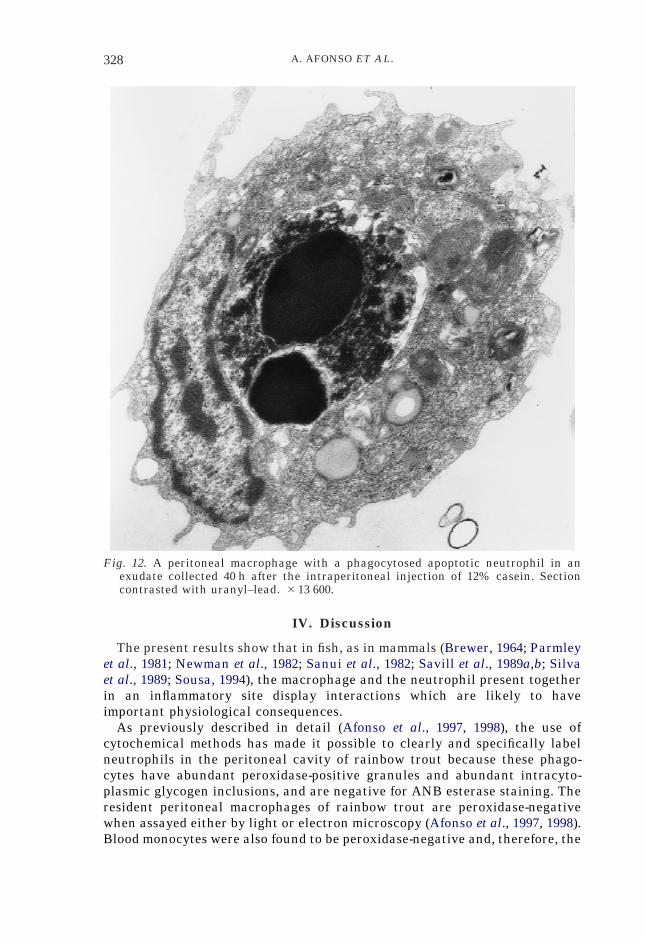
Fig. 12. A peritoneal macrophage with a phagocytosed apoptotic neutrophil in anexudate collected 40 h after the intraperitoneal injection of 12% casein. Sectioncontrasted with uranyl–lead. #13 600.
IV. Discussion
The present results show that in fish, as in mammals (Brewer, 1964; Parmleyet al., 1981; Newman et al., 1982; Sanui et al., 1982; Savill et al., 1989a,b; Silvaet al., 1989; Sousa, 1994), the macrophage and the neutrophil present togetherin an inflammatory site display interactions which are likely to haveimportant physiological consequences.
As previously described in detail (Afonso et al., 1997, 1998), the use ofcytochemical methods has made it possible to clearly and specifically labelneutrophils in the peritoneal cavity of rainbow trout because these phago-cytes have abundant peroxidase-positive granules and abundant intracyto-plasmic glycogen inclusions, and are negative for ANB esterase staining. Theresident peritoneal macrophages of rainbow trout are peroxidase-negativewhen assayed either by light or electron microscopy (Afonso et al., 1997, 1998).Blood monocytes were also found to be peroxidase-negative and, therefore, the

UPTAKE OF NEUTROPHILS BY MACROPHAGES 329
Fig. 13. A peritoneal macrophage with a phagocytosed neutrophil in an exudate of atrout inoculated 6 d before with 1#108 R. salmoninarum. Notice the presence ofbacteria in the cytoplasms of both the macrophage and the neutrophil. Sectioncontrasted with uranyl–lead #12 800.
incoming (exudate) monocytes in the inflamed peritoneal cavity must also bedevoid of peroxidase activity (Afonso et al., 1998). Since the neutrophils arethe only peroxidase-positive leucocyte in the peritoneal exudate, the peroxi-dase activity can be used as a specific marker for neutrophilic material. In thepresent study of inflammatory exudate cells we found some peroxidase-positive large mononuclear leucocytes with the morphology of macrophages.That these leucocytes are indeed macrophages is confirmed by two othercytochemical characteristics: their ANB esterase positivity and scarcity ofintracytoplasmic glycogen granules. Consequently, the presence of peroxidaseactivity in macrophages of the inflamed peritoneal cavity of rainbow trout isan indication of the occurrence of transfer of neutrophilic material to themacrophages. The present observations show two obvious mechanisms forsuch a transfer: the phagocytosis by the macrophage of intact neutrophils andof neutrophil apoptotic bodies.

330 A. AFONSO ET AL.
Fig. 14. An apoptotic neutrophil shedding apoptotic bodies in an exudate collected40 h after the intraperitoneal injection of 12% casein. Section contrasted withuranyl–lead. #23 600.
Four patterns of peroxidase positivity were observed in macrophagespresent in the inflammatory peritoneal exudates. Type 1 refers to the presenceof neutrophils su$ciently intact to be detected by Wright counter-stainingwhen using Antonow’s method. Most probably, this pattern shows neutrophilswhich had been phagocytosed by the macrophage very shortly before sam-pling. In fact, in mammals, it is known that the phagocytosed neutrophils arequickly degraded by the hydrolytic enzymes of the macrophage lysosomes(Newman et al., 1982; Haslett et al., 1989), with the resulting loss of the normalmorphology of the neutrophil. However, the peroxidase activity remains forsome time after the loss of normal neutrophil morphology (Heifets et al., 1980)and thus, the presence of peroxidase-positive material in large vacuoles insidethe macrophages (pattern type 2) is an indication that those vacuolesare phagosomes with remnants of a phagocytosed neutrophil in advanceddegradation.

UPTAKE OF NEUTROPHILS BY MACROPHAGES 331
Fig. 15. Free neutrophil apoptotic bodies in the peritoneal cavity of a rainbow troutinjected intraperitoneally with 5#107 Y. ruckeri. On the left conventional electronmicroscopy; section contrasted with uranyl–lead. #11 600. On the right, sampleprocessed for peroxidase; section contrasted with lead. #18 900. Note the presenceof neutrophil granules with peroxide activity.
It is known that senescent mammalian neutrophils in inflammatory fociundergo apoptosis (Haslett et al., 1989; Savill et al., 1989a,b). One feature ofthis process is the release of apoptotic bodies, sub-cellular particles rich inneutrophil granules that are taken up by macrophages (Hanker & Giammara,1983; Cohen et al., 1992). The presence of neutrophil apoptotic bodies insiderainbow trout macrophages observed here represents the type 3 pattern ofperoxidase positivity in macrophages.
Whether other types of transfer of neutrophilic components to the macro-phage, besides those discussed above, are occurring in inflamed sites ofrainbow trout is not known with certainty, but some possibilities have tobe considered. One regards the neutrophil/macrophage interaction heredescribed whereby the two phagocytes appear closely adherent with thepossible participation of intercellular junctions. The interpretation that thisinteraction could lead to material transfer is supported by two observations.Firstly, the adherence of neutrophils was preferentially to macrophages; and,secondly, the peroxidase activity was sometimes found in the macrophagesassociated with neutrophils. It is interesting that the adherence of neutrophilsto macrophages was seen only in inflammatory exudates but never in thenormal, unstimulated peritoneal cavity. The finding of the junctions bytransmission electron microscopy has been, so far, occasional, but it ispossible that it might occur more frequently than it appears, because thechances of the plane of section passing through the actual location of thejunction are quite small. The occurrence of adhesions between neutrophils

332 A. AFONSO ET AL.
Fig. 16. A peritoneal macrophage with phagocytosed neutrophil apoptotic bodies(arrows) in an exudate of a trout inoculated 12 d before with 1#108 R. salmoni-narum. One of the apoptotic bodies (*) contains bacteria. Section contrasted withlead. #14 500.
and macrophages reported here for inflammatory exudates in rainbow trouthas not been described, to our knowledge, in mammals.
Another possibility for the transfer of neutrophilic material to the macro-phages is the uptake by the macrophage of neutrophil granules or molecules.Phagocytosis of neutrophilic granules and of neutrophilic granular moleculesby macrophages has been reported for rodents (Heifets et al., 1980; Shellitoet al., 1987; Leung & Goren, 1989; Sousa, 1994). The neutrophil granules can bereleased by neutrophils lysing at the inflammatory sites due to leucotoxinsproduced by infectious agents (Cochrane, 1968; Henson & Johnston, 1987). It is

UPTAKE OF NEUTROPHILS BY MACROPHAGES 333
Fig. 17. (a) Adherent neutrophil and macrophage in the peritoneal exudate of arainbow trout injected intraperitoneally with 5#107 Y. ruckeri 24 h before. Sampleprocessed for detection of peroxidase activity. Section contrasted with lead. Notethe numerous peroxidase-positive granules in the cytoplasm of the neutrophil. Inthe cytoplasm of the macrophage a few granules show peroxidase activity. #9000.(b) High magnification of the region of contact indicated in (a) showing intercellularjunctions with the characteristics of gap junctions. #60 800.
also known that, at inflammatory sites, activated neutrophils secrete mol-ecules stored in their granules through the process known as degranulation,and that at those sites neutrophilic molecules, like myeloperoxidase, lacto-ferrin and defensins, can be present extracellularly (Estensen et al., 1974;Goldstein et al., 1975; Pryzwansky & Bréton-Gorius, 1983; Ganz, 1987; Shellitoet al., 1987; Falloon & Gallin, 1988; Leung & Goren, 1989; Esaguy et al., 1991;Densen et al., 1995). The occurrence, also reported here, of macrophages withdiscrete peroxidase-positive granules (pattern type 4) may well correspond tothe transfer of neutrophilic granules or molecules.
The phagocytosis by macrophages of intact neutrophils in inflammatory fociwas described a century ago by Metchniko# in tadpoles, and thereafter it wasfound to occur in mammals, including man (Brewer, 1964; Parmley et al., 1981;Newman et al., 1982; Sanui et al., 1982; Shellito et al., 1987; Silva et al., 1989;Sousa, 1994). However, the physiological importance of such an event is notyet completely understood. On the one hand, it has been described as amechanism of disposing of senescent neutrophils by macrophages and thus

334 A. AFONSO ET AL.
limiting tissue damage in acute inflammation and in the resolution ofinflammation (Haslett et al., 1989; Savill et al., 1989a,b; Cohen et al., 1992). Onthe other hand, the phagocytosis of neutrophils by macrophages at infectiousfoci may have another consequence. Namely, the uptake of the neutrophils byinfected macrophages would have the additional advantage of enhancing thecapacity of the macrophage to cope with infection (Silva et al., 1989; Sousa,1994). Such an increased antimicrobial capacity can result from the acquisi-tion by the macrophage of antimicrobial molecules present in the neutrophiland lacking in the macrophage (Heifets et al., 1980; Locksley et al., 1982;Nogueira et al., 1982; Lima & Kierszenbaum, 1985; Shellito et al., 1987; Leung& Goren, 1989; Silva et al., 1989; Sousa, 1994). That some antimicrobialmolecules operating in neutrophils, like myeloperoxidase, lactoferrin anddefensins, are not present in mammalian macrophages is well documented(Falloon & Gallin, 1988; Ganz et al., 1990; Ogata et al., 1992; Borregaard et al.,1993; Densen et al., 1995), as is the utilisation in antimicrobial activities ofsome of those molecules when transferred to macrophages (Heifets et al., 1980;Nogueira et al., 1982).
One interesting aspect regarding the phagocytosis of neutrophils by macro-phages in infected peritoneal exudates described in this paper regards theingestion of infected neutrophils, and of neutrophil apoptotic bodies, contain-ing bacteria, by the macrophages, as already reported in experimental myco-bacteriosis in mice (Silva et al., 1989). This phagocytosis results in the transferto the macrophages of bacteria initially phagocytosed by the neutrophils.When the host is able to control the growth of the invading microorganism, itis likely that the macrophage’s antimicrobial and degradative activities willbe exerted on these transferred parasites. Since macrophages are much morelong-lived as compared to neutrophils, such a transfer can be advantageous tothe host. Moreover, it is likely that the antimicrobial molecules of theneutrophil transferred to the macrophages with the bacteria will participatein the attack on the microorganism. In fact, it has been shown that neu-trophilic molecules ingested by macrophages are mobilised to parasite-containing macrophage phagosomes (Leung & Goren, 1989; Sousa, 1994) andare used in the macrophage’s antimicrobial processes (Heifets et al., 1980;Nogueira et al., 1982; Silva et al., 1989; Sousa, 1994). On the other hand, in theinstances where the parasite resists the host’s defence mechanisms, thetransfer of the infectious agent to the macrophage through the phagocytosisby this phagocyte of infected neutrophils or neutrophilic apoptotic bodies maybe an e$cient way for the agent to spread in the organism.
A new picture is emerging for the physiology of the mammalian neutrophil.Recent results show that the neutrophil is not just a terminal e#ector cellsolely engaged in the killing of extracellular pathogens. Rather, the neu-trophil appears as a leucocyte that also participates in the defence mecha-nisms against intracellular pathogens (Silva et al., 1989; Conlan & North, 1991,1992, 1994; Rogers & Unanue, 1993; Appelberg et al., 1994, 1995; Czuprynskiet al., 1994a,b; Sjostedt et al., 1994; Sousa, 1994; Barteneva et al., 1996; Newmanet al., 1997). Moreover, neutrophils have been shown to be biosyntheticallyactive leucocytes producing and excreting several cytokines that participatein immune mechanisms, including the modulation of macrophage activity

UPTAKE OF NEUTROPHILS BY MACROPHAGES 335
(Lloyd & Oppenheim, 1992; Cassatella, 1995; Cassatella et al., 1995). Theoccurrence of transfer of neutrophilic components to macrophages, describedhere, is evidence for the view that in fish, like in mammals, cooperationbetween neutrophils and macrophages would occur as part of the defencemechanism against parasites growing inside macrophages. Whether such atransfer occurs in fish as well as in mammals (Silva et al., 1989; Sousa, 1994),actually enhancing the capacity of trout macrophages against fish pathogensremains to be directly demonstrated and is currently under investigation inthis laboratory.
The authors are grateful to Prof. C. J. Secombes (University of Aberdeen, U.K.) forcontinuous help and support; to Prof. Alberto Villena and Dr Jorge Pedrosa for helpfuldiscussions and to Prof. António C. Castro for supplying the fish used in the presentwork.
The excellent technical assistance provided by Mrs Paula M. Macedo, Miss IreneBarros and Mr Adão Silva is gratefully acknowledged.
References
Adams, D. O. & Hamilton, T. A. (1984). The cell biology of macrophage activation.Annual Review of Immunology 2, 283–318.
Afonso, A., Ellis, A. E. & Silva, M. T. (1997). Leucocyte population of the unstimulatedperitoneal cavity of rainbow trout (Oncorhynchus mykiss). Fish & ShellfishImmunology 7, 335–348.
Afonso, A., Lousada, S., Silva, J., Ellis, A. E. & Silva, M. T. (1998). Neutrophil andmacrophage responses to inflammation in the peritoneal cavity of rainbow trout(Oncorhynchus mykiss). A light and electron microscopic cytochemical study.Diseases of Aquatic Organisms, in press.
Ainsworth, A. J. (1992). Fish granulocytes: morphology, distribution and function.Annual Review of Fish Diseases 123–148.
Appelberg, R. (1992a). Mycobacterial infection primes T cells and macrophages forenhanced recruitment of neutrophils. Journal of Leucocyte Biology 51, 472–477.
Appelberg, R. (1992b). Interferon-gamma (IFNã) and macrophage inflammatory pro-teins (MIP)-1 and -2 are involved in the regulation of the T cell-dependentchronic peritoneal neutrophilia of mice infected with mycobacteria. Clinical &Experimental Immunology 89, 269–273.
Appelberg, R. (1992c). Macrophage inflammatory proteins MIP-1 and MIP-2 areinvolved in T cell-mediated neutrophil recruitment. Journal of Leucocyte Biology52, 303–306.
Appelberg, R., Pedrosa, J. M. & Silva, M. T. (1991). Host and bacterial factors controlthe Mycobacterium avium-induced chronic peritoneal granulocytosis in mice.Clinical & Experimental Immunology 83, 231–236.
Appelberg, R., Castro, A. G. & Silva, M. T. (1994). Neutrophils as e#ector cells ofT-cell-mediated, acquired immunity in murine listeriosis. Immunology 83, 302–307.
Appelberg, R., Castro, A. G., Gomes, S., Pedrosa, J. R. & Silva, M. T. (1995).Susceptibility of beige mice to Mycobacterium avium: Role of neutrophils.Infection & Immunity 63, 3381–3387.
Barteneva, N., Theodor, I., Peterson, E. M. & de la Maza, L. M. (1996). Role ofneutrophils in controlling early stages of a Chlamydia trachomatis infection.Infection & Immunity 64, 4830–4833.
Borregaard, N., Lollike, K., Kjeldsen, L., Sengelov, H., Bastholm, L., Nielsen, M. H. &Bainton, D. F. (1993). Human neutrophil granules and secretory vesicles.European Journal of Haematology 51, 187–198.

336 A. AFONSO ET AL.
Boxer, L. A., Haak, R. A., Yang, H.-H., Wolach, J. H., Whitcomb, J. A., Butterick, C. J.& Baehner, R. L. (1982). Membrane-bound lactoferrin alters the surface proper-ties of polymorphonuclear leucocytes. Journal of Clinical Investigation 70,1049–1057.
Bozdech, M. J. & Bainton, D. E. (1981). Identification of alpha naphthyl butyrateesterase as a plasma membrane ectoenzyme of monocytes and as a discreteintracellular membrane bounded organelle in lymphocytes. Journal of Exper-imental Medicine 153, 182–195.
Brewer, D. B. (1964). Electron-microscope phagocytosis of neutrophil polymorpho-nuclear leucocytes by macrophages. Journal of Pathology 88, 307–310.
Cassatella, M. A., Meda, L., Gasperini, S., D’Andrea, A., Ma, X. & Trinchieri, G. (1995).Interleukin-12 production by human polymorphonuclear leucocytes. EuropeanJournal of Immunology 25, 1–5.
Cassatella, M. A. (1995). The production of cytokines by polymorphonuclear neu-trophils. Immunology Today 16, 21–26.
Cochrane, C. G. (1968). Immunologic tissue injury mediated by neutrophilic leucocytes.Advances in Immunology 9, 97–165.
Cohen, J. J., Duke, R. C., Fadok, V. A. & Sellins, K. S. (1992). Apoptosis andprogrammed cell death in immunology. Annual Review of Immunology 10,267–293.
Conlan, J. W. & North, R. J. (1991). Neutrophil-mediated dissolution of infected hostcells as a defense strategy against a facultative intracellular bacterium. Journalof Experimental Medicine 174, 741–744.
Conlan, J. W. & North, R. J. (1992). Early pathogenesis of infection in the liver with thefacultative intracellular bacteria Listeria monocytogenes, Francisella tularensis,and Salmonella typhimurium involves lysis of infected hepatocytes by leuco-cytes. Infection & Immunity 60, 5164–5171.
Conlan, J. W. & North, R. J. (1994). Neutrophils are essential for early anti-listeriadefense in the liver, but not in the spleen or peritoneal cavity, as revealed by agranulocyte-depleting monoclonal antibody. Journal of Experimental Medicine179, 259–268.
Czuprynski, C. J., Brown, J. F., Maroushek, N., Wagner, R. D. & Steinberg, H. (1994a).Administration of anti-granulocyte MAb RB6-8C5 impairs resistance of mice toListeria monocytogenes infection. Journal of Immunology 152, 1836–1846.
Czuprynski, C. J., Brown, J. F., Wagner, R. D. & Steinberg, H. (1994b). Administrationof antigranulocyte monoclonal antibody RB6-8C5 prevents expression ofacquired resistance to Listeria monocytogenes infection in previously immunizedmice. Infection & Immunity 62, 5161–5163.
Densen, P., Clark, R. A. & Nauseef, W. M. (1995). Granulocytic phagocytes. InPrinciples and Practice of Infectious Diseases (G. L. Mandell, J. E. Burnett & R.Dolin, eds) pp. 78–102. New York: Churchill Livingstone.
Edwards, D. & Kirkpatrick, C. H. (1986). The immunology of mycobacterial diseases.American Review of Respiratory Diseases 134, 1062–1071.
Ellis, A. E. (1977). The leucocytes of fish: A review. Journal of Fish Biology 11, 453–491.Esaguy, N., Aguas, A. P., Vilanova, M. & Silva, M. T. (1991). Activation of human
neutrophils by phorbol ester decreases the cytoplasm compactness and thelactoferrin content of the granulocytes. Journal of Leucocyte Biology 50, 444–452.
Estensen, R. D., White, J. G. & Holmes, B. (1974). Specific degranulation of humanpolymorphonuclear leucocytes. Nature 248, 347–348.
Evelyn, T. P. T. (1977). An improved growth medium for the kidney disease bacteriumand some notes on using the medium. Bulletin de l’Office International desEpizooties 87, 511–513.
Falloon, J. & Gallin, J. I. (1988). Neutrophil granules in health and disease. Journal ofAllergy & Clinical Immunology 77, 653–662.
Ganz, T. (1987). Extracellular release of anti-microbial defensins by human polymor-phonuclear leucocytes. Infection & Immunity 55, 568–571.
UPTAKE OF NEUTROPHILS BY MACROPHAGES 337
Ganz, T., Selsted, M. E. & Lehrer, R. I. (1990). Defensins. European Journal ofHaematology 44, 1–8.
Goldstein, I. M., Ho#stein, S. T. & Weissmann, G. (1975). Mechanisms of lysosomalenzyme release from human polymorphonuclear leucocytes. Journal of CellBiology 66, 647–652.
Hanker, J. S. & Giammara, B. L. (1983). Neutrophil pseudoplatelets: their discrimina-tion by myeloperoxidase demonstration. Science 220, 415–417.
Haslett, C., Savill, J. S. & Meagher, L. (1989). The neutrophil. Current Opinion inImmunology 2, 10–18.
Heifets, L., Imai, K. & Goren, M. B. (1980). Expression of peroxidase-dependentiodination by macrophages ingesting neutrophil debris. Journal of theReticuloendothelial Society 28, 391–404.
Henson, P. M. & Johnston, R. B. (1987). Tissue injury in inflammation. Oxidants,proteases and cationic proteins. Journal of Clinical Investigation 79, 669–674.
Hine, P. M. (1992). The granulocytes of fish. Fish & Shellfish Immunology 2, 79–98.Kaplow, L. S. (1965). Simplified myeloperoxidase stain using benzidine dihydro-
chloride. Blood 26, 215–219.Lehrer, R. I. (1988). Neutrophils and host defense. Annals of Internal Medicine 109,
127–142.Leung, K.-P. & Goren, M. B. (1989). Uptake and utilization of human polymorpho-
nuclear leucocyte granule myeloperoxidase by mouse peritoneal macrophages.Cell & Tissue Research 257, 653–656.
Lima, M. F. & Kierszenbaum, F. (1985). Lactoferrin e#ects on phagocytic cell function.I. Increased uptake and killing of an intracellular parasite by murine macro-phages and human monocytes. Journal of Immunology 134, 4176–4183.
Locksley, R. M., Wilson, C. B. & Klebano#, S. J. (1982). Role for endogenous andacquired peroxidase in the toxoplasmacidal activity of murine and humanmononuclear phagocytes. Journal of Clinical Investigation 69, 1099–1111.
Lloyd, A. R. & Oppenheim, J. J. (1992). Poly’s lament. The neglected role of polymor-phonuclear neutrophil in the a#erent limb of the immune response. ImmunologyToday 13, 169–172.
Mackaness, G. B. (1969). The influence of immunologically committed lymphoid cellson macrophage activity in vivo. Journal of Experimental Medicine 129, 973–992.
Newman, S. L., Henson, J. E. & Henson, P. M. (1982). Phagocytosis of senescentneutrophils by human monocyte-derived macrophages and rabbit inflammatorymacrophages. Journal of Experimental Medicine 156, 430–442.
Newman, G. W., Guarnaccia, J. R., Remold, H. G. & Kazanjian, P. H. (1997). Cytokinesenhance neutrophils from Human Immunodeficiency Virus-negative donors andAIDS patients to inhibit the growth of Mycobacterium avium in vitro. Journal ofInfectious Diseases 175, 891–900.
Nogueira, N., Klebano#, S. J. & Cohn, Z. (1982). T. cruzi: sensitization to macrophagekilling by eosinophil peroxidase. Journal of Immunology 128, 1705–1708.
Ogata, K., Linzer, B. A., Zuberi, I., Ganz, T., Lehrer, R. I. & Catanzaro, A. (1992).Activity of defensins from human neutrophilic granulocytes against Mycobacte-rium avium–Mycobacterium intracellulare. Infection & Immunity 60, 4720–4725.
Parmley, R. T., Christ, W. M., Ragab, A. H., Boxer, L. A., Malluh, A. & Findley, H.(1981). Phagocytosis of neutrophils by marrow macrophages in childhood benignnetropenia. Journal of Pediatrics 98, 207–211.
Pryzwansky, K. B. & Bréton-Gorius, J. (1983). Mechanism of specific granule exocytosisof human neutrophils exposed to phorbol myristate acetate. Journal of CellBiology 97, 170a.
Robbins, D., Fahimi, H. D. & Cotran, R. A. (1971). Fine structural cytochemicallocalization of peroxidase activity in rat peritoneal cells, mononuclear cells,eosinophils and mast cells. Journal of Histochemistry & Cytochemistry 19,517–575.
Rogers, H. W. & Unanue, E. R. (1993). Neutrophils are involved in acute, nonspecificresistance to Listeria monocytogenes in mice. Infection & Immunity 61, 5090–5096.

338 A. AFONSO ET AL.
Sanui, H., Yoshida, S. I., Nomoto, K., Ohhara, R. & Adachi, Y. (1982). Peritonealmacrophages which phagocytose autologous polymorphonuclear leucocytes inguinea-pigs. I. Induction by irritants and microorganisms and inhibition bycolchicine. British Journal of Experimental Pathology 63, 278–285.
Savill, J. S., Henson, P. M. & Haslett, C. (1989a). Phagocytosis of aged humanneutrophils by macrophages is mediated by a novel ‘ charge-sensitive ’ recog-nition mechanism. Journal of Clinical Investigation 84, 1518–1527.
Savill, J. S., Wyllie, A. H., Henson, J. E., Walport, M. J., Henson, P. M. & Haslett, C.(1989b). Macrophage phagocytosis of aging neutrophils in inflammation. Pro-grammed cell death in the neutrophil leads to its recognition by macrophages.Journal of Clinical Investigation 83, 865–875.
Secombes, C. J. & Fletcher, T. C. (1992). The role of phagocytes in the protectivemechanisms of fish. Annual Report of Fish Diseases 53–71.
Shellito, J., Sniezek, M. & Warnock, M. (1987). Acquisition of peroxidase activity by ratalveolar macrophages during pulmonary inflammation. American Journal ofPathology 129, 567–577.
Silva, M. T., Santos-Mota, J. M., Melo, J. V. C. & Carvalho-Guerra, F. (1971). Uranylsalts as fixatives for electron microscopy. Study of membrane ultrastructure andphospholipid loss in bacilli. Biochimica et Biophisica Acta 233, 513–520.
Silva, M. T. & Macedo, P. M. (1983). The interpretation of the ultrastructure ofmycobacterial cells in transmission electron microscopy of ultrathin sections.International Journal of Leprosy 51, 225–234.
Silva, M. T. & Macedo, P. M. (1987). Improved Thiéry staining for the ultrastructuraldetection of polysaccharides. Journal of Submicroscopic Cytology 19, 677–681.
Silva, M. T., Appelberg, R., Silva, M. N. T. & Macedo, P. M. (1987). In vivo killing anddegradation of Mycobacterium aurum within mouse peritoneal macrophages.Infection & Immunity 55, 2006–2016.
Silva, M. T., Silva, M. N. & Appelberg, R. (1989). Neutrophil–macrophage cooperationin the host defence against mycobacterial infections. Microbial Pathogenesis 6,369–380.
Sjostedt, A., Conlan, J. W. & North, R. J. (1994). Neutrophils are critical for hostdefense against primary infection with the facultative intracellular bacteriumFrancisella tularensis in mice and participate in defense against reinfection.Infection & Immunity 62, 2779–2783.
Sousa, M. I. (1994). Cooperação Neutrófilo/Macrófago na infecção pelo Toxoplasmagondii. Ph.D. Thesis, University of Porto.
Suzuki, K. (1986). Morphological and phagocytic characteristics of peritoneal exudatecells in tilapia, Oreochromis niloticus (Trewavas), and carp, Cyprinus carpio L.Journal of Fish Biology 29, 349–364.
Taub, D. D., Anver, M., Oppenheimer, J. J., Longo, D. L. & Murphy, W. J. (1996).T lymphocyte recruitment for interleukin-8 (IL-8). IL-8-induced degranulationof neutrophils releases potent chemoattractants for human T lymphocytes bothin vitro and in vivo. Journal of Clinical Investigation 97, 1931–1941.
Thiéry, J. P. (1967). Mise en évidence des polysaccharides sur coupes fines enmicroscopie électronique. Journal de Microscopie 6, 987–1018.
van Furth, R. (1992). Development and distribution of mononuclear phagocytes. InInflammation: Basic Principles and Clinical Correlates (J. I. Gallin, I. M.Goldstein & R. Snyderman, eds), Second edition, pp. 325–339. New York: RavenPress Ltd.