
UPGRADATION OF INTERNAL DOSIMETRY FACILITIES · PDF fileUPGRADATION OF INTERNAL DOSIMETRY...
15
9 Issue no. 296 September 2008 UPGRADATION OF INTERNAL DOSIMETRY FACILITIES AT BARC, TROMBAY INTRODUCTION Monitoring of occupational workers for possible internal radioactive contamination, is an important part of a comprehensive radiological surveillance programme. Body Burden Measurement (BBM) and Bioassay and Biokinetics (BB) Sections of the Health Physics Division (HPD) BARC, are responsible for carrying out internal contamination monitoring of occupational workers at BARC, Trombay using in-vivo (direct) and/or in-vitro (indirect) measurements as applicable. Direct measurement techniques viz. whole body counting, thyroid counting and lung counting are used, to estimate internal contamination due to fission products, activation products and actinides that emit x or g ray photons. In the indirect method, urine or faeces of the occupational worker is analyzed, to determine excretion rate from the body of internally deposited radionuclides, which are pure b or a emitters. Results of both the measurements are used, to estimate Committed Effective Dose (CED), using appropriate biokinetic model and internal dosimetry software. For estimation of internal contamination due to actinides in lungs, dual phosphor detector (phoswich) installed inside a steel room at BARC Hospital has been in use, for the last 30 years. In addition to this, recently we have installed a state-of-the-art system for lung counting, which uses HyperPure Germanium (HPGe) detector as has been K.A. Pendharkar, S. Bhati, I. S. Singh, Pramilla D. Sawant, N. Sathyabama, Minal Y. Nadar, P. Vijayagopal, H.K. Patni, G. N. Kalyane, Supreetha P. Prabhu, Vandana P. Ghare and S. P. Garg Health Physics Division BARC done at most of the internal dosimetry laboratories around the world. In the years 2002-06, we participated in an IAEA intercomparison exercise called ‘Intercomparison of radiological measurements for monitoring purpose– Direct Measurement of Radionuclides, in Simulated Organs’. Under this exercise Knee, JAERI, BOMAB and Thyroid phantoms (representing human body/organs) distributed with unknown amount of radionuclides, were received. Measurement and estimation of each radionuclide were carried out, using appropriate detection system and results reported to IAEA. In the year 2005, the division also participated in IAEA web- based workshop viz. “Intercomparison exercise on internal dose assessment” called IDEAS. This web based intercomparison exercise consisted of six cases covering wide and complex exposure scenarios for internal dose calculation. These cases were solved using various dose evaluation software and results reported to IAEA. Monte Carlo techniques have been employed, for calibration of various direct monitoring systems, using size-dependent mathematical phantoms, representing Indian and ICRP reference man. Computer software was developed/ procured for biokinetic studies of various radionuclides, using the latest Human Respiratory Tract (HRT), Gastro Intestinal Tract (GIT) and element specific biokinetic models.
Transcript of UPGRADATION OF INTERNAL DOSIMETRY FACILITIES · PDF fileUPGRADATION OF INTERNAL DOSIMETRY...

9I s s u e n o . 2 9 6 S e p t e m b e r 2 0 0 8
UPGRADATION OF INTERNAL DOSIMETRY
FACILITIES AT BARC, TROMBAY
INTRODUCTION
Monitoring of occupational workers for possible
internal radioactive contamination, is an important part
of a comprehensive radiological surveillance
programme. Body Burden Measurement (BBM) and
Bioassay and Biokinetics (BB) Sections of the Health
Physics Division (HPD) BARC, are responsible for
carrying out internal contamination monitoring of
occupational workers at BARC, Trombay using
in-vivo (direct) and/or in-vitro (indirect) measurements
as applicable. Direct measurement techniques viz.
whole body counting, thyroid counting and lung
counting are used, to estimate internal contamination
due to fission products, activation products and
actinides that emit x or g ray photons. In the indirect
method, urine or faeces of the occupational worker is
analyzed, to determine excretion rate from the body
of internally deposited radionuclides, which are pure
b or a emitters. Results of both the measurements are
used, to estimate Committed Effective Dose (CED),
using appropriate biokinetic model and internal
dosimetry software. For estimation of internal
contamination due to actinides in lungs, dual phosphor
detector (phoswich) installed inside a steel room at
BARC Hospital has been in use, for the last 30 years.
In addition to this, recently we have installed
a state-of-the-art system for lung counting, which uses
HyperPure Germanium (HPGe) detector as has been
K.A. Pendharkar, S. Bhati, I. S. Singh, Pramilla D. Sawant,N. Sathyabama, Minal Y. Nadar, P. Vijayagopal, H.K. Patni, G. N. Kalyane,Supreetha P. Prabhu, Vandana P. Ghare and S. P. GargHealth Physics DivisionBARC
done at most of the internal dosimetry laboratories
around the world.
In the years 2002-06, we participated in an IAEA
intercomparison exercise called ‘Intercomparison of
radiological measurements for monitoring purpose–
Direct Measurement of Radionuclides, in Simulated
Organs’. Under this exercise Knee, JAERI, BOMAB and
Thyroid phantoms (representing human body/organs)
distributed with unknown amount of radionuclides,
were received. Measurement and estimation of each
radionuclide were carried out, using appropriate
detection system and results reported to IAEA. In the
year 2005, the division also participated in IAEA web-
based workshop viz. “Intercomparison exercise on
internal dose assessment” called IDEAS. This web based
intercomparison exercise consisted of six cases
covering wide and complex exposure scenarios for
internal dose calculation. These cases were solved using
various dose evaluation software and results reported
to IAEA. Monte Carlo techniques have been employed,
for calibration of various direct monitoring systems,
using size-dependent mathematical phantoms,
representing Indian and ICRP reference man. Computer
software was developed/ procured for biokinetic
studies of various radionuclides, using the latest Human
Respiratory Tract (HRT), Gastro Intestinal Tract (GIT)
and element specific biokinetic models.
10 I s s u e n o . 2 9 6 S e p t e m b e r 2 0 0 8
In the indirect methods, Fission Track Analysis (FTA)
technique has been standardized, to detect ultra trace
levels of U and Pu in urine and faeces. Thoron-in-
breath measurement technique, for estimation of
internal contamination due to thorium, has been
standardized for regular use. The division has initiated
intercomparison exercise for response of shadow shield
bed, whole body counters, in operation at various
DAE facilities. All these activities have helped in
upgrading and strengthening of internal dosimetry
infrastructure at BARC Trombay. A brief report of these
activities is given in this article.
UPGRADATION OF LUNG COUNTING FACILITY
Permissible limit of intake through inhalation for
actinides such as U, Pu and Am for an occupational
worker is very low. The direct method of measurement
of internal contamination due to these radionuclides,
is based on the measurement of activity in lungs, by
measuring x/g ray photon emission from them.
Detection of Low Energy Photon (LEP) emitting
radionuclides like 239Pu (17 keV), 241Am (60 keV), and238U (63, 93 keV) in human lungs, at the desired
detection level, is rendered difficult due to low yield
of their photons and their significant attenuation /
absorption in the lungs and the chest wall. For this
purpose, specialized radiation detectors with large area,
good energy resolution and very low background are
required. In order to reduce detector background,
measurements have to be carried out inside totally
shielded massive steel room (weight 100- 150 tons)
with optimized shield thickness of about 20 cm.
Further reduction in the background of the detector
at the region of interest is achieved by lining the inside
of the steel room with graded Z material viz. 3 mm
Pb + 2 mm Cd + 0.5 mm Cu in this order.
As the background of the detector is dependent on
the thickness/ volume of the detector, earlier thin NaI(Tl)
detectors of 200 mm diameter and a few mm thickness
(3 to 12 mm depending on application) were
employed. Later on, in early seventies, to further
reduce the background of these thin detectors, a new
type of detector known as ‘phoswich detector’ was
developed. Phoswich is a combination of a thin primary
NaI(Tl) (3 to 12 mm thick ) and a thick secondary
CsI(Tl) (50 mm thick) coupled to three photomultiplier
tubes. Difference in the decay times of the two
scintillators is used, to reduce the background of the
primary thin detector by about a factor of ten, by
using pulse shape discrimination technique. A lung
counting facility using phoswich detector is in
operation at BARC Hospital for more than thirty years,
for routine monitoring of radiation workers.
In mid eighties, array of planar HPGe detectors was
developed by some laboratories abroad. Although the
detection area of these arrays was much lower as
compared to 200 mm diameter phoswich, their
inherently superior energy resolution, more than
compensated for their smaller area, as the identification
of the radionuclides was possible at much lower level
of radioactivity. However these systems were
prohibitively expensive and because several liquid
nitrogen Dewars had to be used for cooling the
detectors in most of them, their positioning over the
human chest was considered to be little complicated.
Moreover, the energy resolution of planar detectors
was relatively poor as compared to the expected value
and they were not considered as rugged as coaxial
HPGe detectors, used for High Energy Photon (HEP)
detection (137Cs, 60Co, 131I etc). In the late eighties /
early nineties, an improved technology of growing
coaxial HPGe detectors was used, to develop larger
diameter crystals with lower thickness. This special
coaxial geometry, resulted in a reduction of detector
capacitance compared with the earlier conventional
51 mm dia. planar detector. This reduction in
capacitance helped in improving energy resolution
compared with the best available planar detector. These
detectors were designated as LOAX HPGe detectors.

11I s s u e n o . 2 9 6 S e p t e m b e r 2 0 0 8
The superior geometry of LOAX
detectors, provides lower noise,
superior energy resolution, high
peak to background ratio and
much lower background
continuum. Another noteworthy
development took place towards
the end of the last decade, when
3 or 4 crystals of 70 mm dia. each
of LOAX HPGe could be sealed in
a disc-like vacuum tight enclosure
and cooled to liquid nitrogen
temperature, by attaching them
from a side to a single large size
Dewar. This has considerably
improved the convenience in
using these systems for routine
monitoring of radiation workers.
As a result of these developments, the cost of these
systems also came down and they became more
affordable.
We have procured a state-of-the-art system which
comprises three (70 mm dia. x 25 mm thick) LOAX
HPGe detectors in one enclosure,
with side connection to a single
30 litre Dewar which is kept on a
platform fixed to the wall of the
steel room. The detector has 0.8
mm thick carbon entrance
window, which can transmit all
photons above 10 keV energy, to
active area of the detector. The
signal from each detector can be
analyzed separately as well as in
any combination with other
detectors. The spectrum of
individual detector is used, to
obtain information about the
distribution of the contaminant
in the lungs. For this purpose,
three separate MCA cards are
Fig. 1: LOAX HPGe detector and JAERI phantom in counting position
inside the steel room
Fig. 2: A person is being monitored for lung contamination using
LOAX HPGe detector.
used. Figs. 1 and 2 show an array of LOAX HPGe,
installed inside steel room for lung monitoring of
radiation workers. The detector system is movable
vertically and the bed can be moved in all the three
directions, for positioning of the detector above the
chest of the subject to be monitored.
12 I s s u e n o . 2 9 6 S e p t e m b e r 2 0 0 8
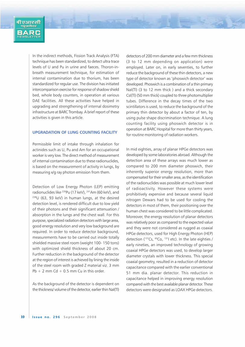
The average energy resolution (FWHM) of the three
detector system at 17 keV (239Pu), 60 keV (241Am), 63
keV (238U), 93 keV (238U) and 185 keV (235U) is
measured to be about 505, 650, 590, 740 and 730
eV, respectively. The energy resolution of 650 eV for241Am (60 keV) may be compared with the resolution
of about 12 keV, obtained with phoswich detector.
The excellent resolution and good sensitivity of the
LOAX detector system provides a more accurate
assessment of internal contamination of low energy
X-rays/ g-emitting actinides of interest, even in
presence of other gamma emitting radionuclides.
Keeping in view these advantages, we have planned
to use LOAX HPGe system for special monitoring and
continue to use phoswich for routine monitoring. The
calculated minimum detectable activity (MDA) of
LOAX HPGe system for a monitoring period of 3600
sec is 4 and 5 Bq for 241Am and 238U, respectively. Fig.
3 shows the spectra for 241Am recorded with LOAX
HPGe detector.
Earlier, the lung counting system was calibrated by an
in-vivo calibration technique, which involved inhalation
of 103Pd-51Cr [20 keV X-ray and 320 keV g-ray] labeled
polystyrene aerosols by human volunteers, as part of
an international intercomparison exercise. The present
calibration factors for assessment of lung burden due
to actinides, are based on measurements carried out
in the mid eighties using realistic thorax phantom,
designed and developed by the Lawrence Livermore
National Laboratory (LLNL), USA and again in the year
1997 using JAERI (Japan Atomic Energy Research
Institute) phantom i.e. Reference Asian phantom.
Recently, the phoswich and HPGe system have been
tested again using JAERI phantom as a part of IAEA
intercomparison exercise.
APPLICATION OF FISSION TRACK ANALYSIS (FTA)
TECHNIQUE, FOR ESTIMATION OF INTERNAL
CONTAMINATION DUE TO PLUTONIUM
In order to carry out internal contamination monitoring
of workers handling plutonium, analysis of urine/
faeces is normally carried out, to determine excretion
rate of plutonium. The method involves chemical
separation of plutonium from the bioassay sample,
followed by electro deposition and final activity
quantification by alpha spectrometry. Fission Track
Analysis (FTA) technique is more sensitive than the
above method. Therefore, it has been standardized
for measurement of trace levels of Pu in bioassay
samples. In this technique, chemically separated
plutonium from the sample and a Pu standard are
electrodeposited on planchettes, covered with Lexan
Solid State Nuclear Track Detector (SSNTD) and
irradiated with thermal neutrons in the APSARA reactor
of BARC. Pu in the samples undergoes fission and the
resulting fission fragments produce tracks in the Lexan
film. After irradiation, the Lexan films are chemically
etched with 6 M NaOH at 60° C for 1 hour. The tracks
thus developed are counted manually, using
400 X magnification optical microscope. The net
fission tracks in the Lexan films of the sample and the
standard are used, to calculate the amount of
Pu in the sample. Presence of uranium in the reagents
used for the chemical separation of Pu can lead to
interferences in the analysis of Pu at trace level.
Therefore, doubly distilled electronic grade reagentsFig. 3: Pulse height spectrum of 241Am in lungs of
JAERI phantom recorded with LOAX HPGe detector
13I s s u e n o . 2 9 6 S e p t e m b e r 2 0 0 8
The minimum amount of Pu that
can be determined by this method,
using doubly distilled electronic
grade reagents, is about 12 mBq/
L. Further efforts are being made,
to improve minimum detection
limit and to automate the time
consuming process of counting of
fission tracks manually by using
image analyzer.
T H O R O N - I N - B R E A T H
MEASUREMENT TECHNIQUE
FOR ESTIMATION OF THORIUM
BODY BURDEN
Thoron (220Rn) is a noble gas and it occurs in
the decay series of 232Th. It is possible to estimate
thorium body burden of a person, by measuring the
thoron content in his breath by using an ElectroStatic
Chamber (ESC). The method is based on
the observation, that more than 88% of decay
products of thoron are positively charged ions
at birth and may be collected on an electrode
maintained at sufficiently high negative
potential. In order to
estimate thoron in
breath, the person is
made to inhale thoron-
free air from a delay
chamber and exhale into
the electrostatic chamber
having a collection
electrode, maintained at
sufficiently high negative
potential. The thoron
progeny atoms formed
due to decay of thoron in
the electrostatic
chamber are collected on
a removable metallic
plate, kept attached
Fig. 4: Fission track analyzer set-up
Fig. 5: Fission tracks per field (1.91 x 10⁻³ cm2) observed under 400X
magnification
are used. The contribution from background level of
uranium present in the urine sample is further
minimized, by carrying out separation using two stages
of ion exchange separation (cation exchange separation
followed by anion exchange). The decontamination
factor achieved by this process is about 1x10⁷. Fig. 4
shows the FTA setup and Fig. 5 shows the tracks
observed with the microscope.

14 I s s u e n o . 2 9 6 S e p t e m b e r 2 0 0 8
to the electrode, which is later assayed for alpha
activity using ZnS(Ag) scintillation detector. By proper
calibration of the system, it is possible to correlate
alpha activity collected on the metallic plate with
thorium body burden. Fig. 6 shows the Thoron In
Breath Measurement (TIBM) setup. The minimum
detectable level of thorium in the body, for this system
is about 4 Bq which is a small fraction of the ALI. A
software has been developed to compute thoron in
breath, thorium in the body and the resultant dose
from the gross alpha counts, obtained from a PC-
based alpha counting system. Recently we have started
thoron-in-breath measurement on some workers from
gas mantle industry, who handle thorium powder.
for estimation of internal contamination due to
actinides and fission/activation products. The detection
systems (Phoswich, Array of HPGe) used for assessment
of lung/liver burden of LEP emitters require calibration
with realistic thorax phantoms (LLNL, JAERI). The
partially and wholly shielded detection systems,
employed for the assessment of HEP emitters, are
calibrated with tissue equivalent BOMAB phantoms.
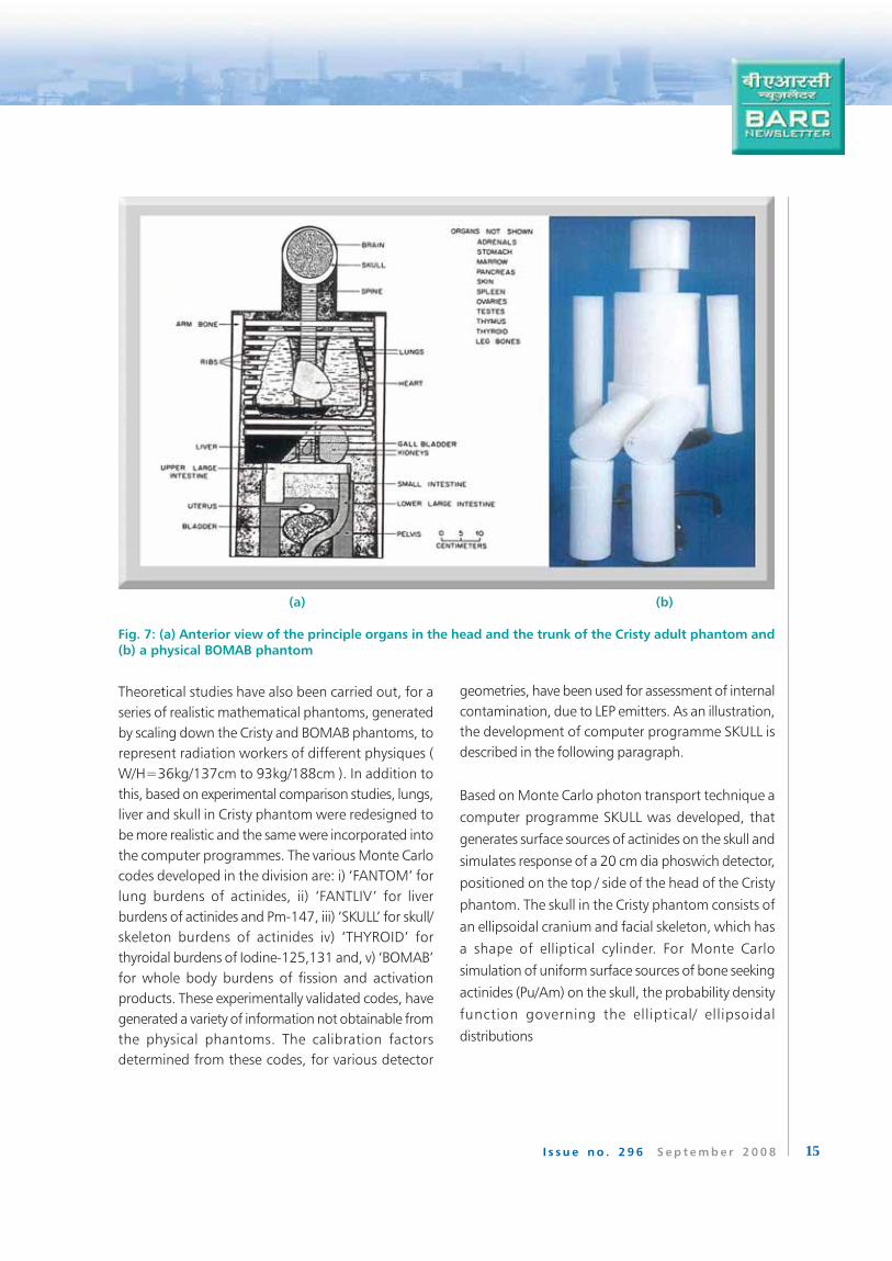
Attractive alternatives to physical phantoms are the
theoretical calibration methods, involving Monte Carlo
techniques in conjunction with mathematical
phantoms, such as MIRD / Cristy and BOMAB
(Fig. 7). An added advantage of theoretical calibration
is the fact, that detection efficiencies can be calculated
for any photon energy, source
distribution, shape and size of the
organ, detection geometry and
the physique of the radiation
workers.
Based on Monte Carlo photon
transport techniques, a number of
specialized software in FORTRAN
language, have been developed
in the division. In brief, the
software generated different types
of source distributions in the
relevant organs/whole body and
simulated photon transport
through different types of tissue
media of the mathematical
phantom, considering possible
interaction processes, namely
photo-electric, Compton and pair-
production, in proportion to their individual
probabilities. A photon is traced in the relevant part of
the phantom (head, neck, thorax, whole body), until
it either gets completely absorbed or escapes the
phantom. The programme finally simulates pulse
height response and the corresponding Detection
Efficiencies (DEs) of the various detection systems,
employed for assessment of internal contamination of
radionuclides.
Fig. 6: A TIBM instrument is being used for monitoring ofa worker due to possible contamination from thorium
THEORETICAL STUDIES IN INTERNAL DOSIMETRY
1. Monte Carlo studies
The Monte Carlo techniques have been utilized with a
great deal of success, in the field of radiation protection,
particularly in internal dosimetry of radionuclides. At
BARC, a variety of detection systems are in operation,
15I s s u e n o . 2 9 6 S e p t e m b e r 2 0 0 8
Theoretical studies have also been carried out, for a
series of realistic mathematical phantoms, generated
by scaling down the Cristy and BOMAB phantoms, to
represent radiation workers of different physiques (
W/H=36kg/137cm to 93kg/188cm ). In addition to
this, based on experimental comparison studies, lungs,
liver and skull in Cristy phantom were redesigned to
be more realistic and the same were incorporated into
the computer programmes. The various Monte Carlo
codes developed in the division are: i) ‘FANTOM’ for
lung burdens of actinides, ii) ‘FANTLIV’ for liver
burdens of actinides and Pm-147, iii) ‘SKULL’ for skull/
skeleton burdens of actinides iv) ‘THYROID’ for
thyroidal burdens of Iodine-125,131 and, v) ‘BOMAB’
for whole body burdens of fission and activation
products. These experimentally validated codes, have
generated a variety of information not obtainable from
the physical phantoms. The calibration factors
determined from these codes, for various detector
geometries, have been used for assessment of internal
contamination, due to LEP emitters. As an illustration,
the development of computer programme SKULL is
described in the following paragraph.
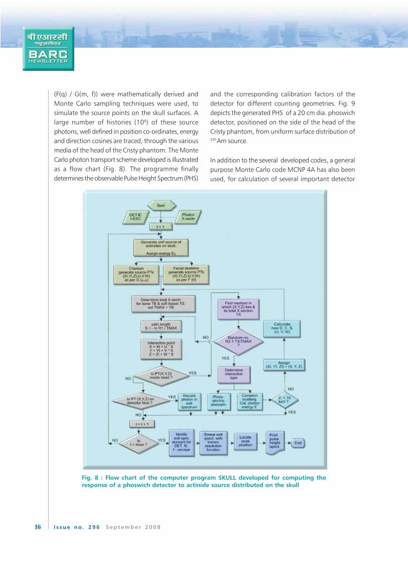
Based on Monte Carlo photon transport technique a
computer programme SKULL was developed, that
generates surface sources of actinides on the skull and
simulates response of a 20 cm dia phoswich detector,
positioned on the top / side of the head of the Cristy
phantom. The skull in the Cristy phantom consists of
an ellipsoidal cranium and facial skeleton, which has
a shape of elliptical cylinder. For Monte Carlo
simulation of uniform surface sources of bone seeking
actinides (Pu/Am) on the skull, the probability density
function governing the elliptical/ ellipsoidal
distributions
(a) (b)
Fig. 7: (a) Anterior view of the principle organs in the head and the trunk of the Cristy adult phantom and(b) a physical BOMAB phantom
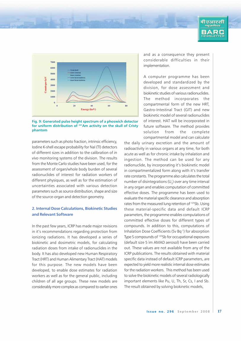
16 I s s u e n o . 2 9 6 S e p t e m b e r 2 0 0 8
and the corresponding calibration factors of the
detector for different counting geometries. Fig. 9
depicts the generated PHS of a 20 cm dia. phoswich
detector, positioned on the side of the head of the
Cristy phantom, from uniform surface distribution of241Am source.
In addition to the several developed codes, a general
purpose Monte Carlo code MCNP 4A has also been
used, for calculation of several important detector
(F(q) / G(m, f)) were mathematically derived and
Monte Carlo sampling techniques were used, to
simulate the source points on the skull surfaces. A
large number of histories (106) of these source
photons, well defined in position co-ordinates, energy
and direction cosines are traced, through the various
media of the head of the Cristy phantom. The Monte
Carlo photon transport scheme developed is illustrated
as a flow chart (Fig. 8). The programme finally
determines the observable Pulse Height Spectrum (PHS)
Fig. 8 : Flow chart of the computer program SKULL developed for computing the
response of a phoswich detector to actinide source distributed on the skull
17I s s u e n o . 2 9 6 S e p t e m b e r 2 0 0 8
parameters such as photo fraction, intrinsic efficiency,
Iodine K-shell escape probability for NaI (Tl) detectors
of different sizes in addition to the calibration of in
vivo monitoring systems of the division. The results
from the Monte Carlo studies have been used, for the
assessment of organ/whole body burden of several
radionuclides of interest for radiation workers of
different physiques, as well as for the estimation of
uncertainties associated with various detection
parameters such as source distribution, shape and size
of the source-organ and detection geometry.
2. Internal Dose Calculations, Biokinetic Studies
and Relevant Software
In the past few years, ICRP has made major revisions
in it’s recommendations regarding protection from
ionizing radiations. It has developed a series of
biokinetic and dosimetric models, for calculating
radiation doses from intake of radionuclides in the
body. It has also developed new Human Respiratory
Tract (HRT) and Human Alimentary Tract (HAT) models
for this purpose. The new models have been
developed, to enable dose estimates for radiation
workers as well as for the general public, including
children of all age groups. These new models are
considerably more complex as compared to earlier ones
and as a consequence they present
considerable difficulties in their
implementation.
A computer programme has been
developed and standardized by the
division, for dose assessment and
biokinetic studies of various radionuclides.
The method incorporates the
compartmental form of the new HRT,
Gastro-Intestinal Tract (GIT) and new
biokinetic model of several radionuclides
of interest. HAT will be incorporated in
future software. The method provides
solution from the complete
compartmental model and can calculate
the daily urinary excretion and the amount of
radioactivity in various organs at any time, for both
acute as well as for chronic intake by inhalation and
ingestion. The method can be used for any
radionuclide, by incorporating it’s biokinetic model
in compartmentalized form along with it’s transfer
rate constants. The programme also calculates the total
number of disintegrations (US) over any time interval
in any organ and enables computation of committed
effective doses. The programme has been used to
evaluate the material specific clearance and absorption
rates from the measured lung retention of 125Sb. Using
these material-specific data and default ICRP
parameters, the programme enables computations of
committed effective doses for different types of
compounds. In addition to this, computations of
Inhalation Dose Coefficients (Sv Bq-1) for absorption
Type S compounds of 125Sb for occupational exposures
(default size 5 ìm AMAD aerosol) have been carried
out. These values are not available from any of the
ICRP publications. The results obtained with material
specific data instead of default ICRP parameters, are
expected to yield more realistic internal dose estimates
for the radiation workers. This method has been used
to solve the biokinetic models of several radiologically
important elements like Pu, U, Th, Sr, Cs, I and Sb.
The result obtained by solving biokinetic models,
Fig. 9: Generated pulse height spectrum of a phoswich detector
for uniform distribution of 241Am activity on the skull of Cristy
phantom
18 I s s u e n o . 2 9 6 S e p t e m b e r 2 0 0 8
was compared with the experimentally obtained values.
This was also used to find out uncertainties in ICRP
biokinetic parameters.
Many software packages like LUDEP, MONDAL/
MONDES, IMIE and IMBA are commercially available
for internal dose evaluation. Among them the most
advanced software package is Integrated Modules for
Bioassay Analysis (IMBA) Professional Plus. This
incorporates latest biokinetic model of various
radionuclides. The division has recently procured this
software. The software is having many advanced
features for standard calculations and all of the ICRP
default values can be selected from built in database
at the touch of a button. For more detailed
calculations, the user can enter individual parameter
values. The IMBA has enhanced the capabilities of
internal dosimetry laboratory further, since certain types
of exposure scenarios can only be handled by this
software. Calculation of internal dose for few case
studies have been carried out using IMBA and
compared with ICRP-78 methodology. The IMBA allows
the user simultaneous fitting of more than one
measured data types i.e. urine, faeces and whole body
of radiation worker, for best estimate of intake. As a
result, it gives realistic estimate of intake and
committed effective dose.
IAEA Intercomparison exercise:
Reference Asian-JAERI Phantom
Assessment of lung burden due to
actinides such as plutonium and
uranium isotopes and 241Am, is based
on the detection of low energy photons
and x-rays emitted in their decays.
These Low Energy Photons (LEP < 100
keV) suffer severe attenuation due to
soft tissues and rib bones overlying the
lungs. Therefore, it is necessary to
calibrate the detection systems used
for lung monitoring of radiation
workers, using realistic phantom. The JAERI phantom
is a realistic phantom representative of thorax of
Reference Asian man as against LLNL phantom which
is a representative of a Caucasian man. The JAERI
phantom was received by the division under IAEA
sponsored intercomparison exercise viz.
“Intercomparison of Radiological Measurements for
Monitoring Purposes – Direct Measurement of
Radionuclides in Simulated Organs”.
Under this exercise, various phantoms were received
for intercomparison purpose viz. Knee, JAERI, BOMAB
and Thyroid phantoms. Knee and JAERI phantoms are
realistic phantoms simulating human knee and torso
of the reference man respectively and are used, for
calibrating the detection systems for the actinides such
as 239Pu, 241Am, Nat. and Enr. U. The BOMAB and
Thyroid phantoms are used for calibration of detection
systems for HEP emitters such as 131I, 137Cs and 60Co.
IAEA circulated two sets of phantoms among
participating laboratories and phantoms were
designated as JAERI I or JAERI II depending on the
number assigned to the set.
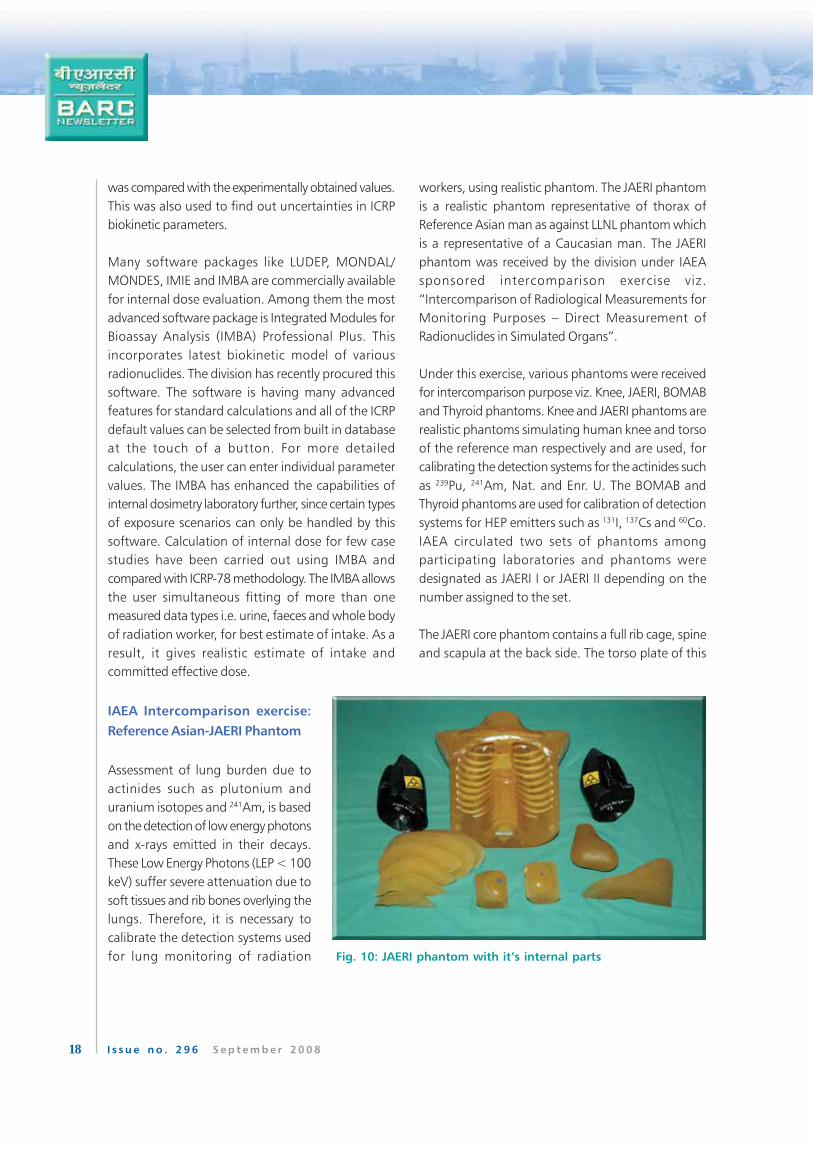
The JAERI core phantom contains a full rib cage, spine
and scapula at the back side. The torso plate of this
Fig. 10: JAERI phantom with it’s internal parts
19I s s u e n o . 2 9 6 S e p t e m b e r 2 0 0 8
phantom is constructed of an adipose-muscle
substitute mixture and contains synthetic bone
and cartilage. The overlay plates of the phantom
are constructed of different adipose-muscle
substitute mixtures, to simulate different Chest
Wall Thicknesses (CWT). The torso cavities contain
lungs, heart, liver and other organs. Six lung sets
were provided with the phantom, which contained
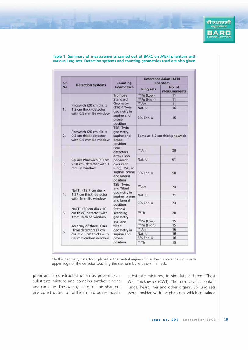
Table 1: Summary of measurements carried out at BARC on JAERI phantom with
various lung sets. Detection systems and counting geometries used are also given.
*In this geometry detector is placed in the central region of the chest, above the lungs withupper edge of the detector touching the sternum bone below the neck.
20 I s s u e n o . 2 9 6 S e p t e m b e r 2 0 0 8
natural thorium, natural uranium, uranium with 3%235U enrichment, 241Am, and two 238Pu sets with
significantly different amount of radioactivity. The JAERI
Phantom with it’s internal parts is shown in Fig. 10.
About 700 measurements were performed with JAERI
phantom in various geometries using several detection
systems viz. phoswich, LOAX HPGe and NaI(Tl). Table
1 gives a summary of these measurements.
Estimation of activity in JAERI II was carried out using
calibration factors obtained from the earlier
measurements carried out on JAERI I phantom during
on earlier IAEA CRP. As the JAERI II phantom was not
provided with the blank lung sets, normal subject
background (subject having weight and height similar
to the JAERI Phantom) was taken to estimate the
activity. The results and other data were reported to
IAEA for intercomparison.
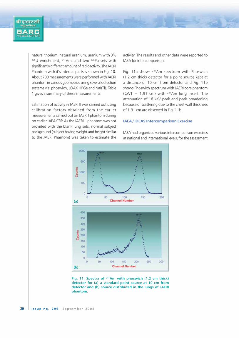
Fig. 11a shows 241Am spectrum with Phoswich
(1.2 cm thick) detector for a point source kept at
a distance of 10 cm from detector and Fig. 11b
shows Phoswich spectrum with JAERI core phantom
(CWT = 1.91 cm) with 241Am lung insert. The
attenuation of 18 keV peak and peak broadening
because of scattering due to the chest wall thickness
of 1.91 cm are observed in Fig. 11b.
IAEA / IDEAS Intercomparison Exercise
IAEA had organized various intercomparison exercises
at national and international levels, for the assessment
(a)
(b)
Fig. 11: Spectra of 241Am with phoswich (1.2 cm thick)
detector for (a) a standard point source at 10 cm from
detector and (b) source distributed in the lungs of JAERIphantom.
21I s s u e n o . 2 9 6 S e p t e m b e r 2 0 0 8
of internal dose, due to intakes of radionuclides. These
exercises revealed significant differences in the
approaches, methods and assumptions used and
consequently in the results obtained by participating
laboratories. This led to the development of ‘General
guidelines for the estimation of committed dose from
incorporation of monitoring data’ by IDEAS project
(A European Union project). The guidelines provide
well defined procedures to obtain best estimate of
dose from the available data, depending upon the
expected level of exposure and the complexity of the
case.
For harmonizing the methods of assessing the
committed effective dose to workers after an intake
of radionuclides using these guidelines, a joint
IDEAS/IAEA intercomparison exercise viz.
”Intercomparison exercise on assessment of
occupational exposure” was organized. The division
has participated in this exercise, which consisted of
six cases covering wide and complex exposure
scenarios. The cases given were (i) acute intake of
HTO, (ii) acute inhalation of fission products 137Cs and90Sr, (iii) acute inhalation of 60Co, (iv) chronic intake
of 131I, (v) enriched uranium intake and (vi) intake of
Pu and Am. The cases were solved using LUDEP,
MONDAL/MONDES and IMBA dose evaluation
software and results were submitted to IAEA. Eighty
one participants from forty two countries submitted
the results to IAEA. Out of these, only thirty five
participants had solved all the cases. Our laboratory is
one of them. IAEA statistically analyzed the results
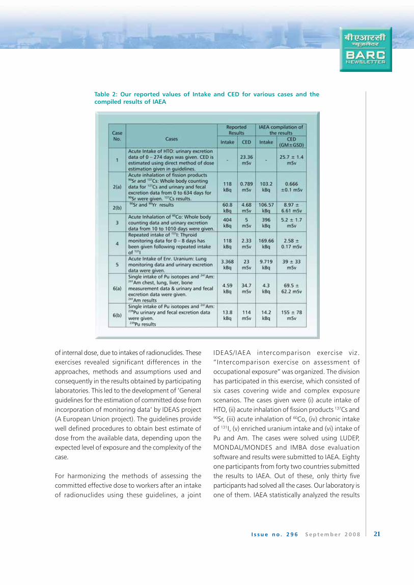
Table 2: Our reported values of Intake and CED for various cases and the
compiled results of IAEA
22 I s s u e n o . 2 9 6 S e p t e m b e r 2 0 0 8
reported by the participating laboratories, using Log-
Normal distribution for Geometric Mean (GM) and
Geometric Standard Deviation (GSD) and for better
graphical representation of the data. Our results were
found to be in good agreement with the IAEA results.
Table 2 shows the Intakes and CED values estimated
by our laboratory and the results compiled by IAEA
for the six cases given in the intercomparison exercise.
This table includes only a brief description of the
exposure cases. The CED values given by IAEA are the
GM of all the values reported by the participating
laboratories. These values are given in this table along
with their GSD.
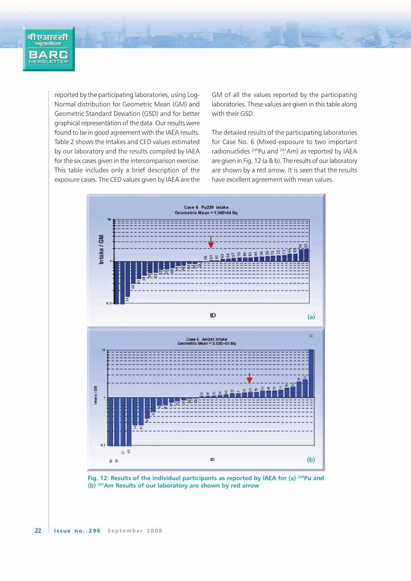
The detailed results of the participating laboratories
for Case No. 6 (Mixed exposure to two important
radionuclides 239Pu and 241Am) as reported by IAEA
are given in Fig. 12 (a & b). The results of our laboratory
are shown by a red arrow. It is seen that the results
have excellent agreement with mean values.
Fig. 12: Results of the individual participants as reported by IAEA for (a) 239Pu and
(b) 241Am Results of our laboratory are shown by red arrow
(b)
(a)
23I s s u e n o . 2 9 6 S e p t e m b e r 2 0 0 8
Intercomparison of shadow shield bed whole
body monitors
Shadow shield bed Whole Body Counters (WBC) using
a NaI(Tl) detector have been installed at various DAE
units like BARC Trombay, Environment Survey
Laboratories attached to Nuclear Power Plants (NPP),
Health Physics Laboratory and also at some of the
other facilities of DAE. The WBCs are used to estimate
internal contamination in workers due to high energy
photon emitters such as 60Co, 131I, 137Cs etc. The
division initiated an intercomparison exercise to
compare the response of WBCs at BARC, Trombay,
ESLs of NPP and Health Physics Laboratory Tarapur.
The work is nearing completion.
Acknowledgements
The authors are thankful to Mr. S. H. Shirke and Mr.
D. Toppo for their technical support.
Publications
1. Garg, S.P., Bhati, S., Haridasan, T.K., Jaiswal,
D.D., Sawant, Pramilla D., Singh, I.S. and
Pendharkar, K.A. “An overview of internal
dosimetry programmes in India”. Paper
presented at 16th Annual Conference of Indian
Nuclear Society (INSAC-2005) Mumbai, Nov.
15-18, (2005).
2. Bhati, S., Patni, H.K., Nandanwar, V. H., Singh,
I. S. and Garg, S. P. “Effect of BOMAB Phantom
size on efficiency calibration of a scanning bed
whole body monitor: A Monte Carlo approach”.
Second Asian and Oceanic (IRPA) congress on
radiological protection (AOCRP-2), Beijing,
China, Oct. 9-13 (2006).
3. Singh, I.S., Nadar, Minal Y., Kalyane, G.N. and
Garg, S.P. “Monitoring Inhalation of Thorium
Environmental Radioactivity in Humans with
Phoswich Detector”. 15th National Symposium
on Environment (NSE-15), Coimbatore, June
5-7 (2007).
4. Nadar, Minal Y., Prabhu, R.S., Haridasan, T.K.,
Garg, S.P. and Pendharkar, K.A. “Assessment of
calibration factors for lung monitoring of
uranium using JAERI phantom”. Radiation
Protection and Environment, Vol. 28 (1/4):
363-365 (2005).
5. Bhati, S., Sharma, R. C. and Venkatraj, V.
“Assessing skull burdens of actinides using a
mathematical phantom: A Monte Carlo
approach”. Radiat. Prot. Dosim. 103(3) 247-
254 (2003).
6. Garg, S. P., Singh, I. S. and Sharma, R. C. “Long
term lung retention studies of 125Sb aerosols in
humans”. Health Phys. 84 (4) 457-468 (2003).
7. Sawant, Pramilla D., Prabhu, Supreetha, Kalsi,
P.C. and Pendharkar, K.A. “Estimation of Trace
Levels of Plutonium in Urine Samples by Fission
Track Technique”. Journal of Radioanalytical and
Nuclear Chemistry, Vol. 279, No. 1 (2009).
8. Sathyabama, N., Eappen, K.P., and Mayya,Y.S.
“Calibration of the electrostatic chamber for
measurements of thoron in exhaled breath”.
Radiat. Prot. Dosim. 118, 61 – 69 (2006).
9. Singh, I.S., Sharma, R.C. and Abani, M. C. “A
Computational Study of Urinary Excretion Rates
for 239Pu using new ICRP Internal Dosimetry
Models”. Radiat. Prot. Dosim. Vol. 105 (1/4)
pp. 361-364 (2003).