Updating the Guidelines - kitasato- · PDF fileUpdating the Guidelines Released online June...
45
Circulation Journal Vol.77, August 2013 Circulation Journal Official Journal of the Japanese Circulation Society http://www.j-circ.or.jp in population proportion of elderly people but also a result from excellent life-saving activity in cardiovascular territory such as that in acute myocardial infarction (AMI). From a cohort study, it is estimated that the number of patients with heart failure will increase by 0.6% every year over at least the next 30 years in Japan. 1 Those patients with heart failure who 1. Preamble The number of patients with acute heart failure who are clini- cally characterized with orthopnea and congestion is steadily increasing in developed countries. This tendency reflects a rise Updating the Guidelines Released online June 12, 2013 Mailing address: Scientific Committee of the Japanese Circulation Society, 18F Imperial Hotel Tower, 1-1-1 Uchisaiwai-cho, Chiyoda-ku, Tokyo 100-0011, Japan. E-mail: [email protected] This English language document is a revised digest version of Guidelines for Treatment of Acute Heart Failure reported at the Japanese Circulation Society Joint Working Groups performed in 2010 (Website: http://www.j-circ.or.jp/guideline/pdf/JCS2011_izumi_d.pdf). Joint Working Groups: The Japanese Circulation Society, The Japanese Association for Thoracic Surgery, The Japanese Society of Hyper- tension, The Japanese Society of Pediatric Cardiology and Cardiac Surgery, The Japanese Society for Cardiovascular Surgery, The Japanese College of Cardiology, The Japanese Association of Cardiac Rehabilitation, The Japanese Society of Electrocardiology, The Japanese Heart Failure Society, The Japan Society of Ultrasonics in Medicine, The Japanese Heart Rhythm Society ISSN-1346-9843 doi:10.1253/circj.CJ-66-0068 All rights are reserved to the Japanese Circulation Society. For permissions, please e-mail: [email protected] Guidelines for Treatment of Acute Heart Failure (JCS 2011) – Digest Version – JCS Joint Working Group Table of Contents Updating the Guidelines ∙∙∙∙∙∙∙∙∙∙∙∙∙∙∙∙∙∙∙∙∙∙∙∙∙∙∙∙∙∙∙∙∙∙∙∙∙∙∙∙∙∙∙∙∙∙ 2157 1. Preamble ∙∙∙∙∙∙∙∙∙∙∙∙∙∙∙∙∙∙∙∙∙∙∙∙∙∙∙∙∙∙∙∙∙∙∙∙∙∙∙∙∙∙∙∙∙∙∙∙∙∙∙∙∙∙∙∙∙∙∙∙∙∙∙∙∙∙∙∙∙ 2157 2. Updating the Guidelines ∙∙∙∙∙∙∙∙∙∙∙∙∙∙∙∙∙∙∙∙∙∙∙∙∙∙∙∙∙∙∙∙∙∙∙∙∙∙∙∙∙∙∙∙∙∙ 2158 I General Matters∙∙∙∙∙∙∙∙∙∙∙∙∙∙∙∙∙∙∙∙∙∙∙∙∙∙∙∙∙∙∙∙∙∙∙∙∙∙∙∙∙∙∙∙∙∙∙∙∙∙∙∙∙∙∙∙∙∙∙ 2159 1. Definition ∙∙∙∙∙∙∙∙∙∙∙∙∙∙∙∙∙∙∙∙∙∙∙∙∙∙∙∙∙∙∙∙∙∙∙∙∙∙∙∙∙∙∙∙∙∙∙∙∙∙∙∙∙∙∙∙∙∙∙∙∙∙∙∙∙∙∙∙∙∙ 2159 2. Epidemiology ∙∙∙∙∙∙∙∙∙∙∙∙∙∙∙∙∙∙∙∙∙∙∙∙∙∙∙∙∙∙∙∙∙∙∙∙∙∙∙∙∙∙∙∙∙∙∙∙∙∙∙∙∙∙∙∙∙∙∙∙∙∙∙ 2163 3. Signs/Symptoms and Causes ∙∙∙∙∙∙∙∙∙∙∙∙∙∙∙∙∙∙∙∙∙∙∙∙∙∙∙∙∙∙∙∙∙∙∙∙∙ 2164 4. Treatment Strategies ∙∙∙∙∙∙∙∙∙∙∙∙∙∙∙∙∙∙∙∙∙∙∙∙∙∙∙∙∙∙∙∙∙∙∙∙∙∙∙∙∙∙∙∙∙∙∙∙∙∙∙ 2165 5. Initial Management of Acute Heart Failure ∙∙∙∙∙∙∙∙∙∙∙∙∙∙∙∙∙∙ 2165 II Diagnosis ∙∙∙∙∙∙∙∙∙∙∙∙∙∙∙∙∙∙∙∙∙∙∙∙∙∙∙∙∙∙∙∙∙∙∙∙∙∙∙∙∙∙∙∙∙∙∙∙∙∙∙∙∙∙∙∙∙∙∙∙∙∙∙∙∙∙∙∙∙ 2166 1. Procedures for Diagnosis and Triage for Treatment ∙∙∙ 2166 2. Procedures for Diagnosis in the Emergency Room ∙∙∙∙ 2166 3. Diagnostic Procedures in ICU/CCU ∙∙∙∙∙∙∙∙∙∙∙∙∙∙∙∙∙∙∙∙∙∙∙∙∙∙∙∙ 2170 III Treatment∙∙∙∙∙∙∙∙∙∙∙∙∙∙∙∙∙∙∙∙∙∙∙∙∙∙∙∙∙∙∙∙∙∙∙∙∙∙∙∙∙∙∙∙∙∙∙∙∙∙∙∙∙∙∙∙∙∙∙∙∙∙∙∙∙∙∙∙ 2170 1. Treatment Strategies ∙∙∙∙∙∙∙∙∙∙∙∙∙∙∙∙∙∙∙∙∙∙∙∙∙∙∙∙∙∙∙∙∙∙∙∙∙∙∙∙∙∙∙∙∙∙∙∙∙∙∙ 2170 2. Early Diagnosis and Treatment ∙∙∙∙∙∙∙∙∙∙∙∙∙∙∙∙∙∙∙∙∙∙∙∙∙∙∙∙∙∙∙∙∙∙∙ 2172 3. Determination of Targets of Treatment and Management∙∙∙∙∙∙∙∙∙∙∙∙∙∙∙∙∙∙∙∙∙∙∙∙∙∙∙∙∙∙∙∙∙∙∙∙∙∙∙∙∙∙∙∙∙∙∙∙∙∙∙∙∙∙∙∙∙∙∙∙∙∙∙∙ 2173 4. Medical Therapy ∙∙∙∙∙∙∙∙∙∙∙∙∙∙∙∙∙∙∙∙∙∙∙∙∙∙∙∙∙∙∙∙∙∙∙∙∙∙∙∙∙∙∙∙∙∙∙∙∙∙∙∙∙∙∙∙∙∙ 2174 5. Nonpharmacologic Treatment ∙∙∙∙∙∙∙∙∙∙∙∙∙∙∙∙∙∙∙∙∙∙∙∙∙∙∙∙∙∙∙∙∙∙∙∙∙ 2178 6. Nursing ∙∙∙∙∙∙∙∙∙∙∙∙∙∙∙∙∙∙∙∙∙∙∙∙∙∙∙∙∙∙∙∙∙∙∙∙∙∙∙∙∙∙∙∙∙∙∙∙∙∙∙∙∙∙∙∙∙∙∙∙∙∙∙∙∙∙∙∙∙∙∙∙ 2183 7. Recommended Requirements for Medical Practice and Equipment ∙∙∙∙∙∙∙∙∙∙∙∙∙∙∙∙∙∙∙∙∙∙∙∙∙∙∙∙∙∙∙∙∙∙∙∙∙∙∙∙∙∙∙∙∙∙∙∙∙∙∙∙∙∙∙∙∙∙∙∙ 2186 IV Treatment Strategies for Heart Failure by Cause ∙∙∙ 2186 1. Ischemic Heart Disease∙∙∙∙∙∙∙∙∙∙∙∙∙∙∙∙∙∙∙∙∙∙∙∙∙∙∙∙∙∙∙∙∙∙∙∙∙∙∙∙∙∙∙∙∙∙∙ 2186 2. Hypertensive Urgency ∙∙∙∙∙∙∙∙∙∙∙∙∙∙∙∙∙∙∙∙∙∙∙∙∙∙∙∙∙∙∙∙∙∙∙∙∙∙∙∙∙∙∙∙∙∙∙∙∙ 2187 3. Idiopathic Cardiomyopathy ∙∙∙∙∙∙∙∙∙∙∙∙∙∙∙∙∙∙∙∙∙∙∙∙∙∙∙∙∙∙∙∙∙∙∙∙∙∙∙∙∙∙ 2187 4. Myocarditis ∙∙∙∙∙∙∙∙∙∙∙∙∙∙∙∙∙∙∙∙∙∙∙∙∙∙∙∙∙∙∙∙∙∙∙∙∙∙∙∙∙∙∙∙∙∙∙∙∙∙∙∙∙∙∙∙∙∙∙∙∙∙∙∙∙∙ 2187 5. Valvular Heart Disease ∙∙∙∙∙∙∙∙∙∙∙∙∙∙∙∙∙∙∙∙∙∙∙∙∙∙∙∙∙∙∙∙∙∙∙∙∙∙∙∙∙∙∙∙∙∙∙∙ 2188 V Concomitant Conditions and Their Management ∙∙∙ 2188 1. Anemia∙∙∙∙∙∙∙∙∙∙∙∙∙∙∙∙∙∙∙∙∙∙∙∙∙∙∙∙∙∙∙∙∙∙∙∙∙∙∙∙∙∙∙∙∙∙∙∙∙∙∙∙∙∙∙∙∙∙∙∙∙∙∙∙∙∙∙∙∙∙∙∙∙ 2188 2. Renal Failure ∙∙∙∙∙∙∙∙∙∙∙∙∙∙∙∙∙∙∙∙∙∙∙∙∙∙∙∙∙∙∙∙∙∙∙∙∙∙∙∙∙∙∙∙∙∙∙∙∙∙∙∙∙∙∙∙∙∙∙∙∙∙∙ 2188 3. Hepatic Congestion ∙∙∙∙∙∙∙∙∙∙∙∙∙∙∙∙∙∙∙∙∙∙∙∙∙∙∙∙∙∙∙∙∙∙∙∙∙∙∙∙∙∙∙∙∙∙∙∙∙∙∙∙∙ 2189 4. Pneumonia ∙∙∙∙∙∙∙∙∙∙∙∙∙∙∙∙∙∙∙∙∙∙∙∙∙∙∙∙∙∙∙∙∙∙∙∙∙∙∙∙∙∙∙∙∙∙∙∙∙∙∙∙∙∙∙∙∙∙∙∙∙∙∙∙∙∙ 2189 5. Pulse Abnormalities ∙∙∙∙∙∙∙∙∙∙∙∙∙∙∙∙∙∙∙∙∙∙∙∙∙∙∙∙∙∙∙∙∙∙∙∙∙∙∙∙∙∙∙∙∙∙∙∙∙∙∙∙∙ 2189 6. Chronic Obstructive Pulmonary Disease∙∙∙∙∙∙∙∙∙∙∙∙∙∙∙∙∙∙∙∙∙ 2192 VI Treatment Strategies for Heart Failure With Preserved EF∙∙∙∙∙∙∙∙∙∙∙∙∙∙∙∙∙∙∙∙∙∙∙∙∙∙∙∙∙∙∙∙∙∙∙∙∙∙∙∙∙∙∙∙∙∙∙∙∙∙∙∙∙∙∙∙∙∙∙∙∙ 2192 1. Definition of Heart Failure With Preserved EF ∙∙∙∙∙∙∙∙∙∙∙∙ 2192 2. Diagnosis in the Acute Phase of Heart Failure With Preserved EF∙∙∙∙∙∙∙∙∙∙∙∙∙∙∙∙∙∙∙∙∙∙∙∙∙∙∙∙∙∙∙∙∙∙∙∙∙∙∙∙∙∙∙∙∙∙∙∙∙∙∙∙∙∙∙∙∙∙∙∙∙∙∙ 2192 3. Treatment in the Acute Phase of Heart Failure With Preserved EF∙∙∙∙∙∙∙∙∙∙∙∙∙∙∙∙∙∙∙∙∙∙∙∙∙∙∙∙∙∙∙∙∙∙∙∙∙∙∙∙∙∙∙∙∙∙∙∙∙∙∙∙∙∙∙∙∙∙∙∙∙∙∙ 2192 4. Blood Pressure Control ∙∙∙∙∙∙∙∙∙∙∙∙∙∙∙∙∙∙∙∙∙∙∙∙∙∙∙∙∙∙∙∙∙∙∙∙∙∙∙∙∙∙∙∙∙∙∙ 2193 5. Rate Control in Atrial Fibrillation ∙∙∙∙∙∙∙∙∙∙∙∙∙∙∙∙∙∙∙∙∙∙∙∙∙∙∙∙∙∙∙∙∙∙ 2193 6. Medical Therapy in the Chronic Phase of Heart Failure With Preserved EF ∙∙∙∙∙∙∙∙∙∙∙∙∙∙∙∙∙∙∙∙∙∙∙∙∙∙∙∙∙∙∙∙∙∙∙∙∙∙∙∙∙∙ 2193 VII Treatment Strategies for Biventricular Failure ∙∙∙ 2193 1. Pathophysiology and Treatment of Biventricular Failure ∙∙∙∙∙∙∙∙∙∙∙∙∙∙∙∙∙∙∙∙∙∙∙∙∙∙∙∙∙∙∙∙∙∙∙∙∙∙∙∙∙∙∙∙∙∙∙∙∙∙∙∙∙∙∙∙∙∙∙∙∙∙∙∙∙∙∙∙∙∙∙∙∙∙ 2193 2. Right Heart Failure ∙∙∙∙∙∙∙∙∙∙∙∙∙∙∙∙∙∙∙∙∙∙∙∙∙∙∙∙∙∙∙∙∙∙∙∙∙∙∙∙∙∙∙∙∙∙∙∙∙∙∙∙∙∙ 2194 VIII Transition to Chronic Heart Failure and Timing of Discharge ∙∙∙∙∙∙∙∙∙∙∙∙∙∙∙∙∙∙∙∙∙∙∙∙∙∙∙∙∙∙∙∙∙∙∙∙∙∙∙∙∙∙∙∙∙ 2194 IX Palliative Care ∙∙∙∙∙∙∙∙∙∙∙∙∙∙∙∙∙∙∙∙∙∙∙∙∙∙∙∙∙∙∙∙∙∙∙∙∙∙∙∙∙∙∙∙∙∙∙∙∙∙∙∙∙∙∙∙∙∙∙ 2195 1. Introduction ∙∙∙∙∙∙∙∙∙∙∙∙∙∙∙∙∙∙∙∙∙∙∙∙∙∙∙∙∙∙∙∙∙∙∙∙∙∙∙∙∙∙∙∙∙∙∙∙∙∙∙∙∙∙∙∙∙∙∙∙∙∙∙∙∙∙ 2195 2. Proposals for Palliative Care (Support Model) ∙∙∙∙∙∙∙∙∙∙∙∙ 2195 3. Summary ∙∙∙∙∙∙∙∙∙∙∙∙∙∙∙∙∙∙∙∙∙∙∙∙∙∙∙∙∙∙∙∙∙∙∙∙∙∙∙∙∙∙∙∙∙∙∙∙∙∙∙∙∙∙∙∙∙∙∙∙∙∙∙∙∙∙∙∙∙ 2196 X Flowchart of Treatment ∙∙∙∙∙∙∙∙∙∙∙∙∙∙∙∙∙∙∙∙∙∙∙∙∙∙∙∙∙∙∙∙∙∙∙∙∙∙∙∙∙∙∙ 2196 XI Summary ∙∙∙∙∙∙∙∙∙∙∙∙∙∙∙∙∙∙∙∙∙∙∙∙∙∙∙∙∙∙∙∙∙∙∙∙∙∙∙∙∙∙∙∙∙∙∙∙∙∙∙∙∙∙∙∙∙∙∙∙∙∙∙∙∙∙∙∙ 2196 References∙∙∙∙∙∙∙∙∙∙∙∙∙∙∙∙∙∙∙∙∙∙∙∙∙∙∙∙∙∙∙∙∙∙∙∙∙∙∙∙∙∙∙∙∙∙∙∙∙∙∙∙∙∙∙∙∙∙∙∙∙∙∙∙∙∙∙∙∙∙∙ 2197 (Circ J 2013; 77: 2157 – 2201) JCS GUIDELINES
Transcript of Updating the Guidelines - kitasato- · PDF fileUpdating the Guidelines Released online June...

Circulation Journal Vol.77, August 2013
Circulation JournalOfficial Journal of the Japanese Circulation Societyhttp://www.j-circ.or.jp
in population proportion of elderly people but also a result from excellent life-saving activity in cardiovascular territory such as that in acute myocardial infarction (AMI). From a cohort study, it is estimated that the number of patients with heart failure will increase by 0.6% every year over at least the next 30 years in Japan.1 Those patients with heart failure who
1. Preamble
The number of patients with acute heart failure who are clini-cally characterized with orthopnea and congestion is steadily increasing in developed countries. This tendency reflects a rise
Updating the Guidelines
Released online June 12, 2013Mailing address: Scientific Committee of the Japanese Circulation Society, 18F Imperial Hotel Tower, 1-1-1 Uchisaiwai-cho, Chiyoda-ku,
Tokyo 100-0011, Japan. E-mail: [email protected] English language document is a revised digest version of Guidelines for Treatment of Acute Heart Failure reported at the Japanese
Circulation Society Joint Working Groups performed in 2010 (Website: http://www.j-circ.or.jp/guideline/pdf/JCS2011_izumi_d.pdf).Joint Working Groups: The Japanese Circulation Society, The Japanese Association for Thoracic Surgery, The Japanese Society of Hyper-
tension, The Japanese Society of Pediatric Cardiology and Cardiac Surgery, The Japanese Society for Cardiovascular Surgery, The Japanese College of Cardiology, The Japanese Association of Cardiac Rehabilitation, The Japanese Society of Electrocardiology, The Japanese Heart Failure Society, The Japan Society of Ultrasonics in Medicine, The Japanese Heart Rhythm Society
ISSN-1346-9843 doi: 10.1253/circj.CJ-66-0068All rights are reserved to the Japanese Circulation Society. For permissions, please e-mail: [email protected]
Guidelines for Treatment of Acute Heart Failure (JCS 2011)– Digest Version –JCS Joint Working Group
Table of ContentsUpdating the Guidelines ∙∙∙∙∙∙∙∙∙∙∙∙∙∙∙∙∙∙∙∙∙∙∙∙∙∙∙∙∙∙∙∙∙∙∙∙∙∙∙∙∙∙∙∙∙∙ 2157 1. Preamble ∙∙∙∙∙∙∙∙∙∙∙∙∙∙∙∙∙∙∙∙∙∙∙∙∙∙∙∙∙∙∙∙∙∙∙∙∙∙∙∙∙∙∙∙∙∙∙∙∙∙∙∙∙∙∙∙∙∙∙∙∙∙∙∙∙∙∙∙∙ 2157 2. Updating the Guidelines ∙∙∙∙∙∙∙∙∙∙∙∙∙∙∙∙∙∙∙∙∙∙∙∙∙∙∙∙∙∙∙∙∙∙∙∙∙∙∙∙∙∙∙∙∙∙ 2158I General Matters ∙∙∙∙∙∙∙∙∙∙∙∙∙∙∙∙∙∙∙∙∙∙∙∙∙∙∙∙∙∙∙∙∙∙∙∙∙∙∙∙∙∙∙∙∙∙∙∙∙∙∙∙∙∙∙∙∙∙∙ 2159 1. Definition ∙∙∙∙∙∙∙∙∙∙∙∙∙∙∙∙∙∙∙∙∙∙∙∙∙∙∙∙∙∙∙∙∙∙∙∙∙∙∙∙∙∙∙∙∙∙∙∙∙∙∙∙∙∙∙∙∙∙∙∙∙∙∙∙∙∙∙∙∙∙ 2159 2. Epidemiology ∙∙∙∙∙∙∙∙∙∙∙∙∙∙∙∙∙∙∙∙∙∙∙∙∙∙∙∙∙∙∙∙∙∙∙∙∙∙∙∙∙∙∙∙∙∙∙∙∙∙∙∙∙∙∙∙∙∙∙∙∙∙∙ 2163 3. Signs/Symptoms and Causes ∙∙∙∙∙∙∙∙∙∙∙∙∙∙∙∙∙∙∙∙∙∙∙∙∙∙∙∙∙∙∙∙∙∙∙∙∙ 2164 4. Treatment Strategies ∙∙∙∙∙∙∙∙∙∙∙∙∙∙∙∙∙∙∙∙∙∙∙∙∙∙∙∙∙∙∙∙∙∙∙∙∙∙∙∙∙∙∙∙∙∙∙∙∙∙∙ 2165 5. Initial Management of Acute Heart Failure ∙∙∙∙∙∙∙∙∙∙∙∙∙∙∙∙∙∙ 2165II Diagnosis ∙∙∙∙∙∙∙∙∙∙∙∙∙∙∙∙∙∙∙∙∙∙∙∙∙∙∙∙∙∙∙∙∙∙∙∙∙∙∙∙∙∙∙∙∙∙∙∙∙∙∙∙∙∙∙∙∙∙∙∙∙∙∙∙∙∙∙∙∙ 2166 1. Procedures for Diagnosis and Triage for Treatment ∙∙∙ 2166 2. Procedures for Diagnosis in the Emergency Room ∙∙∙∙ 2166 3. Diagnostic Procedures in ICU/CCU ∙∙∙∙∙∙∙∙∙∙∙∙∙∙∙∙∙∙∙∙∙∙∙∙∙∙∙∙ 2170III Treatment∙∙∙∙∙∙∙∙∙∙∙∙∙∙∙∙∙∙∙∙∙∙∙∙∙∙∙∙∙∙∙∙∙∙∙∙∙∙∙∙∙∙∙∙∙∙∙∙∙∙∙∙∙∙∙∙∙∙∙∙∙∙∙∙∙∙∙∙ 2170 1. Treatment Strategies ∙∙∙∙∙∙∙∙∙∙∙∙∙∙∙∙∙∙∙∙∙∙∙∙∙∙∙∙∙∙∙∙∙∙∙∙∙∙∙∙∙∙∙∙∙∙∙∙∙∙∙ 2170 2. Early Diagnosis and Treatment ∙∙∙∙∙∙∙∙∙∙∙∙∙∙∙∙∙∙∙∙∙∙∙∙∙∙∙∙∙∙∙∙∙∙∙ 2172 3. Determination of Targets of Treatment and
Management ∙∙∙∙∙∙∙∙∙∙∙∙∙∙∙∙∙∙∙∙∙∙∙∙∙∙∙∙∙∙∙∙∙∙∙∙∙∙∙∙∙∙∙∙∙∙∙∙∙∙∙∙∙∙∙∙∙∙∙∙∙∙∙∙ 2173 4. Medical Therapy ∙∙∙∙∙∙∙∙∙∙∙∙∙∙∙∙∙∙∙∙∙∙∙∙∙∙∙∙∙∙∙∙∙∙∙∙∙∙∙∙∙∙∙∙∙∙∙∙∙∙∙∙∙∙∙∙∙∙ 2174 5. Nonpharmacologic Treatment ∙∙∙∙∙∙∙∙∙∙∙∙∙∙∙∙∙∙∙∙∙∙∙∙∙∙∙∙∙∙∙∙∙∙∙∙∙ 2178 6. Nursing ∙∙∙∙∙∙∙∙∙∙∙∙∙∙∙∙∙∙∙∙∙∙∙∙∙∙∙∙∙∙∙∙∙∙∙∙∙∙∙∙∙∙∙∙∙∙∙∙∙∙∙∙∙∙∙∙∙∙∙∙∙∙∙∙∙∙∙∙∙∙∙∙ 2183 7. Recommended Requirements for Medical Practice
and Equipment ∙∙∙∙∙∙∙∙∙∙∙∙∙∙∙∙∙∙∙∙∙∙∙∙∙∙∙∙∙∙∙∙∙∙∙∙∙∙∙∙∙∙∙∙∙∙∙∙∙∙∙∙∙∙∙∙∙∙∙∙ 2186IV Treatment Strategies for Heart Failure by Cause ∙∙∙ 2186 1. Ischemic Heart Disease∙∙∙∙∙∙∙∙∙∙∙∙∙∙∙∙∙∙∙∙∙∙∙∙∙∙∙∙∙∙∙∙∙∙∙∙∙∙∙∙∙∙∙∙∙∙∙ 2186 2. Hypertensive Urgency ∙∙∙∙∙∙∙∙∙∙∙∙∙∙∙∙∙∙∙∙∙∙∙∙∙∙∙∙∙∙∙∙∙∙∙∙∙∙∙∙∙∙∙∙∙∙∙∙∙ 2187 3. Idiopathic Cardiomyopathy ∙∙∙∙∙∙∙∙∙∙∙∙∙∙∙∙∙∙∙∙∙∙∙∙∙∙∙∙∙∙∙∙∙∙∙∙∙∙∙∙∙∙ 2187 4. Myocarditis ∙∙∙∙∙∙∙∙∙∙∙∙∙∙∙∙∙∙∙∙∙∙∙∙∙∙∙∙∙∙∙∙∙∙∙∙∙∙∙∙∙∙∙∙∙∙∙∙∙∙∙∙∙∙∙∙∙∙∙∙∙∙∙∙∙∙ 2187 5. Valvular Heart Disease ∙∙∙∙∙∙∙∙∙∙∙∙∙∙∙∙∙∙∙∙∙∙∙∙∙∙∙∙∙∙∙∙∙∙∙∙∙∙∙∙∙∙∙∙∙∙∙∙ 2188V Concomitant Conditions and Their Management ∙∙∙ 2188 1. Anemia∙∙∙∙∙∙∙∙∙∙∙∙∙∙∙∙∙∙∙∙∙∙∙∙∙∙∙∙∙∙∙∙∙∙∙∙∙∙∙∙∙∙∙∙∙∙∙∙∙∙∙∙∙∙∙∙∙∙∙∙∙∙∙∙∙∙∙∙∙∙∙∙∙ 2188
2. Renal Failure ∙∙∙∙∙∙∙∙∙∙∙∙∙∙∙∙∙∙∙∙∙∙∙∙∙∙∙∙∙∙∙∙∙∙∙∙∙∙∙∙∙∙∙∙∙∙∙∙∙∙∙∙∙∙∙∙∙∙∙∙∙∙∙ 2188 3. Hepatic Congestion ∙∙∙∙∙∙∙∙∙∙∙∙∙∙∙∙∙∙∙∙∙∙∙∙∙∙∙∙∙∙∙∙∙∙∙∙∙∙∙∙∙∙∙∙∙∙∙∙∙∙∙∙∙ 2189 4. Pneumonia ∙∙∙∙∙∙∙∙∙∙∙∙∙∙∙∙∙∙∙∙∙∙∙∙∙∙∙∙∙∙∙∙∙∙∙∙∙∙∙∙∙∙∙∙∙∙∙∙∙∙∙∙∙∙∙∙∙∙∙∙∙∙∙∙∙∙ 2189 5. Pulse Abnormalities ∙∙∙∙∙∙∙∙∙∙∙∙∙∙∙∙∙∙∙∙∙∙∙∙∙∙∙∙∙∙∙∙∙∙∙∙∙∙∙∙∙∙∙∙∙∙∙∙∙∙∙∙∙ 2189 6. Chronic Obstructive Pulmonary Disease∙∙∙∙∙∙∙∙∙∙∙∙∙∙∙∙∙∙∙∙∙ 2192VI Treatment Strategies for Heart Failure With
Preserved EF ∙∙∙∙∙∙∙∙∙∙∙∙∙∙∙∙∙∙∙∙∙∙∙∙∙∙∙∙∙∙∙∙∙∙∙∙∙∙∙∙∙∙∙∙∙∙∙∙∙∙∙∙∙∙∙∙∙∙∙∙∙ 2192 1. Definition of Heart Failure With Preserved EF ∙∙∙∙∙∙∙∙∙∙∙∙ 2192 2. Diagnosis in the Acute Phase of Heart Failure With
Preserved EF ∙∙∙∙∙∙∙∙∙∙∙∙∙∙∙∙∙∙∙∙∙∙∙∙∙∙∙∙∙∙∙∙∙∙∙∙∙∙∙∙∙∙∙∙∙∙∙∙∙∙∙∙∙∙∙∙∙∙∙∙∙∙∙ 2192 3. Treatment in the Acute Phase of Heart Failure With
Preserved EF ∙∙∙∙∙∙∙∙∙∙∙∙∙∙∙∙∙∙∙∙∙∙∙∙∙∙∙∙∙∙∙∙∙∙∙∙∙∙∙∙∙∙∙∙∙∙∙∙∙∙∙∙∙∙∙∙∙∙∙∙∙∙∙ 2192 4. Blood Pressure Control ∙∙∙∙∙∙∙∙∙∙∙∙∙∙∙∙∙∙∙∙∙∙∙∙∙∙∙∙∙∙∙∙∙∙∙∙∙∙∙∙∙∙∙∙∙∙∙ 2193 5. Rate Control in Atrial Fibrillation ∙∙∙∙∙∙∙∙∙∙∙∙∙∙∙∙∙∙∙∙∙∙∙∙∙∙∙∙∙∙∙∙∙∙ 2193 6. Medical Therapy in the Chronic Phase of Heart
Failure With Preserved EF ∙∙∙∙∙∙∙∙∙∙∙∙∙∙∙∙∙∙∙∙∙∙∙∙∙∙∙∙∙∙∙∙∙∙∙∙∙∙∙∙∙∙ 2193VII Treatment Strategies for Biventricular Failure ∙∙∙ 2193 1. Pathophysiology and Treatment of Biventricular
Failure ∙∙∙∙∙∙∙∙∙∙∙∙∙∙∙∙∙∙∙∙∙∙∙∙∙∙∙∙∙∙∙∙∙∙∙∙∙∙∙∙∙∙∙∙∙∙∙∙∙∙∙∙∙∙∙∙∙∙∙∙∙∙∙∙∙∙∙∙∙∙∙∙∙∙ 2193 2. Right Heart Failure ∙∙∙∙∙∙∙∙∙∙∙∙∙∙∙∙∙∙∙∙∙∙∙∙∙∙∙∙∙∙∙∙∙∙∙∙∙∙∙∙∙∙∙∙∙∙∙∙∙∙∙∙∙∙ 2194VIII Transition to Chronic Heart Failure and
Timing of Discharge ∙∙∙∙∙∙∙∙∙∙∙∙∙∙∙∙∙∙∙∙∙∙∙∙∙∙∙∙∙∙∙∙∙∙∙∙∙∙∙∙∙∙∙∙∙ 2194IX Palliative Care ∙∙∙∙∙∙∙∙∙∙∙∙∙∙∙∙∙∙∙∙∙∙∙∙∙∙∙∙∙∙∙∙∙∙∙∙∙∙∙∙∙∙∙∙∙∙∙∙∙∙∙∙∙∙∙∙∙∙∙ 2195 1. Introduction ∙∙∙∙∙∙∙∙∙∙∙∙∙∙∙∙∙∙∙∙∙∙∙∙∙∙∙∙∙∙∙∙∙∙∙∙∙∙∙∙∙∙∙∙∙∙∙∙∙∙∙∙∙∙∙∙∙∙∙∙∙∙∙∙∙∙ 2195 2. Proposals for Palliative Care (Support Model) ∙∙∙∙∙∙∙∙∙∙∙∙ 2195 3. Summary ∙∙∙∙∙∙∙∙∙∙∙∙∙∙∙∙∙∙∙∙∙∙∙∙∙∙∙∙∙∙∙∙∙∙∙∙∙∙∙∙∙∙∙∙∙∙∙∙∙∙∙∙∙∙∙∙∙∙∙∙∙∙∙∙∙∙∙∙∙ 2196X Flowchart of Treatment ∙∙∙∙∙∙∙∙∙∙∙∙∙∙∙∙∙∙∙∙∙∙∙∙∙∙∙∙∙∙∙∙∙∙∙∙∙∙∙∙∙∙∙ 2196XI Summary ∙∙∙∙∙∙∙∙∙∙∙∙∙∙∙∙∙∙∙∙∙∙∙∙∙∙∙∙∙∙∙∙∙∙∙∙∙∙∙∙∙∙∙∙∙∙∙∙∙∙∙∙∙∙∙∙∙∙∙∙∙∙∙∙∙∙∙∙ 2196References ∙∙∙∙∙∙∙∙∙∙∙∙∙∙∙∙∙∙∙∙∙∙∙∙∙∙∙∙∙∙∙∙∙∙∙∙∙∙∙∙∙∙∙∙∙∙∙∙∙∙∙∙∙∙∙∙∙∙∙∙∙∙∙∙∙∙∙∙∙∙∙ 2197
(Circ J 2013; 77: 2157 – 2201)
JCS GUIDELINES

Circulation Journal Vol.77, August 2013
2158 JCS Joint Working Group
and 19th, 2011, to carefully evaluate where evidence may be lacking and how to improve the current situation. The timing and topics chosen for updating were based on consensual deci-sion making by the Joint Working Groups. The volition of members and collaborators for updating the guidelines is out-lined below, and there is a significant consensus on the follow-ing issues. Considering the comprehensive cost benefit, the recommendations in the Guidelines for Treatment of Acute Heart Failure should reflect not only near-term rescue treat-ment and treatment to overcome cardiac crisis, but also the ideal state of acute heart failure treatment with the perspective of long-term prognosis (e.g., intervention aimed at myocardial reverse remodeling), better ways to facilitate patients’ (includ-ing the elderly’s) abilities to achieve an independent gait at discharge, and appropriate treatment of acute heart failure that truly allows rehabilitation of patients into society (with atten-tion to the importance of cardiac rehabilitation). Base on this ascertained premise, the Joint Working Groups addressed the revision of the following 11 issues: (1) detecting acute heart failure in an early stage; (2) promptly relieving patients’ symp-tom; (3) promptly resolving the cardiopulmonary emergency; (4) identifying the cause of the heart failure; (5) choosing a definitive treatment; (6) achieving stable hemodynamics; (7) intervening in the acute phase with the perspective of long-term prognosis (e.g., cardiac reverse remodeling); (8) promot-ing early ambulation and early discharge; (9) preventing wors-ening and recurrence of disease; (10) providing desirable hospice care and terminal care; and (11) achieving resolution of the noted conflicts. This guideline was incorporated by evi-dence obtained in Japan, although the newest editions of the AHA (American Heart Association) and ESC (European So-ciety of Cardiology) guidelines were consulted. Consistency with other guidelines was ensured, and easily understandable figures and tables were to be provided. All members and col-laborators made efforts to accomplish this task in total coop-eration.
However, as is true in all fields of medicine, scientific ratio-nale is not enough to make recommendations for diagnosis and treatment according to evidence-based medicine when there are insufficient supporting clinical studies in Japan. Ac-cording to precedent in the development of guidelines, the va-lidity of recommendations was tested from various aspects by using the results of literature searches and data obtained from the Japanese people, on the basis of the wisdom and experi-ence of experts in Japan. The results of recent studies were used as much as possible. When there was no evidence avail-able, discussions were frequently held to achieve consensus based on the measures often used by specialists.
Classification of recommendations and levels of evidence are shown explicitly in this set of guidelines. There are 4 class-es of recommendation.
Class I: There is evidence and/or general agreement that a given procedure or treatment is useful/effective.
Class II: There is conflicting evidence and/or divergence of opinion about the usefulness/efficacy of a procedure or treatment. There are two subclasses:
Class IIa: The weight of evidence/opinion is in favor of use-fulness/efficacy.
Class IIb: Usefulness/efficacy is less well established by evi-dence/opinion.
Class III: There is evidence and/or general agreement that a procedure/treatment is not useful/effective and in some cases may be harmful.
are also suffering from various complications demand an enor-mous economic and labor burden on healthcare systems. Be-sides, these burdens will be imposed on the next and subse-quent generations. Therefore, the coming healthcare systems should be designed to avoid increase of the burden in any way. This is one of reasons why some strategies to prevent from worsening and recurrence of heart failure are seriously empha-sized even in the acute phase.
Toward this end, the Japanese Circulation Society (JCS) developed the Guidelines for Treatment of Acute Severe Heart Failure in 2000,2 and issued Guidelines for Treatment of Acute Heart Failure (JCS2006) in 2006.3 These efforts show the real-ized intention of JCS to promote the standardization of thera-py of heart failure to the fullest extent possible. After 5 years of follow-up, a partial revision of the guidelines under the Joint Working Groups on the 2011 Guidelines for Treatment of Acute Heart Failure (Chair, Tohru Izumi) has been request-ed by the 2010 Science Committee of JCS (Chair, Masatsugu Hori).
2. Updating the Guidelines
The Joint Working Groups on the 2011 Guidelines for Treat-ment of Acute Heart Failure was set up in April 2010, com-prising 11 members recommended by the JCS, the Japanese Society of Hypertension, the Japanese Association for Tho-racic Surgery, the Japanese Society of Pediatric Cardiology and Cardiac Surgery, the Japanese Society for Cardiovascular Surgery, the Japanese College of Cardiology, the Japanese Association of Cardiac Rehabilitation, the Japanese Society of Electrocardiology, the Japanese Heart Failure Society, the Japan Society of Ultrasonics in Medicine, and the Japanese Heart Rhythm Society, and 21 study collaborators.
In the previous update, the guidelines were organized bear-ing in mind the following issues: (1) the management of acute heart failure should be described extensively with equal atten-tion to each of the topics; (2) acute-stage management useful for the long-term prognosis should be identified; (3) treatments not covered by insurance should also be described if they are highly useful for patients; (4) the level of advanced medical technology included in the guidelines should remain practical; and (5) thorough discussion among members and collabora-tors, and a consensus of the independent assessment commit-tee, are necessary for adopting topics for a region lacking in evidence. These intentions were accepted favorably, and that subsequently the outcomes of acute treatment of heart failure were improved in clinical practice. In Japan, every patient is covered by health insurance, and has free access to health care. This situation has exerted a positive influence on the forma-tion of a healthcare environment that allows standardization and beneficial advances various aspects, such as the use of risk profiling by the Nohria-Stevenson classification and artificial respiratory management with non-invasive positive-pressure ventilation (NPPV), acute treatment using human atrial natri-uretic polypeptide (hANP), proper use of Swan-Ganz catheter-guided procedures, early introduction of angiotensin-convert-ing enzyme (ACE) inhibitors or β-blockers, spread of cardiac rehabilitation, and guidance at the time of discharge based on brain natriuretic peptide (BNP, N-Terminal pro-BNP [NT-Pro BNP]) assay and so on. However, it is true that there have been still further improvements possible for clinical practice in Japan since the current guideline was published.
A thorough identification and discussion of problems was carried out by members and collaborators on February 18th

Circulation Journal Vol.77, August 2013
2159JCS Guidelines for Management of Acute Heart Failure
In principle, this set of guidelines provides information on procedures or treatments currently feasible or covered by healthcare insurance. Promising methods of diagnosis and treat-ment that may be clinically applicable in the near future are also described briefly for the readers’ reference. New recom-mendations for terminal care and hospice care in the clinical practice of heart failure are also included. These are important issues for a healthcare in Japan, and recommendations on these issues become more refined and acceptable to both pa-tients and healthcare providers through the review process in the near future.
There are 3 levels of evidence:
Level A: Data are derived from multiple, randomized, multi-center, interventional clinical trials that each include at least 400 patients, or from meta-analyses.
Level B: Data are derived from multiple, randomized, multi-center, interventional clinical trials each including less than 400 patients, from well-designed compara-tive studies, or from large-scale cohort studies.
Level C: Recommendations are a consensus opinion of ex-perts, in the absence of data from randomized inter-ventional clinical trials.
(2) Hypertensive acute heart failure: Signs and symptoms of heart failure are accompanied by high blood pressure with a chest X-ray compatible with acute pulmonary conges-tion and pulmonary edema.
(3) Acute cardiogenic pulmonary edema: Pulmonary edema (verified by chest X-ray) accompanied by severe respira-tory distress, with rales over the lung and orthopnea, with O2 saturation usually less than 90% on room air prior to treatment.
(4) Cardiogenic shock: Significant impairment of microcircu-lation of peripheral tissues and major organs induced by heart failure after correction of pre-load. There is a con-tinuum from low output syndrome to cardiogenic shock.
(5) High output heart failure: Characterized by high cardiac output, usually with high heart rate (caused by thyrotoxi-cosis, anemia, shunt disease, beriberi heart, Paget’s dis-ease, iatrogenic, or by other mechanisms), with warm pe-ripheries, pulmonary congestion, and sometimes with low blood pressure as in septic shock.
(6) Right sided acute heart failure: Characterized by low out-
1. Definition
Acute heart failure is defined as the rapid onset or exacerba-tion of signs and symptoms secondary to increased ventricular end-diastolic pressure and decreased perfusion of major or-gans that are caused by acute loss of compensation of pump-ing function of the heart due to its organic and/or functional abnormalities. Acute heart failure may occur as a newly devel-oped condition or an acute exacerbation of chronic heart fail-ure, and may range in severity from mild to life-threatening.
Acute heart failure is classified into the following 6 catego-ries. Table 1 summarizes hemodynamic profiles of the 6 cat-egories of acute heart failure.4
(1) Acute decompensated heart failure: De novo or as decom-pensation of chronic heart failure with signs and symp-toms of acute heart failure, which are mild and do not fulfill criteria for cardiogenic shock, pulmonary edema, or hypertensive acute heart failure.
I General Matters
Table 1. Hemodynamic Profiles of the 6 Categories of Acute Heart Failure
Heart rate (bpm)
SBP (mmHg)
Cardiac index (L/min/m2)
Mean PAWP
(mmHg)
Killip classifi-cation
Forrester classifi-cation
DiuresisPeripheral hypoperfu-
sion
Decreased blood flow in major
organs including the brain
1. Acute decompen-sated heart failure
Increase/decrease
Decrease, normal/increase
Decrease, normal/increase
Slight increase
II II Present/decrease#
Present/absent
Absent
2. Hypertensive acute heart failure
Increase in many cases
Increase Increase/decrease
Increase II to IV II to III Present/decrease
Present/absent
Present, with CNS symptoms*
3. Acute cardiogenic pulmonary edema
Increase Decrease, normal/
increase#
Decrease Increase III II/IV# Present Present/absent
Absent/present#
4. Cardiogenic shock
(1) low output syndrome
Increase Decrease, normal
Decrease Increase III to IV III to IV# Decrease Present Present
(2) Severe cardio-genic shock
>90 <90 Decrease Increase IV IV Oliguria Significant Present
5. High output heart failure
Increase Increase/decrease
Increase Increase/ no increase
II I to II Present Absent Absent
6. Right sided acute heart failure
Decrease in many cases
Decrease Decrease Decrease I# I, III# Present/decrease
Present/absent
Present/absent
CNS, central nervous system; PAWP, pulmonary artery wedge pressure; SBP, systolic blood pressure.An increase in mean PAWP is defined as that by ≥18 mmHg. *Present in patients with hypertensive urgency. #Modified to fit the circumstances in Japan.Adapted from Eur Heart J 2005; 26: 384 – 416,4 with permission from Oxford University Press.

Circulation Journal Vol.77, August 2013
2160 JCS Joint Working Group
Figure 1. Nohria-stevenson classification.
Table 2. Epidemiological Surveys in Japan
HIJC-HF JCARE-CARD ATTEND
Study design Retrospective observational study
Prospective observational study
Prospective observational study
Participants Acute heart failure Exacerbation of heart failure Acute heart failure
Basic statistics
No. of participants 3,578 2,675 1,110
Areas of registration 8 prefectures 47 prefectures 20 prefectures
No. of institutions 15 164 32
Mean age (years) 69.8±13.9 71.0±13.4 73±14
Sex
Males (n, %) – 1,598 (59.8) (58.9)
Females (n, %) 1,287 (40.7) – –
BMI (kg/m2) 21.4±3.7 22.3±4.1 –
Patient characteristics
History of hospitalization for heart failure (n, %) 1,090 (33.5) 1,223 (45.7) (37.4)
Past illness
Hypertension 1,711 (54.1) 1,406 (52.8) (70.6)
Diabetes 993 (31.4) 798 (29.9) (34)
Dyslipidemia 814 (25.7) 657 (24.8) –
Atrial fibrillation 1,151 (36.4) 937 (35.0) (40)
COPD – 175 (6.7) (9)
NYHA classification at hospitalization
Class I – 31 (1.2) 8 (0.7)
Class II – 305 (11.4) 134 (12.1)
Class III (30.4) 1,192 (44.6) 434 (39.1)
Class IV (34.6) 1,147 (42.9) 524 (47.2)
Cardiac disease causing heart failure
Coronary artery disease 1,060 (33.5) 856 (32.0) (33.2)
Cardiomyopathy 658 (20.8) 586 (21.9) (Dilated: 12.7)
Valvular heart disease 731 (23.1) 742 (27.8) (17.3)
Hypertensive heart disease 365 (11.5) 658 (24.4) (18.4)
ATTEND, Acute Decompensated Heart Failure Syndromes; BMI, body mass index; COPD, chronic obstructive pulmonary disease; HIJC-HF, Heart Institute of Japan-Department of Cardiology-Heart Failure; JCARE-CARD, Japanese Cardiac Registry of Heart Failure in Cardiology; NYHA, New York Heart Association.Data are expressed as number of patients and percentage (in parenthesis). The data of the ATTEND study include the results of an interim study in 1,110 patients.

Circulation Journal Vol.77, August 2013
2161JCS Guidelines for Management of Acute Heart Failure
commonly with the New York Heart Association (NYHA) clas-sification,5 and the Killip’s classification that is based on signs and symptoms of AMI,6 and the Forrester classification based on hemodynamic conditions.7 In both classifications, mortal-
put syndrome with increased jugular venous pressure, in-creased liver size and hypotension.
The nature and severity of heart failure are classified most
Table 3. Characteristics of Patients With Acute Heart Failure: Comparison Between Epidemiological Studies in Japan and Western Countries
ATTEND n=1,110
ADHERE n=187,565
OPTIMIZE-HF n=48,612
EHFS II n=3,580
Demographics
Age (years; mean ± SD) 73±14 72±14 73±14 70±13
Males (%) 59 49 48 61
Comorbidities (%)
Hypertension 71 74 71 63
Diabetes 34 44 42 33
Atrial fibrillation/flutter 40 31 31 39
Etiology
Ischemic (%) 33 57 46 30
Hypertensive (%) 18 N/A 23 11
Clinical status on hospitalization
De novo acute heart failure (%) 63 24 13 37
Orthopnea (%) 69 34 27 N/A
Peripheral edema (%) 68 65 65 N/A
Serum creatinine level (mg/dL; mean ± SD) 1.4±1.5 1.8±1.6 1.8±1.8 N/A
Brain natriuretic peptide 1,063±1,158 Median 843 1,273±1,330 N/A
Heart rate (bpm; mean ± SD, median) 99±30 N/A 87±22 Median 95
SBP (mmHg; mean ± SD) 147±38 144±33 143±33 N/A
(median, mmHg) 141 N/A N/A 135 LVEF <40% 57 47 48.8 46
Outcomes
Length of stay (days, median) 21 4.3 N/A 9
(days, mean) 31 N/A 6.4 N/A
In-hospital mortality (%) 7.7 3.8 3.8 6.7
ADHERE, Acute Decompensated Heart Failure Patients National Registry; EHFS II, EuroHeart Failure Survey II; LVEF, left ventricular ejection fraction; N/A, not applicable; OPTIMIZE-HF, Organized Program To Initiate lifesaving treatMent In hospitaliZEd patients with Heart Failure; SD, standard deviation. Laboratory findings at admission are expressed with percentage, mean ± SD or median. Adapted from Am Heart J 2010; 159: 949 – 955,11 with permission from Elsevier Inc.
Table 4. Treatment During the Early Phase of Hospitalization
Items n (%) Items n (%)
No. of patients 1,100 Nonpharmacologic treatment
Intravenous drugs Continuous positive airway pressure 241 (21.7)
Diuretics 894 (80.4) Bilevel positive airway pressure 160 (14.4)
Carperitide 770 (69.4) Endotracheal intubation 123 (11.1)
Isosorbide dinitrate 102 (9.2) Swan-Ganz catheterization 223 (20.1)
Nitroglycerine 289 (26.0) Pacing 52 (4.7)
Nicorandil 118 (10.6) Cardiac resynchronization therapy 27 (2.4)
Inotropes, any 230 (20.7) Implantable cardioverter defibrillators 29 (2.6)
Dobutamine 141 (12.7) Hemodialysis 39 (3.5)
Dopamine 122 (11.0) Continuous hemodiafiltration 41 (3.7)
Norepinephrine 69 (6.2) Percutaneous coronary intervention 107 (9.6) Milrinone 31 (2.8) Coronary artery bypass grafting 15 (1.4)
Olprinone 8 (0.7) Valve replacement 19 (1.7)
Digoxin 72 (6.5) Intraaortic balloon pumping 40 (3.6)
Calcium channel blockers 91 (8.2) Percutaneous cardiopulmonary support 7 (0.6)
Left ventricular assist system 1 (0.1)
Adapted from Am Heart J 2010; 159: 949 – 955,11 with permission from Elsevier Inc.

Circulation Journal Vol.77, August 2013
2162 JCS Joint Working Group
patients were classified into profiles A, B, C, and L (Figure 1), short-term mortality rates (including cases of heart transplan-tation) were higher in patients with profiles B and C.8
ity rates are higher among patients classified into more severe categories.
The Nohria-Stevenson classification is useful for risk profil-ing of patients with heart failure based on peripheral circula-tion and pulmonary auscultation findings. In a study where
Figure 2. Prescriptions at discharge: comparison between Japan and Western countries. ACE, angiotensin-converting enzyme; ARB, angioten-sin II receptor blocker. Adapted from Am Heart J 2010; 159: 949 – 955,11 with permission from Elsevier Inc.
Table 5. Criteria for Diagnosis of Congestive Heart Failure: Framingham Criteria
Diagnosis of heart failure requires the simultaneous presence of at least 2 major criteria or 1 major criterion in conjunction with 2 minor criteria.
[Major criteria]
- Paroxysmal nocturnal dyspnea or orthopnea
- Neck-vein distention
- Rales
- Cardiomegaly
- Acute pulmonary edema
- Protodiastolic gallop (S3 gallop)
- Increased venous pressure (≥16 cm H2O at right atrium)
- Increased circulation time (≥25 sec)
- Hepatojugular reflux
[Minor criteria]
- Ankle edema
- Nocturnal cough
- Dyspnea on ordinary exertion
- Hepatomegaly
- Pleural effusion
- Decrease in vital capacity by one third from maximum recorded
- Tachycardia (heart rate ≥120 bpm)
[Major or minor criteria]
Weight loss of 4.5 kg or more in 5 days in response to treatment. When the weight loss is attributable to the treatment of heart failure, it is considered 1 major criterion. Otherwise it is considered a minor criterion.

Circulation Journal Vol.77, August 2013
2163JCS Guidelines for Management of Acute Heart Failure
2. Epidemiology
Because no formal epidemiological survey of acute heart fail-ure has been conducted in Japan, the actual status and trend of this disease have not yet been clarified. In 2008, the Ministry of Health, Labour and Welfare estimated that there were 47,500 patients with heart failure and 27,900 patients hospital-ized for this disease per day.9 Considering current trends of
Table 6. Signs and Symptoms of Acute Heart Failure
Congestive signs and symptoms
Left heart failure
Symptoms: Dyspnea, shortness of breath, tachypnea, orthopnea
Signs: Bubbling rales, wheezing, pink foamy sputum, third or fourth heart sound
Right heart failure
Symptoms: Right hypochondrium pain, anorexia, abdominal fullness, epigastric discomfort, fatigability
Signs: Hepatomegaly, increased hepatobiliary enzymes, neck-vein distention
Signs of pulmonary congestion are not apparent in patients with severe right heart failure
Signs and symptoms of low output syndrome
Symptoms: Disturbance of consciousness, restlessness, memory disorder
Signs: Cold sweat, cold limbs, cyanosis, hypotension, oliguria, agitated or confused
Table 7. Causes and Precipitating Factors of Acute Heart Failure
1. Acute decompensated heart failure: e.g., cardiomyopathy, specific cardiomyopathy and old myocardial infarction
2. Acute coronary syndromes
a) Myocardial infarction/unstable angina with large extent of ischemia and ischemic dysfunction
b) Complication of acute myocardial infarction (e.g., mitral valve insufficiency, ventricular septal perforation)
c) Right ventricular infarction
3. Hypertension
4. Acute arrhythmia: Ventricular tachycardia, ventricular fibrilla-tion, atrial fibrillation or flutter, other supraventricular tachy-cardia
5. Valvular regurgitation (endocarditis, rupture of chordae tendin-eae, exacerbation of pre-existing valvular regurgitation, aortic dissection)
6. Severe aortic valve stenosis
7. Acute severe myocarditis (fulminant myocarditis)
8. Takotsubo cardiomyopathy
9. Cardiac tamponade, constrictive pericarditis
10. Congenital heart disease: e.g., atrial septal defect, ventricu-lar septal defect
11. Aortic dissection
12. Pulmonary (thrombosis) embolism
13. Pulmonary hypertension
14. Postpartum cardiomyopathy
15. Non-cardiovascular precipitating factors
a) Lack of compliance with medical therapy
b) Excessive water/salt consumption
c) Infections, particularly pneumonia or septicemia
d) Severe brain insult
e) After major surgery
f ) Reduction in renal function
g) Asthma, chronic obstructive pulmonary disease
h) Drug abuse, treatment with drugs that may reduce cardiac function
i ) Alcohol abuse
j ) Phaeochromocytoma
k) Overwork, insomnia, emotional or physical stress
16. High output syndromes
a) Septicemia
b) Thyrotoxicosis
c) Anemia
d) Shunt disease
e) Beriberi heart
f ) Paget’s disease
Table 8. Pathophysiological Mechanisms and Related Factors in Heart Failure
I. Cardiac abnormalities
1. Structural abnormalities
a) Myocardium or myocyte: Abnormal excitation-contraction coupling, β-adrenergic desensitization, hypertrophy, necro-sis, fibrosis, and apoptosis
b) Left ventricular chamber: Remodeling (dilatation, increased sphericity, aneurysmal dilatation, or wall thinning)
c) Coronary arteries: Obstruction, inflammation
2. Functional abnormalities
a) Mitral valve regurgitation
b) Intermittent ischemia, myocardial stunning, or hibernating myocardium
c) Supraventricular and ventricular arrhythmias
d) Altered ventricular interaction
II. Biologically active tissue and circulating substances
1. Humoral factors of the rennin-angiotensin-aldosterone system
2. Sympathetic nervous system: Norepinephrine
3. Vasodilators: Bradykinin, nitric oxide, and prostaglandins
4. Natriuretic peptides
5. Cytokines: Endothelin, tumor necrosis factor, and interleu-kins
6. Vasopressin
7. Matrix metalloproteinases
III. Other factors
1. Genetic background, including effects of sex
2. Age
3. Environmental factors, including use of alcohol, tobacco, and toxic drugs
4. Coexisting conditions: diabetes, hypertension, renal disease, coronary artery disease, anemia, obesity, sleep apnea, and depression
Adapted from N Engl J Med 2003; 348: 2007 – 2018.14 Copyright © 2003 Massachusetts Medical Society. All rights reserved.

Circulation Journal Vol.77, August 2013
2164 JCS Joint Working Group
3. Signs/Symptoms and Causes
1. Signs and SymptomsPhysicians should evaluate patients for signs and symptoms by obtaining a complete medical history taking and conduct-ing a physical examination according to the criteria for con-gestive heart failure used in the Framingham study (Table 5).13
(1) SymptomsSee Table 6. Physicians should note that patients with heart failure may not always have the listed symptoms, and some patients may have “hidden heart failure”, where patients do not notice the presence of heart failure (see Chapter II).
(2) SignsSee Table 6 also.
2. Disorders Causing Acute Heart FailureTable 7 lists disorders causing acute heart failure and the fac-tors that worsen it.2 Table 8 summarizes the pathophysiology and mechanisms of the onset of acute heart failure.14
disorders causing this condition, acute heart failure is highly likely to become more common in the future. A detailed epi-demiological survey of acute heart failure must be conducted promptly and practical measures should be taken according to survey results in the light of the medical expenses in Japan.
1. Characteristics of Patients With Acute Heart FailureData on characteristics of patients are essential to establish standard treatment for acute heart failure. Such data have been reported in three epidemiological surveys, i.e., the HIJC-HF (Heart Institute of Japan-Department of Cardiology-Heart Failure) Registry,10 the ATTEND (Acute Decompensated Heart Failure Syndromes) Registry,11 and the JCARE-CARD (Japanese Cardiac Registry of Heart Failure in Cardiology study) evaluating the patients hospitalized with exacerbating heart failure.12 Table 2 outlines the characteristics of the pa-tients evaluated in these surveys.10–12
2. Comparison With Epidemiological Data in Western Countries
Epidemiological data obtained in the ATTEND Registry are compared with those in Western counries.11 Table 3 shows con-dition at hospitalization and its outcome. Table 4 is content of treatment during the early phase of hospitalization, and Figure 2 is comparative data in prescriptions at discharge among them.
Figure 3. Initial management of acute heart failure. ACLS, advanced cardiac life support; BLS, basic life support; NPPV, non-invasive positive-pressure ventilation; PEEP, positive end-expiratory pressure.

Circulation Journal Vol.77, August 2013
2165JCS Guidelines for Management of Acute Heart Failure
Specifically, measures should be taken to treat dyspnea, as well as congestion and hypoperfusion of organs to save the life.15–17 Patients for whom active interventions were initiated in the emergency room show lower mortality rates, shorter durations of hospitalization, shorter stays in intensive care units/cardiac care units (ICU/CCU), and lower rates of ICU admis-sion as compared with patients for whom treatment was initi-ated in the ward.18
2. Preparations for Accepting Patients and Evaluation of Vital Signs
Before accepting the patient, medical staff members should prepare instruments, such as an automatic blood pressure moni-tor, a pulse oximeter and an ECG monitor, devices to establish intravenous lines, drugs for cardiopulmonary resuscitation, a defibrillator, and devices for endotracheal intubation, to be ready for immediate use, considering the worst scenario such as respi-ratory arrest and cardiopulmonary arrest. When the patient ar-rives, treatment should be started immediately under continuous monitoring of blood pressure, pulse rate, and oxygen saturation.
4. Treatment Strategies
1. Basic PrinciplePatients with acute heart failure are treated to (1) rescue and stabilize vital signs; (2) improve symptoms such as dyspnea; and (3) reduce organ congestion. Treatment must be initiated as soon as possible to stabilize and maintain the condition by using cost-effective and low-risk methods.
2. Points to Be CheckedFigure 3 shows a chart of approach to diagnosis, points to be checked, and emergent treatment methods for acute heart failure.
5. Initial Management of Acute Heart Failure
1. Purpose and SignificanceSaving the life and alleviating distress should be prioritized.
Table 9. Algorithm for the Early In-Hospital Management of Patients With Acute Heart Failure: Clinical Scenario
Management at admission
- Non-invasive monitoring (SaO2, BP, temperature) - Laboratory tests
- O2 - BNP or NT-pro BNP when diagnosis is uncertain
- NPPV as indicated - ECG
- Physical examination - Chest X-Ray
Treatment objectives
- Decrease dyspnea - Decrease heart rate - Maintain/improve SBP
- Improve well being - Urine output >0.5 mL/kg/min - Restore adequate perfusion
ACS, acute coronary syndrome; BNP, brain natriuretic peptide; BP, blood pressure; CS, clinical scenario; IABP, intraaortic balloon pumping; NT-pro BNP, N-terminal pro-brain natriuretic peptide; PAC, pulmonary artery catheter; SaO2, arterial oxygen saturation.Adapted from Crit Care Med 2008; 36(Suppl): S129 – S139,19 with permission from Wolters Kluwer Health.
CS 1 CS 2 CS 3 CS 4 CS 5
SBP > 140 mmHg SBP 100 to 140 mmHg SBP < 100 mmHg ACS Right ventricular failure
- Symptoms develop abruptly
- Predominantly diffuse pulmonary edema
- Minimal systemic edema (patient may be euvolemic or hypo-volemic)
- Acute elevation of filling pressure often with preserved LVEF
- Vascular pathophysi-ology
- Symptoms develop gradually, together with a gradual increase in body weight
- Predominantly systemic edema
- Minimal pulmonary edema
- Chronic elevation of filling pressure, includ-ing increased venous pressure and elevated pulmonary arterial pressure
- Manifestations of organ failure (renal impairment, liver dysfunction, anemia, hypoalbuminemia)
- Rapid or gradual onset of symptoms
- Predominantly signs of hypoperfusion
- Minimal systemic and pulmonary edema
- Elevation of filling pres-sure
Two subsets:(1) Clear hypoperfusion
or cardiogenic shock(2) No hypoperfusion or
cardiogenic shock
- Symptoms and signs of acute heart failure
- Evidence of ACS- Isolated elevation of
cardiac troponin is inadequate for CS 4 classification
- Rapid or gradual onset- No pulmonary edema- Right ventricular
dysfunction- Signs of systemic
venous congestion
Treatments
- NPPV and nitrates- Diuretics are rarely
indicated unless volume overload
- NPPV and nitrates- Diuretics if systemic
chronic fluid retention
- Volume loading with initial fluid challenge if no overt fluid retention;
- Inotrope- PAC if no improvement- If BP fails to improve
above 100 mmHg and hypoperfusion persists, then consider vasocon-strictors
- NPPV- Nitrates- Cardiac catheterization
lab- Follow guideline
recommended management for ACS (aspirin, heparin, reper-fusion therapy)
- IABP
- Avoid volume loading- Diuretics if SBP
>90 mmHg and systemic chronic fluid retention
- Inotropes if SBP <90 mmHg
- If SBP fails to improve above 100 mmHg, then begin vasoconstrictors

Circulation Journal Vol.77, August 2013
2166 JCS Joint Working Group
formed quickly even by general clinicians (see Figure 3 and Table 9).19
3. TreatmentProcedures of early intervention should be able to be per-
2. Signs and Symptoms for Acute Phase Diagnosis(1) SymptomsSigns/symptoms of acute heart failure are classified into those due to congestion and those due to peripheral hypoperfusion resulting from decreased cardiac output (Table 6).
The severity of congestion can be evaluated on the basis of non-invasive estimation of central venous pressure (Figure 5).
(2) Cardiac AuscultationIn patients with low output heart failure, the first heart sound is often diminished, while the third and fourth heart sounds are often audible. A ventricular or atrial gallop may be heard. Car-diac auscultation is useful in the diagnosis of acute mitral re-gurgitation due to ventricular septal perforation or papillary muscle rupture, which requires emergent surgery. Prompt di-agnosis and determination of the appropriate timing of surgery become feasible by using auscultation and echocardiography.
(3) Measurement of Systemic Blood PressureIn patients presenting with both hypertension and acute heart failure, acute heart failure may result from untreated hyperten-sion (see Section 2 of Chapter IV), or hypertension may be
1. Procedures for Diagnosis and Triage for Treatment
Figure 4 shows a flowchart of how to diagnose acute heart failure.
Physicians should evaluate patients to differentiate the pres-ence/absence of common disorders that cause heart failure (Tables 7 and 8) and to distinguish the most treatable ones that may be effectively remedied with emergent surgery and emergent cardiac catheterization.
2. Procedures for Diagnosis in the Emergency Room
1. Assessment of General ConditionAt the time of arrival, the patient should be inspected for gen-eral condition by an evaluation of vital signs and the typical signs and symptoms of acute heart failure.
II Diagnosis
Figure 4. Procedures for diagnosis of acute heart failure.

Circulation Journal Vol.77, August 2013
2167JCS Guidelines for Management of Acute Heart Failure
tients with AMI, physicians should perform Swan-Ganz cath-eterization to predict the prognosis and determine treatment strategies by using the Forrester classification.7 Although the Forrester classification is widely used for patients with acute heart failure, the threshold values for pulmonary artery wedge pressure (PAWP) and cardiac index may not always be helpful for patients with acute decompensated heart failure. Alterna-tively, the Nohria-Stevenson classification categorizes patients with heart failure into 4 hemodynamic profiles based on the clinical findings of congestion and hypoperfusion (Figure 1).21 This classification is useful in the assessment of the severity of acute heart failure.
4. 12-Lead ECG and ECG MonitoringIn patients with acute heart failure, the 12-lead ECG should be repeated at periodic intervals (Table 11). ECG monitoring is
caused by acute heart failure. Physicians should consider the possibility of heart failure with preserved ejection fraction (EF) in the treatment of patients’ acute heart failure resulting from untreated or poorly controlled hypertension (see Chapter VI).
In 2008, Mebazaa, and Gheorghiade et al. proposed a clinical scenario to roughly classify the types of acute heart failure according to the initial systolic blood pressure upon arrival at the hospital or immediately after hospital admission, and design a treatment strategy for each individual patient (Table 9).19 Clear evidence has not been established for the efficacy of the scenario, and validation studies are awaited.
3. Classification of SeveritySwan-Ganz catheterization is not always necessary for all patients with acute heart failure, and should be indicated based upon the characteristics of the patients (Table 10).20 In pa-
Figure 5. Non-invasive estimation of central venous pressure.
Table 10. Indications for Swan-Ganz Catheterization in Patients With Heart Failure
Class I, Level of Evidence: C
- Cardiogenic shock not responding promptly to appropriate fluid administration.
- Pulmonary edema associated with hypotension or shock/near shock that does not respond to appropriate treatment measures.
- A diagnostic measure to confirm whether the cause of pulmonary edema is cardiogenic or noncardiogenic.
Class II, Level of Evidence: C
- Assessment of intravascular volume status, ventricular end-diastolic pressure, and cardiac function in patients with heart failure not responding to conventional treatment.
- Assessment of cardiac hemodynamics in patients with non-metabolic chronic lung disease or examination to rule out left heart failure.
- A diagnostic measure to investigate the cause and clinical/hemodynamic significance of systolic murmur newly developed in patients with acute heart failure.
Class III, Level of Evidence: C
- A routine approach in the assessment, diagnosis, and treatment of heart failure.
Table 11. Assessment at Hospital Admission of Patients With Acute Heart Failure
Class I
- 12-lead ECG, arterial blood gas analysis, hematology and blood chemistry, plasma BNP (NT-pro BNP) (Level of Evidence: C)
- Chest X ray, echocardiography, Doppler echocardiography (Level of Evidence: C)

Circulation Journal Vol.77, August 2013
2168 JCS Joint Working Group
er in the follow-up of patients with acute heart failure. In pa-tients with acute heart failure in whom cardiac contractility is maintained, NT-proBNP levels plays a key role in the diagno-sis of heart failure with preserved EF.21 Aspartate aminotrans-ferase (AST) (glutamate oxaloacetate transaminase [GOT]), alanine aminotransferase (ALT) (glutamic-pyruvic transami-nase [GPT]) and total bilirubin levels increase in patients with right heart failure.
6. Chest X-Ray (Including Portable Chest X-Ray Machines)Chest X-ray should be examined to determine the nature and location of pulmonary congestion in patients to make the di-agnosis and efficacy evaluation of acute heart failure. See Figure 6.
7. Echocardiography and Doppler Echocardiography (Table 12)
The roles of echocardiography and Doppler echocardiography in the diagnosis and treatment of acute heart failure are (1) to
necessary.
5. Arterial Blood Gas Analysis, Hematology, and Blood Chemistry
Arterial blood gas analysis should be performed to diagnose respiratory failure and acidosis. Oxygen administration should be initiated promptly, after a blood sample is obtained when-ever possible (Table 11).
An increase in levels of creatine kinase (CK), especially CK-MB, and an increase in troponin T levels strongly suggest the presence of AMI. An increase in levels of troponin T or I is observed in 30 to 50% of patients with acute heart failure without AMI. Renal/hepatic function tests, and the presence or absence of anemia, electrolyte abnormality, and infections and/or inflammation are also important in the assessment of the causes of heart failure. Because plasma BNP levels in-creases to 100 pg/mL or more in most patients with acute heart failure with substantial pulmonary congestion, it can be used in the diagnosis of the condition. NT-proBNP is a useful mark-
Figure 6. Chest X-ray findings of heart failure: schema.
Table 12. Echocardiographic Parameters Used in the Diagnosis and Treatment of Acute Heart Failure
1. Abnormal left ventricular function
- Left ventricular ejection fraction (LVEF)
2. Increased left ventricular filling pressure
- Left ventricular inflow velocity pattern: The ratio of early diastolic filling velocity to atrial filling velocity (E/A), the deceleration time (DT) of the E wave
- Tissue Doppler imaging: Early diastolic movement of the mitral annulus (E’ wave) (See Figure 7)
- Systolic pressure gradient between right ventricle and right atrium calculated on the basis of the tricuspid regurgitation jet velocity
- Respiratory variation in inferior vena cava diameter (See Figure 8)
- Estimated pulmonary artery systolic pressure (using the above two parameters)
3. Low output syndrome
- Left ventricular outflow tract velocity-time index (velocity-time index, VTI)
4. Abnormal right ventricular function
- The size of the right ventricle and right atrium
- Abnormal findings in at least one of the parameters of the right ventricular systolic function (fractional area change [FAC]; tricuspid annular plane systolic excursion [TAPSE]; and right ventricular index of myocardial performance [RIMP])
- Estimated pulmonary artery systolic pressure

Circulation Journal Vol.77, August 2013
2169JCS Guidelines for Management of Acute Heart Failure
8. Other ExaminationsCardiac magnetic resonance (CMR) imaging and radionuclide (radioisotope) imaging are useful in patients in whom it is dif-ficult to determine whether ischemia is involved in acute heart failure. Contrast computed tomography (CT) is necessary in
detect the presence of hemodynamic abnormalities, abnormal pump function resulting in increased ventricular filling pres-sure, and decreased cardiac output; and (2) to obtain findings of underlying disease. See Figures 7 and 8.
Figure 7. E/E’ before and after treatment of heart failure. E/E’, early diastolic filling velocity/peak early diastolic velocity of the mitral annulus.
Figure 8. Enlargement of the inferior vena cava and respiratory variation in inferior vena cava diameter.

Circulation Journal Vol.77, August 2013
2170 JCS Joint Working Group
2. Evaluation for Causative ConditionsWhen conditions causing acute heart failure can be specified, appropriate treatment strategies can be planned. Patients with acute heart failure should be evaluated for the presence/ab-sence of ischemic heart disease (coronary artery disease) and non-ischemic heart diseases (myocardial disease, valvular heart disease, and others). See Tables 7 and 8.
3. Search for Precipitating Factors of Acute Heart FailureIdentifying precipitating factors of acute heart failure is impor-tant to ensure the most effective treatment of the condition. Recurrence or exacerbation of heart failure cannot be pre-vented without addressing or eliminating the causes and pre-cipitating factors of the acute heart failure. Precipitating fac-tors of acute heart failure include extreme fatigue, infections, anemia, mental stress, and discontinuation of oral drugs. See Tables 7 and 8.
4. Diagnosis of ComplicationsPatients with severe heart failure or diabetes, and elderly pa-tients, should be carefully observed for infections. Renal func-tion may often be decreased in association with acute heart failure. The mortality risk of patients with acute heart failure increases when complications are present.
patients suspected to have aortic dissection.
3. Diagnostic Procedures in ICU/CCU
Patients who do not require emergent surgery or catheteriza-tion are transferred to the ICU/CCU where initial treatment is continued. Comprehensive evaluation through (1) cardiac hemodynamic monitoring, (2) investigation to identify the cause of acute heart failure, (3) identification of precipitating factors of acute heart failure, and (4) evaluation of complica-tions is necessary to ensure appropriate treatment of acute heart failure.
1. Cardiac Hemodynamic Monitoring (Table 13)The pathologic condition of patients with acute heart failure changes moment to moment. Patients should also be moni-tored for efficacy of treatment over time. Patients with acute heart failure should be observed for physical signs/symptoms, blood pressure, heart rate, timed urine volume, arterial partial pressure of oxygen (PaO2), and estimated pulmonary arterial pressure by Doppler echocardiography, among other param-eters.
heart failure by using the Killip classification, the Nohria-Stevenson classification (Figure 1),8 echocardiography, and arterial blood gas analysis, as well as whenever necessary Swan-Ganz catheterization for hemodynamic assessment. Pa-tients with findings suggestive of congestion should be evalu-ated for pulmonary and/or systemic congestion. Patients with pulmonary congestion should be treated with vasodilators and those with systemic congestion should be treated mainly with diuretics.22 Intravenous catecholamines are required for pa-tients with peripheral circulatory failure or hypotension (less than 90 mmHg). Patients with intractable acute heart failure not responding to medical therapy should be treated with re-spiratory support with endotracheal intubation, extracorporeal ultrafiltration method (ECUM), continuous hemodiafiltration (CHDF), intraaortic balloon pumping (IABP), percutaneous cardiopulmonary support (PCPS), and/or ventricular assist sys-tem (VAS), among other methods. After discharge from the emergency room or ICU/CCU, patients are treated in the car-
1. Treatment Strategies
1. Treatment GoalsTreatment goals for acute heart failure are (1) making a prompt diagnosis, evaluating its severity and starting appropriate treat-ment in the acute phase; (2) initiating appropriate medical ther-apy considering the patient’s long-term prognosis to ensure myocardial protection after the patient’s condition is stabi-lized, and promoting early ambulation as possible; and (3) providing comprehensive patient/family education in terms of life style, medications, and diet before discharge to prevent rehospitalization due to exacerbation of heart failure.
2. Basic Policy of TreatmentPatients should be initially treated in the emergency room ac-cording to Figure 3. Treatment methods should be based on the results (Figure 9) of an evaluation of the severity of acute
III Treatment
Table 13. Monitoring of Patients With Acute Heart Failure
Class I (Level of Evidence: C)
- ECG monitoring
- Blood pressure
- Pulse oximeter (SaO2)
- Estimation of hemodynamics using echocardiography or Doppler echocardiography
- Determination of hemodynamics by Swan-Ganz catheterization (for Class I indications listed Table 10)
Class IIa (Level of Evidence: C)
- Arterial line
- Central venous line
- Determination of hemodynamics by Swan-Ganz catheterization (for Class II indications listed in Table 10)
Class III (Level of Evidence: B)
- Routine determination of hemodynamics by Swan-Ganz catheterization in the treatment

Circulation Journal Vol.77, August 2013
2171JCS Guidelines for Management of Acute Heart Failure
promptly. Patients receiving β-blockers should be treated ac-cording to methods shown in Table 14.
(2) Hypertensive Acute Heart FailureHypertensive acute heart failure is caused by hypertension. Blood pressure management is the basis of treatment (see Tables 15 and 32).
(3) Acute Cardiogenic Pulmonary EdemaTable 15 lists recommendations for the treatment of acute cardiogenic pulmonary edema.
(4) Cardiogenic ShockTable 16 lists recommendations for the initial treatment of cardiogenic shock. Patients should be treated according to the basic treatment strategies shown in Figures 3 and 17.
diovascular or general ward. Patients who can walk and are considered to be able to perform light activities of daily living are discharged home from hospital and are managed in the ambulatory setting. Treatment goals for patients in the ambu-latory setting are improving long-term prognosis; preventing rehospitalization; improving quality of life (QOL), and pre-venting the exacerbation of heart failure. Physicians should refer to the guidelines for the diagnosis and treatment of chron-ic heart failure for its management in the ambulatory setting.
(1) Acute Decompensated Heart FailureAcute decompensated heart failure is classified into (1) newly developed acute heart failure, and (2) acute exacerbation of chronic heart failure. In both cases, treatment should follow the basic treatment strategies (Figure 9). For patients with acute decompensated heart failure, it is effective to identify the pre-cipitating factors of heart failure (Table 7) and to treat them
Figure 9. Flow chart for the management of acute heart failure. CHDF, continuous hemodiafiltration; CRT-D, cardiac resynchro-nization therapy defibrillator; ECUM, extracorporeal ultrafiltration method; ICU, intensive care unit; PCPS, percutaneous cardiopul-monary support; QOL, quality of life; SAS, specific activity scale; VAS, ventricular assist system.
Table 14. Use of β-Blockers in Patients With Acute Decompensated Heart Failure
Class IIa
- Continuing β-blocker therapy at the same or reduced doses during acute exacerbation of heart failure in patients receiving β-blocker therapy. (Level of Evidence: B)
- Initiating PDE inhibitors during acute exacerbation of heart failure in patients receiving β-blocker therapy. (Level of Evidence: C)
PDE, phosphodiesterase.

Circulation Journal Vol.77, August 2013
2172 JCS Joint Working Group
2. Early Diagnosis and Treatment
1. Treatment in the Emergency RoomAt the arrival in the emergency room, patients should be checked for vital signs and consciousness, and their conditions, and should be treated according to basic life support (BLS) and advanced cardiac life support (ACLS) guidelines.23 The patients should then receive treatment to alleviate major symp-
(5) Acute Right Heart FailureSee Section 2 of Chapter VII.
(6) High Output Heart FailureThe treatment of the underlying disease/condition that induced the high output heart failure should be prioritized. When no improvement is observed after treatment of the cause, physi-cians should investigate other types of underlying heart dis-ease.
Table 15. Treatment of Acute Cardiogenic Pulmonary Edema
Class I
- Oxygen administration (maintain SaO2 >95% and PaO2 >80 mmHg) (Level of Evidence: C)
- NPPV for patients not responding to oxygen administration (Level of Evidence: A)
- Sublingual, spray, or intravenous nitrates (Level of Evidence: B)
- Intravenous furosemide (Level of Evidence: B)
- Intravenous catecholamines for patients with hypotension (Level of Evidence: C)
- Intravenous nitroprusside for patients with acute heart failure due to hypertensive urgency, aortic valve insufficiency, or mitral valve regur-gitation (Level of Evidence: C)
- Calcium channel blockers (e.g., nicardipine) for patients with acute pulmonary edema associated with significant hypertension (Level of Evidence: C)
- Nitrates for patients with acute pulmonary edema associated with significant hypertension (Level of Evidence: C)
- Loop diuretics for patients with acute pulmonary edema associated with significant hypertension (Level of Evidence: C)
- Carperitide for patients with acute pulmonary edema associated with significant hypertension (Level of Evidence: C)
- Artificial respiration with endotracheal intubation for patients who do not respond well to NPPV, or have disturbance of consciousness or difficulty of expectoration (Level of Evidence: C)
Class IIa
- Intravenous carperitide (Level of Evidence: B)
- Intravenous PDE inhibitors (for patients without ischemia) (Level of Evidence: A)
- Torasemide during transition to chronic phase (Level of Evidence: C)
- Adenylate cyclase activators (for patients without ischemia) (Level of Evidence: C)
Class IIb
- Intravenous PDE inhibitors (for patients with ischemia) (Level of Evidence: A)
- Intravenous carperitide for patients with renal dysfunction (Level of Evidence: B)
- Intravenous morphine (Level of Evidence: B)
- Adenylate cyclase activators (for patients with ischemia) (Level of Evidence: C)
Class III
- Antialdosterones for patients with renal dysfunction and hyperkalemia (Level of Evidence: C)
- Sublingual nifedipine for patients with hypertensive urgency (Level of Evidence: C)
PaO2, arterial partial pressure of oxygen.
Table 16. Treatment of Cardiogenic Shock
Class I
- Oxygen administration (maintain SaO2 >95% and PaO2 >80 mmHg) (Level of Evidence: C)
- Artificial respiration with endotracheal intubation for patients who do not respond well to NPPV, or have disturbance of consciousness or difficulty of expectoration (Level of Evidence: C)
- Volume loading for patients with decreased circulating blood volume (Level of Evidence: C)
- Use of catecholamines (Level of Evidence: C)
- Concomitant use of inotropes (with catecholamines and PDE inhibitors) (Level of Evidence: C)
- Mechanical circulatory support (IABP and PCPS) for patients not responding to medical therapy (Level of Evidence: C)
- Intravenous epinephrine during cardiopulmonary arrest (Level of Evidence: B)
- Endotracheal epinephrine during cardiopulmonary arrest (at a dose 2 to 2.5 times higher than the intravenous dose) (Level of Evidence: C)
Class IIa
- NPPV (Level of Evidence: A)
- VAS for patients with intractable heart failure that is not responding to medical therapy or may be recoverable or is indicated for heart transplantation (Level of Evidence: B)
Class III
- Intracardiac injections during cardiac arrest (Level of Evidence: C)

Circulation Journal Vol.77, August 2013
2173JCS Guidelines for Management of Acute Heart Failure
4. Measurements of Urine Volume and Body Weight
5. Blood Pressure
6. Heart Rate
7. Arterial Oxygen SaturationTable 19 lists the goals of the listed treatment and manage-ment.
8. Cardiac Rehabilitation(1) Significance of Cardiac Rehabilitation in Patients
With Acute Heart FailureThe goals of cardiac rehabilitation in patients with acute heart failure are (1) facilitating early ambulation to prevent conse-quences of prolonged bed rest (e.g., physical/mental decon-ditioning, bedsores, and pulmonary embolism); (2) establish-ing/sharing a plan for a prompt and safe discharge from hospital and social rehabilitation; (3) improving QOL by increasing exercise capacity; and (4) preventing severe heart failure and
toms, and correct pathological conditions (Tables 17 and 18, and Figure 3).
2. Intervention to Correct the Causative Condition and Precipitating Factors
When the causative condition and precipitating factors are iden-tified and require prompt intervention, treatment should be pri-oritized.
3. Determination of Targets of Treatment and Management
1. Bed Rest Level
2. Placement of Urinary Catheter and Establishment of Venous Lines
3. Hydration and Meals
Table 17. Respiratory Management in Patients With Acute Heart Failure
Class I
- Oxygen administration (maintain SaO2 >95% and PaO2 >80 mmHg) (Level of Evidence: C)
- NPPV for patients not responding to oxygen administration (Level of Evidence: A)
- Artificial respiration with endotracheal intubation for patients who do not respond well to NPPV, or have disturbance of consciousness or difficulty of expectoration (Level of Evidence: C)
- Artificial respiration with endotracheal intubation in patients who cannot receive NPPV (Level of Evidence: C)
Table 18. Initial Management of Acute Heart Failure in the Emergency Room
Respiratory management
- Airway management: Class I, Level of Evidence: C
- Oxygen administration: Class I, Level of Evidence: C
- NPPV such as CPAP and bilevel PAP for patients in whom oxygenation is inadequate despite oxygen administration (see Tables 16 and 17): Class I, Level of Evidence: A
- Endotracheal intubation for patients in whom oxygenation is inadequate despite oxygen administration (See Tables 16 and 17): Class I, Level of Evidence: C
Treatment of underlying disease (whenever possible)
- Thrombolysis/percutaneous transluminal coronary angioplasty for acute myocardial infarction: Class I, Level of Evidence: A
- Considering indications of surgery for patients with acute aortic dissection: Class I, Level of Evidence: C
- Temporary pacing for patients with bradyarrhythmia: Class I, Level of Evidence: C
- Pericardiocentesis and drainage for the treatment of cardiac tamponade: Class I, Level of Evidence: C
- Thrombolysis for patients with unstable hemodynamics such as prolonged shock or hypotension in the early stage of acute pulmonary throm-boembolism: Class I, Level of Evidence: C
Medical therapy for each aspect of acute heart failure
- Sublingual, spray, or intravenous nitrates: Class I, Level of Evidence: B
- Intravenous epinephrine during cardiopulmonary arrest: Class I, Level of Evidence: B
- Intravenous diuretics for acute pulmonary edema: Class I, Level of Evidence: C
- Nitroglycerin and calcium channel blockers (e.g., nicardipine) for patients with acute pulmonary edema associated with significant hyper-tension: Class I, Level of Evidence: C
- Catecholamines for cardiogenic shock: Class I, Level of Evidence: C
- Mechanical circulatory support for patients in whom no improvement in hemodynamics has been achieved after medical therapy: Class I, Level of Evidence: C
- Prompt transfer to CCU for the treatment of acute coronary syndrome after initial management in emergency room: Class I, Level of Evidence: C
- Intravenous morphine: Class IIb, Level of Evidence: B
- Sublingual nifedipine during hypertensive urgency: Class III, Level of Evidence: C
- Intracardiac injections during cardiac arrest: Class III, Level of Evidence: C
CCU, cardiac care unit; CPAP, continuous positive airway pressure; PAP, positive airway pressure.

Circulation Journal Vol.77, August 2013
2174 JCS Joint Working Group
after by a multidisciplinary team.24 Ambulatory cardiac reha-bilitation that is performed as a part of the disease manage-ment program after discharge is also effective in improving exercise capacity and QOL, and preventing rehospitalization.25
4. Medical Therapy
Table 21 lists the recommended dosage regimens of intrave-nous drugs used for the treatment of acute heart failure in Japan.
1. Sedation(1) Morphine HydrochlorideBlood pressure may decrease in patients with hypotension, bradycardia, and/or advanced atrioventricular (AV) block. Morphine hydrochloride should not be administered to patients with intracerebral hemorrhage, decreased consciousness, bron-chial asthma, or chronic obstructive pulmonary disease (COPD), in principle. Morphine hydrochloride may worsen the progno-sis in patients complicated with unstable angina. Because mor-phine hydrochloride may cause respiratory arrest, a bag valve mask or other equipment suitable for non-breathing patients should be ready for use (Class IIb, Level of Evidence: B) (Table 18).
rehospitalization through comprehensive patient education and disease management.
(2) Physical/Exercise TherapyPatients with dyspnea at rest due to pulmonary congestion and fever and patients in whom the IABP is inserted through the groin need complete bed rest, and are not recommended to participate in physical or exercise therapy. Patients without symptoms at rest should undergo low-intensity physical/exer-cise therapy.
(3) Mental Support and CounselingMental support during the early stage of acute heart failure is important in alleviating emotional distress and improving QOL of patients during hospitalization. Table 20 summarizes the procedures of mental support.
(4) Patient Education and Disease ManagementComprehensive patient education on how to manage heart fail-ure and prevent rehospitalization should be initiated early dur-ing the hospital visit.
It is effective for patients with acute heart failure at high risk of rehospitalization to receive a “disease management program” that is initiated before discharge and continued there-
Table 19. Targets of Treatment and Management
Bed rest level
- Resting in the Fowler’s position: Class IIa, Level of Evidence: C
- Allowing more physical activity promptly after obtaining stable hemodynamics: Class I, Level of Evidence: C
- Use of elastic stockings to prevent venous thrombosis for patients requiring long-term bed rest: Class I, Level of Evidence: A
Establish venous lines
- Establishing more than one venous lines using large cannulae: Class I, Level of Evidence: C
- Introduction of a Swan-Ganz catheter to monitor hemodynamics and assess treatment efficacy: Class IIb, Level of Evidence: C
Meals and nutrition
- Prohibition of oral nutrition/food intake until circulation and diuresis are stabilized: Class I, Level of Evidence: C
- Limit salt intake
Urine output
- Achieving a urine output of ≥40 mL/hr: Class I, Level of Evidence: C
- Fluid removal to reduce body weight by ≤1 to 1.5 kg/day in patients with congestion: Class I, Level of Evidence: C
Blood pressure
- Drip infusion of nitroglycerin and calcium channel blockers (e.g., nicardipine) for the treatment of significant hypertension: Class IIa, Level of Evidence: C
Heart rate
- Rate control (using digitalis or other drugs) for the treatment of atrial fibrillation: Class I, Level of Evidence: C
- Aggressive rate control therapy for the treatment of sinus tachycardia (excluding severe cases): Class IIb, Level of Evidence: C
Arterial oxygen saturation
- Oxygen administration to ensure an oxygen saturation level of 95 to 98%: Consider CPAP, NPPV and endotracheal intubation for patients with lower oxygen saturation after oxygen therapy: Class I, Level of Evidence: C
Table 20. Mental Support for Patients in the Early Stage of Acute Myocardial Infarction
(1) Allow the patient time to meet his/her family members from an early stage of treatment.
(2) Listen to the patient carefully.
(3) Explain the purposes and methods of examinations and procedures to alleviate the patient’s concern.
(4) Address the patient in a familiar way to encourage the patient to talk about his/her concern and questions.
(5) Ensure a sufficient amount of sleep.
(6) Allow the patient to get a change of pace to prevent excessive stress from bed rest or restriction of visits.
(7) Explain the examinations, treatment, and rehabilitation plans to help the patient to imagine his/her life in the future.
(8) Suspect the possibility of restlessness and CCU syndrome when uneasiness and insomnia continue, and take preventive measures.

Circulation Journal Vol.77, August 2013
2175JCS Guidelines for Management of Acute Heart Failure
treatment of acute heart failure associated with ischemic heart disease.
(3) CarperitideIn prospective surveillance in the clinical setting in Japan, carperitide is often given at a dose of 0.05 to 0.1 μg/kg/min (maximum recommended dose: 0.2 μg/kg/min),28 and is a known, highly effective in the treatment for patients with de-compensated heart failure caused by cardiomyopathy, hyper-tensive heart disease, or valvular heart disease (Class IIa, Level of Evidence: B) (Table 15). Ultra-low doses of carper-itide (0.0125 to 0.025 μg/kg/min) do not always impair renal function.
(4) Phosphodiesterase InhibitorsPhosphodiesterase (PDE) inhibitors selectively inhibit PDEs involved in the decomposition of cyclic adenosine monophos-phate (cAMP), to increase the level of cAMP in the myocar-dium and vascular smooth muscle cells without affecting β
2. DiureticsTable 22 lists recommendations on diuretic treatment for pa-tients with acute heart failure.
3. Vasodilators(1) NitratesThe previous JCS guidelines3 and the current American Col-lege of Cardiology (ACC)/AHA guidelines26 have described that sublingual tablets, nasal sprays, and intravenous injections of nitroglycerin and isosorbide dinitrate (ISDN) are effective in the treatment of pulmonary congestion in patients with acute heart failure or acute decompensated heart failure (Class I, Level of Evidence: B) (Tables 15 and 18).
(2) NicorandilIt has been reported that repeated bolus intravenous adminis-tration of nicorandil prior to reperfusion therapy for AMI may improve coronary microcirculation and attenuate reperfusion injury.27 Nicorandil is thus expected to be effective in the
Table 21. Intravenous Drugs Used for the Treatment of Acute Heart Failure in Japan
Drugs Dosage regimens
Morphine An ampule of 5 to 10 mg is diluted, and 2 to 5 mg is administered intravenously over 3 minutes.
Furosemide The dose of an intravenous infusion is 20 to 120 mg, and the dose of continuous infusion is around 2 to 5 mg/hr.
Digoxin A dose of 0.125 to 0.25 mg is administered slowly. The effective blood concentration is 0.5 to 1.0 ng/mL. The patient should be carefully observed for toxicity.
Dopamine The dose ranges between 0.5 and 20 μg/kg/min. It increases renal blood flow at ≤5 μg/kg/min, exerts positive inotropic action at 2 to 5 μg/kg/min, and constricts blood vessels and increases blood pressure at ≥5 μg/kg/min.
Dobutamine The dose ranges between 0.5 and 20 μg/kg/min. It dilates peripheral blood vessels and decreases pulmonary capil-lary pressure at ≤5 μg/kg/min.
Norepinephrine The dose ranges between 0.03 to 0.3 μg/kg/min.
Milrinone It is given as a bolus injection at 50 μg/kg followed by continuous infusion at 0.1 to 0.75 μg/kg/min. Treatment is started with continuous infusion in many cases.
Olprinone It is given as a bolus injection at 10 μg/kg followed by continuous infusion at 0.1 to 0.3 μg/kg/min. Treatment is started with continuous infusion in many cases.
Colforsin daropate Treatment is started at 0.1 to 0.25 μg/kg/min, which is adjusted according to hemodynamics and heart rate. Care should be taken because it may increase heart rate.
Nitroglycerin It is infused continuously at 0.5 to 10 μg/kg/min. Care should be taken because it may cause vascular resistance.
Isosorbide dinitrate It is given at 1 to 8 mg/hr, 0.5 to 3.3 μg/kg/min. Care should be taken because it may cause vascular resistance.
Nicorandil It is infused continuously at 0.05 to 0.2 mg/kg/hr.
Nitroprusside Continuous infusion is started at 0.5 μg/kg/min, and adjusted according to the hemodynamics (0.5 to 3 μg/kg/min).
Carperitide Continuous infusion is started at 0.025 μg/kg/min (in some cases at 0.0125 μg/kg/min), and adjusted according to the hemodynamics (up to 0.2 μg/kg/min). The most commonly used dose is between 0.05 and 0.1 μg/kg/min.
Table 22. Diuretic Treatment in Patients With Acute Heart Failure
Class I
- Furosemide (intravenous injection or oral administration) for patients with pulmonary congestion or edema associated with acute heart failure (Level of Evidence: B)
- Spironolactone (oral administration) for patients with severe chronic heart failure (NYHA functional Class III or IV) (Level of Evidence: B)
Class IIa
- Intravenous carperitide (Level of Evidence: B)
- Torasemide during transition from acute heart failure to chronic phase management (Level of Evidence: B)
- Continuous infusion of furosemide in patients not responding to repeated bolus intravenous furosemide (Level of Evidence: B)
Class IIb
- Multiple diuretics for patients in whom the effect of furosemide is reduced (concomitant use of loop diuretics and thiazides, or spironolac-tone) (Level of Evidence: C)
- Intravenous carperitide for patients with renal dysfunction (Level of Evidence: B)
Class III
- Antialdosterones for patients with renal dysfunction or hyperkalemia (Level of Evidence: C)

Circulation Journal Vol.77, August 2013
2176 JCS Joint Working Group
(2) DigitalisIn patients with acute heart failure, digitalis is indicated for the treatment of heart failure induced by tachycardias such as atrial fibrillation (Tables 19 and 39). Treatment with digitalis is not recommended for patients with acute heart failure due to AMI or myocarditis. Digitalis is contraindicated for patients with bradycardia, second or third degree AV block, sick sinus syn-drome, Wolff-Parkinson-White (WPW) syndrome, hypertro-phic obstructive cardiomyopathy, hypokalemia, or hypercal-cemia.
(3) PDE InhibitorsPDE inhibitors are effective even in patients not responding to catecholamines because they exert their effects without affect-ing β receptors; possess both vasodilator and positive inotro-pic effects and are associated with a smaller increase in myo-cardial oxygen consumption as compared with catecholamines; and are less prone to induce resistance to nitrates. PDE in-hibitors exert their actions promptly after the initiation of in-travenous administration and improve hemodynamics in a nearly dose related manner (for non-ischemic heart failure, Class IIa, Level of Evidence: A; for ischemic heart failure, Class IIb, Level of Evidence: A)30 (Tables 15 and 40).
In patients who are receiving β-blockers for the treatment of the acute decompensated heart failure, dopamine and dobu-tamine do not exert inotropic effects because β receptors are blocked, while PDE inhibitors and adenylate cyclase activa-tors such as colforsin daropate (for non-ischemic heart failure, Class IIa, Level of Evidence: C; for ischemic heart failure, Class IIb, Level of Evidence: C) favorably increase cardiac output and decrease pulmonary capillary pressure31 (Tables 15 and 40).
(4) Adenylate Cyclase Activators (Colforsin Daropate)Adenylate cyclase activators are inotropes available only in Japan. These drugs act as inodilators as PDE inhibitors do. However, adenylate cyclase activators have slower onset of action and induce a larger increase in heart rate as compared with PDE inhibitors. Physicians should carefully observe for proarrhythmic adverse reactions during treatment. It has been suggested that the co-administration of PDE inhibitors and low-dose adenylate cyclase activators is effective in the treat-ment of acute heart failure. (for non-ischemic heart failure, Class IIa, Level of Evidence: C; for ischemic heart failure, Class IIb, Level of Evidence: C) (Tables 15 and 40)
(5) Calcium Sensitizers (Pimobendan, Levosimendan [Not Approved in Japan])
In the LIDO (Levosimendan Infusion versus Dobutamine) trial where patients with low output heart failure received levosi-mendan or dobutamine, levosimendan improved hemodynam-ics better than dobutamine with less mortality rate.32 In the
receptors, and thereby increase the myocardial contractility and exert a vasodilating effect (see Section 4.4 of Chapter III).
(5) ACE Inhibitors and Angiotensin II Receptor BlockersIn the treatment of chronic heart failure, ACE inhibitors have been established as a first-line therapy for patients ranging from asymptomatic heart failure to symptomatic, severe heart failure. Because patients with acute heart failure may often have an increase in circulating blood volume, but not all pa-tients have excessive fluid retention as is often observed in patients with chronic heart failure, physicians should carefully observe for hypotension due to adverse drug reactions to these drugs when they are given to patients with acute heart failure (Class IIa, Level of Evidence: C) (Table 23).29
4. InotropesInotropes are effective in improving hemodynamics and clin-ical findings for a short period of time, but may negatively affect long-term prognosis by causing arrhythmias, myocar-dial ischemia, or myocardial injury. Physicians should care-fully select inotropes and use them at an appropriate dose and duration.
(1) Catecholamine Inotropes1) DobutamineDobutamine at a dose of 10 μg/kg/min or lower does not in-crease heart rate or myocardial oxygen consumption substan-tially, and may be readily used for patients with ischemic heart disease. However, dobutamine should be given with dopamine or norepinephrine to patients in whom dobutamine mono-therapy cannot maintain blood pressure or ensure adequate diuresis.
2) DopamineDopamine at a dose of 2 μg/kg/min or lower induces diuresis by dilating renal arteries to increase the glomerular filtration rate and directly affects renal tubules. At a dose of 2 to 10 μg/kg/min, it exerts positive inotropic action, increases heart rate and constricts blood vessels. At a dose of 10 to 20 μg/kg/min, it increases vascular resistance.
3) NorepinephrineNorepinephrine is administered to patients with cardiogenic shock that do not respond to other inotropes or restoration of circulating blood volume. Because it increases mean arterial blood pressure by increasing peripheral vascular resistance, it increases myocardial oxygen consumption but decreases blood flow in the kidney, brain, and other organs. Monotherapy with norepinephrine as an inotropes should be avoided. However, norepinephrine is required for patients complicated with septic shock.
Table 23. ACE inhibitors, ARBs, Antialdosterones, and Carperitide as Myocardial Protective Agents
ACE inhibitors and ARBs
- In patients with stabilized acute heart failure, start ACE inhibitors and ARBs from a small dose, and increase the dose gradually (Class I, Level of Evidence: A)
- In the acute phase, use ACE inhibitors and ARBs appropriately according to the type and severity of heart failure (Class IIa, Level of Evidence: B)
- In patients with severely unstable hemodynamics, avoid ACE inhibitors and ARBs (Class IIb, Level of Evidence: C)
Antialdosterones (Class I, Level of Evidence: A)
Carperitide (Class IIa, Level of Evidence: B)

Circulation Journal Vol.77, August 2013
2177JCS Guidelines for Management of Acute Heart Failure
(2) In patients with pulmonary congestion and a systolic blood pressure of less than 90 mmHg, treatment should be start-ed with dopamine 2 to 5 μg/kg/min. Although in most patients blood pressure often increases after the dose of dopamine is increased up to 10 μg/kg/min, patients with no increase in blood pressure after dopamine infusion at 15 μg/kg/min should be treated with norepinephrine con-tinuous drip infusion at 0.03 to 0.3 μg/kg/min. In patients with cardiogenic shock norepinephrine increases afterload on the heart with decreased contractility, and therefore treatment with low-dose norepinephrine must be as short as possible. Patients requiring a large dose of norepineph-rine should start promptly mechanical circulatory support such as IABP and PCPS to decrease the use of norepi-nephrine.
(3) In patients with pulmonary congestion and a systolic blood pressure of less than 70 mmHg, a co-administration of do-pamine and norepinephrine should be considered. Recent reports have suggested a co-administration of dobutamine and norepinephrine is effective in the treatment of multi-ple organ failure. Mechanical circulatory support devices such as IABP and PCPS should be used whenever neces-sary.
6. Myocardial Protective AgentsSurvival should be prioritized in the treatment of acute heart failure. When survival is ensured, treatment should be given
RUSSLAN (Randomized Study on Safety and Effectiveness of Levosimendan in Patients with Left Ventricular Failure due to an Acute Myocardial Infarct) trial in patients with left ven-tricular failure complicating AMI, patients receiving levosi-mendan experienced a lower risk of both death and exacerba-tion of heart failure than did patients receiving placebo.33 However, in the SURVIVE (Survival of Patients with Acute Heart Failure in Need of Intravenous Inotropic Support) trial in patients with acute decompensated heart failure, levosimen-dan did not improve long-term prognosis as compared with dobutamine.34 Further studies should be carried out.
5. Vasopressors(1) In patients with pulmonary congestion and a systolic blood
pressure of 90 mmHg or more, inotropic vasodilators such as PDE inhibitors and adenylate cyclase activators should be considered. When blood pressure decreases, the drugs should be replaced by or used with dobutamine. Dobuta-mine drip infusion should be initiated at a dose of 2 to 5 μg/kg/min, and should be increased whenever necessary up to 10 μg/kg/min. Even when the restoration of blood pressure is insufficient, the drugs should be replaced by or used with dopamine. After blood pressure is stabilized, venous dilators such as nitroglycerin should be adminis-tered concomitantly to patients with severe pulmonary con-gestion. Patients with a timed urine volume of 1 mL/kg/hr or less should be additionally treated with diuretics.
Table 24. The Effects of Antiarrhythmic Drugs on Cardiac Function: Classification of Drugs Proposed by the Members of Sicilian Gambit
Drug
Ion channels Receptors Pumps Clinical efficacy ECG findings
NaCa K If α β M2 A1
Na-K ATPase
LV function
Sinus rhythm
Extra-cardiac PR QRS JT
Fast Med Slow
Lidocaine ○ → → ● ↓Mexiletine ○ → → ● ↓Procainamide A ● ↓ → ● ↑ ↑ ↑Disopyramide A ● ○ ↓ → ● ↑↓ ↑ ↑Quinidine A ● ○ ○ → ↑ ● ↑↓ ↑ ↑Propafenone A ● ↓ ↓ ○ ↑ ↑Aprindine I ○ ○ ○ → → ● ↑ ↑ →Cibenzoline A ○ ● ○ ↓ → ○ ↑ ↑ →Pirmenol A ● ○ ↓ ↑ ○ ↑ ↑ ↑→Flecainide A ○ ↓ → ○ ↑ ↑Pilsicainide A ↓ → ○ ↑ ↑Bepridil ○ ● ● ? → ○ ↑Verapamil ○ ● ● ↓ ↓ ○ ↑Diltiazem ● ↓ ↓ ○ ↑Sotalol ● ● ↓ ↓ ○ ↑ ↑Amiodarone ○ ○ ● ● ● → ↓ ● ↑ ↑Nifekalant ● → → ○ ↑Nadolol ● ↓ ↓ ○ ↑Propranolol ○ ● ↓ ↓ ○ ↑Atropine ● → ↑ ● ↓ATP ■ ? ↓ ○ ↑Digoxin ■ ● ↑ ↓ ● ↑ ↓
Relative intensity of blockage: ○ = low; ● = moderate; and ● = strong.A = activated channel blocker, I = inactivated channel blocker, ■ = agonist.ATP, adenosine triphosphate.Modified from Members of the Sicilian Gambit, Antiarrhythmic therapy: a pathophysiologic approach. New York: Futura Publishing Company, Inc; 1994.

Circulation Journal Vol.77, August 2013
2178 JCS Joint Working Group
unless they have bradycardia, advanced AV block, broncho-spasm, cardiogenic shock, or other intractable conditions. In the latter group of patients, physicians should consider dose reduction or discontinuation of β-blockers (Class IIa, Level of Evidence: B) (See Table 14). The long-term prognosis of patients with heart failure who discontinued β-blockers due to acute exacerbation is poor.37
7. Antiarrhythmic DrugsVarious types of arrhythmias may develop in patients with heart failure. Antiarrhythmic drugs should be carefully used because many antiarrhythmic drugs possess negative inotropic and proarrhythmic effects. Antiarrhythmic treatment in patients with heart failure should be limited to the minimum necessary. Table 24 summarizes the effects of antiarrhythmic drugs on cardiac function.38,39 When antiarrhythmic drugs are used in patients with heart failure, physicians should consider the neg-ative inotropic effects when selecting appropriate medication.
5. Nonpharmacologic Treatment
1. Emergent Treatment and ACLS(1) Patients With Cardiopulmonary ArrestPatients with cardiopulmonary arrest should receive cardio-
to improve long-term prognosis and QOL. Considering the fact that the acute decompensated heart failure accounts for a considerable number of acute heart failures, physicians should try to protect the myocardium during the acute phase to im-prove long-term prognosis.
See Table 23 for the use of (1) ACE inhibitors and angio-tensin II receptor blockers (ARBs); (2) antialdosterones; and (3) carperitide.
(4) β-BlockersAlthough treatment with β-blockers has long been initiated during the stable phase of chronic heart failure, physicians should try to start the treatment in an early stage of acute heart failure.35 Patients with signs/symptoms of heart failure or left ventricular dysfunction due to AMI should especially start treatment with β-blockers in an early stage at least prior to discharge. In patients hospitalized with acute heart failure who were using ACE inhibitors or ARBs, β-blockers should be initiated prior to discharge, unless contraindicated (the ESC Guidelines for the diagnosis and treatment of acute and chron-ic heart failure 2008) (Class I, Level of Evidence: A)36 (See Table 41).
In patients hospitalized with acute decompensated heart failure who were receiving β-blockers, β-blockers should not be discontinued and should be continued whenever possible,
Figure 10. BLS algorithm in the 2010 AHA Guidelines for CPR and ECC. AHA, American Heart Association; CPR, cardiopulmonary resuscitation; ECC, emergency cardiovascular care. Cited from Circulation 2010; 122 (Suppl 3): S685 – S705,41 with permis-sion from Wolters Kluwer Health.

Circulation Journal Vol.77, August 2013
2179JCS Guidelines for Management of Acute Heart Failure
In patients who do not respond to NPPV and treatment of pulmonary congestion (see Section 1 and 2 of Chapter III), and show no improvement in respiratory condition and arterial blood gas, or patients who have a consciousness disorder, loss of cough reflex, or difficulty in expectoration, artificial respira-tion under endotracheal intubation is indicated (Class I, Level of Evidence: C) (Table 17). Table 25 summarizes the criteria for weaning from PEEP and artificial respiration, and those for extubation.
3. Types and Indications of Mechanical Circulatory Support Devices (IABP, PCPS, and VAS)
(1) Purpose and Strategies for Acute Decompensated Heart Failure Treatment by the Use of Mechanical Circulatory Support Devices
Mechanical circulatory support devices are indicated for pa-tients with heart failure who are refractory to medical thera-py.42 Mechanical circulatory support devices include the IABP, cardiopulmonary support devices (PCPS such as veno-arterial [V-A] bypass and extracorporeal membrane oxygenation
pulmonary resuscitation (CPR) on the basis of ACLS accord-ing to “the 2010 AHA Guidelines for Cardiopulmonary Re-suscitation and Emergency Cardiovascular Care”.40 The major change in the 2010 AHA Guidelines is a change in the CPR sequence of steps from “A-B-C” (Airway-Breathing-Chest compressions) to “C-A-B” (Chest compressions-Airway-Breathing) (Figure 10).41 As Figure 11 shows, high-quality CPR should be continued to facilitate complete recovery and ambulatory discharge.40
2. Artificial RespirationIn patients with acute heart failure, oxygen administration should be started with a nasal cannula or face mask at 2 to 6 L/min (Table 16). In patients with a PaO2 of less than 80 mmHg (arterial oxygen saturation [SpO2] of less than 95%) or an arterial partial pressure of carbon dioxide (PaCO2) of 50 mmHg or more, or patients in whom symptoms such as tachypnea, forced respiration, and orthopnea are not improved or are worsening, NPPV should be initiated promptly (see Section 1 and 2 of Chapter III).
Figure 11. ACLS cardiac arrest circular algorithm. ET, endotracheal; IO, intraosseous; IV, intravenous; PETCO2, end-tidal carbon dioxide tension; VF, ventricular fibrillation; VT, ventricular tachycardia. Cited from Circulation 2010; 122 (Suppl 3): S729 – S767,40 with permission from Wolters Kluwer Health.

Circulation Journal Vol.77, August 2013
2180 JCS Joint Working Group
and 12). Enhanced external counterpulsation (EECP), a noninvasive
circulatory support device that is expected to function simi-larly to the IABP, has been developed and is being investi-gated in patients with heart failure.44
(ii) ContraindicationsIABP is contraindicated for patients with moderate or severe aortic valve insufficiency, those with thoracic or abdominal aortic dissection, and those with aortic aneurysm. Careful con-sideration is required to determine whether the IABP should be used in patients with severe aortic atherosclerosis and those with arteriosclerosis obliterans of the lower extremities.
(iii) Major ComplicationsMajor complications of the IABP include leg ischemia, hem-orrhage, balloon rupture, arterial injuries (including arterial dissection), cholesterol embolization, spinal cord injury (spi-nal artery ischemia), and abdominal organ ischemia (intestinal ischemia).
2) PCPS (V-A Bypass, ECMO)PCPS is a method of cardiopulmonary support using a closed-circuit cardiopulmonary bypass system that contains a cen-trifugal pump and an extracorporeal membrane oxygenator (ECMO). Using the centrifugal pump, blood is taken via a cannula inserted into the right atrium through the femoral vein, oxygenized with ECMO, and returned into the femoral artery. Children and patients whose femoral artery and vein are not
[ECMO]), and VAS. Table 26 summarizes the characteristics of these procedures. Short- and mid-term circulatory supports are indicated for patients who cannot be weaned off cardiopul-monary bypass; those with extensive myocardial infarction; those with fulminant myocarditis with hemodynamic compro-mise; and those with severe allograft rejection after heart trans-plantation. Mechanical circulatory support is used as a bridge to recovery or to long-term device. Long-term mechanical circulatory support devices are used to bridge to transplanta-tion in patients with intractable heart failure that meet the cri-teria for heart transplantation (e.g., patients with dilated car-diomyopathy, dilated phase hypertrophic cardiomyopathy [D-HCM], or ischemic cardiomyopathy).
(2) Indications for Mechanical Circulatory Support Devices
1) IABP(i) Indications43
The IABP is a simple circulatory support device that is used in patients with acute heart failure not responding well to medical therapy or those with cardiogenic shock (Class I, Level of Evidence: B) (Table 16). The IABP is also useful in preventing infarct expansion in patients with acute coronary syndrome, alleviating anginal pain, preventing impending in-farction, and treating severe arrhythmias due to ischemia or low output syndrome (Class IIa, Level of Evidence: B) (Figures 9 and 12). It has been reported that prophylactic use of IABP is beneficial in high-risk patients undergoing coronary revascularization (Class IIa, Level of Evidence: B) (Figures 9
Table 25. Indications of PEEP and Criteria for Discontinuation and Extubation
[Indications]
1. A PaO2 ≤60 mmHg during artificial respiration under endotracheal intubation with 50% oxygen.
2. NPPV with or without endotracheal intubation in patients with acute pulmonary edema.
3. Patients with high PCWP and respiratory failure despite treatment with drugs such as diuretics, vasodilators, and inotropes for acute heart failure.
4. Prevention of pulmonary edema in patients requiring volume expansion for the treatment of low output syndrome.
[Criteria for discontinuation of PEEP]
1. A PaO2 ≥80 mmHg with PEEP 0 (ZEEP) and 50% oxygen.
2. Patients with decreased PCWP (<18 mmHg as a guide), and improved physical findings (e.g., rales and abnormal third heart sound [gallop]) and chest X-ray.
[Criteria for extubation]
1. A tidal volume of ≥200 ml.
2. A PaO2 of ≥80 mmHg with ZEEP and 40% oxygen.
3. Patients who can receive NPPV such as CPAP after extubation.
PCWP, pulmonary capillary wedge pressure; ZEEP, zero end-expiratory pressure.
Table 26. Types and Characteristics of Mechanical Circulatory Support Devices
IABP PCPS, V-A bypass, ECMO Extracorporeal VAS Implantable VAS
Placement Percutaneous Percutaneous, surgical Surgical Surgical
Flow volume Increase CO by ≤40% 2.0 to 3.0 L/min 3 to 5 L/min Up to 10 L/min (depend on type of device used)
Assisted ventricle Left heart Left heart/right heart Left heart/right heart Left heart
Pulmonary support No support Possible No support No support
Duration Days to weeks Days to weeks Months (years when the device is replaced appropriately)
Months to years
Place of use Only in hospital Only in hospital Only in hospital May be used after discharge to home
ECMO, extracorporeal membrane oxygenation; V-A, veno-arterial.

Circulation Journal Vol.77, August 2013
2181JCS Guidelines for Management of Acute Heart Failure
while on the waiting list; and (3) the risks of surgery.45–47 For patients who have been repeatedly hospitalized and are resis-tant to medical therapy, physicians should be attuned to the appropriateness and optimal timing of VAS support. The 6-month survival rate of patients who are inotrope-dependent is less than 50%. Such patients should be referred to institu-tions where VAS surgery can be performed in order to ensure timely and successful intervention.48,49 Social workers and psychiatrists should also be consulted to discuss the social and psychological issues for patients indicated for VAS.
[Extracorporeal Ventricular Assist System]Extracorporeal VAS uses diaphragm-type pulsatile pneumatic pumps and can provide biventricular support. Extracorporeal VAS can be used in patients of small body size. The inflow cannula may be placed in either the left atrium or left ventri-cle, and blood is returned to the ascending aorta through the outflow cannula.
(i) IndicationsExtracorporeal VAS placement is indicated for patients who are expected to have enough recovery of their own ventricular function to be eligible for weaning from mechanical support within several months, and for patients who require biven-tricular support. Extracorporeal ventricular support is also in-dicated for patients whose body size is too small for intratho-racic VAD placement and for whom the surgery will be performed for the purpose of bridging to transplantation.
(ii) ContraindicationsExtracorporeal VAS placement is contraindicated for (1) pa-tients with multiple organ failure who are not expected to re-cover despite maximal medical therapy; (2) patients with can-cer or other malignant diseases with poor prognoses; (3) patients with central nervous system disorders with poor prog-noses (including those with cerebral infarction or cerebral hemorrhage); and (4) patients with intractable serious infec-
percutaneously accessible should undergo open chest surgery to cannulate the right atrium and ascending aorta directly (V-A bypass). V-A bypass is commonly maintained for about 1 week, but can be used continuously for several weeks. V-A bypass may also be performed as a respiratory support (ECMO).
(i) IndicationsPCPS is indicated for patients requiring cardiopulmonary re-suscitation for cardiopulmonary arrest or cardiogenic shock (Class I, Level of Evidence: B) (Tables 16 and 18); those with intractable heart failure requiring respiratory and circula-tory support (Class IIa, Level of Evidence: C) (Tables 17 and 26), low output syndrome after open heart surgery (Class IIa, Level of Evidence: C) (Figure 9), drug-resistant intrac-table arrhythmias and severe respiratory failure.
(ii) ContraindicationsPatients with severe arteriosclerosis obliterans, moderate or severe aortic regurgitation, bleeding diathesis, recent cerebro-vascular accident or head injury and drug-resistant sepsis are considered to be relative contraindications for PCPS.
(iii) ComplicationsComplications of PCPS include hemorrhage from the access sites for outflow and inflow cannulae, vascular damage, lower limb thrombosis or ischemia, retroperitoneal hematoma, neu-rological complications, infections, and pulmonary disorders.
3) Ventricular Assist SystemVAS is indicated for patients with low output syndrome that is refractory to maximal medical therapy and short-term me-chanical circulatory supports such as IABP and PCPS, in whom the major organs and peripheral tissues are not sufficiently oxygenated (Class IIa, Level of Evidence: B) (Tables 16 and 18).42 In patients indicated for heart transplantation, VAS should be introduced at the appropriate timing considering (1) the expected waiting time for transplant; (2) the mortality risk
Figure 12. Indications of mechanical circulatory support devices and treatment strategies in patients with acute heart failure.

Circulation Journal Vol.77, August 2013
2182 JCS Joint Working Group
(ii) ContraindicationsContraindications for implantable VAS are similar to those for extracorporeal VAS. In addition, patients who will not be able to manage their devices at home and who are not expected to be socially rehabilitated are also contraindicated for implant-able VAS surgery.
(iii) ComplicationsIn addition to complications commonly observed with the use of extracorporeal VAS, the implantable VAS may be more frequently associated with anorexia and gastrointestinal per-foration due to gastrointestinal compression by the implanted device. Other complications include gastrointestinal hemor-rhage due to degradation of von Willebrand factor, and aortic valve insufficiency.
4. Management With Pacing (Cardiac Resynchronization Therapy and Other Types of Pacing)
See Table 27.
5. Acute HemofiltrationFor those patients with acute heart failure and acute decom-pensated heart failure, hepatic congestion and edema may de-velop in association with pulmonary congestion and excessive fluid retention. During the acute phase treatment, excessive water should be quickly removed from the body. Patients with renal dysfunction in whom diuresis cannot be achieved require acute hemofiltration (Table 28).
6. Indications and Methods of Surgical Treatment in Patients With Acute Heart Failure (Cardiac Tamponade and Acute Valvular Disease)
(1) Cardiac TamponadeCardiac tamponade is a condition caused by the accumulation of fluid in the pericardial space, resulting in decreased venous return during the diastolic phase that reduces ventricular fill-ing. It causes a decrease in cardiac output and venous conges-tion, and patients often show a rapid deterioration and develop cardiogenic shock within minutes from the onset of acute cardiac tamponade.
When cardiac tamponade with overt hemodynamic com-promise requires urgent removal of pericardial fluid, echocar-diography-guided pericardiocentesis is conducted. In patients with unsuccessful pericardiocentesis or recurrence of hemor-rhagic cardiac tamponade, surgical drainage by pericardioto-my through a subxiphoid approach or open chest surgery should be performed. Cardiac tamponade occurring immediately after cardiac surgery, caused by aortic dissection, post-infarction heart rupture, or trauma requires emergent surgical correction.
(2) Acute Valvular Heart DiseaseAcute valvular heart disease may occur in any of the four valves of the heart, but left heart valvular disease is the most common and requires urgent surgical treatment. Figure 13 shows a treatment algorithm for acute valvular heart disease.50–53
tions, severe respiratory failure, or a bleeding diathesis. Extra-corporeal VAS is not recommended for patients with moder-ate or severe aortic valve insufficiency, or patients with severe calcification of the ascending aorta.
(iii) ComplicationsPhysicians should carefully observe patients for complications of extracorporeal VAS including hemorrhage; infections (e.g., at cannula insertion areas or pump pocket, and/or sepsis); cra-nial nerve disorder (e.g., cerebral infarction or hemorrhage); arrhythmias; pericardial effusion (including tamponade); de-vice malfunction; right heart failure; hemolysis; hepatic, renal, pulmonary, and multiple organ failure; and device-related em-bolisms including myocardial infarction. Patient may also ex-perience mood change such as adjustment disorder or depres-sion.
[Implantable Ventricular Assist System]Currently available implantable VAS are continuous flow pumps (centrifugal or axial pumps), and are used for assisting only the left ventricle. In both types of continuous flow pumps, the inflow cannula is placed at the apex of the left ventricle, and the outflow cannula is placed at the ascending aorta.
(i) IndicationsImplantable VAS is used as a bridge to transplant for patients with intractable heart failure who have low output syndrome that is refractory to maximal medical therapy or short-term mechanical circulatory support such as an IABP and in whom short-term devices are not adequate enough to support periph-eral circulation. Patients must be able to self-manage the de-vice at home for a long period of time and are expected to be socially rehabilitated. Patients are also required to understand the limitations and complications associated with VAS place-ment and must have adequate family and social support.
Table 27. Management With Pacing (Cardiac Resynchronization Therapy and Other Types of Pacing)
Class I
- Temporary emergent pacing should be performed promptly in patients with bradycardia causing compromised hemodynamics or transient cerebral ischemia who do not respond to atropine. (Level of Evidence: C)
Class IIb
- There are no established indications of cardiac resynchronization therapy during the very acute phase of heart failure. (Level of Evidence: C)
Table 28. Indications of Hemofiltration Methods for Patients With Acute Heart Failure
Class IIa
1) Hemofiltration
a) Extracorporeal ultrafiltration method, Level of Evidence: B
b) Continuous veno-venous hemofiltration, Level of Evidence: B
Only for patients with volume overload and stable hemo-dynamics.
Class IIb
2) Hemodialysis
a) Hemodialysis, Level of Evidence: B
b) Peritoneal dialysis, Level of Evidence: B
3) Hemodiafiltration
a) Continuous hemodiafiltration, Level of Evidence: C

Circulation Journal Vol.77, August 2013
2183JCS Guidelines for Management of Acute Heart Failure
of the medical team to obtain vital signs, physical findings, and laboratory data that are necessary to establish treatment strategies and goals of treatment according to the clinical scenario (Table 9) and the Nohria-Stevenson classification (Figure 1). These findings should guide selection of the ap-propriate treatment for each individual patient.20,57,58 Nurses should also try to alleviate symptoms, keep the patient in the Fowler position to reduce the cardiac load, and observe the patient continuously for cardiac hemodynamics and signs/symptoms during the treatment (Table 13).
2. Nursing of Patients Under Respiratory Management and/or Mechanical Circulatory Support
Respiratory management in patients with acute heart failure includes oxygen administration using a nasal cannula or an oxygen mask with reservoir bag, NPPV, and artificial respira-tion with endotracheal intubation. While the patient is receiv-ing oxygen administration, nurses should observe the patient’s general conditions including respiration, and should monitor the SpO2 (oxygenation index) continuously to confirm wheth-er SpO2 is being maintained at 95% or more. Prompt introduc-tion of NPPV is recommended for patients with severe venti-
7. Treatment of Mechanical Complications Associated With Acute Myocardial Infarction (Left Ventricular Free Wall Rupture, Ventricular Septal Perforation, and Papillary Muscle Dysfunction)
Mechanical complications associated with AMI are caused by destruction of the fragile myocardium during the early stage of AMI, resulting in left ventricular free wall rupture, ven-tricular septal perforation, or mitral regurgitation due to papil-lary muscle dysfunction, depending on the lesion and size of the infarction. These conditions may lead to cardiogenic shock, and require emergent surgery (Class I, Level of Evidence: C) (Figure 14).54,55
6. Nursing
1. Nursing in the Initial Assessment of Acute Heart FailureIn the initial assessment of acute heart failure, physicians must establish an appropriate treatment strategy and goals of treat-ment without delay. A prolonged duration from the onset of acute heart failure to the initiation of vasoactive drugs will worsen the prognosis.56 Nurses should cooperate as members
Figure 13. Treatment strategies for acute valvular heart disease. TEE, transesophageal echocardiography. Adapted from Circu-lation 2009; 199: 3232 – 3241,53 with permission from Wolters Kluwer Health.

Circulation Journal Vol.77, August 2013
2184 JCS Joint Working Group
PCPS are bedridden and are thus prone to pulmonary throm-boembolism, bedsores, and pulmonary disorders, preventive measures for these complications are necessary.62 Patients using VAS require postoperative management after implantation, as well as observation for the functioning of the device and pres-ence/absence of hemorrhage and thromboembolism, and in-fection control. Because patients using VAS are prone to de-pression, anxiety, feelings of irritation, and fear of death, a team of cardiologists, nurses, psychiatrists, clinical psycho-therapists, and psychiatric liaison nurses should offer mental support.63 Family members of patients using VAS need practi-cal support to reduce the burden of patient care as well as mental support.
Patients who require sedation to stabilize their condition should be evaluated regularly using appropriate criteria such as the Richmond Agitation Sedation Scale (RASS), and ob-served for neurological findings such as convulsion and pupil signs.64
3. Patient Education During the Early Stage of Acute Heart Failure
Prevention of the exacerbation of heart failure is essential to improve the prognosis and QOL, and reduce excessive medi-cal costs.65 The most common precipitating factors of heart failure include insufficient control of the salt and water bal-ance, poor adherence to medical therapy, and insufficient re-striction of activity, all of which are preventable.66
Comprehensive patient education is important to prevent rehospitalization. Appropriate pre-discharge patient education
lation disorder. Patient cooperation is essential for successful implementation of NPPV. Patients and families should be suf-ficiently informed about NPPV, and appropriate care such as eliminating discomfort with the mask should be given. While the patient is receiving NPPV, the respiratory condition, fitting of the mask, and presence/absence of air leakage should be checked. While the patient is receiving artificial respiration, tidal volume, respiratory minute volume, airway pressure, re-spiratory rate, respiratory sounds, SpO2 percentage, end-tidal carbon dioxide concentration (ETCO2), and the presence/ab-sence of patient-ventilator dyssynchrony should be checked. It is also important to prevent the development of atelectasis and ventilation-associated pneumonia (VAP). Frequent pos-tural changes, oral care, regular changing of ventilator circuits, subglottic aspiration, and observation of the VAP bundle are effective in the prevention of these complications.59–61
In patients with mechanical circulatory support, nurses should cooperate with physicians and clinical engineers to maintain stable hemodynamics, ensure safe use of the devices, and continuously check the blood pressure, water balance, level of consciousness, presence/absence of ventricular arrhythmias, and peripheral circulation state. In patients undergoing IABP, nurses should check the position of the balloon catheter and the functioning of the IABP, and also check for hemorrhage at the insertion site and the presence/absence of hematoma. Pa-tients with PCPS should be observed for limb ischemia, pe-ripheral nerve disorder, and thromboembolism, receive ap-propriate infection control, and be maintained with appropriate sedation and analgesia. Because patients receiving an IABP or
Figure 14. Guidelines for treatment of acute heart failure due to acute coronary syndrome. PCI, percutaneous coronary interven-tion; LVAS, left ventricular assist system.

Circulation Journal Vol.77, August 2013
2185JCS Guidelines for Management of Acute Heart Failure
prognosis as well as significant mental stress due to having a life-threatening illness and receiving advanced mechanical support. Nurses should closely communicate with patients and their family members to alleviate their anxiety.
Delirium develops in about 30% of elderly patients hospi-talized for an acute exacerbation of heart failure.76,77 Delirium makes the treatment difficult, prolongs hospitalization, and in-creases mortality rate.78 Prevention of delirium is important to ensure the smooth progress of treatment of acute heart failure. Factors involved in the development of delirium include pre-cipitating factors, predisposing factors, and facilitating factors (Table 30).79 Effective measures to prevent delirium include close communication between the patient, family members, and medical staff members; support to ensure the patient has sufficient sleep at night; early ambulation; avoidance of un-necessary physical restriction; treatment of dehydration; ap-propriate vision and hearing support in elderly patients; ap-propriate sensory stimulation using music; early introduction
by nurses improves the prognosis of heart failure.67 Nurses should provide patient education repeatedly from the early phase of hospitalization to immediately before discharge to facilitate early discharge and prevent recurrence68–70 (Table 29). Patient support using disease management programs by a mul-tidisciplinary team consisting of physicians, nurses, pharma-cists, physiotherapists, dietitians, and clinical psychotherapists is effective.24 Especially in elderly patients with heart failure, a patient’s ability to perform self-care behavior should be as-sessed71 to build appropriate self-care methods that fit the liv-ing environment of individual patients from the early phase of hospitalization, and patients and family members should be educated repeatedly until the time of discharge.
4. Mental and Psychological SupportDepression is common among patients hospitalized for acute heart failure or an acute decompensated heart failure.72,73 De-pression and anxiety worsen the QOL and the long-term prog-nosis of heart failure.74,75 Patients should undergo screening and mental disorders should be appropriately treated and man-aged by a team of psychiatrists, psychosomatic physicians, clinical psychotherapists, and psychiatric liaison nurses. Pa-tients receiving intensive care and those using VAS and their family members may have anxiety about their disease and its
Table 29. Education and Counseling for Patients With Heart Failure and Their Family Members and Caregivers
1. General topics
Explanation of pathophysiology of heart failure
Physical changes (signs and symptoms)
Psychological responses
Prognosis
2. Monitoring and management of symptoms
Symptoms of exacerbation of heart failure
Daily self-monitoring of body weight
Measures to be taken when symptoms are getting worse
Measures to treat psychological symptoms
3. Diet therapy
Restriction of salt and water
Restriction of alcohol
Adherence strategies
4. Medical therapy
Nature of each drug, dosing, and side effects
Concomitant drugs
How to manage complex medical therapy
Cost issues
Adherence strategies
5. Activities and exercise
Work and leisure activities
Exercise therapy
Sexual activities
Adherence strategies
6. Risk factor modification
Smoking cessation
Body weight control for obese patients
Management of dyslipidemia, diabetes, and hypertension
Source: Moser DK, Riegel B. Management of heart failure in the outpatients setting. In: Mann DL, editor. Heart failure: A compari-son to Braunwald’s heart disease. Philadelphia: Saunders, 2004: 772.
Table 30. Risk Factors for Developing Delirium
1. Precipitating factors
CNS disorders
e.g., cerebrovascular disorders, brain tumor, brain injuries, and encephalomeningitis
Non-CNS disorders
Hemodynamic disorders (hypotension, low output syndrome, and heart failure)
Respiratory disorders (e.g., hypopnea, apnea, and pulmo-nary infarction)
Infections
Metabolic disorders (e.g., hyperglycemia/hypoglycemia, dehy-dration, renal/hepatic failure, and electrolyte abnormality)
Endocrine disorders (e.g., thyroid disorders, parathyroid disor-ders, and adrenal disorders)
Collagen disease
Surgical invasion
Addictive and abused substances
e.g., alcohol, cocaine, psychostimulants, and benzodiaze-pines
Drugs
Steroids, anticholinergics, antihistamines, anesthetics, H2 blockers, digitalis, lidocaine, β-blockers, antiparkinson drugs, lithium, and morphine
2. Predisposing factors
Advanced age
Male sex
Cerebrovascular disorders (chronic phase)
Alzheimer’s disease, and other factors
3. Facilitating factors
Change of environment due to hospitalization
Excessive stimuli experienced in ICU/CCU
Sleep disturbing factors
Noises and inappropriate lightning
Mental stress
Physical stress
e.g., pain, itching, pollakiuria
Sensory deprivation
e.g., absence of eyeglass or hearing aid, and eye surgery
Restrained condition
Adapted from Japanese Journal of Nursing Techniques 2011; 57(Suppl): 9 – 16.79

Circulation Journal Vol.77, August 2013
2186 JCS Joint Working Group
the patient from the acute to chronic phase of heart failure, multidisciplinary team of healthcare professionals should pro-vide a discharge plan and patient education to facilitate early discharge and prevent recurrence.70,81
7. Recommended Requirements for Medical Practice and Equipment
Medical institutions treating patients with acute decompen-sated heart failure should assess their conformity to the recom-mended requirements listed in Table 31. Although these re-quirements are established as the goals for medical institutions treating acute decompensated heart failure, these have not been validated using statistical procedures. There are regional and institutional differences in the conformity to these require-ments. Each medical institution should provide appropriate care according to the level of available medical treatment, and build relationships with local clinics to facilitate a smooth transition from before to after discharge.
of self-care activities; and maintaining temporal orientation using clocks and calenders.80,81 Patients with delirium should be assessed objectively,82–85 and receive appropriate medical therapy under the direction of a physician.
5. Multidisciplinary Approaches to the Management of Acute Heart Failure
In the initial assessment of acute heart failure, physicians, nurs-es, and emergency response personnel should cooperate to collect and evaluate the data necessary for the prompt and appropriate assessment of the patient’s clinical condition. Dur-ing intensive care, a multidisciplinary approach involving phy-sicians, nurses, specialized nurses, certified nurses, clinical en-gineers, clinical laboratory technicians, pharmacists, clinical psychotherapists, certified artificial heart technicians, and cer-tified respiratory therapists should be taken to stabilize hemo-dynamics and respiration to ensure patient survival and im-prove the general medical condition by maintaining medical devices safely and appropriately, supporting the activities of daily living, and providing mental support. In the transition of
2. Ischemic CardiomyopathyPatients with acute exacerbation of ischemic cardiomyopathy should be treated similarly to those who have acute decompen-sated heart failure. However, the progression of coronary le-sions may be involved in the acute decompensated heart fail-ure due to previous myocardial infarction. In such patients, physicians should consider revascularization as in the case of
1. Ischemic Heart Disease
1. Acute Myocardial InfarctionFigure 14 summarizes guidelines for the treatment of acute heart failure due to AMI.86
IV Treatment Strategies for Heart Failure by Cause
Table 31. Recommended Requirements for Medical Service and Equipment
Medical service
Class I
- 24-hour emergency service by cardiologists (Level of Evidence: C)
Class II
- Availability of intensive treatment of patients with severe cardiovascular disorders (e.g., CCU, HCU, and ICU) (Level of Evidence: C)
- 24-hour emergency service by cardiac surgeons (emergent surgery) and nephrologists (dialysis and ultrafiltration) (Level of Evidence: C)
- Education of nurses specialized in the treatment of cardiovascular disorders (Level of Evidence: C)
In-hospital examinations
Class I
[Items that should be available 24 hours a day]
- Blood chemistry, arterial blood gas analysis, and chest X-ray (Level of Evidence: C)
- ECG, echocardiography, and angiography (Level of Evidence: C)
Class II
[Special examination items additionally required to determine treatment strategies of sub-acute phase of hospitalization]
- Holter ECG, cardiac electrophysiological study, cardiac radionuclide imaging, X-ray, CT, and CMR (Level of Evidence: C)
Special devices
Class I
- ECG, blood pressure and arterial oxygen saturation monitors (Level of Evidence: C)
- Swan-Ganz catheterization to determine and monitor cardiac output (Level of Evidence: C)
- Artificial respiration devices (Level of Evidence: C)
- External pacemakers (Level of Evidence: C)
- Dialysis and ultrafiltration devices (Level of Evidence: C)
- Intraaortic balloon pumping (Level of Evidence: C)
- Percutaneous cardiopulmonary bypass assist devices (Level of Evidence: C)
- Ventricular assist system (Level of Evidence: C)
CMR, cardiac magnetic resonance; CT, computed tomography; HCU, high care unit.

Circulation Journal Vol.77, August 2013
2187JCS Guidelines for Management of Acute Heart Failure
In some patients HCM progresses to D-HCM. Patients with D-HCM should be treated similarly to those with dilated car-diomyopathy. Because the prognosis of D-HCM is often poor, heart transplantation should be considered for these patients.
2. Dilated CardiomyopathyPatients with acute heart failure due to dilated cardiomyopathy should receive treatment to decrease left ventricular filling pressure and improve low output syndrome, as in the case of heart failure due to other causes. After improvement of con-gestion, ACE inhibitors should be administered at an optimal dose, and β-blockers should be initiated starting with a low dose with careful monitoring of heart rate and blood pressure. Physicians should use diuretics to treat excessive fluid reten-tion, digitalis to control pulse rate during atrial fibrillation, warfarin to prevent thrombosis, and amiodarone to manage severe ventricular arrhythmias.
Patients with heart failure not responding well to medical therapy should be considered for cardiac resynchronization therapy (CRT) or valvuloplasty for the treatment of functional mitral valve insufficiency. Patients with intractable disease who do not responded to these procedures are indicated for VAS as a bridge to heart transplantation. Hospice care is an option for patients who are not indicated for heart transplantation.
3. Specific Cardiomyopathy Requiring Careful AttentionSpecial attention should be paid to reversible or treatable acute heart failure. These types of heart failures (e.g., tachycardia-induced cardiomyopathy, some types of acute myocarditis, takotsubo cardiomyopathy, or drug-induced cardiomyopathy) may improve with catheter ablation, or other interventions to treat tachycardias, treatment with immunosuppressants includ-ing steroids, careful observation of the natural course, and/or discontinuation of culprit drugs whenever possible. Differen-tial diagnoses should be actively performed even in patients in the acute phase to determine appropriate treatment strategies without delay.
4. Myocarditis
1. Intervention According to the CauseNo antiviral drugs are available for the treatment of viral myo-
patients with multivessel disease and hibernating myocardium.
2. Hypertensive Urgency
Patients who need urgent antihypertensive treatment should be given intravenous drugs such as nitroglycerin and calcium channel blockers. The addition of carperitide is effective in patients with severe pulmonary congestion (Table 32). ACE inhibitors, ARBs, and calcium channel blockers are used in the long-term treatment.
In general, patients with hypertensive urgency receive treat-ment to decrease mean atrial pressure by about 25% in the first 1 to 2 hours, and then to decrease gradually to around 160/ 100 mmHg over 2 to 6 hours. An excessive decrease in blood pressure that may cause ischemia of major organs should be avoided. Although immediate-release nifedipine capsules have been administered sublingually to some patients with hyper-tensive urgency, sublingual nifedipine should not be given to these patients because sublingual treatment does not allow for control the speed and degree of the blood pressure reduction.87
3. Idiopathic Cardiomyopathy
1. Hypertrophic CardiomyopathyPatients with acute heart failure due to left ventricular outflow tract stenosis have a decrease in left ventricular output. Pa-tients with HCM are treated to reduce preload using fluid ad-ministration, β-blockers and class I antiarrhythmic drugs, con-trol cardiac contractility and tachycardia.88 Patients with ventricular tachycardias or tachycardiac paroxysmal atrial fi-brillation should be actively treated with defibrillation, and should be considered for amiodarone treatment without delay. Because the incidence of atrial fibrillation is high among pa-tients with HCM, it is important to administer anticoagulants to prevent cardiogenic embolism. Treatment with inotropes for low output syndrome and nitrates for chest pain may wors-en symptoms of HCM.
The pathophysiology of HCM not associated with left ven-tricular outflow tract stenosis is characterized by diastolic dys-function. These patients should be treated similarly to those with heart failure with preserved EF.
Table 32. Drugs for the Treatment of Hypertensive Urgency
Class I
- The use of nitroglycerine, calcium channel blockers (e.g., nicardipine), carperitide, ACE inhibitors, and ARBs for the treatment of hyperten-sive urgency associated with pulmonary edema (Level of Evidence: C)
Class III
- Sublingual nifedipine for the treatment of hypertensive urgency (Level of Evidence: C)
Table 33. Treatment of Acute Heart Failure Due to Fulminant Myocarditis
Class I
- IABP, PCPS, external pacemaker, and LVAS (Level of Evidence: C)
Class IIa
- Catecholamines, and PDE III inhibitors (Level of Evidence: C)
- Steroids for the treatment of giant cell myocarditis or eosinophilic myocarditis (Level of Evidence: C)
Class IIb
- High-dose immunoglobulin therapy, and steroid therapy for patients other than those with special types of myocarditis (Level of Evidence: C)
- Carperitide (Level of Evidence: C)

Circulation Journal Vol.77, August 2013
2188 JCS Joint Working Group
substances by suppressing immune function,90,91 but the effi-cacy of steroid pulse therapy in this patient population has not been confirmed. High-dose immunoglobulin therapy92,93 and plasmapheresis are under investigation.
Patients with myocarditis should be treated by considering the above-described circumstances (Table 33). Especially pa-tients in the acute phase of fulminant myocarditis should be transferred to large hospitals that can provide specialized treat-ment by experienced staff members, and should receive sys-temic treatment including mechanical circulatory support, when necessary.
5. Valvular Heart Disease
Because patients with acute heart failure associated with val-vular heart disease did not recover despite maximal medical therapy, patients who require operative intervention should undergo surgery in a timely manner.94,95 Readers should refer to the “Guidelines for Surgical and Interventional Treatment of Valvular Heart Disease (JCS 2007)” for detailed indications for surgical treatment.94
carditis. Allergy or autoimmune processes may play roles in special types of myocarditis such as giant cell myocarditis and eosinophilic myocarditis, and steroids and immunosuppres-sants are considered effective for these diseases.89 Steroids and immunosuppressants should not be used aggressively unless the specific cause of myocarditis is confirmed with myocar-dial biopsy.
2. Maintaining Hemodynamics to the Time of Spontaneous Improvement
Patients in the acute phase of myocarditis often experience cardiogenic shock, AV block, ventricular tachycardia, ven-tricular fibrillation, and asystole. When arrhythmias such as AV block and ventricular fibrillation develop, external pacing or direct current defibrillation should be used. IABP or PCPS should be introduced when cardiogenic shock or low output syndrome develops (see Section 5.3 of Chapter III).
3. Treatment of Myocardial Dysfunction Due to Inflammatory Substances
Short-term, high-dose steroid therapy (steroid pulse therapy) is used to treat myocardial dysfunction due to inflammatory
2. Renal Failure (Table 35)
Acute heart failure is often complicated with renal dysfunc-tion, which is an independent factor associated with poor prog-nosis, as in the case of chronic heart failure. Excessive use of diuretics may worsen renal function. To protect the kidneys, diuretics should be used at a dose appropriate to alleviate renal congestion without delay.19
Carperitide, a vasodilator, exerts renal protective effects, and has been demonstrated to protect the kidneys during open heart surgery and to prevent contrast-induced nephropathy.96,97 It has not been determined whether carperitide consistently ex-erts renal protective effects in patients with acute heart failure when they receive the drug after the onset of heart failure.98
1. Anemia (Table 34)
Both in patients with acute heart failure and in those with chronic heart failure, anemia is an independent prognostic fac-tor. Transfusion is the main procedure to treat anemia in pa-tients with acute heart failure because of its quick effect. How-ever, the efficacy of transfusion in patients with acute heart failure has not been investigated in detail, and remains uncer-tain. Transfusion is indicated for patients in whom significant anemia is a cause of exacerbation of heart failure, who require prompt improvement of their condition, and in whom only transfusion is expected to improve their condition.
V Concomitant Conditions and Their Management
Table 34. Treatment of Anemia in Patients With Acute Heart Failure
Class IIb
- Treatment of anemia with iron supplementaion, erythropoietin, and/or darbepoetin during the chronic phase of heart failure. (Level of Evidence: B)
- Transfusion is indicated for patients in whom significant anemia is a cause of exacerbation of heart failure, who require prompt improve-ment of the condition, and in whom only transfusion is expected to improve the condition. (Level of Evidence: C)
Table 35. Treatment of Renal Failure in Patients With Acute Heart Failure
Class IIa
- There is no single procedure to protect the kidneys in patients with acute heart failure. Physicians should try the most effective methods to stabilize hemodynamics according to systolic blood pressure and the severity of congestion. (Level of Evidence: B)
Table 36. Treatment of Hepatic Congestion in Patients With Acute Heart Failure
Class IIa
- There is no single procedure to protect the liver in patients with acute heart failure. Physicians should try the most effective methods to stabilize hemodynamics according to systolic blood pressure and the severity of congestion. (Level of Evidence: C)

Circulation Journal Vol.77, August 2013
2189JCS Guidelines for Management of Acute Heart Failure
pain), infiltrates in chest X-ray, inflammation (C-reactive pro-tein [CRP] levels and white blood cell count [WBC]), and the results of bacteriological examination.
1. Selection of Antimicrobial Drugs for Empirical TherapyThe severity of pneumonia is classified, and appropriate anti-microbial drugs are selected, according to Figure 15.
Physicians should refer to the Guidelines for the Manage-ment of Hospital-Acquired Pneumonia in Adults proposed by the Japanese Respiratory Society in 2008 for selection of drugs (Table 37).100
5. Pulse Abnormalities
1. Arrhythmias in Patients With Acute Heart FailureArrhythmias requiring management during the early stage of acute heart failure include (1) severe bradycardia, (2) paroxys-mal supraventricular tachycardia, (3) atrial fibrillation/flutter, and (4) ventricular tachycardias (see Section 4.7 of Chapter III).
(1) Severe BradycardiaTemporary pacing is indicated for patients with advanced or third degree AV block who have disturbance of consciousness or heart failure. Temporary pacing is also indicated for pa-
Drip infusion of inotropes including dopamine, which has been indicated to increase renal blood flow, has not been confirmed to exert renal protective effects in the clinical setting. No drugs have been demonstrated to protect the kidneys in this patient population.
Hemofiltration should be considered for patients who can-not achieve sufficient diuresis and improvement of hemody-namics and symptoms with medical therapy.
3. Hepatic Congestion (Table 36)
In patients with acute heart failure, liver disease develops as a result of hepatic congestion and low output syndrome. Im-provement of hemodynamics is essential to treat the hepatic congestion.99
4. Pneumonia
Most cases of pneumonia developing after hospitalization for acute heart failure are hospital-acquired pneumonia. Hospital-acquired pneumonia is defined as pneumonia developing more than 48 hours after hospitalization. Pneumonia is diagnosed on the basis of symptoms (fever, cough, sputum, and chest
Figure 15. Severity classification of pneumonia. CRP, C-reactive protein; FiO2, fraction of inspired oxygen; MRSA, methicillin-re-sistant Staphylococcus aureus.

Circulation Journal Vol.77, August 2013
2190 JCS Joint Working Group
Table 37. Selection of Antimicrobial Drugs
1. Selection of antimicrobial drugs for mild pneumonia (Group A)
- Ceftriaxone (CTRX, Rocephin®), 1 to 2 g per dose, 1 or 2 doses per day, div (the maximal dose is 4 g/day).
- Sulbactam/ampicillin (SBT/ABPC, Unasyn®-S), 3 g per dose, 2 to 4 doses per day, div.
- Panipenem/betamipron (PAPM/BP, Carbenin®), 0.5 to 1 g per dose, 2 to 4 doses per day (the maximal dose is 2 g/day).
[Alternative drugs]
Ceftriaxone → Cefotaxime (CTX, Claforan®), 1 to 2 g per dose, 2 to 4 doses per day, div (the maximal dose is 4 g/day).
2. Selection of antimicrobial drugs for moderate pneumonia (Group B)
(1) Group 1, monotherapy
- Tazobactam/piperacillin (TAZ/PIPC, Zosyn®), 4.5 g per dose, 3 or 4 doses per day, div.
- Imipenem/cilastatin (IPM/CS, Tienam®), 0.5 to 1 g per dose, 2 to 4 doses per day (the maximal dose is 2 g/day).
- Meropenem (MEPM, Meropen®), 0.5 to 1 g per dose, 2 to 4 doses per day, div (the maximal dose is 2 g).
[Alternative drugs]
Imipenem, meropenem → Doripenem (DRPM, Finibax®), 0.25 to 0.5 g per dose, 2 or 3 doses per day, div (the maximal dose is 1.5 g).
Biapenem (BIPM, Omegacin®), 0.3 g per dose, 2 or 3 doses per day, div (the maximal dose is 1.2 g).
(2) Group 2, concomitant use should be performed in particular conditions*
- Cefepime (CFPM, Maxipime®), 1 to 2 g per dose, 2 to 4 doses per day, div (the maximal dose is 4 g).
±
- Clindamycin (CLDM, Dalacin®S), 600 mg per dose, 2 to 4 doses per day (the maximal dose is 2,400 mg).
[Alternative drugs]
Cefepime → Cefpirome (CPR, Keiten® or Broact®), 1 to 2 g per dose, 2 to 4 doses per day, div (the maximal dose is 4 g/day).
Cefozopran (CZOP, Firstcin®), 1 to 2 g per dose, 2 to 4 doses per day, div (the maximal dose is 4 g/day).
* When aspiration or anaerobic infection is suspected.
(3) Group 3, concomitant use should be performed in principle
- Ceftazidime (CAZ, Modacin®), 1 to 2 g per dose, 2 to 4 doses per day, div (the maximal dose is 4 g).
+
- Clindamycin (CLDM, Dalacin S®), 600 mg per dose, 2 to 4 doses per day (the maximal dose is 2,400 mg).
[Alternative drugs]
Ceftazidime → Aztreonam (AZT, Azactam®), 1 to 2 g per dose, 2 to 4 doses per day, div (the maximal dose is 4 g).
Sulbactam/cefoperazone (SBT/CPZ, Sulperazon®),* 1 to 2 g per dose, 2 to 4 doses per day, div (the maximal dose is 4 g).
* As in the case of clindamycin, sulbactam and cefoperazone are metabolized in the liver and are useful for patients with renal dysfunction.
- Ciprofloxacin (CPFX, Ciproxan®), 300 mg per dose, 2 doses per day, div.
+
- Sulbactam/ampicillin (SBT/ABPC, Unasyn®-S), 3 g per dose, 2 to 4 doses per day, div.
[Alternative drugs]
Ciprofloxacin → Pazufloxacin (PZFX, Pasil® or Pazucross®), 500 mg per dose, 2 doses per day, div.
Sulbactam/ampicillin → Clindamycin (CLDM, Dalacin®S), 600 mg per dose, 2 to 4 doses per day (the maximal dose is 2,400 mg).
3. Selection of antimicrobial drugs for severe pneumonia (Group C)
The following drugs are used in addition to those listed for Group B.
- Amikacin (AMK, Amikacin® or Biklin®), 200 to 400 mg/day divided into 2 doses.
or
- Ciprofloxacin (CPFX, Ciproxan®),*1 300 mg per dose, 2 doses per day, div.
[Alternative drugs]
Ciprofloxacin → Pazufloxacin (PZFX, Pasil® or Pazucross®), 500 mg per dose, 2 doses per day, div.
Amikacin → Gentamicin (GM, GENTACIN®), 80 to 120 mg/day, divided into 2 or 3 doses.
Tobramycin (TOB, Tobracin®), 180 mg/day, divided into 2 to 3 doses.
Isepamicin (ISP, Isepacin® or Exacin®), 400 mg/day, divided into 1 or 2 doses.
Arbekacin (ABK, Habekacin®)*2, 150 to 200 mg/day, once daily.
*1 Use CPFX only when quinolones are not selected from Group B.
*2 Arbekacin is anti-MRSA drug but is also effective against Pseudomonas (P.) aeruginosa.
(Table 37 continued the next page.)
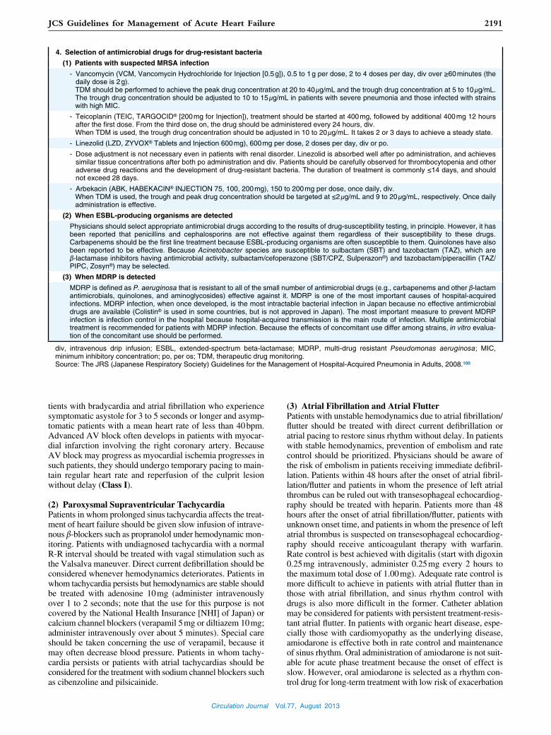
Circulation Journal Vol.77, August 2013
2191JCS Guidelines for Management of Acute Heart Failure
(3) Atrial Fibrillation and Atrial FlutterPatients with unstable hemodynamics due to atrial fibrillation/flutter should be treated with direct current defibrillation or atrial pacing to restore sinus rhythm without delay. In patients with stable hemodynamics, prevention of embolism and rate control should be prioritized. Physicians should be aware of the risk of embolism in patients receiving immediate defibril-lation. Patients within 48 hours after the onset of atrial fibril-lation/flutter and patients in whom the presence of left atrial thrombus can be ruled out with transesophageal echocardiog-raphy should be treated with heparin. Patients more than 48 hours after the onset of atrial fibrillation/flutter, patients with unknown onset time, and patients in whom the presence of left atrial thrombus is suspected on transesophageal echocardiog-raphy should receive anticoagulant therapy with warfarin. Rate control is best achieved with digitalis (start with digoxin 0.25 mg intravenously, administer 0.25 mg every 2 hours to the maximum total dose of 1.00 mg). Adequate rate control is more difficult to achieve in patients with atrial flutter than in those with atrial fibrillation, and sinus rhythm control with drugs is also more difficult in the former. Catheter ablation may be considered for patients with persistent treatment-resis-tant atrial flutter. In patients with organic heart disease, espe-cially those with cardiomyopathy as the underlying disease, amiodarone is effective both in rate control and maintenance of sinus rhythm. Oral administration of amiodarone is not suit-able for acute phase treatment because the onset of effect is slow. However, oral amiodarone is selected as a rhythm con-trol drug for long-term treatment with low risk of exacerbation
tients with bradycardia and atrial fibrillation who experience symptomatic asystole for 3 to 5 seconds or longer and asymp-tomatic patients with a mean heart rate of less than 40 bpm. Advanced AV block often develops in patients with myocar-dial infarction involving the right coronary artery. Because AV block may progress as myocardial ischemia progresses in such patients, they should undergo temporary pacing to main-tain regular heart rate and reperfusion of the culprit lesion without delay (Class I).
(2) Paroxysmal Supraventricular TachycardiaPatients in whom prolonged sinus tachycardia affects the treat-ment of heart failure should be given slow infusion of intrave-nous β-blockers such as propranolol under hemodynamic mon-itoring. Patients with undiagnosed tachycardia with a normal R-R interval should be treated with vagal stimulation such as the Valsalva maneuver. Direct current defibrillation should be considered whenever hemodynamics deteriorates. Patients in whom tachycardia persists but hemodynamics are stable should be treated with adenosine 10 mg (administer intravenously over 1 to 2 seconds; note that the use for this purpose is not covered by the National Health Insurance [NHI] of Japan) or calcium channel blockers (verapamil 5 mg or diltiazem 10 mg; administer intravenously over about 5 minutes). Special care should be taken concerning the use of verapamil, because it may often decrease blood pressure. Patients in whom tachy-cardia persists or patients with atrial tachycardias should be considered for the treatment with sodium channel blockers such as cibenzoline and pilsicainide.
4. Selection of antimicrobial drugs for drug-resistant bacteria
(1) Patients with suspected MRSA infection
- Vancomycin (VCM, Vancomycin Hydrochloride for Injection [0.5 g]), 0.5 to 1 g per dose, 2 to 4 doses per day, div over ≥60 minutes (the daily dose is 2 g).
TDM should be performed to achieve the peak drug concentration at 20 to 40 μg/mL and the trough drug concentration at 5 to 10 μg/mL. The trough drug concentration should be adjusted to 10 to 15 μg/mL in patients with severe pneumonia and those infected with strains with high MIC.
- Teicoplanin (TEIC, TARGOCID® [200 mg for Injection]), treatment should be started at 400 mg, followed by additional 400 mg 12 hours after the first dose. From the third dose on, the drug should be administered every 24 hours, div.
When TDM is used, the trough drug concentration should be adjusted in 10 to 20 μg/mL. It takes 2 or 3 days to achieve a steady state.
- Linezolid (LZD, ZYVOX® Tablets and Injection 600 mg), 600 mg per dose, 2 doses per day, div or po.
- Dose adjustment is not necessary even in patients with renal disorder. Linezolid is absorbed well after po administration, and achieves similar tissue concentrations after both po administration and div. Patients should be carefully observed for thrombocytopenia and other adverse drug reactions and the development of drug-resistant bacteria. The duration of treatment is commonly ≤14 days, and should not exceed 28 days.
- Arbekacin (ABK, HABEKACIN® INJECTION 75, 100, 200 mg), 150 to 200 mg per dose, once daily, div. When TDM is used, the trough and peak drug concentration should be targeted at ≤2 μg/mL and 9 to 20 μg/mL, respectively. Once daily
administration is effective.
(2) When ESBL-producing organisms are detected
Physicians should select appropriate antimicrobial drugs according to the results of drug-susceptibility testing, in principle. However, it has been reported that penicillins and cephalosporins are not effective against them regardless of their susceptibility to these drugs. Carbapenems should be the first line treatment because ESBL-producing organisms are often susceptible to them. Quinolones have also been reported to be effective. Because Acinetobacter species are susceptible to sulbactam (SBT) and tazobactam (TAZ), which are β-lactamase inhibitors having antimicrobial activity, sulbactam/cefoperazone (SBT/CPZ, Sulperazon®) and tazobactam/piperacillin (TAZ/PIPC, Zosyn®) may be selected.
(3) When MDRP is detected
MDRP is defined as P. aeruginosa that is resistant to all of the small number of antimicrobial drugs (e.g., carbapenems and other β-lactam antimicrobials, quinolones, and aminoglycosides) effective against it. MDRP is one of the most important causes of hospital-acquired infections. MDRP infection, when once developed, is the most intractable bacterial infection in Japan because no effective antimicrobial drugs are available (Colistin® is used in some countries, but is not approved in Japan). The most important measure to prevent MDRP infection is infection control in the hospital because hospital-acquired transmission is the main route of infection. Multiple antimicrobial treatment is recommended for patients with MDRP infection. Because the effects of concomitant use differ among strains, in vitro evalua-tion of the concomitant use should be performed.
div, intravenous drip infusion; ESBL, extended-spectrum beta-lactamase; MDRP, multi-drug resistant Pseudomonas aeruginosa; MIC, minimum inhibitory concentration; po, per os; TDM, therapeutic drug monitoring.Source: The JRS (Japanese Respiratory Society) Guidelines for the Management of Hospital-Acquired Pneumonia in Adults, 2008.100

Circulation Journal Vol.77, August 2013
2192 JCS Joint Working Group
fibrillation, it is important to ensure rate control by inhibiting AV conduction.104 When catheter ablation can restore normal sinus rhythm, patients are expected to have recovery to normal cardiac function and improvement in exercise capacity and QOL105 (Class IIa, Level of Evidence: C).
6. Chronic Obstructive Pulmonary Disease (Table 38)
Although COPD is present among 20 to 30% of patients with heart failure,106 and is an independent risk factor for cardiovas-cular death,107 the awareness of COPD is still low. It is diffi-cult to differentiate heart failure from respiratory failure due to an exacerbation of COPD, but BNP or NT-proBNP levels are useful in the diagnosis of heart failure.106,108 In patients with heart failure complicated with COPD, ACE inhibitors, β-blockers, and ARBs are recommended.108 β-blockers can be used safely in most patients with heart failure complicated with COPD. It is preferable to introduce β-blockers at a low dose and increase the dose slowly.109 Careful attention should be given to patients with uncontrolled asthma. Treatment of COPD should be continued in parallel to treatment of the heart failure.110,111
of heart failure (Class IIb, Level of Evidence: C).
(4) Ventricular Tachycardia and Frequent Premature Ventricular Contractions
Patients with premature ventricular contractions (PVCs) or ventricular tachycardia should be assessed for myocardial isch-emia and electrolyte abnormality to make differential diagno-sis. Patients in whom ventricular tachycardia affects hemody-namics should be considered for direct current defibrillation. Patients in whom the origin of tachycardia is localized are indicated for catheter ablation.
2. Heart Failure Due to Tachycardias (Tachycardia-Induced Cardiomyopathy)
Tachycardia-induced cardiomyopathy is a reversible condition where heart failure develops in the presence of persistent or recurrent tachycardia with no other underlying heart disease, and discontinues in the absence of tachycardias resulting in recovery to normal cardiac function.101
Because rhythm or rate control therapy may restore left ven-tricular function, it is important to differentiate this condition from other irreversible myocardial disorders.102,103 The most common cause of tachycardia-induced cardiomyopathy is atri-al fibrillation, but it is also caused by ectopic atrial tachycardia or accelerated idioventricular rhythm. In patients with atrial
mitral valve stenosis, and other conditions meet the definition of “heart failure with preserved EF”, but these conditions are not included in the clinical setting.
2. Diagnosis in the Acute Phase of Heart Failure With Preserved EF
During the acute phase, heart failure with preserved EF is di-agnosed on the basis of the following signs: (1) the presence of heart failure is confirmed or strongly suspected; (2) left ventricular ejection fraction (LVEF) is normal and left ven-tricular enlargement is absent; and (3) the presence of other heart diseases such as valvular heart disease or constrictive pericarditis is ruled out.
Plasma BNP (NT-proBNP) levels increases in patients with heart failure with both reduced and preserved EF.119 Echocar-diography should be performed to determine LVEF and assess the presence/absence of valvular heart disease.
3. Treatment in the Acute Phase of Heart Failure With Preserved EF (Table 39)
Most patients in the acute phase of heart failure with preserved EF show clinical findings of acute pulmonary edema. Refer to Table 15 for the treatment of acute pulmonary edema.
1. Definition of Heart Failure With Preserved EF
Heart failure with reduced EF accounts for about 60% of pa-tients with heart failure, and heart failure with preserved EF accounts for about 40%.112,113 The main feature of heart failure with preserved EF is diastolic dysfunction not associated with left ventricular enlargement.114 Because of the absence of pre-load reserve, left ventricular diastolic pressure readily increas-es in association with an increase in blood pressure or physical activity, which increases the left atrial pressure and pulmonary venous pressure and causes difficulty in breathing. In these pa-tients, pulmonary edema develops in the absence of systolic dysfunction,115 and their prognosis, including rehospitalization due to heart failure, is poor as in the case of heart failure with reduced EF.112,113 Heart failure with preserved EF is relatively common among elderly women, and patients often present with complications of hypertension, diabetes, chronic kidney dis-ease, or atrial fibrillation. The pathophysiology of heart failure with preserved EF is still unclear. It has been pointed out that functional arterial stiffing and functional mitral regurgitation as well as diastolic dysfunction play roles in the development of this condition.116–118 In Western countries, the term “dia-stolic heart failure” is no longer used, and the term “heart fail-ure with preserved EF” is used instead. In the broadest sense of the word, constrictive pericarditis, aortic valve stenosis,
VI Treatment Strategies for Heart Failure With Preserved EF
Table 38. Treatment of Patients With Heart Failure Associated With COPD
Class IIa
- ACE inhibitors, ARBs, β-blockers, and diuretics are recommended for the treatment of heart failure associated with COPD. Treatment of COPD should be continued during treatment of heart failure. (Level of Evidence: B)

Circulation Journal Vol.77, August 2013
2193JCS Guidelines for Management of Acute Heart Failure
tion 5 of Chapter V.
6. Medical Therapy in the Chronic Phase of Heart Failure With Preserved EF
Although treatment of the acute and chronic phase of heart failure with preserved EF should aim at an improvement of the prognosis, no treatment strategies specific to this condition have been proven effective in large-scale clinical studies. The re-sults of large-scale clinical studies have suggested the efficacy of ARBs,120 ACE inhibitors,121 and β-blockers,122 but none of these drugs have been established as treatment options for this condition. Treatment should thus be directed at the cause of heart failure. Antihypertensive treatment should be given to patients with poorly controlled hypertensive heart disease. Treatment of myocardial ischemia is expected to improve he-modynamics and symptoms in patients with ischemic heart disease.
4. Blood Pressure Control
Patients in the acute phase of heart failure with preserved EF often have a significant increase in blood pressure. Blood pressure control should be prioritized, because adequate diuresis may be achieved and pulmonary congestion may be alleviated by control-ling blood pressure. Refer to Section 4 of Chapter III for details.
5. Rate Control in Atrial Fibrillation
In patients with heart failure with preserved EF, left ventricu-lar filling highly depends on atrial contraction. Accordingly, atrial fibrillation significantly affects left ventricular filling and deteriorates hemodynamics due to the absence of effective atrial contraction and the decreased diastolic time associated with tachycardia. Rate control therapy is especially important in these patients. Refer to Section 4.7 of Chapter III and Sec-
VII Treatment Strategies for Biventricular Failure
doses, PDE III inhibitors mainly exert a positive inotropic ac-tion, which is further potentiated by concomitant use of dobu-tamine.125 When PDE III inhibitors are used with dobutamine, both drugs should first be administered at low doses.
Patients not responding well to medical therapy should be considered for heart transplantation at the initial phase of the disease, and physicians should establish detailed treatment strat-
1. Pathophysiology and Treatment of Biventricular Failure
1. Biventricular Failure Mainly Due to Increased Circulating Blood Volume
Diuretic drugs are the primary treatment for patients with bi-ventricular failure accompanied by increased circulating blood volume. Inotropes may also be required in patients with se-verely impaired cardiac function and those with poor re-sponse to diuretics alone (Table 40). Repeated bolus intrave-nous injections of diuretics can be given; however, continuous infusions may be effective for patients not responding well to repeated boluses (Class IIa, Level of Evidence: B) (See Table 21). Treatment with natriuretic peptide analogs such as carperitide which protects renal function is considered valu-able in the treatment of patients who increased circulatory blood volume due to the cardio-renal relationship. Co-administration of heparin should be considered to prevent thrombosis. In pa-tients with diuretic-resistant or severe renal dysfunction, ad-justment of circulating blood volume by extracorporeal ultra-filtration or hemofiltration is effective.123,124
2. Biventricular Failure Mainly Due to Significant Decrease in Cardiac Output
The relative contributions of the positive inotropic and vaso-dilator actions of PDE III inhibitors differ by dose.125 At low
Table 39. Treatment of Heart Failure With Preserved Ejection Fraction
Treatment of pulmonary edema (See Table 15)
Class I
- Treatment of congestion with diuretics, nitrates, and carperitide (Level of Evidence: C)
- Intravenous drip infusion of calcium channel blockers (nicardipine or diltiazem) to control severe hypertension (Level of Evidence: C)
Class IIa
- Treatment with digitalis, calcium channel blockers, and β-blockers to control heart rate in patients with atrial fibrillation (Level of Evidence: C)
Class IIb
- Treatment with inotropes in patients with heart failure with preserved ejection fraction (Level of Evidence: C)
Table 40. Treatment of Biventricular Failure
Biventricular failure with increased circulating blood volume
Class I
- Loop diuretics (Level of Evidence: C)
Class IIa
- Carperitide (Level of Evidence: B)
- Ultrafiltration (Level of Evidence: B)
- Inotropes (dobutamine, PDE inhibitors) (Level of Evidence: C)
Class IIb
- Thiazides (Level of Evidence: C)
- Tolvaptan (Level of Evidence: B)
Biventricular failure with a significant decrease in cardiac output
Class IIb
- Inotropes (concomitant use of dobutamine and PDE inhibi-tors) (Level of Evidence: C)

Circulation Journal Vol.77, August 2013
2194 JCS Joint Working Group
cases secondary to left heart failure, include a decrease in right ventricular contractility due to right ventricular infarction or arrhythmogenic right ventricular dysplasia, right ventricular volume overload due to acute tricuspid valve regurgitation or failure of the Fontan circulation, an increase in right ventricu-lar afterload due to pulmonary embolism or primary pulmo-nary hypertension, and right ventricular diastolic dysfunction due to cardiac tamponade or constrictive pericarditis.
In the treatment of acute right heart failure, physicians should treat the underlying cause, and reduce the right ventricular afterload, enhance the contractility of the right ventricle, and control the right ventricular preload appropriately. PDE III in-hibitors as monotherapy or concomitant use of dobutamine are useful in reducing pulmonary vascular resistance and enhanc-ing the contractility of the right ventricle. In patients with se-vere pulmonary hypertension who need a significant decrease in the right ventricular afterload, concomitant use of calcium channel blockers, prostaglandins, endothelin antagonists, in-haled nitric oxide (NO), PDE V inhibitors, and/or Rho kinase inhibitors are effective.128 Because PCPS may decrease the right ventricular load and maintain PaO2 at an appropriate level, PCPS is quite effective in the treatment of patients with acute right heart failure and poor pulmonary oxygenation.
egies for such patients including the use of mechanical circula-tory support during an acute exacerbation of the condition.
2. Right Heart Failure
1. Right Heart Failure Associated With Left Heart FailureIn many cases, right heart failure develops secondary to left heart failure.126 Right heart failure can also cause bowing of the inter-ventricular septum toward the left due to the increased volume load of the right ventricle, thereby impeding left ventricular dia-stolic function and further reducing cardiac output. The coexis-tence of left ventricular systolic dysfunction and right ventricular systolic dysfunction is the largest independent risk factor of poor prognosis.126,127 As right heart failure progresses, functional tri-cuspid valve regurgitation contributes to hepatic congestion and portal hypertension resulting in resistance to oral medications due to intestinal edema, hepatorenal syndrome. Because veno-venous shunts due to portal hypertension increase mixed venous oxygen saturation, the cardiac output calculated using the Fick method may overestimate the actual cardiac output.
2. Acute Right Heart FailureThe typical causes of acute right heart failure, other than the
has not been established. Patients with heart failure due to systolic dysfunction should start treatment with β-blockers (Table 41). It is preferable to introduce β-blockers when con-gestion is absent and the heart failure is stabilized. Physicians should start β-blockers in the in-hospital setting to achieve the sufficient dose during hospitalization.129 Long-term prognosis is better in patients who start β-blockers during hospitalization rather than after discharge.130 Some patients who cannot toler-ate β-blockers due to exacerbation of heart failure may begin β-blocker treatment safely when the drug is combined with pimobendan 1.25 or 2.5 mg/day.129
When signs/symptoms of heart failure are managed well and euvolemia can be obtained, according to the evidence, treat-ment of chronic heart failure should be initiated and drug doses should be increased to the target doses. During this stage, phy-sicians should assess the severity of the myocardial damage and renal dysfunction that developed after the onset of acute heart failure, and treat these conditions accordingly.
Treatment with ACE inhibitors should be initiated as soon as possible (Table 41). ARBs should be used for patients who cannot receive ACE inhibitors. The efficacy of ACE inhibitors and ARBs in the treatment of heart failure with preserved EF
VIII Transition to Chronic Heart Failure and Timing of Discharge
Table 41. Transition to Chronic Heart Failure
Class I
- During the early stage of acute heart failure, treatment with ACE inhibitors (or ARBs when ACE inhibitors cannot be used) should be started at a low dose and the dose should be increased gradually thereafter. (Level of Evidence: C)
- In patients with heart failure due to systolic dysfunction, treatment with β-blockers should be initiated during hospitalization. (Level of Evidence: A)
Table 42. Criteria for Discharge of Patients With Acute Heart Failure
1. Reaching the target body weight.
2. Achieving target blood pressure.
3. Absence of shortness of breath or dizziness during daily activities.
4. Stable condition for 24 hours or more after addition or change in dose of oral drugs.
5. Stable condition for 48 hours or more after discontinuation of intravenous treatment.
6. Maintaining the optimal circulating blood volume with oral diuretics.
7. Absence of substantial dehydration.
8. Stable or improving renal function.
9. Adequate control of the triggers or precipitating factors of acute heart failure.
10. Implementation or development of treatment strategies according to the guidelines for the treatment of heart failure.
11. Education of the patient and his/her family about heart failure.

Circulation Journal Vol.77, August 2013
2195JCS Guidelines for Management of Acute Heart Failure
failure.Physicians should also refer to the section of “Cardiovascu-
lar Intensive Care” in the “Statement for End-Stage Cardio-vascular Care (JCS 2010)”.133
2. Proposals for Palliative Care (Support Model)
1. Catch the PainPatients suffer from not only physical symptoms such as short-ness of breath and palpitations but the “gap between ambitions and reality” such as that the patient can no longer move as he/she wants, and the patient wants to live longer but his/her time is now limited. Healthcare professionals should be aware of the potential for patients’ physical and emotional pain. The first step is listening to the patient carefully to know his/her pain (to establish the gap between ambitions and reality), in addition to paying attention to visible figures such as vital signs, blood test results, and imaging findings.
1. Introduction
The treatment of heart failure has advanced greatly as the di-agnostic and therapeutic technologies have evolved. Today, the needs for palliative care for patients with end-stage heart failure who are not expected to recover are increasing.132 Pal-liative care was first advocated mainly for patients with end-stage cancer, and was developed to alleviate pain in patients with treatment-resistant disease and to provide mental support. However, palliative care now includes relief of symptoms and phychological care from an acute phase of the disease. When palliative care is defined as a method to support the life and well-being of patients with physical and psychological distress, healthcare professionals should make every effort not only to alleviate pain and provide terminal care, but also support every aspect of patients living with heart failure. There is little evi-dence regarding effective palliative care for patients with heart failure. The present guidelines provide recommendations for approaches to support patients with treatment-resistant heart
IX Palliative Care
should be given for each aspect of heart failure (Figure 16).131
Because acute heart failure may be caused by a wide variety of conditions and diseases, no criteria for discharge have been established. Table 42 summarizes conditions suitable for dis-charge in typical cases.
Treatment of acute heart failure should aim at not only an improvement in congestion and in other symptoms of heart failure but also prevention of rehospitalization and an improve-ment of prognosis through comprehensive assessment of the condition and its complications. Evidence-based treatment
Figure 16. Assessment and targeted implementation of evidence-based therapy in acute heart failure. *Select patients. #Modified to fit the circumstances in Japan. CAD, coronary artery disease; CRT, cardiac resynchronization therapy; Dor procedure, ven-tricular reconstructive surgery to restore aneurysmal left ventricle to its normal; ICD, implantable cardioverter defibrillator; LV, left ventricular. Adapted from J Am Coll Cardiol 2009; 53: 557 – 573,131 with permission from Elsevier Inc.

Circulation Journal Vol.77, August 2013
2196 JCS Joint Working Group
ness when others understand his/her suffering, and should carefully listen to the patient. Healthcare professionals should interact with the patient to listen to him/her carefully using attentive hearing techniques such as repetition, pausing, and asking. Even if the patient is faced with death, he/she can stay calm when the patient understands he/she is fully supported. Even though the keys for support differ among individuals, healthcare professionals can support the well-being of patients when they understand their roles in palliative care.
4. Support for Healthcare ProfessionalsIt is not difficult for healthcare professionals to build a good relationship with the patient if they can help him/her. However, if they cannot help the patient with treatment-resistant disease, it is difficult to interact with him/her continuously. Even when the best available treatment is provided, the patient’s condition deteriorates gradually. When healthcare professionals are over-whelmed by a feeling of helplessness, they may want to dis-tance themselves from the patient. To keep interacting with patients with treatment-resistant heart failure, healthcare pro-fessionals should also be supported emotionally. The true value of healthcare professionals is not the power to solve all prob-lems but the continuity of effort to treat the patient even when it is not helpful. This is difficult to achieve, and healthcare professionals need support and encouragement.
3. Summary
Healthcare professionals involved in the treatment of heart failure should provide a good standard of care, and should also continue to interact with the patient until the end of his/her life. There is little evidence regarding recommendations for palliative care for patients with heart failure. Japan is a coun-try with an aging population and how to care for and interact with end-stage patients with heart failure is a major issue.
2. Catch the KeysHealthcare professional should know the patient’s keys to being him/herself. Such keys differ among individuals, but the most common keys among patients in the clinical setting are dreams for the future (temporal existence), relationships with others (relational existence), and the freedom of making one’s own decisions (autonomous existence).134 These keys help the pa-tient feel peace even when he/she is suffering from pain.135 Healthcare professionals should concentrate not only on alle-viating the patient’s pain but also knowing their keys in the clinical setting.
Dreams for the future encourage the patient to reflect on their experience in the past and live for the future. Even though the time is limited, the patient may be motivated to live to do something he/she wants to do if he/she has dreams for the future. The patient may also be motivated if he/she can imag-ine that the tie with family members will last forever even after his/her death. Relationships with others who fully under-stand the patient are important. The patient is encouraged by the existence of the patient’s family members, friends, and medical staff members who will continue to support him/her throughout the treatment-resistance heart failure. The patient may also be encouraged by connecting with pets and nature. The freedom of making one’s own decisions is a basic human right. It is important to value the patient’s decisions on the treatment strategy, place of care, and other matters related to heart failure treatment.
3. Knowing Healthcare Professionals’ Roles to Support the Patient
Patients with end-stage treatment-resistant heart failure cannot be reassured by just giving words of encouragement and un-wanted life-sustaining treatment. In palliative care, it is quite important to keep good communication with the patient even when the patient receives unfortunate news. The patient is not encouraged by frank and open discussion only. Healthcare professionals should understand that the patient feels happi-
failure.Figure 17 summarizes the outline of treatment for acute heart
X Flowchart of Treatment
present guideline document also describes the clinical scenar-ios and the Nohria-Stevenson classification that are common-ly used in the assessment of the pathophysiology of acute phase heart failure.
Treatment has advanced as well. In addition to conventional medical therapy, new methods of respiratory management have become available. In this document, carperitide and inotropes are positioned more clearly, and new drugs are introduced. It is expected that further studies will facilitate appropriate selec-tion of treatment options for individual patients according to their pathophysiological conditions.
The prevention of acute heart failure is the most important goal, and this depends on the proper management of chronic heart failure, as well as treatment of arrhythmias, ischemia, cardiomyopathy, and valvular heart disease. Physicians should manage cardiac function, but should also take a multidisci-plinary approach to the patient as a whole.
The present revision of the guidelines reflects the rapid ad-vancement of understanding of the pathophysiology of heart failure and treatment methods. Early diagnosis and prompt treatment are essential for patients with acute heart failure, and a misjudgment may lead to life-threatening conditions. It is essential to appropriately understand patients’ pathological condition without delay. Assessment of clinical symptoms and physical findings is especially important as emphasized in the present guideline document. It is also important to use the new-est and most appropriate treatment methods for the varied path-ological conditions encountered in each patient, and to evalu-ate the effects and modify the treatment as needed.
Because the understanding of the pathophysiology of heart failure and treatment techniques has been greatly advanced, the present guideline document covers many aspects of this condition. The pathophysiology of acute heart failure second-ary to diastolic dysfunction has been clarified in detail. The
XI Summary

Circulation Journal Vol.77, August 2013
2197JCS Guidelines for Management of Acute Heart Failure
cians will utilize this guideline document in the clinical setting by adapting the standard treatment strategies appropriately to their patients according to each pathophysiological condition.
Finally, the present guideline document provides standard information regarding the diagnosis and treatment of acute heart failure, and it is the physicians’ responsibility to determine treatment strategies for their patients. We expect that physi-
great vessels, 9th ed. Boston: Little, Brown and Company, 1994; 253 – 256.
6. Killip T 3rd, Kimball JT. Treatment of myocardial infarction in a coronary care unit. A two year experience with 250 patients. Am J Cardiol 1967; 20: 457 – 464.
7. Forrester JS, Diamond G, Chatterjee K, Swan HJ. Medical therapy of acute myocardial infarction by application of hemodynamic sub-sets (second of two parts). N Engl J Med 1976; 295: 1404 – 1413.
8. Nohria A, Tsang SW, Fang JC, Lewis EF, Jarcho JA, Mudge GH, et al. Clinical assessment identifies hemodynamic profiles that pre-dict outcomes in patients admitted with heart failure. J Am Coll Cardiol 2003; 41: 1797 – 1804.
9. Ministry of Health, Labour and Welfare. http://www.mhlw.go.jp/english/
10. Kawashiro N, Kasanuki H, Ogawa H, Matsuda N, Hagiwara N; Heart Institute of Japan--Department of Cardiology (HIJC) Inves-tigators. Clinical characteristics and outcome of hospitalized pa-tients with congestive heart failure: Results of the HIJC-HF regis-try. Circ J 2008; 72: 2015 – 2020.
11. Sato N, Kajimoto K, Asai K, Mizuno M, Minami Y, Nagashima M, et al; ATTEND Investigators. Acute decompensated heart failure
References 1. Okura Y, Ramadan MM, Ohno Y, Mitsuma W, Tanaka K, Ito M,
et al. Impending epidemic: Future projection of heart failure in Japan to the year 2055. Circ J 2008; 72: 489 – 491.
2. Guidelines for the Diagnosis and Treatment of Cardiovascular Dis-eases (1998–1999 Joint Working Groups Report). Guidelines for Treatment of Severe Acute Heart Failure. Jpn Circ J 2000; 64 (Suppl IV): 1129 – 1165 (in Japanese).
3. Guidelines for the Diagnosis and Treatment of Cardiovascular Dis-eases (2004–2005 Joint Working Groups Report). Guidelines for Treatment of Acute Heart Failure (JCS 2006). http://www.j-circ.or.jp/guideline/pdf/JCS2006_maruyama_h.pdf (in Japanese).
4. Nieminen MS, Böhm M, Cowie MR, Drexler H, Filippatos GS, Jondeau G, et al; ESC Committee for Practice Guideline (CPG). Executive summary of the guidelines on the diagnosis and treat-ment of acute heart failure: The Task Force on Acute Heart Failure of the European Society of Cardiology. Eur Heart J 2005; 26: 384 – 416.
5. The Criteria Committee of the New York Heart Association. No-menclature and criteria for diagnosis of diseases of the heart and
Figure 17. Outline of treatment for acute heart failure. ACLS, advanced cardiac life support; ACS, acute coronary syndrome; BLS, basic life support; CVVH, continuous veno-venous hemofiltration; ECLS, extracorporeal life support; ECUM, extracorporeal ultra-filtration method; ET, endotracheal; IABP, intraaortic balloon pumping; LV, left ventricular; LVEF, left ventricular ejection fraction; NPPV, non-invasive positive pressure ventilation; PDE, phosphodiesterase; PEEP, positive-end-expiratory pressure; SaO2, arterial oxygen saturation; SBP, systolic blood pressure; VAS, ventricular assist system.

Circulation Journal Vol.77, August 2013
2198 JCS Joint Working Group
Aldactone Evaluation Study. N Engl J Med 2004; 351: 543 – 551. 30. Seino Y, Momomura S, Takano T, Hayakawa H, Katoh K. Multi-
center, double-blind study of intravenous milrinone for patients with acute heart failure in Japan. Japan Intravenous Milrinone Investiga-tors. Crit Care Med 1996; 24: 1490 – 1497.
31. Lowes BD, Tsvetkova T, Eichhorn EJ, Gilbert EM, Bristow MR. Milrinone versus dobutamine in heart failure subjects treated chron-ically with carvedilol. Int J Cardiol 2001; 81: 141 – 149.
32. Follath F, Cleland JG, Just H, Papp JG, Scholz H, Peuhkurinen K, et al; Steering Committee and Investigators of the Levosimendan Infusion versus Dobutamine (LIDO) Study. Efficacy and safety of intravenous levosimendan compared with dobutamine in severe low-output heart failure (the LIDO study): A randomized double-blind trial. Lancet 2002; 360: 196 – 202.
33. Moiseyev VS, Põder P, Andrejevs N, Ruda MY, Golikov AP, Lazebnik LB, et al; RUSSLAN Study Investigators. Safety and ef-ficacy of a novel calcium sensitizer, levosimendan, in patients with left ventricular failure due to an acute myocardial infarction. A ran-domized, placebo-controlled, double-blind study (RUSSLAN). Eur Heart J 2002; 23: 1422 – 1432.
34. Mebazaa A, Nieminen MS, Packer M, Cohen-Solal A, Kleber FX, Pocock SJ, et al; SURVIVE Investigators. Levosimendan vs dobu-tamine for patients with acute decompensated heart failure: The SURVIVE Randomized Trial. JAMA 2007; 297: 1883 – 1891.
35. Heart Failure Society of America, Lindenfeld J, Albert NM, Boehmer JP, Collins SP, Ezekowitz JA, et al. HFSA 2010 Comprehensive Heart Failure Practice Guideline. J Card Fail 2010; 16: e1 – e194.
36. Dickstein K, Cohen-Solal A, Filippatos G, McMurray JJ, Ponikowski P, Poole-Wilson PA, et al; ESC Committee for Practice Guidelines (CPG). ESC Guidelines for the diagnosis and treatment of acute and chronic heart failure 2008: The Task Force for the Diagnosis and Treatment of Acute and Chronic Heart Failure 2008 of the European Society of Cardiology. Developed in collaboration with the Heart Failure Association of the ESC (HFA) and endorsed by the European Society of Intensive Care Medicine (ESICM). Eur Heart J 2008; 29: 2388 – 2442.
37. Fonarow GC, Abraham WT, Albert NM, Stough WG, Gheorghiade M, Greenberg BH, et al; OPTIMIZE-HF Investigators and Coordi-nators. Influence of beta-blocker continuation or withdrawal on out-comes in patients hospitalized with heart failure: Findings from the OPTIMIZE-HF program. J Am Coll Cardiol 2008; 52: 190 – 199.
38. The Antiarrhythmic Drugs Guideline Committee. The Guidelines for the use of antiarrhythmic drugs: Commentary on the CD-ROM version of the Guidelines and concept of the Sicilian Gambit. Tokyo: Lifemedicom Co., ltd. 2000 (in Japanese).
39. Members of the Sicilian Gambit. Antiarrhythmic therapy: A patho-physiologic approach. New York: Futura Publishing Company, Inc; 1994.
40. Neumar RW, Otto CW, Link MS, Kronick SL, Shuster M, Callaway CW, et al. Part 8: Adult advanced cardiovascular life support: 2010 American Heart Association Guidelines for Cardiopulmonary Re-suscitation and Emergency Cardiovascular Care. Circulation 2010; 122(Suppl 3): S729 – S767.
41. Berg RA, Hemphill R, Abella BS, Aufderheide TP, Cave DM, Hazinski MF, et al. Part 5: Adult basic life support: 2010 American Heart Association Guidelines for Cardiopulmonary Resuscitation and Emergency Cardiovascular Care. Circulation 2010; 122(Suppl 3): S685 – S705.
42. Hunt SA, Abraham WT, Chin MH, Feldman AM, Francis GS, Ganiats TG, et al; American College of Cardiology Foundation; American Heart Association. 2009 Focused update incorporated into the ACC/AHA 2005 Guidelines for the Diagnosis and Management of Heart Failure in Adults. A Report of the American College of Cardiology Foundation/American Heart Association Task Force on Practice Guidelines Developed in Collaboration with the Interna-tional Society for Heart and Lung Transplantation. J Am Coll Car-diol 2009; 53: e1 – e90.
43. Antman EM, Hand M, Armstrong PW, Bates ER, Green LA, Halasyamani LK, et al; Canadian Cardiovascular Society; American Academy of Family Physicians; American College of Cardiology; American Heart Association. 2007 focused update of the ACC/AHA 2004 guidelines for the management of patients with ST-ele-vation myocardial infarction: A report of the American College of Cardiology/American Heart Association Task Force on Practice Guidelines. J Am Coll Cardiol 2008; 51: 210 – 247.
44. Soran O, Kennard ED, Kfoury AG, Kelsey SF; IEPR Investigators. Two-year clinical outcomes after enhanced external counterpulsa-tion (EECP) therapy in patients with refractory angina pectoris and left ventricular dysfunction (report from the International EECP Patient Registry). Am J Cardiol 2006; 97: 17 – 20.
syndromes (ATTEND) registry: A prospective observational mul-ticenter cohort study: Rationale, design, and preliminary data. Am Heart J 2010; 159: 949 – 955. e1.
12. Hamaguchi S, Yokoshiki H, Kinugawa S, Tsuchihashi-Makaya M, Yokota T, Takeshita A, et al; Japanese Cardiac Registry of Heart Failure in Cardiology Investigators. Effects of atrial fibrillation on long-term outcomes in patients hospitalized for heart failure in Japan: A report from the Japanese Cardiac Registry of Heart Failure in Cardiology (JCARE-CARD). Circ J 2009; 73: 2084 – 2090.
13. McKee PA, Castelli WP, McNamara PM, Kannel WB. The natural history of congestive heart failure: The Framingham study. N Engl J Med 1971; 285: 1441 – 1446.
14. Jessup M, Brozena S. Heart failure. N Engl J Med 2003; 348: 2007 – 2018.
15. Felker GM, Pang PS, Adams KF, Cleland JG, Cotter G, Dickstein K, et al; International AHFS Working Group. Clinical trials of pharmacological therapies in acute heart failure syndromes: Les-sons learned and directions forward. Circ Heart Fail 2010; 3: 314 – 325.
16. Gheorghiade M, Filippatos G, De Luca L, Burnett J. Congestion in acute heart failure syndromes: An essential target of evaluation and treatment. Am J Med 2006; 119(Suppl 1): S3 – S10.
17. Allen LA, Hernandez AF, O’Connor CM, Felker GM. End points for clinical trials in acute heart failure syndromes. J Am Coll Car-diol 2009; 53: 2248 – 2258.
18. Peacock WF 4th, Fonarow GC, Emerman CL, Mills RM, Wynne J; ADHERE Scientific Advisory Committee and Investigators; Ad-here Study Group. Impact of early initiation of intravenous therapy for acute decompensated heart failure on outcomes in ADHERE. Cardiology 2007; 107: 44 – 51.
19. Mebazaa A, Gheorghiade M, Piña IL, Harjola VP, Hollenberg SM, Follath F, et al. Practical recommendations for prehospital and early in-hospital management of patients presenting with acute heart fail-ure syndromes. Crit Care Med 2008; 36(Suppl): S129 – S139.
20. Nohria A, Lewis E, Stevenson LW. Medical management of ad-vanced heart failure. JAMA 2002; 287: 628 – 640.
21. Lubien E, DeMaria A, Krishnaswamy P, Clopton P, Koon J, Kazanegra R, et al. Utility of B-natriuretic peptide in detecting dia-stolic dysfunction: Comparison with Doppler velocity recordings. Circulation 2002; 105: 595 – 601.
22. Cotter G, Metra M, Milo-Cotter O, Dittrich HC, Gheorghiade M. Fluid overload in acute heart failure--re-distribution and other mech-anisms beyond fluid accumulation. Eur J Heart Fail 2008; 10: 165 – 169.
23. Field JM, Hazinski MF, Sayre MR, Chameides L, Schexnayder SM, Hemphill R, et al. Part 1: Executive summary: 2010 American Heart Association Guidelines for Cardiopulmonary Resuscitation and Emergency Cardiovascular Care. Circulation 2010; 122(Suppl 3): S640 – S656.
24. McAlister FA, Stewart S, Ferrua S, McMurray JJ. Multidisciplinary strategies for the management of heart failure patients at high risk for admission: A systematic review of randomized trials. J Am Coll Cardiol 2004; 44: 810 – 819.
25. Davidson PM, Cockburn J, Newton PJ, Webster JK, Betihavas V, Howes L, et al. Can a heart failure-specific cardiac rehabilitation program decrease hospitalizations and improve outcomes in high-risk patients? Eur J Cardiovasc Prev Rehabil 2010; 17: 393 – 402.
26. Hunt SA, Abraham WT, Chin MH, Feldman AM, Francis GS, Ganiats TG, et al; American College of Cardiology; American Heart Association Task Force on Practice Guidelines; American College of Chest Physicians; International Society for Heart and Lung Transplantation; Heart Rhythm Society. ACC/AHA 2005 Guide-line Update for the Diagnosis and Management of Chronic Heart Failure in the Adult: A report of the American College of Cardiol-ogy/American Heart Association Task Force on Practice Guidelines (Writing Committee to Update the 2001 Guidelines for the Evalu-ation and Management of Heart Failure): Developed in collabora-tion with the American College of Chest Physicians and the Inter-national Society for Heart and Lung Transplantation: Endorsed by the Heart Rhythm Society. Circulation 2005; 112: e154 – e235.
27. Ishii H, Ichimiya S, Kanashiro M, Amano T, Imai K, Murohara T, et al. Impact of a single intravenous administration of nicorandil before reperfusion in patients with ST-segment-elevation myocar-dial infarction. Circulation 2005; 112: 1284 – 1288.
28. Suwa M, Seino Y, Nomachi Y, Matsuki S, Funahashi K. Multi-center prospective investigation on efficacy and safety of carperit-ide for acute heart failure in the ‘real world’ of therapy. Circ J 2005; 69: 283 – 290.
29. Juurlink DN, Mamdani MM, Lee DS, Kopp A, Austin PC, Laupacis A, et al. Rates of hyperkalemia after publication of the Randomized

Circulation Journal Vol.77, August 2013
2199JCS Guidelines for Management of Acute Heart Failure
failure: Research challenges and opportunities. J Am Coll Cardiol 2010; 56: 343 – 351.
59. Alexiou VG, Ierodiakonou V, Dimopoulos G, Falagas ME. Impact of patient position on the incidence of ventilator-associated pneu-monia: A meta-analysis of randomized controlled trials. J Crit Care 2009; 24: 515 – 522.
60. Muscedere J, Dodek P, Keenan S, Fowler R, Cook D, Heyland D; VAP Guidelines Committee and the Canadian Critical Care Trials Group. Comprehensive evidence-based clinical practice guidelines for ventilator-associated pneumonia: Prevention. J Crit Care 2008; 23: 126 – 137.
61. The Japanese Society of Intensive Care Medicine. Committee for the assessing functional category of ICUs. 2010 Revision of the bundles to prevent ventilator-associated pneumonia (in Japanese).
62. Guidelines for the Diagnosis and Treatment of Cardiovascular Dis-eases (2008 Joint Working Groups Report). Guidelines for the Di-agnosis, Treatment and Prevention of Pulmonary Thromboembo-lism and Deep Vein Thrombosis (JCS 2009). http://www.j-circ.or.jp/guideline/pdf/JCS2009_andod_h.pdf (in Japanese).
63. Baba A, Hirata G, Yokoyama F, Kenmoku K, Tsuchiya M, Kyo S, et al. Psychiatric problems of heart transplant candidates with left ventricular assist devices. J Artif Organs 2006; 9: 203 – 208.
64. Sessler CN, Gosnell MS, Grap MJ, Brophy GM, O’Neal PV, Keane KA, et al. The Richmond Agitation-Sedation Scale: Validity and reliability in adult intensive care unit patients. Am J Respir Crit Care Med 2002; 166: 1338 – 1344.
65. Setoguchi S, Stevenson LW, Schneeweiss S. Repeated hospitaliza-tions predict mortality in the community population with heart fail-ure. Am Heart J 2007; 154: 260 – 266.
66. Tsuchihashi M, Tsutsui H, Kodama K, Kasagi F, Setoguchi S, Mohr M, et al. Medical and socioenvironmental predictors of hospital readmission in patients with congestive heart failure. Am Heart J 2001; 142: E7.
67. Koelling TM, Johnson ML, Cody RJ, Aaronson KD. Discharge edu-cation improves clinical outcomes in patients with chronic heart failure. Circulation 2005; 111: 179 – 185.
68. Paul S. Hospital discharge education for patients with heart failure: What really works and what is the evidence? Crit Care Nurse 2008; 28: 66 – 82.
69. Konick-McMahan J, Bixby B, McKenna C. Heart failure in older adults. Providing nursing care to improve outcomes. J Gerontol Nurs 2003; 29: 35 – 41.
70. Guidelines for the Diagnosis and Treatment of Cardiovascular Dis-eases (2009 Joint Working Groups Report). Guidelines for Treat-ment of Chronic Heart Failure (JCS 2010). http://www.j-circ.or.jp/guideline/pdf/JCS2010_matsuzaki_h.pdf (in Japanese).
71. Kato N, Ito N, Kinugawa K, Kazuma K. Validity and reliability of the Japanese version of the European Heart Failure Self-Care Be-havior Scale. Eur J Cardiovasc Nurs 2008; 7: 284 – 289.
72. Vaccarino V, Kasl SV, Abramson J, Krumholz HM. Depressive symptoms and risk of functional decline and death in patients with heart failure. J Am Coll Cardiol 2001; 38: 199 – 205.
73. Rozzini R, Sabatini T, Frisoni GB, Trabucchi M. Depression and major outcomes in older patients with heart failure. Arch Intern Med 2002; 162: 362 – 364.
74. Kato N, Kinugawa K, Yao A, Hatano M, Shiga T, Kazuma K. Re-lationship of depressive symptoms with hospitalization and death in Japanese patients with heart failure. J Card Fail 2009; 15: 912 – 919.
75. Tsuchihashi-Makaya M, Kato N, Chishaki A, Takeshita A, Tsutsui H. Anxiety and poor social support are independently associated with adverse outcomes in patients with mild heart failure. Circ J 2009; 73: 280 – 287.
76. Hutt E, Frederickson E, Ecord M, Kramer AM. Associations among processes and outcomes of care for Medicare nursing home resi-dents with acute heart failure. J Am Med Dir Assoc 2003; 4: 195 – 199.
77. Pi-Figueras M, Aguilera A, Arellano M, Miralles R, Garcia-Caselles P, Torres R, et al. Prevalence of delirium in a geriatric convales-cence hospitalization unit: Patient’s clinical characteristics and risk precipitating factor analysis. Arch Gerontol Geriatr Suppl 2004; 333 – 337.
78. Ely EW, Shintani A, Truman B, Speroff T, Gordon SM, Harrell FE Jr, et al. Delirium as a predictor of mortality in mechanically venti-lated patients in the intensive care unit. JAMA 2004; 291: 1753 – 1762.
79. Inada K. The pathophysiology and causes. In: Nishimura K, Yamauchi N, editors. The best way to treat delirium: Techniques to avoid worsening. Japanese Journal of Nursing Techniques 2011; 57(Suppl): 9 – 16 (in Japanese).
45. Rao V, Oz MC, Flannery MA, Catanese KA, Argenziano M, Naka Y. Revised screening scale to predict survival after insertion of a left ventricular assist device. J Thorac Cardiovasc Surg 2003; 125: 855 – 862.
46. Lietz K, Long JW, Kfoury AG, Slaughter MS, Silver MA, Milano CA, et al. Outcomes of left ventricular assist device implantation as destination therapy in the post-REMATCH era: Implications for patient selection. Circulation 2007; 116: 497 – 505.
47. Levy WC, Mozaffarian D, Linker DT, Farrar DJ, Miller LW; REMATCH Investigators. Can the Seattle heart failure model be used to risk-stratify heart failure patients for potential left ventricu-lar assist device therapy? J Heart Lung Transplant 2009; 28: 231 – 236.
48. Stevenson LW. Clinical use of inotropic therapy for heart failure: Looking backward or forward? Part I: Inotropic infusions during hospitalization. Circulation 2003; 108: 367 – 372.
49. Stevenson LW. Clinical use of inotropic therapy for heart failure: Looking backward or forward? Part II: Chronic inotropic therapy. Circulation 2003; 108: 492 – 497.
50. Bonow RO, Carabello BA, Chatterjee K, de Leon AC Jr, Faxon DP, Freed MD, et al; 2006 Writing Committee Members; American College of Cardiology/American Heart Association Task Force. 2008 Focused update incorporated into the ACC/AHA 2006 guide-lines for the management of patients with valvular heart disease: A report of the American College of Cardiology/American Heart Association Task Force on Practice Guidelines (Writing Committee to Revise the 1998 Guidelines for the Management of Patients With Valvular Heart Disease): Endorsed by the Society of Cardiovascu-lar Anesthesiologists, Society for Cardiovascular Angiography and Interventions, and Society of Thoracic Surgeons. Circulation 2008; 118: e523 – e661.
51. Bonow RO, Carabello BA, Kanu C, de Leon AC Jr, Faxon DP, Freed MD, et al; American College of Cardiology/American Heart Association Task Force on Practice Guidelines; Society of Cardio-vascular Anesthesiologists; Society for Cardiovascular Angiogra-phy and Interventions; Society of Thoracic Surgeons. ACC/AHA 2006 guidelines for the management of patients with valvular heart disease: A report of the American College of Cardiology/American Heart Association Task Force on Practice Guidelines (writing com-mittee to revise the 1998 Guidelines for the Management of Pa-tients With Valvular Heart Disease): Developed in collaboration with the Society of Cardiovascular Anesthesiologists: Endorsed by the Society for Cardiovascular Angiography and Interventions and the Society of Thoracic Surgeons. Circulation 2006; 114: e84 – e231.
52. Vahanian A, Baumgartner H, Bax J, Butchart E, Dion R, Filippatos G, et al; Task Force on the Management of Valvular Heart Disease of the European Society of Cardiology; ESC Committee for Prac-tice Guidelines. Guidelines on the management of valvular heart disease: The Task Force on the Management of Valvular Heart Disease of the European Society of Cardiology. Eur Heart J 2007; 28: 230 – 268.
53. Stout KK, Verrier ED. Acute valvular regurgitation. Circulation 2009; 119: 3232 – 3241.
54. Antman EM, Hand M, Armstrong PW, Bates ER, Green LA, Halasyamani LK, et al. 2007 Focused Update of the ACC/AHA 2004 Guidelines for the Management of Patients With ST-Elevation Myocardial Infarction: A report of the American College of Cardi-ology/American Heart Association Task Force on Practice Guide-lines: Developed in collaboration With the Canadian Cardiovascu-lar Society endorsed by the American Academy of Family Physicians: 2007 Writing Group to Review New Evidence and Update the ACC/AHA 2004 Guidelines for the Management of Patients With ST-Elevation Myocardial Infarction, Writing on Behalf of the 2004 Writing Committee. Circulation 2008; 117: 296 – 329.
55. Eagle KA, Guyton RA, Davidoff R, Edwards FH, Ewy GA, Gardner TJ, et al; American College of Cardiology; American Heart Asso-ciation. ACC/AHA 2004 guideline update for coronary artery by-pass graft surgery: A report of the American College of Cardiology/American Heart Association Task Force on Practice Guidelines (Committee to Update the 1999 Guidelines for Coronary Artery Bypass Graft Surgery). Circulation 2004; 110: e340 – e437.
56. Emerman CL. Treatment of the acute decompensation of heart fail-ure: Efficacy and pharmacoeconomics of early initiation of therapy in the emergency department. Rev Cardiovasc Med 2003; 4: S13 – S20.
57. Tang WH, Mullens W. Cardiorenal syndrome in decompensated heart failure. Heart 2010; 96: 255 – 260.
58. Peacock WF, Braunwald E, Abraham W, Albert N, Burnett J, Christenson R, et al. National Heart, Lung, and Blood Institute work-ing group on emergency department management of acute heart

Circulation Journal Vol.77, August 2013
2200 JCS Joint Working Group
JM, et al; HeartMate II Clinical Investigators. Renal and hepatic function improve in advanced heart failure patients during contin-uous-flow support with the HeartMate II left ventricular assist de-vice. Circulation 2009; 120: 2352 – 2357.
100. The Japanese Respiratory Society. Writing Committee of the Guide-lines for the Management of Hospital-Acquired Pneumonia in Adults. The JRS Guidelines for the Management of Hospital-Ac-quired Pneumonia in Adults. Tokyo: The Japanese Respiratory So-ciety, 2008 (in Japanese).
101. Shinbane JS, Wood MA, Jensen DN, Ellenbogen KA, Fitzpatrick AP, Scheinman MM. Tachycardia- induced cardiomyopathy: A re-view of animal models and clinical studies. J Am Coll Cardiol 1997; 29: 709 – 715.
102. Van Gelder IC, Crijns HJ, Blanksma PK, Landsman ML, Posma JL, Van Den Berg MP, et al. Time course of hemodynamic changes and improvement of exercise tolerance after cardioversion of chronic atrial fibrillation unassociated with cardiac valve disease. Am J Car-diol 1993; 72: 560 – 566.
103. Umana E, Solares CA, Alpert MA. Tachycardia-induced cardiomy-opathy. Am J Med 2003; 114: 51 – 55.
104. Wyse DG, Waldo AL, DiMarco JP, Domanski MJ, Rosenberg Y, Schron EB, et al; Atrial Fibrillation Follow-up Investigation of Rhythm Management (AFFIRM) Investigators. A comparison of rate control and rhythm control in patients with atrial fibrillation. N Engl J Med 2002; 347: 1825 – 1833.
105. Hsu LF, Jaïs P, Sanders P, Garrigue S, Hocini M, Sacher F, et al. Catheter ablation for atrial fibrillation in congestive heart failure. N Engl J Med 2004; 351: 2373 – 2383.
106. Le Jemtel TH, Padeletti M, Jelic S. Diagnostic and therapeutic chal-lenges in patients with coexistent chronic obstructive pulmonary disease and chronic heart failure. J Am Coll Cardiol 2007; 49: 171 – 180.
107. Sin DD, Man SF. Chronic obstructive pulmonary disease as a risk factor for cardiovascular morbidity and mortality. Proc Am Thorac Soc 2005; 2: 8 – 11.
108. Rutten FH, Cramer MJ, Grobbee DE, Sachs AP, Kirkels JH, Lammers JW, et al. Unrecognized heart failure in elderly patients with stable chronic obstructive pulmonary disease. Eur Heart J 2005; 26: 1887 – 1894.
109. Shelton RJ, Rigby AS, Cleland JG, Clark AL. Effect of a commu-nity heart failure clinic on uptake of beta blockers by patients with obstructive airways disease and heart failure. Heart 2006; 92: 331 – 336.
110. Tashkin DP, Celli B, Senn S, Burkhart D, Kesten S, Menjoge S, et al; UPLIFT Study Investigators. A 4-year trial of tiotropium in chronic obstructive pulmonary disease. N Engl J Med 2008; 359: 1543 – 1554.
111. Calverley PM, Anderson JA, Celli B, Ferguson GT, Jenkins C, Jones PW, et al; TORCH investigators. Salmeterol and fluticasone pro-pionate and survival in chronic obstructive pulmonary disease. N Engl J Med 2007; 356: 775 – 789.
112. Tsuchihashi-Makaya M, Hamaguchi S, Kinugawa S, Yokota T, Goto D, Yokoshiki H, et al; JCARE-CARD Investigators. Characteristics and outcomes of hospitalized patients with heart failure and reduced vs preserved ejection fraction: Report from the Japanese Cardiac Registry of Heart Failure in Cardiology (JCARE-CARD). Circ J 2009; 73: 1893 – 1900.
113. Miyagishima K, Hiramitsu S, Kimura H, Mori K, Ueda T, Kato S, et al. Long term prognosis of chronic heart failure: Reduced vs preserved left ventricular ejection fraction. Circ J 2009; 73: 92 – 99.
114. Yamamoto K, Sakata Y, Ohtani T, Takeda Y, Mano T. Heart failure with preserved ejection fraction. Circ J 2009; 73: 404 – 410.
115. Gandhi SK, Powers JC, Nomeir AM, Fowle K, Kitzman DW, Rankin KM, et al. The pathogenesis of acute pulmonary edema associated with hypertension. N Engl J Med 2001; 344: 17 – 22.
116. Nishio M, Sakata Y, Mano T, Ohtani T, Takeda Y, Hori M, et al. Difference of clinical characteristics between hypertensive patients with and without diastolic heart failure: The roles of diastolic dys-function and renal insufficiency. Hypertens Res 2008; 31: 1865 – 1872.
117. Kawaguchi M, Hay I, Fetics B, Kass DA. Combined ventricular systolic and arterial stiffening in patients with heart failure and pre-served ejection fraction: Implications for systolic and diastolic re-serve limitations. Circulation 2003; 107: 714 – 720.
118. Penicka M, Bartunek J, Trakalova H, Hrabakova H, Maruskova M, Karasek J, et al. Heart failure with preserved ejection fraction in outpatients with unexplained dyspnea: A pressure-volume loop anal-ysis. J Am Coll Cardiol 2010; 55: 1701 – 1710.
119. Yamaguchi H, Yoshida J, Yamamoto K, Sakata Y, Mano T, Akehi N, et al. Elevation of plasma brain natriuretic peptide is a hallmark
80. Inouye SK, Bogardus ST Jr, Charpentier PA, Leo-Summers L, Acampora D, Holford TR, et al. A multicomponent intervention to prevent delirium in hospitalized older patients. N Engl J Med 1999; 340: 669 – 676.
81. Cole MG, McCusker J, Bellavance F, Primeau FJ, Bailey RF, Bonnycastle MJ, et al. Systematic detection and multidisciplinary care of delirium in older medical inpatients: A randomized trial. CMAJ 2002; 167: 753 – 759.
82. Machida I. Screening tools for delirium. Japanese Journal of Gen-eral Hospital Psychiatry 2002; 14: 1 – 8 (in Japanese).
83. Neelon VJ, Champagne MT, Carlson JR, Funk SG. The NEECHAM Confusion Scale: Construction, validation, and clinical testing. Nurs Res 1996; 45: 324 – 330.
84. Ely EW, Margolin R, Francis J, May L, Truman B, Dittus R, et al. Evaluation of delirium in critically ill patients: Validation of the Confusion Assessment Method for the Intensive Care Unit (CAM-ICU). Crit Care Med 2001; 29: 1370 – 1379.
85. Japan Society of Respiratory Care Medicine. Guidelines for seda-tion during artificial respiration. Japanese Journal of Respiratory Care 2007; 24: 146 – 167 (in Japanese).
86. Khan MG. Complications of myocardial infarction and postinfarc-tion care. In: Khan MG, editor. Heart disease diagnosis and therapy: A practical approach, 2nd ed. New York: Human Press Inc., 2005; 69 – 107.
87. Grossman E, Messerli FH, Grodzicki T, Kowey P. Should a mora-torium be placed on sublingual nifedipine capsules given for hyper-tensive emergencies and pseudoemergencies? JAMA 1996; 276: 1328 – 1331.
88. Kajimoto K, Imai T, Minami Y, Kasanuki H. Comparison of acute reduction in left ventricular outflow tract pressure gradient in ob-structive hypertrophic cardiomyopathy by disopyramide versus pil-sicainide versus cibenzoline. Am J Cardiol 2010; 106: 1307 – 1312.
89. Cooper LT Jr, Berry GJ, Shabetai R. Idiopathic giant-cell myocar-ditis--natural history and treatment. N Engl J Med 1997; 336: 1860 – 1866.
90. Ohuchida M, Imaizumi T. Treatment strategies for fulminant myo-carditis: Steroid therapy. In: Izumi T, editor. Clinical practice for fulminant myocarditis. Tokyo: Igaku-Shoin Ltd., 2002; 71 – 80 (in Japanese).
91. Kodama M, Okura Y, Hirono S, Hanawa H, Ogawa Y, Itoh M, et al. A new scoring system to predict the efficacy of steroid therapy for patients with acute myocarditis--a retrospective study. Jpn Circ J 1998; 62: 715 – 720.
92. Kato S, Morimoto S, Hiramitsu S, Uemura A, Ohtsuki M, Kato Y, et al. Successful high-dose intravenous immunoglobulin therapy for a patient with fulminant myocarditis. Heart Vessels 2007; 22: 48 – 51.
93. Robinson JL, Hartling L, Crumley E, Vandermeer B, Klassen TP. A systematic review of intravenous gamma globulin for therapy of acute myocarditis. BMC Cardiovasc Disord 2005; 5: 12.
94. Guidelines for the Diagnosis and Treatment of Cardiovascular Dis-eases (2006 Joint Working Groups Report). Guidelines for Surgical and Interventional Treatment of Valvular Heart Disease (JCS 2007). http://www.j-circ.or.jp/guideline/pdf/JCS2007_matsuda_h.pdf (in Japanese).
95. Bonow RO, Carabello BA, Chatterjee K, de Leon AC Jr, Faxon DP, Freed MD, et al; American College of Cardiology/American Heart Association Task Force on Practice Guidelines. 2008 focused up-date incorporated into the ACC/AHA 2006 guidelines for the man-agement of patients with valvular heart disease: A report of the American College of Cardiology/American Heart Association Task Force on Practice Guidelines (Writing Committee to revise the 1998 guidelines for the management of patients with valvular heart disease). Endorsed by the Society of Cardiovascular Anesthesiolo-gists, Society for Cardiovascular Angiography and Interventions, and Society of Thoracic Surgeons. J Am Coll Cardiol 2008; 52: e1 – e142.
96. Sezai A, Hata M, Niino T, Yoshitake I, Unosawa S, Wakui S, et al. Influence of continuous infusion of low-dose human atrial natri-uretic peptide on renal function during cardiac surgery: A random-ized controlled study. J Am Coll Cardiol 2009; 54: 1058 – 1064.
97. Morikawa S, Sone T, Tsuboi H, Mukawa H, Morishima I, Uesugi M, et al. Renal protective effects and the prevention of contrast-induced nephropathy by atrial natriuretic peptide. J Am Coll Car-diol 2009; 53: 1040 – 1046.
98. Nigwekar SU, Navaneethan SD, Parikh CR, Hix JK. Atrial natri-uretic peptide for management of acute kidney injury: A system-atic review and meta-analysis. Clin J Am Soc Nephrol 2009; 4: 261 – 272.
99. Russell SD, Rogers JG, Milano CA, Dyke DB, Pagani FD, Aranda

Circulation Journal Vol.77, August 2013
2201JCS Guidelines for Management of Acute Heart Failure
•MitsuakiIsobe,DepartmentofCardiovascularMedicine,TokyoMed-ical and Dental University
•HiroshiIto,DepartmentofCardiovascularMedicine,OkayamaUniver-sity Graduate School of Medicine, Dentistry, and Pharmaceutical Sciences
•MasafumiKitakaze,DepartmentofCardiovascularMedicine,NationalCerebral and Cardiovascular Center
•Shin-ichiMomomura,DivisionofCardiovascularMedicine,Depart-ment of Integrated Medicine I, Jichi Medical University Saitama Medical Center
•YoshikiSawa,DepartmentofCardiovascularSurgery,OsakaUniver-sity Graduate School of Medicine
•YoshihikoSeino,DepartmentofCardiology,CardiovascularCenter,Nippon Medical School Chiba Hokusoh Hospital
•HiroyukiTsutsui,DepartmentofCardiovascularMedicine,HokkaidoUniversity Graduate School of Medicine
•KenjiYamazaki,DepartmentofCardiovascularSurgery,TheHeartIn-stitute of Japan, Tokyo Women’s Medical University
•TsutomuYoshikawa,DepartmentofCardiology,SakakibaraHeartIn-stitute
Collaborators: •HitoshiAdachi,DivisionofCardiology,GunmaPrefecturalCardiovas-
cular Center •ToshihisaAnzai, Department of CardiovascularMedicine, National
Cerebral and Cardiovascular Center •NaoyoshiAoyama,DepartmentofCardio-angiology,KitasatoUniver-
sity School of Medicine •YoichiGoto,DepartmentofCardiovascularMedicine,NationalCere-
bral and Cardiovascular Center •KazuhikoHashimura,DepartmentofCardiology,HanwaMemorialHos-
pital •TakayukiInomata,DepartmentofCardio-angiology,KitasatoUniver-
sity School of Medicine •MahotoKato,Division of CardiovascularMedicine,Department of
Medicine, Nihon University School of Medicine •ShintaroKinugawa,DepartmentofCardiovascularMedicine,Hokkaido
University Graduate School of Medicine •KazufumiNakamura,DepartmentofCardiovascularMedicine,Okayama
University Graduate School of Medicine, Dentistry, and Pharmaceu-tical Sciences
•KazuhikoNishigaki,DivisionofCardiology,GifuUniversityGraduateSchool of Medicine
•ShinichiNiwano,DepartmentofCardio-angiology,KitasatoUniver-sity School of Medicine
•KatsuyaOnishi,OnishiHeartClinic •TaketoshiOzawa,MegumiHomeCareClinic •YasushiSakata,DepartmentofCardiovascularMedicine,OsakaUni-
versity Graduate School of Medicine •NaokiSato,InternalMedicine,Cardiology,andIntensiveCareUnit,
Nippon Medical School Musashi-Kosugi Hospital •YukihitoSato,DivisionofCardiology,DepartmentofInternalMedi-
cine, Hyogo Prefectural Amagasaki Hospital •TomokoSugiyamaKato,DepartmentofCardiovascularMedicine,Na-
tional Cerebral and Cardiovascular Center •MiyukiTsuchihashi-Makaya,DepartmentofCardiovascularMedicine,
Hokkaido University Graduate School of Medicine •KazuhiroYamamoto,DivisionofCardiovascularMedicine,Endocri-
nology and Metabolism, Department of Molecular Medicine and Therapeutics, Faculty of Medicine, Tottori University
•YoshioYasumura,DivisionofCardiology,NationalHospitalOrgani-zation Osaka National Hospital
•HiroyukiYokoyama,DepartmentofCardiovascularMedicine,Nation-al Cerebral and Cardiovascular Center
Independent Assessment Committee: •MasatsuguHori,OsakaMedicalCenterforCancerandCardiovascular
Diseases •TsutomuImaizumi,DivisionofCardiovascularMedicine,Department
of Internal Medicine, Kurume University School of Medicine •TakeshiNakatani,DepartmentofTransplantation,NationalCerebral
and Cardiovascular Center •KeijiroSaku,DepartmentofCardiology,FukuokaUniversitySchool
of Medicine •MichihiroYoshimura,DivisionofCardiology,DepartmentofInternal
Medicine, The Jikei University School of Medicine(The affiliations of the members are as of July 2012)
of diastolic heart failure independent of ventricular hypertrophy. J Am Coll Cardiol 2004; 43: 55 – 60.
120. Yusuf S, Pfeffer MA, Swedberg K, Granger CB, Held P, McMurray JJ, et al; CHARM Investigators and Committees. Effects of cande-sartan in patients with chronic heart failure and preserved left-ven-tricular ejection fraction: The CHARM-Preserved Trial. Lancet 2003; 362: 777 – 781.
121. Cleland JG, Tendera M, Adamus J, Freemantle N, Polonski L, Taylor J; PEP-CHF Investigators. The perindopril in elderly people with chronic heart failure (PEP-CHF) study. Eur Heart J 2006; 27: 2338 – 2345.
122. van Veldhuisen DJ, Cohen-Solal A, Böhm M, Anker SD, Babalis D, Roughton M, et al; SENIORS Investigators. Beta-blockade with nebivolol in elderly heart failure patients with impaired and pre-served left ventricular ejection fraction: Data From SENIORS (Study of Effects of Nebivolol Intervention on Outcomes and Rehospital-ization in Seniors With Heart Failure). J Am Coll Cardiol 2009; 53: 2150 – 2158.
123. Costanzo MR, Saltzberg MT, Jessup M, Teerlink JR, Sobotka PA; Ultrafiltration Versus Intravenous Diuretics for Patients Hospital-ized for Acute Decompensated Heart Failure (UNLOAD) Investi-gators. Ultrafiltration is associated with fewer rehospitalizations than continuous diuretic infusion in patients with decompensated heart failure: results from UNLOAD. J Card Fail 2010; 16: 277 – 284.
124. Costanzo MR. Ultrafiltration in the management of heart failure. Curr Opin Crit Care 2008; 14: 524 – 530.
125. Jaski BE, Fifer MA, Wright RF, Braunwald E, Colucci WS. Posi-tive inotropic and vasodilator actions of milrinone in patients with severe congestive heart failure. Dose-response relationships and comparison to nitroprusside. J Clin Invest 1985; 75: 643 – 649.
126. Campana C, Pasotti M, Monti L, Revera M, Serio A, Nespoli L, et al. The evaluation of right ventricular performance in different clin-ical models of heart failure. Eur Heart J Suppl 2004; 6: F61 – F67.
127. Voelkel NF, Quaife RA, Leinwand LA, Barst RJ, McGoon MD, Meldrum DR, et al; National Heart, Lung, and Blood Institute Work-ing Group on Cellular and Molecular Mechanisms of Right Heart Failure. Right ventricular function and failure: Report of a Na-tional Heart, Lung, and Blood Institute working group on cellular and molecular mechanisms of right heart failure. Circulation 2006; 114: 1883 – 1891.
128. Guidelines for the Diagnosis and Treatment of Cardiovascular Dis-eases (2005 Joint Working Groups Report). Guidelines for Treat-ment of Pulmonary Hypertension (JCS 2006). http://www.j-circ. or.jp/guideline/pdf/JCS2006_nakano_h.pdf (in Japanese).
129. Yoshikawa T, Baba A, Suzuki M, Yokozuka H, Okada Y, Nagami K, et al. Effectiveness of carvedilol alone versus carvedilol + pimo-bendan for severe congestive heart failure. For the Keio Interhospi-tal Cardiology Study (KICS) Group. Am J Cardiol 2000; 85: 1495 – 1497; A7.
130. Stevenson LW, Massie BM, Francis GS. Optimizing therapy for complex or refractory heart failure: A management algorithm. Am Heart J 1998; 135(6 Pt 2 Su): S293 – S309.
131. Gheorghiade M, Pang PS. Acute heart failure syndromes. J Am Coll Cardiol 2009; 53: 557 – 573.
132. Ozawa T. Care for end-stage patients. In: Izumi T, Tsutsui H, edi-tors. Treatment strategies for prevention of occurrence and recur-rence of heart failure. Tokyo: Nakayama Shoten Co. Ltd., 2006; 343 – 350 (in Japanese).
133. Guidelines for the Diagnosis and Treatment of Cardiovascular Dis-eases (2008–2009 Joint Working Groups Report). Statement for end-stage cardiovascular care (JCS 2010). http://www.j-circ.or.jp/guideline/pdf/JCS2010_nonogi_h.pdf (in Japanese).
134. Murata H. Spiritual pain and its care in patients with end-stage can-cer: Development of conceptual framework for assessment and care. Japanese Journal of Palliative Medicine 2003; 5: 157 – 165 (in Japanese).
135. Ozawa T. Theoretical approach for spiritual pain and its care. Rinsho Kango 2004; 30: 1076 – 1086 (in Japanese).
AppendixChair: •TohruIzumi,KitasatoUniversity
Members: •AtsushiHirayama,DivisionofCardiovascularMedicine,Department
of Medicine, Nihon University School of Medicine


![[digest version] Guidelines for Radiation Safety in Interventional …j-circ.or.jp/english/activities/pdf/gl/ryozo_nagai2006.pdf · 1 I General Issues 1 In formulating the guidelines](https://static.fdocuments.in/doc/165x107/5f79d1bb248ca05f027cc211/digest-version-guidelines-for-radiation-safety-in-interventional-j-circorjpenglishactivitiespdfglryozo.jpg)


![[digest version] Guidelines for Diagnosis and Management ...j-circ.or.jp/english/activities/pdf/gl/shunichi_ogawa2008l.pdf · in Kawasaki Disease (JCS 2008) Joint Working Groups:](https://static.fdocuments.in/doc/165x107/5d60c57f88c993cf698b8f8e/digest-version-guidelines-for-diagnosis-and-management-j-circorjpenglishactivitiespdfglshunichi.jpg)