Updates in diagnostic evaluation and management … · Updates in diagnostic evaluation and...
29
Updates in diagnostic evaluation and management of Normal pressure hydrocephalus Petra M. Klinge, International Neuroscience Institute Hannover, Germany Corresponding author: Petra Klinge, M.D.PhD. International Neuroscience Institute Hannover Alexis-Carrel-Str.4 30625 Hannover Germany E-mail: [email protected] Phone/Fax: 0049 (0)511-27092-877 Introduction In the three cases of Normal pressure hydrocephalus (NPH) described by Adams and Hakim in the sixties 1, two secondary after head trauma and one idiopathic, all were clinically improved by the removal of cerebrospinal fluid. 40 years after the initial findings, it is beyond question, that NPH remains an important treatable syndrome of dementia and of gait disorder associated with an enlargement of the cerebral ventricles, though, in the eighties, the enthusiasm for treating NPH disappeared: Responses to cerebrospinal fluid (CSF) diversion by shunt treatment appeared to be highly variable and unpredictable, while patients were facing a considerable risk of shunt treatment and high rates of transient or residual morbidity related surgical complications. Moreover, only a limited treatment effect was observed affected by much co-morbidity occurring in both idiopathic and secondary NPH 2. Over the years, so called “diagnostic tests” were introduced to improve decision making for shunt treatment, thereby identifying those patients likely beeing a "shunt responsive NPH". Traditionally, those tests consisted on the detection of a CSF circulatory failure by invasive procedures, such as measurement of resistance to CSF outflow (Rout) and/or intracranial pressure-volume index 3 4 5 6 7, analysis of intracranial pressure oscillations (“B-waves”) 8. Or they were based on a temporary CSF diversion, such as high-volume CSF-tap test 9 10 11 and the controlled continuous lumbar drainage (CCLD) 12 13. Measurement of cerebral blood flow function 14 and of CSF- and brain- metabolism and metabolic products 15 16 17 18 19 20, though they were encouraging with regard to a more comprehensive understanding of NPH mechanisms, i.e. impairment of local cerebral blood flow in both periventricular and distinct cortical areas and certain changes at the neuronal transmitter level, are presently not considered a clinical routine tool. Both the more traditional and non-traditional methods have failed to contribute to an accurate diagnosis and to reliably predict outcome after shunting. As a result, there are no standard criteria for the diagnosis and clinical evaluation of Normal pressure hydrocephalus. Likewise, in treatment and postoperative management, multiple divergent opinions and only little consensus exist.
Transcript of Updates in diagnostic evaluation and management … · Updates in diagnostic evaluation and...
Updates in diagnostic evaluation and management of Normal pressure hydrocephalus Petra M. Klinge, International Neuroscience Institute Hannover, Germany Corresponding author: Petra Klinge, M.D.PhD. International Neuroscience Institute Hannover Alexis-Carrel-Str.4 30625 Hannover Germany E-mail: [email protected] Phone/Fax: 0049 (0)511-27092-877
Introduction
In the three cases of Normal pressure hydrocephalus (NPH) described by Adams and Hakim in the sixties 1, two secondary after head trauma and one idiopathic,
all were clinically improved by the removal of cerebrospinal fluid. 40 years after the initial findings, it is beyond question, that NPH remains an important
treatable syndrome of dementia and of gait disorder associated with an enlargement of the cerebral ventricles, though, in the eighties, the enthusiasm
for treating NPH disappeared: Responses to cerebrospinal fluid (CSF) diversion by shunt treatment appeared to be highly variable and unpredictable, while patients were facing a considerable risk of shunt treatment and high rates of
transient or residual morbidity related surgical complications. Moreover, only a limited treatment effect was observed affected by much co-morbidity occurring in
both idiopathic and secondary NPH 2. Over the years, so called “diagnostic tests” were introduced to improve decision
making for shunt treatment, thereby identifying those patients likely beeing a "shunt responsive NPH". Traditionally, those tests consisted on the detection of a
CSF circulatory failure by invasive procedures, such as measurement of resistance to CSF outflow (Rout) and/or intracranial pressure-volume index 3 4 5 6 7, analysis of intracranial pressure oscillations (“B-waves”) 8. Or they were
based on a temporary CSF diversion, such as high-volume CSF-tap test 9 10 11 and the controlled continuous lumbar drainage (CCLD) 12 13.
Measurement of cerebral blood flow function 14 and of CSF- and brain-metabolism and metabolic products 15 16 17 18 19 20, though they were
encouraging with regard to a more comprehensive understanding of NPH mechanisms, i.e. impairment of local cerebral blood flow in both periventricular
and distinct cortical areas and certain changes at the neuronal transmitter level, are presently not considered a clinical routine tool.
Both the more traditional and non-traditional methods have failed to contribute to an accurate diagnosis and to reliably predict outcome after shunting. As a
result, there are no standard criteria for the diagnosis and clinical evaluation of Normal pressure hydrocephalus. Likewise, in treatment and postoperative management, multiple divergent opinions and only little consensus exist.
The present article should address important aspects of the current
understanding in the diagnostic evaluation and management of the NPH syndrome. However, pointing on the fact that still no "state of the art" exists,
it´s potential reasons will be discussed outlining but some of the major drawbacks in NPH diagnosis and management. In this regard, advantages and potential future applications of modern neuroimaging techniques will be
presented, based on the author’s and others´ clinical and experimental studies of cerebral blood flow, cerebrovascular reserve capacity and of brain function in
NPH and in hydrocephalus. In the synthesis of this article, current initiatives providing perspectives for an improved management of the NPH patient, the evidence-based NPH practice guidelines and the European multicenter study on
prediction of outcome in idiopathic NPH, are introduced.
While no clear figures on the incidence of both idiopathic and secondary NPH exist, in many services patient numbers presently seem to increase, however, not only due to a better standard of care and an increased longevity, but also
patients are now seeking an improved quality of life. Though, among clinicians, there is an ongoing battle between those, which think that NPH is just a
conglomerate of diseases with a lot of co-morbidity and not worthwhile treating, and those who still believe that a shunt can cure. Fortunately, most of us believe
that adequate selection of patients may bring about great alleviation of NPH related symptoms, and, if undetected, NPH may cause significant social disability, e.g. in the elderly population.
NPH Diagnosis
The association between non-obstructive ventricular enlargement and the triad of gait, cognitive and urinary disturbances have been observed in many clinical
studies over the past decades 21 22 23 24 25 26 27. Thus, identification of ventriculomegaly by neuroimaging and findings of the triad is a mainstay of
establishing the diagnosis of NPH. Patients with ventriculomegaly in the absence of any of the symptoms associated with the disorder are generally not diagnosed of having NPH and, vice versa, patients without evidence of ventriculomegaly
even if they present some of the clinical signs and symptoms would be not considered an NPH 21. NPH is classified into secondary NPH, in which
intracerebral and subarachnoidal hemorrhage is among the leading causes, followed by posttraumatic, post infectious or brain tumor associated NPH 28 29 30 31 32, and into idiopathic NPH without an identifiable antecedent 33 .
As diagnosis of secondary NPH seem to be more straight forward due to the
presence of potential causative factors, diagnosing idiopathic NPH remains a difficult task as the differential diagnosis includes the wide spectrum of the classic forms of neurodegenerative diseases, in particular vascular dementia 34
35, M. Alzheimer 36 37 38 39, M. Parkinson 40 41 42 43, less frequently Lewy body disease, ALS, progressive supranuclear palsy and others. Furthermore, in
this group, age-related orthopedic problems, degenerative changes of the spine, metabolic secondary neuropathies, prostatism in men and gynecological problems in women may variably confound the diagnosis of the symptoms
related to the NPH syndrome.
The Clinical Triad
Ojemann and Fisher 44 were among the first to emphasize the gait disturbance
as a primary clinical manifestation of NPH. The Gait-profile has been described as an "abasia-astasia"- syndrome due to the frequently observed abnormal standing base, difficulty ascending or descending stairs and/or rising from a chair and, as
the disease progresses, turning in place becomes quite difficult and typically requires multiple steps 45 46. Others have stressed on the importance of
problems of posture and balance in NPH as after shunting most marked improvements in the postural than in the remaining motor functions have been observed 47. In a study of postural function in 52 patients with normal pressure
hydrocephalus, compared to the healthy control, the NPH patients had a larger sway area, however, the direction of the inclination in the saggital plane was
neutral or forward in the NPH patients (identical to patients with SAE) while it was backward in the controls 48.
Generally, investigators most readily look at motor and gait difficulties, e.g. using computer aided video-analysis, which has not only shown valuable in the
clinical evaluation of NPH, however, has furthermore helped to identify some "typical" criteria of gait disturbances in NPH: A reduced cadence, decreased step
height, reduced counter rotation of the shoulders relative to the pelvis during ambulation, an upward angular movement of the foot during stepping and a diminished stride length was observed 49. Compared to healthy controls,
patients with NPH walked with more variable strides, while the step width and foot rotation angles showed less step-to-step variability 50. Interestingly,
following a diagnostic spinal tap, only the stride length has improved 42 50. It would be valuable, however, to relate the changes before and after a tap to gait features before and after shunting in order to determine the potential value of
different gait findings for predicting clinical improvement after shunting 41.
Gait and postural disturbances, however, are not the only finding of movement disorders in NPH 40 43 47: a more general involvement of the motor system including disturbances of the upper limb kinetics was evidenced by an
optoelectronic movement analysis of the postural, loco motor and manual (PLM) performance 43 done before intervention, after CSF tap test and 3 months post
CSF shunt. Comparison was made to data from 10 age matched normal subjects and 10 patients with Parkinson's disease (PD). Shunt operation of the NPH patients and l-dopa treatment of the Parkinson patients improved the PLM
performance to a comparable degree, however, the NPH patients improved the speed in the PLM test after the operation, while the PD patients additionally
improved the co-ordination after l-dopa treatment. This is not the only indication of an association between NPH and Parkinson's symptoms in humans as Parkinson-like symptoms were reported in 118 patients of different types of
chronic hydrocephalus40: 75% of the patients had additional akinetic, tremulous, hypertonic and /or hyperkinetic movement disorders. Their prevalence was
highest in patients with idiopathic normal pressure hydrocephalus (56/65 patients, 86%) and, supporting the suggestion of a more generalized motor disturbance in NPH, the most frequent movement disorder was upper extremity
bradykinesia, which responded favorably to CSF diversion in 80% of a subset of this group.
Focal signs, pyramidal tract signs, spasticity and abnormal reflexes are seldom
reported, however, may be either suggestive of an advanced stage of the disease or reflect a co-morbid condition, and have shown to be associated with a poorer
outcome of shunt treatment 34 51. Electromyography and motor-evoked potentials 52 53 have contributed to an
understanding of the potential origin of motor disturbances suggesting that the walking and motor dysfunction in hydrocephalus are rather extrapyramidal
disturbances than a result of a major pyramidal tract involvement. The studies of motor-evoked potentials 52 and the above reported study of postural function 48 in NPH patients both have indicated an involvement of the sensorimotor
integration leading to normal gait by a misinterpretation of afferent visual stimuli in the brainstem postural centre. In addition, the Rhomberg quotient in NPH was
recently found to be lower than in patients with subcortical encephalopathy or in controls, thereby indicating that the visual information cannot or only mildly improve posture and motor coordination in NPH 54. This is controversial in
Parkinson’s disease where external visual cues were highly effective in raising the stride length and cadence 42 suggesting different mechanisms behind the
motor performances in NPH and Parkinson’s disease 43.
Based on the findings, the gait disorder in normal pressure hydrocephalus is suggested to reflect a subcortical motor control disorder affecting the substantia nigra and the basal ganglia output connection pathways to structures in the
frontal lobes, brought about by abnormal pulsatile CSF flow while fibers are passing in proximity of the lateral ventricles 42 49.
In contrast, the incontinence and/or urinary dysfunction is no longer regarded a specific symptom due to a considerable co morbidity in the elderly, though, it is
only observed in about 43% of patients 30. Although urodynamic measures have found "hyperactivity" and "contractibility" with low volumes in patients, i.e. a
"neurogenic bladder", they have not contributed to differential diagnosis of incontinence in NPH 55. While urgency has been observed more frequently in early stages of the disease, gait disturbances and frontal indifferences may lead
the disturbances of incontinence at later stages of the disease.
The cognitive profile of NPH patients has been described as a deficit in attention, a slowing of thought process, apathy, inertness, loss of motivation associated with memory disorders. However, compared to the gait, there seem to be the
highest variability in cognitive disturbances before and also in their changes after shunting 23 26 25 56 57 58 59. Furthermore, psychiatric problems such as
hostility, aggressiveness and depression were frequently reported in patients before shunting and also in patients, which responded favorably to shunting 57 60. The appearance of depression in patients with NPH could not only be a
consequence of the underlying brain disease, however, could also arise in response to the physical and mental disabilities associated with NPH and to the
psychomotor retardation commonly seen. In early studies, cognitive symptoms have been emphasized as the primary
clinical manifestation of NPH 61, however, questions about the prevalence and the evolution of cognitive symptoms in the NPH syndrome relative to the gait and
motor signs still remain unanswered unless this has never been tested formally, i.e. using neuropsychological testing 57. Giving the impression of a variable
improvement, cognitive symptoms can be subtle and also changes after shunting
may go undetected, however, may not have been reliably assessed without the aid of psychometric measures 24. A fact yet not has been paid much attention to
by clinicians, is that a certain “profile” of cognitive symptoms has also been detected using the various neuropsychological tests 9 10 25 26 58 59 62 63 64:
Overall, verbal performance was better than non-verbal performance showing an impairment of attention, reaction time, executive function, visuospatial
performance, object memory and perception and visuoconstructive function. Authors suggest that this may point to a bilateral kortiko-frontal lobe defect 65, however, the origin and pathophysiology of NPH dementia clearly needs further
understanding, and so far, no consensus has been reached by the various neuropsychological studies with regard to the “neuroanatomical” localization of
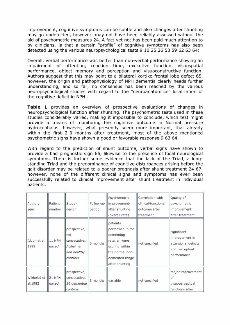
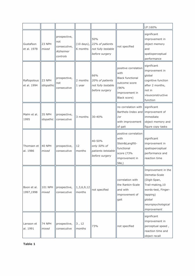
the cognitive deficit in NPH . Table 1 provides an overview of prospective evaluations of changes in
neuropsychological function after shunting. The psychometric tests used in these studies considerably varied, making it impossible to conclude, which test might
provide a means of monitoring the cognitive outcome in Normal pressure hydrocephalus, however, what presently seem more important, that already
within the first 2-3 months after treatment, most of the above mentioned psychometric signs have shown a good or favorable response 9 63 64.
With regard to the prediction of shunt outcome, verbal signs have shown to provide a bad prognostic sign 66, likewise to the presence of focal neurological
symptoms. There is further some evidence that the lack of the Triad, a long-standing Triad and the predominance of cognitive disturbances arising before the gait disorder may be related to a poorer prognosis after shunt treatment 24 67,
however, none of the different clinical signs and symptoms has ever been successfully related to clinical improvement after shunt treatment in individual
patients.
Author,
year
Patient
number
Study-
design
Follow-up
period
Psychometric
improvement-
after shunting
(overall rate)
Correlation with
clinical/functional
outcome after
treatment
Quality of
psychometric
improvement
after treatment
Iddon et al.
1999
11 NPH
mixed
prospective,
not
consecutive,
Alzheimer
and healthy
controls
6 months
patients
performed in the
dementing
rate, all were
scoring within
the normal non-
demented range
after shunting
not specified
significant
improvement in
attentional deficits
and perceptual
performance
Wikkelsö et
al.1982
21 NPH
mixed
prospective,
consecutive,
16 demented
controls
3 months variable not specified
major improvement
of
visuoperceptual
functions after
LP:160%
Gustafson
et al. 1978
23 NPH
mixed
prospective,
not
consecutive,
Alzheimer
controls
(10 days),
6 months
50%
22% of patients
not fully testable
before surgery
not specified
significant
improvement in
object memory
and
spatioperceptual
performance
Raftopolous
et al. 1994
23 NPH
idiopathic
prospective,
not
consecutive
2 months
1 year
66%
20% of patients
not fully testable
before surgery
positive correlation
with
Black functional
outcome score
(96%
improvement in
Black score)
significant
improvement in
global
cognitive function
after 2 months,
not in
visuoconstructive
function
Malm et al.
1995
35 NPH
idopathic
prospective,
consecutive 3 months 30-40%
no correlation with
Barthels-Index and
/or
with improvement
of gait
significant
improvement of
immediate
object memory and
figure copy tasks
Thomsen et
al. 1986
40 NPH
mixed
prospective,
consecutive
12
months
40-50%
only 50% of
patients tetstable
before surgery
positive correlation
with
Stein&Langfitt-
functional
score (73%
improvement in
S&L)
significant
improvement in
spatioperceptual
performance and
reaction time
Boon et al.
1997,1998
101 NPH
mixed
prospective,
consecutive
1,3,6,9,12
months not specified
correlation with
the Rankin-Scale
and with
improvement of
gait
Improvement in the
Demetia-Scale
(Digit-Span,
Trail-making,10
words-test, Finger-
tapping)
global
neuropsychological
improvement
Larsson et
al. 1991
74 NPH
mixed
prospective,
consecutive
3 , 12
months 73% not specified
significant
improvement in
perceptual speed ,
reaction time and
object recall
Table 1
Neuroimaging
Neuroimaging (CCT and MRI) should identify ventriculomegaly and rule out other
pathology. The Evan's Index, which is determined by the largest diameter of the frontal horns divided by the diameter of the internal skull at the level of the frontal horns, if above 0.3 indicates ventriculomegaly, may possibly be the only
finding obligatory for establishing the diagnosis of NPH. All the other imaging findings, i.e. large temporal horns, dilated third ventricle, enlarged perisylvian
fissures or both focal dilation and obliteration of cortical sulci, cortical and subcortial atrophy and the "so-called" periventricular edema have never been consistently reported in larger patients series 3 68 69 70 71 72, apart from the
fact that none of these studies have evidenced a positive relation of any of the imaging criteria with clinical improvement. Some of the findings have been
suggested to relate to NPH pathophysiology by comparison made in the pre- post shunting CT´s, e.g. focal sulcal dilation, which has been “misinterpreted” as cortical atrophy in the previous times and therefore even been used as a criterion
to exclude patients from undergoing a shunting procedure 71 73. In a study of five cases of patients who presented with focal dilation of cortical fissures and
sulci, in three of the cases, there was a paradoxical decrease in the size of the dilated fissures and sulci that paralleled the decrease in the size of the lateral
ventricles following successful shunting. The authors concluded that focal fissural and sulcal dilation may represent “atypical” reservoirs of cerebrospinal fluid analogous to the ventricular system 71.
Nevertheless, CT with regard to ventricular width and the “so called”
periventricular edema has shown a variable change after shunting 74 and was not supposed to play any role for the clinical outcome, particularly in idiopathic NPH 69 72 75. Reduced compliance seemed to be the best predictor of rapid and
marked reduction in ventricular size 76, while others suggested that reduction of ventricular size following CSF shunting is not exclusively related to CSF dynamics
and more likely depending upon the intrinsic elastic properties of the cerebral parenchyma, which vary with age and/or co-existing pathologies in the brain 74 77 78. Others argue that unless not assessed volumetrically, changes of the
ventricles may go undetected questioning any statement about the significance of ventricular changes after shunting observed in NPH patients 73 79 80.
With the advantage of a more detailed anatomy provided by MRI analysis of the brain, some more findings could have been associated with NPH 81, i.e.
increased aqueductal flow void and/or stroke volume indicating a disturbed and “hyperdynamic” CSF flow 82 83 84 85, increased callosal angle, preserved
hippocampal fissures and hippocampal volume different from Alzheimer’s disease 86 87 and, last but not least periventricular and deep white-matter lesions, the so-called PWMLs and DWMLs 88 89 90.
Very few results are promising with regard to both the diagnostic evaluation of
NPH and the prediction of shunt response, e.g. MR-based quantitative phase-contrast cerebrospinal fluid velocity imaging of the aqueductal flow volume, which has indicated a significant relationship between CSF stroke volume greater
than 42 microL and a favorable response to VP shunting 91 . Furthermore, in a large study of aqueductal flow in 236 individuals including normal elderly,
patients with different types of dementia and patients with the clinical diagnosis
of idiopathic NPH, CSF flow of greater than 18 ml/min was suggestive of
idiopathic NPH 92.
Finally, the periventricular (PWMLs) and deep white matter lesions (DWMLs) observed in T2-weighted MRI have been clearly associated to cerebrovascular co-morbidity with systemic hypertension as a main contributing factor in patients
with idiopathic NPH 89 93. In a study of Tullberg et al. comparing idiopathic NPH and Binswanger´s disease 93 both groups shared the major part of MRI changes
with regard to WMLs, which the authors suggested indicates a common pathophysiological pattern. Also others claim that cerebrovascular risk factors and cerebrovascular disease play a role in the pathogenesis of idiopathic NPH
explained by an increased pulse pressure in periventricular and subcortical arteriosclerotic vessels and the subsequent changes in the elastic properties of
the periventricular tissue, which may cause enlargement of ventricles in the absence of raised intracranial pressure 35 94 95 96. Contributing to this theory is the high prevalence of systemic hypertension in idiopathic NPH patients of 83%
34 97 . A negative correlation of DWMLs and PWMLs with the outcome in patients was found 88, though, an exclusion of an individual patient based on the finding
of the white-matter lesions is not justified as both PWMLs and DWMLs before shunting did not differ between outcome groups in a study by Tullberg et al. 90.
In this study and other studies 81 87, no single MR imaging variable could predict the clinical effect of shunt surgery.
Most crucial, however, is the accurate selection of those patients which may benefit from shunting based on both the clinical and imaging findings. In a
retrospective analysis of supportive and non-supportive imaging and clinical findings Vanneste et al. 98 were the first, which introduced a definition of NPH classified using the terms "probable", "possible" and "unlikely" based on an
ordinal global scale derived from combined clinical and CT data, which predicted the clinical outcome in 112 patients shunted for presumed normal pressure
hydrocephalus (NPH). The best strategy was to shunt only patients with “probable shunt-responsive NPH” providing “typical CT and clinical findings“ with a positive predictive value was 0.65. Surprisingly, these data were supported by
a more recent analysis made by Boon et al. 51, suggesting that best predictive rates based on a combination of “typical” clinical and imaging findings may not
exceed 60%. Practically, however, this has resulted in different strategies of decision making
for shunt treatment: A more strict investigator only considering shunting in patients presenting with the complete triad, marked ventriculomegaly and other
supportive clinical and imaging findings, and a more liberal investigator, which shunts even in the presence of an incomplete triad, co-morbid conditions and/or unexpected clinical and radiological findings trying to avoid any delay of
treatment. The first investigator may probably miss an occasional treatable patient and the second may perform many useless and risky surgical
interventions. Neither of the strategies will help improving the quality of our treatment of NPH, however, may rather increase the risk of an upcoming "therapeutic nihilism".
Diagnostic Tests
The so-called "diagnostic-tests" may be more helped with the term "prognostic
tests" as they have been set up to improve the prediction of the result of shunting. Thus, they were supposed to support the selection of suitable shunt candidates, particularly in those patients diagnosed with a “questionable” or
“possible” NPH providing the majority of our cases, as supported by the observations made in an outpatient multidisciplinary memory clinic 99.
It is discussed, however, whether an increased Rout and/or the B-waves may be linked to NPH pathophysiology and therefore required for establishing the
diagnosis8 100. Early investigations have shown an association of an elevated Rout and of a reduced compliance with cerebrospinal fluid malabsorption4 101
102. Furthermore, these parameters have been associated with Normal pressure hydrocephalus 3 4 103, and were then identified as an important diagnostic tool 5 104 105. As a fact, however, findings in Rout have neither correlated to the
size and visibility of cortical sulci nor to the amount of atrophy or the ventricular width seen in the patients´ CT 72 . In addition, NPH biopsy findings 36 39
questioned the value of Rout for diagnosing NPH as, for example, meningeal fibrosis was found in 12 of 25 biopsies containing arachnoid tissue, but no
correlation with an increased R(out) or B-waves was found 106, although, arachnoid fibrosis was previously supposed to be the morphological correlate causing CSF malabsorption. It is questioned, however, whether a single brain
biopsy may explain all changes in the hydrocephalic brain related to impairment of the CSF absorption.
"Diagnostic tests" may be divided into “traditional” and “non-traditional” tests. The traditional tests all reflect the traditional idea about NPH pathophysiology
being a primary disorder of cerebrospinal fluid absorption 4 103 and, as a consequence, require invasive testing in order to identify a defective CSF-flow
and a CSF circulation disorder, or they mimic the shunt: Cisternography 107, long-term ICP measurement 8 100 108 109 110 111, measurement of resistance to CSF outflow (Rout) or compliance11 102 105 112, TAP-test 10 11 62 and
controlled continuous lumbar drainage 12 13 110 113. In many services the invasive tests are considered a standard investigation, however, only a few tests
have been studied prospectively in a manner to allow a reliable determination of their accuracy in predicting the result of shunting. A general observation in most of the tests is a relatively high positive predictive value, while the negative
predictive value is disappointingly low.
The cisternography, although initially a mainstay of the diagnostic protocol for NPH, has not improved the diagnostic accuracy of combined clinical and computed tomographic criteria in patients with suspected normal-pressure
hydrocephalus107. Suggestive for NPH was a stagnation of isotopic activity in the cerebral ventricles for more than 72 hours without any appearance at the
convexity, however, similar features were found both in age-matched controls and other non-hydrocephalic conditions. In the absence of experience in using other supplemental tests, clinicians still commonly use cisternography worldwide.
It has been proposed that the increased frequency in B waves (rhythmic
oscillations of the CSF-pressure with a wavelength of 0.5 to 2/min)114, measured either by the lumbar or ventricular route with and without combination
of transcranial ultrasound 108 114 115, is indicative of lowered compliance and
may therefore play an important role in the pathophysiology of the ventriculomegaly and neuronal dysfunction 8. If frequent during the overnight
measurement, i.e. more 50%, they are thought to be a good predictor of a positive shunt response 64 109. Others found that B-waves poorly predicted outcome 110 116. Still, there is no normative data and no definite understanding
of the origin of those waves in NPH. Vascular factors such as vasodilatation and consequent changes in arterial blood pressure were suggested to cause the
observed rhythmic changes of ICP 117, however, others have shown that B-wave activity has preceded occurrence of blood pressure changes in patients with Normal pressure hydrocephalus 114. A more recent and quantitative study of the
relationship of the vasogenic components of the ICP wave with resistance to CSF outflow has demonstrated that the magnitude of vasogenic waves of ICP both
before and their increase during an infusion test positively correlated with Rout, which may finally indicate a vasogenic origin of the CSF circulatory failure in Normal pressure hydrocephalus 118. Even more important is the fact that the
occurrence of B-wave activity could be clearly associated with REM-sleep stages100. This was supposed to confound the results of the overnight
measurements and clearly doubts on the suggestion that a relative frequency of less than 80% B-wave activity can be a valid indicator for shunt responsiveness.
The author’s suggest that ICP recordings in suspected NPH should be accompanied by polysomnography to avoid misleading results due to variability of B-wave appearance dependent on the sleep pattern 100.
The Tap-test is in worldwide use, is most easy to perform and cheap. It has been
indicated, that the higher the volume drained (40-50 cc), the more likely the patient is going to improve, i.e. sensitivity and specificity increases as more fluid is tapped 9 10. However, reports vary with regard to its usefulness in selecting
best shunt candidates. If definitely positive, it has shown a helpful indicator of shunt improvement and a positive predictive value of more than 70% has been
reported 11, however if patients would have been selected based only on a positive Tap-test, a considerable part of shunt-responsive NPH patients would have been missed. In other words, NPH candidates should not be excluded based
on a negative spinal tap test. In combination with SPECT measurements of cerebral blood flow before and after the tap-test, studies promise higher
predictive rates 119 120: Increase of more than 80% in CBF after CSF removal was predictive of response to shunt surgery with an accuracy of 77% 120.
Controlled continuous lumbar drainage (CCLD) was initially described by Haan and Thomeer 12 and consisted of draining 10 cc of CSF per hour for a period of
72 hours. In Haan´s study 12, values for sensitivity, specificity and PPV and NPV resulted in 100% as all patients with a positive test improved after shunting and vice versa. However, the potential risks and disadvantages (hospitalization and
costs) are still not calculated and have to be further assessed. Reported complication rates with ELD are generally considered low 13 113 121, however,
from the existing studies so far a 5 - 20% rate of risks can be estimated, i.e. severe nerve root irritations and infections have been described requiring a removal of the external drainage. Still, studies are few and do not provide us
with sufficient patient numbers.
Regardless of the methodology used to estimate Ro and/or intracranial compliance 103 111 122 123 124 125 126, it is acknowledged that Ro naturally
increases with age in otherwise healthy individuals 78. This may be a potential
confounding variable that has not been adequately addressed in many of the previous clinical studies. In a prospective study of the Dutch multicenter group
including both secondary and idiopathic NPH patients, retrospectively, the authors recommended a Rout value of 18 mmHg/ml/min as the threshold based on an optimal positive predictive value for clinical improvement of 92% and
highest likelihood ratio 51 112 127. Such high values of Rout, however, will be rarely observed in most of the patients, moreover, patients show a wide range
and a more variable resistance profile, particularly observed in idiopathic NPH 111. This holds also true for the remaining physiological values and CSF dynamic parameter, such as compliance, elastance and intracranial pressure profiles,
which is the evidence of a heterogeneous patient population suggesting that possibly no constant morphological element in the NPH syndrome does exist 39
36 106 128 129. It becomes clear to us that the results, methods, and thresholds of the
“diagnostic tests” are center specific and are subject to wide variation. In addition to the differences in patient selection and inclusion these are two of
many possible reasons why results remain disappointing with regard to the ability and reliability to predict outcome after shunting. Particularly in the
“possible” and/or “questionable” NPH patients, the validity of these tests is not known. As a consequence, a "diagnostic puzzle" results from the necessity to include more than on test in a stepwise manner in order to increase the
prognostic accuracy 67. Regardless the reason of advocating such combinations of test procedures in the individual patient, the practicability of performing all the
different tests is questioned by many clinicians. PET Cerebral Blood Flow and Brain Metabolic Studies
The non-traditional methods, such as cerebral blood flow measurements and
metabolic imaging have not been widely available in the earlier decades. Cerebral blood flow measurement has been performed using XENON-CT, HMPAO-Spect, PET and Doppler investigations 130 131 132 133 134 135 136 137 138
139 140 and have recently been systematically reviewed by Owler and Pickard 14 . As a common finding, regional CBF measures indicated reduced blood flow in
frontal and temporal regions130 133 141, however, global blood flow turned out to be both decreased or normal in patients14. Disappointingly, there was a variable correlation of the preoperative CBF profile and CBF changes after
surgery with the clinical outcome, providing another reason why no definite diagnostic value of blood flow measurements has yet been demonstrated 14.
Using 15-0-H20-PET before and after application of 1g Diamox®, global cerebral blood flow and cerebrovascular reserve capacity have been quantified before,
one week and seven month after surgery in 60 patients with idiopathic NPH 138. In the earlier series 136, lower global blood flow values were indicative of clinical
improvement after shunt treatment. Although a cut-off level has not been stated 136, lower values before surgery were observed in clinical responders compared to non responders (36±8 vs. 41±11 ml/100 ml/min; p= 0.04) and to the
controls (48± 6 ml/100ml/min; p<0.01). Thus, retrospectively, the prognostic accuracy obtained by additional blood flow measurement was increased to 88%
compared to an accuracy of 76% obtained only by the combination of clinical, imaging findings and Rout measurement 138. Furthermore, cerebrovascular
reserve capacity has correlated with clinical changes after shunt placement 137
142: At one week after shunting, shunt responders have shown considerable improvement in reserve capacity and non-responders did not, suggesting that
neurological improvement may be related to early restoration of the hemodynamic reserve. Improvement of cerebrovascular reserve capacity was particularly important in those patients with a high cerebrovascular co morbidity
and with a high prevalence of cerebrovascular risk factors142 underlining the significance of the pathophysiology of chronic infarction in idiopathic NPH.
Moreover, it evidences that shunting may reverse a chronic hypoxic environment existing in the hydrocephalic brain, as also indicated by the findings of others 89 97 143.
Contributing to the role of cerebrovascular disease in idiopathic NPH
pathophysiology, a very recent PET study144 using an anatomic region-of-interest analysis on co-registered magnetic resonance done in both secondary and idiopathic NPH demonstrated highest reductions of CBF in the basal ganglia
and the thalamus but not in white matter regions, contrary to studies in the past 134 141 145. In addition, CBF in the putamen of idiopathic NPH patients was
correlated with a poorer level of functioning. As the lenticulostriate vessels are the main arterial supply to the regions of the thalamus, caudate and putamen,
and as these are the vessels most likely at risk to be affected by arteriosclerotic changes, the author’s concluded that the reduction of CBF seen in patients with idiopathic NPH compared to controls may support the role for cerebrovascular
disease. Also, the results suggest that the role of the basal ganglia and thalamus may be more prominent than currently appreciated, thereby, they contribute to
the above mentioned clinical observations of a subcortical extrapyramidal motor disturbances in NPH 43 53 .
Using statistical parametric mapping (SPM 99, Wellcome Department of Cognitive Neurology, London, UK) in the own series of idiopathic NPH significant regional
changes at a voxel level were investigated and after transformation into a standard stereotaxic space, a decreased flow in prefrontal, frontomesial and temporomesial cortical areas was found, which positively correlated with the
functional impairment using a score 22 that assigns the severity of symptoms based on a formal assessment of gait and mental function. Also, after shunting
clinical responders have shown an increase in regional blood flow in some of these areas in contrast to the non-responders suggesting an importance of local metabolic disturbances in the NPH brain (Berding G, Klinge P, Brooks DJ, et al.
Cerebral blood flow before and after ventriculoperitoneal shunting in normal pressure hydrocephalus; submitted for publication).
Preliminary 1H- and 31P- MRI Spectroscopy investigations in NPH patients by Braun et al.146 have also evidenced patterns of disturbances in brain and
neuronal metabolism by demonstrating both reversible and irreversible neuronal damage: lactate peaks in periventricular areas and reduced NAA/tCr ratios in the
cortex different from controls and other forms of dementia have indicated a reduced, however, a reversible deterioration of neuronal metabolism in NPH patients 20. Likewise, very recent studies in micro dialysis by Agren-Wilson et al.
147 have found a situation of postischaemic recovery in patients with NPH. They investigated PtiO(2), glucose, lactate, pyruvate, and glutamate in the
periventricular white matter and in the frontal brain before and after drainage
and found a significant rise in lactate and pyruvate trend towards a lowering of
glucose and glutamate after removal of CSF.
In the light of these observations, the findings in CSF contents of neuronal metabolites and peptides, neurotransmitters and of products reflecting neuronal degeneration may underline the potential advantage of investigating neuronal
and brain metabolism being a more effective diagnostic marker of NPH than CSF dynamic testing 15 16 17 18 19 148. Analyzing metabolic markers of neuronal
and axonal degeneration in the CSF of patients with idiopathic NPH 15 148, i.e. sulfatide, neurofilament, neuron specific enolase and Tau-proteins, the authors found a negative correlation of clinical improvement after shunt treatment with
the concentration of some of the markers: In patients with sulfatide levels higher than 400 nmol/l clinical improvement was unlikely. Moreover, the CSF sulfatide
concentrations distinguished between patients with vascular dementia and those with idiopathic NPH with a sensitivity of 0.47 and a specificity of 0.94.
Thus, assess to the CSF metabolic products and to modern non-invasive functional neuroimaging techniques, as they may detail structural and functional
profile of neuronal impairment, may give rise to both, a more comprehensive diagnostic approach to the “brain disease” in NPH and to an understanding of the
origin of symptoms in the hydrocephalic brain (“locus of dysfunction”). Functional and brain metabolic imaging using both positron emission tomography
and single photon emission computed tomography has already been widely applied to the study of other dementing diseases and has contributed to both
differential diagnosis and the quantification of treatment effects on disturbed metabolism 149 150 151 152 153 154 155. As the diseases progressed, metabolism was affected in certain corticlal and subcortical areas, which showed
a different profile among the various dementia types, e.g. frontopolar and temporal in M.Pick, biparietal in Parkinson's dementia, temporoparietal in
Alzheimer's disease or bitemporal in Creutzfeld´s disease, however, may encourage subsequent studies in NPH – Figure 1. Using positron emission tomography and [18F]Fluorodeoxyglucose (FDG) in three patients with normal
pressure hydrocephalus, 17 patients with Alzheimer-type dementia, and seven healthy elderly controls, patterns of metabolic abnormality were distinctly
different in the two dementia groups, with Alzheimer-type dementia subjects demonstrating bilateral temporoparietal hypometabolism while normal pressure hydrocephalus subjects showed globally diminished glucose use 156.
In experimental hydrocephalus, however, findings have already evidenced a
more complex impairment of neuronal metabolism and neuronal transmitter function indicating a role for chronic ischemia 157 158 159 160 161 162, e.g. in a recent study of kaolin-hydrocephalus author's have described a prolonged
moderate ischemia assessed by autoradiographic studies that might have caused a delayed neuronal injury in vulnerable brain regions, e.g. the hippocampus 163.
Others have indicated that in the chronic hydrocephalic state cerebrovascular adaptive processes, e.g. angiogenensis and neuronal plasticity, might play a more important role for the disease process than ventricular enlargement and
cerebrospinal fluid dynamics 164. It is clear, that the physics about the relationship of intracranial pressure, impaired CSF-circulation, blood flow and
neuronal dysfunction in the normal and hydrocephalic brain need further investigations 1 79 165.
Apart from the significance of cerebrovascular processes for neuronal dysfunction in the hydrocephalic brain, lately, an age-related impairment of the CSF-
turnover has been suggested to decrease the clearance or wash-out of potentially toxic products of brain metabolism, e.g. the beta amyloid, from brain interstitial fluid. Thus, it may predispose amyloid accumulation, plaque
aggregation and the amyloid-associated neurotoxicities 166. This was suggested to be critical to the onset and progression of both Alzheimer´s disease and of
dementia in Normal pressure hydrocephalus as an impaired CSF- production rate and CSF-turnover has been found among both Alzheimer’s and NPH patients 167. This might be but one of the most exciting explanations for the recent
observations of a frequent coincidence of Alzheimer's-type neuropathology among patients with idiopathic NPH 36 37 39 168.
NPH Management and Outcome Assessment
Some advocate that a positive response to shunt treatment might be the diagnostic gold standard and favor the term “shunt-proven” NPH. This, however,
would lead to a number of incorrect classifications as, e.g. false negatives would occur when patients have an absent or diminished response to shunt placement
due to co-existing conditions, unpredictable effects of the underlying disease in secondary NPH, post-operative complications, inadequate treatment or mechanical failures of the shunt. Furthermore, the clinical improvement to shunt
treatment considerably varies: Across studies, overall improvement rates to shunt treatment were ranging between bounds of 30% to 96 % 22 24 33 64 169
170 171 172, however, are mainly reported as a matter of treatment associated shunt complications, the selection of the valve-type and shunting procedures, or of the preoperative selection of shunt-candidates 2.
An issue, which has not been very well addressed in the past, is the fact, that the
postoperative management still lacks protocols for patients follow-up and the assessment of clinical outcome and clinical improvement. As long as there is no clear definition for, how long and how often a patient must be observed and,
which criteria are applied in order to assess the shunt success, the definite value of shunting NPH may never be clarified. In the literature, clinical outcome is
reported within different time periods of up to 60 months or even within a range of follow-up 10 173 174, however, outcome figures given are based on these variable observation times, while long-term and short-term outcome are mixed-
up. It is known from studies of long-term outcome in idiopathic NPH, for example, that particularly in that elderly age group it is the co-morbid factors,
e.g. ischemic brain or heart diseases, that do affect the long-term outcome, and clinical deterioration occurs unrelated to the shunt 33. Malm et al 175 have shown a decreased overall functional outcome from 64% to 26% when
comparing outcomes at three months with those at three years. In that study, survival curves were compared with those of age-matched healthy elderly
subjects and patients with first-ever ischemic stroke and the relative risk of death among idiopathic NPH patients compared to a general elderly population was 3.3. Savolainen et al. 174 reported a decrease of clinical improvement over
a period of five years when compared with one year. In a study by Larsson et al. 67 deterioration already occurred at 12 months.
Similarly, the assessment of clinical improvement lacks a unique figure, and
criteria for improvement are generally based on a subjective grading made according to the investigators clinical judgment. By some authors, scales were
introduced 22 24 88 either by rating the degree of cognitive impairment, gait and urinary disturbances before shunting or by grading the magnitude of improvement in each symptom after surgery. Others document clinical
improvement and outcome of shunt treatment using functional grades, however, none of this scales has gained a wide acceptance and have shown of advantage
for the clinical assessment. Therefore, some authors have used scales from the rehabilitation field, e.g. the Rankin-Scale, the Barthel-Index and the Katz-Index, or other neurological scales, e.g. the MMSE, or the GCOS, however, these have
not been validated for the assessment of NPH.
Neuropsychological methods have been used in order to assess the clinical improvement more reliably and to measure improvement of function after shunt treatment 10 64 67 172. It has been shown by the various investigators, that
there is considerable improvement in neuropsychological function, which has correlated with an improvement in the activities of daily living and function 9 59
64 172 . Raftopoulos et al. 64 , for example, have shown a 66% improvement of psychometric function, which contributed to an improvement in the Black
functional outcome scale 24 of 96% - Table 1. Thereby, neuropsychological measures may provide early and reliable measures of functional outcome in NPH in addition to gait. In follow-up examination, these tests may offer a practical
and standardized tool to monitor patient status, to reset adjustable valves 176 and to compare results from different centers.
However, the majority of test procedures require expertise and are usually time consuming, particularly in the more disabled patients 58 59 64. This may be but
one of the main reasons why neuropsychological measures have not found integration in the neurosurgical and clinical routine. Moreover, it has been
reported that nearly 50% of the patients have been not testable with the applied battery, which makes a proper assessment again difficult 59 – Table 1. Clinicians therefore tend to rely on the more simple measurement of gait function only.
However, assessment of gait function alone might not contribute sufficiently to monitoring of the patients' functional outcome after shunt placement.
More simple tests are needed 177, i.e. a set of tests or a test-battery, that are assessable in a larger cohort of patients, that are simple and that can be easily
applied as bedside tests to define the degree of dementia pre- and postoperatively. Efforts should be made to assess the reliability of these tests for
assessing functional improvement to shunt treatment in NPH, e.g. in a preliminary study of psychometric functions before and early after shunt treatment, early changes in a simple “pencil-paper” test measuring visual
attention were most sensitive indicating a positive response to shunt treatment 178. The implementation of standard tools for measuring the NPH deficits will be
critical if there is to be an "evidenced-based" approach to CSF shunting NPH. Finally, the management of Non-responders has virtually not been addressed in
the literature. Still, it is not clear, whether there is ineffectiveness of a shunt (blockage or under drainage), selection of a bad candidate or irreversible
damage, which has occurred in a patient. Presently, these questions cannot be answered properly as long as follow-up studies have not been systematically
performed (Petra Klinge, M.D., Anthony Marmarou, Ph.D., Marvin Bergsneider,
M.D., Norman Relkin, M.D., Ph.D., Peter Black, M.D., Ph.D. Outcome of shunting in Idiopathic Normal Pressure Hydrocephalus (iNPH) and the value of Outcome
assessment in shunted patients; submitted for publication). In a recent review by Hebb and Cusimano179, which have first systematically analyzed the world literature, it was stated that there is clearly a lack of well-
designed prospective clinical studies that analyze sufficient patient numbers in a standardized fashion in many fields of NPH diagnosis, management and
treatment. Recent Efforts and Future Perspectives
In September 2000, a group of scientists chaired by Anthony Marmarou, MCV
Neurosciences Center, Richmond Virginia, and co-chaired by Peter Black, Brigham& Women's Hospital, Boston was assembled to determine the feasibility of developing standardized guidelines for current clinical practice based on the
available scientific evidence. The so-called “NPH-advisory board” presented its first results during an NPH consensus conference in San Diego, CA, February
2001, and discussed with a small group of NPH consultants from the U.S. and Europe. The consensus and the synthesis of literature evidence were in the
following period presented at several international meetings, e.g. during the international Hydrocephalus Workshop in KOS, May 2001, and to a group of European NPH experts and scientists in Hannover in November 2001. The
available literature was traced back to 1965 and studies were classified into classes of evidence based on the methods used for the AANS Head Injury
Guidelines (J Neurotrauma, 17 Number 6/7 June/July 2000): Class I, prospective randomized controlled trials, the "gold standard" of clinical trials, Class II, prospective studies with retrospective data analysis, and Class III, purely
retrospective studies. Based on this classification, recommendations for diagnosis and management of NPH were made subdivided into Standards, Guidelines and
Options. The initial guidelines refer to diagnosis and management of the idiopathic NPH (iNPH) and therefore, the search was limited to papers that describe the results selectively for iNPH. The guidelines were finalized and
submitted for publication recently. As a key result of the guidelines, no Class I study in iNPH existed, subsequently, absolutely no accepted standard for
diagnosis, treatment and management may exist. There was clearly a lack of both well-designed prospective and retrospective clinical studies in idiopathic NPH. A problem making it impossible to extract certain data for idiopathic NPH
was based on the fact that studies were mixed with regard to idiopathic and secondary NPH, and were mostly, if not in almost all of the available studies, not
segregating out the figures and the results for the both groups. One of the projects, which has evolved from the guideline initiative, is the
upcoming prospective European multicenter study on the prediction of outcome in patients idiopathic NPH. This initiative is currently shared by different centers
from 10 to 11 European countries. In May 2002, during the Hydrocephalus workshop held in Lund, Sweden, a group of European NPH experts was assembled for a first meeting on the feasibility of a European NPH multicenter
study. During the first meeting, there was agreement that the study should be set out to investigate the predictive value of different diagnostic tests and a
formal agreement was reached, that all patients included in the study should receive a shunt irrespective of the test results and should be included into the
study solely based on clinical and radiological criteria. An executive committee
was assembled to proceed with formulating and proposing a study protocol. At least 200 patients should be evaluated during a 2 years period on a prospective
basis, and all included patients are planned to be operated by a VP shunt using the same valve system. Follow-up will be done using primary and secondary outcome measures applied at 1,3 and 12 months after surgery in a standardized
manner, and outcome results will be correlated with the preoperative test variables in order to calculate their predictive rates. The obligatory diagnostic
tests consist of the CSF tap-test, resistance to outflow (Rout) and different CSF metabolic markers, which are assessed in a combined infusion and drainage test. Optional test, i.e. PET or SPECT, MRI investigations, B-wave analysis and
continuous lumbar drainage are supposed to be included upon agreement between different centers. Anonymized data will be electronically registered in a
central web-based database, which will be presented through a world-wide-web application over the internet, protected by a username/password based authentication mechanism.
Epilogue
The present article has not intended to present a detailed and systematic
analysis of literature, therefore, not every aspect and contribution regarding NPH diagnostic evaluation and NPH management may have been covered.
As the author is a member of the “NPH advisory board”, the results are in part reflecting both the knowledge and the experiences acquired during the process of
“guideline work” made on the basis of a meta-analysis and during the many valuable discussions with the colleagues and with experts during the meetings and consensus conferences. Mainly, aspects of the article relate to the authors
own work and findings in the diagnosis and in the pathophysiology of chronic hydrocephalus having performed PET-CBF studies in idiopathic NPH and
experimental histological studies in the adult kaolin-hydrocephalus animal model. The evidence based NPH guidelines should encourage many investigators to
continue with seeking an improved medical and scientific evidence in the diagnosis and management of NPH. This, in part, requires the implementation of
well-designed prospective clinical trials as currently initiated with the European multicenter study.
Modern functional neuroimaging techniques, however, seem a realistic approach to measuring neuronal and brain function in the hydrocephalic brain and to a
more comprehensive understanding of NPH pathophysiology. As this has been achieved, however, a “re-classification” or at least a “re-thinking” of Normal pressure hydrocephalus might become necessary. Finally, basic research should
proceed with detailed studies of both degenerative and regenerative processes in the hydrocephalic brain in addition to evaluating the potential value of an adjunct
pharmacological and “neuroprotective” treatment for restoring brain function after a "hydrocephalic trauma".
Legends
Figure 1) Imaging of regional brain metabolism and brain function in dementia.
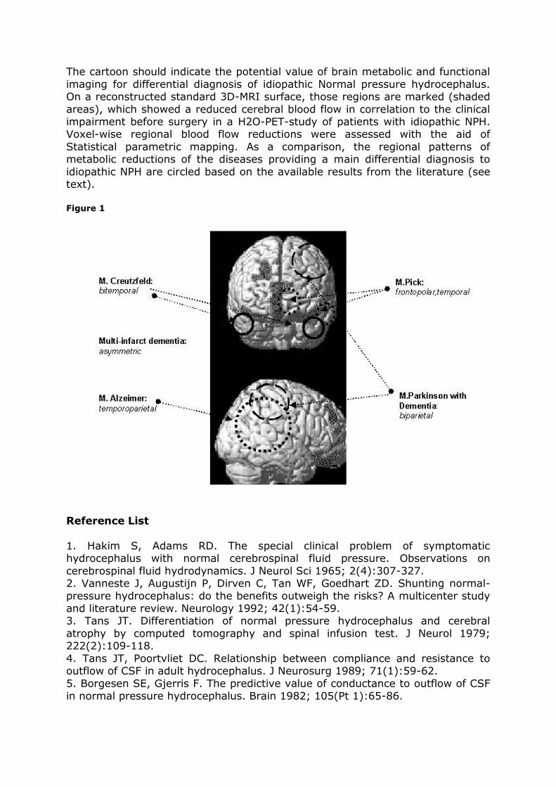
The cartoon should indicate the potential value of brain metabolic and functional
imaging for differential diagnosis of idiopathic Normal pressure hydrocephalus. On a reconstructed standard 3D-MRI surface, those regions are marked (shaded
areas), which showed a reduced cerebral blood flow in correlation to the clinical impairment before surgery in a H2O-PET-study of patients with idiopathic NPH. Voxel-wise regional blood flow reductions were assessed with the aid of
Statistical parametric mapping. As a comparison, the regional patterns of metabolic reductions of the diseases providing a main differential diagnosis to
idiopathic NPH are circled based on the available results from the literature (see text). Figure 1
Reference List
1. Hakim S, Adams RD. The special clinical problem of symptomatic hydrocephalus with normal cerebrospinal fluid pressure. Observations on
cerebrospinal fluid hydrodynamics. J Neurol Sci 1965; 2(4):307-327. 2. Vanneste J, Augustijn P, Dirven C, Tan WF, Goedhart ZD. Shunting normal-
pressure hydrocephalus: do the benefits outweigh the risks? A multicenter study and literature review. Neurology 1992; 42(1):54-59. 3. Tans JT. Differentiation of normal pressure hydrocephalus and cerebral
atrophy by computed tomography and spinal infusion test. J Neurol 1979; 222(2):109-118.
4. Tans JT, Poortvliet DC. Relationship between compliance and resistance to outflow of CSF in adult hydrocephalus. J Neurosurg 1989; 71(1):59-62.
5. Borgesen SE, Gjerris F. The predictive value of conductance to outflow of CSF in normal pressure hydrocephalus. Brain 1982; 105(Pt 1):65-86.
6. Kosteljanetz M. CSF dynamics and pressure-volume relationships in
communicating hydrocephalus. J Neurosurg 1986; 64(1):45-52. 7. Kosteljanetz M, Nehen AM, Kaalund J. Cerebrospinal fluid outflow resistance
measurements in the selection of patients for shunt surgery in the normal pressure hydrocephalus syndrome. A controlled trial. Acta Neurochir (Wien ) 1990; 104(1-2):48-53.
8. Raftopoulos C, Chaskis C, Delecluse F, Cantraine F, Bidaut L, Brotchi J. Morphological quantitative analysis of intracranial pressure waves in normal
pressure hydrocephalus. Neurol Res 1992; 14(5):389-396. 9. Wikkelso C, Andersson H, Blomstrand C, Lindqvist G. The clinical effect of lumbar puncture in normal pressure hydrocephalus. J Neurol Neurosurg
Psychiatry 1982; 45(1):64-69. 10. Malm J, Kristensen B, Karlsson T, Fagerlund M, Elfverson J, Ekstedt J. The
predictive value of cerebrospinal fluid dynamic tests in patients with th idiopathic adult hydrocephalus syndrome. Arch Neurol 1995; 52(8):783-789. 11. Kahlon B, Sundbarg G, Rehncrona S. Comparison between the lumbar
infusion and CSF tap tests to predict outcome after shunt surgery in suspected normal pressure hydrocephalus. J Neurol Neurosurg Psychiatry 2002; 73(6):721-
726. 12. Haan J, Thomeer RT. Predictive value of temporary external lumbar drainage
in normal pressure hydrocephalus. Neurosurgery 1988; 22(2):388-391. 13. Walchenbach R, Geiger E, Thomeer RT, Vanneste JA. The value of temporary external lumbar CSF drainage in predicting the outcome of shunting on normal
pressure hydrocephalus. J Neurol Neurosurg Psychiatry 2002; 72(4):503-506. 14. Owler BK, Pickard JD. Normal pressure hydrocephalus and cerebral blood
flow: a review. Acta Neurol Scand 2001; 104(6):325-342. 15. Tullberg M, Mansson JE, Fredman P, Lekman A, Blennow K, Ekman R et al. CSF sulfatide distinguishes between normal pressure hydrocephalus and
subcortical arteriosclerotic encephalopathy. J Neurol Neurosurg Psychiatry 2000; 69(1):74-81.
16. Malm J, Kristensen B, Ekstedt J, Adolfsson R, Wester P. CSF monoamine metabolites, cholinesterases and lactate in the adult hydrocephalus syndrome (normal pressure hydrocephalus) related to CSF hydrodynamic parameters. J
Neurol Neurosurg Psychiatry 1991; 54(3):252-259. 17. Wikkelso C, Ekman R, Westergren I, Johansson B. Neuropeptides in
cerebrospinal fluid in normal-pressure hydrocephalus and dementia. Eur Neurol 1991; 31(2):88-93. 18. Poca MA, Mataro M, Sahuquillo J, Catalan R, Ibanez J, Galard R. Shunt
related changes in somatostatin, neuropeptide Y, and corticotropin releasing factor concentrations in patients with normal pressure hydrocephalus. J Neurol
Neurosurg Psychiatry 2001; 70(3):298-304. 19. Spanu G, Santagostino G, Marzatico F, Gaetani P, Silvani V, Baena R. Idiopathic hydrocephalic dementia in aging brain the neurosurgical approach.
Funct Neurol 1989; 4(3):293-298. 20. Braun KP, Gooskens RH, Vandertop WP, Tulleken CA, van der GJ. 1H
magnetic resonance spectroscopy in human hydrocephalus. J Magn Reson Imaging 2003; 17(3):291-299. 21. Mulrow CD, Feussner JR, Williams BC, Vokaty KA. The value of clinical
findings in the detection of normal pressure hydrocephalus. J Gerontol 1987; 42(3):277-279.
22. Stein SC, Langfitt TW. Normal-pressure hydrocephalus. Predicting the results of cerebrospinal fluid shunting. J Neurosurg 1974; 41(4):463-470.
23. Black PM, Ojemann RG, Tzouras A. CSF shunts for dementia, incontinence,
and gait disturbance. Clin Neurosurg 1985; 32:632-651. 24. Black PM. Idiopathic normal-pressure hydrocephalus. Results of shunting in
62 patients. J Neurosurg 1980; 52(3):371-377. 25. Boon AJW, Tans JT, Delwel EJ, Egeler-Peerdeman SM, Hanlo PW, Wurzer JAL et al. Dutch Normal Pressure hydrocephalus study:baseline characteristics with
emphasis on clinical findings. European Journal of Neurology 1997; 4:39-47. 26. De Mol J. [Neuropsychological symptomatology in normal pressure
hydrocephalus] Semiologie neuropsychologique dans l'hydrocephalie a pression normale. Schweiz Arch Neurol Psychiatr 1986; 137(4):33-45.
27. Spanu G, Sangiovanni G, Locatelli D. Normal-pressure hydrocephalus: twelve years experience. Neurochirurgia (Stuttg) 1986; 29(1):15-19.
28. Meier U, Zeilinger FS, Kintzel D. [Psychophysiology, signs and symptoms of disease the course of normal pressure hydrocephalus] Pathophysiologie, Klinik und Krankheitsverlauf beim Normaldruckhydrozephalus.
Fortschr Neurol Psychiatr 1998; 66(4):176-191. 29. Black PM. Hydrocephalus and vasospasm after subarachnoid hemorrhage
from ruptured intracranial aneurysms. Neurosurgery 1986; 18(1):12-16. 30. Meier U, Zeilinger FS, Kintzel D. Signs, symptoms and course of normal
pressure hydrocephalus in comparison with cerebral atrophy. Acta Neurochir (Wien ) 1999; 141(10):1039-1048. 31. Paoletti P, Pezzotta S, Spanu G. Diagnosis and treatment of post-traumatic
hydrocephalus. J Neurosurg Sci 1983; 27(3):171-175. 32. Chahlavi A, El Babaa SK, Luciano MG. Adult-onset hydrocephalus. Neurosurg
Clin N Am 2001; 12(4):753-760. 33. Greenberg JO, Shenkin HA, Adam R. Idiopathic normal pressure hydrocephalus-- a report of 73 patients. J Neurol Neurosurg Psychiatry 1977;
40(4):336-341. 34. Boon AJ, Tans JT, Delwel EJ, Egeler-Peerdeman SM, Hanlo PW, Wurzer HA et
al. Dutch Normal-Pressure Hydrocephalus Study: the role of cerebrovascular disease. J Neurosurg 1999; 90(2):221-226. 35. Jellinger K. Neuropathological aspects of dementias resulting from abnormal
blood and cerebrospinal fluid dynamics. Acta Neurol Belg 1976; 76(2):83-102. 36. Savolainen S, Paljarvi L, Vapalahti M. Prevalence of Alzheimer's disease in
patients investigated for presumed normal pressure hydrocephalus: a clinical and neuropathological study. Acta Neurochir (Wien ) 1999; 141(8):849-853. 37. George AE, Holodny A, Golomb J, de Leon MJ. The differential diagnosis of
Alzheimer's disease. Cerebral atrophy versus normal pressure hydrocephalus. Neuroimaging Clin N Am 1995; 5(1):19-31.
38. Golomb J, Wisoff J, Miller DC, Boksay I, Kluger A, Weiner H et al. Alzheimer's disease comorbidity in normal pressure hydrocephalus: prevalence and shunt response. J Neurol Neurosurg Psychiatry 2000; 68(6):778-781.
39. Del Bigio MR, Cardoso ER, Halliday WC. Neuropathological changes in chronic adult hydrocephalus: cortical biopsies and autopsy findings. Can J Neurol Sci
1997; 24(2):121-126. 40. Krauss JK, Regel JP, Droste DW, Orszagh M, Borremans JJ, Vach W. Movement disorders in adult hydrocephalus. Mov Disord 1997; 12(1):53-60.
41. Krauss JK, Faist M, Schubert M, Borremans JJ, Lucking CH, Berger W. Evaluation of gait in normal pressure hydrocephalus before and after shunting.
Adv Neurol 2001; 87:301-310.
42. Stolze H, Kuhtz-Buschbeck JP, Drucke H, Johnk K, Illert M, Deuschl G.
Comparative analysis of the gait disorder of normal pressure hydrocephalus and Parkinson's disease. J Neurol Neurosurg Psychiatry 2001; 70(3):289-297.
43. Matousek M, Wikkelso C, Blomsterwall E, Johnels B, Steg G. Motor performance in normal pressure hydrocephalus assessed with an optoelectronic measurement technique. Acta Neurol Scand 1995; 91(6):500-505.
44. Ojemann RG, Fisher CM, Adams RD, Sweet WH, New PF. Further experience with the syndrome of "normal" pressure hydrocephalus. J Neurosurg 1969;
31(3):279-294. 45. Fisher CM. Hydrocephalus in walking problems of the elderly. Trans Am Neurol Assoc 1980; 105:29-33.
46. Fisher CM. Hydrocephalus as a cause of disturbances of gait in the elderly. Neurology 1982; 32(12):1358-1363.
47. Blomsterwall E, Bilting M, Stephensen H, Wikkelso C. Gait abnormality is not the only motor disturbance in normal pressure hydrocephalus. Scand J Rehabil Med 1995; 27(4):205-209.
48. Blomsterwall E, Svantesson U, Carlsson U, Tullberg M, Wikkelso C. Postural disturbance in patients with normal pressure hydrocephalus. Acta Neurol Scand
2000; 102(5):284-291. 49. Sudarsky L, Simon S. Gait disorder in late-life hydrocephalus. Arch Neurol
1987; 44(3):263-267. 50. Stolze H, Kuhtz-Buschbeck JP, Drucke H, Johnk K, Diercks C, Palmie S et al. Gait analysis in idiopathic normal pressure hydrocephalus--which parameters
respond to the CSF tap test? Clin Neurophysiol 2000; 111(9):1678-1686. 51. Boon AJ, Tans JT, Delwel EJ, Egeler-Peerdeman SM, Hanlo PW, Wurzer HA et
al. The Dutch normal-pressure hydrocephalus study. How to select patients for shunting? An analysis of four diagnostic criteria. Surg Neurol 2000; 53(3):201-207.
52. Zaaroor M, Bleich N, Chistyakov A, Pratt H, Feinsod M. Motor evoked potentials in the preoperative and postoperative assessment of normal pressure
hydrocephalus. J Neurol Neurosurg Psychiatry 1997; 62(5):517-521. 53. Knutsson E, Lying-Tunell U. Gait apraxia in normal-pressure hydrocephalus: patterns of movement and muscle activation. Neurology 1985; 35(2):155-160.
54. Wikkelso C, Blomsterwall E, Frisen L. Subjective visual vertical and Romberg's test correlations in hydrocephalus. J Neurol 2003; 250(6):741-745.
55. Jonas S, Brown J. Neurogenic bladder in normal pressure hydrocephalus. Urology 1975; 5(1):44-50. 56. De Mol J. [Neuropsychological study of mental troubles in normal pressure
hydrocephaly and their short term evolution after spinal fluid derivation (author's transl)]
Etude neuropsychologique des troubles mentaux dans l'hydrocephalie normotensive et de leur evolution a court terme apres derivation du liquide cephalo-rachidien. Acta Psychiatr Belg 1977; 77(2):228-253.
57. De Mol J. [Psychic disturbance in normal pressure hydrocephalus (author's transl)]
Troubles psychiques au cours d'hydrocephalie normotensive. Acta Neurol Belg 1978; 78(6):321-340. 58. Gustafson L, Hagberg B. Recovery in hydrocephalic dementia after shunt
operation. J Neurol Neurosurg Psychiatry 1978; 41(10):940-947. 59. Thomsen AM, Borgesen SE, Bruhn P, Gjerris F. Prognosis of dementia in
normal-pressure hydrocephalus after a shunt operation. Ann Neurol 1986; 20(3):304-310.
60. Lindqvist G, Andersson H, Bilting M, Blomstrand C, Malmgren H, Wikkelso C.
Normal pressure hydrocephalus: psychiatric findings before and after shunt operation classified in a new diagnostic system for organic psychiatry. Acta
Psychiatr Scand Suppl 1993; 373:18-32. 61. Adams RD. Further observations on normal pressure hydrocephalus. Proc R Soc Med 1966; 59(11 Part 1):1135-1140.
62. Wikkelso C, Andersson H, Blomstrand C, Lindqvist G, Svendsen P. Normal pressure hydrocephalus. Predictive value of the cerebrospinal fluid tap-test. Acta
Neurol Scand 1986; 73(6):566-573. 63. Iddon JL, Pickard JD, Cross JJ, Griffiths PD, Czosnyka M, Sahakian BJ. Specific patterns of cognitive impairment in patients with idiopathic normal
pressure hydrocephalus and Alzheimer's disease: a pilot study. J Neurol Neurosurg Psychiatry 1999; 67(6):723-732.
64. Raftopoulos C, Deleval J, Chaskis C, Leonard A, Cantraine F, Desmyttere F et al. Cognitive recovery in idiopathic normal pressure hydrocephalus: a prospective study. Neurosurgery 1994; 35(3):397-404.
65. Caltagirone C, Gainotti G, Masullo C, Villa G. Neurophysiological study of normal pressure hydrocephalus. Acta Psychiatr Scand 1982; 65(2):93-100.
66. De Mol J. [Prognostic factors for therapeutic outcome in normal-pressure hydrocephalus. Review of the literature and personal study]
Facteurs pronostiques du resultat therapeutique dans l'hydrocephalie a pression normale. Revue de la litterature et etude personnelle. Acta Neurol Belg 1985; 85(1):13-29.
67. Larsson A, Wikkelso C, Bilting M, Stephensen H. Clinical parameters in 74 consecutive patients shunt operated for normal pressure hydrocephalus. Acta
Neurol Scand 1991; 84(6):475-482. 68. Wikkelso C, Andersson H, Blomstrand C, Matousek M, Svendsen P. Computed tomography of the brain in the diagnosis of and prognosis in normal pressure
hydrocephalus. Neuroradiology 1989; 31(2):160-165. 69. Petersen RC, Mokri B, Laws ER, Jr. Surgical treatment of idiopathic
hydrocephalus in elderly patients. Neurology 1985; 35(3):307-311. 70. Mori K, Handa H, Murata T, Nakano Y. Periventricular lucency in computed tomography of hydrocephalus and cerebral atrophy. J Comput Assist Tomogr
1980; 4(2):204-209. 71. Holodny AI, George AE, de Leon MJ, Golomb J, Kalnin AJ, Cooper PR. Focal
dilation and paradoxical collapse of cortical fissures and sulci in patients with normal-pressure hydrocephalus. J Neurosurg 1998; 89(5):742-747. 72. Takeuchi T, Kasahara E, Iwasaki M, Mima T, Mori K. Indications for shunting
in patients with idiopathic normal pressure hydrocephalus presenting with dementia and brain atrophy (atypical idiopathic normal pressure hydrocephalus).
Neurol Med Chir (Tokyo) 2000; 40(1):38-46. 73. Kitagaki H, Mori E, Ishii K, Yamaji S, Hirono N, Imamura T. CSF spaces in idiopathic normal pressure hydrocephalus: morphology and volumetry. AJNR Am
J Neuroradiol 1998; 19(7):1277-1284. 74. Tans JT, Poortvliet DC. Reduction of ventricular size after shunting for normal
pressure hydrocephalus related to CSF dynamics before shunting. J Neurol Neurosurg Psychiatry 1988; 51(4):521-525. 75. Meier U, Paris S, Grawe A, Stockheim D, Hajdukova A, Mutze S. Is there a
correlation between operative results and change in ventricular volume after shunt placement? A study of 60 cases of idiopathic normal-pressure
hydrocephalus. Neuroradiology 2003; 45(6):377-380.
76. Tans JT, Poortvliet DC. Does compliance predict ventricular reduction after
shunting for normal pressure hydrocephalus. Neurol Res 1989; 11(3):136-138. 77. Cardoso ER, Del Bigio MR. Age-related changes of cerebral ventricular size.
Part II: Normalization of ventricular size following shunting. Acta Neurochir (Wien ) 1989; 97(3-4):135-138. 78. Czosnyka M, Czosnyka ZH, Whitfield PC, Donovan T, Pickard JD. Age
dependence of cerebrospinal pressure-volume compensation in patients with hydrocephalus. J Neurosurg 2001; 94(3):482-486.
79. Penar PL, Lakin WD, Yu J. Normal pressure hydrocephalus: an analysis of aetiology and response to shunting based on mathematical modeling. Neurol Res 1995; 17(2):83-88.
80. Anderson RC, Grant JJ, de la PR, Frucht S, Goodman RR. Volumetric measurements in the detection of reduced ventricular volume in patients with
normal-pressure hydrocephalus whose clinical condition improved after ventriculoperitoneal shunt placement. J Neurosurg 2002; 97(1):73-79. 81. Jack CR, Jr., Mokri B, Laws ER, Jr., Houser OW, Baker HL, Jr., Petersen RC.
MR findings in normal-pressure hydrocephalus: significance and comparison with other forms of dementia. J Comput Assist Tomogr 1987; 11(6):923-931.
82. Krauss JK, Regel JP, Vach W, Jungling FD, Droste DW, Wakhloo AK. Flow void of cerebrospinal fluid in idiopathic normal pressure hydrocephalus of the elderly:
can it predict outcome after shunting? Neurosurgery 1997; 40(1):67-73. 83. Bradley WG, Jr., Scalzo D, Queralt J, Nitz WN, Atkinson DJ, Wong P. Normal-pressure hydrocephalus: evaluation with cerebrospinal fluid flow measurements
at MR imaging. Radiology 1996; 198(2):523-529. 84. Dixon GR, Friedman JA, Luetmer PH, Quast LM, McClelland RL, Petersen RC
et al. Use of cerebrospinal fluid flow rates measured by phase-contrast MR to predict outcome of ventriculoperitoneal shunting for idiopathic normal-pressure hydrocephalus. Mayo Clin Proc 2002; 77(6):509-514.
85. Poca MA, Sahuquillo J, Busto M, Rovira A, Capellades J, Mataro M et al. Agreement between CSF flow dynamics in MRI and ICP monitoring in the
diagnosis of normal pressure hydrocephalus. Sensitivity and specificity of CSF dynamics to predict outcome. Acta Neurochir Suppl 2002; 81:7-10. 86. Holodny AI, Waxman R, George AE, Rusinek H, Kalnin AJ, de Leon M. MR
differential diagnosis of normal-pressure hydrocephalus and Alzheimer disease: significance of perihippocampal fissures. AJNR Am J Neuroradiol 1998;
19(5):813-819. 87. Savolainen S, Laakso MP, Paljarvi L, Alafuzoff I, Hurskainen H, Partanen K et al. MR imaging of the hippocampus in normal pressure hydrocephalus:
correlations with cortical Alzheimer's disease confirmed by pathologic analysis. AJNR Am J Neuroradiol 2000; 21(2):409-414.
88. Krauss JK, Regel JP, Vach W, Orszagh M, Jungling FD, Bohus M et al. White matter lesions in patients with idiopathic normal pressure hydrocephalus and in an age-matched control group: a comparative study. Neurosurgery 1997;
40(3):491-495. 89. Bradley WG, Jr., Whittemore AR, Watanabe AS, Davis SJ, Teresi LM, Homyak
M. Association of deep white matter infarction with chronic communicating hydrocephalus: implications regarding the possible origin of normal-pressure hydrocephalus. AJNR Am J Neuroradiol 1991; 12(1):31-39.
90. Tullberg M, Jensen C, Ekholm S, Wikkelso C. Normal pressure hydrocephalus: vascular white matter changes on MR images must not exclude
patients from shunt surgery. AJNR Am J Neuroradiol 2001; 22(9):1665-1673.
91. Bradley WG, Jr., Scalzo D, Queralt J, Nitz WN, Atkinson DJ, Wong P. Normal-
pressure hydrocephalus: evaluation with cerebrospinal fluid flow measurements at MR imaging. Radiology 1996; 198(2):523-529.
92. Luetmer PH, Huston J, Friedman JA, Dixon GR, Petersen RC, Jack CR et al. Measurement of cerebrospinal fluid flow at the cerebral aqueduct by use of phase-contrast magnetic resonance imaging: technique validation and utility in
diagnosing idiopathic normal pressure hydrocephalus. Neurosurgery 2002; 50(3):534-543.
93. Tullberg M, Hultin L, Ekholm S, Mansson JE, Fredman P, Wikkelso C. White matter changes in normal pressure hydrocephalus and Binswanger disease: specificity, predictive value and correlations to axonal degeneration and
demyelination. Acta Neurol Scand 2002; 105(6):417-426. 94. Bradley WG. Normal pressure hydrocephalus and deep white matter
ischemia: which is the chicken, and which is the egg? AJNR Am J Neuroradiol 2001; 22(9):1638-1640. 95. Casmiro M, Cacciatore FM, D'Alessandro R. The pathogenesis of idiopathic
normal pressure hydrocephalus: an open problem. Funct Neurol 1989; 4(4):403-410.
96. Casmiro M, D'Alessandro R, Cacciatore FM, Daidone R, Calbucci F, Lugaresi E. Risk factors for the syndrome of ventricular enlargement with gait apraxia
(idiopathic normal pressure hydrocephalus): a case-control study. J Neurol Neurosurg Psychiatry 1989; 52(7):847-852. 97. Krauss JK, Regel JP, Vach W, Droste DW, Borremans JJ, Mergner T. Vascular
risk factors and arteriosclerotic disease in idiopathic normal-pressure hydrocephalus of the elderly. Stroke 1996; 27(1):24-29.
98. Vanneste J, Augustijn P, Tan WF, Dirven C. Shunting normal pressure hydrocephalus: the predictive value of combined clinical and CT data. J Neurol Neurosurg Psychiatry 1993; 56(3):251-256.
99. Bech-Azeddine R, Waldemar G, Knudsen GM, Hogh P, Bruhn P, Wildschiodtz G et al. Idiopathic normal-pressure hydrocephalus: evaluation and findings in a
multidisciplinary memory clinic. Eur J Neurol 2001; 8(6):601-611. 100. Krauss JK, Droste DW, Bohus M, Regel JP, Scheremet R, Riemann D et al. The relation of intracranial pressure B-waves to different sleep stages in patients
with suspected normal pressure hydrocephalus. Acta Neurochir (Wien ) 1995; 136(3-4):195-203.
101. Borgesen SE, Gjerris F, Srensen SC. The resistance to cerebrospinal fluid absorption in humans. A method of evaluation by lumbo-ventricular perfusion, with particular reference to normal pressure hydrocephalus. Acta Neurol Scand
1978; 57(1):88-96. 102. Borgesen SE, Gjerris F, Sorensen SC. Intracranial pressure and conductance
to outflow of cerebrospinal fluid in normal-pressure hydrocephalus. J Neurosurg 1979; 50(4):489-493. 103. Borgesen SE. Conductance to outflow of CSF in normal pressure
hydrocephalus. Acta Neurochir (Wien ) 1984; 71(1-2):1-45. 104. Borgesen SE, Gjerris F. Relationships between intracranial pressure,
ventricular size, and resistance to CSF outflow. J Neurosurg 1987; 67(4):535-539. 105. Meier U, Bartels P. The importance of the intrathecal infusion test in the
diagnosis of normal pressure hydrocephalus. J Clin Neurosci 2002; 9(3):260-267. 106. Bech RA, Juhler M, Waldemar G, Klinken L, Gjerris F. Frontal brain and
leptomeningeal biopsy specimens correlated with cerebrospinal fluid outflow
resistance and B-wave activity in patients suspected of normal-pressure
hydrocephalus. Neurosurgery 1997; 40(3):497-502. 107. Vanneste J, Augustijn P, Davies GA, Dirven C, Tan WF. Normal-pressure
hydrocephalus. Is cisternography still useful in selecting patients for a shunt? Arch Neurol 1992; 49(4):366-370. 108. Krauss JK, Droste DW. Predictability of intracranial pressure oscillations in
patients with suspected normal pressure hydrocephalus by transcranial Doppler ultrasound. Neurol Res 1994; 16(5):398-402.
109. Pickard JD. Adult communicating hydrocephalus. Br J Hosp Med 1982; 27(1):35, 37-8, 40, passim. 110. Williams MA, Razumovsky AY, Hanley DF. Comparison of Pcsf monitoring
and controlled CSF drainage diagnose normal pressure hydrocephalus. Acta Neurochir Suppl (Wien ) 1998; 71:328-330.
111. Sahuquillo J, Rubio E, Codina A, Molins A, Guitart JM, Poca MA et al. Reappraisal of the intracranial pressure and cerebrospinal fluid dynamics in patients with the so-called "normal pressure hydrocephalus" syndrome. Acta
Neurochir (Wien ) 1991; 112(1-2):50-61. 112. Boon AJ, Tans JT, Delwel EJ, Egeler-Peerdeman SM, Hanlo PW, Wurzer HA
et al. Dutch normal-pressure hydrocephalus study: prediction of outcome after shunting by resistance to outflow of cerebrospinal fluid. J Neurosurg 1997;
87(5):687-693. 113. Chen IH, Huang CI, Liu HC, Chen KK. Effectiveness of shunting in patients with normal pressure hydrocephalus predicted by temporary, controlled-
resistance, continuous lumbar drainage: a pilot study. J Neurol Neurosurg Psychiatry 1994; 57(11):1430-1432.
114. Droste DW, Krauss JK. Intracranial pressure B-waves precede corresponding arterial blood pressure oscillations in patients with suspected normal pressure hydrocephalus. Neurol Res 1999; 21(7):627-630.
115. Droste DW, Krauss JK. Simultaneous recording of cerebrospinal fluid pressure and middle cerebral artery blood flow velocity in patients with
suspected symptomatic normal pressure hydrocephalus. J Neurol Neurosurg Psychiatry 1993; 56(1):75-79. 116. Droste DW, Krauss JK, Berger W, Schuler E, Brown MM. Rhythmic
oscillations with a wavelength of 0.5-2 min in transcranial Doppler recordings. Acta Neurol Scand 1994; 90(2):99-104.
117. Czosnyka M, Richards HK, Czosnyka Z, Piechnik S, Pickard JD, Chir M. Vascular components of cerebrospinal fluid compensation. J Neurosurg 1999; 90(4):752-759.
118. Momjian S, Czosnyka Z, Czosnyka M, Pickard JD. Link between vasogenic waves of intracranial pressure and cerebrospinal fluid outflow resistance in
normal pressure hydrocephalus. Br J Neurosurg 2004; 18(1):56-61. 119. Hertel F, Walter C, Schmitt M, Morsdorf M, Jammers W, Busch HP et al. Is a combination of Tc-SPECT or perfusion weighted magnetic resonance imaging with
spinal tap test helpful in the diagnosis of normal pressure hydrocephalus? J Neurol Neurosurg Psychiatry 2003; 74(4):479-484.
120. Mori K, Maeda M, Asegawa S, Iwata J. Quantitative Local Cerebral Blood Flow Change after Cerebrospinal Fluid Removal in Patients with Normal Pressure Hydrocephalus Measured by a Double Injection Method with N-Isopropyl-p-
[(123)I] Iodoamphetamine. Acta Neurochir (Wien ) 2002; 144(3):255-263. 121. Williams MA, Razumovsky AY, Hanley DF. Comparison of Pcsf monitoring
and controlled CSF drainage diagnose normal pressure hydrocephalus. Acta Neurochir Suppl (Wien ) 1998; 71:328-330.
122. Katzman R, Hussey F. A simple constant-infusion manometric test for
measurement of CSF absorption. I. Rationale and method. Neurology 1970; 20(6):534-544.
123. Marmarou A, Foda MA, Bandoh K, Yoshihara M, Yamamoto T, Tsuji O et al. Posttraumatic ventriculomegaly: hydrocephalus or atrophy? A new approach for diagnosis using CSF dynamics. J Neurosurg 1996; 85(6):1026-1035.
124. Czosnyka M, Whitehouse H, Smielewski P, Simac S, Pickard JD. Testing of cerebrospinal compensatory reserve in shunted and non-shunted patients: a
guide to interpretation based on an observational study. J Neurol Neurosurg Psychiatry 1996; 60(5):549-558. 125. Meier U, Zeilinger FS, Kintzel D. Diagnostic in normal pressure
hydrocephalus: A mathematical model for determination of the ICP-dependent resistance and compliance. Acta Neurochir (Wien ) 1999; 141(9):941-947.
126. Meier U, Kunzel B, Zeilinger FS, Riederer A. [Pressure-dependent flow resistance in craniospinal cerebrospinal fluid dynamics: a calculation model for diagnosis of normal pressurehydrocephalus]Der druckabhangige
Abflusswiderstand in der kraniospinalen Liquordynamik: ein Berechnungsmodell zur Diagnostik des Normaldruckhydrozephalus. Biomed Tech (Berl) 2000; 45(1-
2):26-33. 127. Boon AJ, Tans JT, Delwel EJ, Egeler-Peerdeman SM, Hanlo PW, Wurzer JA et
al. Does CSF outflow resistance predict the response to shunting in patients with normal pressure hydrocephalus? Acta Neurochir Suppl (Wien ) 1998; 71:331-333.
128. Bradley WG. Normal pressure hydrocephalus: new concepts on etiology and diagnosis. AJNR Am J Neuroradiol 2000; 21(9):1586-1590.
129. Mori K. Management of idiopathic normal-pressure hydrocephalus: a multiinstitutional study conducted in Japan. J Neurosurg 2001; 95(6):970-973. 130. Graff-Radford NR, Rezai K, Godersky JC, Eslinger P, Damasio H, Kirchner
PT. Regional cerebral blood flow in normal pressure hydrocephalus. J Neurol Neurosurg Psychiatry 1987; 50(12):1589-1596.
131. Kristensen B, Malm J, Fagerland M, Hietala SO, Johansson B, Ekstedt J et al. Regional cerebral blood flow, white matter abnormalities, and cerebrospinal fluid hydrodynamics in patients with idiopathic adult hydrocephalus syndrome. J
Neurol Neurosurg Psychiatry 1996; 60(3):282-288. 132. Waldemar G, Schmidt JF, Delecluse F, Andersen AR, Gjerris F, Paulson OB.
High resolution SPECT with [99mTc]-d,l-HMPAO in normal pressure hydrocephalus before and after shunt operation. J Neurol Neurosurg Psychiatry 1993; 56(6):655-664.
133. Vorstrup S, Christensen J, Gjerris F, Sorensen PS, Thomsen AM, Paulson OB. Cerebral blood flow in patients with normal-pressure hydrocephalus before
and after shunting. J Neurosurg 1987; 66(3):379-387. 134. Tanaka A, Kimura M, Nakayama Y, Yoshinaga S, Tomonaga M. Cerebral blood flow and autoregulation in normal pressure hydrocephalus. Neurosurgery
1997; 40(6):1161-1165. 135. Mataro M, Poca MA, Salgado-Pineda P, Castell-Conesa J, Sahuquillo J, Diez-
Castro MJ et al. Postsurgical cerebral perfusion changes in idiopathic normal pressure hydrocephalus: a statistical parametric mapping study of SPECT images. J Nucl Med 2003; 44(12):1884-1889.
136. Klinge P, Fischer J, Brinker T, Heissler HE, Burchert W, Berding G et al. PET and CBF studies of chronic hydrocephalus: a contribution to surgical indication
and prognosis. J Neuroimaging 1998; 8(4):205-209.
137. Klinge PM, Berding G, Brinker T, Knapp WH, Samii M. A positron emission
tomography study of cerebrovascular reserve before and after shunt surgery in patients with idiopathic chronic hydrocephalus. J Neurosurg 1999; 91(4):605-
609. 138. Klinge P, Berding G, Brinker T, Schuhmann M, Weckesser E, Knapp WH et al. The role of cerebral blood flow and cerebrovascular reserve capacity in the
diagnosis of chronic hydrocephalus--a PET-study on 60 patients. Acta Neurochir Suppl 2002; 81:39-41.
139. Lee EJ, Hung YC, Chang CH, Pai MC, Chen HH. Cerebral blood flow velocity and vasomotor reactivity before and after shunting surgery in patients with normal pressure hydrocephalus. Acta Neurochir (Wien ) 1998; 140(6):599-604.
140. Bakker SL, Boon AJ, Wijnhoud AD, Dippel DW, Delwel EJ, Koudstaal PJ. Cerebral hemodynamics before and after shunting in normal pressure
hydrocephalus. Acta Neurol Scand 2002; 106(3):123-127. 141. Larsson A, Bergh AC, Bilting M, Arlig A, Jacobsson L, Stephensen H et al. Regional cerebral blood flow in normal pressure hydrocephalus: diagnostic and
prognostic aspects. Eur J Nucl Med 1994; 21(2):118-123. 142. Klinge P, Berding G, Brinker T, Schuhmann M, Knapp WH, Samii M. PET-
studies in idiopathic chronic hydrocephalus before and after shunt-treatment: the role of risk factors for cerebrovascular disease (CVD) on cerebral hemodynamics.
Acta Neurochir Suppl 2002; 81:43-45. 143. Corkill RG, Garnett MR, Blamire AM, Rajagopalan B, Cadoux-Hudson TA, Styles P. Multi-modal MRI in normal pressure hydrocephalus identifies pre-
operative haemodynamic and diffusion coefficient changes in normal appearing white matter correlating with surgical outcome. Clin Neurol Neurosurg 2003;
105(3):193-202. 144. Owler BK, Momjian S, Czosnyka Z, Czosnyka M, Pena A, Harris NG et al. Normal pressure hydrocephalus and cerebral blood flow: a PET study of baseline
values. J Cereb Blood Flow Metab 2004; 24(1):17-23. 145. Kimura M, Tanaka A, Yoshinaga S. Significance of periventricular
hemodynamics in normal pressure hydrocephalus. Neurosurgery 1992; 30(5):701-704. 146. Braun KP, Vandertop WP, Gooskens RH, Tulleken KA, Nicolay K. NMR
spectroscopic evaluation of cerebral metabolism in hydrocephalus: a review. Neurol Res 2000; 22(1):51-64.
147. Agren-Wilsson A, Roslin M, Eklund A, Koskinen LO, Bergenheim AT, Malm J. Intracerebral microdialysis and CSF hydrodynamics in idiopathic adult hydrocephalus syndrome. J Neurol Neurosurg Psychiatry 2003; 74(2):217-221.
148. Tullberg M, Rosengren L, Blomsterwall E, Karlsson JE, Wikkelso C. CSF neurofilament and glial fibrillary acidic protein in normal pressure hydrocephalus.
Neurology 1998; 50(4):1122-1127. 149. Heiss WD, Herholz K, Pawlik G, Hebold I, Klinkhammer P, Szelies B. Positron emission tomography findings in dementia disorders: contributions to
differential diagnosis and objectivizing of therapeutic effects. Keio J Med 1989; 38(2):111-135.
150. Herholz K. FDG PET and differential diagnosis of dementia. Alzheimer Dis Assoc Disord 1995; 9(1):6-16. 151. Jagust WJ. Functional imaging in dementia: an overview. J Clin Psychiatry
1994; 55 Suppl:5-11. 152. Jagust WJ. Neuroimaging in dementia. Neurol Clin 2000; 18(4):885-902.
153. Kumar A. Functional brain imaging in late-life depression and dementia. J Clin Psychiatry 1993; 54 Suppl:21-25.
154. Mazziotta JC, Frackowiak RS, Phelps ME. The use of positron emission
tomography in the clinical assessment of dementia. Semin Nucl Med 1992; 22(4):233-246.
155. Lang C, Herholz K, Huk W, Feistel H. [Diagnostic differentiation of dementia diseases by modern imaging procedures] Die diagnostische Differenzierung dementieller Erkrankungen durch moderne
bildgebende Verfahren. Fortschr Neurol Psychiatr 1990; 58(10):380-398. 156. Jagust WJ, Friedland RP, Budinger TF. Positron emission tomography with
[18F]fluorodeoxyglucose differentiates normal pressure hydrocephalus from Alzheimer-type dementia. J Neurol Neurosurg Psychiatry 1985; 48(11):1091-1096.
157. Del Bigio MR. Calcium-mediated proteolytic damage in white matter of hydrocephalic rats? J Neuropathol Exp Neurol 2000; 59(11):946-954.
158. Del Bigio MR, Bruni JE, Vriend JP. Monoamine neurotransmitters and their metabolites in the mature rabbit brain following induction of hydrocephalus. Neurochem Res 1998; 23(11):1379-1386.
159. Del Bigio MR, Massicotte EM. Protective effect of nimodipine on behavior and white matter of rats with hydrocephalus. J Neurosurg 2001; 94(5):788-794.
160. Tashiro Y, Chakrabortty S, Drake JM, Hattori T. Progressive loss of glutamic acid decarboxylase, parvalbumin, and calbindin D28K immunoreactive neurons in
the cerebral cortex and hippocampus of adult rat with experimental hydrocephalus. J Neurosurg 1997; 86(2):263-271. 161. Tashiro Y, Drake JM, Chakrabortty S, Hattori T. Functional injury of
cholinergic, GABAergic and dopaminergic systems in the basal ganglia of adult rat with kaolin-induced hydrocephalus. Brain Res 1997; 770(1-2):45-52.
162. Tashiro Y, Drake JM. Reversibility of functionally injured neurotransmitter systems with shunt placement in hydrocephalic rats: implications for intellectual impairment in hydrocephalus. J Neurosurg 1998; 88(4):709-717.
163. Klinge P, Samii A, Mühlendyck A, Visnyei K, Meyer GJ, Walter GF et al. Cerebral hypoperfusion and delayed hippocampal response after induction of
adult kaolin hydrocephalus. Stroke 2003; Jan; (34):139-9. 164. Luciano MG, Skarupa DJ, Booth AM, Wood AS, Brant CL, Gdowski MJ. Cerebrovascular adaptation in chronic hydrocephalus. J Cereb Blood Flow Metab
2001; 21(3):285-294. 165. Hakim CA, Hakim R, Hakim S. Normal-pressure hydrocephalus. Neurosurg
Clin N Am 2001; 12(4):761-773. 166. Silverberg GD, Huhn S, Jaffe RA. Cerebrospinal fluid production is down-regulated in chronic hydrocephalus. J Neurosurg 2002; 97(6):1271-5.
167. Silverberg GD, Mayo M, Saul T, Rubenstein E, McGuire D. Alzheimer's disease, normal-pressure hydrocephalus, and senescent changes in CSF
circulatory physiology: a hypothesis. Lancet Neurol 2003; 2(8):506-511. 168. Bech RA, Waldemar G, Gjerris F, Klinken L, Juhler M. Shunting effects in patients with idiopathic normal pressure hydrocephalus; correlation with cerebral
and leptomeningeal biopsy findings. Acta Neurochir (Wien ) 1999; 141(6):633-639.
169. Hughes CP, Siegel BA, Coxe WS, Gado MH, Grubb RL, Coleman RE et al. Adult idiopathic communicating hydrocephalus with and without shunting. J Neurol Neurosurg Psychiatry 1978; 41(11):961-971.
170. Magnaes B. Communicating hydrocephalus in adults. Diagnostic tests and results of treatment with medium pressure shunts. Neurology 1978; 28(5):478-
484.
171. Salmon JH. Adult hydrocephalus. Evaluation of shunt therapy in 80 patients.
J Neurosurg 1972; 37(4):423-428. 172. Boon AJ, Tans JT, Delwel EJ, Egeler-Peerdeman SM, Hanlo PW, Wurzer HA
et al. Dutch Normal-Pressure Hydrocephalus Study: randomized comparison of low- and medium-pressure shunts. J Neurosurg 1998; 88(3):490-495. 173. Raftopoulos C, Massager N, Baleriaux D, Deleval J, Clarysse S, Brotchi J.
Prospective analysis by computed tomography and long-term outcome of 23 adult patients with chronic idiopathic hydrocephalus. Neurosurgery 1996;
38(1):51-59. 174. Savolainen S, Hurskainen H, Paljarvi L, Alafuzoff I, Vapalahti M. Five-year outcome of normal pressure hydrocephalus with or without a shunt: predictive
value of the clinical signs, neuropsychological evaluation and infusion test. Acta Neurochir (Wien ) 2002; 144(6):515-523.
175. Malm J, Kristensen B, Stegmayr B, Fagerlund M, Koskinen LO. Three-year survival and functional outcome of patients with idiopathic adult hydrocephalus syndrome. Neurology 2000; 55(4):576-578.
176. Zemack G, Romner B. Adjustable valves in normal-pressure hydrocephalus: a retrospective study of 218 patients. Neurosurgery 2002; 51(6):1392-1400.
177. Merten T. [Neuropsychology of normal pressure hydrocephalus] Neuropsychologie des Normaldruckhydrozephalus. Nervenarzt 1999; 70(6):496-
503. 178. Klinge P, Ruckert N, Schuhmann M, Dorner L, Brinker T, Samii M. Neuropsychological testing to improve surgical management of patients with
chronic hydrocephalus after shunt treatment. Acta Neurochir Suppl 2002; 81:51-53.
Hebb AO, Cusimano MD. Idiopathic normal pressure hydrocephalus: a systematic review of diagnosis and outcome. Neurosurgery 2001; 49(5):1166-1184.
Acknowledgements
The author wants to thank the chairs of the „NPH advisory board“, Dr. Anthony Marmarou, MCV Neurosciences Center, Richmond Virginia and Dr. Peter Black, Brigham& Women's Hospital, Boston, and the co-members, Dr. Marvin
Bergsneider, UCLA Adult Hydrocephalus Center, Los Angeles, and Dr. Norman Relkin, New York Presbytarian Hospital, Weill Medical College of Cornell
University, New York, for being friends the past years throughout a constant learning process having experienced encouraging scientific work at the very instructive NPH advisory board meetings. The effort of this article lies in these
experiences.
Warm thanks also to Dr. Joseph Th. J. Tans, Westeinde Hospital, Den Haag and to Dr. Carsten Wikkelso, University of Gøteborg, for being friends and tireless companions not only in the realization of the upcoming and exciting project of a
prospective European NPH multicenter study.