Update on tick-borne bacterial diseases in Europe - Parasite
15
259 Mise au point Parasite, 2009, 16, 259-273 UPDATE ON TICK-BORNE BACTERIAL DISEASES IN EUROPE SOCOLOVSCHI C.*, MEDIANNIKOV O.*, RAOULT D.* & PAROLA P.* Summary : In recent years, the prevalence of tick-borne bacterial diseases has significantly increased in European countries. The emergence and reemergence of these illnesses are attributed to changes in the environment and human behavior. Several diseases are caused by bacteria initially isolated from ticks and subsequently considered pathogenic. It is necessary to consider the bacteria found in arthropods capable of biting humans as potential human pathogens. Here we review the clinical and epidemiological data on bacterial tick-borne diseases in European countries. We focus on the epidemiological and clinical aspects of tick-borne rickettsioses and give an overview of other tick-borne illnesses as well as the emergence and re-emergence of these diseases. Résumé : ACTUALITÉ SUR LES MALADIES BACTÉRIENNES TRANSMISES PAR PIQÛRE DE TIQUE EN EUROPE Au cours des dernières années, la prédominance des maladies bactériennes transmises par piqûre de tique a sensiblement augmentée dans les pays européens. L’émergence et la réémergence de ces maladies sont attribuées aux changements de l’environnement et du comportement humain. Des bactéries initialement isolées dans des tiques ont provoqué plusieurs maladies et sont considérées maintenant comme pathogènes. Il faudrait considérer toutes les bactéries trouvées dans les arthropodes capables de piquer l’homme comme des bactéries potentiellement pathogènes. Ici, nous examinons les données cliniques et épidémiologiques des pays européens sur les rickettsioses transmises par piqûre de tique, de l’ehrlichiose, la tularémie, la maladie de Lyme, la fièvre Q, la bartonellose, de l’apparition et de la réémergence de ces maladies. KEY WORDS : Europe, ticks, Rickettsia, Tularemia, Borrelia, Anaplasma, Bartonella, Q fever. MOTS CLÉS : Europe, tiques, rickettsies, tularémie, Borrelia, Anaplasma, Bartonella, fièvre Q. * Unité de recherche en maladies infectieuses et tropicales émer- gentes (URMITE), UMR CNRS-IRD 6236-198, WHO Collaborative cen- ter for rickettsial diseases and other arthropod-borne bacterial disea- ses, Faculté de Médecine, 27, boulevard Jean Moulin, 13385 Marseille Cedex 5, France. Correspondence: Dr Philippe Parola. Tel.: 33 (0)4 91 32 43 75 – Fax: 33 (0)4 91 38 77 72. E-mail: [email protected] I n European countries, Ixodid ticks are considered the main vector of human infectious diseases. Ticks transmit a number of different pathogens that cause disease in humans, including bacteria, viruses, and pro- tozoa (Parola & Raoult, 2001b). These obligate hemato- phagous arthropods parasitize every class of vertebrates in almost every region of the world and occasionally bite humans (Parola & Raoult, 2001b). All species of ticks (Acaris: Ixodida) are grouped into three families: Argasidae (186 species), Ixodidae (692 species) and Nuttalliellidae (monotypic) (Nava et al., 2009). Each tick species lives in optimal environmental conditions that define particular biotopes and more or less strictly adhe- red to a specific host; therefore, the geographic distri- bution of ticks is usually restricted to specific areas (small or large), which explains why tick-borne diseases are often regional (Parola & Raoult, 2001b). Tick-borne diseases are emerging zoonoses with a re-emergence of “old” diseases. The history of tick-borne illnesses is one of constant renewal, with discoveries of new patho- gens associated with descriptions of novel diseases. There are several factors in the emergence of tick- borne diseases, and among these may be climate change (Parola & Raoult, 2001a). It has been suggested that global warming has led to a northward expansion of several tick species, including Ixodes ricinus, the vector of Lyme disease in Europe, and Rhipicephalus sanguineus, the vector of Mediterranean spotted fever (MSF) (Suss et al., 2008; Estrada-Pena & Venzal, 2007). However, the impact of climate change on tick-borne diseases remains a topic of much debate in the scien- tific literature (Suss et al., 2008; Randolph, 2004). In this review, we describe the bacterial tick-borne diseases in European countries, focusing on the epi- demiological and clinical aspects of tick-borne ric- kettsioses, with overviews of other tick-borne diseases. For diagnostic strategies, we refer to recent reviews (Brouqui et al., 2004; Dana, 2009). TICK-BORNE RICKETTSIOSES T ick-borne rickettsioses are caused by obligate intracellular bacteria belonging to the spotted fever group (SFG) of the genus Rickettsia within Article available at http://www.parasite-journal.org or http://dx.doi.org/10.1051/parasite/2009164259
Transcript of Update on tick-borne bacterial diseases in Europe - Parasite
259Mise au pointParasite, 2009, 16, 259-273
UPDATE ON TICK-BORNE BACTERIAL DISEASES IN EUROPESOCOLOVSCHI C.*, MEDIANNIKOV O.*, RAOULT D.* & PAROLA P.*
Summary:
In recent years, the prevalence of tick-borne bacterial diseases hassignificantly increased in European countries. The emergence andreemergence of these illnesses are attributed to changes in theenvironment and human behavior. Several diseases are caused bybacteria initially isolated from ticks and subsequently consideredpathogenic. It is necessary to consider the bacteria found inarthropods capable of biting humans as potential humanpathogens. Here we review the clinical and epidemiological dataon bacterial tick-borne diseases in European countries. We focuson the epidemiological and clinical aspects of tick-bornerickettsioses and give an overview of other tick-borne illnesses aswell as the emergence and re-emergence of these diseases.
Résumé : ACTUALITÉ SUR LES MALADIES BACTÉRIENNES TRANSMISES PARPIQÛRE DE TIQUE EN EUROPE
Au cours des dernières années, la prédominance des maladiesbactériennes transmises par piqûre de tique a sensiblementaugmentée dans les pays européens. L’émergence et laréémergence de ces maladies sont attribuées aux changements del’environnement et du comportement humain. Des bactériesinitialement isolées dans des tiques ont provoqué plusieursmaladies et sont considérées maintenant comme pathogènes.Il faudrait considérer toutes les bactéries trouvées dans lesarthropodes capables de piquer l’homme comme des bactériespotentiellement pathogènes. Ici, nous examinons les donnéescliniques et épidémiologiques des pays européens sur lesrickettsioses transmises par piqûre de tique, de l’ehrlichiose, latularémie, la maladie de Lyme, la fièvre Q, la bartonellose, del’apparition et de la réémergence de ces maladies.
KEY WORDS : Europe, ticks, Rickettsia, Tularemia, Borrelia, Anaplasma,Bartonella, Q fever.
MOTS CLÉS : Europe, tiques, rickettsies, tularémie, Borrelia, Anaplasma,Bartonella, fièvre Q.
* Unité de recherche en maladies infectieuses et tropicales émer-gentes (URMITE), UMR CNRS-IRD 6236-198, WHO Collaborative cen-ter for rickettsial diseases and other arthropod-borne bacterial disea-ses, Faculté de Médecine, 27, boulevard Jean Moulin, 13385 MarseilleCedex 5, France.Correspondence: Dr Philippe Parola.Tel.: 33 (0)4 91 32 43 75 – Fax: 33 (0)4 91 38 77 72.E-mail: [email protected]
In European countries, Ixodid ticks are consideredthe main vector of human infectious diseases. Tickstransmit a number of different pathogens that cause
disease in humans, including bacteria, viruses, and pro-tozoa (Parola & Raoult, 2001b). These obligate hemato-phagous arthropods parasitize every class of vertebratesin almost every region of the world and occasionallybite humans (Parola & Raoult, 2001b). All species ofticks (Acaris: Ixodida) are grouped into three families:Argasidae (186 species), Ixodidae (692 species) andNuttalliellidae (monotypic) (Nava et al., 2009). Each tickspecies lives in optimal environmental conditions thatdefine particular biotopes and more or less strictly adhe-red to a specific host; therefore, the geographic distri-bution of ticks is usually restricted to specific areas(small or large), which explains why tick-borne diseasesare often regional (Parola & Raoult, 2001b). Tick-bornediseases are emerging zoonoses with a re-emergenceof “old” diseases. The history of tick-borne illnesses is
one of constant renewal, with discoveries of new patho-gens associated with descriptions of novel diseases.There are several factors in the emergence of tick-borne diseases, and among these may be climatechange (Parola & Raoult, 2001a). It has been suggestedthat global warming has led to a northward expansionof several tick species, including Ixodes ricinus, thevector of Lyme disease in Europe, and Rhipicephalussanguineus, the vector of Mediterranean spotted fever(MSF) (Suss et al., 2008; Estrada-Pena & Venzal, 2007).However, the impact of climate change on tick-bornediseases remains a topic of much debate in the scien-tific literature (Suss et al., 2008; Randolph, 2004). Inthis review, we describe the bacterial tick-bornediseases in European countries, focusing on the epi-demiological and clinical aspects of tick-borne ric-kettsioses, with overviews of other tick-borne diseases.For diagnostic strategies, we refer to recent reviews(Brouqui et al., 2004; Dana, 2009).
TICK-BORNE RICKETTSIOSES
Tick-borne rickettsioses are caused by obligateintracellular bacteria belonging to the spottedfever group (SFG) of the genus Rickettsia within
Article available at http://www.parasite-journal.org or http://dx.doi.org/10.1051/parasite/2009164259
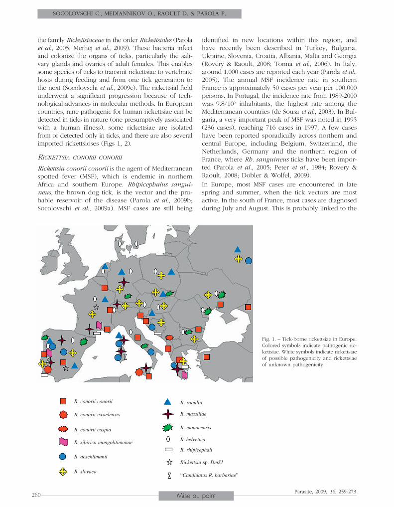
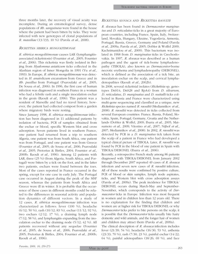
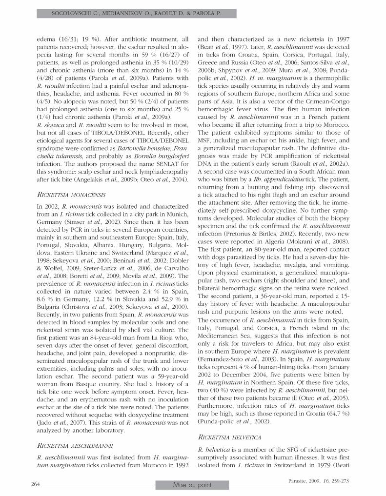
the family Rickettsiaceae in the order Rickettsiales (Parolaet al., 2005; Merhej et al., 2009). These bacteria infectand colonize the organs of ticks, particularly the sali-vary glands and ovaries of adult females. This enablessome species of ticks to transmit rickettsiae to vertebratehosts during feeding and from one tick generation tothe next (Socolovschi et al., 2009c). The rickettsial fieldunderwent a significant progression because of tech-nological advances in molecular methods. In Europeancountries, nine pathogenic for human rickettsiae can bedetected in ticks in nature (one presumptively associatedwith a human illness), some rickettsiae are isolatedfrom or detected only in ticks, and there are also severalimported rickettsioses (Figs 1, 2).
RICKETTSIA CONORII CONORII
Rickettsia conorii conorii is the agent of Mediterraneanspotted fever (MSF), which is endemic in northernAfrica and southern Europe. Rhipicephalus sangui-neus, the brown dog tick, is the vector and the pro-bable reservoir of the disease (Parola et al., 2009b;Socolovschi et al., 2009a). MSF cases are still being
identified in new locations within this region, andhave recently been described in Turkey, Bulgaria,Ukraine, Slovenia, Croatia, Albania, Malta and Georgia(Rovery & Raoult, 2008; Tonna et al., 2006). In Italy,around 1,000 cases are reported each year (Parola et al.,2005). The annual MSF incidence rate in southernFrance is approximately 50 cases per year per 100,000persons. In Portugal, the incidence rate from 1989-2000was 9.8/105 inhabitants, the highest rate among theMediterranean countries (de Sousa et al., 2003). In Bul-garia, a very important peak of MSF was noted in 1995(236 cases), reaching 716 cases in 1997. A few caseshave been reported sporadically across northern andcentral Europe, including Belgium, Switzerland, theNetherlands, Germany and the northern region ofFrance, where Rh. sanguineus ticks have been impor-ted (Parola et al., 2005; Peter et al., 1984; Rovery &Raoult, 2008; Dobler & Wolfel, 2009).In Europe, most MSF cases are encountered in latespring and summer, when the tick vectors are mostactive. In the south of France, most cases are diagnosedduring July and August. This is probably linked to the
SOCOLOVSCHI C., MEDIANNIKOV O., RAOULT D. & PAROLA P.
260 Mise au pointParasite, 2009, 16, 259-273
Fig. 1. – Tick-borne rickettsiae in Europe.Colored symbols indicate pathogenic ric-kettsiae. White symbols indicate rickettsiaeof possible pathogenicity and rickettsiaeof unknown pathogenicity.
▲
R. conorii conorii
R. conorii israelensis
R. conorii caspia
R. sibirica mongolitimonae
R. aeschlimanii
R. slovaca
R. raoultii
R. massiliae
R. monacensis
R. helvetica
R. rhipicephali
Rickettsia sp. DmS1
“Candidatus R. barbariae”

increased aggressiveness and propensity of Rh. san-guineus to bite hosts in warmer conditions. During theFrench heat wave in August 2003, which was the hot-test summer of the preceding 50 years, 22 Rh. sangui-neus (including specimens infected by R. conorii andR. massiliae) were found attached to a homeless personwho died of MSF (Hemmersbach-Miller et al., 2004).Recently, the investigation of a cluster of rickettsiosesin southern France during the exceptionally warmmonths of April and May 2007 was reported. Patientssuffered from severe R. conorii and R. massiliae infec-tions, and the authors found that this group of casesresulted from the unexpected proliferation and aggres-sive behavior of Rh. sanguineus ticks infected withthese rickettsiae. The authors also demonstrated expe-rimentally that Rh. sanguineus readily bites humanswhen exposed to higher temperatures (Parola et al.,2008). In Croatia, more than 80 % of cases occur bet-ween July and September, with a peak in August(Punda-Polic et al., 2003). During the 1970s, an increasein MSF cases was observed in France, Italy, Spain, andPortugal. The increase was correlated with higher tem-peratures and lower rainfall in Spain and with adecrease in the number of days of frost during the pre-ceding year in France (Parola et al., 2008; Parola et al.,2005). In Portugal, the number of positive cases of MSFdoubled in 2004-2005, and the authors speculated thatit was probably associated with climate change, parti-cularly low precipitation (de Sousa et al., 2006). In
addition, an undetected reservoir-vector system mayhave emerged (Parola et al., 2009b). The increasedincidence of MSF could also be explained by the useof improved diagnostic methods and by new interestin several countries after reconsidering the severity andhigh mortality rates of the disease (Rovery & Raoult,2008). Recently, in a prospective study conducted inAlgeria, North Africa, 49 % of patients were hospitali-zed with a severe form of MSF, and the global deathrate was 3.6 %. Furthermore, the MSF incidence was54.5 % in patients hospitalized with major neurologicalmanifestations and multiorgan involvement (Mouffoket al., 2009).After an incubation of around six days, the onset ofMSF is abrupt. Typical patients have a high fever, flu-like symptoms, a local necrotic inflammation with ablack crust called an eschar (the “tache noire”) at thetick bite site and a maculopapular rash (Rovery &Raoult, 2008; Parola et al., 2005). Occasionally, theeschar is not found, and it is seen rarely in multiples(Rovery & Raoult, 2008). Recently, clinical symptomsof MSF were described in 415 Sicilian children. Fever,rash and “tache noire” were present in 386 (93 %), 392(94.5 %) and 263 (63.4 %) cases, respectively, and 18(4.6 %) children had atypical exanthema (Colombaet al., 2006). Severe forms, including major neurolo-gical manifestations and multiorgan involvement, mayoccur in 5 to 6 % of cases. In Portugal from 1994-2006,the case fatality rate among 71 MSF patients was 13 %,
TICK-BORNE BACTERIAL DISEASES IN EUROPE
261Mise au pointParasite, 2009, 16, 259-273
Rickettsia sibirica sibirica (Dermacentor spp.)
Rickettsia parkeri (Amblyomma spp.)
Rickettsia sibirica mongolitimonae (Hyalomma spp.)
Rickettsia conorii conorii (Rhipicephalus sanguineus)
Rickettsia rickettsii (Dermacentor spp.)
Rickettsia conorii israelensis (Rhipicephalus sanguineus)
Rickettsia conorii caspiensis (Rhipicephalus pumilio)
Rickettsia slovaca (Dermacentor spp.)
“Candidatus Rickettsia barbariae” (Rhipicephalus spp.)
Rickettsia africae (Amblyomma spp.)
Rickettsia massiliae (Rhipicephalus spp.)
Rickettsia Rhipicephali (Rhipicephalus spp.)
“Candidatus Rickettsia kulagini” (Rhipicephalus sanguineus)
Rickettsia aeschlimannii (Hyalomma marginatum marginatum)
Rickettsia raoultii (Dermacentor spp.)
“Rickettsia sp. DmS1” (Dermacentor marginatus)
Rickettsia helvetica (Ixodes ricinus)
Rickettsia monacensis (Ixodes ricinus)
Rickettsia hoogstraalii (Haemaphysalis sulcata)
Rickettsia typhii (fleas)
“Candidatus Rickettsia tarasevichiae” (Ixodes persulcatus)
Rickettsia bellii (Ixodes spp.)
3640
51
4386
100
4383
4156
74
5992
55
46
100
Fig. 2. – Phylogenetic trees for members of the genus Rickettsia of derived from gltA sequence data with Tamura-Nei genetic distancemethod (Tick vector of each rickettsia).
more than five times higher than the conventionallyrecognized mortality rate (Sousa et al., 2008; Parolaet al., 2005). The highest MSF mortality rate (32.3 %)was described in southern Portugal in 1997 (de Sousaet al., 2003). Although one may speculate that the patho-genic strain of Israeli tick typhus isolated in 1997 couldbe responsible for this increase in the fatality rate, inhe-rited patient factors might also be strongly associatedwith mortality. The classic risk factors for severe formsinclude advanced age, cirrhosis, chronic alcoholism,G6PD deficiency, immunocompromised situations,prior prescription of an inappropriate antibiotic, anddelay of treatment (Parola et al., 2005). Recently, a dia-gnostic score was established to help clinicians dia-gnose MSF (Raoult & Roux, 1997).
RICKETTSIA CONORII ISRAELENSIS
Israeli spotted fever (ISF) was reported in 1946 inIsrael, and the number of cases increased following thedevelopment of new settlements in the rural areas ofthe country (Raoult & Roux, 1997). R. conorii israelensishas been detected in Rh. sanguineus specimens col-lected in Sicily and Portugal, and several clinical caseswere described in the same countries (Giammancoet al., 2003; Santos-Silva et al., 2006a; Sousa et al.,2008). The clinical manifestations of ISF are similar tothose of other spotted fever group infections, but aninoculation eschar is rarely observed and a history oftick exposure is not always present. In Israel, escharscaused by infection with the ISF strain have been des-cribed in only 4 % of cases (Gross &Yagupsky, 1987).A prospective study in Portugal conducted during1994-2006 identified 69 patients with ISF, confirmedeither by isolation of R. conorii israelensis from bloodcultures or detection of rickettsial DNA in skin biopsysamples by polymerase chain reaction (PCR). The cli-nical characteristics were statistically similar with MSF.A history of a recognized tick bite was more commonin patients infected with R. conorii conorii than in thoseinfected with ISF, and an eschar was observed in asignificantly higher percentage of MSF patients (Sousaet al., 2008).Several fatal cases and severe forms of ISF have beendescribed, especially in children, as well as in trave-lers and those with G6PD deficiency (Sousa et al., 2008;Chai et al., 2008; Boillat et al., 2008). In the Portugalstudy, the case fatality rate for ISF was significantlygreater than that for MSF, and a greater percentage ofpatients with ISF required admission to the intensivecare unit compared to MSF patients. Analysis of therelationship between co-morbidities and fatal MSF out-comes demonstrated that alcoholism was a statisticallysignificant risk factor for a fatal outcome (Sousa et al.,2008). A retrospective analysis in Sicily from 1987 to2001 identified five of 24 patients infected with R. cono-
rii israelensis by molecular tools; three of them pre-sented severe forms and one died (Giammanco et al.,2005). These recently reported cases show that ISF isa severe disease.
RICKETTSIA CONORII CASPIENSIS
Astrakhan fever is a summer spotted fever resemblingMSF that is endemic to the Astrakhan and nearbyregions of Russia near the Caspian Sea. It is transmittedto humans through the bites of Rh. pumilio ticks. Astra-khan spotted fever was later serologically diagnosedin Kalmykia (Russia), the agent was isolated also fromRh. pumilio from Western Kazakhstan; however, no cli-nical cases have yet been reported (Tarasevich & Median-nikov, 2006). Similar rickettsiae were identified as acause of febrile illness in a patient from Chad anddetected in Rh. sanguineus ticks from Kosovo (Four-nier et al., 2003a; Fournier et al., 2003b). Thus, Astra-khan fever might be a cause of spotted fever in Kosovo,and the area of distribution of this rickettsia could bewider than initially suspected in Astrakhan. Clinically,Astrakhan fever is characterized by clinical symptomsresembling those of MSF, that is, elevated fever, amaculopapular rash of the trunk, and severe myalgias.However, an inoculation eschar at the site of the tickbite is present in only 23 % of patients (Tarasevich et al.,1991).
RICKETTSIA MASSILIAE
In 1992, a novel rickettsial agent was isolated fromRh. sanguineus ticks collected near Marseille (France).It was characterized as a distinct species within the SFGgroup of rickettsiae and named R. massiliae (Beati &Raoult, 1993). In Europe, this rickettsia has been detec-ted by molecular methods in Rh. sanguineus fromGreece, Switzerland, Spain, and Italy; in Rh. turanicusfrom Portugal, the Greek Islands, Spain, and Italy; inI. ricinus from Spain and Germany; and in Rh. pusillusfrom Spain (Bernasconi et al., 2002; Psaroulaki et al.,2003; Fernandez-Soto et al., 2006; Psaroulaki et al.,2006; Mura et al., 2008). The first human case of infec-tion with R. massiliae was confirmed in 2005, 20 yearsafter the isolate was obtained from the patient. In 1985,this patient was hospitalized in Palermo, Italy, with fever,a necrotic eschar on his right ankle, a maculopapularrash on his palms and soles and mild hepatomegaly.He recovered completely after receiving tetracycline(Vitale et al., 2006). Recently, the second case of R. mas-siliae infection was diagnosed by serological and mole-cular tools. A 25-year-old man developed fever, nightsweats, headache, two necrotic skin lesions on the but-tocks and the thighs, and a maculopapular rash invol-ving the palms and soles. A few days later, he com-plained of acute visual loss and bilateral chorioretinitiswas diagnosed. The clinical course was favorable, but
SOCOLOVSCHI C., MEDIANNIKOV O., RAOULT D. & PAROLA P.
262 Mise au pointParasite, 2009, 16, 259-273
three months later, the recovery of visual acuity wasincomplete. During an entomological survey, densepopulations of Rh. sanguineus were found in the housewhere the patient had been bitten by ticks. They wereinfected with new genotypes of clonal populations ofR. massiliae (13/133; 10 %) (Parola et al., 2008).
RICKETTSIA SIBIRICA MONGOLITIMONAE
R. sibirica mongolitimonae causes LAR (lymphangitis-associated rickettsiosis) (Fournier et al., 2005; Fournieret al., 2006). This rickettsia was firstly isolated in Bei-jing from Hyalomma asiaticum ticks collected in theAlashian region of Inner Mongolia in 1991 (Yu et al.,1993). In Europe, R. sibirica mongolitimonae was detec-ted in H. anatolicum excavatum from Greece and inRh. pusillus from Portugal (Psaroulaki et al., 2005;De Sousa et al., 2006). In 1996, the first case of humaninfection was diagnosed in southern France in a womanwho had a febrile rash and a single inoculation escharin the groin (Raoult et al., 1996). This patient was aresident of Marseille and had no travel history; how-ever, the patient had collected compost from a gardenwhere migratory birds were resting.Since January 1998, R. sibirica mongolitimonae infec-tion has been diagnosed in 11 additional patients byisolation of bacteria, PCR of samples obtained fromthe eschar or blood, specific Western blot and crossadsorption. Seven patients lived in southern France,one patient had returned from a trip to southernAlgeria, one patient was from South Africa, one patientwas from Portugal, and one patient was from Greece(Fournier et al., 2005; de Sousa et al., 2006; Psaroulakiet al., 2005; Pretorius & Birtles, 2004; Fournier et al.,2000b; Raoult et al., 1996). Among 12 patients withLAR, three (25 %) (from Algeria, South Africa, and Por-tugal) were bitten by a tick on the foot, and in the lattertwo patients, eschars were found between the toes.Most of the cases reported in France occurred in thespring, except for one case in early July. The Portugalcase occurred in August during the peak of the MSFseason, whereas the patients from South Africa andGreece were ill in winter. It is probable that the occur-rence of these cases in different months could be rela-ted to the differences in seasonal activity and popula-tion dynamics of different vectors. In a study of12 cases, R. sibirica mongolitimonae infection wascharacterized as follows: fever (100 %), headache(6/12; 50 %), rash (10/12; 83 %), eschar (11/12; 92 %),two eschars (2/12; 17 %), a draining lymph node(7/12; 58 %), and lymphangitis expanding from the ino-culation eschar to the draining node (5/12; 42 %). Allpatients recovered without any sequelae (Fournieret al., 2005; de Sousa et al., 2006; Psaroulaki et al.,2005; Pretorius & Birtles, 2004; Fournier et al., 2000b;Raoult et al., 1996).
RICKETTSIA SLOVACA AND RICKETTSIA RAOULTII
R. slovaca has been found in Dermacentor margina-tus and D. reticulatus ticks in a great majority of Euro-pean countries, including France, Spain, Italy, Switzer-land, Slovakia, Hungary, Ukraine, Yugoslavia, Armenia,Portugal, Russia, Greece, Germany and Poland (Parolaet al., 2009a; Parola et al., 2005; Dobler & Wolfel 2009;Kachrimanidou et al., 2009). This bacterium was iso-lated in 1968 from D. marginatus ticks in Czechoslo-vakia. In 1997, R. slovaca was described as a humanpathogen and the agent of tick-borne lymphadeno-pathy (TIBOLA), also known as Dermacentor-bornenecrotic erythema and lymphadenopathy (DEBONEL),which is defined as the association of a tick bite, aninoculation eschar on the scalp, and cervical lympha-denopathies (Raoult et al., 2002b).In 2008, several rickettsial isolates (Rickettsia sp. geno-types DnS14, DnS28 and RpA4) from D. silvarum,D. reticulatus, D. marginatus and D. nuttalli ticks col-lected in Russia and France were characterized usingmulti-gene sequencing and classified as a unique, newRickettsia species named R. raoultii (Mediannikov et al.,2008). R. raoultii was detected in ticks collected fromseveral European countries: France, Russia, Poland, Slo-vakia, Spain, Portugal, Germany, Croatia and the Nether-lands (Dobler & Wolfel, 2009; Ibarra et al., 2005; Mat-sumoto et al., 2009; Vitorino et al., 2007; Nijhof et al.,2007; Mediannikov et al., 2008). In 2002, R. raoultii wasdetected by PCR in a D. marginatus tick taken fromthe scalp of a patient in France who had developed atypical clinical picture of TIBOLA. Later, R. raoultii wasfound by PCR in the blood of one patient in Spain withTIBOLA/DEBONEL (Ibarra et al., 2006).Recently, a retrospective French study among 86 patientsdiagnosed with TIBOLA/DEBONEL from January 2002through December 2007 reported 49 cases of R. slovacainfection and seven new cases of R. raoultii infection.All of these results were confirmed by positive culture,PCR of blood or skin samples, lymph node aspirates,ticks, and Western blot with cross adsorption assays(Parola et al., 2009a). The peak incidence for TIBOLA/DEBONEL occurs during March-May and September-November, which corresponds to the activity of Der-macentor ticks in Europe. Infection was most frequentin women and in children less than 12 years old. Thereis no explanation for the finding that children andwomen are at higher risk for TIBOLA/DEBONEL or whyDermacentor ticks prefer to bite persons on the scalp; itis possible that the Dermacentor ticks usually bite hairydomestic and wild animals, and the longer hair of womenand children may attract them (Parola et al., 2009a).The clinical description of R. slovaca infection includesfever (21/39; 54 %), headache (16/30; 53 %), asthenia(23/33; 70 %), rash (7/30; 23 %), painful eschar (14/22;64 %), painful adenopathies (18/26; 69 %), and face
TICK-BORNE BACTERIAL DISEASES IN EUROPE
263Mise au pointParasite, 2009, 16, 259-273
edema (16/31; 19 %). After antibiotic treatment, allpatients recovered; however, the eschar resulted in alo-pecia lasting for several months in 59 % (16/27) ofpatients, as well as prolonged asthenia in 35 % (10/29)and chronic asthenia (more than six months) in 14 %(4/28) of patients (Parola et al., 2009a). Patients withR. raoultii infection had a painful eschar and adenopa-thies, headache, and asthenia. Fever occurred in 80 %(4/5). No alopecia was noted, but 50 % (2/4) of patientshad prolonged asthenia (one to six months) and 25 %(1/4) had chronic asthenia (Parola et al., 2009a).R. slovaca and R. raoultii seem to be involved in most,but not all cases of TIBOLA/DEBONEL. Recently, otheretiological agents for several cases of TIBOLA/DEBONELsyndrome were confirmed as Bartonella henselae, Fran-cisella tularensis, and probably as Borrelia burgdorferiinfection. The authors proposed the name SENLAT forthis syndrome: scalp eschar and neck lymphadenopathyafter tick bite (Angelakis et al., 2009b; Oteo et al., 2004).
RICKETTSIA MONACENSIS
In 2002, R. monacensis was isolated and characterizedfrom an I. ricinus tick collected in a city park in Munich,Germany (Simser et al., 2002). Since then, it has beendetected by PCR in ticks in several European countries,mainly in southern and southeastern Europe: Spain, Italy,Portugal, Slovakia, Albania, Hungary, Bulgaria, Mol-dova, Eastern Ukraine and Switzerland (Marquez et al.,1998; Sekeyova et al., 2000; Beninati et al., 2002; Dobler& Wolfel, 2009; Sreter-Lancz et al., 2006; de Carvalhoet al., 2008; Boretti et al., 2009; Movila et al., 2009). Theprevalence of R. monacensis infection in I. ricinus tickscollected in nature varied between 2.4 % in Spain,8.6 % in Germany, 12.2 % in Slovakia and 52.9 % inBulgaria (Christova et al., 2003; Sekeyova et al., 2000).Recently, in two patients from Spain, R. monacensis wasdetected in blood samples by molecular tools and onerickettsial strain was isolated by shell vial culture. Thefirst patient was an 84-year-old man from La Rioja who,seven days after the onset of fever, general discomfort,headache, and joint pain, developed a nonpruritic, dis-seminated maculopapular rash of the trunk and lowerextremities, including palms and soles, with no inocu-lation eschar. The second patient was a 59-year-oldwoman from Basque country. She had a history of atick bite one week before symptom onset. Fever, hea-dache, and an erythematous rash with no inoculationeschar at the site of a tick bite were noted. The patientsrecovered without sequelae with doxycycline treatment(Jado et al., 2007). This strain of R. monacensis was notanalyzed by another laboratory.
RICKETTSIA AESCHLIMANNII
R. aeschlimannii was first isolated from H. margina-tum marginatum ticks collected from Morocco in 1992
and then characterized as a new rickettsia in 1997(Beati et al., 1997). Later, R. aeschlimannii was detectedin ticks from Croatia, Spain, Corsica, Portugal, Italy,Greece and Russia (Oteo et al., 2006; Santos-Silva et al.,2006b; Shpynov et al., 2009; Mura et al., 2008; Punda-polic et al., 2002). H. m. marginatum is a thermophilictick species usually occurring in relatively dry and warmregions of southern Europe, northern Africa and someparts of Asia. It is also a vector of the Crimean-Congohemorrhagic fever virus. The first human infectioncaused by R. aeschlimannii was in a French patientwho became ill after returning from a trip to Morocco.The patient exhibited symptoms similar to those ofMSF, including an eschar on his ankle, high fever, anda generalized maculopapular rash. The definitive dia-gnosis was made by PCR amplification of rickettsialDNA in the patient’s early serum (Raoult et al., 2002a).A second case was documented in a South African manwho was bitten by a Rh. appendiculatus tick. The patient,returning from a hunting and fishing trip, discovereda tick attached to his right thigh and an eschar aroundthe attachment site. After removing the tick, he imme-diately self-prescribed doxycycline. No further symp-toms developed. Molecular studies of both the biopsyspecimen and the tick confirmed the R. aeschlimanniiinfection (Pretorius & Birtles, 2002). Recently, two newcases were reported in Algeria (Mokrani et al., 2008).The first patient, an 80-year-old man, reported contactwith dogs parasitized by ticks. He had a seven-day his-tory of high fever, headache, myalgia, and vomiting.Upon physical examination, a generalized maculopa-pular rash, two eschars (right shoulder and knee), andbilateral hemorrhagic signs on the retina were noticed.The second patient, a 36-year-old man, reported a 15-day history of fever with headache. A maculopapularrash and purpuric lesions on the arms were noted.The occurrence of R. aeschlimannii in ticks from Spain,Italy, Portugal, and Corsica, a French island in theMediterranean Sea, suggests that this infection is notonly a risk for travelers to Africa, but may also existin southern Europe where H. marginatum is prevalent(Fernandez-Soto et al., 2003). In Spain, H. marginatumticks represent 4 % of human-biting ticks. From January2002 to December 2004, five patients were bitten byH. marginatum in Northern Spain. Of these five ticks,two (40 %) were infected by R. aeschlimannii, but nei-ther of these two patients became ill (Oteo et al., 2005).Furthermore, infection rates of H. marginatum ticksmay be high, such as those reported in Croatia (64.7 %)(Punda-polic et al., 2002).
RICKETTSIA HELVETICA
R. helvetica is a member of the SFG of rickettsiae pre-sumptively associated with human illnesses. It was firstisolated from I. ricinus in Switzerland in 1979 (Beati
SOCOLOVSCHI C., MEDIANNIKOV O., RAOULT D. & PAROLA P.
264 Mise au pointParasite, 2009, 16, 259-273
et al., 1993). R. helvetica has been detected in I. ricinusin many European countries, including France, Spain,Portugal, Austria, Denmark, the Netherlands, Switzer-land, Sweden, Portugal, Germany, Bulgaria, Slovenia,Poland, Hungary, Moldova, Eastern Ukraine and Italy(Nijhof et al., 2007; Christova et al., 2003; Dobler &Wolfel, 2009; Marquez, 2008; Parola et al., 2005; Nilssonet al., 1999; Movila et al., 2009). In Sweden, two patientswith clinical symptoms of fatal perimyocarditis wereinfected with R. helvetica, as confirmed by electronmicroscopy, PCR and serology (Nilsson et al., 1999).Subsequently, an association between R. helvetica andsarcoidosis in Sweden was reported by the sameresearchers (Nilsson et al., 2002). However, the validityof these associations has been questioned by some ric-kettsiologists (Walker et al., 2003), and additional stu-dies did not reveal anti-rickettsial antibodies in a groupof Scandinavian sarcoidosis patients (Planck et al., 2004).In 2000, seroconversion to R. helvetica was describedin a 37-year-old immunocompetent French man fourweeks after the onset of an unexplained febrile illnesswith prolonged fever, fatigue, myalgias, and headache.No rash, lymphadenopathy or inoculation eschar wasnoted. The serology result was confirmed by cross-adsorption and Western blotting. Results of a serosur-vey of forest workers from the area where the patientlived showed a 9.2 % seroprevalence against R. hel-vetica (Fournier et al., 2000a). In eight of 75 patients(11 %) in Switzerland, serological findings suggestedpossible acute or past R. helvetica infection (Baumannet al., 2003). In 2004, eight patients from France, Italy,and Thailand showed serological evidence of R. hel-vetica infection. The infection presented as a milddisease in the warm season and was associated withfever, headache, and myalgia but without a cutaneousrash. Only one patient developed an eschar (Fournieret al., 2004). Interestingly, one case of R. helvetica infec-tion was reported in Sweden with the presence of amacular rash involving the arms and legs. A 57-year-old, healthy, immunocompetent man had a fever, myal-gias, arthralgias and severe headache with photopho-bia, but no lymphadenopathy or inoculation eschar.The infection was confirmed by PCR, blood culture, sero-logy and Western blot assay (Nilsson, 2009). Among4,604 clinical rickettsial cases reported in Italy from1998 to 2002, three cases of a mild form of rickettsiosiswere serologically attributed to R. helvetica (Ciceroniet al., 2006). Additional evaluation and isolation of thebacterium from clinical samples are needed to confirmthe pathogenicity of R. helvetica.
OTHER TICK-BORNE RICKETTSIAE
The most recently discovered rickettsiae were firstdetected and isolated in ticks and subsequently cha-racterized as human pathogens, including R. massiliae,
R. aeschlimannii, R. monacensis and R. slovaca. To beconsidered a human pathogen, a rickettsia must beeither isolated in cell culture from or detected by mole-cular methods in blood or tissue from patients with ill-nesses clinically compatible with spotted fever rickettsio-ses who are also seropositive using standard referencelaboratory methods (Parola et al., 2005). Several addi-tional rickettsiae have been detected in ticks, but theirpathogenicity is not known. R. rhipicephali was detec-ted in Rh. sanguineus ticks collected from France, Por-tugal, Greece and Croatia (Drancourt et al., 1992). In2000, a new rickettsial genotype, Rickettsia sp. DmS1,was detected in one out of 70 D. marginatus ticks col-lected from game pigs (Sus scrofa) in southern France.This genotype was later detected in the same tick spe-cies in Spain (Sanogo et al., 2003; Fernandez-Soto et al.,2006). Based on preliminary phylogenetic studies, thisnew rickettsia appears to be within the R. massiliaegroup. Recently, another new rickettsia was describedin Rh. sanguineus ticks collected from Sardinia; thisorganism was previously identified in four Rh. turani-cus ticks in Portugal and referred to as PoTiRb169. Thisnew species was further characterized by amplificationand sequencing of several genes, and the authors pro-posed the name “Candidatus Rickettsia barbariae”(Mura et al., 2008). In addition, the existence of ano-ther novel SFG rickettsiae was demonstrated in Hae-maphysalis sulcata ticks collected from sheep andgoats in Croatia. This new rickettsia was named R. hoog-straali, and was isolated and deposited in two differentcollections (Duh et al., 2009). A rickettsial genotypefrom the R. massiliae genogroup called CandidatusRickettsia kulagini was identified in Rh. sanguineusticks from Crimean peninsula, Ukraine (Mediannikovet al., 2007). Candidatus Rickettsia tarasevichiae wasidentified in Ixodes persulcatus ticks with a high pre-valence (Shpynov et al., 2003). This tick replaces I. rici-nus in Northern Russia and Finland (Jaaskelainen et al.,2006).
IMPORTED RICKETTSIOSES
Case reports and retrospective studies showed theimportance of rickettsioses imported by internationaltourists. Usually, the diagnosis can be made based onthe presence of flu-like symptoms following a recentvisit to tick-infested areas, a history of a tick bite, thepresence of an eschar and the type of rash. Severecomplications and fatalities are occasionally seen.During the last decade, approximately 400 cases oftick-borne rickettsioses have been reported in inter-national travelers. The vast majority of these illnesseswere either African tick bite fever (ATBF) caused byR. africae in patients returning from sub-Saharan Africaand the West Indies or Mediterranean spotted fevercaused by R. conorii in patients retuning from the
TICK-BORNE BACTERIAL DISEASES IN EUROPE
265Mise au pointParasite, 2009, 16, 259-273
Mediterranean area (Jensenius et al., 2006). ATBF istransmitted by Amblyomma ticks that are aggressiveand actually attack hosts, which leads to clustered casesof infection (Socolovschi et al., 2009b). In more thanhalf of the cases, multiple inoculation eschars occur.Indeed, several ticks may attack at the same time, andthe rickettsiae infection rate among ticks may reach100 % (Jensenius et al., 2003a). Furthermore, thirty-eight travelers, 4.0 % of the cohort and 26.6 % of thosereporting flulike symptoms, of 940 Norwegian trave-lers to rural sub-Equatorial Africa were diagnosed withATBF. Game hunting, travel to Southern Africa andtravel during November through April were found tobe independent risk factors (Jensenius et al., 2003b).Finally, all new rickettsioses described throughout theworld may be imported into European countries.
HUMAN GRANULOCYTIC ANAPLASMOSIS
Human granulocytotropic anaplasmosis (HGA) isa tick-borne zoonotic infection caused by Ana-plasma phagocytophilum, which is transmitted
in Europe by I. ricinus ticks. I. ricinus is also the vector ofLyme borreliosis, it harbours R. helvetica and R. mona-censis. Across Europe, the prevalence of Anaplasmaphagocytophilum infection in I. ricinus ticks is variableand can be between 0.4 to 66.7 %. It is not transmittedtransovarially in ticks; small mammals, particularly Apo-demus sylvaticus (wood mouse), A. flavicollis (yellow-necked mouse), Sorex araneus (common shrew) andespecially Clethrionomys glareolus (bank vole), havebeen implicated as HGA reservoirs in Europe.A limited number of laboratory-confirmed HGA caseshave been reported in Austria, Italy, Latvia, the Nether-lands, Norway, Poland, Slovenia, Spain, France, Russiaand Sweden, (Bakken & Dumler, 2008; Parola, 2004;Lotric-Furlan et al., 2006). Serologic studies also supportthe presence of HGA infection, much of which maybe asymptomatic, in potentially exposed adults in Ger-many, Bulgaria, Denmark, Spain, Italy, Estonia, Greece,and France (Parola, 2004). The first patient to haverecognized HGA was reported in the U.S. in 1990; thefirst European case was reported in Slovenia in 1997(Petrovec et al., 1997). By the end of December 2004,at least 2,871 cases of HGA were reported from 13 U.S.states to the Centers for Disease Control and Preven-tion (CDC) (Bakken & Dumler, 2008). In 2002, Blancoand Oteo (Blanco & Oteo, 2002) compared the clinicaland laboratory features of 15 European patients tothose reported for U.S. patients. Most patients reportexposure to ticks or a tick bite between seven and30 days before the onset of the disease. The majorityof cases occurred between June and August (73 %), aperiod of higher vector activity in these areas. Euro-
pean HGA patients generally have a less severe coursethan U.S. patients, and the presence of morulae isuncommon. In most patients, the disease consists offlu-like symptoms: fever, myalgias, arthralgias, andheadache. Physical examination reveals a few abnor-malities, such as conjunctivitis and lymphadenopathy.Other manifestations that can accompany the illness arepneumonia (13 %) and gastrointestinal symptoms suchas nausea (53 %), vomiting (20 %), diarrhea (13 %), andabdominal pain (20 %). Hepatomegaly, splenomegalyor both are also occasionally described. Laboratory teststypically show leukopenia and thrombocytopenia, as wellas elevated serum hepatic aminotransferase levels, ery-throcyte sedimentation rate and C-reactive protein levels.Between January 1996 and December 2004, 24 adultpatients with confirmed HGA were identified in a prospec-tive study conducted in Slovenia. The clinical charac-teristics and laboratory findings were similar to thosereported from other European countries. All of the patientshad an acute febrile illness with headache, malaise,myalgia and/or arthralgia. Laboratory findings includedleukopenia (16/24; 66.7 %), thrombocytopenia (20/24;83.3 %), abnormal liver function (23/24; 95.8 %), ele-vated erythrocyte sedimentation rates (18/24; 75 %),and elevated concentrations of C-reactive protein (23/24;95.8 %) (Lotric-Furlan et al., 2006). In Europe, verifica-tion of HGA has been based on PCR and immuno-fluorescence antibody tests, because no isolation of theetiologic agent from humans has yet been reported(Blanco & Oteo 2002).
BORRELIOSIS
Borrelia species are Gram-negative spriochetes,or helically coiled bacteria, which cause severalarthropod-borne diseases in European coun-
tries, including Lyme disease and tick-borne relapsingfever (TBRF).
LYME BORRELIOSIS (LB)Lyme disease is caused by three of the at least 14 diffe-rent Borrelia species belonging to the complex B. burg-dorferi: B. burgdorferi sensu stricto (ss), B. garinii, andB. afzelii (Stanek & Strle, 2003).However, B. valaisiana, B. lusitaniae, B. spielmanii andB. bissettii have been detected and isolated in samplesof human origin (Rudenko et al., 2009). LB is the mostfrequent Ixodid tick-borne human disease in the world,with an estimated 85,500 patients annually and 65,500in Europe alone (Hubalek, 2009). In Europe, most casesoccur in Scandinavian countries (up to 155 cases per100,000 individuals) and in central Europe (especiallyGermany, Austria and Switzerland). However, LB caseshave been reported throughout the region, including theUnited Kingdom, where many cases occur in the South
SOCOLOVSCHI C., MEDIANNIKOV O., RAOULT D. & PAROLA P.
266 Mise au pointParasite, 2009, 16, 259-273

Downs or New Forest areas. B. burgdorferi ss and relatedBorrelia species exist in nature in enzootic cycles pri-marily involving Ixodes ticks and a wide range of animalhosts. In Europe, small mammals, such as mice andvoles, are reservoirs of B. afzelii, B. burgdorferi ss andB. garinii serotype 4, while birds are the primary reser-voirs of B. garinii and B. valaisiana (Stanek & Strle, 2003;Nadelman & Wormser, 1998). A recent study in Hungaryidentified green lizards (Lacerta viridis) as potential hostsof I. ricinus ticks, and therefore a possible reservoir ofBorrelia burgdorferi sensu lato (Foldvari et al., 2009).As with other spirochete infections, human Lyme diseaseoccurs in stages, with remissions, exacerbations, and dif-ferent clinical manifestations at each stage. Althoughinfection with any of three pathogenic Borrelia speciesmay occur in the skin, nervous system, or joints, the fre-quency, severity, and duration of these symptoms varieswith each species. B. burgdorferi ss seems to be asso-ciated with most rheumatologic disorders, B. gariniiappears to be the most neutropic and causes chronicborrelial encephalomyelitis, and B. afzelii is moststrongly associated with skin manifestations, includingacrodermatitis chronica atrophicans. The clinical andmicrobiological characteristics of LB have been recentlyreviewed (Nadelman & Wormser, 1998; Stanek & Strle,2003). Microbial or serological confirmation of infectionis needed for all manifestations of LB except for typicalearly skin lesions. The culture of Borrelia species frompatient specimens permits definitive diagnosis, butusually is obtained only in the early stage of disease.
RELAPSING FEVER BORRELIOSIS
Tick-borne relapsing fever (TBRF) is a zoonotic disease,caused by spirochetes of the genus Borrelia and trans-mitted to humans through the bite of soft ticks of thegenus Ornithodoros, family Argasidae (Rebaudet &Parola, 2006). Six species of borrelia causing TBRF are
known to occur in Europe, including B. hispanica,transmitted by the O. erraticus tick; B. crocidurae,transmitted by the O. sonrai tick; B. persica, transmittedby the O. tholozani tick; B. caucasica, transmitted bythe O. asperus tick; B. latyschevii, transmitted by theO. tartakovskyi tick; and a new Borrelia species, iso-lated in southern Spain from three patients with TBRFand from the O. erraticus tick (Anda et al., 1996)(Fig. 3). Two additional Borrelia pathogenic to humansare present in Eurasia: B. microtii and B. baltazardiwere described in 1946 and 1979, respectively. Noother scientific data are known at this time regardingthese species (Assous & Wilamowski, 2009).In Israel, a total of 606 cases of TBRF were confirmedby association between febrile illness and a positive per-ipheral blood smear. Most cases occurred during summerand fall and correlated with Ornithodoros tick activity,high human outdoor activity and light clothes wornduring these seasons (Sidi et al., 2005). TBRF is a diseasecharacterized by relapsing or recurring episodes of fever,often accompanied by headache, muscle and joint achesand nausea. The incidence of TBRF appears to be rarein Europe. However, European physicians should be onalert for imported cases of TBRF. Several confirmed caseshave been reported in the literature: immigrants fromEthiopia three months after they arrived in Israel, fourFrench patients with unexplained fever from Senegal anda Dutch woman that developed symptoms after a two-week trip to Guatemala and Belize (Sidi et al., 2005;Patrat-Delon et al., 2008; Heerdink et al., 2006).
TULAREMIA
Tularemia is a zoonotic infection caused by thebacterium Francisella tularensis. In 1923, tick-borne tularemia was reported for the first time
TICK-BORNE BACTERIAL DISEASES IN EUROPE
267Mise au pointParasite, 2009, 16, 259-273
Fig. 3. – Relapsing fever borreliosis inEurope.
by physicians in Idaho, and one year later it was isola-ted from ticks (Petersen et al., 2009). Moreover, infec-tions with F. tularensis in nature have been documen-ted in various arthropods, including fleas, lice, midges,bedbugs, mosquitoes, ticks and flies. In Europe, F. tula-rensis has been detected in I. ricinus, D. reticulates andD. marginatus ticks (Aberer, 2009; Hubalek et al., 1996).In the U.S., Sweden, Finland and Russia, arthropodbites are a common mode of tularemia transmissionto humans. In Central Europe, contact with infectedanimals and ingestion of contaminated food or waterare more common modes of transmission (Hubaleket al., 1996; Petersen et al., 2009). The prevalence oftularemia in ticks is very low: only 0.01 % of 120,000I. ricinus adult ticks in Russia were positive (Sjostedt,2007a). In Slovakia, 34 F. tularensis strains were iso-lated from 4,542 starving ticks, predominantly fromD. reticulatus (Gurycova et al., 1995). Recently, a pos-sible reservoir of F. tularensis was investigated in Ger-many. F. tularensis was detected in five different rodentspecies with carrier rates of 2.04, 6.94 and 10.87 % pertrapping area, and two strains were isolated fromwater voles. None of the ticks tested positive forF. tularensis (Kaysser et al., 2008). In central Europeanfoci, hares appeared to be the main carrier and sourceof tularemia in humans, whereas ticks harbored byhares were the perennial reservoir of F. tularensis (Sjos-tedt, 2007b).Several outbreaks of tularemia have been reported inthe literature, but they were not associated with trans-mission by ticks (Kantardjiev et al., 2006; Reintjes et al.,2002; Payne et al., 2005; Perez-Castrillon et al., 2001).The clinical symptoms of tularemia depend upon theroute of infection. When infected by a tick, an indo-lent ulcer often occurs at the site of the bite followedby swelling of the regional lymph nodes. The ulcer isusually also followed by a fever.
Q FEVER
Qfever is a zoonosis caused by Coxiella burnetii(Raoult et al., 2005). C. burnetii has been detec-ted in 140 species of ticks in 12 genera throu-ghout the word (absent in New Zealand), but
the role of ticks in Q fever transmission to humans isvery small (Parola & Raoult, 2001b; Tissot-Dupont &Raoult, 2008). Recently, none of 862 Dermacentor tickstested positive for C. burnetii in Germany, and one spe-cimen of Haemaphysalis punctata from 691 questingadult ixodid ticks was positive in Spain (Hartelt et al.,2008; Barandika et al., 2008). Q fever is usually acqui-red by the ingestion or inhalation of virulent organismsfrom infected mammals and their products, most fre-quently goats, sheep, and cats. Infection in humans is
often asymptomatic, but it can manifest as an acutedisease, such as pneumonia, hepatitis or isolated fever.Q fever can also appear in a chronic form, mainly invol-ving endocarditis and vascular infection, as well ashepatitis and chronic infection after pregnancy (Raoultet al., 2005).
BARTONELLA
Bartonella species are Gram-negative bacilli or coc-cobacilli belonging to the alpha 2 subgroup ofProteobacteria, family Bartonellaceae. These
bacteria are transmitted by blood-sucking arthropods,including fleas, lice, sandflies and ticks (Angelakis et al.,2009a). In European countries, Bartonella species havebeen detected by molecular tools in D. reticulates andIxodes spp. ticks. The prevalence of Bartonella-infectedticks can vary from 1.2 % in the Czech Republic to 60 %in the Netherlands (Schouls et al., 1999; Hercik et al.,2007). Recently, I. ricinus ticks were demonstrated tobe a competent vector for B. henselae (Cotte et al.,2008). Five Bartonella species were found in ticks col-lected in European countries: B. quintana, B. henselae,B. capreoli, B. bacilliformis-like bacterium and B. schoen-buchensis. In Russia, Slovenia, Poland and France, seve-ral clinical studies confirmed Bartonella transmissionfrom ticks to humans (Podsiadly et al., 2009; Angelakiset al., 2009b). In the study performed in Slovenia,86 febrile children were screened with serological toolsfor multiple tick-borne antigens after a history of tickbite. B. henselae infection was detected in five children,B. quintana infection was found in four children, andboth infections were present in one child (Arnez et al.,2003). In Poland, 17 patients with clinical symptoms ofneuroborrelioses were tested for Bartonella infection.Among these patients, B. henselae was detected in onecase, and B. burgdorferi and B. henselae infections werefound in two patients (Podsiadly et al., 2003). In France,B. henselae infection was confirmed in three patientsafter a tick bite history. All patients presented scalpeschar and neck lymphadenopathy (Angelakis et al.,2009b).
CONCLUSION
Tick-borne illnesses are emergent and re-emer-gent zoonotic diseases. The progression of tick-borne diseases and their increased morbidity is
associated with outdoor activities and travels (Parola,2004). Several climate models predict a global warming,more in the northern and altitude range, such as in Ger-many, with the emergence of Lyme diseases and tick-borne encephalitis in the last years (Suss et al., 2008).
SOCOLOVSCHI C., MEDIANNIKOV O., RAOULT D. & PAROLA P.
268 Mise au pointParasite, 2009, 16, 259-273
Increasing public education on tick-borne diseasesand avoidance of tick bites (such as using protectiveclothing, insect repellents, and early detection andremoval of ticks) are the best prevention. The earlyremoval of ticks can reduce the spread of infectiousorganisms. Antibiotic prophylactic therapy after a tickbite is not generally recommended (Aberer, 2009).Europeans who have spent time in tick-endemic areasshould inspect themselves frequently for ticks. Com-plete removal of attached ticks is indicated with twee-zers or forceps close to the skin. Routine disinfectionof the bite wound is recommended to avoid contami-nation of the bite site with skin bacteria (Bakken &Dumler, 2008; Aberer, 2009). European cliniciansshould be aware of the clinical signs of tick-transmitteddiseases in patients with unexplained febrile illnesses,and a careful travel and tick exposure history shouldbe taken. Empiric antimicrobial therapy (most oftenwith doxycycline) is appropriate in most cases of signi-ficant clinical illness in which rickettsioses, anaplas-mosis, tularemia, bartonelloses or borrelial infectionsare suspected by epidemiological and clinical mani-festations, and may be lifesaving.
REFERENCES
ABERER E. What should one do in case of a tick bite? Curr.Probl. Dermatol., 2009, 37, 155-166.
ANDA P., SANCHEZ-YEBRA W., DEL MAR VITUTIA M., PÉREZ PAS-TRANA E., RODRIGUEZ I., MILLER N.S., BACKENSON P.B. & BENACH
J.L. A new Borrelia species isolated from patients withrelapsing fever in Spain. Lancet, 1996, 348, 162-165.
ANGELAKIS E., BILLETER S.A., BREITSCHWERDT B., CHOMEL B. &RAOULT D. Is there tick-borne Bartonelloses? Emerg. Infect.Dis., 2009a (Submitted).
ANGELAKIS E., PULCINI C., WATON J., IMBERT P., SOCOLOVSCHI C.,EDOUARD S., DELLAMONICA P. & RAOULT D. Scalp eschar andneck lymphadenopathy caused by Bartonella henselaeafter tick bite. CID, 2009b (In press).
ARNEZ M., LUZNIK-BUFON T., AVSIC-ZUPANC T., RUZIC-SABLJIC E.,PETROVEC M., LOTRIC-FURLAN S. & STRLE F. Causes of febrileillnesses after a tick bite in Slovenian children. Pediatr.Infect. Dis. J., 2003, 22, 1078-1083.
ASSOUS M.V. & WILAMOWSKI A. Relapsing fever borreliosis inEurasia-forgotten, but certainly not gone! Clin. Microbiol.Infect., 2009, 15, 407-414.
BAKKEN J.S. & DUMLER S. Human granulocytic anaplasmosis.Infect. Dis. Clin. North Am., 2008, 22, 433-448, viii.
BARANDIKA J.F., HURTADO A., GARCIA-SANMARTIN J., JUSTE R.A.,ANDA P. & GARCIA-PEREZ A.L. Prevalence of tick-borne zoo-notic bacteria in questing adult ticks from northern Spain.Vector. Borne. Zoonotic. Dis., 2008, 8, 829-835.
BAUMANN D., PUSTERLA N., PETER O., GRIMM F., FOURNIER P.E.,SCHAR G., BOSSART W., LUTZ H. & WEBER R. Fever after atick bite: clinical manifestations and diagnosis of acute tick
bite-associated infections in northeastern Switzerland.Dtsch. Med. Wochenschr., 2003, 128, 1042-1047.
BEATI L., MESKINI M., THIERS B. & RAOULT D. Rickettsia aes-chlimannii sp. nov., a new spotted fever group rickettsiaassociated with Hyalomma marginatum ticks. Int. J. Syst.Bacteriol., 1997, 47, 548-554.
BEATI L., PETER O., BURGDORFER W., AESCHLIMANN A. & RAOULT D.Confirmation that Rickettsia helvetica sp. nov. is a distinctspecies of the spotted fever group of rickettsiae. Int. J. Syst.Bact., 1993, 43, 521-526.
BEATI L. & RAOULT L. Rickettsia massiliae sp. nov., a newspotted fever group rickettsia. Int. J. Syst. Bact., 1993, 43,839-840.
BENINATI T., LO N., NODA H., ESPOSITO F., RIZZOLI A., FAVIA G.& GENCHI C. First detection of spotted fever group Ric-kettsiae in Ixodes ricinus from Italy. Emerg. Infect. Dis.,2002, 8, 983-986.
BERNASCONI M.V., CASATI S., PETER O. & PIFFARETTI J.C. Rhipi-cephalus ticks infected with Rickettsia and Coxiella in Sou-thern Switzerland (Canton Ticino). Infect. Genet. Evol.,2002, 2, 111-120.
BLANCO J.R. & OTEO J.A. Human granulocytic ehrlichiosis inEurope. Clin. Microbiol. Infect., 2002, 8, 763-772.
BOILLAT N., GENTON B., D’ACREMONT V., RAOULT D. & GREUB G.Fatal case of Israeli spotted fever after Mediterranean cruise.Emerg. Infect. Dis., 2008, 14, 1944-1946.
BORETTI F.S., PERRETEN A., MELI M.L., CATTORI V., WILLI B.,WENGI N., HORNOK S., HONEGGER H., HEGGLIN D., WOELFEL R.,REUSCH C.E., LUTZ H. & HOFMANN-LEHMANN R. Molecularinvestigations of Rickettsia helvetica infection in dogs,foxes, humans, and Ixodes ticks. Applied and Environ-mental Microbiology, 2009, 75, 3230-3237.
BROUQUI P., BACELLAR F., BARANTON G., BIRTLES R.J., BJOERS-DORFF A., BLANCO J.R., CARUSO G., CINCO M., FOURNIER P.E.,FRANCAVILLA E., JENSENIUS M., KAZAR J., LAFERL H., LAKOS A.,LOTRIC F.S., MAURIN M., OTEO J.A., PAROLA P., PEREZ-EID C.,PETER O., POSTIC D., RAOULT D., TELLEZ A., TSELENTIS Y. &WILSKE B. Guidelines for the diagnosis of tick-borne bac-terial diseases in Europe. Clin. Microbiol. Infect., 2004, 10,1108-1132.
CHAI J.T., EREMEEVA M.E., BORLAND C.D. & KARAS J.A. FatalIsraeli spotted fever in a UK traveler to South Portugal.J. Travel. Med., 2008, 15, 122-123.
CHRISTOVA I., POL J. VAN DE, YAZAR S., VELO E. & SCHOULS L.Identification of Borrelia burgdorferi sensu lato, Ana-plasma and Ehrlichia species, and spotted fever group Ric-kettsiae in ticks from Southeastern Europe. Eur. J. Clin.Microbiol. Infect. Dis., 2003, 22, 535-542.
CICERONI L., PINTO A., CIARROCCHI S. & CIERVO A. Current know-ledge of rickettsial diseases in Italy. Annals of the New YorkAcademy of Sciences, 2006, 1078, 143-149.
COLOMBA C., SAPORITO L., POLARA V.F., RUBINO R. & TITONE L.Mediterranean spotted fever: clinical and laboratory charac-teristics of 415 Sicilian children. BMC. Infect. Dis., 2006, 6, 60.
COTTE V., BONNET S., RHUN D. LE, NAOUR E. LE, CHAUVIN A.,BOULOUIS H.J., LECUELLE B., LILIN T. & VAYSSIER-TAUSSAT M.Transmission of Bartonella henselae by Ixodes ricinus.Emerg. Infect. Dis., 2008, 14, 1074-1080.
TICK-BORNE BACTERIAL DISEASES IN EUROPE
269Mise au pointParasite, 2009, 16, 259-273
DANA A.N. Diagnosis and treatment of tick infestation andtick-borne diseases with cutaneous manifestations. Derma-tol. Ther., 2009, 22, 293-326.
DE CARVALHO I.L., MILHANO N., SANTOS A.S., ALMEIDA V., BARROS
S.C., DE SOUSA R. & NUNCIO M.S. Detection of Borrelia lusi-taniae, Rickettsia sp. IRS3, Rickettsia monacensis, andAnaplasma phagocytophilum in Ixodes ricinus collected inMadeira Island, Portugal. Vector. Borne. Zoonotic. Dis.,2008, 8, 575-579.
DE SOUSA R., BARATA C., VITORINO L., SANTOS-SILVA M., CARRA-PATO C., TORGAL J., WALKER D. & BACELLAR F. Rickettsia sibi-rica isolation from a patient and detection in ticks, Por-tugal. Emerg. Infect. Dis., 2006, 12, 1103-1108.
DE SOUSA R., LUZ T., PARREIRA P., SANTOS-SILVA M. & BACELLAR F.Boutonneuse fever and climate variability. Annals of theNew York Academy of Sciences, 2006, 1078, 162-169.
DE SOUSA R., NOBREGA S.D., BACELLAR F. & TORGAL J. Mediter-ranean spotted fever in Portugal: risk factors for fatal out-come in 105 hospitalized patients. Ann. N.Y. Acad. Sci.,2003, 990, 285-294.
DOBLER G. & WOLFEL R. Typhus and other rickettsioses: emer-ging infections in Germany. Dtsch. Arztebl. Int., 2009,106, 348-354.
DRANCOURT M., KELLY P.J., REGNERY R.L. & RAOULT D. Identifi-cation of spotted fever group rickettsiae using polymerasechain reaction and restriction-endonuclease length poly-morphism analysis. Acta Virol., 1992, 36, 1-6.
DUH D., PUNDA-POLIC V., AVSIC-ZUPANC T., BOUYER D., WALKER
D.H., POPOV V.L., JELOVSEK M., GRACNER M., TRILAR T., BRA-DARIC N., KURTTI T.J. & STRUS J. Rickettsia hoogstraaliisp. nov., isolated from hard- and soft-bodied ticks. Int. J.Syst. Evol. Microbiol., 2009.
ESTRADA-PENA A. & VENZAL J.M. Climate niches of tick speciesin the Mediterranean region: modeling of occurrence data,distributional constraints, and impact of climate change.J. Med. Entomology, 2007, 44, 1130-1138.
FERNANDEZ-SOTO P., PEREZ-SANCHEZ R., DIAZ MARTIN V., ENCINAS-GRANDES A. & ALAMO SANZ R. Rickettsia massiliae in ticksremoved from humans in Castilla y León, Spain. Eur. J.Clin. Microbiol. Infect. Dis., 2006, 217-219.
FERNANDEZ-SOTO P., ENCINAS-GRANDES A. & PEREZ-SANCHEZ R.Rickettsia aeschlimannii in Spain: molecular evidence inHyalomma marginatum and five other tick species thatfeed on humans. Emerg. Infect. Dis., 2003, 9, 889-890.
FERNANDEZ-SOTO P., PEREZ-SANCHEZ R., ALAMO-SANZ R. & ENCINAS-GRANDES A. Spotted fever group rickettsiae in ticks feedingon humans in northwestern Spain: is Rickettsia conoriivanishing? Ann. N.Y. Acad. Sci., 2006, 331-333.
FOLDVARI G., RIGO K., MAJLATHOVA V., MAJLATH I., FARKAS R. &PET’KO B.. Detection of Borrelia burgdorferi sensu lato inlizards and their ticks from Hungary. Vector. Borne. Zoo-notic. Dis., 2009, 9, 331-336.
FOURNIER P.E., ALLOMBERT C., SUPPUTAMONGKOL Y., CARUSO G.,BROUQUI P. & RAOULT D. Aneruptive fever associated withantibodies to Rickettsia helvetica in Europe and Thailand.J. Clin. Microbiol., 2004, 42, 816-818.
FOURNIER P.E., DURAND J.P., ROLAIN J.M., CAMICAS J.L., TOLOU H.& RAOULT D. Detection of Astrakhan fever rickettsia fromticks in Kosovo. Ann. N.Y. Acad. Sci., 2003a, 990, 158-161.
FOURNIER P.E., GOURIET F., BROUQUI P., LUCHT F. & RAOULT D.Lymphangitis-associated rickettsiosis, a new rickettsiosiscaused by Rickettsia sibirica mongolotimonae: Seven newcases and review of the literature. Clin. Infect. Dis., 2005,40, 1435-1444.
FOURNIER P.E., GUNNENBERGER F., JAULHAC B., GASTINGER G. &RAOULT D. Evidence of Rickettsia helvetica infection inhumans, Eastern France. Emerg. Infect. Dis., 2000a, 6,389-392.
FOURNIER P.E., TISSOT-DUPONT H., GALLAIS H. & RAOULT D. Ric-kettsia mongolotimonae: a rare pathogen in France. Emerg.Infect. Dis., 2000b, 6, 290-292.
FOURNIER P.E., XERIDAT B. & RAOULT D. Isolation of a rickettsiarelated to Astrakhan fever rickettsia from a patient inChad. Ann. N.Y. Acad. Sci., 2003b, 990, 152-157.
FOURNIER P.E., ZHU Y., YU X. & RAOULT D. Proposal to createsubspecies of Rickettsia sibirica and an emended des-cription of Rickettsia sibirica. Ann. N.Y. Acad. Sci., 2006,597-606.
GIAMMANCO G., MANSUETO S., AMMATUNA P. VITALE G. Israelispotted fever Rickettsia in Sicilian Rhipicephalus sangui-neus ticks. Emerg. Infect. Dis., 2003, 9, 892-893.
GIAMMANCO G.M., VITALE G., MANSUETO S., CAPRA G., CALECA
M.P. & AMMATUNA P. Presence of Rickettsia conorii subsp.israelensis, the causative agent of Israeli spotted fever, inSicily, Italy, ascertained in a retrospective study. J. Clin.Microbiol., 2005, 43, 6027-6031.
GROSS E.M. & YAGUPSKY P. Israeli rickettsial spotted fever inchildren. A review of 54 cases. Acta Trop., 1987, 44, 91-96.
GURYCOVA D., KOCIANOVA E., VYROSTEKOVA V. & REHACEK J. Pre-valence of ticks infected with Francisella tularensis innatural foci of tularemia in western Slovakia. Eur. J. Epi-demiol., 1995, 11, 469-474.
HARTELT K., PLUTA S., OEHME R. & KIMMIG P. Spread of ticksand tick-borne diseases in Germany due to global war-ming. Parasitol. Res., 2008, 103 (Suppl. 1), S109-S116.
HEERDINK G., PETIT P.L., HOFWEGEN H. & VAN GENDEREN P.J.A patient with fever following a visit to the tropics: tick-borne relapsing fever discovered in a thick blood smearpreparation. Ned. Tijdschr. Geneeskd., 2006, 150, 2386-2389.
HEMMERSBACH-MILLER M., PAROLA P., RAOULT D. & BROUQUI P.A homeless man with maculopapular rash who died inMarseille, France. Clin. Infect. Dis., 2004, 38, 1493-1494.
HERCIK K., HASOVA V., JANECEK J. & BRANNY P. Molecular evi-dence of Bartonella DNA in ixodid ticks in Czechia. FoliaMicrobiol. (Praha), 2007, 52, 503-509.
HUBALEK Z. Epidemiology of lyme borreliosis. Curr. Probl.Dermatol., 2009, 37, 31-50.
HUBALEK Z., TREML F., HALOUZKA J., JURICOVA Z., HUNADY M. &JANIK V. Frequent isolation of Francisella tularensis fromDermacentor reticulatus ticks in an enzootic focus oftularaemia. Med. Vet. Entomol., 1996, 10, 241-246.
IBARRA V., OTEO J.A., PORTILLO A., SANTIBANEZ S., BLANCO J.R.,METOLA L., EIROS J.M., PEREZ-MARTINEZ L. & SANZ M. Rickettsiaslovaca infection: DEBONEL/TIBOLA. Ann. N.Y. Acad.Sci., 2006, 1078, 206-214.
SOCOLOVSCHI C., MEDIANNIKOV O., RAOULT D. & PAROLA P.
270 Mise au pointParasite, 2009, 16, 259-273

IBARRA V., PORTILLO A., SANTIBANEZ S., BLANCO J.R., PEREZ-MAR-TINEZ L., MARQUEZ J. & OTEO J.A. DEBONEL/TIBOLA: is Ric-kettsia slovaca the only etiological agent? Ann. N.Y. Acad.Sci., 2005, 1063, 346-348.
JAASKELAINEN A.E., TIKKAKOSKI T., UZCATEGUI N.Y., ALEKSEEV
A.N., VAHERI A. & VAPALAHTI O. Siberian subtype tickborneencephalitis virus, Finland. Emerg. Infect. Dis., 2006, 12,1568-1571.
JADO I., OTEO J.A., ALDAMIZ M., GIL H., ESCUDERO R., IBARRA V.,PORTU J., PORTILLO A., LEZAUN J.M., GARCIA-AMIL C., RODRI-GUEZ-MORENO I. & ANDA P. Rickettsia monacensis and humandisease, Spain. Emerg. Infect. Dis., 2007, 13, 1405-1407.
JENSENIUS M., FOURNIER P.E., KELLY P., MYRVANG B. & RAOULT D.Afican tick bite fever. Lancet Infect. Dis., 2003a, 3, 557-564.
JENSENIUS M., FOURNIER P.E., VENE S., HOEL T., HASLE G., HEN-RIKSEN A.Z., HELLUM K.B., RAOULT D. & MYRVANG B. Africantick bite fever in travelers to rural sub-equatorial Africa.Clin. Infect. Dis., 2003b, 36, 1411-1417.
JENSENIUS M., PAROLA P. & RAOULT D. Threats to internationaltravellers posed by tick-borne diseases. Travel. Med. Infect.Dis., 2006, 4, 4-13.
KACHRIMANIDOU M., SOULIOU E., PAVLIDOU V., ANTONIADIS A. &PAPA A. First detection of Rickettsia slovaca in Greece.Experimental & Applied Acarology, 2009.
KANTARDJIEV T., IVANOV I., VELINOV T., PADESHKI P., POPOV B.,NENOVA R. & MINCHEFF M. Tularemia outbreak, Bulgaria,1997-2005. Emerg. Infect. Dis., 2006, 12, 678-680.
KAYSSER P., SEIBOLD E., MATZ-RENSING K., PFEFFER M., ESSBAUER S.& SPLETTSTOESSER W.D. Re-emergence of tularemia in Ger-many: presence of Francisella tularensis in different rodentspecies in endemic areas. BMC Infect. Dis., 2008, 8, 157.
LOTRIC-FURLAN S., ROJKO T., PETROVEC M., AVSIC-ZUPANC T. &STRLE F. Epidemiological, clinical and laboratory charac-teristics of patients with human granulocytic anaplasmosisin Slovenia. Wien. Klin. Wochenschr., 2006, 118, 708-713.
MARQUEZ F.J. Spotted fever group Rickettsia in ticks from sou-theastern Spain natural parks. Exp. Appl. Acarol., 2008, 45,185-194.
MARQUEZ F.J., MUNIAIN M.A., SORIGUER R.C., IZQUIERDO G., RODRI-GUEZ-BANO J. & BOROBIO M.V. Genotypic identification ofan undescribed spotted fever group Rickettsia in Ixodesricinus from southwestern Spain. Am. J. Trop. Med. Hyg.,1998, 58, 570-577.
MATSUMOTO K., GRZESZCZUK A., BROUQUI P. & RAOULT D. Ric-kettsia raoultii and Anaplasma phagocytophilum in Der-macentor reticulatus ticks collected from Bialowieza Pri-meval Forest European bison (Bison bonasus bonasus),Poland. Clin. Microbiol. Infect., 2009 (Epub ahead of print).
MEDIANNIKOV O., MATSUMOTO K., SAMOYLENKO I., DRANCOURT M.,ROUX V., RYDKINA E., DAVOUST B., TARASEVICH I., BROUQUI P.& FOURNIER P.E. Rickettsia raoultii sp. nov., a spotted fevergroup rickettsia associated with Dermacentor ticks inEurope and Russia. Int. J. Syst. Evol. Microbiol., 2008, 58,1635-1639.
MEDIANNIKOV O.Y., PAROLA P. & RAOULT D. Other tick-bornerickettsioses, in: Rickettsial diseases, Infectious diseases andtherapy. Cuhna B.A. (ed.), Informa Healthcare, New York,2007, 139-162.
MERHEJ V., KARKOURI K. EL & RAOULT D. Whole genome-basedphylogenetic analysis of Rickettsiae. Clin. Microbiol. Infect.,2009 (Epub ahead of print).
MOKRANI N., PAROLA P., TEBBAL S., DALICHAOUCHE M., AOUATI A.& RAOULT D. Rickettsia aeschlimannii infection, Algeria.Emerg. Infect. Dis., 2008, 14, 1814-1815.
MOUFFOK N., PAROLA P., LEPIDI H. & RAOULT D. Mediterraneanspotted fever in Algeria – new trends. Int. J. Infect. Dis.,2009, 13, 227-235.
MOVILA A., ROLAIN J.M., PODAVALENKO A., TODERAS I., TKA-CHENCO L., NAGLOV V. & RAOULT D. Detection of spottedfever group rickettsiae and family Anaplasmataceae inIxodes ricinus ticks from Republic of Moldova and EasternUkraine. Clin. Microbiol. Infect., 2009 (Epub ahead of print).
MURA A., MASALA G., TOLA S., SATTA G., FOIS F., PIRAS P., ROLAIN
J.M., RAOULT D. & PAROLA P. First direct detection of ric-kettsial pathogens and a new rickettsia, “Candidatus Ric-kettsia barbariae”, in ticks from Sardinia, Italy. Clin. Micro-biol. Infect., 2008, 14, 1028-1033.
Nadelman R.B. & Wormser G.P. Lyme borreliosis. Lancet,1998, 352, 557-565.
NAVA S., GUGLIELMONE A.A. & MANGOLD A.J. An overview ofsystematics and evolution of ticks. Front. Biosci., 2009, 14,2857-2877.
NIJHOF A.M., BODAAN C., POSTIGO M., NIEUWENHUIJS H., OPS-TEEGH M., FRANSSEN L., JEBBINK F. & JONGEJAN F. Ticks andassociated pathogens collected from domestic animals inthe Netherlands. Vector. Borne. Zoonotic. Dis., 2007, 7, 585-596.
NILSSON K. Septicaemia with Rickettsia helvetica in a patientwith acute febrile illness, rash and myasthenia. J. Infect.,2009, 1,79-82.
NILSSON K., LINDQUIST O. & PAHLSON C. Association of Rickettsiahelvetica with chronic perimyocarditis in sudden cardiacdeath. Lancet, 1999, 354, 1169-1173.
NILSSON K., PAHLSON C., LUKINIUS A., ERIKSSON L., NILSSON L. &LINDQUIST O. Presence of Rickettsia helvetica in granulo-matous tissue from patients with sarcoidosis. J. Infect.Dis., 2002, 185, 1128-1138.
OTEO J.A., IBARRA V., BLANCO J.R., MARTINEZ DE ARTOLA V., MAR-QUEZ F.J., PORTILLO A., RAOULT D. & ANDA P. Dermacentor-borne necrosis erythema and lymphadenopathy: clinicaland epidemiological features of a new tick-borne disease.Clin. Microbiol. Infect., 2004, 10, 327-331.
OTEO J.A., PORTILLO A., BLANCO J.R., IBARRA V., PEREZ-MARTI-NEZ L., IZCO C., PEREZ-PALACIOS A. & JIMENEZ S. Low risk ofdeveloping human Rickettsia aeschlimannii infection in thenorth of Spain. Ann. N.Y. Acad. Sci., 2005, 1063, 349-351.
OTEO J.A., PORTILLO A., SANTIBANEZ S., BLANCO J.R., JIMENEZ S.,IBARRA V., PEREZ-PALACIOS A. & SANZ M. Prevalence ofSptted Fever Group Rickettsia species detected in ticks inLa Rioja, Spain. Ann. N.Y. Acad. Sci., 2006, 1078, 320-323.
PAROLA P., PADDOCK C.D. & RAOULT D.Tick borne rickettsiosesaround the world: emerging diseases challenging oldconcepts. Clin. Microbiol. Rev., 2005, 18, 719-756.
PAROLA P., ROVERY C., ROLAIN J.M., BROUQUI B., DAVOUST B. &RAOULT D. Rickettsia slovaca and R. raoultii in tick-bornerickettioses. Emerg. Infect. Dis., 2009a, 15, 1105-1108.
TICK-BORNE BACTERIAL DISEASES IN EUROPE
271Mise au pointParasite, 2009, 16, 259-273
PAROLA P. Tick-borne rickettsial diseases: emerging risks inEurope. Comp. Immunol. Microbiol. Infect. Dis., 2004, 27,297-304.
PAROLA P. & RAOULT D. Tick-borne bacterial diseases emergingin Europe. Clin. Microbiol. Infect., 2001a, 7, 80-83.
PAROLA P. & RAOULT D. Ticks and tickborne bacterial diseasesin humans: an emerging infectious threat. Clin. Infect. Dis.,2001b, 32, 897-928.
PAROLA P., SOCOLOVSCHI C., JEANJEAN L., BITAM I., FOURNIER P.E.,SOTTO A., LABAUGE P. & RAOULT D. Warmer weather linkedto tick attack and emergence of severe rickettsioses. PLoSNegl. Trop. Dis., 2008, 2.e338.
PAROLA P., SOCOLOVSCHI C. & RAOULT D. Deciphering the rela-tionships between Rickettsia conorii conorii and Rhipice-phalus sanguineus in the ecology and epidemiology ofMediterranean spotted fever. Ann. N.Y. Acad. Sci., 2009b,1166, 49-54.
PATRAT-DELON S., DROGOUL A.S., HO H. LE, BIZIRAGUZENYUKA J.,RABIER V., ARVIEUX C., MICHELET C., CHEVRIER S. & TATTEVIN P.Recurrent tick-borne fever: a possible diagnosis in patientsreturning from Senegal. Med. Mal. Infect., 2008, 38, 396-399.
PAYNE L., ARNEBORN M., TEGNELL A. & GIESECKE J. Endemic tula-remia, Sweden, 2003. Emerg. Infect. Dis., 2005, 11, 1440-1442.
PEREZ-CASTRILLON J.L., BACHILLER-LUQUE P., MARTIN-LUQUERO M.,MENA-MARTIN F.J. & HERREROS V. Tularemia epidemic in nor-thwestern Spain: clinical description and therapeutic res-ponse. Clin. Infect. Dis., 2001, 33, 573-576.
PETER O., BURGDORFER W., AESCHLIMANN A. & CHATELANAT P.Rickettsia conorii isolated from Rhipicephalus sanguineusintroduced into Switzerland on a pet dog. Z. Parasitenkd.,1984, 70, 265-270.
PETERSEN J.M., MEAD P.S. & SCHRIEFER M.E. Francisella tularen-sis: an arthropod-borne pathogen. Vet. Res., 2009, 40, 7.
PETROVEC M., LOTRIC FURLAN S., AVSIC ZUPANC T., STRLE F., BROU-QUI P., ROUX V. & DUMLER J.S. Human disease in Europecaused by a granulocytic Ehrlichia species. J. Clin. Micro-biol., 1997, 35, 1556-1559.
PLANCK A., EKLUND A., GRUNEWALD J. & VENE S. No serologicalevidence of Rickettsia helvetica infection in Scandinaviansarcoidosis patients. Eur. Respir. J., 2004, 24, 811-813.
PODSIADLY E., CHMIELEWSKI T. & TYLEWSKA-WIERZBANOWSKA S.Bartonella henselae and Borrelia burgdorferi infections ofthe central nervous system. Ann. N.Y. Acad. Sci., 2003,990, 404-406.
PODSIADLY E., KARBOWIAK G. & TYLEWSKA-WIERZBANOWSKA S.Presence of Bartonella spp. in Ixodidae ticks. Clin. Micro-biol. Infect., 2009 (Epub ahead of print).
PRETORIUS A.M. & BIRTLES R.J. Rickettsia aeschlimannii: a newpathogenetic spotted fever group Rickettsia, South Africa.Emerg. Infect. Dis., 2002, 8, 874.
PRETORIUS A.M. & BIRTLES R.J. Rickettsia mongolotimonae infec-tion in South Africa. Emerg. Infect. Dis., 2004, 10, 125-126.
PSAROULAKI A., GERMANAKIS A., GIKAS A., SCOULICA E. & TSE-LENTIS Y. Simultaneous detection of Rickettsia mongoloti-monae in a patient and in a tick in Greece. J. Clin. Micro-biol., 2005, 43, 3558-3559.
PSAROULAKI A., RAGIADAKOU D., KOURIS G., PAPADOPOULOS B.,CHANIOTIS B. & TSELENTIS Y. Ticks, tick-borne Rickettsiae,and Coxiella burnetii in the Greek Island of Cephalonia.Ann. N.Y. Acad. Sci., 2006, 1078, 389-399.
PSAROULAKI A., SPYRIDAKI I., IOANNIDIS A., BABALIS T., GIKAS A.& TSELENTIS Y. First isolation and identification of Rickettsiaconorii from ticks collected in the region of Fokida in Cen-tral Greece. J. Clin. Microbiol., 2003, 41, 3317-3319.
PUNDA-POLIC V., KLISMANIC Z. & CAPKUN V. Prevalence of anti-bodies to spotted fever group rickettsiae in the region ofSplit (southern Croatia). Eur. J. Epidemiol., 2003, 18, 451-455.
PUNDA-POLIC V., PETROVEC M., TRILAR T., DUH D., BRADARIC N.,KLISMANIC Z. & AVSIC-ZUPANC T. Detection and identifica-tion of spotted fever group rickettsiae in ticks collectedin southern Croatia. Exp. Appl. Acarol., 2002, 28, 169-176.
RANDOLPH S.E. Evidence that climate change has caused “emer-gence” of tick-borne diseases in Europe? Int. J. Med. Micro-biol., 2004, 293 (Suppl. 37), 5-15.
RAOULT D., BROUQUI P. & ROUX V. A new spotted-fever-grouprickettsiosis. Lancet, 1996, 348, 412.
RAOULT D., FOURNIER P.E., ABBOUD P. & CARON F. First docu-mented human Rickettsia aeschlimannii infection. Emerg.Infect. Dis., 2002a, 8, 748-749.
RAOULT D., LAKOS A., FENOLLAR F., BEYTOUT J., BROUQUI P. &FOURNIER P.E. Spotless rickettsiosis caused by Rickettsia slo-vaca and associated with Dermatocentor ticks. Clin. Infect.Dis., 2002b, 34, 1331-1336.
RAOULT D., MARRIE T. & MEGE J. Natural history and patho-physiology of Q fever. Lancet Infect. Dis., 2005, 5, 219-226.
RAOULT D. & ROUX V. Rickettsioses as paradigms of new oremerging infectious diseases. Clin. Microbiol. Rev., 1997,10, 694-719.
REBAUDET S. & PAROLA P. Epidemiology of relapsing fever bor-reliosis in Europe. FEMS Immunol. Med. Microbiol., 2006,48, 11-15.
REINTJES R., DEDUSHAJ I., GJINI A., JORGENSEN T.R., COTTER B.,LIEFTUCHT A., D’ANCONA F., DENNIS D.T., KOSOY M.A., MULLIQI-OSMANI G., GRUNOW R., KALAVESHI A., GASHI L. & HUMOLLI I.Tularemia outbreak investigation in Kosovo: case controland environmental studies. Emerg. Infect. Dis., 2002, 8, 69-73.
ROVERY C. & RAOULT D. Mediterranean spotted fever. Infect.Dis. Clin. North Am., 2008, 22, 515-530, ix.
RUDENKO N., GOLOVCHENKO M., RUZEK D., PISKUNOVA N., MAL-LATOVA N. & GRUBHOFFER L. Molecular detection of Borreliabissettii DNA in serum samples from patients in the CzechRepublic with suspected borreliosis. FEMS Microbio. Let-ters, 2009, 292, 274-281.
SANOGO Y.O., DAVOUST B., PAROLA P., CAMICAS J.L., BROUQUI P.& RAOULT D. Prevalence of Rickettsia spp. in Dermacentormarginatus ticks removed from game pigs (Sus scrofa) insouthern France. Ann. N.Y. Acad. Sci., 2003, 990, 191-195.
SANTOS-SILVA M., SOUSA R., SANTOS A.S., LOPES D., QUEIJO E.,DORETA A., VITORINO L. & BACELLAR F. Ticks and tick-bornerickettsiae surveillance in montesinho natural park, por-tugal. Ann. N.Y. Acad. Sci., 2006a, 1078, 137-142.
SOCOLOVSCHI C., MEDIANNIKOV O., RAOULT D. & PAROLA P.
272 Mise au pointParasite, 2009, 16, 259-273
SANTOS-SILVA M.M., SOUSA R., SANTOS A.S., MELO P., ENCARNACAO V.& BACELLAR F. Ticks parasitizing wild birds in Portugal:detection of Rickettsia aeschlimannii, R. helvetica andR. massiliae. Exp. Appl. Acarol., 2006b, 39, 331-338.
SCHOULS L.M., DE POL VAN I, RIJPKEMA S.G. & SCHOT C.S. Detec-tion and identification of Ehrlichia, Borrelia burgdorferisensu lato, and Bartonella species in Dutch Ixodes ricinusticks. J. Clin. Microbiol., 1999, 37, 2215-2222.
SEKEYOVA Z., FOURNIER P.E., REHACEK J. & RAOULT D. Charac-terization of a new spotted fever group Rickettsia detectedin Ixodes ricinus (Acari: Ixodidae) collected in Slovakia.J. Med. Entomol., 2000, 37, 707-713.
SHPYNOV S., FOURNIER P.E., RUDAKOV N. & RAOULT D. “Candi-datus Rickettsia tarasevichiae” in Ixodes persulcatus tickscollected in Russia. Ann. N.Y. Acad. Sci., 2003, 990, 162-172.
SHPYNOV S., RUDAKOV N., TOHKOV Y., MATUSHCHENKO A., TARA-SEVICH I., RAOULT D. & FOURNIER P.E. Detection of Rickettsiaaeschlimannii in Hyalomma marginatum ticks in westernRussia. Clin. Microbiol. Infect., 2009 (Epub ahead of print).
SIDI G., DAVIDOVITCH N., BALICER R.D., ANIS E., GROTTO I. &SCHWARTZ E. Tickborne relapsing fever in Israel. Emerg.Infect. Dis., 2005, 11, 1784-1786.
SIMSER J.A., PALMER A.T., FINGERLE V., WILSKE B., KURTTI T.J. &MUNDERLOH U.G. Rickettsia monacensis sp. nov., a spottedfever group Rickettsia, from ticks (Ixodes ricinus) collectedin a european city park. Appl. Environ. Microbiol., 2002,68, 4559-4566.
SJOSTEDT A. Tularemia: history, epidemiology, pathogen phy-siology, and clinical manifestations. Ann. N.Y. Acad. Sci.,2007a, 1105, 1-29.
SJOSTEDT A. Tularemia: history, epidemiology, pathogen phy-siology, and clinical manifestations. Ann. N.Y. Acad. Sci.,2007b, 1105, 1-29.
SOCOLOVSCHI C., BITAM I., RAOULT D. & PAROLA P. Transmissionof Rickettsia conorii conorii in naturally infected Rhipice-phalus sanguineus. CMI, 2009a (In press).
SOCOLOVSCHI C., HUYNH T.PH., DAVOUST B., GOMEZ J., RAOULT D.& PAROLA P. Transovarial and transstadial transmission ofRickettsia africae in Amblyomma variegatum ticks. CMI,2009b (In press).
SOCOLOVSCHI C., MEDIANNIKOV O., RAOULT D. & PAROLA P. Therelationship between spotted fever group Rickettsiae andIxodid ticks. Vet. Res., 2009c, 40, 34.
SOUSA R., FRANCA A., NOBREGA S. DORIA, BELO A., AMARO M.,ABREU T., POCAS J., PROENCA P., VAZ J., TORGAL J., BACELLAR F.,ISMAIL N. & WALKER D.H. Host- and microbe-related risk fac-tors for and pathophysiology of fatal Rickettsia conoriiinfection in Portuguese patients. J. Infect. Dis., 2008, 198,576-585.
SRETER-LANCZ Z., SZELL Z., KOVACS G., EGYED L., MARIALIGETI K.& SRETER T. Rickettsiae of the spotted-fever group in ixodidticks from Hungary: identification of a new genotype“Candidatus Rickettsia kotlanii”. Ann. Trop. Med. Parasi-tol., 2006, 100, 229-236.
STANEK G. & STRLE F. Lyme borreliosis. Lancet, 2003, 362,1639-1647.
TICK-BORNE BACTERIAL DISEASES IN EUROPE
273Mise au pointParasite, 2009, 16, 259-273
SUSS J., KLAUS C., GERSTENGARBE F.W. & WERNER P.C. Whatmakes ticks tick? Climate change, ticks, and tick-bornediseases. J. Travel. Med., 2008, 15, 39-45.
TARASEVICH I.V., MAKAROVA V., FETISOVA N F., STEPANOV A., MIST-KAROVA E., BALAYEVA N.M. & RAOULT D. Astrakhan fever: newspotted fever group rickettsiosis. Lancet, 1991, 337, 172-173.
TARASEVICH I.V. & MEDIANNIKOV O.Y. Rickettsial diseases inRussia. Ann. N.Y. Acad. Sci., 2006, 1078, 48-59.
TISSOT-DUPONT H. & RAOULT D. Q fever. Infect. Dis. Clin. NorthAm., 2008, 22, 505-514, ix.
TONNA I., AZZOPARDI C. MALLIA, PISCOPO T., CUSCHIERI P.,FENOLLAR F. & RAOULT D. Characterisation of rickettsial disea-ses in a hospital-based population in Malta. J. Infect., 2006,53, 394-402.
VITALE G., MANSUETO S., ROLAIN J.M. & RAOULT D. Rickettsia mas-siliae human isolation. Emerg. Infect. Dis., 2006, 12, 174-175.
VITORINO L., DE SOUSA R., BACELLAR F. & ZE-ZE L. Rickettsia sp.strain RpA4 detected in Portuguese Dermacentor margi-natus Ticks. Vector. Borne. Zoonotic. Dis., 2007, 7, 217-220.
WALKER D.H., VALBUENA G.A. & OLANO J.P. Pathogenic mecha-nisms of diseases caused by Rickettsia. Ann. N.Y. Acad.Sci., 2003, 990, 1-11.
YU X., JIN Y., FAN M., XU G., LIU Q. & RAOULT D. Genotypicand antigenic identification of two new strains of spottedfever group rickettsiae isolated from China. J. Clin. Micro-biol., 1993, 31, 83-88.
Reçu le 10 septembre 2009Accepté le 26 septembre 2009