UNRAVELING THE CHROMOSOME - fas.org · Mapping the Genome/Unraveling the Chromosome (Figure 3)....
10
Mapping the Genome/Unraveling the Chromosome UNRAVELING THE CHROMOSOME E. Morton Bradbury Central to biology is an understanding of the organization, structure, and functions of the chromosomes of higher organisms. Chromosomes contain the DNA molecules of the genome and are themselves contained within the cell nuclei of all eukaryotes, from single-celled yeast all the way up the evolutionary ladder to human beings. As pointed out by David Galas (pages 164–165 of “Mapping the Genome”), to understand the functions of the multitude of protein-coding and noncoding DNA sequences that will be determined by the Human Genome Project, we will need detailed knowledge of the three-dimensional structure of chromosomes and the structural changes that chromosomes undergo during the various phases of the cell cycle. Major advances in biology will be at the interfaces between the Human Genome Project, structural biology, and molecular biology of the cell. The size of the human genome suggests the magnitude of the problem. The diploid human genome contains 6x 10⁹ base pairs or 204 centimeters of DNA molecules packaged into 46 chromosomes. It is generally believed that each chromosome con- 168 Figure 1. Human Metaphase Chromosome A scanning transmission electron micrograph of a metaphase chromosome showing two sister chromatics attached at the centromeres. Each compact projection is thought to be a long loop of DNA (see Figure 2) packaged along with various proteins into a thick chromatin fiber. (Reprinted courtesy of U.K. Laemmli, University de Geneve.) tains a single DNA molecule several centimeters in length. Studies of the yeast S. cere- visiae, a lower eukaryote that can be easily manipulated, have revealed three chromosomal el- ements that are essential to the faithful replication of each chromosome and to the subse- quent separation of the two du- plicate chromosomes into daugh- ter cells during cell division. These are: (1) the very ends of chromosomes, called the telom- eres; (2) a central region of con- striction called the centromere that, after replication of a chro- mosome, is the last point of at- tachment between the resulting pair of sister chromatics; and (3) a DNA sequence required to initiate DNA replication, called an origin of replication. Figure 1 is a scanning transmission electron micrograph of a human metaphase chromosome, the highly condensed structure adopted by the chromosome during metaphase, one of the last phases of cell division. The chromosome has already replicated into two sister chromatics. The centromere connecting the sister chromatics (seen in the micrograph as a region of constriction) provides the point of attachment Los Alamos Science Number 20 1992
Transcript of UNRAVELING THE CHROMOSOME - fas.org · Mapping the Genome/Unraveling the Chromosome (Figure 3)....

Mapping the Genome/Unraveling the Chromosome
UNRAVELING THE CHROMOSOMEE. Morton Bradbury
Central to biology is an understanding of the organization, structure, and functions
of the chromosomes of higher organisms. Chromosomes contain the DNA molecules
of the genome and are themselves contained within the cell nuclei of all eukaryotes,
from single-celled yeast all the way up the evolutionary ladder to human beings. As
pointed out by David Galas (pages 164–165 of “Mapping the Genome”), to understand
the functions of the multitude of protein-coding and noncoding DNA sequences that
will be determined by the Human Genome Project, we will need detailed knowledge
of the three-dimensional structure of chromosomes and the structural changes that
chromosomes undergo during the various phases of the cell cycle. Major advances
in biology will be at the interfaces between the Human Genome Project, structural
biology, and molecular biology of the cell.
The size of the human genome suggests the magnitude of the problem. The diploid
human genome contains 6x 10⁹ base pairs or 204 centimeters of DNA molecules
packaged into 46 chromosomes. It is generally believed that each chromosome con-
168
Figure 1. Human Metaphase ChromosomeA scanning transmission electron micrograph of a metaphase chromosome showing two
sister chromatics attached at the centromeres. Each compact projection is thought to be a
long loop of DNA (see Figure 2) packaged along with various proteins into a thick chromatin
fiber. (Reprinted courtesy of U.K. Laemmli, University de Geneve.)
tains a single DNA molecule
several centimeters in length.
Studies of the yeast S. cere-
visiae, a lower eukaryote that
can be easily manipulated, have
revealed three chromosomal el-
ements that are essential to
the faithful replication of each
chromosome and to the subse-
quent separation of the two du-
plicate chromosomes into daugh-
ter cells during cell division.
These are: (1) the very ends of
chromosomes, called the telom-
eres; (2) a central region of con-
striction called the centromere
that, after replication of a chro-
mosome, is the last point of at-
tachment between the resulting
pair of sister chromatics; and
(3) a DNA sequence required to
initiate DNA replication, called
an origin of replication.
Figure 1 is a scanning transmission electron micrograph of a human metaphase
chromosome, the highly condensed structure adopted by the chromosome during
metaphase, one of the last phases of cell division. The chromosome has already
replicated into two sister chromatics. The centromere connecting the sister chromatics
(seen in the micrograph as a region of constriction) provides the point of attachment
Los Alamos Science Number 20 1992
Mapping the Genome/Unraveling the Ch~omosome
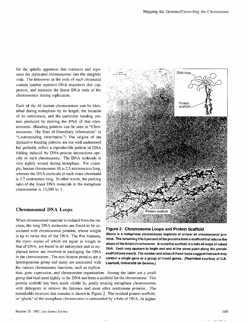
for the spindle apparatus that contracts and sepa-
rates the replicated chromosomes into the daughter
cells. The telomeres at the ends of each chromatid
contain tandem repeated DNA sequences that cap,
protect, and maintain the linear DNA ends of the
chromosomes during replication.
Each of the 46 human chromosomes can be iden-
tified during metaphase by its length, the location
of its centromere, and the particular banding pat-
tern produced by staining the DNA of that chro-
mosome. (Banding patterns can be seen in “Chro-
mosomes: The Sites of Hereditary Information” in
“Understanding Inheritance.”) The origins of the
distinctive banding patterns are not well understood
but probably reflect a reproducible pattern of DNA
folding induced by DNA-protein interactions spe-
cific to each chromosome. The DNA molecule is
very tightly wound during metaphase. For exam-
ple, human chromosome 16 is 2.5 micrometers long,
whereas the DNA molecule in each sister chromatid
is 3.7 centimeters long. In other words, the packing
ratio of the linear DNA molecule in the metaphase
chromosome is 15,000 to 1.
Chromosomal DNA Loops
When chromosomal material is isolated from the nu-
cleus, the long DNA molecules are found to be as-
sociated with chromosomal proteins, whose weight
-,,!, <W--- , ,
Figure 2. Chromosome Loops and Protein ScaffoldAbove is a metaphase chromosome depleted of almost all chromosomal pro-
is up to twice that of the DNA. The five histones,
the many copies of which are equal in weight toteins. The remaining 2to 3 percent of the proteins form a scaffold that retains the
that of DNA, are found in all eukaryotes and as ex-shape of the intact chromosome. Around the scaffold is a halo of loops of naked
DNA. Each loop appears to begin and end at the same point along the proteinplained below are involved in packaging the DNA scaffold (sac insert). The number and sizes of these loops suggest that each mayin the chromosomes. The non-histone proteins are a contain a single gene or a group of linked genes. (Reprinted courtesy of U.K.
heterogeneous group and many are associated with Laemrnli, University de Gen&e,)
the various chromosome functions, such as replica-
tion, gene expression, and chromosome organization. Among the latter are a small
group that bind most tightly to the DNA and form a scaffold for the chromosome. This
protein scaffold has been made visible by gently treating metapliase chromosomes
with detergents to remove the histones and most other nonhistoric proteins. The
remarkable structure that remains is shown in Figure 2. The residual protein scaffold,
or “ghost,” of the metaphase chromosome is surrounded by a halo of DNA. At higher
Number 20 1992 Los Alamos Science 169
Mapping the Genome/Unraveling the Chromosome
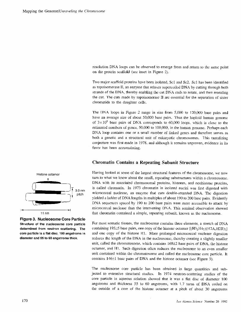
Histone octamer
t
1- *I
llnm
Figure 3. Nucleosome Core ParticleStructure of the nucleosome core particle
determined from neutron scattering. The
core particle is a flat disc, 100 angstroms in
diameter and 55 to 60 angstroms thick.
resolution DNA loops can be observed to emerge from and return to the same point
on the protein scaffold (see inset in Figure 2).
Two major scaffold proteins have been isolated, SC1 and SC2. Scl has been identified
as topoisomerase II, an enzyme that relaxes supercooled DNA by cutting through both
strands of the DNA, thereby enabling the cut DNA ends to rotate, and then resealing
the cut. The cuts made by topoisomerase II are essential for the separation of sister
chromatics to the daughter cells.
The DNA loops in Figure 2 range in size from 5,000 to 120,000 base pairs and
have an average size of about 50,000 base pairs. Thus the haploid human genome
of 3 x 10⁹ base pairs of DNA corresponds to 60,000 loops, which is close to the
estimated numbers of genes, 50,000 to 100,000, in the human genome. Perhaps each
DNA loop contains one or a small number of linked genes and therefore serves as
both a genetic and a structural unit of eukaryotic chromosomes. This tantalizing
conjecture was first made in 1978, and although it remains unproven, evidence in its
favor has been accumulating.
Chromatin Contains a Repeating Subunit Structure
Having looked at some of the largest structural features of the chromosome, we now
turn to what we know about the small, repeating substructures within a chromosome.
DNA with its associated chromosomal proteins, histones, and nonhistoric proteins,
is called chromatin. In 1973 chromatin in isolated nuclei was first digested with
micrococcal nuclease, an enzyme that cuts double-stranded DNA. The digestion
yielded a ladder of DNA lengths in multiples of about 190 to 200 base pairs. Evidently
DNA sequences spaced by 190 to 200 base pairs were more accessible to attack by
micrococcal nuclease than the intervening DNA. This seminal observation showed
that chromatin contained a simple, repeating subunit, known as the nucleosome.
For most somatic tissues, the nucleosome contains three elements, a stretch of DNAcontaining 195+5 base pairs, one copy of the histone octamer [(H3zH4z)(H2A,H2B )z]
and one copy of the histone H 1. More prolonged micrococcal nuclease digestion
reduces the length of the DNA in the nucleosome, thereby creating a slightly smaller
unit, called the chromatosome, which contains 168+2 base pairs of DNA, the hi stone
octamer, and H 1. Such digestion often reduces the nucleosome to an even smaller
unit contained within the chromatosome and called the nucleosome core particle. It
contains 146*1 base pairs of DNA and the histone octamer (see Figure 3).
The nucleosome core particle has been obtained in large quantities and sub-
jected to extensive structural studies. In 1974 neutron-scattering studies of the
core particle in aqueous solution showed that it was a flat disc of diameter 100
angstroms and thickness 55 to 60 angstroms, with 1.7 turns of DNA coiled on
the outside of a core of the histone octamer at a pitch of about 30 angstroms
170 Los Alamos Science Number 20 1992

Mapping the Genome/Unraveling the Chromosome
(Figure 3). Subsequent x-ray-diffraction studies of crystallized
core particles achieved a resolution of 6 to 7 angstroms. The
crystal structure (Figure 4) not only confirmed the lower resolution
solution structure achieved by neutron scattering but also showed
that histones are in contact with the minor groove of DNA and
leave the major groove available for interactions with the proteins
that regulate gene expression and other DNA functions. The 7-
angstrom-resolution crystal structure also revealed that DNA does
not bend uniformly but rather bends gently and then more sharply
around the histone octamer. Such a path implies that flexibility,
or bendability, of DNA may be sequence-dependent and that the
underlying DNA sequence along the molecule may determine
the positions of some nucleosomes. The most recent work on
nucleosome positioning shows that the bulk of nucleosome core
particles are able to move along the DNA molecule between a
cluster of positions separated by about 10 base pairs. This mobility
is probably required during DNA replication and transcription to
allow DNA polymerases and other enzymes access to specific
DNA sequences.
Despite considerable effort to achieve higher resolution, the best
data for the core particle structure is at a resolution of about 6
angstroms. However, the crystal structure of the isolated hi stone
octamer has been solved to the higher resolution of 3.3 angstroms.
This structure shows shapes of the individual histones and the
nature of interhistone interaction of most but not all of the histone
polypeptide chains. In particular, the basic N-terminal domains,
comprising some 20 to 25 percent of the histone octamer, are
not “seen” in the crystal structure, probably because they bind to
Figure 4. Crystal Structure of Core ParticleThe structure of the nucieosome core particle as determined by
x-ray diffraction is shown above. At a resolution of 6 to 7
angstroms, this top view of the core particle shows that the DNA
(brown) does not follow a smooth path around the histone
octamer (blue and turquoise) but rather bends sharply and then
more gently. (Reprinted courtesy of Uberbacherand Bunick, Oak
Ridge National Laboratory.)
DNA, and in the absence of DNA, they are disordered. These N-terminal domains
contain all of the sites of the cell-cycle-dependent acetylation of lysines and phospho-
rylation of serines or threonines. Acetylation of Iysine converts it from a positively
charged residue, which can therefore bind to DNA, to a neutral acetylysine. It has
been shown first that lysine acetylation is strictly correlated with transcription and
DNA replication, and more recently, that histone acetylation drives the uncoiling
of part of the DNA from the nucleosome to allow the initiation and progression of
DNA replication and transcription.
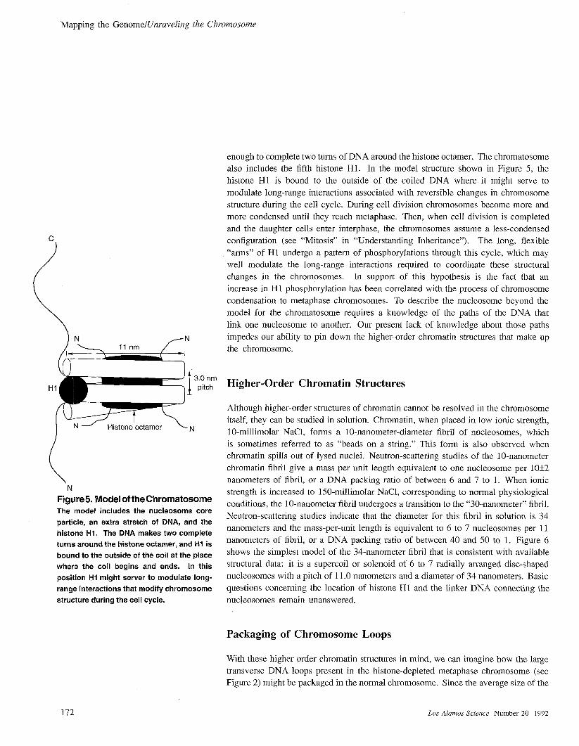
Chromatosomes and Nucleosomes
A model of the structure of the chromatosome (Figure 5) has been inferred from
the structures of the nucleosome core particle and the histone H 1. The core particle
has 1.7 turns of DNA at a pitch of 3.0 nanometers (30 angstroms) coiled around the
histone octamer. Consequently, the chromatosome’s 168 base pairs of DNA are long
Number 20 1992 Los Alamos Science 171
Mapping the GenorneIUnraveling lhe Chromosome
“i!!zR?3””\ N - I-listorw’octa.rnet’ ~jq
N
Figure5. Model of the ChromatosomeThe model includes the nucleosome core
particle, an extra stretch of DNA, and the
histone HI, The DNA makes two complete
turns around the histone octamer, and HI is
bound to the outside of the coil at the place
where the coil begins and ends. In this
position HI might server to modulate long-
range interactions that modify chromosome
structure during the cell cycle.
enough to complete two turns of DNA around the histone octamer. The chromatosome
also includes the fifth histone H 1. In the model structure shown in Figure 5, the
histone HI is bound to the outside of the coiled DNA where it might serve to
modulate long-range interactions associated with reversible changes in chromosome
structure during the cell cycle. During cell division chromosomes become more and
more condensed until they reach metaphase. Then, when cell division is completed
and the daughter cells enter interphase, the chromosomes assume a less-condensed
configuration (see “Mitosis” in “Understanding Inheritance”). The long, flexible
“arms” of H 1 undergo a pattern of phosphorylations through this cycle, which may
well modulate the long-range interactions required to coordinate these structural
changes in the chromosomes. In support of this hypothesis is the fact that an
increase in H 1 phosphorylation has been correlated with the process of chromosome
condensation to metaphase chromosomes. To describe the nucleosome beyond the
model for the chromatosome requires a knowledge of the paths of the DNA that
link one nucleosome to another. Our present lack of knowledge about those paths
impedes our ability to pin down the higher-order chromatin structures that make up
the chromosome.
Higher-Order Chromatin Structures
Although higher-order structures of chromatin cannot be resolved in the chromosome
itself, they can be studied in solution. Chromatin, when placed in low ionic strength,
10-millimolar NaCl, forms a 10-nanometer-diameter fibril of nucleosomes, which
is sometimes referred to as “beads on a string.” This form is also observed when
chromatin spills out of lysed nuclei. Neutron-scattering studies of the 10-nanometer
chromatin fibril give a mass per unit length equivalent to one nucleosome per 10i2
nanometers of fibril, or a DNA packing ratio of between 6 and 7 to 1. When ionic
strength is increased to 150-millimolar NaCl, corresponding to normal physiological
conditions, the 10-nanometer fibril undergoes a transition to the “30-nanometer” fibril.
Neutron-scattering studies indicate that the diameter for this fibril in solution is 34
nanometers and the mass-per-unit length is equivalent to 6 to 7 nucleosomes per 11
nanometers of fibril, or a DNA packing ratio of between 40 and 50 to 1. Figure 6
shows the simplest model of the 34-nanometer fibril that is consistent with available
structural data: it is a supercoil or solenoid of 6 to 7 radially arranged disc-shaped
nucleosomes with a pitch of 11.0 nanometers and a diameter of 34 nanometers. Basic
questions concerning the location of histone HI and the linker DNA connecting the
nucleosomes remain unanswered.
Packaging of Chromosome Loops
With these higher order chromatin structures in mind, we can imagine how the large
transverse DNA loops present in the histone-depleted metaphase chromosome (see
Figure 2) might be packaged in the normal chromosome. Since the average size of the
172 Los A[amos Sciwzce Number 20 1992

Mapping the Genome/Unraveling the Chromosome
//,,/
,
i\
‘\,:‘.
\.
,.-.,..,, ~’
,, Metaphase
DNA 100p
Figure 6. The Packagingof a DNA Loop
Artist’s rendition shows the
packaging of a DNA loop,
first into the different
orders of chrornatin, and
then into a twisted loop
within a metaphase
chromosome. The
DNAs double helix
makes two turns about
a histone octamer to
form the nucleosome,
the repeating unit in
chromatin. The
chromatosomes, or
nucieosomes bound to
H1, are shown forming a
thick chromatin fiber 34
nanometers in diameter.
In this model, the supercoil
of chromatosomes is further
condensed into a twisted loop
attached at a single point to the
protein scaffold. The spiraling
array of twisted loops constitutes
the familiar metaphase chromosome,
which is visible through the light
microscope.
Mapping the Genome/Unraveling the Ch~omosome
DNA loops is 50,000 base pairs, or 17 micrometers in length, each loop of DNA can
form a string of nucleosomes that are either coiled to form 2.6 micrometers of a 10
nanometer fiber, or supercooled into 0.4 micrometers of a 34 nanometer fiber. Thus, to
create the thickness of a sister chromatid (Figure 1), which is 1 micron in diameter,
would require just one more order of chromatin folding above the 34 nanometer
supercoil. Figure 6 shows a possible model of this final level of chromatin folding.
How is the packaging of DNA loops controlled in response to chromosome functions?
Evidence suggests that the inactive form of chromatin is the 34-nanometer supercoil
or solenoid of nucleosomes. For both DNA transcription and genome replication this
supercoil of nucleosomes must first be uncoiled to the linear array of nucleosomes
and then the DNA must uncoil even further to allow access of the transcriptional
machinery or the replication machinery to the DNA sequences. Whenever DNA is
constrained by proteins to form a loop, DNA supercooling becomes an important con-
sideration in understanding DNA structure-function relationships. DNA supercooling
has been subjected to extensive experimental and mathematical analysis,
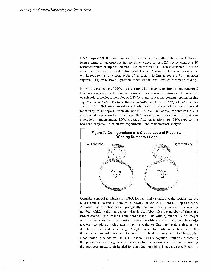
Figure 7. Configurations of a Closed Loop of Ribbon withWinding Numbers +1 and -1
Consider a model in which each DNA loop is firmly attached to the protein scaffold
of a chromosome and is therefore somewhat analogous to a closed loop of ribbon.
A closed loop of ribbon has a topologically invariant property known as the winding
number, which is the number of twists in the ribbon plus the number of times the
ribbon crosses itself, that is, coils about itself. The winding number is an integer
or half-integer and remains constant unless the ribbon is cut. Each complete twist
and each complete crossing adds + 1 or – 1 to the winding number depending on the
direction of the twist or crossing. A right-handed twist (the same direction as the
thread of a standard screw and the standard helical structure of a double-stranded
DNA molecule) is positive, and a left-handed twist is negative. Similarly, a crossing
that produces an extra right-handed loop in a loop of ribbon is positive, and a crossing
that produces an extra left-handed loop in a loop of ribbon is negative (see Figure 7).
174 Los Alamos Science Number 20 1992

Mapping the GenomeJUnraveling the Chromosome
Now consider a loop of double-stranded DNA. Unconstrained DNA has 10.4 to 10.6
base pairs in each complete turn of the double helix. Taking the value 10.6 base
pairs per helical turn, the twist (Ti) of a loop of unconstrained DNA consisting of N
base pairs would be N/10.6. Because a double-stranded DNA molecule already has
a helical structure, a loop of DNA further coiled about itself is said to be supercooled.
The linking number (UC) of a closed loop of DNA is defined in terms of the twist
and the number of supercools, or writhe (W-), through the equation Lk = Tw + W.
Twists can be converted into supercools, but Lk must remain constant in a DNA loop
whose ends are fixed, in analogy with the constancy of the winding number of the
loop of ribbon. If the loop is closed, the linking number must be an integer.
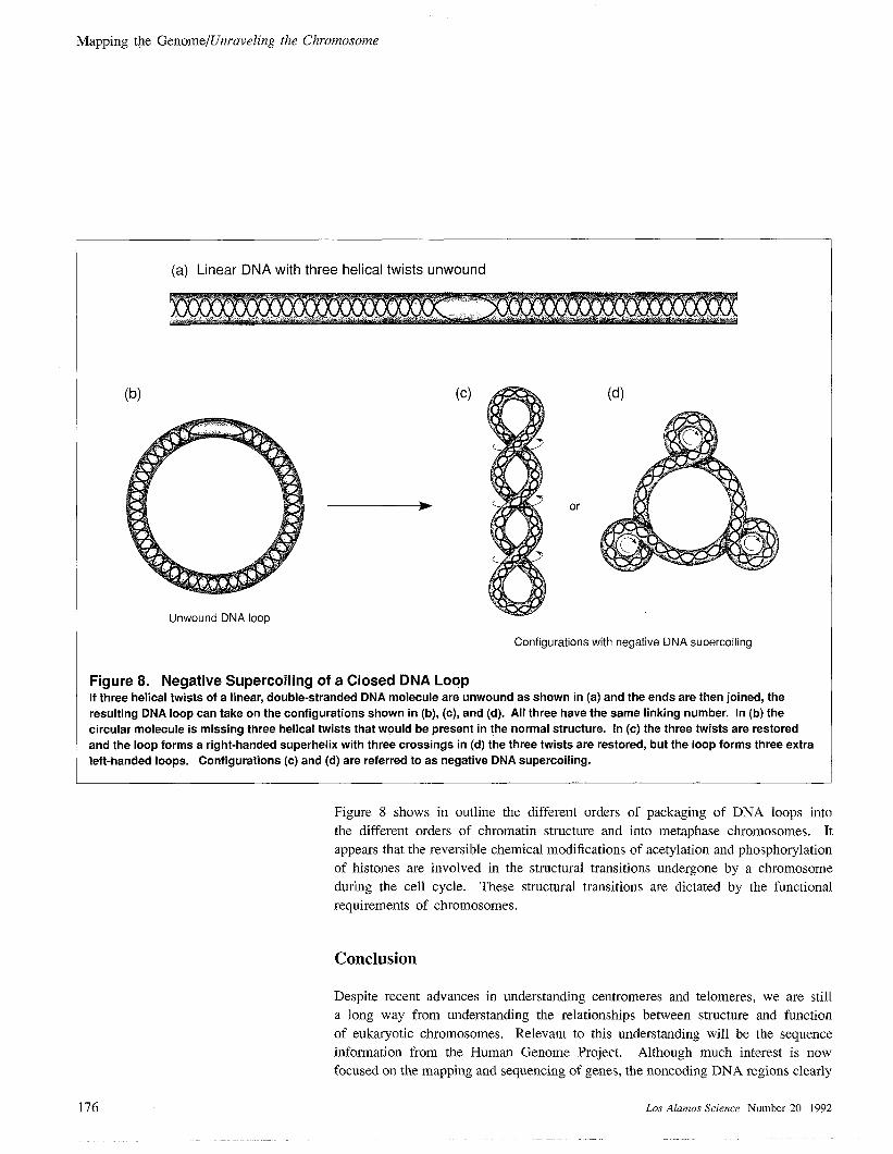
As an example, suppose three helical turns of a linear stretch of DNA are unwound and
the ends are then joined. The linking-number change resulting from the unwinding
is – 3, and the loop can take on any of the three configurations shown in Figure 8.
Moreover, the three configurations can be converted into one another without cutting
the DNA. DNA configured as in (b) and (c) is said to be negatively supercooled.
As shown in Figure 3, the DNA in the nucleosome core particle has 1.7 left-
handed supercools and in early studies it was expected that the linking-number
change associated with the dissociation of a core particle would be —1.7. However,
the experimentally determined linking-number change was – 1.02. Although this
difference was unexpected and initially controversial, it is easily explained by the
change in twist between the DNA constrained in the core particle and free DNA in
solution. The average DNA helical repeat on the core particle as measured from its
crystal structure is 10.1 base pairs per turn. If we take the average helical repeat of
free DNA as 10.6 base pairs per turn, the difference in twist between the DNA in the
core particle and free DNA would be 146/10.1 – 146/10.6 that is, 0.68. Thus the
linking-number change associated with the core particle ALk = – 1.7+0.68 = – 1.02
as observed.
Now we can suggest how a DNA loop packaged as a 34-nanometer supercoil
of nucleosomes (see Figure 6) could be unwound during interphase. If negative
supercools previously constrained by the nucleosomes are released, then negative
supercooling must be taken up by the linker DNA joining one nucleosome to another.
This negative supercooling would favor the unwinding of a 34-nanometer supercoil of
nucleosomes. As suggested above, the acetylation of histones releases DNA that was
negatively supercooled about the histone octamer, presumably by unwinding DNA
from the ends of the nucleosome.
The reverse process of chromosome condensation to the metaphase configuration (see
Figure 1) requires that the 34-nanometer supercoil be further coiled into higher orders
of coiling(s). Perhaps histone-H 1 phosphorylation introduces additional supercooling
into a packaged DNA loop causing the higher order of ceilings of metaphase
chromosomes.
Number 20 1992 Los Akzmos Science 175
Mapping the GenomelUnraveling the Chromosome
(b)
(a) Linear DNA with three helical twists unwound
Unwound DNA loop
(c)
or
(d)
Configurations with negative DNA supercooling
Figure 8. Negative Supercooling of a Closed DNA LoopIf three helical twists of a linear, double-stranded DNA molecule are unwound as shown in (a) and the ends are then joined, the
resulting DNA loop can take on the configurations shown in (b), (c), and (d). All three have the same linking number. In (b) the
circular molecule is missing three helical twists that would be present in the normal structure. In (c) the three twists are restoredand the loop forms a right-handed superhelix with three crossings in (d) the three twists are restored, but the loop forms three extraleft-handed loops. Configurations (c) and (d) are referred to as negative DNA supercooling.
Figure 8 shows in outline the different orders of packaging of DNA loops into
the different orders of chromatin structure and into metaphase chromosomes. It
appears that the reversible chemical modifications of acetylation and phosphorylation
of histones are involved in the structural transitions undergone by a chromosome
during the cell cycle. These structural transitions are dictated by the functional
requirements of chromosomes.
Conclusion
Despite recent advances in understanding centromeres and telomeres, we are still
a long way from understanding the relationships between structure and function
of eukaryotic chromosomes. Relevant to this understanding will be the sequence
information from the Human Genome Project. Although much interest is now
focused on the mapping and sequencing of genes, the noncoding DNA regions clearly
176 Los A[amos Science Number 20 1992
Mapping the Genome/Unraveling the Ch~omosome
contain information involved in the organization and functions of chromosomes. The
constancy of the banding patterns of individual metaphase chromosomes reflects
a highly reproducible pattern of long-range DNA folding, most probably directed
by specific DNA-protein interactions and possibly by unusual DNA structures such
as bent DNA segments. Superimposed on the very long-range order suggested by
banding patterns is the packaging of the DNA loops by the histones together with
other structural and regulatory proteins.
The existence of several subtypes of each histone raises the possibility that DNA loops
containing different gene families could be packaged with different types of histones
according to the requirements of the different cells. DNA control regions of active
genes must be packaged in a fashion that makes them accessible to gene-regulating
proteins, whereas regions containing permanently repressed genes of a particular cell
type may be packaged so that they are inaccessible to such proteins. Such packaging
may also determine the availability of DNA regions to chemical damage. Thus a
knowledge of the organization of chromosomes is essential to an understanding of
the central processes of cell differentiation and the orderly development of complex
organisms as well as the processes of DNA damage in chromosomes. ■
Further Reading
E, Morton Bradbury, “Reversible Histone Modifications and the Chromosome Cell Cycle.” BioEssaw,Volume 14, No. 1: January 1992,
Morton Bradbury received a bachelor of sciencedegree in physics and a Ph.D. in biophysics fromKing’s College, University of London, in 1955 and1958, respectively. After completing his postdoc-toral research at Courtauld Research Laboratory, hewas appointed head of the Department of Molecu-lar Biology at Portsmouth (England) Polytechnic in1962, where he remained until his appointment at UCDavis in 1979. He became leader of the Life Sci-ences Division at Los Alamos in 1988. Bradbury ’sresearch has been devoted to understanding whetherchromosome organization and chromosome structureare involved in determining how a cell looks and be-haves; the structure and function of active chromatin;and the process by which chromosomes condenseprior to cell division. In pursuing his investiga-tions, Bradbury has combined the results of measure-ments derived from the use of a wide range of techniques, including optical spectroscopy,nuclear magnetic resonance, x-ray diffraction, electron diffraction, and neutron diffraction.The recipient of numerous award and honors, Bradbury has also chaired a number of scientificorganizations, including the British Biophysical Society, the International Council for MagneticResonance in Biology, and the Neutron/Biology Committee of the Institut Laue-Langevin.Bradbury is a member of HERAC and a member of the HERAC subcommittee on structuralbiology.
Number20 1992 Los Akrmos Science 177