UNIVERSITY OF SÃO PAULO FACULTY OF ... - teses.usp.br
114
UNIVERSITY OF SÃO PAULO FACULTY OF PHARMACEUTICAL SCIENCES Department of Biochemical and Pharmaceutical Technology Fermentation Technology Production of L-asparaginase of pharmaceutical interest from yeasts isolated from the Antarctic continent Ignacio Sánchez Moguel Thesis to obtain the degree of DOCTOR Thesis director: Prof. Dr. Adalberto Pessoa Junior SÃO PAULO 2018
Transcript of UNIVERSITY OF SÃO PAULO FACULTY OF ... - teses.usp.br

UNIVERSITY OF SÃO PAULO
FACULTY OF PHARMACEUTICAL SCIENCES
Department of Biochemical and Pharmaceutical Technology
Fermentation Technology
Production of L-asparaginase of pharmaceutical interest from yeasts
isolated from the Antarctic continent
Ignacio Sánchez Moguel
Thesis to obtain the degree of DOCTOR
Thesis director: Prof. Dr. Adalberto Pessoa Junior
SÃO PAULO
2018

UNIVERSITY OF SÃO PAULO
FACULTY OF PHARMACEUTICAL SCIENCES
Department of Biochemical and Pharmaceutical Technology
Fermentation Technology
Production of L-asparaginase of pharmaceutical interest from yeasts
isolated from the Antarctic continent
Ignacio Sánchez Moguel
Original Version
Thesis to obtain the degree of DOCTOR
Thesis director: Prof. Dr. Adalberto Pessoa Junior
SÃO PAULO
2018

SÁNCHEZ-MOGUEL, I. Production of L-asparaginase of pharmaceutical interes from yeasts
isolated from Antarctic continent. 2018. 120 p. Thesis (Ph.D). –Faculty of Pharmaceutical
Sciences, University of São Paulo, 2017.
Examining Board
__________________________________________
Prof. Dr. Adalberto Pessoa Junior
(Thesis director/President)
___________________________________________
1º examiner
____________________________________________
2º examiner
_____________________________________________
3º examiner
_____________________________________________
4º examiner
São Paulo_____________ de_____________ de 2018.

In memory of my father, Macario
To my mother, Mónica

AKNOWLEGDMENTS
Thank you,
To life for this experience, for having brought me here, for having taken a great professional
step, but even more personally, where I got great friendships, and life lessons.
To my family, Judith, Ariadna, Alonso, David, Saiyd, Zayra, Jacob, André, Rodolfo, Simón,
to all of you even when we were away, you were present all this way, my dreams became
yours, you always They supported me in everything. I love you brothers.
To my father, Macario for guiding me in this walk of constant questions and search for
answers.
To my mother, for all your love and care.
To my teachers Adalberto, Solange for everything I learned from them and for being an
essential part of my professional and personal preparation.
To all the people who made this long road, a more enjoyable, more cheerful, more vibrant, of
great experiences, of many laughs and that we share great moments and we share the same
dreams, we learn together and grew professionally and personally. Thank you for teaching me
that borders do not exist.
To all my friends
To Mexico, to CONACYT for the scholarship granted for the realization of this doctorate.
To Brazil, for this great experience

CONTENTS
1 CHAPTER I. LITERATURE REVIEW ............................................................................. 18
1.1 General Introduction .................................................................................................. 18
1.2 L-Asparaginase As A Biopharmaceutical Drug. ....................................................... 21
1.2.1 Anti-Tumor Action Of L-Asparaginase ............................................................... 21
1.2.2 Adverse Effects Of L-Asparaginase Treatment .................................................... 23
1.3 Asparaginases Types And Structural Aspects ........................................................... 25
1.3.1 Mechanism Of Enzymatic Action And Structural Aspects Of L-Asparaginase ... 25
1.3.2 Asparaginases Classification. ................................................................................ 26
1.4 Fonts Of Asparaginases ............................................................................................. 28
1.5 General Objetives ...................................................................................................... 29
1.5.1 Specific Objetives .................................................................................................. 30
2 CHAPTER II. SCREENING AND CHARACTERIZATION OF L-ASPARAGINASE
PRODUCED BY A PSYCHROTOLERANT YEAST Leucosporidium Scottii L115 ............ 31
2.1 Introduction ............................................................................................................... 31
2.1.1 Psychrotolerant and Psychophiles Microorganisms .............................................. 31
2.1.1 Psychrophyilic and Psychrotolerant Enzymes ...................................................... 31
2.1.2 Yeast isolated from Antartic ecosystems .............................................................. 33
2.1.3 Asparaginases of eukaryotic microorganisms ....................................................... 34
2.1.4 Yeast Asparaginases .............................................................................................. 35
2.2 Materials and Methods .............................................................................................. 36
2.2.1 Microorganisms ..................................................................................................... 36
2.2.2 Preparation of inoculum culture and enzyme production ..................................... 36
2.2.3 Assay of periplasmic activity of ASNase and GLNase by hydroxylaminolysis
reaction ............................................................................................................................ 36
2.2.4 Thin Layer Chromatography (TLC) of reaction products from L. scottii cells ..... 37

2.2.5 ASNase assay by hydrolysis reaction .................................................................... 37
2.2.6 Enzyme Extraction ................................................................................................ 37
2.2.7 Enzyme Purification .............................................................................................. 38
2.2.8 Molecular weight determination by Size Exclusion Chromatography ................. 38
2.2.9 Determination of molecular weight in reduced conditions and purity of fractions
obtained ........................................................................................................................... 39
2.2.1 Enzyme Glycosylation Analysis By SDS-PAGE .................................................. 39
2.2.1 Determination of specific activity of ASNase ....................................................... 39
2.2.1 Effect of pH and Temperature ............................................................................... 40
2.2.1 Effect of metal ions on enzyme activity ................................................................ 40
2.2.14 ... Circular Dichroism for secondary structure comparison of LsASNase and E.coli
ASNase type II (ELSPAR®) .......................................................................................... 40
2.2.1 Circular Dichroism for thermal stability of LsASNase ......................................... 41
2.2.1 Determination of kinetic enzymatic parameters .................................................... 41
2.3 Results And Discussion ............................................................................................. 42
2.3.1 Screening Of enzyme activity ............................................................................... 42
2.3.2 Enzyme Purification .............................................................................................. 45
2.3.3 Determination of Molecular Weight by SDS-PAGE and SEC ............................. 48
2.3.4 Glycosylation analysis of LsASNase by SDS-PAGE .......................................... 49
2.3.5 Determination of specific activity ......................................................................... 50
2.3.6 Effect of pH and Temperature on LsASNase ........................................................ 51
2.3.7 Effect of the metal ions in the LsASNase Activity ............................................... 53
2.3.8 Secondary structure and thermal stability assays .................................................. 53
2.3.9 Kinetic parameters determination ......................................................................... 56
2.4 Conclusion. ................................................................................................................ 59
3 CHAPTER III. Optimization of medium components for production of L-Asparaginase
Enzyme by a psychrotolerant yeast Leucosporidium scottii L115 .......................................... 60
3.1 Introduction ............................................................................................................... 60

3.1.1 Development Of Culture Medium ......................................................................... 61
3.1.2 Carbon Sources ...................................................................................................... 62
3.1.3 Nitrogen Sources ................................................................................................... 62
3.1.1 Experimental Design in the Optimization of Medium Composition .................... 64
3.2 Materials And Methods ............................................................................................. 65
3.2.1 Microorganism and inoculum preparation for L-asparaginase production for PB
Design ........................................................................................................................ 65
3.2.2 Assay of ASNase activity ...................................................................................... 65
3.2.3 Plackett-Burman Design ........................................................................................ 66
3.2.4 Inoculum preparation and evaluation of ASNase production by CCD study ....... 67
3.2.5 Response Surface Methodology ............................................................................ 67
3.2.6 Validation of Statistical Model .............................................................................. 68
3.3 Results And Discussions. .......................................................................................... 69
3.3.1 Evaluation of different founts of Nitrogen and Carbon on ASNase Production
using the Placket-Burman Design ................................................................................... 69
3.3.2 Optimization of nutritional factors on ASNase production using Central
Composite Design ........................................................................................................... 71
3.3.3 Validation of the Model ........................................................................................ 75
5 CHAPTER IV ...................................................................................................................... 77
EVALUATION OF ASNASE PRODUCTION AND LIPID ACCUMULATION BY THE
PSYCHROTOLERANT YEAST Leucosporidium Scottii L115 ............................................. 77
5.1 Introduction ............................................................................................................... 77
5.2 Materials And Methods ............................................................................................. 79
5.2.1 Microorganism and Inoculum ............................................................................... 79
5.2.2 Bioreactor assays ................................................................................................... 79
5.2.3 Effect of Initial Cell Concentration In Enzyme Production .................................. 79
5.2.4 Effect of the Carbon Source in the Enzyme Production ....................................... 80
5.2.5 CCD Design for optimum kla determination ......................................................... 80

5.2.6 Analysis ................................................................................................................. 81
5.2.7 Staining Of Yeast Cell With Nile Red Dye For Microscopy Analysis ................. 81
5.2.8 Lipid Extraction ..................................................................................................... 82
5.2.9 Total Lipids Quantification And Determination Of Fatty Acid Composition ...... 82
5.2.1 Fermentation Parameters ....................................................................................... 82
5.2.1 Determination of kla............................................................................................... 83
5.3 Results And Discusion............................................................................................... 85
5.3.1 Effect of Initial Cell Concentration in ASNase Production .................................. 85
5.3.2 Effect of Carbon Source In Enzyme Production ................................................... 86
5.3.3 Effect of Aeration And Agitation .......................................................................... 87
5.3.4 Lipids Accumulation ............................................................................................. 92
5.3.5 Fatty Acid Profile produced by Leucosporidium scottii L115 .............................. 93
5.4 Conclusion ................................................................................................................. 95
6 GENERAL CONCLUSION ................................................................................................ 96
7 FUTURE PERSPECTIVES ................................................................................................ 97
8 REFERENCES .................................................................................................................... 98

LIST OF FIGURES
Figure 1. Therapeutic action of L-asparaginase ...................................................................... 21
Figure 2. Mechanism of L-asparginase enzymes. ................................................................. 26
Figure 3. General classification of Asparaginases. ................................................................. 27
Figure 4. Enzyme activity of ASNase and GLNase in yeast strains isolated from samples
collected in Antarctic Peninsula. .............................................................................................. 43
Figure 5. TLC of reaction products of cell suspension L. scottii L115. ................................. 45
Figure 6. DEAE chromatogram, SEC chromatogram. ............................................................ 46
Figure 7. Molecular weight determination of the purified enzyme LsASNase by SDS-PAGE
in reduced conditions ............................................................................................................. 48
Figure 8. Molecular weight determination of the purified enzyme LsASNase in native
conditions ................................................................................................................................ 49
Figure 9. SDS-PAGE in reduced conditions for each step of the enzyme purification and
LsASNase purified by SEC treated with PGNase F enzyme. ................................................. 50
Figure 10. Determination of specific activity for LsASNase enzyme. ASNase specific
activity, GLNase specific activity. ........................................................................................... 51
Figure 11. Effect of physical parameters on purified LsASNase. Temperature and pH ......... 52
Figure 12. Effect of metal ions and EDTA on the enzyme activity of the LsASNase. ........... 53
Figure 13. Far-UVCD spectrum (190 -260 nm) of LsASNase and EcASNase II ................... 54
Figure 14. Thermal stability assay of: A) EcASNase II (control) B) LsASNase. CD spectrum
at 222 nm and temperatures ranging from 20-95oC. ................................................................ 55
Figure 15. The LsASNase kinetic behavior with L-asparagine as substrate at pH 7.0. .......... 57
Figure 16. Contour plots for ASNase (U L-1) and cell dry weight (g L-1) production. ........... 74
Figure 17 Substrate comsumption and ASNase production in the optimized medium........... 74
Figure 18. Experimental data from the batch cultures of L. scottii L115 (ASNase;
cell;fructose; glucose; and glycerol) . ........................................................................ 88
Figure 19. Experimental data from the batch cultures of L. scottii L115 to assess the carbon
source A) Glycerol, B) Sucrose, C) Glycerol + sucrose. ......................................................... 89
Figure 20. Countor surface of kLa determination at different conditions of aeration and
agitantion in Bioractor 1L. Biostat. Sartourius ........................................................................ 90
Figure 21. Lipid accumulation of Nile red stained cells of Leucosporidium scottii L115
observed trough phase-contrast microscopy and fluorescence microscopy (63x) . ................. 92

LIST OF TABLES
Table 1. Characteristics of Biopharmaceutical L- Asparaginases. .......................................... 23
Table 2. Adverse reactions produced by the L-Asparaginase treatment. ................................ 24
Table 3. Enzymes reported produces by psychrotolerant and psychorphile yeasts. ................ 33
Table 4. Eukaryotic microorganisms reported as producers of ASNase. ................................ 34
Table 5. Yeast strain isolated from samples collected in Antarctic Peninsula tested for the
production of ASNase and GLNase ......................................................................................... 42
Table 6. Purification scheme of ASNase from Leucosporidium scottii . ............................... 47
Table 7. Kinetic parameters of different ASNase enzymes reported that presents allosteric
regulation and the E. coli enzyme. ........................................................................................... 58
Table 8. Average composition of microorganisms. (% dry mass) ......................................... 61
Table 9. Medium composition for ASNase production by eukaryotic microorganisms ......... 63
Table 10. Nitrogen, carbon and complex fonts tested and levels used in PB design .............. 66
Table 11. Plackett-Burman design and responses of ASNase and Biomass production ......... 70
Table 12. Effects of the variables and statistical analysis of the Plackett-Burman design ...... 71
Table 13. Experimental design and responses from ASNase production and biomass
production ................................................................................................................................. 72
Table 14. ANOVA analysis of the production of L-asparaginase by L. scottii according to
CCD design .............................................................................................................................. 73
Table 15. Factor Levels for a CCD Design ............................................................................. 80
Table 16. Experimental design for the optimization of enzyme production ........................... 80
Table 17. Enzyme and lipid production at different concentration of inoculum ..................... 86
Table 18. CCD design to optimize the ASNase production and results of productivity and
conversion ................................................................................................................................ 91
Table 19. Fatty acids composition produced by Leucosporidium scottii L115 ....................... 93

LIST OF ABREVIATIONS
AHA β-aspartohydroxamic acid
ALL Acute lymphoblastic leukemia
ANVISA National Health Surveillance Agency
AsnSynt Asparagine Synthetase enzyme
ASNase L-asparaginase
CCD Central composited design
CD Circular dichroism
cdw Cell dry weight
DEAE Diethylaminoethyl cellulose
H Entalpy
DHA Docohexanoic acid
DoE Design of experiments
EcASNase Escherichia coli Asparaginase
FAME Fatty acid methylesters
GC Gas chromatography
GDH Glutarate dehydronase enzyme
GHA γ-glutamohydroxamic acid
GLNase L-glutaminase
GOT Glutamic oxaloacetic transaminase enzyme
GRAS General Recognized As Safe
hASNase Human asparaginase
Kav Partition coefficient
kcat Turn over constant
KLa Oxygen transference coeficient
K0.5 Constant
KM Michaelis –Menten constant
LDH Lactate dehydrogenase enzyme
LsASNase Leucospordium scotti L- Asparaginase
MUFA Monounsaturated Fatty Acids
MW Molecular weight
β-NADH Nicotin Adenosin des

nH Hill coefficient
OD Optic density
P Product concentration
Pf Final product concentration
P0 Initial product concentration
PB Plackett –Burman design
PBS Phospate buffer solution
PEG Polyethylene glicol
PEG-ASNase Pegylated L-asparaginase
PMSF Phenyl metil sulfonyl F
PUFAs Polyunsaturated fatty acids
Q Volumetric productivity
rDNA Ribosomal DNA
Rf Migration distance
S Substrate concentration
S0 Initial substrate concentration
Sf Final substrate concentration
ScASNase Scharomyces cerevisiae Asparaginase
SCOs Single cell oils
SD Standard deviation
SDS-PAGE SDS- PolyAcrylamide Gel Electrophoresis
SEC Size Exclusion Chromatography
TCA Trichloroacetic acid
TGA Triacylglycerol
TLC Thin Layer Chromatography
Ve Elution volumen
V0 Void volumen
VC Column volume
Vmax Máximum velocity
X Cell mass concentration
X0 Initial cell mass concentration
Xf Final cell mass concentration
YPD Yeast peptone dextrose

YX/S Conversion factor of substrate to cells
YP/S Conversion factor of substrate to products

ABSTRACT
Production of L-asparaginase of pharmaceutical interest from yeasts isolated from the
Antarctic continent
The L-asparaginase (ASNase) obtained from yeasts species has been poorly studied
and a new yeast ASNase could be an alternative to minimize the side effect in the treatment of
lymphoblastic leukemia. The Antarctic ecosystems have a great potential to obtain novel
enzymes produced from psychrophilic and psychrotolerant microorganisms. Yeasts isolated
from samples collected in the Antarctic Peninsula by the PROANTAR expedition team were
tested for the production of ASNase and L-glutaminase (GLNase). From this screening, the
strain Leucosporidium scottii L115 presented the highest ASNase activity (6.24 U g-1 of dried
cell weight (dcw)) with a combination of low GLNase activity (0.41 U g-1 dcw). The ASNase
belonging to L. scottii L115 (LsASNase) was purified 227 fold with a specific activity of
137.01 U mg-1 at 37 ºC, and with 0.93 U mg-1 for GLNase. Moreover, the maximum activity
was observed at pH 7.5 at 55 ºC. The enzyme is a multimer presenting a single band of 54.5
kDa of molecular weight in reduced conditions and 462 kDa by size exclusion
chromatography. The LsASNase is a glycosylated enzyme that presented a band lower at 25
kDa when was treated with PGNase F. The enzymatic kinetic reveals an allosteric regulation
of the enzyme and the kinetic parameters were determined at 37º C, pH 7.0 as K0.5 = 233 μM,
kcat = 54.7 s-1 and nH = 1.52 demonstrating a positive cooperativity by the enzyme and the
substrate. The ASNase production by L. scottii L115 was improved by applying DoE for the
culture medium development. The PB and CDD designs were used to optimize the ASNase
production providing the nutrient values of 6.15 g L-1 of proline, 28.34 g L-1 sucrose, and
15.61 g L-1 of glycerol for a maximal production. The synthetic medium containing the
optimized quantities was added with the salts: KCl, 0.52 g L-1; MgSO4.7H2O, 0.52 g L-1;
CuNO3.3H2O, 0.001 g L-1; ZnSO4.7H2O, 0.001 g L-1; FeSO4.7H2O, 0.001 g L-1.The optimized
medium produces a 23.75 ULh-1 of ASNase in shake flask culture. Furthermore, L. scottii is
characterized as an oleaginous yeast that accumulates lipids with a suitable fatty acid profile.
The production of ASNase and lipids were scaled up in the 1 L bioreactor to evaluate the
initial cell concentration, carbon source, and oxygen transfer rate (kLa).The experiments were
performed at 15ºC in the bioreactor BIOSTAT®Q plus (Sartorius Stedim, Germany) in batch
mode, using 0.5 L of the optimized medium culture in phosphate buffer 50 mM pH 7.0. The
initial cell concentration was evaluated at 1%, 3%, and 5% (v/v). Sucrose and glycerol were
tested alone to examine if the combination of both is mandatory to produce ASNase. All these
assays were carried in duplicate. The kLa was assessed through a CCD design in the range of
1.42 – 123.0 h-1. The performance in bioreactor showed the productivity of 36.95 ULh-1of
ASNase under the optimized conditions (growth temperature 15º C, X0: 5 g L-1, pH 7.0, 48 h,
kLa 89-92 h-1). The cultivation of L. scottii L115 at 15ºC in sucrose and glycerol as carbon
sources generate an interesting lipid profile, where it presents monounsaturated and
polyunsaturated lipids.
Keywords: L-asparginase; Leucosporidium scottii, psychrotolerant yeast, purification;
characterization, purification, production, DoE, bioreactor, lipid accumulation.
Resumo:
Produção de L-asparaginase de interesse farmacêutico a partir de leveduras isoladas do
continente Antártico
A L-asparaginase (ASNase) obtida a partir de espécies de leveduras tem sido pouco estudada
e uma nova ASNase de levedura pode ser uma alternativa para minimizar os efeitos adversos
no tratamento da leucemia linfoblástica. Os ecossistemas Antárticos têm um grande potencial
para obter novas enzimas produzidas a partir de microorganismos psicrofílicos e
psicotrolerantes. As leveduras isoladas de amostras coletadas na Península Antártica pela
equipe de expedição do PROANTAR foram testadas para a produção de ASNase e L-
glutaminase (GLNase). A partir desta triagem, a cepa Leucosporidium scottii L115 apresentou
a maior atividade de ASNase (6,24 U g-1 dcw) com uma combinação de baixa atividade de
GLNase (0,41 U g-1 dcw). A ASNase pertencente a L. scottii L115 (LsASNase) foi purificada
227 vezes com uma atividade específica de 137,01 U mg-1 a 37 ºC e com 0,93 U mg-1 de
GLNase. A atividade máxima foi observada a pH 7,5 a 55 ºC. A enzima é um multímero que
apresenta uma banda única de 54,5 kDa de peso molecular em condições redutoras e 462 kDa
por cromatografia de exclusão molecular. A LsASNase é uma enzima glicosilada que
apresentou uma banda menor a 25 kDa quando tratada com PGNase F. A cinética enzimática
revela uma regulação alostérica da enzima e os parâmetros cinéticos foram determinados a
37º C, pH 7,0 como K0,5 = 233 μM, kcat = 54,7 s-1 e nH = 1,52 demonstrando uma
cooperatividade positiva pela enzima e o substrato. A produção de ASNase por L. scottii L115
foi melhorada aplicando DoE para o desenvolvimento do meio de cultura. Os desenhos
experimentais de PB e CDD forma usados para otimizar a produção de ASNase e forneceram
os valores de nutrientes de 6,15 gL-1 de prolina, 28,34 gL-1 de sacarose e 15,61 gL-1 de
glicerol para uma produção máxima. O meio sintético contendo as quantidades otimizadas foi
adicionado com os sais: : KCl, 0.52 g L-1; MgSO4.7H2O, 0.52 g L-1; CuNO3.3H2O, 0.001 g L-
1; ZnSO4.7H2O, 0.001 g L-1; FeSO4.7H2O, 0.001 g L-1.O meio otimizado produz 23.75 ULh-1
de ASNase em cultivo em frasco agitado. Além disso, L. scottii é caracterizada como uma
levedura oleaginosa que acumula lipídios com um perfil adequado de ácidos graxos. A
produção de ASNase e lipídios foi ampliada no biorreator de 1 L para avaliar a concentração
celular inicial, fonte de carbono e taxa de transferência de oxigênio (kLa). Os experimentos
foram realizados a 15ºC no biorreator BIOSTAT®Q plus (Sartorius Stedim) em modo
batelada, utilizando 0,5 L da cultura de meio otimizado em tampão fosfato 50 mM pH 7,0. A
concentração celular inicial foi avaliada em 1%, 3% e 5% (v / v). Sacarose e glicerol foram
testados isoladamente para examinar se a combinação de ambos é obrigatória para produzir
ASNase. Todos esses ensaios foram realizados em duplicado. O kLa foi avaliado através de
um planejamento CCD na faixa de 1,42-123,0 h-1. O desempenho no biorreator mostrou a
produtividade de 36,95 ULh-1 de ASNase sob condições otimizadas (temperatura de
crescimento 15º C, X0: 5 g L-1, pH 7,0, 48 h, kLa 89-92 h-1). O cultivo de L. scottii L115 a
15ºC em sacarose e glicerol como fontes de carbono gera um perfil lipídico interessante, onde
apresenta lipídios monoinsaturados e poliinsaturados.
Palavras-chave: L-asparginase; Leucosporidium scottii, levedura psicotrolerizante,
purificação; caracterização, purificação, produção, DoE, biorreator, acumulação de lipídios.
18
1 CHAPTER I. LITERATURE REVIEW
1.1 GENERAL INTRODUCTION
The bacterial type II enzyme L-asparaginase (ASNase) is an antitumor agent used in
the treatment of acute lymphoblastic leukemia (ALL). In December 2012 there was a shortage
of ASNase by the interruption in the production of the biopharmaceutical drug, mobilizing the
Brazilian health agencies to search a biosimilar drug. This shortage of ASNase could have
compromised the therapy of 3300 patients treated every year in Brazil, by the lack of a drug
without a substitute in the Brazilian market. The eventual absence of ASNase forced suppliers
to seek the drug in alternative markets such as India, Europe, and China (CANAL SAÚDE /
FIOCRUZ, 2014).
In the current year, the use of a Chinese ASNase formulation again attracted the
attention of media, researchers and the National Health Surveillance Agency (ANVISA) in
the country due to the high content of impurities in the imported drug used in the treatment of
LLA patients. Currently, ASNase formulations are produced around the world but a few have
the quality specifications of the reference drug. These events have placed the ASNase in the
focus of Brazilian researchers and government, interested in the national production of the
enzyme. Although the ASNase has approximately 40 years of use for the treatment of ALL,
there are still deficiencies and problems to be solved. The adverse effects, the short half-life,
the low levels of production at the industrial level are the main topics concerning to be solved
for the clinical use of the enzyme (ZUO et al, 2015).
Approximately in 60% of the patients treated with ASNase is reported the formation
of antibodies anti-ASNase, producing immunogenic reactions or neutralizing the enzyme
affecting the half-life of the drug. Also, the toxicity of ASNase is attributable to the
Glutaminase (GLNase) activity of the enzyme producing several side effects such as
leucopenia, neuralgia immunosuppression, acute pancreatitis, thromboembolism and,
hyperglycemia (OHNUMA et al, 1970; PANOSYAN et al, 2004). Being a serendipitous
biopharmaceutical drug born before the era of genetic engineering, the re-study and
production improvement of the enzyme during the next years could solve the drawbacks of
the ASNase therapy.
19
The microorganisms have been considered as the most important source of ASNase, a
wide range such as bacteria, fungi, yeasts, actinomycetes, and algae have proved to be
proficient sources of this enzyme and, are easily cultivated in bioreactors, with controlled
conditions such as pH, temperature, aeration, medium composition and other parameters,
leading to high reproducibility titters of the enzyme. The bioprospection for new
microorganisms producing ASNase as potential candidates for the formulation of new drugs
is needed to improve the production, activity, and specificity of the enzyme (SAVITRI and
AZMI, 2003).
The Brazilian biomes are an important hotspot for biodiversity, occurring also in the
microbial life of Brazilian soils, such as those from Amazonia, Catinga, Atlantic rainforest,
Pantanal, and Pampa (ANDREOTE et al 2017). Moreover, trough the Brazilian Antarctic
Program (PROANTAR), Brazil participates in the scientific research in the Antarctic realizing
scientific explorations and bioprospection of this little-explored biome with an incredible
biotechnological potential. The Antarctic cold environments were successfully colonized by
numerous microorganisms, including yeasts, fungus and bacteria. Organisms from these
habitats may provide unique biomolecules for industry and medicine. These organisms have
no temperature regulation and their internal temperature is close, if not identical, to that of the
environment. Therefore, they have developed several adaptations through structural changes
at the level of membranes, proteins and constitutive enzymes that allow them to compensate
the effects of low temperatures (GERDAY, 2000).
The enzymes are essential for the adaptation of an organism to a cold environment.
This fundamental aspect is closely associated with a strong biotechnological interest in the
unique properties of these enzymes produced by microorganisms capable of growing at
temperatures close to 0 ° C. Most psychrotolerant and psychrophilic enzymes are
characterized by a change in the apparent optimum temperature of activity, a high rate of
reaction (up to 10 times higher in comparison with the kcat of homologous mesophilic
enzymes) and many cold adapted proteins have flexible point regions, around the active site.
The high flexibility of the enzyme is reflected in reduction of H, high kcat and in most cases
a higher KM (SIDDIQUI, 2006).
20
The yeasts present some characteristics that make them attractive for the production of
biopharmaceuticals, because they are organisms well studied, most are listed as GRAS
(General Recognized As Safe) organisms and have been used in industrial applications, grow
relatively fast in simple media,and there are on the market fermentation equipment and
technologies that operate on industrial scales (WALSH 2007) .
In this context, due to the possibility of finding in yeasts species producers of ASNase
in combination with the property of the psychro-tolerant and psychrophile microorganisms to
generate enzymes with improved kinetic properties that could have different characteristics of
the current ASNases. The present study aims the screening of ASNase production in yeast
isolated from samples collected in the Antarctic Peninsula, during the expeditions realized by
the PROANTAR program. The search and studied the new ASNases enzymes could allow the
generation of a new biopharmaceutical that could be used for the treatment of ALL.

21
1.2 L-ASPARAGINASE AS A BIOPHARMACEUTICAL DRUG.
1.2.1 Anti-tumor action of L-Asparaginase
The LLA and other tumor cells are unable to produce the enzyme Asparagine
Synthetase (AsnSynt) responsible for the de novo synthesis of L-asparagine in normal cells.
This dependence of exogenous L-asparagine, to survive turns it essential for malignant cells
and not for normal cells, this metabolic deficiency makes possible to attack cancer cells with
specificity. The presence of ASNase in the blood stream, limit the intake of L-asparagine for
tumor cells necessary for the synthesis of proteins triggering a metabolic imbalance in the cell
resulted in the cell death via apoptosis (AVRAMIS and TIVARI, 2006; COVINI et al, 2012).
For this reason, the use of ASNase represents a potent anti-leukemic agent, especially when
used in combination with other chemotherapeutic agents such as vincristine, methotrexate,
cytarabine, daunorobicin and doxorubicin (VAN DEN BERG, 2011).
Figure 1. Therapeutic action of L-asparaginase (Modified from Van der Berg et al 2011).

22
The history of ASNase begins in 1953 when Kidd of Cornell Medical College
discovers that guinea pig serum reverses induced leukemia in rats, latter in 1961 Broome and
Cornell identified the ASNase as the inhibitory agent present in guinea pig serum, the
presences of ASNase in the blood of guinea pigs was previously reported by Clementi in
1922, who suggested that ASNase was an adaptation of the herbivore animals to a diet rich in
L-asparagine. Due to the large amounts of serum required for the inhibition of tumors,
alternative sources of the enzyme were sought. In 1964 Mashbur and Wriston reported the
production of ASNase by Escherichia coli, this discovery allowed the supply of quantities
needed to conduct of ASNase clinical studies (COONEY and HANDSCHUMACHER, 1970;
LIVINGSTON and KRAKOW, 1970). Finally in 1978 after many clinical trials, the FDA
approved ASNase as a drug for the treatment of leukemia. And, in 2011 the ASNase from E.
chrysanthemi was approved for its use in patients with hypersensitivity to E. coli ASNase
(GERVAIS et al 2013).
The bacterial type II ASNase are the only currently type of enzyme uses for
therapeutic treatment of leukemic lymphoid and other cancers. There are three dosage forms
commercially available, the ASNase from Escherichia coli, (Elspar®, Kidrolase® Leunase®,
MedacTM, CrastininTM), some of these formulations there are no longer available, the enzyme
isolated from Erwinia chrisantemy (Erwinase®) and the pegylated E. coli enzyme
(Oncospar®) that presents fewer hypersensitivity reactions that native form (PIETERS et al,
2012). The enzyme as a drug from both sources has identical mechanism of action, although,
the pharmacokinetic properties of the two enzymes differ, for example, Erwinia's ASNase is
considered less toxic and is often employed when there are allergic reactions to the E. coli
ASNase. However, it has a shorter half-life that E. coli enzyme, suggesting the need to
discover new ASNases that are serologically different and have similar therapeutic effects
(NARTA et al, 2007). Since then the search for ASNases has been based on the kinetic
properties and the physicochemical and biochemical conditions (such as optimum pH,
optimum temperature, substrate specificity, inhibition patterns, toxicity) and this varies in
each microorganism, these differences stimulate the demand for the search the best ASNase
for a better treatment of ALL, that is, an enzyme with the highest activity, lower adverse
effects and a higher half-life. This requires the screening of samples from several potential
sources for the isolation of microorganisms that have the desired capacity to produce the
enzyme (VERMAN et al, 2007).
23
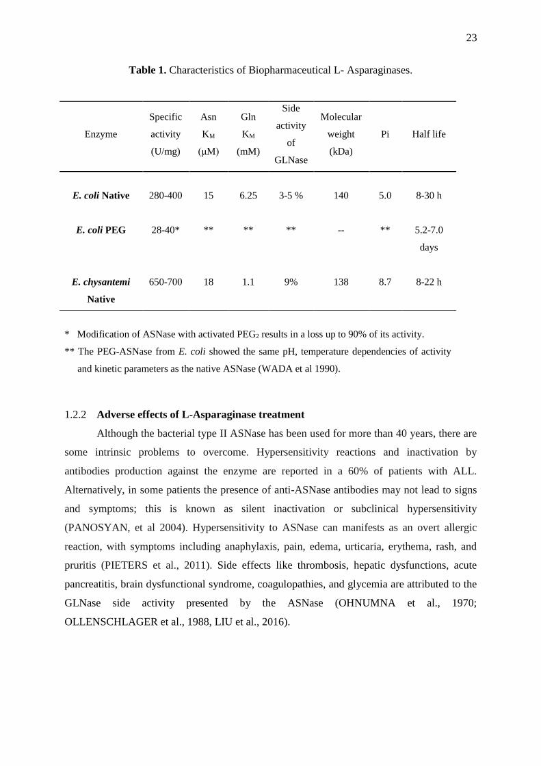
Table 1. Characteristics of Biopharmaceutical L- Asparaginases.
Enzyme
Specific
activity
(U/mg)
Asn
KM
(μM)
Gln
KM
(mM)
Side
activity
of
GLNase
Molecular
weight
(kDa)
Pi Half life
E. coli Native
280-400
15
6.25
3-5 %
140
5.0
8-30 h
E. coli PEG 28-40* ** ** ** -- **
5.2-7.0
days
E. chysantemi
Native
650-700
18
1.1
9%
138
8.7
8-22 h
* Modification of ASNase with activated PEG2 results in a loss up to 90% of its activity.
** The PEG-ASNase from E. coli showed the same pH, temperature dependencies of activity
and kinetic parameters as the native ASNase (WADA et al 1990).
1.2.2 Adverse effects of L-Asparaginase treatment
Although the bacterial type II ASNase has been used for more than 40 years, there are
some intrinsic problems to overcome. Hypersensitivity reactions and inactivation by
antibodies production against the enzyme are reported in a 60% of patients with ALL.
Alternatively, in some patients the presence of anti-ASNase antibodies may not lead to signs
and symptoms; this is known as silent inactivation or subclinical hypersensitivity
(PANOSYAN, et al 2004). Hypersensitivity to ASNase can manifests as an overt allergic
reaction, with symptoms including anaphylaxis, pain, edema, urticaria, erythema, rash, and
pruritis (PIETERS et al., 2011). Side effects like thrombosis, hepatic dysfunctions, acute
pancreatitis, brain dysfunctional syndrome, coagulopathies, and glycemia are attributed to the
GLNase side activity presented by the ASNase (OHNUMNA et al., 1970;
OLLENSCHLAGER et al., 1988, LIU et al., 2016).

24
Table 2. Adverse reactions produced by the L-Asparaginase treatment.
Adverse reaction % of patients affected by
the ASNase treatment
(evaluated/affected)
• Immediate
1. Nausea, vomiting, and chills (27/38) 71
• Delayed
1. Hepatic: Increase in bilirubin, GOT,
LDH, alkaline phosphatase, decrease in
albumin, cholesterol, fibrogen, and other
hepatic coagulation factor- fatty
metamorphosis.
(32/33) 97
2. Loss of weight (24/30) 80
3. Azotemia (25/38) 68
4. Neurological: Headache, drowsiness,
depression, disorientation, confusion.
(13/39) 33
5. Pancreatic: Abdominal pains, increase in
amylase or lipase, hyperglycemia,
hypoinsulinemia, malabsorption
syndrome
(6/39) 15
6. Inmunological: Positive skin test,
anaphylactoid reaction
(5/39) 13
* Modified from Ohnumna et al 1970.
Asparagine and glutamine differ structurally in only one methyl group, and hence L-
asparaginases have the dual substrate specificity resulting in a decrease in the concentrations
of both amino acids in the body. Therefore, the production of an ASNase with a lower affinity
by L-glutamine and regimens with fewer doses need to be reconsidered in order to avoid these
difficulties. Hence, present-day researchers are mainly focused on minimizing or completely
eliminating the L-glutaminase activity of the enzyme L-asparaginase (RAMYA et al., 2012;
KIN CHAN et al., 2014).
But, the need to have a GLNase activity together with ASNase to achieve a therapeutic
effect has generated controversy since in the last decade there has been suggested that
GLNase activity generally increases the efficiency of ASNase and this activity is required to
produce an antitumor effect (ANISHKIN et al., 2015). Clinical studies of an ASNase with
high GLNase activity obtained from Acinetobacter glutaminasificans shows considerable
toxicity, suggesting that the decrease in glutamine produces greater toxicity rather than the
improvement in antitumor activity.

25
On the other hand, there is a hypothesis that an improvement in the treatment with
ASNase can occur if it diminishes the GLNase activity. A "GLNase free" ASNase isolated
from Wolinella succinogenes has been studied by the US National Cancer Institute Rapid
Access to Intervention Development. Unexpectedly this ASNase showed GLNase activity and
was toxic to the patients (NGUYEN et al, 2016). Whereas this enzyme was not actually free
of GLNase activity the question of whether the reduction of GLNase activity improves the
therapeutic index is still unresponsive (CHAN. et al 2014).
1.3 ASPARAGINASES TYPES AND STRUCTURAL ASPECTS
1.3.1 Mechanism of enzymatic action and structural aspects of L-asparaginase
L-Asparaginases (EC. 3.5.1.1) are enzymes that catalyze the hydrolysis of L-
asparagine to L-aspartate and ammonia, the proposed mechanism of enzymatic action of the
ASNase is analogous to that of serine proteases. In the first step, a nucleophilic attack on the
amide carbon atom of the L-asparagine substrate, launched by the hydroxyl group of a
catalytic residue, Thr12 or Thr89, leads via a tetrahedral transition state to a β-acyl-enzyme
intermediate, which in the second step undergoes another nucleophilic attack from the water
molecule, with conversion of the second tetrahedral species into the final product. Structural
and functional studies revealed that the so-called catalytic triad composed of three polar
amino acids, namely Thr-Lys-Asp is essential for enzyme activity (BOREK, et al, 2014).
Although the active site residues of L-asparginases have been identified in the early
crystallographic studies, and the structural as well as kinetic experiments resulted in a
definition of two basic requirements for suitable active site ligand as its size and chemical
composition, details of specificity determinants for both the enzyme and ligand molecules are
just beginning to emerge. This knowledge is instrumental for providing more complete
understanding of the biological properties of ASNase and well as for guiding ist possible
modifications that would allow creation of more efficient therapeutic molecules.
(AGHAIÝPOUR et al 2001).

26
Figure 2. Mechanism of L-asparginase enzymes (Sanson and Jaskolsky, 2004).
1.3.2 Asparaginases classification.
From an amino acid sequence analysis, the ASNases can be classified as bacterial
type, plant type and enzymes similar to Rhizobium etli ASNase (Figure 2) (BOREK and
JASKÓLSKI, 2001). Bacterial ASNases can be further subdivided into two types: type I,
which are expressed constitutively and display enzymatic activity towards both L-
asparaginase and L-glutaminase, and type II, induced by anaerobic condition, which have a
higher specific activity towards L-asparagine. The bacterial-type enzymes frequently exhibit
other activities as well, and this family may be significantly larger than the collection of
sequences deposited as asparaginases. In particular, enzymes such as glutamin-(asparagin)-
ases (EC 3.5.1.38), lysophospholipases (EC 3.1.1.5), and the α-subunit of Glu-tRNA
amidotransferase (EC 6.3.5.-) can also be considered part of the bacterial asparaginase family
(BOREK et al, 2004).

27
Figure 3. General classification of Asparaginases (Borek and Jalskolski 2001).
The bacterial ASNase are the more studied and the best characterized of this enzyme
family, they are composed of four identical subunits, each monomer consist of about 330
amino acid residues, each monomer is formed by two easy identifiable α/β domains, a larger
N-terminal and a smaller C-terminal domain, connected by a loop consisting of about 20
residues. It has been suggested that the flexible loop has an important role in the catalytic
reaction. In an open conformation, it assists in substrate recognition, once the substrate is
bound, the loop undergoes a series of conformational changes allowing several residues to
interact with the substrate molecule and to determine its proper orientation with respect to the
rigid part of the active site. Therefore, loop flexibility is considered to be fundamentally
involved with the enzymatic activity of the type II ASNases. Conversely, is observed that in
the structure of type I ASNases this region is stabilized by the formation of a β-hairpin that
augmented its rigidity, decreasing the affinity for the substrate (SANCHES et al 2007).

28
The monomers are able to associate tightly with each other forming intimate dimers
characterized by an extensive interface between the subunits that are held together by several
interactions, mainly van der Waals and electrostatic interactions. Four independent ASNase
catalytic sites are located at the intersubunit interface of the intimate dimers and, the
association of the two dimers results in the tetrameric biological unit with 222-symmetry and
a molecular mass in the range of 140-150 kDa, which is kept together by molecular
interactions similar to those found in the homodimers, more accurately the ASNase is
described as dimers of intimate dimers (KOTZIA et al 2007; JASKOLSKI et al 2011).
1.4 FONTS OF ASPARAGINASES
The interest for studied new fonts of asparaginases is promoted basically by the
applications anti-carcerigenics properties of these enzymes. The ASNase is present in a wide
variety of organisms including animals, microbes, plants and the serum of some rodents.
Although, ASNase has been found in some species of plants and animals, due to the difficulty
in extracting and purifying other sources such as micro-organisms are preferred (N. EL-
AHMADY EL-NAGGAR et al 2014). Microorganisms are the best sources of ASNases
because they can be grown easily and extraction and purification can be carried out on a large
scale and relatively easily. A wide variety of bacteria, fungi, yeasts, actinomycetes, algae have
been reported as producers of ASNase. Even though not all enzymes of microorganisms have
anti-tumor properties, the variation of this activity has been related to their affinity for the
substrate and the rate of elimination of particular types of enzymes (EL-GONEMY 2014).
According to the literature, the production of ASNase can occur under different conditions by
different microorganisms (SAVITRI and AZMI, 2003).

29
1.5 GENERAL OBJETIVES
Current reviews listed the producer yeasts of ASNases, but none goes beyond that a
few ASNases of yeasts reports in past decades, even so little it is known about the properties
of yeast ASNases as biopharmaceutical, it is expected that the ASNases of yeasts will exhibit
different biochemical and serological characteristics but offering similar or better therapeutics
effects derived from microorganisms that not been studied yet for this purpose.
In this context of the possibility of finding in yeasts species producers of ASNase in
combination with the property of the psychro-tolerant and psychrophile microorganisms to
generate enzymes with improved kinetic properties that could have different characteristics of
the current ASNases. The present study aims the screening, characterization and production of
ASNase from yeast isolated from samples collected in the Antarctic Peninsula, during the
expeditions realized by the PROANTAR program. The search and studied the new ASNases
enzymes could allow the generation of a new biopharmaceutical that could be used for the
treatment of ALL.
30
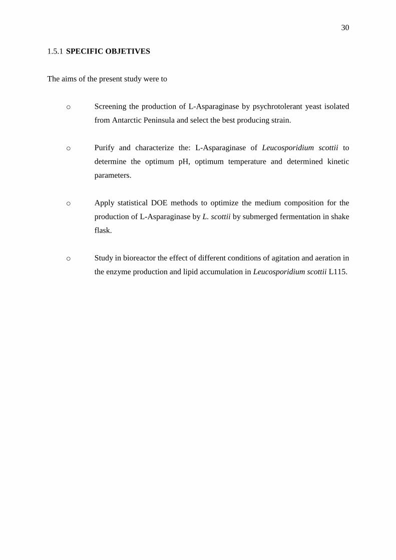
1.5.1 SPECIFIC OBJETIVES
The aims of the present study were to
o Screening the production of L-Asparaginase by psychrotolerant yeast isolated
from Antarctic Peninsula and select the best producing strain.
o Purify and characterize the: L-Asparaginase of Leucosporidium scottii to
determine the optimum pH, optimum temperature and determined kinetic
parameters.
o Apply statistical DOE methods to optimize the medium composition for the
production of L-Asparaginase by L. scottii by submerged fermentation in shake
flask.
o Study in bioreactor the effect of different conditions of agitation and aeration in
the enzyme production and lipid accumulation in Leucosporidium scottii L115.
31
CHAPTER II. SCREENING AND CHARACTERIZATION OF L-
ASPARAGINASE PRODUCED BY A PSYCHROTOLERANT YEAST
Leucosporidium scottii L115
1.6 INTRODUCTION
1.6.1 Psychrotolerant and psychophiles microorganisms
Cold environments were successfully colonized by numerous organisms, particularly
bacteria, single-celled algae, fungi, and yeasts. Ecological niches little explored like the
Antarctic continent has received attention due to the biotechnological potential of the complex
metabolic diversity and unique and particular biomolecules that can present the
microorganisms that inhabit these ecosystems (SHIVAJI and PRASAD 2009). The Antarctic
continent is characterized by its extreme environmental conditions, presenting the coldest and
driest climates known on the planet, very low temperatures (average temperatures below 0 °
C), frequent freeze-thaw cycles, low precipitation and low availability of nutrients, as well as
high salinity and high UV radiation alternated with prolonged periods of obscurity, these
characteristics represent a state of constant stress for the microorganisms that inhabit there
(ONOFRI et al. 2007; MARGESIN and MITEVA 2011). The ability of microorganisms to
survive and grow in cold environments is the result of a range of molecular and physiological
adaptations. Considering that these organisms do not have temperature regulation, their
internal temperature is close, if not identical, to that of the environment. Therefore, they have
developed several adaptations in the form of structural changes at, for example, their
membranes, proteins and constitutive enzymes, to compensate for the drastic effects caused
by the low temperatures (GERDAY et al 2000).
1.6.1 Psychrophyilic and psychrotolerant enzymes
The enzymes are an essential target for adapting an organism to a cold environment.
Therefore, the easiest strategy to maintain a permanent activity at low temperature is the
production of enzymes adapted to cold environments capable of increasing its catalytic
efficiency. The cold-adapted enzymes tend to have an increase in the structural flexibility
resulting in a reduction of the activation energy and consequently increasing the catalytic
efficiency (kcat/KM ratio). In general, the specific activity of psychrophilic enzymes is higher
at temperatures of 0 to 30 ° C than those isolated from mesophiles (FELLER and GERDAY,
1997; HOYOUX et al., 2004)

32
Most psychrophilic enzymes are characterized by a change in the apparent optimum
temperature of activity, a high rate of reaction (up to 10 times higher in comparison with the
kcat of homologous mesophilic enzymes), and many cold-adapted proteins have flexible point
regions around the active site. The high flexibility of the enzyme is reflected in the reduction
of H, high kcat and in most cases a higher KM (SIDDIQUI, 2006). Loops are considered a
diverse class of secondary structures comprising turns, random coils, and stands which
connect the main secondary structures (α-helices and β-strands), and these regions belong to
the most flexible parts of enzyme structures and sequential changes in these regions are
frequently related to the evolution of the enzymes (NESTL and HAUER, 2014).
From a structural point, these proteins have a higher content of α-helix relative to the
β-sheets, which is considered an important factor to maintain the flexibility even at low
temperatures. At low temperature, the low kinetic energy of the molecules involved in the
reaction is compensated by the flexibility in the structures of the enzymes. This flexibility is a
function of the combination of structural characteristics, such as reduction in the
hydrophobicity of the catalytic core, decrease of electrostatic and ionic interactions, increased
loading of surface residues that promote the interaction of solvents, additional loops on the
surface, substitution of proline residues by glycines in these surface loops, decreased
arginine/lysine ratio, less interactions between subunits and interdomains, and less aromatic
interactions. The final effect of these modifications is that the active site and adjacent regions
remain flexible. This increase in conformational flexibility is accompanied by an increase in
thermolability (CAVICCHIOLI et al 2002).
For extracellular enzymes that work at substrate saturation concentrations, the
adaptation mainly consists of increasing kcat. In the contrary, for intracellular enzymes which
may be at conditions where there are low substrate concentrations, the adaptation is observed
in a decreased KM, which means a higher affinity for the substrate. A compilation of available
data indicates that the enzymes isolated from psychrophilic and psychrotolerant
microorganisms optimize their catalytic efficiency (kcat / KM ) mainly by increasing the kcat, in
some cases the KM reduction is achieved and in the best panorama the modification of both
parameters. Thus, an appropriate physiological adaptation at enzyme level is a key aspect for
microorganism survival, being these biocatalysts of great biotechnological interest for the
industry (GERDAY et al., 2000; D´AMICO et al., 2002; CAVICCHIOLI et al., 2011).

33
1.6.2 Yeast Isolated from Antartic ecosystems
About 90% of the yeasts isolated from Antarctica and other cold environments are
from the basidiomycetous origin (DE GARCÍA et al., 2006; SHIVAJI and PRASAD, 2009;
CARRASCO et al., 2012; BUZZINI et al., 2012, DUARTE et al., 2013). The
basidiomycetous yeasts are been poorly explored for the industrial applications but they are
capable to produce valuable metabolites such as enzymes, terpenoids, and carotenoids among
others biomolecules used in pharmaceutical and chemical industries (JOHNSON, 2013). The
Table 3 shows some enzymes isolated from yeasts isolated from Antarctic samples.
Table 3. Enzymes reported produces by psychrotolerant and psychorphile yeasts.
Enzyme Yeast Reference
Serine protease Leucosporidium antarticum Turkiewicz et al 2003
Subtilase Leucosporidum antarcticum Pazgier et al 2003
Lipase A e B Pseudozyma Antarctica
(Candida antarctica)
Michiyo 1986
Pectinase Mrakia frigida Margesin et al 2005
-Amilase Candida Antarctica De Mot and Verachtert,
1987
Invertase, α-
glucosidase
Leucosporidium antarcticum Turkiewicz et al 2005
Acid β-
galactosidase
Guehomyces pullulans Nakagawa et al 2006
Xylanase Crytococcus adelaie Petrescu et al 2000
Xylanase
(termolabile)
Crytococcus adelaie Gomes et al 2000
Polygalacturonases Cryptococcus aquaticus,
Cryptococcus macerans,
Cystofilobasidium
capitatum,
Cystofilobasidium lari-
marini
Birgisson et al 2003
β-galactosidase Guehomyces pullulans 17-1 Song et al 2010
Acid protease Rhodotorula mucilaginosa
L7
Lario et al 2015
Aspartic protease Sporobolomyces roseus Krysiak et al 2016
Pectinases Cystofilobasidium
infirmominiatum,
Cryptococcus adeliensis,
Guehomyces pullulans
Cavello et al 2016
34
The lipase B isolated from Candida Antarctica is used in a very large number of
organic synthesis applications related to food, pharmaceutical, and cosmetics industries,
generating diverse patents, demonstrating the potential of yeasts species that inhabit cold
environments to produces novel molecules with industrial (JOSHEP et al 2008). The yeasts
isolated from Antarctic ecosystems are mainly species belonging to the genus Cryptococcus,
Rhodotorula, Leucosporidiella, Sporobolomyces, Leucosporidium, Candida, Mrakia, and
Meyerozyma.
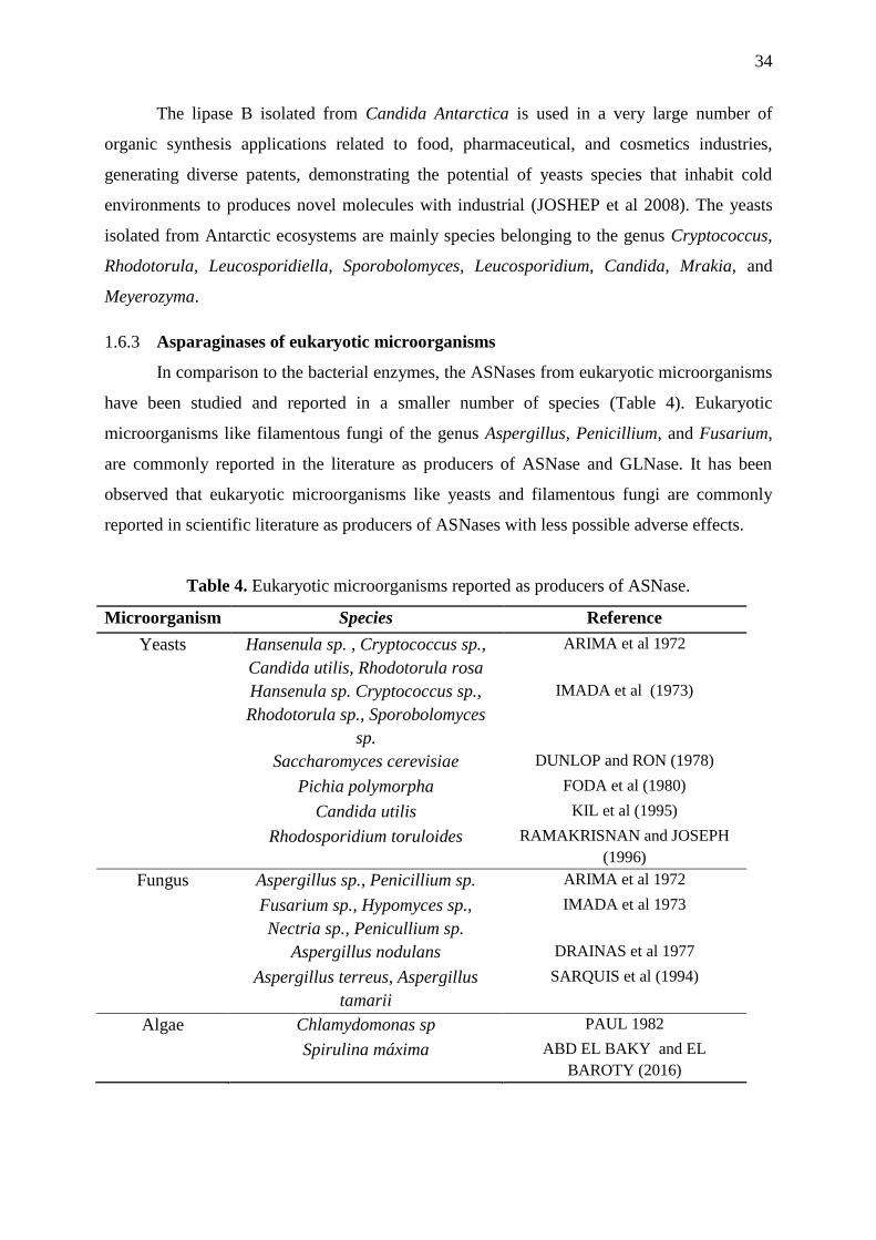
1.6.3 Asparaginases of eukaryotic microorganisms
In comparison to the bacterial enzymes, the ASNases from eukaryotic microorganisms
have been studied and reported in a smaller number of species (Table 4). Eukaryotic
microorganisms like filamentous fungi of the genus Aspergillus, Penicillium, and Fusarium,
are commonly reported in the literature as producers of ASNase and GLNase. It has been
observed that eukaryotic microorganisms like yeasts and filamentous fungi are commonly
reported in scientific literature as producers of ASNases with less possible adverse effects.
Table 4. Eukaryotic microorganisms reported as producers of ASNase.
Microorganism Species Reference
Yeasts Hansenula sp. , Cryptococcus sp.,
Candida utilis, Rhodotorula rosa
ARIMA et al 1972
Hansenula sp. Cryptococcus sp.,
Rhodotorula sp., Sporobolomyces
sp.
IMADA et al (1973)
Saccharomyces cerevisiae DUNLOP and RON (1978)
Pichia polymorpha FODA et al (1980)
Candida utilis KIL et al (1995)
Rhodosporidium toruloides RAMAKRISNAN and JOSEPH
(1996)
Fungus Aspergillus sp., Penicillium sp. ARIMA et al 1972
Fusarium sp., Hypomyces sp.,
Nectria sp., Penicullium sp.
IMADA et al 1973
Aspergillus nodulans DRAINAS et al 1977
Aspergillus terreus, Aspergillus
tamarii
SARQUIS et al (1994)
Algae Chlamydomonas sp PAUL 1982
Spirulina máxima ABD EL BAKY and EL
BAROTY (2016)

35
1.6.4 Yeast Asparaginases
There are few studies on the production and characterization of anti-ALL ASNases by
yeasts. By far, the most studied asparaginase of yeast is the ASNase II from Saccharomyces
cerevisiae. The production of ASNases in Saccharomyces cerevisiae was studied by Dunlop
and Roon (1978, 1980), in which was reported two forms of ASNases: ScASNase I, an
internal constitutive enzyme, and ScASNase II, a mannan glycoprotein located in the cell wall
an external enzyme which is regulated by the amount and source of nitrogen available in the
growth medium. This pattern of intracellular and secreted enzymes is shared with the ASNase
production in bacteria, where just the type II extracellular enzyme is effective against ALL.
Even though, the phylogenetic analyses suggested that these enzymes evolved in an
independent way in prokaryotes and eukaryotes. And, the amino-acid sequences analysis of
the two types of ASNases (the cytosolic and the cell wall glycoprotein) present in
Saccharomyces cerevisiae and Schizosaccharomyces pombe, classifies both enzymes as
bacterial type II enzymes (BONTHORN AND JASKÓLSKI, 1997; BOREK AND
JASKOLSKI, 2001; MICHALSKA AND JASKOLSKI, 2006). Recently, Costa et al., (2016)
reported the cytotoxicity activity in the MOLT-4 leukemic cells of a recombinant ScASNase I
(cytosolic) demonstrating the promising antineoplastic properties of these kinds of enzymes.
In this context, the present study aims the screening and characterization of ASNase
produced in yeast isolated from samples collected in the Antarctic Peninsula, during the
expeditions realized by the PROANTAR program. The search and studied of new ASNases
enzymes with improved kinetic properties that could generate a new biopharmaceutical used
for the treatment of ALL.
36
1.7 MATERIALS AND METHODS
1.7.1 Microorganisms
The 40 yeast strains used in this study were isolated from diverse samples obtained
during an expedition to Antarctica peninsula in the austral summer of 2010 by the Brazilian
Antarctic Program team (Table 5) and the microorganisms were identified using the 26S
rDNA sequencing (DUARTE et al, 2013).
1.7.2 Preparation of inoculum culture and enzyme production
The yeast strains were reactivated in potato dextrose agar (PDA; DifcoTM) and
incubated at 15°C for 72 h. The activated strains were incubated in submerged culture in
potato dextrose broth (DifcoTM) at 15°C and 150 rpm in an orbital shaker during 48 hours.
The cells were harvested by centrifugation at 3400 xg for 15 minutes at 5°C, and washed with
sterile water. For ASNase production the yeast cells were inoculated at 5 g L-1 of initial
inoculum in 100 mL flasks containing 50 mL of modified Czapeck Dox’s medium (GULATI
et al, 1997). After 24, 48, 72 and 96 hours, samples of the culture were collected and the cells
and medium culture were separated by centrifugation. At each point, the ASNase and GLNase
activity was determined in the culture medium and in the cells.
1.7.3 Assay of periplasmic activity of ASNase and GLNase by hydroxylaminolysis
reaction
The ASNase and GLNase activity were determined by the hydroxylaminolysis
reaction and using the whole cell for the enzymatic reaction (GROSSOWICZ et al, 1950;
FERRARA et al., 2004). In a eppendorf tube was added, 1.6 mL of cells suspension at 1.0 of
DO in 50 mM Tris-HCl buffer pH 7.0, 0.2 mL of 0.1 M L-asparagine and 0.2 mL of 1.0 M
solution of hydroxylamine hydrochloride at pH 7.0, the mixture was agitated on a
thermomixer at 850 rpm at 37ºC, after 30 minutes the enzymatic reaction was stopped by
adding 0.25 mL of TCA/FeCl3 reagent (100 g L-1 FeCl3, 50 g L-1 TCA in 0.66 M HCl). The
tube was centrifuged at 3400 xg for 5 minutes and 1.0 mL of the supernatant was measured at
500 nm. Controls were prepared in the same way as samples, but the hydroxylamine and
asparagine solutions were added after the ferric chloride reagent. The GLNase activity was
realized using the same process but 0.1M of asparagine was substituted for 0.1 M glutamine
solution. One unit of ASNase and GLNase was defined as the amount of enzyme that
produces 1μmol of -aspartohydroxamic acid or 1μmol of γ-glutamohydroxamic acid
correspondently, formed by minute by gram of dried cell weight (U g-1cdw).

37
1.7.4 Thin Layer Chromatography (TLC) of reaction products from Leucosporidium
scottii cells
The reaction of ASNase was performed by the addition of 0.2 ml of 0.1M L-
asparagine, 0.2 mL of 1.0 M hydroxylamine pH 7.0 and 1.6 mL of cells suspension at 1.0 of
OD in 50 mM Tris-HCl buffer pH 7.0. The mixture was agitated on a thermomixer at 850 rpm
at 37ºC.After 30 minutes the enzymatic reaction was stopped by centrifuged the reaction at
3400 x g for 5 minutes, and 1.0 mL of supernatant was sample and stored at 5°C.The blank
reaction was prepared in the same way but yeast cells were inactivated by heating them at 80
°C for 15 minutes. The reaction and controls were run on a silica gel plate according to the
method reported by Ramakrishnan et al., (1996). It was used phenol/water (4:1) (w/v) as the
mobile phase. The plate was revealed spraying 0.5% (w/v) of ninhydrin dissolved in acetone
and heated using a hair dryer until the appearance of the points.
1.7.5 ASNase assay by hydrolysis reaction
The enzyme activity was assayed by the quantification of the ammonia released using
the Nessler’s reagent (IMADA et al., 1973). In a eppendorf tube, 50 μL of enzyme were
incubated in 0.5 mL of 50 mMTris-HCl buffer pH 8.0, with 50 μL of 189 mM L-asparagine
and 0.45 mL of ultrapure water, the mixture was agitated on a thermomixer at 850 rpm at
37ºC, after 30 minutes the reaction was interrupted by the addition of 50 μL of 1.5 M
trichloroacetic acid (TCA). The solutions were mixed by inversion and centrifuged at 3400 g
for 5 minutes. The release of ammonia was determined by adding 0.25 mL of Nessler's
reagent to 0.1 mL of the sample diluted in the final volume of 2.5 mL of ultrapure water. The
color developed was quantified at 436 nm. One unit of ASNase activity is the amount of
enzyme, which produced 1 μmol of ammonia per minute.
1.7.6 Enzyme extraction
Cultures of L. scottii were centrifuged at 3400 xg, the cells were harvest and washed
twice with sterile water, resuspended at 50% (v/v) in lysis buffer containing 20 mM Tris-HCl
pH 8.0, 1 mM EDTA, 10 mM cysteine, and 1 mM PMSF. 25 mL of cells suspension was
passed through a French press homogenizer at 1500 psi during 10 cycles, maintaining the
suspension in an ice bath between each cycle to avoid the rise of temperature. The
homogenized suspension was centrifuged at 18514 xg for 20 minutes and the supernatant was
collected.

38
1.7.7 Enzyme purification
1.7.7.1 Polyethylene glycol 4000 for extract clarification
A Polyethylene glycol 4000 solution (PEG 4000) (66 % w/v) was added to the
supernatant the cell homogenate obtained to reach the 6.6% of final concentration of PEG
4000. The mixture was left in an ice bath for 30 minutes and latter centrifuged at 18514 x g
for 15 minutes. The supernatant was filtered tough 0.45 μm membrane and stored at 4º C for
subsequent analysis.
1.7.7.2 Ion exchange chromatography
The enzyme extract was passed through to a DEAE-HiTrap GE 5 mL column
(DEAE-sepharose) using the AKTA purifier system. The column was previously equilibrated
with 20 mM Tris-HCl (pH 8.0) buffer and the protein was eluted with step gradients of NaCl
(50, 100, 150, 200, 250 and 500 mM) in 20 mM Tris-HCl pH 8.0 buffer, fractions of 1 mL
were collected. Protein quantification was measured using Bradford reagent and the ASNase
activity was assayed by the quantification of the aspartic hydroxamic acid formed, using the
method described before. The fractions with greater activity were pooled and concentrated
within an ultrafiltration membrane cartridge of 10 kDa of molecular weight cut-off.
1.7.7.3 Size exclusion chromatography (SEC)
The concentrated protein fraction from the previous step was passed through
Superdex® 200 Increase 10/300 GL column (cross-linked agarose-dextran resin) using the
AKTA purifier system. The column was equilibrated with 20 mM Tris-HCl (pH 8.0) buffer
added with 100 mM glycerol and eluted with the same buffer at the flow of 0.50 mL min-1.
Fractions of 0.25 mL were collected and the protein fractions were assayed for ASNase
activity. SDS-PAGE was realized to check the purity of protein in each fraction collected.
1.7.8 Molecular weight determination by size exclusion chromatography
The molecular weight of the enzyme was determined using the elution volume (Ve)
presented in the Superdex® 200 Increase 10/300 GL for the fraction that presents the enzyme
activity. The column was calibrated by molecular weight size-marker of proteins:
thyroglobulin 669 kDa, ferritin 440 kDa, aldolase 232 kDa, ribonuclease 137 kDa,
conalbumin 75 kDa, ovalbumin 45 kDa, and the void volume (V0) was determined using blue
dextran. The standard proteins were run at the same conditions assayed for the enzyme
fraction, buffer 20mM Tris-HCl pH 8.0 added with 100 mM glycerol, at 0.75 mL min-1 of
flow rate. The molecular mass of the enzyme was determined by the linear relationship

39
obtained by plotting the Kav value of the proteins calculated by the equation 1, and the
logarithms of their molecular weights (MW) (TAYYAB et al., 1991) See Figure 8.
𝐾𝑎𝑣 =𝑉𝑒−𝑉0
𝑉𝐶−𝑉0 equation (1)
1.7.9 Determination of molecular weight in reduced conditions and purity of the
fractions obtained
The different fractions obtained from the subsequent steps of purification were
analyzed by the SDS-PAGE technique in a 12% polyacrylamide gel under reducing
conditions in accordance with the method reported by Laemmli (1970). Molecular weight
protein marker Precision Plus Protein was used (BioRad). Gels were stained with Coomassie
Blue R-250 and subsequently with silver staining. The approximate enzyme molecular weight
was estimated by determining the relative migration distance (Rf) and interpolating the value
from the linear relation of LogMw vs Rf. The purity of the enzyme was confirmed by the
presence of a single band in the fraction analyzed.
1.7.10 Enzyme glycosylation analysis by SDS-PAGE
The purified enzyme was denatured by adding 1μL of SDS 5%, 1μL of DDT 1M, and
heating at 95°C for 5 minutes and cooled for 5 minutes at room temperature. The pH of the
reaction was adjusted by adding 2μL of sodium phosphate buffer 0.5 M, pH 7.5 and added
with 2μL of Triton X-100 at 10%. Finally, 2μL of peptide N-glycosidase F (PNGase F;
PromegaTM) was added to the reaction and incubated at 37°C for 3 hours. The samples were
treated and visualized by SDS-PAGE.
1.7.11 Determination of specific activity of ASNase
The protein concentration of enzyme fractions was assayed using the QuantiProTM
BCA assay kit (Sigma Aldrich®) To determine the ASNase specific activity quantities of the
enzyme purified (0.2-1.5 μg) were incubated at 37ºC for 30 minutes in the presence of L-
asparagine 20 mM and the ammonium released was quantified through Nessler’s reaction as
described above. The glutaminase activity was determined the same conditions at the same
concentrations of the enzyme using L-glutaminase at 20 mM instead L-asparagine.

40
1.7.12 Effect of pH and Temperature
To determined the optimum pH for LsASNase I were used the following buffers at 50
mM concentration: acetate (pH 4.0- 5.5) phosphate (6.0-7.0) Tris-HCl buffer (7.5-9.0),
Glycine-Sodium hydroxide (pH 9.5-10.5), Sodium bicarbonate (11.0). The enzyme was
incubated in the corresponding buffer for 24 hours at 4 ºC and after the activity was determine
by the hydrolysis reaction described above. To determine the optimum temperature, the
enzymatic reaction was performed at different temperatures from 5°C to 65° C and after 30
minutes the enzyme activity was determined by the hydroxylaminolysis reaction, method
reported by Grossowicz et al. (1950) described above.
1.7.13 Effect of metal ions on enzyme activity
The effect of different metal ions including K+ (50mM), Mg+2 (50 mM), Na+ (50mM,
1M), Ca+2 (10mM), Ni+2 (10mM), Cu+ (10 mM), and EDTA (1 mM) on the partial purified
ASNase was studied. The enzyme was incubated during 30 minutes at 37°C in the phosphate
buffer added with the corresponding metal ion and L-asparagine 20 mM, the residual activity
was assayed and compared with the control, which was considered as 100% activity.
1.7.14 Circular Dichroism for secondary structure comparison of LsASNase and E.coli
ASNase type II (ELSPAR®)
Circular Dichroism (CD) spectra of the samples were obtained in a Jasco J-815
Spectropolarimeter (Jasco, Tokyo, Japan). The final spectra were the average of 6 scans,
following subtraction of the spectrum of the buffer 20 mM Tris-HCl pH 8.0 obtained under
the same conditions. CD spectra were obtained in the far-UV range (190-260 nm). Samples
were placed in 5.00 mm optical length quartz cells with a concentration of 0.28 µM
(LsASNase) and 1.92 µM (Control – EcASNase II). Spectra intensities (θ, mdeg) were
converted to residual molar ellipticity ([θ], deg.cm2.dmol-1) using the equation 2. Where “C”
is the protein concentration in mol L-1, “l” is the optical length in cm and “n” is the estimated
number of residues in the protein.
[θ] = θ
10×𝐶×𝑙×𝑛 equation (2)

41
1.7.15 Circular Dichroism for thermal stability of LsASNase
Thermal stability studies of the enzyme samples were performed in a Jasco J-815
spectropolarimeter (Jasco, Tokyo, Japan).The temperature was scanned from 30°C to 95°C, at
a rate of 1°C/minute, and back from 95°C to 30°C to study unfolding and refolding processes,
respectively. Samples with a concentration of 0.28 µM (LsASNase) or 1.92 µM (Control –
EcASNase II) were placed in a 5.00 mm optical length quartz cells, and the intensities of
ellipticity at 222 nm (θ222, mdeg) were registered throughout the experiment. Intensities of
ellipticity at 222 nm were converted to residual molar ellipticity ([θ]222, deg.cm2.dmol-1) using
the equation 3. Where “C” is the protein concentration in mol L-1, “l” is the optical length in
cm and “n” is the estimated number of residues in the protein.
[θ]222= θ222
10×𝐶×𝑙×𝑛 equation (3)
1.7.16 Determination of kinetic enzymatic parameters
The enzyme kinetic behavior was followed by oxidation of β-NADH in present of
glutamate dehydrogenase and ammonia, a product of L-asparagine hydrolysis by the ASNase
action. The β-NADH depletion was monitored spectrophotometrically at 340 nm and the
change in absorbance over time is proportional to the ASNase rate reaction (BALCÃO et al.,
2001). The reaction quantities were modified for its use in microtiter plate. The L- asparagine
concentration was varied from 0 to 2 mM to determine the kinetic parameters. In each well
the enzyme reaction mixture was prepared by combining 87.5 μL of 200 mM Tris-HCl buffer
pH 7.5, 53.75 μL destilled water, 3.5 μL of 110 μMα-ketoglutarate (solved in 100 mM Tris-
HCl buffer pH 8.0), 8.75 μL of 128 μMβ-NADH, and 17.5 μL of 0.148 μM of GDH (solved
in phosphate buffer pH 7.5 with glycerol at 50% (v/v)) and 159 μL of the corresponding L-
asparagine solution (2.0, 1.0, 0.75, 0.5, 0.25 0.1, 0.075, 0.05, 0.025, 0.0125, and 0 mM). For
started the reaction were added 20 μL of the purified LsASNase I (≈ 2.7 x 10⁻⁸ M). The final
volume per well was 350 μL. The molar extinction coefficient of β-NADH was
experimentally determined as 6100 mol-1 cm-1. The substrate affinity and turnover number
were estimated using non-linear regression fitting in the GraphPhad Prism software version
5.0.

42
1.8 RESULTS AND DISCUSSION
1.8.1 Screening of enzyme activity
The activity for ASNase and GLNase by the yeast strains was determinate by the
corresponding formation of -aspartohydroxamic acid (AHA) and -glutamohydroxamic acid
(GHA) by the action of the enzyme in the presence of hydroxylamine and the corresponding
amide. Also, extracellular activity in the cell-free cultures was tested, however, no
extracellular activity was detected in any yeast strain cultures. From the 40 strains analyzed
23 corresponding to Ascomycota and 17 to Basidiomycota phylum. In this screening, 16
strains were able to form the corresponding hydroxamic acid from one or both substrates, and
15 of these 16 strains correspond to the genus Basidiomycota.
Table 5. Yeast strain isolated from samples collected in Antarctic Peninsula tested for the
production of ASNase and GLNase
Strain code Specie Phylum
L3, L6 Debaryomyces hansenii Ascomycota
L4, L5, L9, L10, L11, L13,
L17,L18,L19, L20, L21
Myerozyma guillermondii Ascomycota
L7, L26, L38 Rhodotorula mucilaginosa Basydiomycota
L32, L42, L43, L92, L124,
L97, L122, L123
Cryptococcus victoriae Basidiomycota
L35 Cryptococcus laurentii Basidiomycota
L36, L37a, L 55 Wickerhamomyces anomalus Ascomycota
L58 Candida sake Ascomycota
L79, L83, L84 Candida glaebosa Ascomycota
L81, L82, Debaryomyces macquariensis Ascomycota
L88 Guhomyces pullulans Basidiomycota
L94 Cryptococcus albidiosimilis Basidiomycota
L114 Candidada viasina Ascomycota
L115, L117, L118 Leucosporidium scottii Basidiomycota
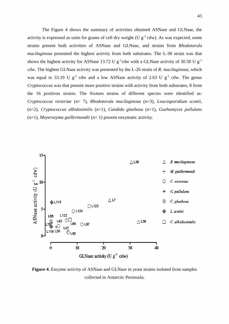
43
The Figure 4 shows the summary of activities obtained ASNase and GLNase, the
activity is expressed as units for grams of cell dry weight (U g-1 cdw). As was expected, some
strains present both activities of ASNase and GLNase, and strains from Rhodotorula
mucilaginosa presented the highest activity from both substrates. The L-38 strain was that
shows the highest activity for ASNase 13.72 U g-1cdw with a GLNase activity of 30.50 U g-1
cdw. The highest GLNase activity was presented by the L-26 strain of R. mucilaginosa, which
was equal to 33.19 U g-1 cdw and a low ASNase activity of 2.63 U g-1 cdw. The genus
Cryptococcus was that present more positive strains with activity from both substrates, 8 from
the 16 positives strains. The Sixteen strains of different species were identified as:
Cryptococcus victoriae (n= 7), Rhodotorula mucilaginosa (n=3), Leucosporidium scottii,
(n=2), Cryptococcus albidosimilis (n=1), Candida glaebosa (n=1), Guehomyces pullulans
(n=1), Meyerozyma guillermondii (n= 1) present enzymatic activity.
Figure 4. Enzyme activity of ASNase and GLNase in yeast strains isolated from samples
collected in Antarctic Peninsula.
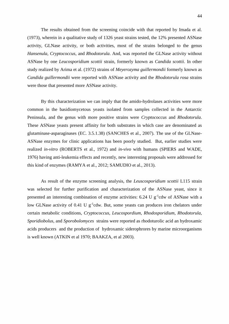
44
The results obtained from the screening coincide with that reported by Imada et al.
(1973), wherein in a qualitative study of 1326 yeast strains tested, the 12% presented ASNase
activity, GLNase activity, or both activities, most of the strains belonged to the genus
Hansenula, Cryptococcus, and Rhodotorula. And, was reported the GLNase activity without
ASNase by one Leucosporidium scottii strain, formerly known as Candida scottii. In other
study realized by Arima et al. (1972) strains of Meyerozyma guillermondii formerly known as
Candida guillermondii were reported with ASNase activity and the Rhodotorula rosa strains
were those that presented more ASNase activity.
By this characterization we can imply that the amido-hydrolases activities were more
common in the basidiomycetous yeasts isolated from samples collected in the Antarctic
Peninsula, and the genus with more positive strains were Cryptococcus and Rhodotorula.
These ASNase yeasts present affinity for both substrates in which case are denominated as
glutaminase-asparaginases (EC. 3.5.1.38) (SANCHES et al., 2007). The use of the GLNase-
ASNase enzymes for clinic applications has been poorly studied. But, earlier studies were
realized in-vitro (ROBERTS et al., 1972) and in-vivo with humans (SPIERS and WADE,
1976) having anti-leukemia effects and recently, new interesting proposals were addressed for
this kind of enzymes (RAMYA et al., 2012; SAMUDIO et al., 2013).
As result of the enzyme screening analysis, the Leuscosporidium scottii L115 strain
was selected for further purification and characterization of the ASNase yeast, since it
presented an interesting combination of enzyme activities: 6.24 U g-1cdw of ASNase with a
low GLNase activity of 0.41 U g-1cdw. But, some yeasts can produces iron chelators under
certain metabolic conditions, Cryptococcus, Leucospordium, Rhodosporidium, Rhodotorula,
Sporidiobolus, and Sporobolomyces strains were reported as rhodoturolic acid an hydroxamic
acids producers and the production of hydroxamic siderophrores by marine microorganisms
is well known (ATKIN et al 1970; BAAKZA, et al 2003).

45
To confirm the production of the enzyme by L. scottii L115 strain was evaluated
qualitatively by TLC the formation of -AHA and the depletion of L-asparagine during the
enzyme reaction. As can be seen in the Figure 5, the first track (ASN) and the second track
(-AHA) correspond to the L-asparagine (20 mM) and the -aspartohydroxamic acid (20
mM) standards respectivetly. In the reaction blank track (B) just the presence of L-asparagine
is detected, but in the reaction track (R) the depletion of L-asparagine is detected after 30
minutes reaction and can be observed the presence of -AHA formed by the reaction of L-
asparagine with hydroxylamine in presence of the enzyme, proving the presences of ASNase
enzyme in the yeast cells.
Figure 5. Thin layer chromatography of the reaction with cell suspension of L. scottii L115.
1.8.2 Enzyme purification
The enzyme was extracted by mechanical disruption by high-pressure homogenization
of the yeast cells and the purification was carried out the following separation steps:
homogenized extract PEG treatment, anionic exchange, and size exclusion chromatography.
The polyethylene glycol 4000 (PEG 4000) at 6.6 % (w/v) of saturation was added to remove
the unwanted macromolecules and lipids, clarifying the cell homogenize prior to further
processing by chromatography methods. The PEG can be easily removed during the
subsequent ion-exchange chromatography step since it does not absorb to the column resin
(HONING and KULA, 1976; INGHMAN, 1990).

46
A
B
.
Figure 6. DEAE chromatogram, enzyme eluted at 50 mM of NaCl gradient step, (A) SEC
chromatogram, the enzyme elute at 9.8 mL of the column volume (B).

47
The extract clarified was subjected to anionic exchange chromatography passed
through a DEAE column and eluted using step gradient at 10, 50, 100, 500 mM of NaCl. The
enzyme activity was found only in the 50 mM eluted fractions with a 25 % fold of recovery
and a specific activity of 14.65 U mg-1 was obtained (Figure 6 A). The fractions with activity
obtained by DEAE chromatography were pooled and concentrated within an ultrafiltration
cartridge of 10 kDa of molecular weight of cut-off, and passed through SEC column
Superdex® 200 Increase 10/300 GL, and the enzyme was eluted at 9.9 mL of the column
volume (Figure 6B), obtaining a 228 fold of purification and a specific activity of 137.0 U
mg-1. The results of the different steps of the purification of LsASNase are shown in Table 6.
Table 6. Purification scheme of ASNase from Leucosporidium scottii .
Step Activity
(U mL-1 )
Protein
(mg/mL)
Total
proteins
(mg)
Total
Units
(U)
Specific
activity
(U mg-1 )
Purification
fold
Recovery
(%)
Culture extract* 7.23 12.01 1082.79 650.80 0.60 1 100
anion exchange
chromatography
DEAE
11.02 0.75 11.28 165.33 14.65 24.38 25.40
Size Exclusion
chromatography
(SEC)
6.85 0.05 0.25 34.25 137.01 227.95 5.26
*From an initial 90 mL of culture extrac

48
1.8.3 Determination of molecular weight by SDS-PAGE and SEC
The purity of the purified enzyme fractions was analyzed by SDS-PAGE and the
equation the of molecular weight was determined by plotting the relative protein mobility (Rf)
to the dye front versus the natural logarithm of molecular weight (MW) of the protein
standards. The MW of the single band corresponding to the LsASNase under reduced
conditions was 54.5 kDa of MW and the native MW by SEC was 462 kDa. This provides the
envidence that the enzyme purified from LsASNase is a high-mass multimeric enzyme that
could be an octamer (Figure 7, 8). This result is in agreement with the intracellular ASNase
reported from S. cerevisiae which is also reported as a decameric enzyme with a molecular
weight of 400 kDa determined by SEC and 41.4 kDa in SDS-PAGE (DUNLOP and ROON,
1978; SINCLAIR et al., 1994), demonstrating that even among different groups
(basidiomycetous and ascomycetous), yeast species could share the same ASNase enzyme.
Figure 7. Molecular weight determination of the purified enzyme LsASNase (A) SDS-PAGE
in reduced conditions of the corresponding fractions purified by SEC fraction 18 (2), fraction
19 (3), fraction 20 (4)
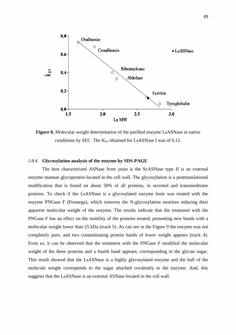
49
Figure 8. Molecular weight determination of the purified enzyme LsASNase in native
conditions by SEC The Kav obtained for LsASNase I was of 0.12.
1.8.4 Glycosylation analysis of the enzyme by SDS-PAGE
The best characterized ASNase from yeast is the ScASNase type II is an external
enzyme mannan glycoprotein located in the cell wall. The glycosylation is a posttranslational
modification that is found on about 50% of all proteins, in secreted and transmenbrane
proteins. To check if the LsASNase is a glycosylated enzyme form was treated with the
enzyme PNGase F (Promega), which removes the N-glycosylation moieties reducing their
apparent molecular weight of the enzyme. The results indicate that the treatment with the
PNGase F has an effect on the mobility of the proteins treated, presenting new bands with a
molecular weight lower than 25 kDa (track 5). As can see in the Figure 9 the enzyme was not
completely pure, and two contaminating protein bands of lower weight appears (track 4).
Even so, it can be observed that the treatment with the PNGase F modified the molecular
weight of the three proteins and a fourth band appears, corresponding to the glycan sugar.
This result showed that the LsASNase is a highly glycosylated enzyme and the half of the
molecule weight corresponds to the sugar attached covalently to the enzyme. And, this
suggests that the LsASNase is an external ASNase located in the cell wall.

50
Figure 9. SDS-PAGE in reduced conditions for each step of the enzyme purification.(1)
Standard proteins, (2) Homogenized extract treated with PEG 4000, (3) Anionic exchange
chromatography DEAE-column,(4) LsASNase purified by SEC (5) ) LsASNase purified by
SEC treated with PGNase F enzyme.
1.8.5 Determination of specific activity
In order to determine the specific activity was used the purified enzyme by SEC. The
activity was measured by the hydrolysis reaction with the Nessler’s reagent and the protein
concentration was determined by the micro BCA method. The specific activity of LsASNase
obtained was 139.8 ± 5.094 U·mg-1 of protein and GLNase activity was 0.93 U·mg-1,
reasonable activity compared with the bacterial ASNase (270- 700 U·mg-1) (NARTA et al
2001) and the yeast ASNase (196 U·mg-1) (COSTAS et al, 2016). Both graphics were made
by plotting the initial velocity of the substrate hydrolysis vs the mg of purified enzyme. The
specific activity corresponds to the slope obtained (Figure 10). But, an interesting point is that
LsASNase present a very low GLNase activity, just the 0.6% of the activity in comparison
with the bacterial ASNases that have 2 until 10 % of the GLNase side activity. That makes an
interesting topic to continue studying the ASNase from yeasts species.
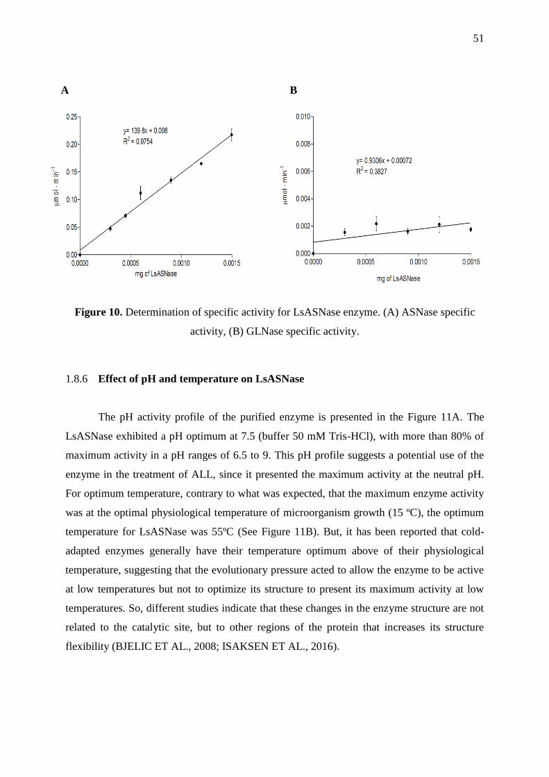
51
A B
Figure 10. Determination of specific activity for LsASNase enzyme. (A) ASNase specific
activity, (B) GLNase specific activity.
1.8.6 Effect of pH and temperature on LsASNase
The pH activity profile of the purified enzyme is presented in the Figure 11A. The
LsASNase exhibited a pH optimum at 7.5 (buffer 50 mM Tris-HCl), with more than 80% of
maximum activity in a pH ranges of 6.5 to 9. This pH profile suggests a potential use of the
enzyme in the treatment of ALL, since it presented the maximum activity at the neutral pH.
For optimum temperature, contrary to what was expected, that the maximum enzyme activity
was at the optimal physiological temperature of microorganism growth (15 ºC), the optimum
temperature for LsASNase was 55ºC (See Figure 11B). But, it has been reported that cold-
adapted enzymes generally have their temperature optimum above of their physiological
temperature, suggesting that the evolutionary pressure acted to allow the enzyme to be active
at low temperatures but not to optimize its structure to present its maximum activity at low
temperatures. So, different studies indicate that these changes in the enzyme structure are not
related to the catalytic site, but to other regions of the protein that increases its structure
flexibility (BJELIC ET AL., 2008; ISAKSEN ET AL., 2016).

52
A
B
Figure 11. Effect of physical parameters on purified LsASNase. (A) Influence of temperature
on the assay reaction. (B) Influence of pH on enzymatic activity determined in different
buffers: acetate (pH 4.0- 5.5) phosphate (6.0-7.0) Tris-HCl buffer (7.5-9.0), Glycine-Sodium
hydroxide (pH 9.5-10.5), Sodium bicarbonate (11.0).

53
1.8.7 Effect of the metal ions in the LsASNase activity
The effect of K+, Na+, Mg+2, Ni+2, Ca+2, Cu+2, Zn+2 on the partial purified ASNase
(DEAE ion-exchange chromatoghaphy step) was studied. Compared to the control, Ni+2, Ca+2,
Cu+2, Zn+2, the divalent ions affect the enzyme activity being the Cu+2 ions the highest
inhibitor producing 40% of the inhibition of activity. Unlike, the of K+, Na+, Mg+2, do not
affect the enzyme activity, and interestingly the activity in present of Na+ 1 M just was
slightly reduced by a 10 %. And, in the presence of EDTA 1 mM the activity is not affected.
This results indicated that the enzyme not dependent on metallic ions.
Figure 12. Effect of metal ions and EDTA on the enzyme activity of the LsASNase.
1.8.8 Secondary structure and thermal stability assays
Since LsASNase has never been reported in the literature, it is important to understand
the secondary structure of this novel enzyme. As it can be seen in Figure 13, the spectra
indicated that the conformation of LsASNase is similar to EcASNase II (ELSPAR®
commercial biopharmaceutical produced by Escherichia coli). Thus, confirms that the
integrity of ASNase was not affected by increasing in monomers, being the secondary
structure preserved when compared to other ASNase. Moreover, thermal stability assays were
performed in order to identify the denaturation temperature of the protein and understand the
refolding process. The Fig. 14A and 15B depicted respectively the CD spectra of both
EcASNase II and LsASNase. From the gather results, the EcASNase II has a slightly higher
melting temperature (around 60°C), when compared to this novel enzyme (around 50°C).
54
Figure 13. Far-UVCD spectrum (190 -260 nm) of LsASNase and EcASNase II (control).
Moreover, it is also possible to conclude that the refolding process of the EcASNase II
is much more efficient when compared to LsASNase, this can easily justify the higher amount
of monomers that the novel enzyme possess, making it difficult to refolding, due to the higher
structural complexity of this latter enzyme. Also, is reported that the cold-adapted enzymes
unfold according to an all-or-none process, because these enzyme structures are stabilized
with fewer weak interactions and the disruption of some interactions strongly influence the
whole structure, triggering the unfolding of all molecule (FELLER, 2013) for this reason can
be observed a sharply loss of the activity after the reaching the melting point (Fig. 5B).
A

55
B
Figure 14. Thermal stability assay of: A) EcASNase II (control) B) LsASNase. CD spectrum
at 222 nm and temperatures ranging from 20-95oC.

56
1.8.9 Kinetic parameters determination
The kinetic behavior of the enzyme was better fitted to the allosteric sigmoidal model
(R2 = 0.9856; p< 0.0001) compared to the Michaelian model (R2= 0.9653) using the F-test.
The kinetic parameters obtained were K0.5 = 233 μM a kcat= 53 s-1 and a Hill coefficient (nH)
of 1.52. The nH indicate the kind of cooperativity between the enzyme subunits and also, the
number of substrate molecules that bind to the enzyme complex, a value of nH> 1 pointed that
the L-asparagine not only acts as substrate also acts as positive effectors of the enzyme
(homotropic regulation). In general, the cytoplasmatic ASNases (ASNases type I) reported in
yeast and bacteria species has a higher KM or K0.5 compared with the extracellular ASNases
(ASNases type II) and presents an allosteric regulation, without L-glutamine affinity (See
Table 7).
This behavior is shared by metabolic enzymes that act as control devices for flux
alterations in metabolic pathways (HELMSTAEDT et al 2001). So, this regulation is
necessary to maintain an intracellular concentration of L-asparagine for protein synthesis, and
is effective at concentrations under 1 mM levels of L-asparagine (DUNLOP and ROON,
1978; YUN et al., 2007). Costa et al. (2016) report this regulation in ScASNase I at substrate
levels below at 0.3 mM and for the human L-asparaginase (hASNase I) this allosteric
behavior occurs at levels of 5 mM, for the LsASNase the allosteric regulation was not
observed markedly but could be present at level below 50 μM (Figure 15). In Table 7 can be
observed the comparison of LsASNase with the properties reported for others intracellular
ASNases from yeast (ScASNase I) and bacterial (EcASNase I). For extracellular ASNases
recently has been reported the heterotropic regulation by the L-glutamine, this kind regulation
was important for the depletion of L-asparagine at μM levels (ANISHKIN et al., 2015;
COSTA et al., 2016). In our case the affinity of the purified enzyme by L-glutamine was not
observed at levels below at 10.0 mM.

57
Figure 15. The LsASNase kinetic behavior with L-asparagine as substrate at pH 7.0. Points
on the graphic represent the mean (n=3). The kinetic parameters were: K0.5 = 233 μM, kcat =
53 s-1 and nH = 1.52.
The analysis of the crystal structures obtained for type I and type II ASNases exhibit a
difference in the flexibility of the loop structures found in the N-terminal region between the
oligomers; this might produce a change in the accessibility of the substrate to the active site.
This structural features could produce a large difference in the affinity constant value (KM or
K0.5) between the two enzyme types (YUN et al., 2007). The enzyme-substrate interactions
are disrupted by both increases and drecreases in temperature (SOMERO, 1977). It was
observed that the enzyme reaches the highest activity close to the melting point, we
speculated that as a cold-adapted enzyme the flexibility of the structure will be reduced as the
temperature increases, in this rigid state the allosteric regulation will be modified, because the
allosteric interactions frequently appear to be correlated with alterations of the quaternary
structure of the proteins. These conformational changes switch the allosteric regulation,
closing the nH to 1, and the enzyme could present a non-cooperative behavior following a
Michaelis-Menten kinetics.

58
The glycans can alter the substrate recognition, the specificity and binding affinity as
well as the turnover rates of the enzymes. In recent years, increasing evidence was found that
glycans have distinct effects on the activity of many enzymes, in particular as regulatory
modules for substrate binding and turnover can have both stimulating and inhibiting effects
on activity (GOETTING, 2016). The interactions of glycosylation with the substrate may be a
possible explanation for the behavior of the LsASNase enzyme. Because, being an
extracellular enzyme present an allosteric behavior, when a Michaelian regulation should be
expected.
The Table 7 shows the efficient catalytic (kcat /K0.5) of diverse ASNase isolated from
different species. It is generally believed that to achieve a therapeutic effect the catalytic
efficiency has to reach or be close to a 106 ratio, but there are other factors that must be taken
into account such as the half-life, antigenicity, optimum pH, that determine the usefulness of
the enzyme for therapeutic purposes. The LsASNase has an efficient catalytic of 2.3 x 10⁵,
only an order of magnitude lower than that of the bacterial ASNase, and one greater than the
ASNase of Saccharomyces cerevisiae, that one that demonstrated anti ALL activity in vitro
studies. This results proves the possibility that the enzyme has anti-ALL activity.
Table 7. Kinetic parameters of different ASNase enzymes reported that presents allosteric
regulation and the E. coli enzyme.
ASNase type KM ,
K0.5*
(μM)
kcat
(s-1)
Vmax
U min-1
nH Specific
activity
(U mg-1)
kcat /K0.5
(M-1 s-1)
Reference
EcASNase II 18* 60 ------ 1.5 ------ 3.3 x 10⁶ Anishkin et
al., 2015
EcASNase I 1200* ----- 0.016 2.6 173 ------- Yun et al.,
2007
ScASNase I 75* 217 0.042 2.2 196.2 1.6 x 10⁴ Costa et al.,
2016
ScASNase I 740 ----- 0.06 ---- 5.4 ------- Dunlop and
Roon, 1978
ScASNase II 240 ----- 57.8 ---- 30.5 ------- Dunlop et al,
1980`
hASNase I 11500* 6.7 ------ 3.9 ------ 5.8 x 10² Karamitros et
al., 2014
LsASNase I 233* 54.7 0.007 1.52 150 2.3 x 10⁵

59
1.9 CONCLUSION.
Few ASNases in yeasts have been described, so far the results show that the enzyme
isolated from the psychrotolerant yeast strain Leucosporidium scottii L115 presents different
characteristics from those reported in bacteria and other yeast species. As a cold-adapted
enzyme the LsASNase present a characteristic behavior affecting the enzyme kinetic
parameters. This particular behavior can still be studied at the structural level by
characterizing the structures that increase the flexibility of the protein and the catalytic site
that has low affinity for the L-glutamine.
A further structural characterization will allow elucidate the characteristics and
similarities with other ASNases and analyze the use of this enzyme as a possible anti-ALL
drug. Being a biopharmaceutical born before the era of genetic engineering, the re-study of
the ASNase can improvement all drawbacks intrinsic to the enzyme and can be re-planned to
generate new ASNases engineered from the accumulated knowledge of the study and
characterization of the different ASNase enzymes. The knowledge of the structure and
function is necessary in order to increase activity, selectivity, and stability of the enzyme, and
this will improve the treatment of the ALL.
.

60
2 CHAPTER III. MEDIUM CULTURE COMPONENTS
OPTIMIZATION FOR PRODUCTION OF L-ASPARAGINASE
ENZYME BY A PSYCHROTOLERANT YEAST Leucosporidium
scottii L115
2.1 INTRODUCTION
Leucosporidium scottii is a psychrotolerant yeast, has been isolated mainly from cold
environment around the world, including terrestrial and marine Antarctic ecosystems
(SUMMERBELL, 1982; DUARTE et al, 2013). Marine and psychrophilic yeasts provide a
unique potential for the synthesis of new biomolecules. These yeasts have been investigated
for the production of pharmaceutical and enzymatic products but still remain poorly explored,
besides, the marine and cold-adapted microbial enzymes can offer novel biocatalyst with
extraordinary properties (ZHANG and KIM, 2010; ABDELRAHMAN et al, 2014). Few yeast
strains have been studied as producers of ASNase and still many potential microorganisms in
diverse ecosystems remain uninvestigated. To date, L. scotti has not been reported as L-
asparaginase producer, for this reason, the yeasts from Antarctic continent could be an
interesting eukaryotic source of ASNase.
The interest for studied new fonts of asparaginases is promoted basically by the
applications anti-cancerogenic properties of these enzymes. It has been observed that
eukaryotic microorganisms like yeast and filamentous fungi are commonly reported in
scientific literature for produce asparaginase with possible less adverse effects (PINHEIRO, et
al 2002). The demand for ASNase could increase in the coming years due to its potential
applications if the adverse effects are reduced. Also, the use of ASNase for acrylamide
mitigation in food industry, makes the application of ASNase more attractive for industrial
commerciallization in other fields aside from biopharmaceutical industry (XU et al., 2016).
To develop a low-cost production is an important step in the development of this
biopharmaceutical (GURUNATHAN AND SAHADEVAN 2011).

61
2.1.1 Development of culture medium
In the development of industrial biotechnological processes, the design of
fermentation medium is the critical importance. The composition of the culture medium is the
most important factor in the production of enzymes, growth, and physiology of the
microorganism and allows the formation of bioproducts. The carbon and nitrogen sources can
dramatically influence the metabolite formation, changes in nutrients and their concentrations
have different effects on the accumulation of different metabolites, which are controlled by
intracellular effectors (WANG and XU, 2005; DINARVAND et al 2013). The screening of
different sources and selection of the optimum concentration of medium components are very
important to determine the production and economic feasibility of the production process.
Nonetheless, each organism has their particular nutritional and environmental condition for
maximum enzyme production (SHARMA and HUSAIN, 2015). Table 8 shows the average
composition of the microorganisms, these quantities must be taken into account to develop a
medium with enough nutrients so as not limit the growth of the microorganisms.
Table 8. Average composition of microorganisms. (% dry mass)
Component Bacteria Yeast Fungi
Carbon 48 (46-52) 48 (46-52) 48 (45-55)
Nitrogen 12.5 (10-14) 7.5 (6- 8.5) 6 (4-7)
Proteins 55 (50-60) 40 (35-45) 32 (25-40)
Carbohidrates 9 (6-15) 38 (30-45) 49 (40- 55)
Lipids 7 (5-10) 8 (5-10) 8 (5-10)
Nucleic acids 23 (15-25) 8 (5-10) 5 ( 2-8)
Minerals ( same composition for the three microorganisms)
Phosphorous 1.0- 2.5
Sulfur, Magnesium 0.3-1.0
Potassium, Sodium 0.1- 0.5
Ferrum 0.01-0.1
Zinc, Copper, Manganesium 0.001- 0.01
62
As previously mentioned, the medium composition plays an important role in the
improvement of productivity of the enzyme and it is also important to reduce the cost of the
medium as much as possible. A diverse medium composition has been reported for the
production of ASNase in eukaryotic microorganisms like yeasts and fungi species (Table 10).
2.1.2 Carbon sources
Carbohydrates are an excellent carbon and energy sources for microbial fermentations.
However, although the patways of sugar utilization follow the same theme in all yeasts,
important biochemical and genetic variations on it exists The diffent sugars produce signals
which modify the conformation of centain proteins that in turn, directly or through regulatory
cascade affect the expression of the genes subject to catabolite repression (GANCEDO 1998).
Most yeasts use sugar hexoses, where only a few use lactose. The use of molasses, beet
molasses, cereal grains, starch, glucose, sucrose and lactose as carbon sources and salts of
ammonia, urea, nitrates, corn solids, bean meal, slaugther house waste and fermentation
residues as source tend to be the production criteria because they are cheap substrates
(OKAFOR , 2007).
2.1.3 Nitrogen sources
The nitrogen source in the culture medium regulates several genes in bacteria, yeast,
and filamentous fungi. This phenomenon is known as regulation of nitrogen and occurs when
the micro-organisms are able to select more easily metabolizable source, for example,
ammonia, glutamine, and asparagine, and inhibit the expression of transporters and enzyme
systems that allow the assimilation of sources known as poor such as nitrate, urea, and proline
(ARIMA et al, 1972; GODARD et al, 2007) . The nitrogen source regulation enzymes
typically under such control are proteases, amidases, ureases and those that degrade amino
acids. The nitrogen font selected results in the expression of the permeases and the activation
of the enzymes responsible for the uptake, synthesis, and interconversion of the compounds,
and additionally the transcription factors and membrane trafficking proteins that regulate the
activity of these enzymes and permeases (MAGASANIK and KAISER, 2002). For this, the
correct selection of a nitrogen font for culture medium plays an important role in the
production of bio-industrial products such as intra and extracellular enzymes (SANCHEZ
and DEMAIN, 2002). This is the case of L-ASNase and different enzymes regulation that his
production is closely related to the nitrogen fonts present in the culture medium.

63
Table 9. Medium composition for ASNase production by eukaryotic microorganisms
Microorganisms Medium composition (g/L) Reference
Different species of yeasts 20 g glucose, 5.0 g asparagine, 1.0 g K2HPO4, 0.5 g KCl, 0.01 g MgSO4. 7H2O, 1.0 g yeast extract , pH
6.0.
Arima &
Tamura 1972
Different species of yeasts 30 g Sucrose, 5 g polypepton, 5 g beef extract, 2 g yeast extract, 2 g malt extract, 5 g KH2PO4, 1.5 g
K2HPO4, 0.5g MgSO4.7H2O.
Imada 1973
S. cerevisiae X-2180-A2 Growth medium: 20 g of D-glucose, 2 g of yeast nitrogen base (without amino acids and ammonium
sulfate), and 1.32 g of ammonium sulfate.
Nitrogen-free medium (production medium): 20 mM potassium phosphate buffer, pH 7.0 and 3%
glucose and allowed to incubate 3h at 25º C.
Dunlop and
Roon 1975
Candida utilis 20 g of D-glucose, 2g of yeast nitrogen base (without amino acids and ammonium sulfate), and 2 g of
peptone
Kil J. O 1995
Rhodosporidium toruloides CBS14,
ATCC10788, Rhodotorula glutinis
NCYC59, ATCC90950, Rhodotorula
rubra MTCC248
20 g D-mannitol, 5g L-asparagine, 1,2 g K2HPO4, 6 g KH2PO4, 0.5 g KCl, 0.01 g MgSO4•7H2O, 1 g
yeast extract, pH 6.0.
Rmamakrishnan
e Joseph 1996.
Aspergillus terreus MTCC1782 Soy bean meal fluor (not reported) glucose 6 g, L-asparagine 1.0 g, NaNO3 4.0 g, KCl 1.0g, 0.052 g
MgSO4.7H2O and 0.02 g FeSO4.7H2O pH 6.
Gurunathan and
Sahadevan
2011.
Aspergillus tamarii, Aspergillus
terreus.
Czapek- Dox modified with different nitrogen sources: L-asparagine 0.1g, L- proline 0.02 g or 0.2g,
urea 0.02g, L- glutamine 0.1 g.
Sarquis et al
2004.
Fusarium ssp. First grown in M agar : sucrose 30g, Polypeptone 5g, beef extract 2g, yeast extract 5g, malt extract 2g.
Production medium: dextrin 30g, Pharmamedia 40g, KH2PO4 5g, K2HPO4 1.5 g, MgSO4•7H2O 0.5 g,
NaCl 5 g.
Nakahama et al
1973.
64
2.1.1 Experimental design in the optimization of medium composition
The use of mathematical modeling by using design of experiments (DOE) followed by
response surface methodology (RSM) is commonly used by many companies in order to
achieve the best combination of factors that will render the best characteristic of a product and
or process response. In the initial formulation of the culture medium, a study has to be made
to select the best source of carbon and nitrogen to the production of the enzyme. The
screening of the culture medium compounds is time-consuming and costly, this is due to a
large number of substrates available and the number of possible component combinations is
immense. The Plackett-Burman (PB) design may be an excellent option, once it has been
widely used to develop process conditions and to allow the understanding of the effects of
various physicochemical, biochemical and sensory variables using a minimum number of
experiments. The Placket Burman design allows the screening of main factors from a lage
number of variables that can be retained in the further optimization process (HYMAVATHIM
et al, 2013; GRANATO and CALADO, 2014).
Since the optimization of nutritional requirements and operating conditions is a very
important step for bioprocess development. So far, most applications of DoE have concerned
optimization of the composition of growth and production culture media. The intention is to
find the most favorable mix of nutrient factors to maximize the cellular productivity by
supplying a well balanced composition of nutrients that enhances the maximum yield of the
product molecule (MANDENIUS and BRUNDIN, 2008).
The objective of the present work was to apply statistical DOE methods to optimize
the medium composition for the production of ASNase by L. scottii by submerged
fermentation. Several nutritional factors for enzyme production were screened by factorial
fractional design and the optimum medium composition was obtained by response surface
methodology.
65
2.2 MATERIALS AND METHODS
2.2.1 Microorganism and inoculum preparation for ASNase production for PB design
The yeast L115 strain used in this study was isolated from Antarctic marine sediments
obtained during expeditions to Antarctica in the austral summer (2009 and 2010) by the
Brazilian Antarctic Program team and identified as Leucosporidium scottii on the basis of 26S
rDNA gene (Duarte et al, 2013). The L-115 L. scottii stock culture was maintained on Potato-
dextrose broth (PDB, DifcoTM) added with glycerol (20% final volume) and stored at -80ºC.
The Inoculum was prepared by incubating the microorganism in 200 mL of PDB in 500 mL
baffled Erlenmeyer flask for 72 hours (log phase) incubated at 15ºC in a rotatory shaker at
200 rpm of agitation. The different production medium (Table 10) were inoculated at 1 % v/v
of final concentration with the cells harvested. After 24, 48, 72, 96, and 120 hours of culture
the cells were harvest by centrifugation at 3400 X g for 15 minutes and washed with distilled
water for further ASNase activity determination. All medium were supplemented with the
same salt base composed by KCl, 0.52 g L-1; MgSO4.7H2O, 0.52 g L-1; CuNO3.3H2O, 0.001 g
L-1; ZnSO4.7H2O, 0.001 g L-1; FeSO4.7H2O, 0.001 g L-1.
2.2.2 Assay of ASNase Activity
The ASNase activity was determined by the hydroxylaminolysis reaction and using
the whole cell for the enzymatic reaction (GROSSOWICZ et al, 1950; DRAINAS et al, 1977;
FERRARA et al., 2004). In a eppendorf tube was added, 1.6 mL of cells suspension at 1.0 of
OD in 50 mM Tris-HCl buffer pH 7.0, 0.2 mL of 0.1 M L-asparagine and 0.2 mL of 1.0 M
solution of hydroxylamine hydrochloride at pH 7.0, the mixture was agitated on a
thermomixer at 850 rpm at 37ºC, after 30 minutes the enzymatic reaction was stopped by
adding 0.25 mL of TCA/FeCl3 reagent (100 g L-1 FeCl3, 50 g L-1 TCA in 0.66 M HCl). The
tube was centrifuged at 3400 xg for 5 minutes and 1.0 mL of the supernatant was measured at
500 nm. Controls were prepared in the same way as samples, but the hydroxylamine and
asparagine solutions were added after the ferric chloride reagent. One unit of ASNase was
defined as the amount of enzyme that produces 1μmol of -aspartohydroxamic acid formed
by minute by gram of dried cell weight (U g-1cdw).

66
2.2.3 Plackett-Burman design
To evaluate the effect of 16 factors of medium components on the ASNase production
a PB design of 20 experimental runs was carried out. The factors were tested at two levels (-1,
+1). Table 10 shows the variables studied and the concentration tested. Three unassigned
variables (commonly referred as dummy variables) were evaluated in the design and used to
estimate the experimental error by pooling into the error the effect values obtained for each
variable. The initial concentrations of each nutrient were selected empirically based on the
literature survey.
Table 10. Nitrogen, carbon and complex fonts tested and levels used in PB design
Code Variables +1 (g L-1) -1 (g L-1)
Nutrients considered like nitrogen fonts
A Proline 5 0
B Urea 5 0
C Aspartate 5 0
D Chloride ammonium 5 0
E Arginine 5 0
F Glutamine 5 0
G Asparagine 5 0
H Glutamate 5 0
Nutrients considered like carbon fonts
I Maltose 10 0
J Glycerol 10 0
K Sucrose 10 0
L Citric acid 10 0
Complex fonts
M Yeast extract 2.5 0
N Soybean peptone 2.5 0
O Step corn solids 2.5 0
P Potato dextrose broth 2.5 0
Dummy variables
Q D1 0 0
R D2 0 0
S D3 0 0

67
The effect of each variable on the enzyme production and biomass production was
evaluated using the next equation:
𝐸𝑓𝑓𝑒𝑐𝑡(𝑥𝑖) =∑ 𝑋𝑖+−∑ 𝑋𝑖−
𝑁 equation (4)
Where the effect of each variable (Xi) is the response effect for determination of
ASNase ; the Xi+ is the yield of ASNase and biomass from the experiments when variables
tested were at high levels; Xi- the yield of ASNase and biomass when variables tested were at
low concentrations; and N the number of experiments. The significance was determined by
applying the Student’s t-test, P-value, and the significance levels. The variables which were
significant at 5% level (P< 0.05) from the regression analysis were considered to have a
greater impact on ASNase production. The statistical analysis were generated using Statistica
10(StatSoft, UK).
2.2.4 Inoculum preparation and production evaluation of L-Asparaginase for CCD
design study
The pre-inoculum was prepared by incubating the microorganism in 200 mL of PDB
in 500 mL baffled Erlenmeyer flask for 72 hours (log phase) incubated at 15ºC in a rotatory
shaker at 200 rpm of agitation. The Inoculum was prepared by adding 5 g L-1 the
microorganism in 200 mL of medium containing proline 5 g L-1, sucrose 10 g L-1, glycerol 10
g L-1in 500 mL baffled Erlenmeyer flask for 72 hours (log phase) incubated at 15ºC in a
rotatory shaker at 200 rpm of agitation. The different production medium (Table 13) were
inoculated with the cells harvested at 1 % v/v of final concentration. After 24, 48, 72, 96, and
120 hours of culture the cells were harvest by centrifugation at 3400 X g for 15 minutes and
washed with distilled water for the further ASNase activity determination.
2.2.5 Response surface methodology
The most important factors found from the PB screening were examined in more detail
using response surface methodology. The concentration of variables selected was optimized
by using a central composite design (CCD). A set of 16 experiments was performed with
three variables, and each variable at five levels (-α,-1, 0, 1, α) (α = 1.6817). The error was
calculated incorporating two replicates of the medial point (000). In Total 17 assays were
performed (Table 13). The quadratic equation for the variables was as follows:
𝑌 = 𝛽0 + ∑ 𝛽𝑖𝑋𝑖 + ∑ 𝛽𝑖𝑖 𝑋𝑖2 + ∑ 𝛽𝑖𝑗𝑋𝑖𝑋𝑗 + 𝜀 equation (5)

68
Where: Y is the predicted response; β0 the intercept; βi the linear coefficients for linear
effects; βii the squared coefficient for quadratic effects; βij the cross-product coefficient for the
effects; X the independent variables; and ε the experimental error. The response surface plots
and statistical analysis were generated using Statistica 10. ANOVA analysis was employed to
determine the model fitness.
2.2.6 Validation of statistical model
To validate the composition of the medium, an additional experiment in triplicate was
conducted under conditions predicted for enzyme production. ASNase activity and cell mass
production were used as a dependent variable in the comparative study.

69
2.3 RESULTS AND DISCUSSIONS.
2.3.1 Evaluation of influence of different founts of nitrogen and carbon for L-
asparaginase production using the Placket-Burman design
The PB design was very useful for finding the main substrates for the culture medium
by studying the main effect of each factor in the enzyme production, which is time-saving and
can be evaluated more than one factor in a single experiment. The orthogonality of the design
implying that the effect of each variable worked out is pure in nature and not confounded with
interaction among variables. The results founded from the screening are shown in Table 11.
According to the results of the experimental design, the nitrogen sources that have a positive
effect in the production of ASNase were proline and ammonium chloride at 5 g L-1 and the
corn steep liquor at 2.5 g L-1. The carbon sources that have a positive effect were sucrose,
glycerol, citric acid at 10 g L-1 and those that affected the production were urea, arginine at 5 g
L-1 and yeast extract at 2.5 g L-1, the other sources were not statistically significant for the
production of ASNase. The nutrients that contribute effectively to the production of biomass
were sucrose and glycerol at 10 g L-1 and proline at 5 g L-1. The urea at 5 g L-1was the only
nutrient that has materially affected the production of biomass, the other sources were not
significant.
In the conducted screening is clearly observed that the enzyme production is affected
by the different sources of nitrogen and carbon selected and shows that the ASNase produced
by Leucosporidium scottii is a nitrogen-regulated enzyme as the enzyme produced by
Saccharomyces cerevisiae. Commonly the L-proline is repoted as the non-preferred sources
of nitrogen used in most nitrogen catabolic repression studies realized in yeasts
(MAGASANIK and KAISER, 2002). Also, in S. cerevisiae is reported that glycerol is a
solute compatible with the amino acid proline in terms of efficiency of energy metabolism
and redox balance of the cells (TAKAGI, 2008). The PB design allowed the reduction of
sixteen possible nitrogen and carbon sources preselected from literature to only three. As was
expected, an augment in the enzyme production was not observed in the presence of good
sources of nitrogen such as asparagine, glutamine, aspartate, and glutamate. The urea
considered a not preferred nitrogen source did not produce an increase in the enzyme
production, on the contrary, it affected the production of the enzyme significantly.
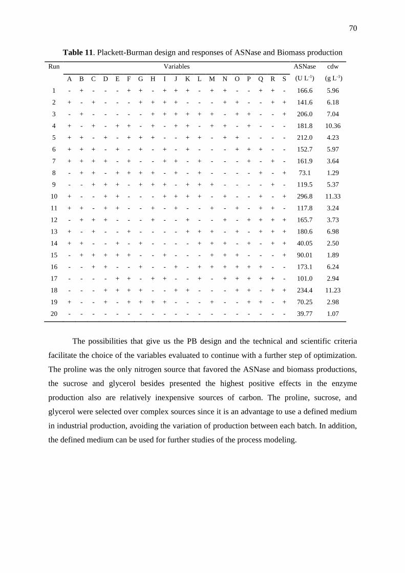
70
Table 11. Plackett-Burman design and responses of ASNase and Biomass production
Run Variables ASNase
(U L-1)
cdw
(g L-1) A B C D E F G H I J K L M N O P Q R S
1 - + - - - + + - + + + - + + - - + + - 166.6 5.96
2 + - + - - - + + + + - - - + + - - + + 141.6 6.18
3 - + - - - - - + + + + + + - + + - - + 206.0 7.04
4 + - + - + + - + - + + - + + - + - - - 181.8 10.36
5 + + - + - + + + - - + + - + + - - - - 212.0 4.23
6 + + + - + - + - + - + - - - + + + - - 152.7 5.97
7 + + + + - + - - + + - + - - - + - + - 161.9 3.64
8 - + + - + + + + - + - + - - - - + - + 73.1 1.29
9 - - + + + - + + + - + + + - - - - + - 119.5 5.37
10 + - - + + - - - + + + + - + - - + - + 296.8 11.33
11 + + - + + - - + - + - - + - + - + + - 117.8 3.24
12 - + + + - - - + - - + - - + - + + + + 165.7 3.73
13 + - + - - + - - - - + + + - + - + + + 180.6 6.98
14 + + - - + - + - - - - + + + - + - + + 40.05 2.50
15 - + + + + + - - + - - - + + + - - - + 90.01 1.89
16 - - + + - - + - - + - + + + + + + - - 173.1 6.24
17 - - - - + + - + + - - + - + + + + + - 101.0 2.94
18 - - - + + + + - - + + - - - + + - + + 234.4 11.23
19 + - - + - + + + + - - - + - - + + - + 70.25 2.98
20 - - - - - - - - - - - - - - - - - - - 39.77 1.07
The possibilities that give us the PB design and the technical and scientific criteria
facilitate the choice of the variables evaluated to continue with a further step of optimization.
The proline was the only nitrogen source that favored the ASNase and biomass productions,
the sucrose and glycerol besides presented the highest positive effects in the enzyme
production also are relatively inexpensive sources of carbon. The proline, sucrose, and
glycerol were selected over complex sources since it is an advantage to use a defined medium
in industrial production, avoiding the variation of production between each batch. In addition,
the defined medium can be used for further studies of the process modeling.

71
Table 12. Effects of the variables and statistical analysis of the Plackett-Burman design
ASNase production (U L-1 ) cdw production (g L-1 )
Effect S.E t student p-Value Effect S.E t student p-Value
Intercept 146.27 5.82 50.21 0.00001 5.20 0.31 33.77 0.00005
YE -33.39 5.82 -5.73 0.010* -0.07 0.31 -0.21 0.841
SP 11.20 5.82 1.92 0.150 0.49 0.31 1.59 0.209
CSS 19.33 5.82 3.31 0.045* 0.60 0.31 1.96 0.144
PDB 4.91 5.82 0.84 0.461 0.90 0.31 2.94 0.060
Glutamine -8.17 5.82 -1.40 0.255 -0.28 0.31 -0.91 0.429
Asparagine -15.79 5.82 -2.71 0.073 -0.02 0.31 -0.08 0.939
Maltose -1.24 5.82 -0.21 0.845 0.08 0.31 0.26 0.808
Glycerol 58.18 5.82 9.99 0.002* 2.89 0.31 9.36 0.002*
Glutamate -14.71 5.82 -2.52 0.085 -0.94 0.31 -3.06 0.054
Sucrose 90.78 5.82 15.58 0.001* 4.02 0.31 13.04 0.000*
Proline 18.63 5.82 3.20 0.049* 1.06 0.31 3.46 0.040*
Urea -25.31 5.82 -4.34 0.022* -2.68 0.31 -8.70 0.003*
Aspartate 14.51 5.82 -2.49 0.088 -0.25 0.31 -0.80 0.478
Citrate 20.33 5.82 3.49 0.039* -0.10 0.31 -0.34 0.755
Arginine -21.08 5.82 -3.62 0.036* 0.64 0.31 2.08 0.128
Ammonium
Chloride
25.76 5.82 4.42 0.021* 0.19 0.31 0.63 0.570
*significance at 5 %
R2= 0.9920, R adjusted 0.9499
PDB- Potato Dextrose broth, CSS- Corn step solids, SP- soybean peptone, YE- yeast extract.
2.3.2 Optimization of nutritional factors on L-asparaginase production using Central
Composite Design
The 3 nutrients selected from the 16 evaluated were significant for the both responses
evaluated, this means that proline, glycerol and sucrose contributive significant for the
enzyme and cell mass production. The CCD was employed to evaluate the interaction
between these three variables and to determine their optimal concentrations for ASNase
production. The CCD is formed from two-level factorials by the addition of just enough
points to estimate curvature and interaction effects. The designs can be viewed as partial
factorials with factors at five levels and is extensively used to build second-order response
surface models (KENNEDY and KROUSE, 1999).
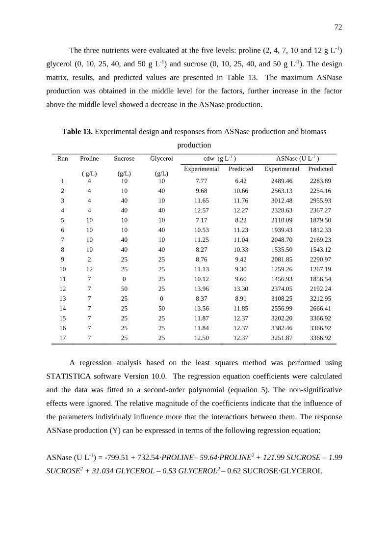
72
The three nutrients were evaluated at the five levels: proline (2, 4, 7, 10 and 12 g L-1)
glycerol (0, 10, 25, 40, and 50 g L-1) and sucrose (0, 10, 25, 40, and 50 g L-1). The design
matrix, results, and predicted values are presented in Table 13. The maximum ASNase
production was obtained in the middle level for the factors, further increase in the factor
above the middle level showed a decrease in the ASNase production.
Table 13. Experimental design and responses from ASNase production and biomass
production
Run Proline
( g/L)
Sucrose
(g/L)
Glycerol
(g/L)
cdw (g L-1 ) ASNase (U L-1 )
Experimental Predicted Experimental Predicted
1 4 10 10 7.77 6.42 2489.46 2283.89
2 4 10 40 9.68 10.66 2563.13 2254.16
3 4 40 10 11.65 11.76 3012.48 2955.93
4 4 40 40 12.57 12.27 2328.63 2367.27
5 10 10 10 7.17 8.22 2110.09 1879.50
6 10 10 40 10.53 11.23 1939.43 1812.33
7 10 40 10 11.25 11.04 2048.70 2169.23
8 10 40 40 8.27 10.33 1535.50 1543.12
9 2 25 25 8.76 9.42 2081.85 2290.97
10 12 25 25 11.13 9.30 1259.26 1267.19
11 7 0 25 10.12 9.60 1456.93 1856.54
12 7 50 25 13.96 13.30 2374.05 2192.24
13 7 25 0 8.37 8.91 3108.25 3212.95
14 7 25 50 13.56 11.85 2556.99 2666.41
15 7 25 25 11.87 12.37 3202.20 3366.92
16 7 25 25 11.84 12.37 3382.46 3366.92
17 7 25 25 12.50 12.37 3251.87 3366.92
A regression analysis based on the least squares method was performed using
STATISTICA software Version 10.0. The regression equation coefficients were calculated
and the data was fitted to a second-order polynomial (equation 5). The non-significative
effects were ignored. The relative magnitude of the coefficients indicate that the influence of
the parameters individualy influence more that the interactions between them. The response
ASNase production (Y) can be expressed in terms of the following regression equation:
ASNase (U L-1) = -799.51 + 732.54·PROLINE– 59.64·PROLINE2 + 121.99 SUCROSE – 1.99
SUCROSE2 + 31.034 GLYCEROL – 0.53 GLYCEROL2 – 0.62 SUCROSE·GLYCEROL

73
The statistical analysis of the model was performed by the analysis of variance
(ANOVA) performance at the 90% confidence limit. The ANOVA results of the CCD
experiment and can be observed in Table 15. The results indicate that the concentration of the
proline, glycerol and sucrose has a direct relation to the enzyme production. The Linear terms
of proline, sucrose and glycerol ,the quadratic terms of proline, sucrose and glycerol and just
the sucrose-glycerol interaction was significative for the ASNase production.for the ASNase
production were significative (p < 0.1). The ANOVA also suggested that the obtained
experimental was a good fit whit the model presenting a correlation coefficient R2= 0.93 and
R2adj = 0.840. Removing the not significant terms, was obtained a lack of fit p-value of
0.1087 (p>0.1), implying that the lack of fit of model was not significant relative to the pure
error, it means that the proposed model fit with the experimental data. The contour graphs
(Figure 16) show in the dark regions the optimal ASNase production. As can be observed the
best ASNase production is localized near to the central region, contrary for the cell biomass
production were the optimal zones are present at higher levels of carbon sources, these higher
concentrations of sucrose and glycerol have a negative impact in the enzyme production.
Table 14. ANOVA analysis of the production of L-asparaginase by L. scottii according to
CCD design
SS DF ,MS F p- value
Proline (L) 1251591 1 1251591 144.37 0.0068*
Proline (Q) 3141439 1 3141439 362.37 0.0027*
Sucrose (L) 137444 1 137444 15.85 0.0576*
Sucrose (Q) 2195471 1 2195471 253.25 0.0039*
Glycerol (L) 360911 1 360911 41.63 0.0231*
Glycerol (Q) 154271 1 154271 17.79 0.0518*
Proline x Sucrose 70724 1 70724 8.15 0.1038
Proline x Glycerol 679 1 679 0.07 0.8059
Sucrose x Glycerol 151267 1 15267 17.44 0.0528*
Lack off it 452698 5 90540 10.44 0.1087
Pure error 17338 2 8669
Total SS 6734472 16
*significance at 10 % ; R2= 0.932

74
Figure 16. Contour plots for ASNase (U L-1) and cell dry weight (g L-1) production.
Figure 17 Substrate comsumption and ASNase production in the optimized medium.

75
2.3.3 Validation of the model
The CCD design used to optimize the ASNase production provided the predicted
values of 6.15 g L-1 of proline, 28.34 g L-1 sucrose, and 15.61 g L-1 of glycerol for a maximal
production. The culture was performed using 200 mL of the synthetic medium containing the
optimized quantities added with the salts: KCl, 0.52 g L-1; MgSO4.7H2O, 0.52 g L-1;
CuNO3.3H2O, 0.001 g L-1; ZnSO4.7H2O, 0.001 g L-1; FeSO4.7H2O, 0.001 g L-1, the pH was
adjusted at 5.0. Under these conditions the predicte response for ASNase production was
2850 U L-1 the predicted response was 3379.41 ± 337.9 U L-1, even the model fail in predict
the response in around to 200 U L to be inside the optimized confidence interval (90%) the
model can improved the production of the enzyme in a almost in 50 fold, compared with the
started medium used by Gulati et al (1997). The figure 17 shows the comsuption of substrates
during the culture process, is observed that the sucrose finish at the 120 hours of culture,
unlike the glycerol was not completely comsumed. Due to this it is necessary to continue the
study in bioreactor to work in the required oxygenation conditions and to observe the
consumption of the different carbon sources present in the culture medium.

76
CONCLUSION
The results show the potential of the Antarctic yeast to produces new molecules, in
particular, the ASNase, that has potential uses in the pharmaceutical and food industries It is
to highlight that the psychrotolerant yeasts are few demanding in terms of nutritional
requirements and can grow well in a simple culture medium. The production of ASNase by
Leucosporidium scottii was improved in flask cultures after using different DoE
methodologies. The application of two well-planed set of experiments increased the enzyme
production till in 50-fold compared whit the initial culture medium used. The rational use of
DoE in bioprocess optimization and understanding how the nutrients and its concentration
influence in the microbial metabolite production during fermentation is a powerful tool to
develop a culture medium. This study allowed to develop a defined culture medium with 3
main components to increase the ASNase production with substrates such as glycerol and
sucrose a relative cheap components easily available at industrial levels, and that it can be
used for further metabolic studies.

77
4 CHAPTER IV EVALUATION OF L-ASPARAGINASE
PRODUCTION AND LIPID ACCUMULATION BY THE
PSYCHROTOLERANT YEAST Leucosporidium scottii L115
4.1 INTRODUCTION
Leucosporidium scottii is a non-fermentative, aerobic basiodiomycetous yeast
considered as a psychrotolerant microorganism, isolated from low –temperature
environments, generally in polar regions or temperate regions during the cold weather
seasons, and in different habitats in the Antarctic such as seawater, soils and mosses and
saline lakes. Able to thrive in relatively extreme environmental conditions, like high salinity,
low temperature or a high concentration of aromatic compounds (SUMMERBELL, 1983;
SAMPAIO, 2011). Psychrotolerant and psychrophilic yeasts have attracted the attention of
scientists for their potential application in various industries because the capacity to produces
valuable metabolites such as extracellular hydrolytic enzymes, pigments, polyunsaturated
fatty acids PUFAs, polyols, antifreeze proteins, and single cell oils (SCOs) (BRIZZIO, et al
2007; de GARCIA, et al 2007, SPENCER, et al 2002, ALCAINO et al 2015). For this
reason, the study of yeasts isolated from cold environments could provide new strains with
biotechnological potential (MARGESIN and SCHINNER, 1994).
The Leucosporidium scottii L115 strain was isolated from marine sediment collected in
the Antarctic Peninsula and our research group found it as capable of produces L-asparaginase
(ASNase) enzyme. This enzyme has gained attention for its anti-leukemia potential and for its
uses in the mitigation of acrylamide production during the food processing. Also, L. scotti is
reported as producer of valuable metabolites like lipases and coenzymes Q9 and Q10 (WOO-
HONG, 1991; DUARTE, et al 2013) is nutritionally versatile and capable of utilizes
alternative carbon sources as xylose and glycerol (SAMPAIO, 2011). And, recently was
characterized as an oleaginous yeast species able to accumulated lipids with a suitable fatty
acid profile for the production of biodiesel (PEREYRA et al 2014). The oleaginous yeasts are
advantageous hosts for the development of platforms to produce lipid-based chemicals and,
new molecular techniques are studied to exploit the potential of microorganisms as a lipid
producer for biodiesel production (ADRIO 2017; TSAI et al 2017). The yeasts have the
benefits of single-cell fermentation and there are a large number of genetic tools to study and
78
manipulate them and, exist a continuously increasing interest for the discovery of new yeast
strains capable of processing low-cost feedstocks like glycerol, molasses, and lignocellulosic
hydrolysates, to transform and storing high levels of SCOs amenable to being converted into
2nd generation biodiesel (TCHAKOUTEU, et al 2015; HUANG, et al 2017).
Microbial oils are an attractive alternative source of PUFAs and according to
experimental evidence, psychrophilic microorganisms like yeast may be are a promising
source (RATLEDGE 2004). And, as cold-adapted yeasts are able to synthesize
polyunsaturated fatty acids (PUFAs) of medical and dietetical interest, over-expressed at low
temperatures The production of yeast lipids with composition similarities with high added-
value has been considered as a process potentially economically viable, specifically if various
low or negative cost raw materials are utilized as substrates (ROSSI, et al 2009;
PAPANIKOLAU and AGGELIS, 2011; AGEITOS, et al 2011).
The aims of this work were to evaluate the ASNase production by L. scottii L115 in a
bioreactor under different conditions of initial cell concentration (X0) and carbon source, and
optimization of process parameters for maximum enzyme production. This work contributed
to add knowledge about psychrotolerant yeasts as potential producer of enzymes and fatty
acids.
79
4.2 MATERIALS AND METHODS
4.2.1 Microorganism and inoculum
Psychrotolerant yeast Leucosporidium scottii L115 was isolated from marine sediment
of Antarctic Peninsula by the expedition team of the Brazilian Antarctic Program
(PROANTAR) and was identified using the 26S rDNA sequencing (DUARTE et al , 2013).
The yeast strain was incubated in 250 mL Erlenmeyer flask for 72 h at 15°C and 200 rpm in
an orbital shaker (New Brunswick™ Innova® 42) in YPD medium: dextrose 20 gL-1,
peptone 20 g L-1, yeast extract 10 g L-1. The cells were harvested by centrifugation (Thermo
Scientific, Multifuge X3 FR) at 3400 xgfor 15 minutesat 5°C,and washed with milli-Q water.
These cells were growth in Erlenmeyer flasks to produce the mass required to inoculate in the
bioreactors. The propagation media was composed by: sucrose, 28.34 g L-1; glycerol, 15.6 g
L-1; L-proline, 6.12 g L-1; KCl, 0.52 g L-1; MgSO4.7H2O, 0.52 g L-1; CuNO3.3H2O, 0.001 g
L-1; ZnSO4.7H2O, 0.001 g L-1; FeSO4.7H2O, 0.001 g L-1, in phosphate buffer 50 mM pH 7.0.
The propagation was carried at the same condition described above. After 48 h, the cells were
harvested by centrifugation and suspended in milli-Q water to inoculate the bioreactors.
4.2.2 Bioreactor assays
All batch cultures were performed in a multi-bioreactor system (BIOSTAT®Qplus,
Sartorius Stedim, Germany) of 1.0 L equipped with automatic monitoring and control units
for temperature, pH, aeration, and agitation. The bioreactor vessel was equipped with two
six/bladed Rushton turbines and, the working volume used was 0.5 L.
4.2.3 Effect of initial cell concentration in enzyme production
All experiments to assess the initial cell concentration on enzyme production were
performed at 15°C under cascade control at 30% of oxygen saturation using compressed air
where the range of agitation was set up at 50 – 500 rpm, and the airflow was set up at 0.1 – 1
Lmin-1.The initial cell concentration was evaluated at 1, 3, and 5 g L-1 (dry basis). The media
used was the same for the propagation. Antifoam was used in the case of need. Samples were
collected regularly and frozen at -20°C immediately for further analysis.
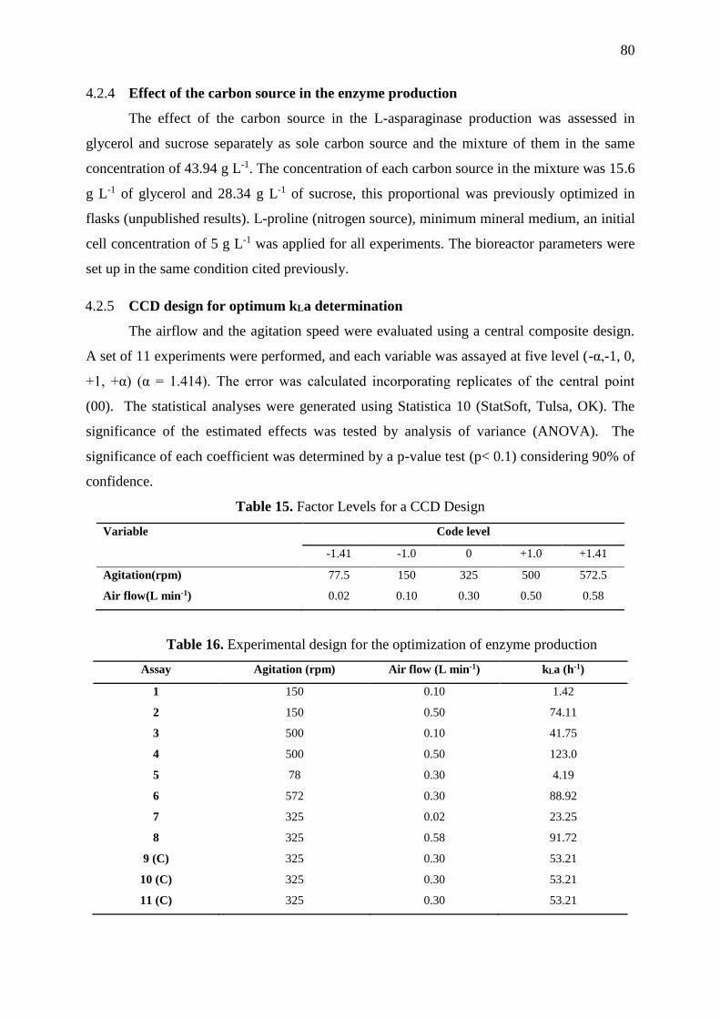
80
4.2.4 Effect of the carbon source in the enzyme production
The effect of the carbon source in the L-asparaginase production was assessed in
glycerol and sucrose separately as sole carbon source and the mixture of them in the same
concentration of 43.94 g L-1. The concentration of each carbon source in the mixture was 15.6
g L-1 of glycerol and 28.34 g L-1 of sucrose, this proportional was previously optimized in
flasks (unpublished results). L-proline (nitrogen source), minimum mineral medium, an initial
cell concentration of 5 g L-1 was applied for all experiments. The bioreactor parameters were
set up in the same condition cited previously.
4.2.5 CCD design for optimum kLa determination
The airflow and the agitation speed were evaluated using a central composite design.
A set of 11 experiments were performed, and each variable was assayed at five level (-α,-1, 0,
+1, +α) (α = 1.414). The error was calculated incorporating replicates of the central point
(00). The statistical analyses were generated using Statistica 10 (StatSoft, Tulsa, OK). The
significance of the estimated effects was tested by analysis of variance (ANOVA). The
significance of each coefficient was determined by a p-value test (p< 0.1) considering 90% of
confidence.
Table 15. Factor Levels for a CCD Design
Variable
Code level
-1.41 -1.0 0 +1.0 +1.41
Agitation(rpm) 77.5 150 325 500 572.5
Air flow(L min-1) 0.02 0.10 0.30 0.50 0.58
Table 16. Experimental design for the optimization of enzyme production
Assay Agitation (rpm) Air flow (L min-1) kLa (h-1)
1 150 0.10 1.42
2 150 0.50 74.11
3 500 0.10 41.75
4 500 0.50 123.0
5 78 0.30 4.19
6 572 0.30 88.92
7 325 0.02 23.25
8 325 0.58 91.72
9 (C) 325 0.30 53.21
10 (C) 325 0.30 53.21
11 (C) 325 0.30 53.21

81
4.2.6 Analysis
4.2.6.1 ASNase assay by the hydroxylaminolysis reaction
The ASNase activity was determined by the hydroxylaminolysis reaction and using
the whole cell for the enzymatic reaction (Ferrara et al., 2004). In a eppendorf tube was
added, 0.8 mL of cells suspension at 1.0 of DO in 25 mMTris-HCl buffer pH 8.0, 0.1 mL of
0.1 M L-asparagine and 0.1 mL of 1.0 M solution of hydroxylamine hydrochloride at pH 7.0,
the mixture was agitated on a Thermomixer at 850 rpm at 37ºC, after 30 minutes the
enzymatic reaction was stopped by adding 0.25 mL of TCA/FeCl3 (100 g L-1 FeCl3, 50 g L-1
TCA in 0.66 M HCl).The tube was centrifuged at 3400 x g for 5 min and 0.3 mL of the
supernatant was measured at 500 nm in a plate reader (Synergy MX, Biotek). Controls were
prepared in the same way as samples, but the hydroxylamine and asparagine solutions were
added after the ferric chloride reagent. One unit of ASNase was defined as the amount of
enzyme that produces 1μmol of -aspartohydroxamic acid, formed for a minute by gram of
dried cell weight (U g-1cdw).
4.2.6.2 Quantification of biomass production
The cells growth was accompanied by measuring the optical cell density (OD) at 600
nm using spectrometer detector (Synergy MX, Biotek) using water as a blank. The dry
weight was measure through gravimetrical analysis, 1 mL of each sample was centrifuged at
3400 g, re-suspended twice in milli-Q water, and placed to dry at 60°C in the oven until
constant weight in the analytical balance.
4.2.6.3 Substrate quantification
The samples were centrifuged at 5000 g and the supernatant was collected. The
glycerol and sucrose concentrations were determined by HPLC (Ultimate 3000, Dionex) with
IR detector 50 °C, Aminex column HPX-87H 300 mm x 7.8 mm at 60º C, and 0.5 mL.min-1
of 5mM H2SO4 as eluent phase. The D-glucose and D-fructose were determined using the
enzymatic assay kit K-SUFRG (Megazyme) according to the manufacturer’s protocol (K-
SUFRG 06/14).
4.2.7 Staining of yeast cell with Nile Red Dye for microscopy analysis
In order to detect the accumulation of lipids, the method of staining neutral lipids in
yeast cells by Nile red was used according to Rostronand Lawrence (2017). A stock solution
of Nile Red (2 mgmL-1) in acetone was prepared and stored at -20oC. Yeast cells were re-

82
suspended and washed twice in the in PBS 1X buffer pH 7.4 and the final concentration was
adjusted at 1.0 of DO. At 250 µL of cell suspension was added 25 μL of DMSO:PBS 1:1 and
25 μLof Nile Red in acetone at 60 µg mL-1 mixed and incubated at room temperature in the
dark for 5 min. The cells were centrifuged at 5000 x g, the supernatant discarded and the cell
washed twice with PBS 1X. The cells were re-suspended in 100 µL of 10% of formaldehyde
(37%) and fix for 15 min at room temperature in the dark. The cells were centrifuged and
washed twice with PBS 1X buffer and re-suspended in 100 µL of the same buffer. The
stained cells were analyzed under a fluorescence microscope (Leica, Germany) using the filter
EGFP.
4.2.8 Lipid extraction
The yeast cultures at different conditions were harvest and washed twice with distilled
water and the final concentration of cells suspension was adjusted at the 10- 30 mg· mL-1 of
dcw, 2 mL of the suspension were transferred to an eppendorf tube, centrifuged at 4.7 x g for
5 minutes the supernatant was discarded, 500 μL of glass beads (0.22mm) and 500 uL of
distilled water were added. The yeast cell were homogenized using an homogenizer Precellys
24 (BertinTechnologies) during 10 cycles of 6000 rpm for 20 sec each, with a period of 10 sec
in bath ice avoiding the over heat of the samples. The homogenized was recovered washing
the eppendorf tube with 10 mL of methanol, and stored at -20ºC.
4.2.9 Total lipids quantification and determination of fatty acid composition
Lipids were extracted from all samples using a modified form of the method described
by Bligh and Dyer (1959), with a reduced amount of solvent (30.0 mL methanol and
chloroform, 1:1). Lipid content was determined by gravimetry after evaporation of
chloroform and is expressed as the percentage. Lipid extracts were used for the preparation of
fatty acid methyl esters using AOCS Official Method CE 2-66 (1998). C 23:0 methyl ester
was used as an internal standard. Fatty acid methyl esters (FAME) were analyzed using the
Agilent 7890A GC system equipped with a column X from Supelco, using AOCS official
method Ce 1b-89 (1998).
4.2.10 Fermentation parameters
Considering a determined time of culture in the bioreactor, the corresponding values
of cells (X), substrate (S) and product (P) concentrations, can be related though the
conversion factors. For comparison of the trials, the production parameters were determined
using the equation described below.

83
• Conversion factor of limitant substrate to cells (Yx/s)
𝑌𝑋 𝑆⁄ =𝑋𝑓−𝑋0
𝑆0−𝑆𝑓 equation (6)
Where: Xf = cell concentration at the time evaluated (g L-1); X0 = initial cell concentration (g
L-1); Sf = substrate in the medium at the time evaluated (g L-1); S0= initial substrate
concentration (g L-1);
• Conversion factor of limitant substrate to product (YP/s)
𝑌𝑃 𝑆⁄ = −𝑃𝑓−𝑃0
𝑆0−𝑆𝑓 equation (7)
Where: Pf = concentration of product formed at the time evaluated (g L-1); P0 = initial
concentration of the product (g L-1); Sf = substrate in the medium at the time evaluated (g L-1);
S0= initial substrate concentration (g L-1);
• Volumetric productivity (Q)
𝑄 = −𝑃𝑓−𝑃0
𝑉×𝑡 , (
𝑔
𝐿∙ℎ) equation ( 8)
Where P, is the maximum of enzyme produced and for lipid was measured at the end
of the culture, S0 is the initial concentration of glycerol + sucrose, Xf is cell concentration at
time t of maximum production of the enzyme, and X0 is the initial cell concentration, V the
volume of liquid culture and t is the culture time for maximum enzyme production.
4.2.11 Determination of kLa
The volumetric oxygen transfer coefficient (kLa) was determined experimentally using
the method of “gas out- gas in” (TRIBE et al, 1994), in a 1L bioreactor(BIOSTAT®Qplus,
Sartorius Stedim, Germany) at 15ºC containing the 0.5 L of medium culture. The oxygen
probe was set to zero in each determination by bubbling nitrongen into the medium culture.
The aeration an agitation parameter were seted accoerding to the experiment design
conditions (Table 17) and the values of oxygen saturation were register at different times until
the probe reads 100% of saturation.
The variation of the dissolved oxygen (DO) concentration with the time can be expressed as
𝑑𝐶
𝑑𝑡º= −𝑘𝐿𝑎 ∙ (𝐶𝑠 − 𝐶) equation (9)

84
Integrating the equation 9 is obtained the equation 1
𝐿𝑛 (1 −𝐶
𝐶𝑠) = −𝑘𝐿𝑎 ∙ 𝑡 equation (10)
Were Cs is the oxygen saturation concentration (mg L-1) at the set temperature, and C is the
dissolved oxygen concentration during the gas-out period. Thus, the kLa can be determinted
from the slope of Ln(1- C/Cs) versus time.

85
4.3 RESULTS AND DISCUSION
4.3.1 Effect of initial cell concentration in the L-asparaginase production
Figure 18 shows the substrate consumption, biomass and enzyme production, as can be
observed the consumption of substrates is sequential and not in parallel. The glucose was
preferred consumed, followed by the fructose and glycerol. The uptake of glucose was clearly
observed during the first hours of the culture the sucrose was broken down and the glucose
was consumed simultaneously, in contrast, the fructose was consumed until the glucose was
almost depleted. The lag phase was absent in all experiments, and the ASNase was
concurrently produced with cellular growth. All sugars were depleted in 114 h under initial
cell concentration of 1 g L-1 and 3 g L-1, whereas starting with 5 g L-1 of inoculum, the carbon
sources were depleted in approximately 70 h. The sequential metabolism of the carbon founts
suggests that the L. scottii yeast produces an extracellular invertase or a fructofuranosidase
located in the cell wall, breaking first the sucrose present in the medium and latter occurs the
sequential consumption of substrates. We observed that the uptake of glycerol occurs during
the stationary phase, this means that glycerol is not used for the growth of the cell, instead is
used for a stage of accumulation, it is reflected in the increase of intracellular lipids during the
stationary phase. In addition, it was not observed a marked diauxic growth phenomenon
during the consumption of the substrates.
At 5% of initial inoculum the substrates are consumed in 70 hours with an enzyme
productivity of 17.4 U·L-1·h-1, with an initial inoculum of 3%, the carbon sources ends around
94 hours with a productivity of 10.45 U·L-1·h-1 and with 1% of inoculums, the substrates are
consumed in 114 hours with a productivity of 8.93 U·L-1·h-1. As can be observed the
inoculum size at 5% increases the productivity of the process. Also, can be observed biomass
production was not significantly different, however, the production of ASNase was affected
by the concentration of the initial inoculum, the experiments showed that the production of
enzyme increases with the cell growth reaching the pick with the stationary phase. The
maximum production of the ASNase and yeast growth were achieved with an inoculums size
of 5% (v/v) producing 2595 U·L-1 at the end of the culture.

86
As shown in Table 17, the production of the enzyme per gram of cell produced
increases relative to the concentration of the inoculum increasing by almost 50 Ug-1 from 1 to
5% of the initial inoculums increasing. In addition, the initial cell concentration influenced the
ASNase volumetric and activity yields. Therefore, the yield increases linearly with cell
concentration and it suggests that the higher value could attain under high cell density.
Table 17. Enzyme and lipid production at different concentration of inoculum
X0 Time μmax QX PASNase QASNase YX/S YASNase/S YE/X YL/S YL/X
g L-1 h h-1 g L-1 h-1 U L-1 U L-1 h-1 g g-1 U g-1 U g-1 g g-1 g g-1
1 114 0.05 0.081±
0.002
1699 ±
85
13.99 ±
0.74
0.31±
0.01
39.03±
8.25
109.3±
9.07
0.21±
0.04
0.60±
0.04
3 94 0.036 0.0089±
0.008
2199
±180
16.79±
1.91
0.29±
0.04
39.74±
4.09
116.7±
18.09
0.18±
0.03
0.56±
0.05
5 70 0.030 0.133 ±
0.007
2595
±350
27.24±
5.12
0.31±
0.02
40.93±
6.53
122.2±
21.82
0.14±
0.01
0.52±
0.02
YASNase/S ,enzyme yield, U g-1 substrate assimilated, X/S , cell yield, g g-1 substrate assimilated, L/S , lipids
yield, g g-1 substrate assimilated, QASNase , volumetric enzyme productivity , U L-1h-1, QX , volumetric cell
productivity , U L-1h-1
4.3.2 Effect of carbon source in enzyme production
The composition of culture medium was previously optimized in shaker resulting in
the ratio sucrose: glycerol (28.34:15.6 g L-1) for the best production of the enzyme. Even so,
the effect of glycerol and sucrose were tested individually at the initial concentration of 43.94
g L-1. This study was conducted to evaluate in bioreactor the influence of both carbon sources
in the ASNase and lipid production. According to Figure 18, the cell growth was similar
independently of carbon source, and the enzyme production initiated since the inoculation.
For the conditions tested, the production of the enzyme was gradually increased during the
exponential phase and the maximum was reached in the stationary phase it means that the
yeast grew firstly due to the availability of nitrogen and the carbon source, then when were
exhausted the enzyme production was increased by the activation of the genes for survival
mechanism. The ASNase production using glycerol (1085 ± 55 U L-1) or sucrose (1534 ± 358
U L-1) separately was less than their mixture (2595 ± 359 U L-1) and the final cell
concentration ranged from 16.1 to 17.45 g L-1 the biomass production was not significantly
different from that obtained using sucrose or the mixture of both substrates.

87
The glycerol and sucrose together caused a positive synergistic effect on enzyme
production, this means that combination of both substrates improves the production of the
enzyme modifying the internal metabolism of the yeast. This interaction could be explained
because in high sugar concentration medium with a high expression of invertase the
hydrolysis of sucrose to glucose and fructose augment the osmotic pressure in the medium,
this stressing osmotic pressure can inhibit the yeast growth triggering a complex response that
enhanced the glycerol production and retention and the ASNase production. Osmotolerant
yeasts are able to retain and synthesize glycerol to act as an osmoregulator, and some yeasta
have active glycerol uptake pumps (FLORES et al, 2000). This behavior was observed in the
L. scottii culture were was produced a small quantity of glycerol in the presences of sucrose
at 49 g L-1(figure 2A). Synthesis and retention of glycerol by yeast strains has been shown
previously to correlate strongly with fermentation in media with high concentrations of either
glucose and fructose, or equivalent levels of sucrose (MYERS, et al 1997). In S. cerevisiae
is reported that the ability to adapt to high osmotic pressures is critical to the efficiency of
fermentation of high sugar concentration media producing high levels of glycerol 3-phosphate
dehydrogenase (GD3P) (ATTFIELD and KLETSAS, 2000). This is in agreement the
capacity of the L. scottii to survive in marine environments where intracellular osmotic
regulation is very important due to the hypertonic characteristics of the sea water.
4.3.3 Effect of aeration and agitation
The oxygen is a factor that influences the growth and biosynthesis of the yeast
enzymes, as well as the production of different metabolites such neutral lipids. In particular,
oxygen acts as a terminal electron acceptor for oxidative reactions to provide energy for
cellular activities, however, each microorganism varies in its oxygen requirements.
Measurements of kLa provide important information about a bioprocess on bioreactor and the
determination of the coefficient ensure that processing conditions are such that an adequate
supply of oxygen is available for cell growth (GARCIA-OCHOA et al, 2010). Being non-
fermentative and strictly aerobic yeast (WATSON et al, 1978) is interesting the study of the
behavior of Leucosporidium scottii L115 under different kLa conditions and at 15°C of culture
temperature which favors the dissolution of oxygen in the medium.

88
.
Figure 18. Experimental data from the batch cultures of L. scottii L115 (ASNase;
cell;fructose; glucose; and glycerol) to assess the initial cell concentration A) X0: 1 g
L-1, B) X0: 3 g L-1, C) X0: 5 g L-1.
0 24 48 72 96 1200
2
4
6
8
10
12
14
16
18
20
0
500
1000
1500
2000
2500
3000
3500
Time (h)
Su
bst
rate
an
d c
ell
(g L
-1)
(A)
AS
Nas
e (U
L-1)
0 24 48 72 96 1200
2
4
6
8
10
12
14
16
18
20
0
500
1000
1500
2000
2500
3000
3500
Time (h)
Su
bst
rate
an
d c
ell
(g L
-1)
(B)
AS
Nas
e (U
L-1)
0 24 48 72 96 1200
2
4
6
8
10
12
14
16
18
20
0
500
1000
1500
2000
2500
3000
3500
Time (h)
Subst
rate
and c
ell
(g L
-1)
AS
Nas
e (U
L-1)
(C)

89
Figure 19. Experimental data from the batch cultures of L. scottii L115 (ASNase; cell;
and substrate) to assess the carbon source A) Glycerol, B) Sucrose, C) Glycerol + sucrose.
From the bioprocess point of view, the effect of oxygen transfer condition on L. scottii
L115 culture was evaluated in the bioreactor. The effect of the aeration and agitation on the
production of the enzyme and the lipids accumulation was studied through RSM using a
Central Composition Design (CCD). According to Table 18, higher values of kLa benefited
ASNase and lipids production. The maximum ASNase production, approximately 2613 U L-1,
corresponded to assay 6 (kLa: 89 h-1) and assay 8 (kLa: 92 h-1), and the maximum productivity
of the enzyme (36.30U·L-1·h-1) was obtained with a kLa coefficient of 89 h-1 obtained at 572.5
rpm of agitation speed and 0.3 L min-1 of airflow. These conditions were more efficient to
produce cell as well, 0.31 and 0.27 g g-1, respectively, also, more efficient in terms of specific
enzyme production (U g-1 cell) that corresponded to 169 and 200 U g-1, respectively. The
conditions of the assay 8 (325 rpm, 0.58 L min-1) are more suitable parameters due to less
foam was produced and in consequence, less anti-foam will be required, instead of assay 6
that corresponds to higher agitation. At lower agitation rate, the oxygen transfer is limited
resulting in decreased cell mass growth, while at higher agitation rate, although growth is
more, there is a reduction in enzyme activity. It may be because of the variation in the
agitation speed influences in the nutrient uptake. Thus, analyzing the data obtained we can
establish that the working conditions for the optimization of work for the ASNase production
were 88.92 – 91.72 h-1 of kLa.
0 24 48 720
10
20
30
40
50
0 24 48 72 0 24 48 720
500
1000
1500
2000
2500
3000
3500(C)(B)
Su
bst
rate
an
d c
ell
(g L
-1)
(A)
Time (h)
AS
Nas
e (U
L-1)
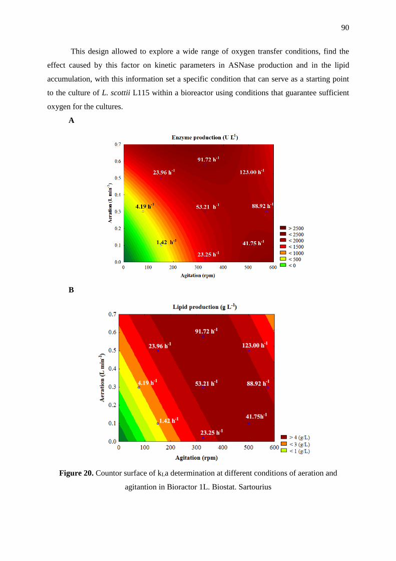
90
This design allowed to explore a wide range of oxygen transfer conditions, find the
effect caused by this factor on kinetic parameters in ASNase production and in the lipid
accumulation, with this information set a specific condition that can serve as a starting point
to the culture of L. scottii L115 within a bioreactor using conditions that guarantee sufficient
oxygen for the cultures.
A
B
Figure 20. Countor surface of kLa determination at different conditions of aeration and
agitantion in Bioractor 1L. Biostat. Sartourius

91
Table 18. CCD design to optimize the ASNase production and results of productivity and conversion
Assay Agitation Airflow kLa μmax ASNase Lipids QCell QASNase PL YX/S YASNase/S YL/S YASNase/X YL/X
rpm L min-1 h-1 h-1 U L-1 g L-1 g L-1 h-1 g L-1 h-1 g L-1 h-1 g g-1 U g-1 g g-1 U g-1 g g-1
1 150 0.1 1.42 0.028 523 1.67 0.065 7.30 0.03 0.13 13.78 0.05 105 0.41
2 150 0.5 74.11 0.046 1893 5.08 0.151 26.30 0.07 0.25 38.04 0.10 155 0.42
3 500 0.1 41.75 0.035 1880 5.98 0.160 26.10 0.08 0.24 37.82 0.12 155 0.51
4 500 0.5 123.0 0.053 1880 5.56 0.175 26.10 0.08 0.26 38.66 0.12 147 0.46
5 78 0.3 4.19 0.025 772 1.91 0.078 10.70 0.03 0.20 27.09 0.07 133 0.35
6 572 0.3 88.92 0.057 2613 5.04 0.183 36.30 0.06 0.31 52.93 0.09 169 0.33
7 325 0.02 23.25 0.041 2062 5.11 0.183 28.60 0.06 0.26 40.47 0.09 154 0.34
8 325 0.58 91.72 0.046 2583 5.27 0.176 35.90 0.07 0.27 53.13 0.10 200 0.39
9 325 0.3 53.21 0.044 2179 5.66 0.156 22.70 0.08 0.24 34.11 0.12 143 0.49
YASNase/S ,enzyme yield, U g-1 substrate assimilated, X/S , cell yield, g g-1 substrate assimilated, L/S , lipids yield, g g-1 substrate
assimilated, QX , volumetric cell productivity , g L-1h-1, QASNase , volumetric enzyme productivity , U L-1h-1.
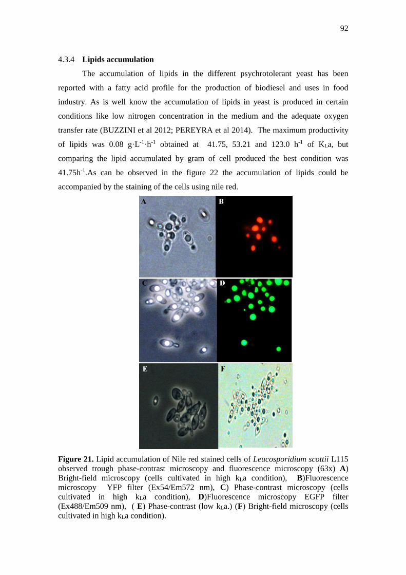
92
4.3.4 Lipids accumulation
The accumulation of lipids in the different psychrotolerant yeast has been
reported with a fatty acid profile for the production of biodiesel and uses in food
industry. As is well know the accumulation of lipids in yeast is produced in certain
conditions like low nitrogen concentration in the medium and the adequate oxygen
transfer rate (BUZZINI et al 2012; PEREYRA et al 2014). The maximum productivity
of lipids was 0.08 g·L-1·h-1 obtained at 41.75, 53.21 and 123.0 h-1 of KLa, but
comparing the lipid accumulated by gram of cell produced the best condition was
41.75h-1.As can be observed in the figure 22 the accumulation of lipids could be
accompanied by the staining of the cells using nile red.
Figure 21. Lipid accumulation of Nile red stained cells of Leucosporidium scottii L115
observed trough phase-contrast microscopy and fluorescence microscopy (63x) A)
Bright-field microscopy (cells cultivated in high kLa condition), B)Fluorescence
microscopy YFP filter (Ex54/Em572 nm), C) Phase-contrast microscopy (cells
cultivated in high kLa condition), D)Fluorescence microscopy EGFP filter
(Ex488/Em509 nm), ( E) Phase-contrast (low kLa.) (F) Bright-field microscopy (cells
cultivated in high kLa condition).
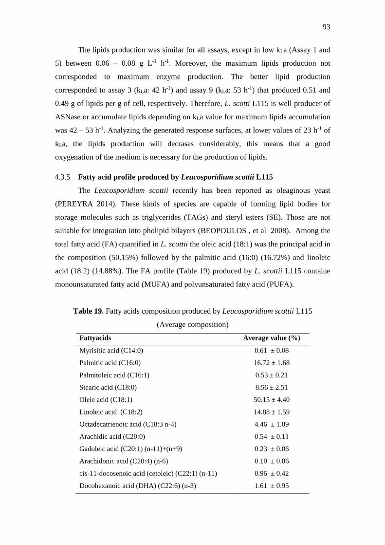
93
The lipids production was similar for all assays, except in low kLa (Assay 1 and
5) between 0.06 – 0.08 g L-1 h-1. Moreover, the maximum lipids production not
corresponded to maximum enzyme production. The better lipid production
corresponded to assay 3 (kLa: 42 h-1) and assay 9 (kLa: 53 h-1) that produced 0.51 and
0.49 g of lipids per g of cell, respectively. Therefore, L. scotti L115 is well producer of
ASNase or accumulate lipids depending on kLa value for maximum lipids accumulation
was 42 – 53 h-1. Analyzing the generated response surfaces, at lower values of 23 h-1 of
kLa, the lipids production will decrases considerably, this means that a good
oxygenation of the medium is necessary for the production of lipids.
4.3.5 Fatty acid profile produced by Leucosporidium scottii L115
The Leucosporidium scottii recently has been reported as oleaginous yeast
(PEREYRA 2014). These kinds of species are capable of forming lipid bodies for
storage molecules such as triglycerides (TAGs) and steryl esters (SE). Those are not
suitable for integration into pholipid bilayers (BEOPOULOS , et al 2008). Among the
total fatty acid (FA) quantified in L. scottii the oleic acid (18:1) was the principal acid in
the composition (50.15%) followed by the palmitic acid (16:0) (16.72%) and linoleic
acid (18:2) (14.88%). The FA profile (Table 19) produced by L. scottii L115 containe
monounsaturated fatty acid (MUFA) and polyunsaturated fatty acid (PUFA).
Table 19. Fatty acids composition produced by Leucosporidium scottii L115
(Average composition)
Fattyacids Average value (%)
Myrisitic acid (C14:0) 0.61 ± 0.08
Palmitic acid (C16:0) 16.72 ± 1.68
Palmitoleic acid (C16:1) 0.53 ± 0.21
Stearic acid (C18:0) 8.56 ± 2.51
Oleic acid (C18:1) 50.15 ± 4.40
Linoleic acid (C18:2) 14.88 ± 1.59
Octadecatrienoic acid (C18:3 n-4) 4.46 ± 1.09
Arachidic acid (C20:0) 0.54 ± 0.11
Gadoleic acid (C20:1) (n-11)+(n+9) 0.23 ± 0.06
Arachidonic acid (C20:4) (n-6) 0.10 ± 0.06
cis-11-docosenoic acid (cetoleic) (C22:1) (n-11) 0.96 ± 0.42
Docohexanoic acid (DHA) (C22:6) (n-3) 1.61 ± 0.95

94
The total lipid quantified reaches the 50 % of the total weight and the results of
the composition obtained are similar with that reported by Pereyra et al (2004), but
unlike, we found that L. scotti L115 produces small amounts of DHA at a level of 1%,
whereas in others studies the DHA was not detected in psychrotolerant yeasts species.
This could be explained because the previous studies the temperature of culture was at
25°C, instead our experiments were carried at 15°C and it is know that the lipid profile
can be modified by adjusting the culture temperature because the degree of saturation
generally decreases with the temperature. The psychrophiles are generally endowed
with a higher proportion of unsaturated fatty acids, especially hexadecenoic (16:1) and
octadecenoic acid (18:1), that mesophilic and increase in the DHA (22:6) and/or
eicosapentanoic acid (20:5) is observed in psychrophiles and barophiles when grown at
temperatures closed to 0°C (RUSSELL 1992). From the samples of cultures obtained at
different oxygen transfer rates analyzed the composition of the fatty acids not changes,
but the production was affected significantly.

95
4.4 CONCLUSION
In conclusion, the study confirms that the Leucosporidium scottii isolated from
Peninsula Antarctica show the capacity to produce ASNase and accumulate lipids using
substrates like glycerol and sucrose, substrates that could be used for a large-scale
production. These findings emphasize that the cold-adapted yeasts are a potential source
of enzymes suitable for biotechnological processes at low and mild temperatures. The
development of the bioprocess for ASNase production by the psychrotolerant yeast
Leucosporidium scottii using the DOE strategies for increasing the volumetric yield of
the enzyme, the optimization of the dissolved oxygen level, results in an increment of
the enzyme production. Under optimized conditions, the highest enzyme production
was obtained (growth temperature 15º C, X0: 5 g L-1, pH 7.0, 48 h, kLa 89-92 h-1).There
are few psychrotolerant yeasts studies in bioreactor cultures at temperatures of 15ºC, for
this very reason, this study can be taken into account for the next studies of different
psychrotolerant yeasts and to better understand the culture process of these
microorganisms.
The lipid accumulation critically depends on the carbon used and the dissolved
oxygen levels. The cultivation of L. scottii L115 at 15ºC in cheap and convenient carbon
sources generating an interesting lipid profile, where it presents monounsaturated and
polyunsaturated lipids that can be studied in greater depth for their possible use in the
production of biodiesel or with uses in the food industry. Also can be explored the use
of molasses from biomass production and recycling glycerol from the biodiesel industry
to produces biodisel in a sustainable way.

96
5 GENERAL CONCLUSION
The L-Asparaginase from Leucosporidium scottii L115 has potential to uses at
therapeuctic level and in the food industry, in further years the need for new ASNase
molecules with improved properties will need to get a better ALL treatment. Further
studies of the LsASNase molecule can help to understand the mechanisms whereby
presents a low glutamine affinity and the structure to favore its kinetics parameters.
Also this non-conventional yeast have the capacity to use alternative sources of carbon,
to generate value-added molecules, such as enzymes and lipids.
The microbiological potential of Antarctica can be exploited through the Brazilian
Antartic Program (PROANTAR) which can provide microorganisms that have been
little studied and that can generate new molecules of great interest for the
pharmaceutical, chemical and food industries. The psychrophiles and psychrotolerant
microorganism, in special the yeasts have shown that they are capable of producing new
enzymes used at industrial levels, these cold adapted enzymes presents different
characteristics that deservs be studied in deep for the improved structures and kinetic
parameters that can result in the generation of new molecules with more specificity and
greated catalitc efficience.
For this reason, the study of the cold-adapted microorganisms opens a wide range
of options to generate new molecules, through the study of the mechanisms and
molecular structures developed to compensate the cold-environments.

97
6 FUTURE PERSPECTIVES
The production of recombinant LsASNase is necessary to study the effect of
glycosylation on the structure and its change in kinetic behavior since the allosteric
behavior can be produced by the presence of glycosylations. These could also affect the
affinity of the substrate and the Kcat can increase by double considering the reduction
of molecular weight by almost half.
The structural analysis of the enzyme, will identificate the the sequences that
increase the flexibility of the structure and improve the kinetic parameters, not only of
this but also of other enzymes as well.
In relation to the lipid profile obtained from L. scottii, studies can be made of the
possible applications of these as SCOs or for the production of biodiesel from the uses
of cheap carbon sources such as glycerol, molasses or xylose.

98
7 REFERENCES
1. Abd El Baky H.H., El Baroty G. S., Optimization of Growth Conditions for
purification and production of L-asparaginase by Spirulina maxima, Evidence-
Based complementary and Alternative Medicine, Article ID 1785930, Vol. 2016.
2. Adrio, J.L., Oleaginous Yeasts: Promising platforms for the production of
Oleochemicals and Biofuels, Biotechnology and Bioengineering, Vol. 114, No. 9,
pp. 1915-1920, 2017.
3. Aghaiypour K., Wlodawer A., Lubkowski J., Structural basis for the activity and
substrate Specificity of Erwinia chrysanthemi L-Asparaginase, Biochemistry, 15
40 (19) pp. 5655-5664, 2001.
4. Alcaino J., Cifuentes V., Baeza M., Physiological adaptations of yeast living in
cold environments and their potential applications, World J Microbiol Biotechnol,
31, pp 1467-1473, 2015.
5. Aldelrahman S. Z. Gregory A. Chenyu D., Marine yeast isolation and industrial
application, FEMS Yeast Research 14 pp 813-825, 2014.
6. Andreote F.D., Pereira S. M., Melo M. V., Roesch L., The Brazilian Soil
Microbiome, in Pylro V., Roesch L., (eds) Soil Biodiversity in Amazonian and
Other Brazilian Ecosystems, Current status and Perspectives, Springer
International Publishing AG, 2017.
7. Anishkin,A., Sukharev, S., Rempe, S.B., Catalytic role of the substrate defines
specificity of therapeutic L-asparaginase, J Mol Biol, 427, 2867-2885, 2015.
8. Arima, K., Sakamoto, T., Araki, C., Tamura, G., Production of extracellular
asparaginases by microorganisms. Agric Biol Chem 36, 356-361, 1972.
9. Atkin C.L., Neilands J.B., Phaff H.J., Rhodotorulic Acid from Species of
Leucospordium, Rhodosporidum, Rhodotorula, Sporidiobolus, and
Sporobolomyces, and a new alanine-containing ferrichrome from Crytococcus
melibiosum, Journal of Bacteriology, Vol. 103, No.3., pp. 722-733, 1970.
10. Attfield, P.V., Kletsas, S., Hyperosmotic stress response by strains of baker’s
yeasts in high sugar concentration medium, Letters in Applied Microbiology, 31,
323-327, 2000.

99
11. Avramis V.I., Tiwari, P. N., Asparaginase (native ASNase or pegylated ASNase)
in the treatment of acute lymphoblastic leukemia, International Journal of
Nanomedicine, I (3) 241-254, 2006.
12. Baakza A., Vala A. K., Dave B. P., Dube H.C., A comparative study of
siderophore production by fungi from marine and terrestrial habitats, Journal of
Experimental Marine Biology and Ecology 311, pp 1-9, 2004.
13. Balcão,V.M., Mateo, C., Fernandez-Lafuente, R., Malcata, X., Guisán, J.M.,
Structural and functional stabilization of asparaginase via multisubunit
immobilization onto highly activated supports. Biotechnol Prog 17(3), 537- 542,
2001.
14. Beopoulos A., Mrozova, Z., Thevenieau, F., Le Dall M.T., Hapala, I.,
Papanikolau, S., Chardot, T., Nicaud, J.M., Control of lipid accumulation in yeast
Yarrowia lipolitica, Applied and Enviromental Microbiology, pp. 7779-7789,
2001.
15. Birgisson H., Delgado, O., García A., L., Hatti-Kaul, R., Mattiasson, B., Cold-
adapted yeasts as producers of cold-active polygalacturonases, Extremophiles (7),
pp. 185-193, 2003.
16. Bjelic, S., Brandsdal, B.O., Aqvist, J., Cold adaptation of enzyme reaction rates.
Biochem 47, 10049-10057, 2008.
17. Bonthron, D.T., Jaskólski, M., Why a benign mutation kills enzyme activity.
Structure-based analysis of the a176v mutant of Saccharomyces cerevisiae
asparaginase I.Acta Biochim Pol 44(3),491-504, 1997.
18. Borek, D. Michalska K., Brzezinski K., Kisiel A., Podkowinski J., Bonthron D.T.,
Krowarsch D., Otlewski J., Jaskólski, M., Expression, purification and catalytic
activity of Lupinus luteus asparagine β-amidohydrolase and its Escherichia coli
homolog, European Journal of Biochemistry, 271, 15, pp 3215-3226, 2004.
19. Borek, D., Jaskólski, M., Sequence analysis of enzymes with asparaginase
activity. Acta Biochim Pol 48(4), 893-902, 2001.
20. Borek, D., Kozak, M., Pei J., Jaskolski M., Crystal structure of active site mutant
of antileukemic L-asparaginase reveals conserved zinc-binding site, FEBS Jounal,
281, pp 4097-4111, 2014.
21. Brizzio, S., Turchetti, B., de García, V., Libkind, D., Buzzini, P., van Broock, M.,
Extracellular enzymatic activities of basidiomycetous yeasts isolated from glacial

100
and subglacial waters of northwest Patagonia (Argentina), Can. J. Microbiol. Vol.
53, pp. 519-525, 2007.
22. Buzzini, P., Branda, E., Goretti, M., Turchetti, B., Psychrophilic yeast from
worldwide glacial habitats: diversity, adaptation strategies and biotechnological
potential. FEMS Microbiol Ecol 82 (2), 217-241, 2012.
23. Canal Saude/ Fiocruz, Ministerio de Saúde garante medicamento contra leucemia
infantil, consulta: 15 de maio 2014.
http://www.canal.fiocruz.br/destaque/index.php?id=1430#head-brasil
24. Carrasco, M., Rozas, J.M., Barahona, S., Alcaíno, J., Cifuentes, V., Baeza, M.,
Diversity and extracellular enzymatic activities of yeasts isolated from King
George island, the sub-antartic region. BMC Microbiol 12, 251, 2012.
25. Cavello, I., Albanesi, A., Fratebianchi, D., Garmedia, G., Vero S., Cavalitto, S.,
Pectinolytic yeasts from cold enviroments: novel findings of Guehomyces
pullulans, Cystofilobasidium infirmominiatum and Cryptococcus adeliensis
producing pectinases, Extremophiles (21) pp. 319-329, 2017.
26. Cavicchioli, R., Charlton, T., Ertan, H., Mohdomar, S., Siddiqui, S., Williams,
T.J., Biotechnological use of enzymes from psycrhrophiles. Microbiol Biotechnol
4(4), 449-460, 2011.
27. Chan, W. K. et al, The Glutaminase Activity of L-Asparaginase Is Not Required
for Anticarcer Activity against ASNS-Negative Cells, Blod , v. 123, n. 23, p.
3596- 3606., 2014.
28. Cooney D.A., Handschumacher R.E., L-asparaginase and L-aspagine Metabolism,
Annu. Rev. Pharmacol., v. 10, 421-440, 1970.
29. Costa, I.M., Schultz, L., De Araujo, B., Leite, M.S., Farsky, S.H. De Oliveira,
M.A., Pessoa, A., Monteiro, G., Recombinant L-asparaginase 1 from
Saccharomyces cerevisiae: an allosteric enzyme with antineoplastic activity. Sci
Rep 6: 36239, doi:10.1038/srep36239, 2016.
30. Covini,D., Tardito, S., Bussolati, O., Chiarelli, L.R., Pasquetto, M.V. Diglio, R.,
Valentini, G., Scotti,C., Expanding targets for a metabolic therapy of cancer: L-
asparaginase. Recent Pat Anticancer Drug Discover 7,4-13, 2012.
31. D´amico, S., Gerday, C., Molecular basis of cold adaptation. Phil. Trans. R. Soc
Lond B 357, 917- 925, 2002.

101
32. De García, V., Brizzio, S., Libkind, D., Buzzini, P., Van Broock, M., Biodiversity
of cold–adapted yeast from glacial melt water rivers in Patagonia, Argentina.
FEMS Microbiol Ecol 59(2), 331-341, 2007.
33. De Mot, R., Verachtert, H., Purification and characterization of extracellular α-
amylase and glucoamylase from yeast Candida Antarctica CBS 6678, Eur. J.
Biochem, 164, pp. 643-654, 1987.
34. Dinarvand M. Ariff A. B., Effects of C/N Ratio and Media Optimization through
Response Surface Methodology on Simultaneous Productions of Intra- and
Extracellular Inulinase and Invertase from Aspergillus Níger ATCC 20611,
BioMedical Research International Journal. Article ID 508968. p.13, 2013
35. Drainas C., Kinghorn J.R., Pateman J.A., Aspartic Hydroxamate Resistance and
Asparaginase Regulation in the Fungus Aspergillus nidulans, Journal of General
Microbiology , Vol. 98, 493-501, 1977.
36. Drainas, C., Pateman, J.A., L-Asparaginase Activity in the Fungus Aspergillus
nidulans, Biochemical Society Transactions,v. 5, p. 259- 261, 1977.
37. Duarte, A.W.F., Dayo-Owoyemi,I., Nobre, F.S., Pagnocca, F.C., Chaud, L.C.S,
Pessoa, A., Felipe, M.G.A., Sette, L.D., Taxonomic assessment and enzymes
production by yeast isolated from marine and terrestrial Antarctic samples.
Extremophiles 17, 1023-1035, 2013.
38. Dunlop, P.C., Roon, R.J., Characterization of two forms of asparaginase in
Saccharomyces cerevisiae.J Biol Chem 253(4), 1297- 1304, 1978.
39. Dunlop,P.C., Meyer, G.M., Roon, R.J., Reactions of asparaginase II of
Saccharomyces cerevisiae. A mechanistic analyisis of hydrolysis and
hydroxylaminolysis. J Biol Chem 255(4), 1542-1546, 1980.
40. Egler, R.A., Ahuja, S.P., Matloub, Y., Asparaginase in the treatment of patients
with acute lymphoblastic leukemia. J Pharmacol Pharmacother 7,62-71, 2016.
41. El-Ahmady N., El-Nagga R, Microbial L-asparaginase as a Potential Therapeutic
Agent for the Treatment of Acute Lymphoblastic Leukemia: The Pros and Cons,
Internal Journal of Pharmacology v. 10, n.4,p.182-199, 2014.
42. El-Ghonemy D., Microbial Amidases and their Industrial Applications: A Review,
Journal of Medical Microbiology & Diagnosis, vol. 4:1, 2014.
43. Feller, G., Psychrophilic enzymes: from folding to function and biotechnology.
Scientifica (Cairo) 2013, 512840, 2013.

102
44. Feller, G., Gerday, C., Pychrophilic enzymes: molecular basis of adaptation. Cell.
Mol Life Sci53, 830-841, 1997.
45. Ferrara, M.A., Pereira, N.J., Kinetics of Asparaginase II fermentation in
Saccharomyces cerevisiae ure2dal80 mutant. Appl Biochem Biotechnol 113-116,
299-305, 2004.
46. Flores C-L., Rodríguez C., Petit T., Gancedo C., Carbohydrate and energy-
yielding metabolism in non-conventional yeasts, FEMS, Microbiology Reviews,
24,pp. 507-529, 2000.
47. Foda, M.S., Zedan, H.H., Hashem, S.A., Formation and properties of L-
glutaminase and L-asparaginase activities in Pichia polymorpha, Acta
Microbiologica Polonica 29 (4): 343-352, 1980.
48. Gancedo J. M., Yeast carbon catabolite repression, Mricrobial Molecular Biology
Reviews, v. 62 (2), pp.334-361, 1998.
49. Garcia-Ochoa F., Santos M. V.E., Merchuk, J.C., Oxygen uptake rate in microbial
processes: An overview, Biochemical Engineering Journal, 49, pp. 289-307, 2010.
50. Gerday, C., Aittaleb, M., Arpigny, J.L., Baise, E., Chessa, J.P., Garsoux, G.,
Petrescu, I., Feller, G., Psychrophilic enzymes: a thermodynamic challenge.
Biochim Biophys Acta1342(2), 119-131, 1997.
51. Gerday,C., Aittaleb, M., Bentahir, M., Chessa, J.P., Claverie, P., Collins, T.,
D´Amico, S., Dumont, J., Garsoux, G., Georlette, D., Hoyoux, A., Lonhienne, T.,
Meuwis, M.A., Feller, G., Cold adapted enzymes: from fundamentals to
Biotechnology.Trends Biotechnol 18(3), 103-107, 2000.
52. Gervais D., Allison N., Jennings A., Jones S., Marks T., Validation of a 30-years
old process for the manufacture of L-asparaginase from Erwinia chrysanthemi ,
Bioprocess Biosyst Eng, 36, 453-460, 2013.
53. Goetting, P., Effects of Glycosylation on the Enzymatic Activity and Mechanisms
of Proteases, Int. J.Mol. Sci. 17,1969, 2016
54. Gomes, J., Gomes, I., Steiner, W., Thermolabile xylanase of the Antarctic yeast
Crytococcus adeliae: production and properties, Extremophiles, 4: 227-235, 2000.
55. Godard P., André B., Effect of 21 Different Nitrogen Sources on Global Gene
Expression in the Yeast Saccharomyces cerevisiae, Molecular and Cellular
Biology, Vol. 27, No. 8, p. 3065-3086, 2007.
56. Granato D., Araújo Calado V.M., The use and importance of design of
expermients (DOE) in process modelling in food science and technology, in

103
Granato D., Ares G., (eds) Mathematical and Statistical Methods Food Science
and Technology, First Edition, Wiley & Sons, 2014.
57. Grossowicz, N., Wainfan, E., Borek, E., Waelsch, H., The enzymatic formation of
hydroxamic acids from glutamine and asparagine. J Biol Chem187, 111-125,
1950.
58. Gulati,R., Saxena, R.K., Gupta, R., A rapid plate assay for screening asparaginase
producing micro-organisms. Lett Appl Microbiol 24, 23-26, 1997.
59. Gurunathan B., Sahadevan R., Optimization of Media Components and Operating
Conditions for Exogenous Production of Fungal L-asparaginase, Chiang Mai
Journal Science, v. 38, n. 2, p. 270-279, 2011.
60. Helmstaedt, K., Krappmann, S., Braus, G.H., Allosteric regulation of catalytic
activity: Escherichia coli asparate transcarbamoylase versus yeast chorismate
mutase. Microbiol Mol Biol Rev 65(3), 404-421, 2001.
61. Huang, C., Chen X., Xiong L., Chen X., Ma L., Chen Y., Single cell oil
production from low-cost substrates: The possibility and potential of its
industrialization, Biotechnology Advances, 31, pp. 129-139, 2017.
62. Honing, W., Kula, M.R., Selectivity of protein precipitation with polyethylene
glycol fractions of various molecular weights. Anal Biochem 72, 502-512, 1976.
63. Hoyoux, A., Gerday, C., Review Extreme Catalysts from Low-Temperature
Enviroments., J Biosci Bioeng 98 (5), 317-330, 2004.
64. Hymavathi M. Et Al, Impact Of Carbon and Nitrogen Sources on L-Asparaginase
Production by Isolated Bacillus circulans (MTCC 8574): Application of Saturated
Plackett-Burman Design., Chem. Biochem. Eng. Q. v.24, n. 4, p. 473-480, 2010. .
65. Isaksen,G.V., Aqvist, J., Brandsdal, B.O., Enzyme surface rigidity tunes the
temperature dependence of catalytic rates. Proc Nat l Acad Sci USA 113(28),
7822-7827, 2016.
66. Inghman, K.C., Precipitation of proteins with polyethylene glycol. Methods
Enzymol 182, 301-306, 1990.
67. Imada, A., Igarasi, S., Nakahama, K., Isono, M., Asparaginase and glutaminase
activities of micro-organisms. J Gen Microbiol 76, 85-99, 1973.
68. Johnson, E.A., Biotechnology of non-saccharomyces yeast- the basidiomycetes.
Appl Microbiol Biotechnol 97, 7563- 7577, 2013.
69. Joseph B., Ramteke, P.W., Thomas G., Cold active microbial lipases: Some hot
issues and recent developments, Biotechnology advances, 26, pp. 457-470, 2008.

104
70. Karamitros, C.S., Konrad, M., Human 60 kDa lysophospholipase contains an N-
terminal asparaginase domain that is allosterically regulated by L-asparagine. J
Biol Chem 289(19), 12922-12975, 2014.
71. Kennedy, M.J., Krouse, D., Strategies for improving fermentation medium
performance: A review, Journal of Industrial Microbiology and Biotechnology,
23, pp. 456-475, 1999.
72. Kil, J. O., Kim, G.N., Park, I., Extraction of Extracellular L-asparaginase from
Candida utilis, Biosci, Biosc. Biotech. Biochem., 59 (4), 749-750, 1995.
73. Kin Chan, W., Lorenzi, L.P., Anishkin, A., Purwaha, P., Rogers, D.M, Sukharev,
S., Rempe, S.B., Weinstein, J.N., 2014. The glutaminase activity of asparaginase
is not required for anticancer activity against ASNs-negative cells. Blood 123(23),
3596-3606, 2014.
74. Kotzia, G.A., Lappa K., Labrou N.E., Tailoring structure-function properties of L-
asparaginase: engineering resistance to trypsin cleavage, Biochem. J., 404, pp.
337-343, 2007.
75. Krysiak, J., Bialkowska, A., Turkiewicz, M., Aspartic protease- Unique enzyme
from psychrotolerant yeast Sporobolomyces roseus, New Biotechnology, v. 33,
p.109.
76. Laemmli, U., Cleavage of structural proteins during the assembly of the head of
bacteriophage T4. Nature 227, 670–685, 1970.
77. Lario, L.D., Chaud, L., Almeida, G. M., Converti, A., Sette, D. L, Pessoa Jr., A.,
Production, purification, and characterization of an extracellular acid protease
from the marine Antarctic yeast Rhodotorula mucilaginosa L7, Fungal Biology,
v. 119, (11), pp. 1129-1136, 2015.
78. Livingston B., Krakoff I., L-Asparaginase: A new type of anticancer Drug, The
American Journal of Nursing, v. 70, n. 9, p. 1910-1915, 1970.
79. Liu, C., Yang, W., Devidas, M., Cheng, C., Pei, D., Smith, C., Carroll, W.L.,
Raetz, E.A., Bowman, M.P., Larsen, E.C., Maloney, K.W., Martin, P.L., Mattano,
L.A., Winik, N.J. Mardis, E.R. Relling, M.V., Clinical and genetic risk factors for
acute pancreatitis in patients with acute lymphoblastic leukemia. J Clin Onco
l34(18), 2133-2140, 2016.
80. Magasanik B., Kaiser C. Nitrogen regulation in Saccharomyces cerevisiae , Gene
290, pp.1-18, 2002

105
81. Mandenius C.F, Brundin A., Bioprocess Optimization Using Design of
Experiments Methodology, Biotechnology Progress, 24, pp.1191-1203, 2008.
82. Margesin, R., Fauster, V., Fonteyne, P.A., Characterization of cold-active pectate
lyases from psychrophilic Mrakia frigida, Letters in Applied Microbiology , 40,
pp. 453-459, 2005.
83. Margesin, R., Miteva, V., Diversity and ecology of psychrophiic microorganisms.
Res Microbiol162(3), 346-361, 2011.
84. Michalska, K., Jaskolski, M., Structural aspects of L-asparaginases, their friends
and relations. Acta Biochim Pol 53 (4), 627-640, 2006.
85. Michiyo, M., WO, Patent 8802775, 1986.
86. Myers, D.K., Lawlor, D.T.M., Attfield P.V., Influence of Invertase activity and
Glycerol Synthesis and retention on fermentacion of media with a high sugar
concentration by Saccharomyces cerevisiae, Applied and Enviromental
Microbiology, v.63, No.1, pp. 145-150, 1997.
87. Nakahama K., Imada A., Igasari S., Formation of L-Asparaginase by Fusarium
Species, Journal of General Microbiology, Vol 75, p 269-273, 1973.
88. Narta U. K., Kanwar S.S., Azmi W., Pharmacological and clinical evaluation of
L-asparaginase in the treatment of leukemia, Critical Reviews in
Oncology/Hematology 61, pp. 208-221, 2007.
89. Nakagawa, T., Ikehata, R., Uchino, M., Miyaji, T., Takano, K., Tomizuka, N.,
Cold-active acid β-glactosidase activity of isolated pyschrophilic-baidiomycetous
yeast Guehomyces pullulans, Microbiological Research, v. 161 (1), pp. 75-79,
2006.
90. Nestl, B.M., Hauer B., Engineering of Flexible loops in Enzymes , ACS Catal, 4,
pp. 3201-3211, 2014.
91. Nguyen H.A, Durden D.L., Lavie A., The differential ability of asparagine and
glutamine in promoting the closed/active enzyme conformation rationales the
Wolinella succinogenes L-asparaginase substrate specificity. Scientific reports
7:41643, 2016.
92. Okafor N., Modern Industrial Microbiology and Biotechnology, Science
Publishers, USA, pp. 54-55, 2007.
93. Ohnuma, T., Holland, J.F., Freeman, A., Sinks, L.F., 1970. Biochemical and
pharmacological studies with asparaginase in man. Cancer Res 30(9), 2297-2305.

106
94. Ollenschlager, G., Roth, E., Linkesch, W., Jansen, S., Simmel, A., Modder, B.,
Asparaginase-induced derangements of glutamine metabolism: the pathogenetic
basis for some drug-related side-effects. Eur J Clin Invest 18, 512-516, 1988.
95. Panosyan EH, Avramis VI., Asparaginase antibody and asparaginase activity in
children with higher-risk acute lymphoblastic leukemia: Children’s Cancer Group
study CCG-1961. J Pediatr Hematol Oncol,, 26 (4) pp. 217-226, 2004.
96. Pazgier, M., Turkiewicz, M., Kalinowska, H., Bielecki, S., The unique cold-
adapted extracellular subtilase from psychrophilic yeast Leucosporidium
antarcticum, Journal of Molecular catalysis B: Enzymatic, v. 21 (1-2), pp. 39-42,
2003.
97. Pereyra, V., Martinez, A., Rufo, C., Vero, S., Oleaginous yeasts form Uruguay
and Antarctica as renewable raw material for biodiesel production, American
Journal of BioScience, v.2, n 6 , pp. 251-257, 2014.
98. Petrescu, I., Lamotte-Brasseur J., Chessa, J.P., Ntarima, P., Claeyssens, M.,
Devreese, B., Marino, G., Gerday, C., Xylanase from the psychrophilic yeast
Cryptococcus adeliae , Extremophiles, 4: 137-144, 2000.
99. Paul J.H., Isolation and characterization of a Chlamydomonas L-asparaginase,
Biochem. J. 203, 109-115, 1982.
100. Pieters, R., Hunger, S.P., Boss, J., Rizzari, C., Silverman, L., Baruchel, A.,
Goekbuget, N., Schrappe M., Pui, CH., L-asparaginase treatment in acute
lymphoblastic leukemia. A focus on Erwinia asparaginase. Cancer, 117 (2), 238-
249, 2011.
101. Ramakrishnan, M., Joseph, R., Characterization of an extracellular asparaginase
of Rhodosporidium toruloides CBS14 exhibiting unique physicochemical
properties. Can J Microbiol 42, 316-225, 1996.
102. Ramya, L.N., Pulicherla, K.K., L-asparaginase as potent anti-leukemic agent and
its signficance of having reduced glutaminase side activity for better treatment of
acute lymphoblastic leukaemia. Appl Biochem Biotechnol 167, 2144-2159, 2012.
103. Roberts, J., Holcenberg, J.H., Dolowy, W.C., Isolation, Crystallization, and
Properties of Achromobacteraceae Glutaminase-Asparaginase with antitumor
activity. J Biol Chem 247 (1), 84-90, 1972.
104. Rossi, M., Buzzini, P., Cordisco, L., Amaretti, A., Sala, M., Raimondi, S.,
Ponzoni, C., Pagnoni, U.M., Matteuzzi D., Growth, lipid accumulation, and fatty

107
acid composition in obligate psychrophilic, facultative psychrophilic, and
mesophilic yeasts, FEMS MicrobiolEcol 69, pp. 363-372, 2009.
105. Russell, N. J. Physiology and molecular biology of psychrophilic microorganisms.
In molecular biology and biotechnology of Extremphiles (R. A. Herbert and R. J.
Sharp, Eds), 1992.
106. Sampaio, J.P., 2011, Leucosporidium Fell, Statzell, I.L. Hunter &Phaff (1969),
In: Kurtzman, C.P., Fell, J.W, Boekhout, T. (Eds), The Yeast. A taxonomic study
v1, Elsevier, 5thedn, pp. 1485-1494, 2011.
107. Samudio I., Kopopleva M., Asparaginase unveils glutamine-addicted AML,
Blood, v.122, n. 20. pp. 3388-3400, 2013.
108. Sanches, M., Krauchenco, S., Polikarpov, I., Structure, substrate complexation
and reaction mechanism of bacterial asparaginases.CurrChemBiol1, 75-86, 2007.
109. Sánchez S., Demain A.L., Metabolic regulation and overproduction of primary
metabolites, Microbial biotechnology, 1 (4), pp . 283-319, 2008.
110. Sanson, E., Jaskolski, M., Structure, Dynamics and Electrostatics of the L-
asparaginase Catalytic Centre: Implications for Reaction Mechanism,
Departament of Crystallography, Bickberg College, London and Venus Internet
Ltd., London, 2004.
111. Santiago,M., Ramirez-Sarmiento, C.A., Zamora, R.A., Parra, L.P. Discovery,
molecular mechanisms and industrial applications of cold-active enzymes, Front
Microbiol 7, 1408, doi: 10.3389/fmicb.2016.01408.
112. Sarquis M. I. , Oliveira M., E. M., Santos S., A., Costa L., G., Production of L-
asparaginase by Filamentous Fungi, Mem Inst Oswaldo Cruz, v. 99 (5). Pp
489/492, 2004.
113. Savitri, Asthana N., Azmi W., Microbial L-Asparaginase: A Potent Antitumour
Enzyme, Indian Journal of Biotechnology, v. 2, p. 184-194, 2003.
114. Shivaji, S., Prasad, G.S., Antarctic Yeasts: Biodiversity and Potential
Applications, in: Satyanarayana, T., Kunze, G. (Eds.), Yeast biotechnology:
diversity and applications, Springer., Amsterdam, pp. 3-18, 2009.
115. Siddiqui K. S., Cavicchioli R., Cold-Adapted Ezymes, Annual Review of
Biochemestry, n. 75 p. 403-433, 2006.
116. Sinclair, K., Warner, J.P., Bonthron, D., The ASP1 gene of Saccharomyces
cerevisiae, encoding the intracellular isozyme of asparaginase. Gene 144, 37-43,
1994.

108
117. Shrivastava, A., Singhal, P.K., Recent developments in asparaginase discovery
and its potential as anticancer agent. Crit Rev Oncol Hematol100, 1-10, 2015.
118. Spencer J.F.T., Ragout de Spencer, A. L., Laluce, C., Non-conventional yeasts,
ApplMicrobiolBiotechnol, 58: 147-156, 2002.
119. Spiers, A.S., Wade, H.E., Bacterial glutaminase in treatment of acute leukemia. Br
Med J 1, 1317-1319, 1976.
120. Somero G. N., Temperature as a selective factor in protein evolution: the strategy
of “compromise”. J. Exp. Zool. 194. Pp. 175-188, 1977.
121. Song, C., Liu G.L., Xu, J.L., Chi, Z.M., Purification and characterization of
extracellular β- galactosidase from the psychrotolerant yeast Guehomyces
pullulans 17-1 isolated from sea sediment in Antarctica, Process Biochemistry,
V. 45, (6) , pp. 954-960, 2010.
122. Summerbell R., The heterobasidiomycetous yeast genus Leucosporidium in an
area of temperate climate, Canadian Journal of Botany 61: 1402-1410, 1983.
123. Takagi H., Proline as a stress protectant in yeast: physiological functions,
metabolic regulations, and biotechnological applications, Appl Microbiol
Biotechnology, v. 81, p. 211-223, 2008.
124. Tayyab, S., Qamar, S., Islam, M., Size exclusion chormatography and size
exclusion HPLC of proteins. BiochemEduc 19(3), 149-152, 1991.
125. Tchakouteu S.S., Kalantzi, O., Gardeli Chr., Koutinas, A. A., Aggelis G.,
Papanikolau s., Lipid production by yeast growing on biodiesel-derived crude
glycerol: strain selection and impact of substrate concentration on the
fermentation efficiency, Journal of Applied Microbiology , 118, 911-927. 2015.
126. Tribe L.A., Briens C.L, Margaritis A., Determination of the volumetric mass
transfer coefficient (kLa) using the dynamic “gas out- gas in” method: Analysis of
errors caused by dissolved oxygen probes, Biotechnology and Bioengineering, v.
46, pp. 388-392, 1994.
127. Tsai Y. Y., Ohashi, T., Kanazawa, T., Polbureee, P. Misaki R., Limtong, S.,
Fujiyama, K., Development of a sufficient and effective procedure for
transformation of an oleaginous yeast, Rhodosporidium toruloides DMKU3-
TK16, Curr Genet v.63, pp. 359-371, 2017.
128. Turkiewicz M., Pazgier, M., Kalinowska, H., Bielecki, S., A cold-adapted
extracellular serine proteinase of the yeast Leucosporidium antarcticum,
Extremophiles,7: 435-442 , 2003.
109
129. Turkiewicz M., Pazgier, M., Donachie S. P. , Kalinowska H., Invertase and α-
glucosidase production by the endemic Antarctic marine yeast Leucosporidum
antarticticum, Polish Polar Research, v. 26 no. 2, pp 125-136, 2005.
130. Van Den Berg H., Asparaginase revisited, Leukemia and Lymphoma, v. 52 n. 2,
p. 168-178, 2011.
131. Verma N. et al, L-Asparaginase: A Promising Chemotherapeutic Agent, Critical
Reviews in Biotechnology, v. 27, p. 45-62, 2007.
132. Wada H., Imamura I., Sako M., Katagiri S., Tarui S., Nishimura H., Inada Y.,
Antitumor Enzyme: Polyethylene Glycol-modified Asparaginase, Annals of the
New York Academy of Sciences, Vol 613, Enzyme Engieering 10, pp. 95-108,
1990.
133. Walsh, G. Pharmaceutical Biotechnology- Concepts and Applications, John Wiley
& Sons Ltd, p. 131-135, 2007.
134. Watson K., Arthur H., Morton H., Thermal adaptation in yeast: Obligate
psychrphiles are obligate aerobes, and obligate thermophiles are facultative
anaerobes, Journal of Bacteriology, v.136, n.2, pp. 815-817, 1978.
135. Woo-Hong, J., Ubiquinone compounds of the strains in Leucosporidium scottii
and its related taxa, Kor. J. Mycol. Vol. 19, No. 4, pp 258-265,1991.
136. Xu, F., Oruna-Concha, M.J., Elmore, J.S., 2016. The use of asparaginase to
reduce acrylamide levels in cooked food. Food Chem 210, 163-171.
137. Yun, M.K., Nouse, A., White, S.W., Rock, C.O., Heath, R.J., Cristal structure and
allosteric regulation of the cytoplasmic Escherichia coli L-asparaginase I.J Mol
Biol369(3), 794-811, 2007.
138. Zhang C., Kim S.K., Research and Application of Marine Microbial Enzymes:
Status and Prospects, Marine drugs, 8, 1920-1934, 2010.
139. Zuo S., Zhang T., Jiang B., Mu W. Recent research progress on microbial L-
asparaginases, Applied Microbiology and Biotechnology, n. 99, p. 1069-1079,
2015.

110
ANEXO A
DISPENSA DE ANÁLISE DO COMITÈ DE ÈTICA EM PESQUISA

111
ANEXO B
FICHA DO ALUNO

112

113

114
ANEXO C
CURRICULUM LATTES