UNIVERSITY OF MARYLAND CARDIOTHORACIC ANESTHESIOLOGY … · 2019-06-17 · UNIVERSITY OF MARYLAND...
38
UNIVERSITY OF MARYLAND CARDIOTHORACIC ANESTHESIOLOGY HANDBOOK 2019-2020 Attending Staff & Specialties Kenichi Tanaka, MD, MSc, Professor, Director, Cardiothoracic Anesthesiology Patrick Odonkor, MBChB, Associate Professor, Clinical Director, Cardiothoracic Anesthesiology Michael Mazzeffi, MD, MPH, Associate Professor, Cardiothoracic Anesthesiology and Director, Critical Care Seema Deshpande, MBBS, Assistant Professor, Program Director, Cardiothoracic Anesthesiology Fellowship Susan Sankova, MD, Assistant Professor, Associate Program Director, Cardiothoracic Anesthesiology Fellowship, Director of Perioperative TEE Ashanpreet Grewal, MD, Assistant Professor, Cardiothoracic Anesthesiology Reney Henderson, Jr., MD, Assistant Professor, Director, Resident Cardiac Rotation, Cardiothoracic Anesthesiology and Adult Multispecialty Service Kimberly Hollander, MD, Assistant Professor, Cardiothoracic Anesthesiology and Adult Multispecialty Service Samhati Mondal, MD, Assistant Professor, Cardiothoracic Anesthesiology and Adult Multispecialty Service Erik Strauss, MD, Assistant Professor, Cardiothoracic Anesthesiology Brittney Williams, MD, Assistant Professor, Cardiothoracic Anesthesiology Jeongae Yoon, MD, Assistant Professor, Cardiothoracic Anesthesiology Anne Savarese, MD, Assistant Professor, Director, Pediatric Anesthesiology, Pediatric Cardiac Anesthesiology Stephanie Kahntroff, MD, Assistant Professor, Pediatric Cardiac Anesthesiology Teresa Niemiec, DO, Assistant Professor, Pediatric Cardiac Anesthesiology Yonmee Chang, MD, Assistant Professor, Pediatric Cardiac Anesthesiology Inna Shats, RDCS - Supervisor, TEE Bobby Ezzati, RCS - TEE Sonographer Kristen Jordan- TEE Sonographer Sarah Krejcik- TEE Sonographer Glenda March – TEE Sonographer
Transcript of UNIVERSITY OF MARYLAND CARDIOTHORACIC ANESTHESIOLOGY … · 2019-06-17 · UNIVERSITY OF MARYLAND...

UNIVERSITY OF MARYLAND
CARDIOTHORACIC ANESTHESIOLOGY
HANDBOOK
2019-2020
Attending Staff & Specialties
Kenichi Tanaka, MD, MSc, Professor, Director, Cardiothoracic Anesthesiology
Patrick Odonkor, MBChB, Associate Professor, Clinical Director, Cardiothoracic Anesthesiology
Michael Mazzeffi, MD, MPH, Associate Professor, Cardiothoracic Anesthesiology and Director, Critical Care
Seema Deshpande, MBBS, Assistant Professor, Program Director, Cardiothoracic Anesthesiology Fellowship
Susan Sankova, MD, Assistant Professor, Associate Program Director, Cardiothoracic Anesthesiology Fellowship,
Director of Perioperative TEE
Ashanpreet Grewal, MD, Assistant Professor, Cardiothoracic Anesthesiology
Reney Henderson, Jr., MD, Assistant Professor, Director, Resident Cardiac Rotation, Cardiothoracic Anesthesiology
and Adult Multispecialty Service
Kimberly Hollander, MD, Assistant Professor, Cardiothoracic Anesthesiology and Adult Multispecialty Service
Samhati Mondal, MD, Assistant Professor, Cardiothoracic Anesthesiology and Adult Multispecialty Service
Erik Strauss, MD, Assistant Professor, Cardiothoracic Anesthesiology
Brittney Williams, MD, Assistant Professor, Cardiothoracic Anesthesiology
Jeongae Yoon, MD, Assistant Professor, Cardiothoracic Anesthesiology
Anne Savarese, MD, Assistant Professor, Director, Pediatric Anesthesiology, Pediatric Cardiac Anesthesiology
Stephanie Kahntroff, MD, Assistant Professor, Pediatric Cardiac Anesthesiology
Teresa Niemiec, DO, Assistant Professor, Pediatric Cardiac Anesthesiology
Yonmee Chang, MD, Assistant Professor, Pediatric Cardiac Anesthesiology
Inna Shats, RDCS - Supervisor, TEE
Bobby Ezzati, RCS - TEE Sonographer
Kristen Jordan- TEE Sonographer
Sarah Krejcik- TEE Sonographer
Glenda March – TEE Sonographer

i
Table of Contents
Page
Introduction………………………………………………………………… …… 1
Overview…………………………………………………………………… …… 1
Pre-Operative Planning and Management………………………………………. 2
Pre-Operative Assessment………………………………………………………. 2
Cardiac OR Setup……………………………………………………………….. 5
Infusions…………………………………………………………………………. 7
Syringes to be Drawn Up and Ready……………………………………………. 9
Pre-Operative Patient Preparation………………………………………………. 10
Patient Management in the Cardiac OR…………………………………………. 11
Transport to CSICU……………………………………………………………… 21
Special Cases and Procedures…………………………………………………... 22
Algorithm for Treatment of Cerebral Oxygen Desaturation……………………. 24
Cerebrospinal Fluid Drainage Protocol…………………………………………. 26
Procedures in Electrophysiology (EP) Lab……………………………………….. 28
Call Responsibilities….......................................................................................... 29
Education and Divisional Activities……………………………………………. 29
Cardiac Anesthesia……………………………………………………………… 30
Evaluation………………………………………………………………………. 35
Phone Directory………………………………………………………………… 36

1
INTRODUCTION
Welcome to your rotation with the Cardiothoracic (CT) Anesthesiology team! During your one or two
month rotation block on this exceptionally active and challenging service you will be exposed to a
variety of adult and pediatric cardiac, thoracic, vascular and transplant procedures. You are expected to
become familiar with cardiac and pulmonary pathophysiology, the anesthetic implications and
management of these disease processes and the clinical pharmacology of drugs that affect the
cardiovascular system. You will also have the opportunity to become proficient in invasive monitoring
techniques (i.e., arterial lines and pulmonary artery catheters) and be exposed to specialized
equipment, including transesophageal echocardiography (TEE), cardiopulmonary bypass (CPB),
pacemakers, intra-aortic balloon pumps (IABP), implantable cardioverter/defibrillators (ICD),
extracorporeal membrane oxygenator (ECMO) and ventricular assist devices (VAD). Though it
appears overwhelming, you will find that with this handbook, weekly tutorials, suggested reading list,
textbooks and daily discussions with your attending and/or fellow these goals can be accomplished.
You should review and commit to memory both basic cardiac life support (BCLS) and advanced
cardiac life support (ACLS) skills and algorithms because some aspect of this training will be used on
a daily basis.
OVERVIEW
This handbook is designed only to give you a general overview. Because it cannot cover every topic,
you are expected to ask questions when you are unsure about a particular item or need more
information. Your attending is always available for consultation.
The patients you will take care of are mostly unstable and fragile. Notify your attending whenever
changes occur in a patient’s condition, major hemodynamic perturbation occurs, or imminent
important events are anticipated; crossclamp coming off, coming off CPB, etc. The beeper list for all
attendings is posted in each CT room. The overhead paging system cannot be heard when your
attending is another Operating Room (OR), so please refrain from using it. The cardiac anesthesia
attending on call can be reached directly on Spectralink phone (83823), in the event that you are unable
to contact your attending for some reason.
2
Formatted: Font: Times New Roman, (no proofing)
A team approach to patient care is an essential part of the CT service. There is close interaction
between the anesthesiology team, surgeons, perfusionists, nurses, echocardiography technicians and
other support staff. You will become an integral part of this team during the rotation. It is very
important to keep your work area neat and organized so that if an emergency arises, any responding
provider can quickly locate the proper equipment or medications. Please strive to reduce waste of
expensive supplies and drugs, and to improve efficiency in case management and patient flow.
Therefore, as you prepare for cases, be aware of the relative cost of drugs/supplies and discuss with the
attending the drugs to have prepared and drips to be spiked (i.e., drawn up vs. left unopened, but on the
cart).Also, please charge for any drugs used from Pyxis, even if an infusion bag was just used to draw
emergency drugs.
PRE-OPERATIVE PLANNING AND MANAGEMENT
The resident's responsibilities begin the night before each scheduled case, with a thorough assessment
of the patient(s) for the coming day, and a plan formed after discussion with the attending or fellow.
All posted cardiac cases for the following day need a pre-operative assessment, preferably by their
assigned resident (or designee if assigned resident is post-call). All scheduled and assigned CT pre-ops
must be discussed with the faculty member assigned to the case. For CT add-on cases, the resident
performing the pre-op assessment must discuss the case with the cardiac faculty member on-call, if
possible and document this on the pre-op form. When CT cases are in progress and faculty are in-
house, call 83823.
As the cardiac schedule can change rapidly, the most updated version can be accessed by calling the
automated cardiac surgery phone line 82949, #1 giving the posted schedule and #2 the last update.
PRE-OPERATIVE ASSESSMENT
In addition to a standard pre-operative assessment, there should be particular focus on the evaluation of
ventricular and valvular function, the cardiac conduction system and extent of coronary artery disease
(CAD). Points to focus on include:
3
Formatted: Font: Times New Roman, (no proofing)
Cardiovascular System (CVS) - A history of angina, congestive heart failure (CHF), syncope,
arrhythmias, medications and treatment. In particular, those patients with severe multi-vessel disease
or left main [including “left main equivalent,” defined by severe proximal lesions in both the left
anterior descending (LAD) and left circumflex (LCX) arteries], severe aortic stenosis and/or severe left
ventricular dysfunction are at highest risk of acute hemodynamic decompensation during anesthetic
induction.
Central Nervous System (CNS) - Neurologic function and symptomatology [transient ischemic
attacks (TIA), cerebrovascular accidents (CVA), syncope or bruits] should be documented, since CPB
may severely exacerbate CNS disease.
Vascular system - Concurrent vascular disease may affect the choice of cannulation sites and location
of arterial or central venous lines.
Gastrointestinal (GI) - Esophageal disease, gastric reflux, hiatal hernia, peptic ulcer disease or
difficulty swallowing, since almost all patients undergo insertion of a TEE probe. Esophageal stricture,
Meckel’s diverticulum, and previous esophageal surgery are some of the contraindications for insertion
of a TEE probe.
Other systems - Renal, hepatic and endocrine diseases are often concurrent with CAD/CHF, and
significantly alter electrolyte and glucose management during CPB.
Work-up available may include, electrocardiograms (ECG), transthoracic or transesophageal
echocardiograms, “stress” ECG or echo, multiple uptake gated acquisition (MUGA), cardiac
catheterization, other angiography (e.g., carotid or femoral arteries), or Doppler ultrasonic
examinations. Track down as much of this information as possible. Record a height and weight on the
patient since this information is essential in the calculation of the cardiac index.
SDS Patients
Many patients are now being admitted on the day of surgery for their major CT procedures. Many
thoracic patients and elective cardiac patients are evaluated by the PREP center, where the same
4
Formatted: Font: Times New Roman, (no proofing)
principles generally apply. Obtaining a good historical evaluation of the patient's ventricular function is
important, as these patients may not have the results of special procedures (i.e., cath or echo) on their
charts. Workup generally includes pulmonary function tests and room air arterial blood gas results on
the thoracotomy patients. The PREP center attending is available for consultation.
Patient Education
It is important to explain all expected anesthetic procedures to the patient and their family in easily
understandable terms. This may be the only time you have to establish a good rapport with the patient
and their family. Specifically, discuss the insertion of arterial and central venous lines and the
requirement for post-op mechanical ventilation (for all cardiac cases and any major thoracic cases).
Anxiety from awakening with an endotracheal tube can be reduced significantly by coaching the
patient properly.
Patients who are candidates for early extubation (known as “Cardiac Accelerated Pathway” (CAP), or
“fast-tracking”) should be identified pre-operatively, so that the overall anesthetic plan (including short
acting premedications and reduced dosing of narcotics and benzodiazepines intraoperatively) can
achieve this goal. In general, “fast track” patients are those with good ventricular function, minimal
pulmonary disease, and undergoing routine procedures at low risk of bleeding or arrhythmias.
Preoperative Medications
In general, most cardiovascular medications are continued until the time of surgery, including beta
blockers, calcium entry blockers, antiarrhythmics, nitroglycerin and heparin (although some surgeons
may discontinue heparin a few hours prior to OR). Usually heparin drips are discontinued on arrival to
the OR, if they are still infusing, for patients who are having routine bypass surgery.
Patients for coronary artery bypass grafting (CABG), who are on beta blockers and have not taken
their medications on the morning of surgery should be given their medications with a sip of water.
Atenolol and metoprolol pills are available in the pharmacy. Beta blockade has to be documented in
Metavision (it is a SCIP measure)-it can be found as a tab under “Review”. For those patients who
have contraindications to beta blockade, the same can be explained under the beta blockade tab.

5
Formatted: Font: Times New Roman, (no proofing)
Diuretics, ACE inhibitors, Angiotensin receptor blockers (ARBs), oral hypoglycemics, and long acting
insulin (i.e., NPH) are held on the morning of surgery. Diabetics should be treated aggressively to
maintain a glucose level < 150 mg/dl. Most diabetics will require an insulin infusion throughout their
cardiac procedure as blood glucose intolerance increases from surgical stress and CPB (see Bypass
section below for details). Please obtain an insulin infusion from Pharmacy while setting up your room
as it is to be taken with the patient to the ICU.
Premedication
Premedication for morning cases can be given prior to transport to the OR from SDS (if the circulating
nurse has checked the patient). Various premedicants are acceptable, as long as oversedation, hypoxia
or hypercapnia do not result. The age and concurrent diseases of a patient and the expected length of
the procedure usually dictate the choice of premedicants; generally a benzodiazepine or a
benzodiazepine and opiod in intubated patients from the Intensive Care Unit (ICU).
CARDIAC “OR” SETUP
A timely arrival in the morning (usually by 6:00 a.m.) to draw up all anesthetic drugs, assemble
necessary equipment and ensure proper room set-up is necessary. First case in-patients arrive in the
PACU holding area around 6:30 a.m. (except Thursdays at 8:00 a.m.). It is expected to have the patient
in the room at 6:45 a.m, with a 7:30 ready time.
Equipment Setup
The anesthesia technicians will assist you and there should be transducers and IVs already assembled
by them in the room.
Additional Equipment in the Cardiac ORs
1. Somanetics cerebral oximeter monitor - We use two sensors over the forehead for monitoring
of cerebral oxygen saturation
2. Bispectral Index (BIS) monitor cable and sensor - The BIS sensor is placed very low on the
forehead when using with cerebral oximeter sensor on the same side
6
Formatted: Font: Times New Roman, (no proofing)
3. Two temperature cables - We monitor both nasopharyngeal temperature (T1) and bladder
Temperature ( T2) for all cardiac cases. For the nasopharyngeal temperature monitoring, use askin
temperature probe with the flat surface at the tip removed, or a pediatric esophageal temperature
probe with lubrication AND BEFORE HEPARINIZATION. Some patients can have troublesome
epistaxis after heparinization, which is difficult to control, so please exercise caution
4. Infusion pumps - for routine cases, you will need only one Alaris IV pump with four channels.
For more complex cases, have two four channel Alaris IV pumps. Be sure to plug each pump into
wall outlets to keep internal batteries recharged, and set all pumps in “anesthesia mode” to
eliminate alarms when the pumps are set to “pause.” An Alaris pump tubing with a manifold (unit
with multiple infusion ports) at the end will be prepared by the techs to be connected to the
designated infusion line (Vasoactive Infusion Port [VIP] - on the pulmonary artery (PA) catheter,
or the second lumen of a MAC catheter). Amicar (Epsilon aminocaproic acid) infusion, run at 50
ml/hr (1 gm/hr) is the carrier used. In event of a shortage of Amicar, tranexamic acid maybe used.
Tranexamic acid is compatible with all infusions, except penicillin and blood. The bolus dose of
tranexamic acid is 10 mg/kg over 20 min followed by 1 mg/kg/h infusion continued for 2 hours in
ICU (no bolus on CBP). The bolus dose is drawn from a vial in the omnicell and generally diluted
in a 100-150 ml bag to be given over 20 minutes, while the infusion bag is obtained from the
pharmacy.
5. Three pressure transducers - routinely, with a fourth transducer for certain cases; femoral
arterial pressure monitoring for thoraco-abdominal aortic aneurysms (TAAs) and occasionally,
bilateral radial artery monitoring for aortic dissections or circulatory arrest cases. Confirm with
surgeons in circulatory arrest cases.
6. Continuous cardiac output monitor. Enter height and weight for calculation of cardiac index
7. Pacemakers should be checked to see if working properly and adequate battery.

7
Formatted: Font: Times New Roman, (no proofing)
Infusions
The Pharmacy has premixed infusions (mixed by the pharmacist) of vasoactive drugs; epinephrine,
vasopressin and norepinephrine. Nitroglycerin, Dobutamine, Milrinone and Nicardipine infusions are
also available in the Omnicell (check at the start of the day). Epinephrine, Norepinephrine, Amicar and
Insulin infusions, as well as Nitroglycerin, Nicardipine and Vasopressin syringes are to be picked up
from the pharmacy. Vasopressin infusion is very expensive, hence used only as a second line
vasopressor after high doses of Norepinephrine are being used. The pharmacy opens at 6:00 a.m. so,
prior to 6:00 a.m. equipment and other set-up can be completed before picking up infusions.
Infusions to be Programmed on the Alaris Pump
Amicar (500 ml bag) is to be spiked on the Alaris pump tubing with the manifold at the end and
programmed to be infused at 50 ml/hr (1 gm/hr). If tranexamic acid is used, the dose is 1 mg/kg/h,
infusion continued for 2 hours in ICU (no bolus on CBP). Discuss with your attending about the
other infusions to be kept ready on the pump.
PLEASE ENSURE THAT THE INFUSION PUMP TUBING IS INSERTED INTO ALARIS
PUMP CHANNEL AND PUMP PROGRAMMED BEFORE CONNECTING INFUSION
TUBING TO PATIENT. THIS MINIMIZES INADVERTENT ADMINISTRATION OF
MEDICATIONS TO THE PATIENT, WHICH COULD POTENTIALLY RESULT IN HARM
TO PATIENT.
For programming the pumps, search for the medication in the drug library on the pump under Critical
Care set-up. Enter patient weight into Alaris pumps.
VERIFY MEDICATION BAG CONCENTRATION AND CORRELATE TO SETTING IN
ALARIS PUMP, TO AVOID INCORRECT DOSING AND MEDICATION ERROR.
Put corresponding drug labels at the end of all pump tubing and also on the channels, to avoid
medication errors when things get busy. Put the pump on “anesthesia mode” (to prevent the alarms
going off) and “pause,” after programming the drips. Also, give the perfusionist one bag of
phenylephrine for use during CPB. Have an extra bag available for their use, in case required.

8
Formatted: Font: Times New Roman, (no proofing)
INFUSIONS
Medication Concentration Starting infusion rate
OR concentration
Phenylephrine 10 mg in 100 ml (100 mcg/ml) - pharmacy
concentration
0.2 mcg/kg/min
OR 10-20 mcg/min
Nitroglycerin (premixed
infusion bottle) 100 mg in 500 ml bottle = 200 mcg/ml 5-10 mcg/min
Nitroprusside (mixed by
pharmacy with order) 100 mg in 250 ml bag = 400 mcg/ml 0.2 mcg/kg/min
Dopamine (premixed bag) 800 mg in 250 ml bag = 3200 mcg/ml 2-3 mcg/kg/min
Dobutamine (premixed bag) 250 mg in 250 ml bag = 1 mg/ml 2-3 mcg/kg/min
Epinephrine (premixed bag
made by pharmacy) 2 mg in 250 ml bag = 8 mcg/ml 0.03-0.04 mcg/kg/min
Milrinone (premixed bag) 20 mg in 250 ml bag 0.3 mcg/kg/min
Vasopressin (premixed bag by
pharmacy) 0.2 units/ml 0.04 units/min
Norepinephrine(premixed by
pharmacy) 16 mg in 500 ml(32 mcg/ml) 0.02-0.04 mcg/kg/min
E-aminocaproic acid
(Amicar)- premixed by
pharmacy
10 gms in 500 ml 1 gm/hr ( 50 ml/hr)
Medications
Drugs are obtained from the specialized cardiac Pyxis machines in the OR (Rooms 29, 30, 31 and 32).
Please remember to sign out and charge all drugs used in the Pyxis and in these additional boxes,
and drawers. If you do not do so, it is difficult for pharmacy to keep track of medications and
they will not be replaced.

9
Formatted: Font: Times New Roman, (no proofing)
SYRINGES TO BE DRAWN UP AND READY
Pre-packaged syringes are denoted with a •
Drug Concentration Syringe Amount
Heparin1
1000 units/ml (100 units=
1mg) 30 ml
Ephedrine (now in syringes
drawn up by pharmacy) 5 mg/ml 10 ml
Phenylephrine (now in
syringes drawn up by
pharmacy)
100 mcg/ml 10 ml , keep 2-3 syringes ready
Calcium chloride• 100 mg/ml 10 ml
Epinephrine • 100 mcg/ml 10 ml
Epinephrine 8 mcg/ml 10 ml, draw out of premixed infusion bag
made by pharmacy
Norepinephrine 16 mcg/ml 10 ml, dilute to 16 mcg/ml, from 32 mcg/ml
in infusion bag
Nitroglycerin 20 mcg/ml
10 ml (draw 1 ml i.e.200 mcg out of pre-
mixed syringe and dilute to 10 ml
i.e., 20 mcg/ml)
Atropine• 1 mg/ml 1 ml
Lidocaine hydrochloride • 2% 2 ml
Succinylcholine (now in
syringes drawn up by
pharmacy)
20 mg/ml 10 ml
Rocuronium bromide (now in
a syringe drawn up by
pharmacy)
10 mg/ml 10 ml
Fentanyl citrate 50 mcg/ml CHECK WITH ATTENDING
Midazolam 1 mg/ml CHECK WITH ATTENDING
Etomidate, if indicated 2 mg/ml 10ml
Propofol 10mg/ml 10 ml
Amicar 250 mg/ml 20 ml (1 vial/5 gms)
1Heparin is supplied from two different species; porcine intestine at 90 units/mg and bovine lung at
110 units/mg (University of Maryland Medical System uses primarily bovine heparin). Use a generic
approximation of 100 units = 1 mg. Note that heparin doses can be listed in terms of volume (mls),
mass (mgs) or units - all are different!

25
Antibiotics: To be drawn up and kept ready, per Metavision antibiotic recommendations
Additional Drugs
For hypothermic circulatory arrest, ice packs and steroids should be available.
For circulatory arrest cases, pharmacological neuroprotection varies from attending to
attending. Please check with your attending. A commonly used regimen is:
o Methylprednisolone (Solumedrol) 1000mg
o Magnesium 5g given by perfusionist on CPB
o Lidocaine 400mg by perfusionist on CPB (200mg at start of CPB and 200mg when
rewarming)
In heart and lung transplants, 500-1000 mg of methylprednisolone (Solumedrol) is given (500 mg
for each lung and 500 mg for heart transplants) immediately before reperfusion, upon surgeon
request.
DDAVP can be obtained directly through the pharmacy or PACU omnicell. Dose is typically 0.3
mcg/kg. Please remember to charge all drugs used in the omnicell and in these additional boxes and
drawers. After hours, drugs (such as Factor VII) are available through the shock trauma pharmacy
(85222)
PRE-OPERATIVE PATIENT PREPARATION
Patient Location
Depending on the time of the day and the condition of the patient, the inpatient may be brought to the
PACU holding area (first cases) or directly outside the OR (after 2 pm). Occasionally, you will have to
transport a patient directly from a critical care environment (e.g., CS-ICU, CCU, cardiac cath lab,
trauma resuscitation unit). In these cases, make sure your room is completely prepared to receive the
patient, and you have appropriate resuscitation drugs (phenylephrine, ephedrine, epinephrine, calcium
chloride, etc.), medications such as fentanyl, midazolam, rocuronium and airway equipment with you
for transport. Transport is a very critical period.
If the patient has an IABP or Extracorporeal Membrane Oxygenation (ECMO) in place, a perfusionist
is required to accompany the patient. If the patient has a VAD, a biomedical engineer has to be present
for transport. The cardiac charge nurse (phone 83664) will help coordinate the availability of the
required personnel.

11
Formatted: Font: Times New Roman, (no proofing)
Patient Preparation
Ensure that the preoperative evaluation is complete including consent.
One large bore (14-18g) IV is in place, occasionally inpatients may have a 20g IV which is adequate if
it runs well.
An arterial line may be placed outside the OR or in the OR after adequate local anesthesia with 1%
lidocaine, or after induction of anesthesia- attending preference. Please check with your attending (Be
sure that the surgeon is not planning to resect a radial artery for CABG. A radial artery graft is taken
from the non-dominant arm hence, ask the patient if they are right or left handed). Occasionally, the
arterial line is placed after the monitors are on, while or after the patient is induced. An arterial line is
almost always placed first in the right radial artery (unless it will be harvested for conduit). Left radial
arterial lines are placed for minimally invasive mitral valve surgery (Dr. Gammie) and bilateral radial
arterial lines are placed occasionally for aortic dissections or circulatory arrest cases. A right radial
arterial line is inserted for TAAs, as well as a femoral arterial line. After the arterial line is placed,
baseline arterial blood gas (ABG) and activated clotting time (ACT) should be drawn and sent to the
lab for analysis. If you remove the Typenex® band for any reason, you must give it to the circulating
nurse.
Remember to check your existing access sites; patients who arrive emergently from the cath lab may
have a sheath in place.
PATIENT MANAGEMENT IN THE CARDIAC OR
Monitors
Standard ASA monitors - Pulse oximeter, electrocardiogram (EKG), non-invasive blood pressure
(NIBP) (preferably on the side opposite to the arterial line placement, unless the radial artery is being
used as conduit). The three lead EKG for the TEE is also placed, if needed.
Somanetics cerebral oximeter monitor - The Somanetics sensors are to be placed on the forehead
before induction, and a baseline obtained on room air or nasal cannula, if the patient is on oxygen

12
Formatted: Font: Times New Roman, (no proofing)
BIS index - The BIS sensor is placed very low on the forehead when using with cerebral oximeter
sensor on the same side.
Arterial line - See Previous Patient Preparation section for details.
PA catheter - A continuous cardiac output (CCO) Swan Ganz catheter is inserted through a 9 Fr MAC
catheter (Multilumen 9 Fr cordis - with an extra 12 G IV port). The usual site of insertion is the right
IJ. The PA catheter should never be advanced beyond 52 cm without informing the anesthesia
attending for the case. Occasionally, a SvO2 CCO PA catheter is used; again, depending on the
complexity of the case and attending preference. Occasionally, a SLIC (single lumen infusion) catheter
is placed in lieu of a SG catheter.
TEE - TEE is used in most cardiac cases unless there is a contraindication, or the patient does not need
one, e.g., re-exploration for bleeding, etc. An orogastric (OG) tube is inserted and the stomach
decompressed. The OG tube is then removed. The TEE probe, if used, is then inserted carefully
through the oral cavity, directed midline and posteriorly. Residents may only pass the TEE probe after
receiving permission and guidance from their attending, as the probes cost in excess of $35,000 and
can be easily broken, if misused. If any resistance is encountered during insertion of TEE probe, do
NOT force it! Get your attending for assistance.
Temperature - Both nasopharyngeal temperature (T1) and bladder temperature (T2) are monitored for
all cardiac cases.
Induction
This is a critical period during the case. The goal is to keep the patient’s hemodynamic variables –
heart rate, blood pressure and cardiac output, at the level appropriate for the patient’s clinical condition
and heart disease. Ensure that all emergency drugs and vasoactive infusions required are immediately
available prior to induction. The actual method of induction will, of course, depend on variables such
as degree of left ventricular (LV) dysfunction, hemodynamic stability, severe AS, etc., as well as
attending preference.
13
Formatted: Font: Times New Roman, (no proofing)
Medications to have ready for induction:
Propofol (etomidate/ ketamine in cases of patient with poor LV function or critically ill patients- check
with attending)
Lidocaine 1% (5 ml syringe)
Fentanyl Attending dependent
Midazolam, attending dependent
Rocuronium syringe
Succinylcholine syringe, if indicated
Phenylephrine 100mcg/ml syringe
Ephedrine 5mg/ml syringe, especially if patient has a low resting heart rate
Calcium chloride
Always have the premixed epinephrine bristajet-100mcg/ml available in the top drawer of omnicell, if
required
The most important attribute and skill in the cardiac OR is maintaining constant vigilance and
awareness of your patient’s vital signs and ever-changing conditions.
Sequence of Events
=>Monitors, +/- awake arterial line
=>Induction and airway management
=>Lines-arterial line (if not inserted earlier), MAC catheter, PA catheter
=>OG tube
=>TEE placement (the TEE probe maybe placed prior to neck line, as per attending preference)
=> Connect the IVs hotline to the brown port of the MAC, infusion line (Amicar carrier) to the second
port of the MAC catheter or VIP port of the PA catheter, continuous cardiac output cables to obtain a
cardiac output.
=>Patient positioning-arms are tucked by the side.
=>Allow the echocardiographer access to the head as soon as practical, as this is prime "TEE-time" to
view the clearest images without interference from the electrosurgical units. Watch for hypotension
and/or bradycardia during this period, since there is roughly ½-hour of preparation time with minimal
stimulation (other than vagal, i.e. OGT & TEE).

14
Formatted: Font: Times New Roman, (no proofing)
=>Pre-incision prophylactic antibiotics – See Metavision
=> Isovolemic Hemodilution (Autologous Blood Collection)- Typically done in patients with good
Hct.
Autologous blood from patient prior to CPB importantly also has factors, platelets and fibrinogen that
have not been through the CPB machine. Process of autologous blood collection started after MAC
insertion. Patient’s blood drained into CPDA bags connected to white/brown port of MAC catheter via
a spike and male to male connector. 1000 units of heparin maybe added to CPDA bag, depending on
attending preference-check with attending.
Patient may need norepinephrine infusion to support BP. Check with attending about having it spiked
and ready.
For most normal CABG’s we now use the Hemobank and blood availability in Hemobank MUST be
verified, typically 2 units for patient starting with normal Hgb. For total amounts please check with the
attending anesthesiologist. For redo sternotomies, blood is ordered in a cooler, to be physically present
in the OR at time of incision. Surgical mishaps may occur upon sternotomy, especially redo
sternotomy, requiring rapid volume and blood infusion.
Pre-Bypass
During the case, while busy charting, connecting infusions, looking at the TEE, etc., it is very
important to be constantly watching the patient’s vital signs and be aware of any change in the clinical
condition. Also, watching the surgical field in cardiac surgery is very important for synchronizing and
optimizing the anesthetic management with the steps of the surgical procedure.
The greatest surgical stimulation to the patient in the pre-CPB phase are skin incision and sternotomy.
Prior to sternotomy with an oscillating foot plate saw, the surgeons should ask you to deflate the lungs
(i.e., ‘lungs down’) during a primary sternotomy; the reason for this maneuver is to reduce the
possibility of lung injury by the sternal saw. For redo-sternotomies, a different saw is generally used
and the lungs are usually kept inflated to prevent injury to the heart that lies directly beneath the
sternum. Re-confirm that blood units have been checked and readily available prior to incision, in case
profuse blood loss occurs from perforating a heart chamber or a previous graft.

15
Formatted: Font: Times New Roman, (no proofing)
Antifibrinolytic Agents
An antifibrinolytic drug is used routinely for all CPB cases, unless the patient has a known thrombotic
disorder (e.g., anti-thrombin III deficiency, Factor V Leiden deficiency, protein C or S deficiencies,
pulmonary emboli). The antifibrinolytic we routinely use is epsilon-aminocaproic acid (Amicar).
When used, epsilon-aminocaproic acid (Amicar) is dosed as follows:
(1) 1 gm/hr IV infusion for the case
(2) 10gm in the CPB pump prime (by the perfusionist)
(3) 5 gm slowly IV bolus (=1vial) before protamine
If Amicar is not available, tranexamic acid (TXA) is used.
The bolus dose of tranexamic acid is 10 mg/kg over 20 min followed by 1 mg/kg/h infusion continued
for 2 hours in ICU (no bolus on CBP). The bolus dose is drawn from a vial in the omnicell and
generally diluted in a 100-150 ml bag to be given over 20 minutes, while the infusion bag is obtained
from the pharmacy.
Heparin and ACT
Once the surgical exposure is completed, the surgeon will request heparin. The dose is always given
centrally after first aspirating blood, to ensure intravenous delivery. Along with the baseline ACT, a
predicted heparin dose is also provided by the lab, using the Hepcon machine. Typical bypass dose of
heparin is usually 300-400 units/kg (i.e., 30-40ml). This will provide adequate anticoagulation to
prevent thrombus formation while on bypass. The effect of heparin is measured by the ACT.
Measured in seconds, baseline ACT is generally 120-130 seconds in a previously non-heparinized
patient. An ACT of >200 seconds is required to begin scavenging blood into the CPB apparatus or
cannulate the major vessels, and an ACT >400 seconds is required to initiate any CPB flow. For off
CPB cases, ACT should be >300 seconds and should be checked every 30 minutes.
At UMMS, the perfusionists also use the Hepcon, that estimates the dose of heparin, that will achieve
a specific target heparin concentration (e.g., 3 mg/kg) based on patient height, weight, sex and baseline
ACT, using the heparin dose response curve. In case this heparin value is not available and urgent
cannulation is indicated, empirically give 300 units/kg IV heparin bolus. Additional heparin will also
be given in the CPB machine. The post-heparin ACT is drawn 2-3 minutes after heparin injection is
16
Formatted: Font: Times New Roman, (no proofing)
complete. When the ACT is >200, the surgeon may cannulate the major vessels, generally beginning
with the inflow (aortic) line first, so that in the event of a mishap during placement of the venous
cannula/cannulae, the patient can be placed on “sucker” bypass using the pump suckers.
Aortic cannulation - During aortic cannulation, aim to keep the systolic blood pressure at 90-
100mmHg to avoid potential aortic dissection from increased shear stress in the aorta. Occasionally,
for redos and some aortic repair procedures, the femoral artery and/or vein may be cannulated.
Sometimes, the surgeons request an epiaortic probe for evaluation of the aorta for cannulation. Inform
your attending and the TEE tech in that event.
Venous cannulation - Venous cannulae (generally 1 “dual-stage” for CABG or AVR, or 2 “single-
stage” for MVR) are placed into the right atrial appendage, during which time there can be significant
blood loss or atrial dysrhythmias. Keep a close watch on both blood pressure tracing and surgical field
for excessive bleeding, and you may have to ask the perfusionist to transfuse volume through the aortic
cannula to replace shed volume. Rapid SVT or a-fib may even require direct cardioversion.
Cardioplegia line - A cardioplegia line, antegrade or retrograde, may be placed by the surgeon.
Depending on the extent of CAD and presence of AI, cardioplegia can be given anterograde, retrograde
or both. Routinely, anterograde cardioplegia is given through a small catheter placed distal to the aortic
valve, in the ascending aorta. If subtotal or total CAD exists or significant AI exists, retrograde
cardioplegia via a transatrially placed coronary sinus catheter (CSC) is used. In this case, the surgeon
will manipulate the catheter through the right atrium, and request that you transduce the distal CSC
pressure; flush the tubing free of air that is passed over the drapes by attaching it to the extra stopcock
on the PA pressure transducer. The surgeon may also ask for confirmation of position of the
cardioplegia catheter using TEE. Call your attending to confirm placement.
Prior to Onset of CPB
Attempt to limit total pre-bypass fluids to ~1500ml to prevent further hemodilution when going on
bypass, as the circuit is primed with ~2000 ml of fluid. Occasionally, 1-2 units of blood are used to
prime the circuit if the pre-CPB Hgb is low. Since PA catheters tend to migrate distally during CPB,
pull back 2-4 cm of catheter while constantly monitoring the PA tracing; the desired position is just

17
Formatted: Font: Times New Roman, (no proofing)
across the pulmonic valve, not in the RV or RA. Discuss this with your attending about drugs to be
given after going on CPB, as it also depends on attending preference. (usually half of this is given after
going on CPB and the other half during rewarming but depends on attending preference). Also give the
perfusionist phenylephrine infusion.
Bypass
Going on bypass is another critical period. When the perfusionist states that the patient is on full CPB
(“full flow”), turn the ventilator and vaporizer off and initiate the cardiopulmonary bypass mode on the
main menu. Check the patient's face for edema suggestive of impaired venous flow, and patient’s eyes
for anisocoria suggestive of ipsilateral arterial hypoperfusion—both of which can occur from
malpositioned cannulae.
Discontinue all IV fluids and document pre-CPB fluid and urine totals. Continue the Amicar infusion,
except while on circulatory arrest.
Parameters to Monitor and Document During CPB Include
Vitals are automatically recorded in Metavision.
Other parameters to be recorded are: urine output during CPB, BIS and cerebral oximeter readings,
medications given to the perfusionist (midazolam, fentanyl, rocuronium) and transfusions, if any.
Note the time of going on CPB and aortic crossclamp application.
The perfusionist will send blood gases and ACTs every 30 minutes while on CPB, and provide any
additional heparinization, as well as manage the metabolic status. The hematocrit will decrease from
hemodilution, which is usually not detrimental, since oxygen delivery is generally maintained from
improved flow and decreased viscosity, and oxygen consumption is depressed by systemic
hypothermia. We aim for a Hct of 25% or a target Hgb of 8.5-9 gm/dl on CPB, occasionally less.
Anesthetic gases (isoflurane) are administered directly through the pump oxygenator. The patient is at
greatest risk for intraoperative awareness during initiation of CPB and during rewarming; therefore,
make sure that amnestics, opioids and muscle relaxants are re-dosed, as discussed with your attending.

18
Formatted: Font: Times New Roman, (no proofing)
All bolus drugs can be given directly through the pump by the perfusionist, while infusions may still be
given via the central venous introducer side-port or CVP port of the PA catheter and these should be
recorded on the chart.
Because of insulin resistance during hypothermia, hyperglycemia may occur, even in the non-diabetic
patient, requiring an insulin infusion. Maintain glucose levels between roughly around 150-160 mg/dl
throughout the operative period, as both very low and higher levels are associated with poorer
neurological outcome.
Use any additional CPB time for discussion with attending staff; pre-CPB problems, anticipated
separation techniques, etc. If other cases are to follow, prepare new drugs/infusions and clean up the
anesthetic areas to speed turn-over time.
Nearing the end of CPB, protamine sulfate (25 ml/250 mg) should be drawn up to reverse the heparin,
and (very important!) CLEARLY MARK THE SYRINGE so that it cannot be erroneously given while
on CPB. Also, keep the protamine syringe away from the other drugs to avoid inadvertant
administration. Protamine given while on CPB can cause massive thrombosis and potentially death.
The actual reversal dose will be conferred at the time of CPB separation from the final CPB heparin
level. Also draw up antibiotic redose if indicated.
Separation from CPB
This is a critical period in the operation. The anesthetic management can be planned based on the
preoperative data and events before and during CPB. If you anticipate difficulty weaning from CPB,
choose your inotropes in advance with input from your attending and surgical staff and begin
administration after the crossclamp or partial occlusion crossclamp is removed, spontaneous rhythm is
established, and at least several minutes prior to actual separation.
General Checklist for CPB Separation
1. Check electrolytes and Hgb/Hct - correct any abnormalities, particularly K+ and Ca++.
Check with the perfusionist, as they usually administer calcium to correct hypocalcemia
before coming off CPB

19
Formatted: Font: Times New Roman, (no proofing)
2. Level and re-zero transducers (do not forget to switch back to PA from retrograde CS, if used)
3. Check temperature - nasopharyngeal temperature 37.0 C, Bladder T> 36.0o C (rectal often
inaccurate, PA next best, or venous return To to CPB)
4. Check HR and rhythm for signs of ischemia - if HR<80 bpm, pace the ventricle at 80-90/min if
only V wires are present, or A pace if A wires are present, unless there is an Atrioventricular
(AV) conduction block. If so, pace AV sequential (Mode DDD). Make sure both A wires and V
wires are working by pacing at a rate higher than the patient's intrinsic rate. Treat SVT or A-fib,
if it occurs
5. Ventilate - watch both lungs inflate while you gently bag with 100% oxygen. You can
disrupt new coronary grafts (especially the LIMA) if you over-inflate too vigorously. Make
sure that the surgeons are watching the grafts as well. After a few large capacity breaths to
relieve atelectasis, return the patient to mechanical ventilation
6. Check all infusions and vaporizers - ensure that the necessary infusions
(inotropes/vasopressors, etc.) are running
7. Vasoactive medications and calcium - make sure you have syringes of vasopressors
available; phenylephrine, ephedrine, norepinephrine, epinephrine, calcium chloride, etc.
8. Volume - check the available blood and make sure you have enough volume
(crystalloid/colloid).
9. Restart pulse oximetry
10. Discuss final plans with surgical team to prevent miscommunications
11. Notify the attending - ideally, the attending anesthesiologist should be present at the
separation from CPB. You must notify them when separation is imminent. Call the
echocardiography technician (pager numbers are on the TEE machine or Spectralink
phone 83670) a few minutes before separation from CPB, particularly if a valvular
procedure was performed (so they can check for residual intracardiac air and valve
competency)
12. Availability of heparin - ensure that there is enough heparin in the Pyxis to reheparinize if
it is necessary to go back on CPB emergently after the protamine has been administered
Once you have completed your checklist and the surgeons are ready, separation from CPB begins with
the perfusionist gradually decreasing flow while retarding venous return. Always watch the ventricular

20
Formatted: Font: Times New Roman, (no proofing)
chambers to prevent overdistention. Central pressure monitors and TEE are very useful at this stage to
determine pump function and filling.
Generally, the goal is to keep the MAP appropriate for the patient, HR 80-100, CO in the normal range
and PA, CVP in a range appropriate for the patient. For greater understanding, discuss separation from
CPB with the attending and the perfusionist.
Post-Bypass
In the post-CPB period, the patient's hemodynamics may fluctuate very rapidly, so keep a close eye on
your monitors and on the surgical field. Restart the CCO monitor immediately post-bypass. When the
surgeon asks for protamine, give a 1-2 ml test dose. Protamine should be administered slowly over a
period of 10 minutes. It can cause different side effects:
Transient hypotension (due to histamine release)
Hypotension with increased PA pressures (due to prostaglandin release)
Anaphylaxis (IgE mediated)
Late reaction with hypotension (complement mediated)
Inform the perfusionist and surgical team when protamine is started and when approximately 20% of
the protamine dose has been administered, so that all suctioning to the CPB apparatus will be
discontinued to protect the pump from clotting. Check the ACT and ABG (to be sent to our OR stat
lab) three minutes after completing the protamine infusion. ROTEM (rotational thromboelastometry)
to be sent per attending preference (blue tube, same as that used for coag tests). Also, make a note of
blood and blood products transfused after the labs were sent, so as to communicate this with the ICU
team. Redose antibiotics, if appropriate.Fill out OR to CSICU handoff sheet in EPIC.
During surgical hemostasis and prior to closure, watch out for hypotension as the surgeons insert laps
into the chest cavity. During re-approximation of the sternum, also watch the hemodynamics closely as
the chest is closed and intrathoracic pressure changes. After the chest is closed, check the cardiac
output again. Adjust central filling pressures to optimize cardiac output and function. If TEE is used, a
final examination follows to detect any significant changes with the sternum closed. After the TEE
probe is removed (notify the TEE tech, so that it goes directly to the decontamination area—do not

21
Formatted: Font: Times New Roman, (no proofing)
leave the probe hanging or laying unprotected on the TEE machine), insert an orogastric tube,
decompress the stomach and leave it in.
TRANSPORT TO CSICU
Preparation for Transport
During closing, prepare for transport. Call for a transport monitor, if there is not one available in the
room. A report to the CSICU should be completed in EPIC( Cardiac Anesthesiology to ICU hand-off).
Also, print out the OR record prior to transport to the CSICU.
Disconnect the IV tubing from the blood warmer and untangle all lines. Unplug IV pumps from wall
source. Lower the top end of the IV pole used for transport to ensure clearance through the doorway.
Bring all emergency and controlled drugs plus adequate volume with you for transport. Keep your
lines organized, free and readily available for injecting medications, if necessary.
Transport to the CSICU - moving the patient from the OR table to the bed can be hazardous, and even
this slight movement may dislodge occult intracardiac air with resultant arrhythmias, ST elevation, or
even cardiac arrest. Watch your monitors closely and move the brick after the transport monitor is
turned on and the patient is stable. Additionally, a portable defibrillator and pacemaker must
accompany every patient during transport.
Arrival in CSICU - Make sure that the patient is stable, and when the nurses are ready, give a directed
report. The anesthesiologist (in coordination with the surgeon) is responsible for the hemodynamic
management of the patient until such time when nursing and the ICU team can assume care of the
patient. Communicate expected ventilatory parameters and airway issues to the respiratory therapist
(including if the patient is a “CAP”). Anesthesia technicians should accompany you on all transports to
the CSICU. If they do not, you are responsible for returning the transport monitor to the OR and
bringing a replacement brick back to the room for the next case, which is kept ready by the ICU nurse.
Also, if you leave behind any extra cable/pacemaker in the ICU, please bring a replacement back for
the OR.
22
Formatted: Font: Times New Roman, (no proofing)
SPECIAL CASES AND PROCEDURES
Off-Pump CABG
Off pump CABG(OPCAB), a technique of bypassing the vessels of the heart without use of the
extracorporeal circulation circuit (i.e., no pump) is occasionally performed at our institution. It
obviates the need for CPB, full heparinization, aortic cross-clamping, protamine, and other necessary
CPB issues.
All previously discussed issues apply, except that:
1. Generally, only a small dose of heparin (usually 1 mg/kg) is administered (ensuring ACT > 300-
350 seconds) before beginning the actual revascularization
2. Complete protamine reversal is often not necessary or given in one-half dose
3. Greater vigilance by the anesthesia team is necessary due to the lack of protection by the CPB
pump and rapid swings in hemodynamics as the heart is lifted and torqued
4. Great emphasis on keeping the patient warm by using a Bair Hugger, keeping the OR warm, etc.,
as patients tend to get cold
5. Greater emphasis is placed on early (in the OR, even!) extubation
Remember, be ready to proceed to CPB at any time as some (<10%) OP-CABGs must be converted
to routine CPB procedures because of technical problems, patient instability, or surgeon preference.
MIDCAB (Off pump CABG)
MIDCAB is a type of OPCAB via small antero-lateral thoracotomy, after use of DaVinci robot for
mammary takedown.
Prior to start of case, Regional team to be notified for Serratus Anterior block.
Use of a double lumen tube is required for one lung ventilation (left lung is collapsed to allow surgeon
access to the LIMA or RIMA Insufflation of the chest with CO2 can lead to hemodynamic
compromise - exercise greater vigilance during that time. At the end of the case, the double lumen ETT
is changed to a single lumen tube prior to transport to the ICU. Frequently, a PA catheter is not used, if
the patient’s LV function is good.
Possibility of extubation at end of case, if patient meets criteria. Discuss with attending.

23
Formatted: Font: Times New Roman, (no proofing)
Surgery for Aortic Aneurysms
Medical therapy for control of blood pressure usually with nicardipine and esmolol infusions, or
Clevidipine and esmolol.
Ascending - Proximal to innominate artery; cardiopulmonary bypass, usual monitors.
Bentall procedure - Ascending aorta replacement with AVR and coronary implants.
Arch - Between innominate and left subclavian; deep hypothermic circulatory arrest, antegrade or
retrograde cerebral perfusion. In case of antegrade cerebral perfusion, the surgeons cannulate the right
axillary artery for the arterial cannula.
Descending - Distal to left subclavian; endovascular versus open procedure (partial bypass may be
used or “clamp and sew”).
If open TAA repair - OLV, Lumbar drain, right radial Aline, right femoral Aline.
24
Formatted: Font: Times New Roman, (no proofing)
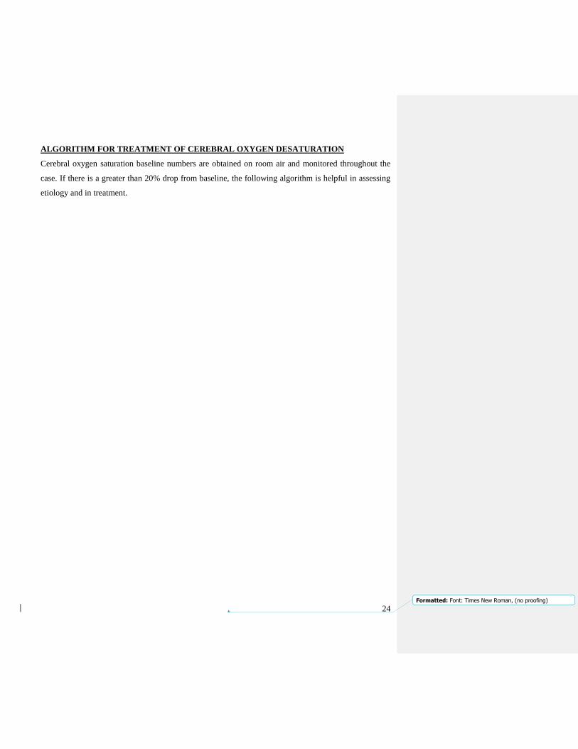
ALGORITHM FOR TREATMENT OF CEREBRAL OXYGEN DESATURATION
Cerebral oxygen saturation baseline numbers are obtained on room air and monitored throughout the
case. If there is a greater than 20% drop from baseline, the following algorithm is helpful in assessing
etiology and in treatment.

25

26
CEREBROSPINAL FLUID DRAINAGE PROTOCOL
Intraoperative Considerations:
SCPP=MAP- (CSFP of CVP whichever is greater)
1. Maintain MAP ≥ 80-90 mmHg as much as possible.
2. Avoid CVP >15 mmHg or acute increase of CVP.
3. CSF drainage system zeroed at the level of RA (not tragus).
4. CSF drain connected to both transducer and drainage system (set up at 10 mmHg), continuously
monitored, either passively or actively drained.
5. Cut the blue rubber of the CSF transducer to eliminate the risk of flushing CSF line by mistake.
6. Adequate label CSF catheter.
7. If 150 ml CSF drained and CSF pressure still high, reassess; check the CSF spinal drain position
(consider pulling back 2 -3 cm), re-zero CSF transducer.
Post-operative Considerations:
1. For CSF pressure higher than 10 mmHg open to passive drainage.
2. Reassess CSF pressure after each 15 ml CSF drained.
3. Do not drain more than 250 ml CSF/day.
4. If 150 ml CSF drained and CSF pressure still high, reassess; check the CSF spinal drain position
(consider pulling back 2 -3 cm), re-zero CSF transducer.
5. If sign of spinal cord ischemia increase MAP > 90 mmHg and drain CSF to < 10 mmHg.
6. If no signs of spinal cord ischemia, clamp the drain after 24-36 hrs and monitor the patient for 12- 24
hours.
7. Remove the CSF catheter at 48 hours if platelet count >100K, INR < 1.3 and a normal PTT. If
patient on SQ heparin, wait 2-4 hours after last dose of heparin, or 12 hours after last dose of
Lovenox.
8. Brain imaging for persistent bloody CSF drainage (more than 4 hours).
Removal of spinal drains:
Spinal drains can be removed if:
1. platelet count >100K, INR < 1.4, normal aPTT, 2-4 h after last dose of SQ heparin or 12 Hrs after last
dose of Lovenox
2. no anticoagulation to be used for the following 2 hours after removal of the drain
Crawford classification of thoracoabdominal aortic
aneurysms

27
Formatted: Font: Times New Roman, (no proofing)
Thoracic Endovascular Aortic Repair (TEVAR)
I. Surgical considerations
• These surgeries include aneurysms, dissections, and hybrid debranching procedures (arch or
abdominal debranching)
• Trans-femoral access usually via groin cutdown (occasionally iliac conduit via lower quadrant
retroperitoneal abdominal approach if femoral arteries not suitable)
• Angiographic access through contralateral femoral artery or brachial artery
• Minimum amount of aorta is covered to exclude the aneurysm with an adequate seal
• Necessity of a relatively straight section of aorta with
good “landing zones”
• Prior to graft, left carotid-subclavian bypass can be
performed if plan to cover left subclavian artery and
feel that this will compromise either left vertebral or
left arm circulation (10% of cases where left subclavian
coverage)
• Other “debranching” options include laparotomy for
bypass to renals, SMA, and celiac axis for open/endo
TAAA repair as well as carotid-carotid or ascending
aorta-carotid and innominate bypass for arch aneurysms
• Monitoring
• Arterial access: CHECK WITH SURGICAL TEAM. Right radial A-line, ± left radial A-line;
bilateral radial a-lines required in all cases where left subclavian artery coverage planned: will need
both radials on screen on separate channels.
• Venous access: Double lumen MAC in right IJV only and large bore IV (≥16G)
• Do not place PA catheter unless indicated by surgery/anesthesia attending; if in doubt, check with
surgeon. PA catheters tend to get in the way of imaging. So, unless essential, avoid using them.
• Temperature monitoring: Nasopharygeal and bladder
• Pulse oximeter: on ear
• Lumbar CSF drain in L3-L4 (on high-risk of spinal ischemia cases): check posting whether CSF
drain is needed for the case.
• SSEP monitoring: measures integrity of posterior columns
• MEP monitoring: monitors integrity of anterior columns
II. Set-up
• No EKG leads on chest: keep all lines away from field of fluro.
• Adenosine 6/12mg (used in selective cases): especially for transections.
o Transducer set-up: A-line, CVP, Lumbar CSF Pressure (on high-risk cases)
• Warming blanket on all cases
• Special considerations for debranching hybrid cases:
o Cerebral oximetry (Get pre-induction baseline!)
• Lumbar drain insertion
• CSF Catheter is not required in all TEVAR cases. Arch debranching cases usually do NOT require
CSF drains.
III. Anesthesia considerations
• Challenges:
o Keep track of blood loss (especially in debranching cases)
o Need for emergency conversion to open repair
A: Arch debranching B: Abdominal debranching

28
Formatted: Font: Times New Roman, (no proofing)
o Strategies to manage paraplegia
• Avoid muscle relaxants if possible (MEP monitoring) or keep to two twitches
• Use TIVA for anesthesia maintenance
• Blood in the room!
• Aim for ACT 200-250s; check at 30-minute intervals; redoes if necessary
• CSF drainage protocol: CSF is drained for lumbar CSF pressure >10 mmHg in 10ml aliquots;
maximum drainage: 10-20ml/hour
• Maintain postoperative MAP using a Vasopressor or a vasodilator (Nicardipine) at minimum of 70
mmHg until protamine given: Increase MAP to 90s after protamine (discuss with surgeon) or at a
MAP where SEP signals are intact
• Avoid use of Nipride (associated with spinal cord perfusion steal and high incidence of paraplegia)
• Management of Spinal Cord Ischemia: [Spinal cord perfusion pressure = MAP – CSFpressure]
o Increase MAP to >90 mmHg using vasopressor
o Drain CSF for CSF pressure >10 mmHg (max 20ml/hr)
o If no Neurologic recovery: increase MAP further
o Give steroids only with persistent EP abnormalities – check with surgeon.
• Send TEG, Fibrinogen and platelet count after stent deployment in debranching cases; not required
in regular TEVARs
IV. TEE
• Check aorta for atheromas; Grading; location of aortic lesion; communicate atheroma findings to
surgeon to assist with planning of stent deployment.
• Monitor LV for ischemia, hypovolemia
• Document absence or type of Endoleaks
• Check for new AI after deployment.
PROCEDURES IN ELECTROPHYSIOLOGY (EP) LAB
When the cardiologists request our services for elective special procedures, the cases are usually listed in EPIC
in off floor procedures. Off floor procedures include cardioversions, electrophysiology studies/arrhythmia
pathway ablations, and ICD testing, which generally take place in the electrophysiology lab (located behind the
GI labs on the third floor north hospital, 87801). Cardioversions are occasionally performed at the bedside,
particularly the CCU or CS-ICU. The EP lab is equipped with a portable anesthesia machine with vapors, wall
oxygen/N20 sources, pneumatic ventilator, medication cart, airway supplies and capnograph. EPS will usually
supply suction, ECG, BP and pulse oximetry, and start an IV. Have the anesthesia technician check your
equipment and supplies before proceeding with an elective case. Additional medications, such as
benzodiazepines and narcotics are available in Pyxis in EP lab 1 and 2. Alternatively, the EP nurses can also
provide you with midazolam or fentanyl. As with cases in the OR, discuss the anesthetic considerations and
management pre-operatively with your attending. EP studies, ICD placements and conduction pathway
ablations take considerably longer and the issues surrounding the perioperative management of these patients
are rather unique, and must be discussed with the attending staff on a case-by-case basis.
For details, refer to Guidelines for EP cases, found on Department Intranet.

29
Formatted: Font: Times New Roman, (no proofing)
CALL RESPONSIBILITIES
The cardiac anesthesia faculty provide anesthesia care for all cardiac cases, some major thoracic and major
vascular cases, and maintain a separate call schedule from the general OR staff. It is posted monthly, and copies
are located in the cardiac ORs, at the OR desk and in the anesthesia office. It also lists each attending, fellow
and rotating resident home phone and beeper numbers. In general, the first call attending arrives at 6:30 a.m.
and stays until all cases are completed. The second call attending also arrives at 6:30 a.m. and usually stays until
the next to last case is completed, or at the discretion of the call attending. The attendings return from home for
emergency cases as needed.
Residents who are on their CT rotation may have longer days but every effort will be made to relieve non-call
residents by 17:00-18:00, such that all duties (including pre-ops) are completed by no later than 19:00.
Emergencies and transplants occur at unpredictable times, and patients can suffer needlessly if everyone does
not work together during such dire times, even though it means stressful work and long hours.
EDUCATION AND DIVISIONAL ACTIVITIES
Education in the cardiothoracic subspecialty of anesthesiology is multifaceted.
Cardiac Conferences - 6:15 a.m. on Thursday mornings. This is the cardiac anesthesiology fellow and resident
conference. All residents on the cardiac rotation are required to attend.
Discussion - in addition, much of what you will learn will come from direct patient care and daily discussions
with attendings and fellows. Encourage your attending and fellow to discuss areas of interest, provide germane
articles, review TEE studies, etc. with you. Speak with the perfusionists about extracorporeal circulation, how
the pump is set-up and operated, and other CPB topics.
TEE Exam
The senior residents will be taught TEE in the operating room by the attendings during their two-month
rotation. They will be evaluated during the second month of their rotation by means of a basic intraoperative
TEE exam, where they are questioned about TEE views and findings. They are not required to drive the TEE
probe. A total of three exams on real patients are to be completed, the forms filled out with patient names with
feedback and comments and signature of the examiner entered on the forms. These forms must be turned in to
Dr. Hong’s office at the end of the rotation. It is the responsibility of the resident to make sure these are
completed and turned in, in time. A copy of the evaluation form will be emailed to you at the beginning of the
30
Formatted: Font: Times New Roman, (no proofing)
rotation. This evaluation is separate from the TEE log (for ACGME) that all the residents are required to
maintain.
Cardiothoracic Rotation Topics
The cardiothoracic faculty have developed a list of cardiothoracic topics; an outline of the expected knowledge
base for a resident on the CT rotation. Faculty and residents should use this to facilitate clinical teaching
and operating room discussions. In order to track individual progress, the resident should place a check
mark after a topic has been covered. The list can be found in each residents’ individual directory, on the
G: drive and is also included in this handbook.
Cardiovascular and Thoracic Anesthesia Resident Rotation Learning Objectives
CARDIAC ANESTHESIA
MEDICAL KNOWLEDGE
1. Preoperative evaluation of cardiac patient
a. Identifying and assessing the risk factors for adverse outcomes
b. Identifying other coexisting diseases and their implication during perioperative management
of cardiac patient
c. Interpretation of diagnostic tests of cardiac function and their perioperative significance
i. TTE/TEE echocardiography
ii. Coronary angiography (distribution of coronary arteries and the clinical implication of
various degrees and locations of coronary lesions)
iii. Stress tests (Exercise, Dobutamine, Adenosine/SESTA MIBI)
d. Interpretation of coagulation panel
i. PT
ii. PTT
iii. Platelet function
iv. TEG/ROTEM
2. Understanding the physiology and physiopathology of valvular diseases as well as anesthetic/
induction considerations
a. Aortic valve disease
i. Stenosis
ii. Regurgitation
b. Mitral valve disease
i. Stenosis
ii. Regurgitation
c. Tricuspid valve disease
i. Stenosis
ii. Regurgitation
d. Pulmonary valve disease `
i. Stenosis
ii. Regurgitation
31
Formatted: Font: Times New Roman, (no proofing)
3. Understanding the physiology and physiopathology of adult congenital heart diseases as well as
anesthetic/induction considerations
a. Atrial septal defects (ASD)
b. Ventricular septal defects (VAD)
c. Atrioventricular septal defects (AVD)
d. Left ventricular outflow tract obstruction
e. Coarctation of the aorta (CoA)
f. Congenital coronary artery anomalies
g. Transposition of great vessels (d-TGA, l-TGA)
h. Ebstein’s anomaly
i. Left superior vena cava (LSVC)
j. Patent ductus arteriosus (PDA)
k. Tetralogy of Fallot (TOF)
l. Truncus arteriosus
m. Single ventricle
n. Pulmonary stenosis
4. Understanding the physiology and physiopathology of constrictive/restrictive pericarditis
5. Understanding the pharmacology, indications, side effects, contraindications of
a. Antidysrhytmics
b. Inotropics
c. Vasopressors
d. Beta blockers
e. Diuretics
f. Vasodilators
g. Recombinant Factor VIIa
h. Antiplatelet agents (clopidogrel, integrilin)
i. ACE inhibitors
j. Angiotensin converting enzyme drugs
k. Statins
l. Calcium channel blockers
6. Understanding the CPB machine
7. Understanding anticoagulation management
a. Heparin
i. Dose
ii. Mechanism of action
iii. Pharmacology
b. Protamine
i. Dose
ii. Mechanism of action
iii. Indications
iv. Contraindications
c. Antifibrinolytic agents
i. epsilon-aminocaproic acid
ii. tranexamic acid
d. Antiplatelet agents

32
Formatted: Font: Times New Roman, (no proofing)
i. Aspirin
ii. Clopidogrel / Ticlid
iii. RePro, Aggrastat, Integrillin
e. DDAVP
8. Understanding the hematologic effect and inflammatory response of CPB.
9. Understanding normovolemic hemodilution/autotransfusion
10. Understanding physiopathology and side effects of hypothermia
11. Understanding the physiopathology of deep hypothermic circulatory arrest
12. Understanding the physiopathology of organ protection during CPB
13. Understanding the physiopathology of alpha stat versus pH stat management during CPB
14. Understanding different techniques of myocardial protection
a. Antegrade versus Retrograde cardioplegia
b. Crystalloid versus Blood cardioplegia
15. Understanding the myocardial reperfusion injury, stunned myocardium and hibernating
myocardium
16. Understanding the mechanism of action of intra-aortic balloon pumps (IABP), indication and
contraindications.
PATIENT CARE
1. Set up, use, and trouble shoot drug infusion pumps.
2. Placement and interpretation of the clinical information from
a. arterial line
b. central line
c. pulmonary artery catheters
3. Preoperative management including knowledge of the role of patient education and
premedication in treating anxiety.
4. Weaning a patient from CPB in routine cases
5. Use the following drips:
a. Epinephrine
b. Dobutamine
c. Norepinephrine
d. Vasopressin
e. Milrinone
6. Use of nitric oxide
7. Intraoperative treatment of left ventricular failure

33
Formatted: Font: Times New Roman, (no proofing)
8. Intraoperative treatment of right ventricular failure
9. Treatment of arrhythmias
10. Diagnostic and treatment of intraoperative bleeding
11. Transfusion therapy; When and How
12. Diagnostic and treatment of intraoperative platelet dysfunction
13. Diagnostic and treatment of heparin rebound syndrome
14. Management of patients undergoing off pump CABG
15. Intraoperative management of resection and repair of aneurysms of ascending aorta
and aortic arch.
16. Management of patients with IABP
17. Management of patients with postoperative bleeding/ cardiac tamponade
18. Management of postoperative pain in cardiac patients
VASCULAR ANESTHESIA
MEDICAL KNOWLEDGE
1. Understanding the pathophysiology of vascular disease and co-morbid conditions.
2. Evaluation of vascular surgical patients preoperatively, including assessment of cardiovascular
and respiratory risk and function.
3. Understanding the potential effects of vascular surgery on other organs such as heart, kidneys,
brain, spinal cord.
4. Understanding the role of regional anesthesia, alone or in combination with general anesthesia,
in vascular surgical patients.
5. Understanding the pathophysiology and intraoperative management of aortic cross-clamping
and unclamping.
6. Understanding the indications and intraoperative management of left heart bypass.
7. Understanding the mechanisms of spinal cord protection during thoracic aortic surgery
PATIENT CARE
1. Preoperative evaluation of vascular surgical patients including ordering appropriate tests of
cardiovascular and respiratory function; assessment of preoperative risk factors
34
Formatted: Font: Times New Roman, (no proofing)
2. Intraoperative management of ruptured AAA
3. Intraoperative management of aorto-bifemoral bypass
4. Placement and intraoperative management of spinal drains
5. Intraoperative management of thoracic aortic surgery (Crawford Type I, II, III, IV)
6. Management of patients undergoing endovascular surgery
THORACIC ANESTHESIA
MEDICAL KNOWLEDGE
1. Understanding the principles of preoperative evaluation in thoracic surgical patients including
interpretation of pulmonary function studies and assessment of tolerable limits of pulmonary
resection.
2. Understanding various disease processes that cause esophageal dysfunction and the risks of
regurgitation and pulmonary aspiration.
3. Understanding the anatomy of the respiratory tract and the upper gastrointestinal tract.
4. Understanding of respiratory physiology including ventilation and perfusion and changes
with one lung ventilation.
5. Understanding the pathophysiology and intraoperative management of hypoxic pulmonary
vasoconstriction.
6. Understanding the advantages and disadvantages of methods/techniques to achieve
one-lung ventilation (OLV).
7. Understanding the absolute and relative indications for OLV.
8. The resident must knowledgeably discuss the systematic approach to hypoxemia during OLV.
9. Understanding approaches to postoperative pain management and the relative advantages and
disadvantages of each.
10. Understanding potential complications of vascular access and describe modalities of therapy
should such complications arise.
PATIENT CARE
1. Intraoperative management of routine thoracic surgical cases.
2. Placement of double lumen endotracheal tubes (DLETTs) and bronchial blockers (BBs)
with a high rate of success.

35
Formatted: Font: Times New Roman, (no proofing)
3. Intraoperative assessment of proper positioning of DLETTs and BBs using fiberoptic
bronchoscopy and correct malposition.
4. Intraoperative management of ruptured pulmonary artery
5. Intraoperative management of ventilation during one- and two-lung ventilation.
6. Intraoperative treatment of hypoxemia.
7. Placement of thoracic and lumbar epidural catheters safely and with a high rate of success.
Reading: All residents receive a copy of the book by Hensley, Gravlee, et al on the CT rotation. This is
distributed on a rotating basis from the residency office. Residents are required to pickup/sign out the books for
the rotation. As new textbooks are published frequently, you should consult with the staff regarding the current
“better” books. Perennial favorites are Cardiac Anesthesia by Frederick Hensley and Martin. Your individual
and directed reading of this text should form the basis of education in CT Anesthesiology. The goals and
objectives for the CT rotation experience for residents are posted on the department intranet, under resident
education development criteria/REDC.
EVALUATION
It is highly recommended that residents seek feedback from the attendings on a daily or regular basis, especially
during mid-rotation. With this, areas of improvement, if any, can be identified and worked on. The final overall
evaluation of your performance over the three months (as mandated by the Clinical Competency Committee)
consists of several categories, including knowledge, judgement, clinical skills and character attributes.
Attendance at conferences, punctuality and teamwork are particularly acknowledged. You will be individually
evaluated by each cardiac attending and will receive feedback/evaluation by the program director, Dr. Hong,
during your meeting with her.

36
Formatted: Font: Times New Roman, (no proofing)
PHONE DIRECTORY
Location Phone Location Phone
OR 29 2-3829 Perfusion /lab 85171
OR 30 2-3830 CT surgery office 85842
OR 31 2-3831 CS-ICU 7G west 85382
OR 32 2-3832 CS-ICU 7G east 82676
Cath lab 8-6618 CS-ICU Fax 85156
EP lab 8-7801 Echo 3G 86072
CCU 3G 8-5426 MIEMSS OR 86850
Name Home Phone Pager
Kenichi Tanaka, MD, MSc 404-983-3770 410-232-3267
Seema Deshpande, MBBS 410-480-4943 410-471-7801
Ashanpreet Grewal, MD 216-544-8485 410-283-0304
Reney Henderson, MD 240-354-6635 410-232-2932
Kimberly Hollander, MD 410-598-1999 410-232-5939
Susan Sankova, MD 857-544-3008 410-232-5466
Erik Strauss, MD 443-386-2030 410-232-2115
Michael Mazzeffi, MD, MPH 504-232-0242 410-283-0063
Samhati Mondal, MD 216-233-4778 410-232-0123
Patrick Odonkor, MBChB 301-617-4177 410-806-9008
Brittney Williams, MD 240-416-1730 410-232-5424
Jeongae Yoon, MBBS 202-431-0598 410-232-3471
Anne Savarese, MD 301-570-1849 410-471-5654
Inna Shats -- 7670 (8-BEEP)
Cardiac Attending on Call Phone 83823
Cardiac Charge Nurse Work ph: 410-328-3664
Cardiac Anesthesia Tech Work ph: 410-328-3661