ELECTROCHEMISTRY Electrical and Chemical Energy Interconversion.

University of Groningen
THE EFFECT OF TRYPSIN TREATMENT ON THE INCORPORATION AND ENERGY-TRANSDUCING PROPERTIES OF BACTERIORHODOPSIN IN LIPOSOMESDriessen, Arnold; Hellingwerf, Klaas J.; Konings, Wil N.
Published in:Biochimica et biophysica acta
DOI:10.1016/0005-2728(87)90009-0
IMPORTANT NOTE: You are advised to consult the publisher's version (publisher's PDF) if you wish to cite fromit. Please check the document version below.
Document VersionPublisher's PDF, also known as Version of record
Publication date:1987
Link to publication in University of Groningen/UMCG research database
Citation for published version (APA):Driessen, A. J. M., Hellingwerf, K. J., & Konings, W. N. (1987). THE EFFECT OF TRYPSIN TREATMENTON THE INCORPORATION AND ENERGY-TRANSDUCING PROPERTIES OF BACTERIORHODOPSININ LIPOSOMES. Biochimica et biophysica acta, 891(2), 165-176. DOI: 10.1016/0005-2728(87)90009-0
CopyrightOther than for strictly personal use, it is not permitted to download or to forward/distribute the text or part of it without the consent of theauthor(s) and/or copyright holder(s), unless the work is under an open content license (like Creative Commons).
Take-down policyIf you believe that this document breaches copyright please contact us providing details, and we will remove access to the work immediatelyand investigate your claim.
Downloaded from the University of Groningen/UMCG research database (Pure): http://www.rug.nl/research/portal. For technical reasons thenumber of authors shown on this cover page is limited to 10 maximum.
Download date: 10-02-2018

Biochimica et Biophysica Acta 891 (1987) 165-176 165 Elsevier
BBA 42516
The effect of trypsin treatment on the incorporation and energy-transducing properties of bacteriorhodopsin in liposomes
Arnold J.M. Driessen, Klaas J. Hellingwerf and Wil N. Konings Laboratorium voor Microbiologie, Rijksuniversiteit Groningen, Haren (The Netherlands)
(Received 19 January 1987)
Key words: Bacteriorhodopsin; Right-side-out liposome; Reconstitution; Protonmotive force
Bacteriorhodopsin and trypsin-modified bacteriorhodopsin have been reconstituted into liposomes by means of a low pH-sonication procedure. The incorporation of bacteriorhodopsin in these proteoliposomes is predominantly in the same direction as in vivo and the direction of proton pumping is from inside to outside the iiposomes. The direction of proton translocation and electrical potential generation was studied as a function of the reconstitution pH. Light-dependent proton extrusion and generation of a Ap, interior negative and alkaline was observed at a reconstitution pH below 3.0 using bacteriorhodopsin, and at a pH below 3.5 using trypsin-modified bacteriorhodopsin. The shift in inflection point is explained in terms of differences between bacteriorhodopsin and trypsin-modified bacteriorhodopsin in a specific protein-phos- pholipid interaction which depends on the surface charge density of the cytoplasmic side of bacteriorhodop- sin. The magnitude of the protonmotive force (Ap) generated by trypsin-modified bacteriorhodopsin in liposomes was quantitated. Illumination of the proteoliposomes resulted in the generation of a high Ap (135 mV, inside negative and alkaline), with a major contribution of the pH gradient. The ionophores nigericin and valinomycin induced, respectively, a compensatory interconversion of ApH into A~ and vice versa. If no endogenous proton permeability of the membrane would exist, a protonmotive force could be generated of
- 143 mV as electrical potential alone or - 162 mV as pH gradient alone.
Introduction
Bacteriorhodopsin contains one single poly- peptide and catalyzes light-driven proton translo- cation across the cytoplasmic membrane in Halobacter ium halobium [1]. Bacteriorhodopsin has
Abbreviations: A~k, transmembrane electrical potential; ApH, transmembrane pH gradient; Ap, protonmotive force, A/~n.; Ph4P +, tetraphenylphosphonium; Ph4B-, tetraphenylbo- ron; S-13, 5-chloro-3-tert-butyl-2'-chloro-4"-nitrosalicylanilide; Hepes, 4-(2-hydroxyethyl)-l-piperazineethanesulphonic acid.
Correspondence: W.N. Konings, Laboratorium voor Microbio- logie, Rijksuniversiteit Groningen, Kerklaan 30, 9751 NN Haren, The Netherlands.
been isolated and purified to homogeneity and various techniques have been described for the reconstitution of this protein into phospholipid vesicles (liposomes). Most procedures yield bacteriorhodopsin proteoliposomes which show light-dependent proton uptake [2-5]. Conse- quently a protonmotive force (Ap), inside acid and positive, will be generated by bacteriorho- dopsin upon illumination of these liposomes. Two different methods have been described for the reconstitution of bacteriorhodopsin into liposomes in the in vivo orientation, e.g., bacteriorhodopsin proteoliposomes that show light-dependent proton extrusion [6,7]. A direct incorporation of bacterio- rhodopsin into liposomes, facilitated by short-
0005-2728/87/$03.50 © 1987 Elsevier Science Publishers B.V. (Biomedical Division)

166
chain lecithines, has been described [6]. On the other hand, 'right-side out' bacteriorhodopsin pro- teoliposomes can also be obtained when purple membranes are cosonicated with liposomes con- taining acidic phospholipids at low pH (2.4-3.0) for a short period of time [7]. During the sonica- tion step a considerable part of bacteriorhodopsin becomes denaturated. Although these bacteriorho- dopsin proteoliposomes show light-dependent pro- ton extrusion, part of the bacteriorhodopsin pro- teoliposomes have the 'inside-out' orientation. Furthermore, the extent of proton extrusion varies considerably between different batches of purple membranes.
It has been suggested that the distribution of charges over the two sides, e.g., cytoplasmic and extracelhilar side in bacteriorhodopsin is an im- portant factor in determining the orientation of this protein in the reconstituted membrane [8]. Under native conditions bacteriorhodopsin is highly resistant against proteolytic treatment. Only the C-terminal part of the peptide chain is sensi- tive to trypsin treatment [9] and this treatment results in the removal of 21 amino acids. This polypeptide fragment contains 5 carboxylic acid groups (3 glutamate, 1 aspartate and the C-termi- nal serine) which are negatively charged at neutral pH. Removal of this peptide will therefore in- fluence the surface-charge density of the cyto- plasmic side of bacteriorhodopsin. In an attempt to optimalize the right-side out reconstitution of bacteriorhodopsin into liposomes, we studied the effect of trypsin treatment of bacteriorhodopsin in purple membranes on the direction of proton pumping and electrical potential generation. In addition, the protonmotive force generated by bacteriorhodopsin in these proteoliposomes was quantitated by the simultaneous measurement of the electrical potential and pH gradient using ion-selective electrodes.
Materials and Methods
Growth of cells and isolation of purple mem- branes. H. halobium NRL (strain R1) [10] was grown according to Danon and Stoeckenius [11]. Purple membranes were isolated by extensive washing as described [12], except that during all steps 5 # g / m l phenylmethylsulfonylfluoride
(PMSF) and 1 mM EDTA were present. Purple membranes resuspended in distilled water (12 mg protein/m l) were stored in liquid nitrogen.
Trypsin treatment of purple membranes. Purple membranes (5 mg protein/ml) were treated with trypsin (0.5 mg protein/ml) for 2 h at 37°C in 20 mM Tris-HC1 (pH 7.0) supplemented with 5 mM CaC12- Proteolysis was terminated by the addition of a 4-fold (w/w) excess of trypsin-inhibitor (i.e., 2 mg protein/m 1). Control experiments were car- ried out at 4°C in the presence of trypsin-inhibi- tor. Trypsin-treated bacteriorhodopsin and native (bacteriorhodopsin) purple membranes were washed twice in a 20-fold volume of 150 mM NaC1 (KC1) (pH 6.5) (centrifugation: 30 min; 48 200 × g; 4 o C).
Preparation of bacteriorhodopsin proteolipo- somes. Bacteriorhodopsin was incorporated into liposomes in the inside-out mode by the sonica- tion method as described [12]. Purple membranes (2.5 mg protein/ml) and cardiolipin (6.25 mg/ml) were cosonicated in 150 mM NaC1 (pH 6.5).
Bacteriorhodopsin was reconstituted into lipo- somes in the rightside out mode as described [7,8]. Purple membranes (2.5 mg protein/ml) were mixed with sonicated cardiolipin liposomes (6.25 mg phospholipid/ml) in 150 mM NaC1 (or 150 mM KC1) (pH 6.5). The mixture was acidified to the desired pH, using 1 M HCI, sonicated with a probe-type sonicator (MSE Scientific Instruments, West Sussex, U.K) two times 10 s (interrupted for 10 s) at an amplitude of 2 #m (peak to peak), and the pH was rapidly readjusted to pH 6.5, using 1 M NaOH (KOH).
Measurement of the protonmotioe force. A~, in- terior negative, was measured with a tetraphenyl- phosphonium (Ph4P +) selective electrode [13] which was inserted in a thermostatted, magneti- cally stirred vessel. A final concentration of 2 ArM Ph4P + was used.
A~p, interior positive, was measured with the same electrode using tetraphenylboron (Ph4B-) as the indicator ion [14]. A final concentration of 1 /~M Ph4B- was used. Measurements were per- formed in the presence of 0.1 /~M Ph4P + to in- crease the permeability of the negatively charged membranes for Ph4B-. This also improved the stability of the electrode and enhanced its re- sponse to Ph4B-.

ApH, interior alkaline, was measured with an ion-selective salicylate electrode as described [15]. A final salicylic acid concentration of 200 #M was used.
Extravesicular pH changes were measured as described [12]. All measurements were performed in a final volume of 2.0 ml. A bacteriorhodopsin concentration of 0.25 mg protein/ml in 150 mM NaC1 (pH 6.5) or 150 mM KC1 (pH 6.5) was used at 25 o C, unless stated otherwise. For pH measure- ments 150 mM KC1 (pH 6.5) was used and experi- ments were performed in the presence of valinomycin (0.8 nmol/mg phospholipid).
For determination of the maximal Ap gener- ated by bacteriorhodopsin in liposomes, A~k and ApH were measured simultaneously in a 5 ml thermostated polyvinylchloride vessel, in which both an ion-selective Ph4P + electrode and an ion-selective salicylate electrode were inserted [16]. Measurements were performed using a bacterio- rhodopsin concentration of 0.98 mg protein/ml (ApH) or 0.25 mg protein/ml (A~k) in 10 mM potassium Hepes (pH 7.0), supplemented with 45 mM KC1 and 10 mM MgSO 4, 2 ml final volume. Valinomycin (400 nM) or nigericin (20 nM) were added as indicated.
Liposomes were illuminated with a slide projec- tor (150 Watt) equipped with fiber optics. The maximal light intensity was 2000 W / m E .
Calculations. The magnitude of the membrane potential was calculated with the Nernst equation. A correction for Ph4P + and Ph4B- binding to the liposomes as described in Ref. 17 was applied. A binding constant of 15-22 was used for Ph4P +, and of 3-7 for Ph4B- (see Refs. 17 and 18). The magnitude of the pH gradient was calculated with the Nernst equation from the distribution of salicylate between bulk phase and intraliposomal space. A correction for non-linearity of the salicy- late-selective electrode response was applied based on a polynomal fit of the calibration curve of the electrode [15]. The sensitivity of the electrodes is expressed as their Z-value: the electrode response to a 10-fold increase in probe concentration. A value of 2.0 btl/mg phospholipid was used as internal volume of the bacteriorhodopsin proteo- liposomes [19]. The concentration of native bacteriorhodopsin was determined using a molar extinction coefficient at 560 nm of 63000 M -1-
167
cm-1 [20]. Denaturation of bacteriorhodopsin was estimated from the decrease in absorption at 560 nm as described [8].
Other analytical methods. Protein [21] was as- sayed as described. SDS polyacrylamide gel elec- trophoresis was performed as described [22]. Sucrose gradient density centrifugation was per- formed as described elsewhere [23].
Materials. Bovine heart cardiolipin, trypsin and trypsin-inhibitor were obtained from Sigma Chemical Co. All other materials were of analytial grade.
R ~
Incorporation of trypsin-modified bacteriorhodopsin into liposomes
Cosonication of purple membranes and cardiolipin liposomes at pH 2.7 yielded bacterio- rhodopsin proteoliposomes which acidify their suspending medium upon illumination (Fig. 1A). Only a short period of sonication was used, since under the conditions employed bacteriorhodopsin molecules become dissolved from their two-di- mensional crystal lattice and diffuse laterally in the plane of the membrane [8]. In its monomeric configuration, bacteriorhodopsin is highly suscep- tible to low pH [8]. Interestingly prior to sonica- tion, purple membrane fragments can be stored on ice at pH 2.7 for several hours without any signifi- cant denaturation (not shown; Ref. 8). Upon trypsin treatment, purple membranes become ag- gregated as indicated by the turbidity of the sus- pension. Trypsin treatment resulted in a complete removal of the C-terminal peptide fragment as could be deduced from SDS (16%) gel electro- phoresis. One single large product could be identi- fied with a molecular weight of approx. 24 500, e.g., 1500 less than the molecular weight of bacteriorhodopsin (not shown). Flocculation of trypsin-treated bacteriorhodopsin was observed upon mixing with cardiolipin liposomes at pH 2.7. However, after a short burst of sonication and pH readjustment a typical blue shift in the visible absorption was observed and a homogeneous sus- pension was obtained. Illuminated trypsin-treated bacteriorhodopsin proteoliposomes showed a max- imal extent of proton extrusion which was consid- erably higher than observed for native bacterio-

168
o J
A RSO bR-liposomes
!
A
~u
z ( I_
- - L
-train-
B RSO trpbR-tiposomes !
E ISO bR-tlposomes
5:
!
Fig. 1. Light-dependent pH changes, Ph4B- and Ph4P + electrode responses in the presence of proteoliposomes reconstituted from cardiolipin and purple membranes. (A) Purple membranes reconstituted at pH 2.7 by the low pH-sonication procedure. (B) As (A), except that trypsin-modified purple membranes were used. (C) Purple membranes reconstituted at pH 6.5 by the sortication procedure, pH changes were recorded in the presence of 0.8 nmol valinomycin per mg of phospholipid. Ph4P + and Ph4B- electrode responses were recorded in the presence of 20 pmol triphenyltin per mg phospholipid. Light was switched on and off at maximal light intensity as indicated by the arrows.
rhodopsin (Fig. 1B). Light-induced proton extru- sion by trypsin-treated bacteriorhodopsin proteo- liposomes was stimulated 1.5- to 2-fold by the addition of the A~ k dissipating ionophore valinomycin. This was not observed with bacteri- orhodopsin proteoliposomes reconstituted with native bacteriorhodopsin.
Removal of the C-terminal peptide fragment did not significantly alter the capacity of bacteriorhodopsin to pump protons, as was shown by others [24]. Reconstitution of bacteriorhodop- sin and trypsin-treated bacteriorhodopsin into cardiolipin liposomes in the inside-out mode by sonication at neutral pH, yielded comparable val- ues of proton uptake of 4.5-5.3 H+/bacteriorho- dopsin (Fig. 1C).
The right-side-out bacteriorhodopsin proteo- liposomes were further characterized for their abil-
ity to generate a A~,. Upon illumination a tran- sient uptake of Ph4P + was observed in both types of liposome reconstituted at low pH. In the pres- ence of triphenyltin, which collapses the ApH by an electroneutral exchange of C1- for OH- [25], a steady-state A~ was generated by the trypsin- treated bacteriorhodopsin proteoliposomes (Fig. 1B). Illuminated bacteriorhodopsin proteolipo- somes showed an overshoot in Ph4P + accumula- tion, while the steady-state level was low com- pared to the trypsin-treated bacteriorhodopsin proteoliposomes (Fig. 1A). This overshoot is most likely explained by the presence of inside-out bacteriorhodopsin proteoliposomes, since low levels of Ph4B- accumulation were observed in the same preparation upon illumination (Fig. 1A). The trypsin-treated bacteriorhodopsin proteo- liposomes on the other hand extruded Ph~B- upon

illumination (Fig. 1B) which must imply that the majority of trypsin-treated bacteriorhodopsin par- titles generate a A~k, interior negative, upon il- lumination.
Inside-out bacteriorhodopsin proteoliposomes showed fight-induced extrusion of Ph4P ÷ and ac- cumulation of Ph4B- (Fig. 1C).
In agreement with previous studies [7,8] an inversion of the direction of proton translocation upon illumination could be observed when the pH of the acid stage of the reconstitution procedure was varied (Fig. 2A). The inflection point was found around pH 3.0 for bacteriorhodopsin pro- teoliposomes. When trypsin-treated bacteriorho- dopsin was used instead of native bacteriorho- dopsin, the extent of light-dependent proton ex- trusion over the whole pH range tested was found to be larger (Fig. 2A). Furthermore, a shift of the inflection point to a higher pH (approx. 3.4) was observed. To substantiate the improved homo- geneity of the transmembranous incorporation of trypsin-treated bacteriorhodopsin in the right- side-out mode, light-dependent Ph4 P÷ and Ph4B- uptake was investigated, while the pH of the acidic
169
phase of the reconstitution was varied, trypsin- treated bacteriorhodopsin proteoliposomes were found to accumulate Ph4P + over about the whole reconstitution pH range with an apparent inflec- tion point around pH 3.5 (Fig. 2B). Bacteriorho- dopsin proteoliposomes already started to extrude Ph4 P+ at a reconstitution pH of about 3.2, while Ph4B- accumulation was already found at pH 2.6 (Fig. 2B and C). Trypsin-treated bacteriorhodop- sin proteoliposomes extruded Ph4B- at a recon- stitution pH below 3.2 (Fig. 2C). Uptake of both Ph4 P+ and Ph4B- as found for bacteriorhodopsin proteoliposomes implies that a large degree of heterogeneity with respect to the orientation of bacteriorhodopsin exists in this preparation (pH < 3.3). On the other hand, uptake of Ph4 P+ and extrusion of Ph+B- as found for trypsin-treated bacteriorhodopsin proteoliposomes reconstituted at pH < 3.3 suggests that these preparations are virtually completely devoid of bacteriorhodopsin proteoliposomes that generate a Ap, interior acid and positive, upon illumination. When trypsin- treated bacteriorhodopsin proteoliposomes and bacteriorhodopsin proteoliposomes, reconstituted
2
0
-1
x -3
A
>
-o
+ o.
~p14
B
-- 1
+
'm
~ p 8
C
~ 5
~pH
Fig. 2. Dependence of the direction of proton-transiocation and A6 generation by proteoliposomes on the pH during reconstitution with cardiolipin. (A) Extent of proton extrusion expressed in mol H + per mol native bacteriorhodopsin. A negative extent indicates proton uptake. (B) Generation of a At, interior negative, expressed as Ph+P + electrode response in inV. A positive response indicates Ph4P+ accumulation. The z of the electrode was 52 mV. (C) Generation of a At, interior positive, expressed as Ph4B- electrode response in mV. The z of the electrode was 12.4 inV. Filled symbols represent the responses found for bacteriorhodopsin proteoliposomes and the open symbols for trypsin-treated bacteriorhodopsin proteoliposomes. Experimental conditions were as described in the legend to Fig. 1 and Materials and Methods.

170
at pH 2.7 are compared by sucrose-density gradi- ent centrifugation no difference was observed (not shown). A small fraction of protein-free liposomes was visible. On the other hand, virtually no free bacteriorhodopsin was detectable, indicating that the differences observed between native bacterio- rhodopsin and trypsin-treated bacteriorhodopsin are not due to different amounts of bacteriorho- dopsin reconstituted. The degree of denaturation increased linearly with the pH of the acid stage from 55% at pH 3.0 to 70% at pH 2.3. In this respect no difference was found between bacterio- rhodopsin and trypsin-treated bacteriorhodopsin (not shown).
Quantitation of A p in trypsin-treated bacteriorho- dopsin proteoliposomes
The homogeneous, in vivo incorporation of bacteriorhodopsin into liposomes with respect to Ap generation, allows a quantitation of the mag- nitude of the protonmotive force (Ap), generated upon illumination. In addition to measurements of the A~, interior negative, by the use of a Ph4P +- selective electrode [13], the ApH, interior alkaline, can most conveniently be measured by the use of an anion-selective electrode [15]. This electrode senses the external concentration of the dissoci- ated form of the weak acid, salicylic acid. The use of ion-selective electrodes offers the possibility to measure the external concentration of the probes continuously with a high sensitivity without the need to separate the liposomes from their external medium. Furthermore, both components of the Ap can be measured simultaneously.
Illumination of trypsin-treated bacteriorho- dopsin proteoliposomes (reconstituted at pH 2.7) results in a transient generation of a Aft, as dem- onstrated by the rapid accumulation of Ph4P +, which is followed by a slow release (Fig. 3). Simultaneously, a ApH was generated, as demon- strated by the uptake of salicylate (Fig. 3). Both A~ and ApH collapsed when the light was turned off.
Upon addition of the trypsin-treated bacterio- rhodopsin proteoliposomes to medium, the exter- nal Ph4 P+ concentration not only decreased as a result of dilution of the suspending medium, but also as a result of energy independent binding of Ph4 P+ to the membranes. As reported earlier
I I / / , I J ~ , I i J ~ I
0 5 10 15
TiME (MIN)
Fig. 3. Simultaneous registration of the external Ph4P + and salicylate concentration in trypsin-treated bacteriorhodopsin proteoliposomes. Light was switched on and off at maximal intensity as indicated by the arrows 37.7 #M bactefiorhodopsin (about 70 pmol bacteriorhodopsin per mg phosphofipid) was used. Experimental details were as described in Materials and Methods.
[18,26], binding of Ph4 P+ was largely diminished by the addition of Mg 2+. Energy-independent binding of Ph4P + increased linearly with increas- ing external Ph4P + concentration. Even in the presence of 100 #M Ph4 P+ no saturation of bind- ing was observed (not shown). Binding of Ph4P + appears to be equally distributed over the internal and external surface of the membrane. Imposition of a A~, interior positive, by the use of an in- wardly directed valinomycin-mediated potassium diffusion gradient resulted in a 48% + 8% decrease of the amount Ph4P + bound to the mombranes. Therefore, to quantitate the A~, Ph4P + accumula- tion was corrected for concentration-di?pendent binding of Ph4P +, according to the model of Lolkema et al. [17]. Recently, we showed that a

good correlation exists between the imposed and calculated potentials in liposomes, when a correc- tion was made for binding of Ph4 P÷ [18]. The external salicylate concentration decreased only by dilution of the medium upon addition of the membrane suspension. No binding of salicylate was observed, therefore no correction for probe binding was made.
At pH 7.0, the main component of the Ap is the ApH (Table I). The transient generation of the A~ and the low steady-state value of the Aft (less than 20 mV) suggests that rapid charge-com- pensating ion movements occur. A nearly com- plete interconversion of the ApH into Ark could be induced by the ionophore rtigericin (Table I). Nigericin catalyzes electrically neutral exchange of protons for K ÷ or Na +, thereby collapsing ApH with a compensatory increase in A~k. On the other hand, dissipation of the A~k by valinomycin re- suited in a compensatory increase of ApH (Table I). Although further addition of valinomycin re- suited in a complete dissipation of Ark, also a decrease of Ap (ApH) was observed. At high concentrations, valinomycin does not only act as an ionophore, but it also directly inhibits the turnover of bacteriorhodopsin [27]. In order to evaluate the efficiency of energy conversion in bacteriorhodopsin proteoliposomes, Westerhoff and Van Dam [28] and Hellingwerf and his co- workers [29] studied the effect of increased proton permeability on the protonmotive force. Using a mosaic non-equilibrium thermodynamic descrip- tion of ion translocation in bacteriorhodopsin pro-
TABLE I
QUANTITATION OF Aft ( INTERIOR NEGATIVE) AND ApH (INTERIOR ALKALINE) IN TRYPSIN-TREATED BACTERIORHODOPSIN PROTEOLIPOSOMES UPON IL- LUMINATION
37.7 #M bacteriorhodopsin (about 70 pmol bacteriorhodopsin per nag phospholipid) was used for the Ap measurements. Nigericin and valinomycin were used at a final concentrations of 80 nM and 400 nM, respectively.
Ionophore At/, - Z ApH Ap (my)
No additions - 19 - 115 - 134 Nigericin - 137 0 - 137, Valinomycin - 3 - 125 - 128
171
teoliposomes, it was predicted that a linear rela- tion exists between the reciprocal Ap and the proton leakage (L~) of the membrane, according to Eqn. 1:
1 1 -L' 1 (mV) (!) Ap n ( l - 2 a ) L . A , H ( 1 - 2 a ) A ~
in which n is the number of protons pumped by bacteriorhodopsin per fully coupled photochem- ical cycle; a, the fraction of bacteriorhodopsin molecules in the right-side out orientation, e.g., for the in vivo orientation of bacteriorhodopsin, or a represents the fraction of inside-out oriented bacteriorhodopsin molecules; L,, the activity of bacteriorhodopsin, which is proportional to the light intensity; and A~, the effective thermody- namic force on bacteriorhodopsin exerted by il- lumination. Measurement of the Ap with different amounts of uncoupler present (to vary LtH) at different light intensities (to vary L,), resulted in a series of lines, intersecting in one point. The re- ciprocal Ap at this point equals:
( 1 - 2 a ) A , (2)
In general terms, from this tYpe of experiment an estimate can be made of the Ap generated under conditions that the endogenous ion leakage of the membrane would equal zero. Under those condi- tions, the Ap generated would only depend on the properties 9 f the proton pump. This maximal Ap can only be estimated if the proton permeability of the membranes increases linearly with the amount of added uncoupler. The linearity of the proton permeability with uncoupler concentration has been demonstrated by Arents et al. [29] for the uncoupler S-13. Eqn. 1 has been verified using bacteriorhodopsin proteoliposomes with an inside- out orientation [28,29].
The effect of S-13 on steady-state ApH (Fig. 4) and A~ (Fig. 5) at different light intensities (ob- tained by using neutral density filters) by trypsin- treated bacteriorhodopsin proteoliposomes has been evaluated. ApH was measured in the pres- ence of valinomycin and a high concentration of K + (55 mM). Under these conditions the Atp was found to be smaller than - 5 mV. The reciprocal of the ApH plotted against the uncoupler con-
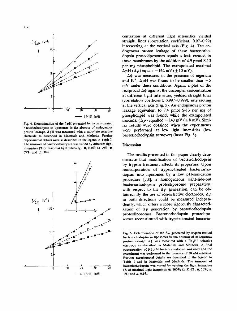
0
.. 1"3
~/ApH (v-l)
I 2s
15
0
172
I I I I
10 20 30
[ s-13] (nil)
Fig. 4. Determination of the ApH generated by trypsin-treated bacteriorhodopsin in liposomes in the absence of endogenous proton leakage. ApH was measured with a salicylate selective electrode as described in Materials and Methods. Further experimental details were as described in the legend to Table I. The turnover of bacteriorhodopsin was varied by different light intensities (% of maximal light intensity): U, 100%; n, 79%; O, 57%; and O, 38%.
. ~ Cv 4)
I 2s
15
5
/ N
I I o lb 2o ,'o [S-13] (nil)
centration at different light intensities yielded straight lines (correlation coefficient, 0.97-0.99) intersecting at the vertical axis (Fig. 4). The en- dogenous proton leakage of these bacteriorho- dopsin proteoliposomes equals a leak created in these membranes by the addition of 4.9 pmol S-13 per mg phospholipid. The extrapolated maximal ApH (Ap) equals - 1 6 2 mV ( + 1 0 mV).
A~b was measured in the presence of nigericin and K ÷. ApH was found to be smaller than - 5 mV under these conditions. Again, a plot of the reciprocal A~ against the uncoupler concentration at different light intensities, yielded straight lines (correlation coefficient, 0.997-0.999), intersecting at the vertical axis (Fig. 5). An endogenous proton leakage equivalent to 7.4 pmol S-13 per mg of phospholipid was found, while the extrapolated maximal (Ap) equalled - 143 mV ( ± 8 mV). Simi- lar results were obtained when the experiments were performed at low light intensities (low bacteriorhodopsin turnover) (inset Fig. 5).
Discussion
The results presented in this paper clearly dem- onstrate that modification of bacteriorhodopsin by trypsin treatment affects its properties. Upon reincorporation of trypsin-treated bacteriorho- dopsin into liposomes by a low pH-sonication procedure [7,8], a homogeneous fight-side-out bacteriorhodopsin proteoliposome preparation, with respect t o the Ap generation, can be ob- tained. By the use of ion-selective electrodes, Ap in both directions could be measured indepen- dently, which offers a more rigorously characteri- zation of Ap generation by bacteriorhodopsin proteoliposomes. Bacteriorhodopsin proteolipo- somes reconstituted with trypsin-treated bacterio-
Fig. 5. Determination of the A~ generated by trypsin-treated bacteriorhodopsin in liposomes in the absence of endogenous proton leakage. A~ was measured with a Ph4P + selective electrode as described in Materials and Methods. A final concentration of 9.6 pM bacteriorhodopsin was used and the experiment was performed in the presence of 20 nM nigericin. Further experimental details are described in the legend to Table I and in Materials and Methods. The turnover of bacteriorhodopsin was varied by varying the light intensities (% of maximal light intensity): L 100%; D, 31.6%; o, 10%; zx, 1%; and A, 0.1%.

rhodopsin at a pH between 2.5 and 3.0 showed in the light: (i) valinomycin-stimulated proton extru- sion, (ii) Ph4P + accumulation, (iii) Ph4B- extru- sion and (iv) salicylate accumulation. No dif- ferences in incorporation efficiency or denatura- tion due to the low pH treatment were observed between native and trypsin-treated bacteriorho- dopsin. These observations strongly suggest that these bacteriorhodopsin proteoliposomes are vir- tually free from inside-out bacteriorhodopsin pro- teoliposomes. Bacteriorhodopsin proteoliposomes reconstituted with native purple membrane frag- ments had distinctly different properties. An intri- guing question is why the extent of proton uptake by native bacteriorhodopsin proteoliposomes was found to vary considerably from batch to batch of purple membrane fragments, as reported by Happe et al. [7] even in one batch from experiment to experiment. Recently it was found by Arrio et al. [30] that purple membrane fragments contain pro- tease activity which was inhibited by benzamidine indicating trypsin-like activity. They suggested that stacking of purple membranes is a result of low- level proteolysis. This stacking phenomenon is also observed upon trypsin treatment, while at acidic pH the purple membrane fragments even tend to flocculate. Slow, but uncontrolled pro- teolysis during storage might explain the dif- ferences in reconstitution behaviour between the different batches of purple membranes. In this respect it should be emphasized that purple mem- branes isolated from H. halobium in the presence of protease inhibitors are very poor samples for reconstitution in the right-side-out mode.
The observation that the inflection pH of re- constitution shifts to a higher pH when bacterio- rhodopsin was treated with trypsin is of interest. Removal of the highly negatively charged C-termi- nal peptide most probably decreases the net nega- tive surface charge density of the cytoplasmic side of bacteriorhodopsin. At neutral pH, bacteriorho- dopsin is mainly negatively charged with the highest charge density at the cytoplasmic side. At low pH an inversion of the side with the highest negative charge density takes place, since at pH 3 preferentially, binding of the extracellular side to polylysine-coated glass is observed [29]. The posi- tively charged lysine residues at the cytoplasmic side will promote binding of this side to the
173
negatively charged liposomes. After successful re- constitution the cytoplasmic side will face the inner surface of the phospholipid bilayer. After removal of the highly negatively charged C-termi- nal peptide by trypsin treatment, inversion of the side with the highest negative charge density will occur at a higher pH. This implies that a specific interaction between bacteriorhodopsin and the negatively charged phospholipids can take place at a higher pH. This is exactly what has been ob- served.
An alternative explanation is that incorporation of bacteriorhodopsin into liposomes is the result of low pH induced fusion between purple mem- brane fragments and the negatively charged lipo- somes. Cardiolipin liposomes fuse at pH values below the pK of this phospholipid (3.5 [8]). It is, however, difficult to envision how fusion can be so specific that bacteriorhodopsin is unidirection- ally right-side out incorporated into liposomes. Furthermore, it is not clear how trypsin modifica- tion of bacteriorhodopsin would affect this fusion event. The effect of trypsin treatment of bacterio- rhodopsin on its orientation in liposomes recon- stituted by the low pH-sonication procedure are in agreement with a previous proposition [8], that the distribution of charges over the two sides, e.g., the cytoplasmic side and external side of bacteriorho- dopsin, determines the orientation.
The second part of the paper deals with the quantitation of the Ap in trypsin-treated bacterio- rhodopsin proteoliposomes. The homogeneity of the direction of proton pumping by these proteo- liposomes offers the possibility to quantitate the Ap generated upon illumination. Although more than 55% of the bacteriorhodopsin molecules be- come denaturated due to the low pH treatment, still Ap values of approx. -135 mV were found. The magnitude of Ap in these proteoliposomes is in the same order as found for the reduction of cytochrome c mediated by cytochrome c oxidase, reconstituted in liposomes [18,24]. In contrast to cytochrome-c oxidase, only a minor contribution of the A~b to the steady state Ap was observed in bacteriorhodopsin proteoliposomes.
Ionophores are able to induce a nearly com- plete interconversion of A~b into ApH, and vice versa in trypsin-treated bacteriorhodopsin proteo- liposomes. In contrast, collapse of a Aft of about
174
- 100 mV by valinomycin in cytochrome-c-oxidase proteoliposomes, results only in a small increase (20-30 mV) in ApH [18,26]. Interconversion of ApH into A~ was found to be nearly complete. The magnitude of the ApH will be determined by the activity of the Ap-generating system and the internal buffer capacity. The latter is most prob- ably not very different for bacteriorhodopsin and cytochrome-c oxidase when reconstituted into cardiolipin liposomes. Therefore, the activity of cytochrome-c oxidase is most likely the limiting factor in the conversion of A~b into ApH. In this respect, ApH and A~b appear to affect respiratory control in a similar manner, since dissipation of the A~b with a small increase in ApH by valinomycin decreases the coupling ratio. On the other hand, dissipation of ApH by nigericin with a compensatory increase of A~b does not alter the coupling ratio (De Vrij, W., unpublished results).
The trypsin-treated bacteriorhodopsin pro- teoliposomes were analysed for the protonmotive force which can be generated maximally upon illumination (A,). The use of ion-selective elec- trodes for these measurements allows a more accu- rate determination of A~. A correction was made for the endogenous proton permeability of the membrane using an uncoupler/turn-over (L I /L~) tritration as described [8,28,29]. A value of -143 mV was found for the A~b, and a value of - 162 mV (2.7 pH units) was found for the A~, and a value of - 162 mV (2.7 pH units) was found for the ApH. These values should be interpreted as lower limits, since in order to determine the A~, the fraction of bacteriorhodopsin molecules 1 - a pumping proton in the vesicle interior should be known (see Eqns. 1 and 2). Hellingwerf and co- workers [29] made an estimate of a for bacterio- rhodopsin molecules with the in vivo orientation in a population of inside-out bacteriorhodopsin proteoliposomes. The amount of the C-terminal amino-acid serine accessible to carboxypeptidase was determined. In view of the results of Arrio et al. [30], which indicate that part of the bacterio- rhodopsin molecules in purple membranes are al- ready devoid of their C-terminal peptide, the value of a determined by the carboxypeptidase assay should be considered as a lower limit, which might result in a lower A, than reported (Table II). The maximal protonmotive force, uncorrected for
bacteriorhodopsin molecules with an opposite di- rection of proton pumping in inside-out bacterio- rhodopsin proteoliposomes, is in the same order as reported in this study for right-side-out bacteriorhodopsin proteoliposomes (Table II). The uncertain factor of bacteriorhodopsin molecules with an opposite direction not only affects A~ determinations by L~H/L~ titrations, but also af- fects the effect of an external electrical field on A~b generation by bacteriorhodopsin reconstituted into planar bilayers (Table II). It should be em- phasized that a two-fold increase in internal volume results in a decrease in the calculated ApH values of 18 mV, while a 2-fold decrease in the internal volume results in an increase of the calculated ApH with 18 inV. However, since Ap is determined by extrapolation, A~ is hardly affected by either a two-fold increase or decrease in the internal volume. This additional error hardly adds to the already indicated error of +10 mY. Aq~ measurements are even less affected by a possible error in the internal volume due to nonspecific binding of Ph4 p+, which largely determines the amount of accumulated probe [17]. The A~ de- termined with proteoliposomes absorbed or fused to planar bilayers [32,33] is relatively high com- pared to the A~ values listed in Table II, the method with planar bilayers has the drawback that part of the externally applied electrical field drops across the supporting bilayer phospholipid membrane [34]. In this respect, the patch pipet method [35] seems to have several advantages. This method has been applied to cytochrome-o oxidase from Escherichia coll. But also in this case, the uncertainty remains how large the contri- bution of opposite-oriented oxidase molecules in the Ap generation is. This problem does not exist for cytochrome-c oxidase when it is reduced by the membrane impermeable electron-donor cyto- chrome c. The A~ determined for bovine heart [18] and Bacillus subtilis cytochrome-c oxidase by the LIH/L~ titration procedure is in the same order of magnitude as found for bacteriorhodopsin with the L~/L, titration procedure or cytochrome-o oxidase with the patch pipet procedure (Table II).
One critical aspect of LI/L~ titrations is that L~ should increase in a linear fashion with the amount of added uncoupler. This has been experi- mentally verified for inside-out bacteriorhodopsin

175
TABLE II
FUNCTIONAL COMPARISON OF PROTONMOTIVE-FORCE-GENERATING SYSTEMS
(+), no specified direction of Ap generation required. ( ), Protonmotive force corrected for orientation (A~).
Method Procedure Maximal Ap Ref.
Bacteriorhodopsin A¢
ApH
planar bilayer planar bilayer/prot eoliposome proteoliposomes proteoliposomes
Cytochrome-c oxidase Bovine heart
A~k planar bilayer/proteoliposome proteofiposomes
B. subtilis A Lk proteofiposomes
Cytochrome-o-oxidase E. coil
A~k patch pipet
ext. field ( + )200 mV ext. field ( + )230 mV L ~ / L , t i t . Ph4 P+ - 143 mV L ~ / L , t i t . CH3NH 3 + 122 mV
(+200 mV) L ~ / L , t i t . pH electrode + 160 mV
( + 270 mV) Lh/L, t i t . salicylate - 162 mV
ext. field ( 5:)210 mV 34 L ~ / L , t i t . a ph4p+ - 130 mV 18
L h / L , t i t . b Ph4P + - 130 mV 18
ext. field ( 5:)150 mV 35
38 33,34 this paper 29
29
this paper
a Cytoehrome c was clamped at 20% reduction. b Cytoehrome c was clamped at 50% reduction.
proteoliposomes [29], in which it was shown that the initial proton efflux rate at fixed ApH in- creased linearly with the uncoupler concentration. On the other hand, the proton permeability of the membrane should be a linear function of the Ap. The proton conductivity of the membrane has been found to be a linear function of the ApH [36,37]. However, the proton conductivity in- creased in a non-linear fashion with the A~k [36,37], although a linear relation was found as long as the A~ k did not exceed 140 inV. This is the situation in our experiments. Furthermore, the lines obtained from L1/L, titrations (Fig. 5) suggest that a possible nonlinear relation between the A~b and the proton conductivity does not lead to a signifi- cant deviation of the linear relationship.
In conclusion, trypsin modification of bacterio- rhodopsin improves the reconstitution of bacterio- rhodopsin into liposomes in the in vivo orienta- tion, as demonstrated by Ap measurements in both directions. Bacteriorhodopsin is able to gen- erate a large Ap in these liposomes upon illumina- tion. This makes bacteriorhodopsin an attractive
protein for generating a protonmotive force in liposomes or fused membranes for studies on en- ergy-transducing systems [23,26].
Acknowledgements
This study has been made possible by the Stichting voor Biophysica with financial support from The Netherlands Organization for the Ad- vancement of Pure Scientific Research (Z.W.O.).
References
1 Stoeckenius, W. (1975) Sci. Am. 243, 38-46 2 Racker, E. (1973) Biochem. Biophys. Res. Commun. 55,
224-230 3 Kayushin, L.P. and Skulachev, V.P. (1974) FEBS Lett. 39,
39-42 4 Van Dijck, P.W.M., Nicolay, K., Leunissen-Bijvelt, J., Van
Dam, K. and Kaptein, R. (1981) Eur. J. Biochem. 117, 639-645
5 Rigand, J.L., Bluzat, A. and Buschlen, S. (1983) Biochem. Biophys. Res. Comm. 111, 373-382
6 Dencher, N.A. (1986) Biochem. 25, 1195-1200

176
7 Happe, M., Teather, R.M., Overath, P., Knobling, A. and Oesterhelt, D. (1977) Biochim. Biophys. Acta 465, 415-420
8 Hellingwerf, K.J., Tegelaers, F.P.W., Westerhoff, H.V., Arents, J.C. and Van Dam, K. (1978) in Energetics and Structure of Halophilic Microorganisms (Caplan, S.R. and Ginzburg, M., eds.), pp. 283-290, Elsevier/North-Holland, Amsterdam
9 Gerber, G.E., Gray, C.P., Wildenauer, D. and Khorana, H.G. (1977) Proc. Natl. Acad. Sci. USA 74, 5426-5430
10 Stoeckenius, W. and Kunau, W.H. (1968) J. Cell. Biol. 38, 337-357
11 Danon, A. and Stoeckenius, W. (1974) Proc. Natl. Acad. Sci. USA 74, 5426-5430
12 Driessen, A.J.M., HeUingwerf, K.J. and Konings, W.N. (1985) Biochim. Biophys. Acta 808, 1-12
13 Shinbo, T., Kama, N., Kuribana, K. and Kobatake, Y. (1978) Arch. Biochem. Biophys. 187, 414-422
14 Casadio, R., Venturoli, G. and Melandri, B.A. (1981) Pho- tobiochem. Photobiophys. 2, 245-253
15 Hellingwerf, K.J. and Van Hoorn, P. (1985) J. Biochem. Biophys. Methods 11, 83-93
16 Elferink, M.G.L., Hellingwerf, K.J. and Konings, W.N. (1986) Biochim. Biophys. Acta 848, 58-68
17 Lolkema, J.S., Hellingwerf, K.J. and Konings, W.N. (1982) Biochim. Biophys. Acta 681, 85-94
18 De Vrij, W., Driessen, A.J.M., Hellingwerf, K.J. and Ko- nings, W.N. (1986) Eur. J. Biochem. 156, 431-440
19 Hellingwerf, K.J., Arents, J.C., Scholte, B.J. and West- erhoff, H.V. (1979) Biochim. Biophys. Acta 547, 561-579
20 Oesterhelt, D. and Hess, B. (1973) Eur. J. Biochem. 37, 316-321
21 Lowry, O.H., Rosebrough, N.J., Farr, A.L. and Randall, R.J. (1951) J. Biol. Chem. 193, 265-275
22 Laemmli, U.K. and Faure, K. (1973) J. Mol. Biol. 80, 575-599
23 Driessen, A.J.M., De Vrij, W. and Konings, W.N. (1985) Proc. Natl. Acad. Sci. USA 82, 7555-7559
24 Ovchinnikov, Y.A., Abdulaev, N.G., Kiselev, A.V., Drachev, L.A., Kaulen, A.D. and Skulachev, V.P. (1986) FEBS Lett. 194, 16-20
25 Selwyn, M.J., Dawson, A.D., Stoeckdale, M. and Gains, N. (1980) Eur. J. Biochem. 14, 120-128
26 Driessen, A.J.M., De Vrij, W. and Konings, W.N. (1986) Eur. J. Biochem. 154, 617-624
27 Rott, R. and Avi-Dor, Y. (1977) FEBS Lett. 81,267-270 28 Westerhoff, H.V. and Van Dam, K. (1985) Non-equi-
librium Thermodynamics and Control of Free-Energy Metabolism, Elsevier, Amsterdam
29 Arents, J.C., Hellingwerf, K.J., Van Dam, K. and West- erhoff, H.V. (1981) J. Membr. Biol. 60, 95-104
30 Arrio, B., Johannin, G., Voifin, P., Lefort-Tran, M., Packer, L., Robinson, A.E. and Hrabeta, E. (1986) Arch. Biochem. Biophys. 246, 185-191
31 Fisher, K.A., Yanagimoto, K. and Stoeckenius, W. (1978) J. Cell. Biol. 77, 611-621
32 Bamberg, F., ApeU, H.J., Dencher, N.A., Sperling, W., Stieve, H. and L~iuger, P. (1979) Biophys. Struct. Mech. 5, 277-292
33 Drachev, L.A., Frolow, V.N, Kaulen, A.D., Liberman, E.A., Ostroumov, S.A., Plakunova, V.G., Semenova, A.Y. and Skulachev, V.P. (1976) J. Biol. Chem. 251, 7059-7065
34 Drachev, L.A., Jasaitis, A.A., Kaulen, A.D., Kondrashin, A.A., La Van Chu, Semenova, A.Y., Severina, I.I. and Skulachev, V.P. (1976) J. Biol. Chem. 251, 7072-7076
35 Hamamoto, T., Carrasco, N., Matsuskita, K., Kaback, H.R. and Montal, M. (1985) Proc. Natl. Acad. Sci. USA 82, 2570-2573
36 O'Shea, p.s., Thelen, S., Petrone, G. and Azzi, A. (1984) FEBS Lett. 172, 103-108
37 Krishnamoorthy, G. and Hinkle, P.C. (1984) Biochemistry 23, 1640-1645
38 Bamberg, E., Dencher, N.A., Fahr, A. and Heyn, M.P. (1981) Proc. Natl. Acad. Sci. USA 78, 7502-7506